Today’s Objectives Be able to explain location, size and position of the heart Be able to explain the definitions Be able to label the pictures of the heart

Today’s Objectives Be able to explain location, size and position of the heart Be able to explain the definitions Be able to label the pictures of the.
Jan 03, 2016
Welcome message from author
This document is posted to help you gain knowledge. Please leave a comment to let me know what you think about it! Share it to your friends and learn new things together.
Transcript
Today’s Objectives
Be able to explain location, size and position of the heart
Be able to explain the definitions
Be able to label the pictures of the heart
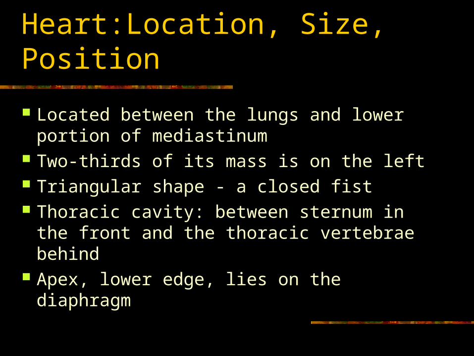
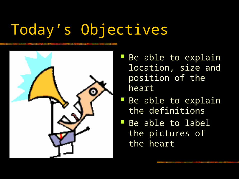
Heart:Location, Size, Position
Located between the lungs and lower portion of mediastinum
Two-thirds of its mass is on the left Triangular shape - a closed fist Thoracic cavity: between sternum in the
front and the thoracic vertebrae behind Apex, lower edge, lies on the diaphragm

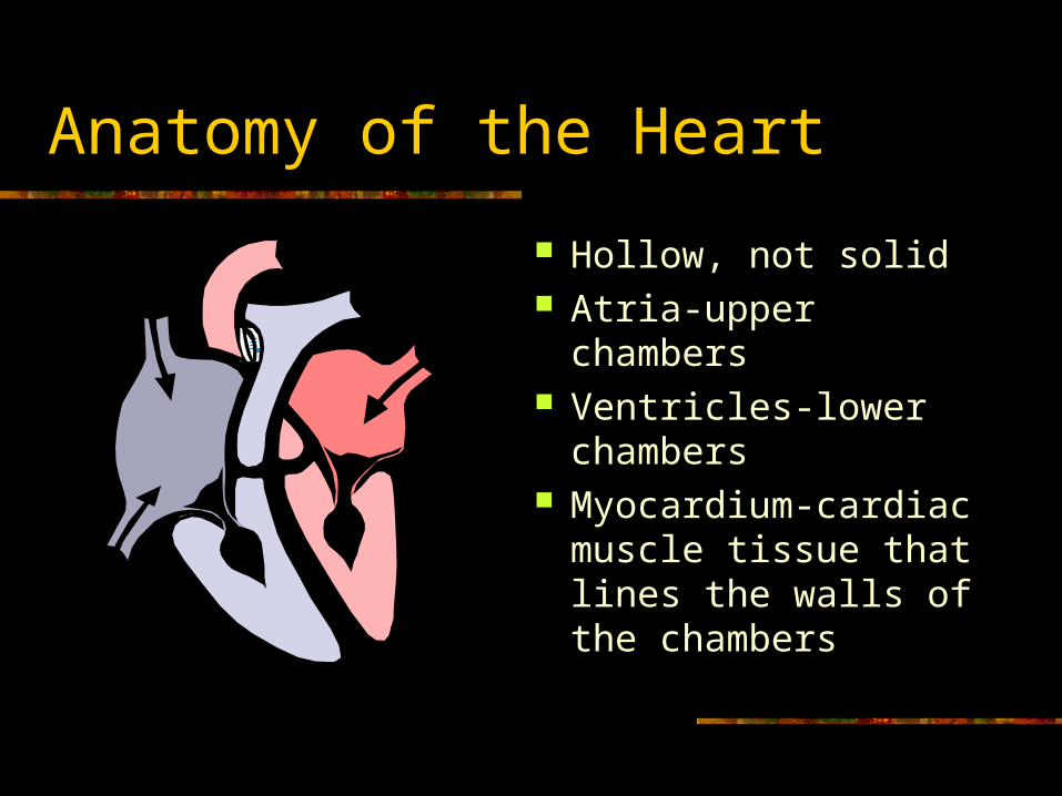
Anatomy of the Heart
Hollow, not solid Atria-upper chambers Ventricles-lower
chambers Myocardium-cardiac
muscle tissue that lines the walls of the chambers
Interatrial septum- septum (divider) between the atrial chambers
Interventricular septum-separates the ventricles Endocardium-thin layer of very smooth tissue
that lines each chamber (“endo” means within or inside)
Endocarditis-inflamed lining; rough and abrasive to RBC’s passing over its surface- thrombus- fatal blood clot

Pericardium – covering of the heart that consists of two layers of fibrous tissue:
1. Inner layer-epicardium (visceral pericardium) Covers the heart like skin on an apple (“epi” means upon or on)
2. Outer layer- parietal pericardium (loose fitting sac that allows the heart to beat)
No friction because the two layers are moist serous membranes
Pericardial fluid lubricates between heart and pericardial sac


Pericarditis
Inflammation of the pericardium Trauma, bacterial and viral infections, tumors Often visceral and parietal pericardium rub
together – severe chest pain Pericardial effusion- pericardial fluid, pus, or
blood accumulate in the space between the two layers and impairs pumping action of the heart
Cardiac tamponade-compression of the heart
Objectives
1. Be able to describe the conduction system of the heart
2. Be able to identify the structures of the heart involved in electrical conduction


Conduction System of the Heart
View the animation and take the quiz http://highered.mcgraw-hill.com/sites/0072
495855/student_view0/chapter22/animation__conducting_system_of_the_heart.html

Conduction System of the Heart
The intercalated disks are electrical connectors.
Thus, both atrial walls will contract at about the same time and both ventricular walls will contract at about the same time.

Conduction System Sequence
1. SA node (pacemaker) 2. All directions through atria causing
atrial fibers to contract 3. AV node 4. Bundle of His 5. Purkinje fibers
Related Documents