Image 1. Metatarsus adductus. Note the kidney bean or C-shaped foot due to the angulated mid- and forefoot. Image 2. Heel bisector line for determining severity of metatarsus adductus. Image 3. Internal tibial torsion. Note the inward-facing toes but forward- facing patella. Image 4. The position to assess thigh-foot angle.

Welcome message from author
This document is posted to help you gain knowledge. Please leave a comment to let me know what you think about it! Share it to your friends and learn new things together.
Transcript
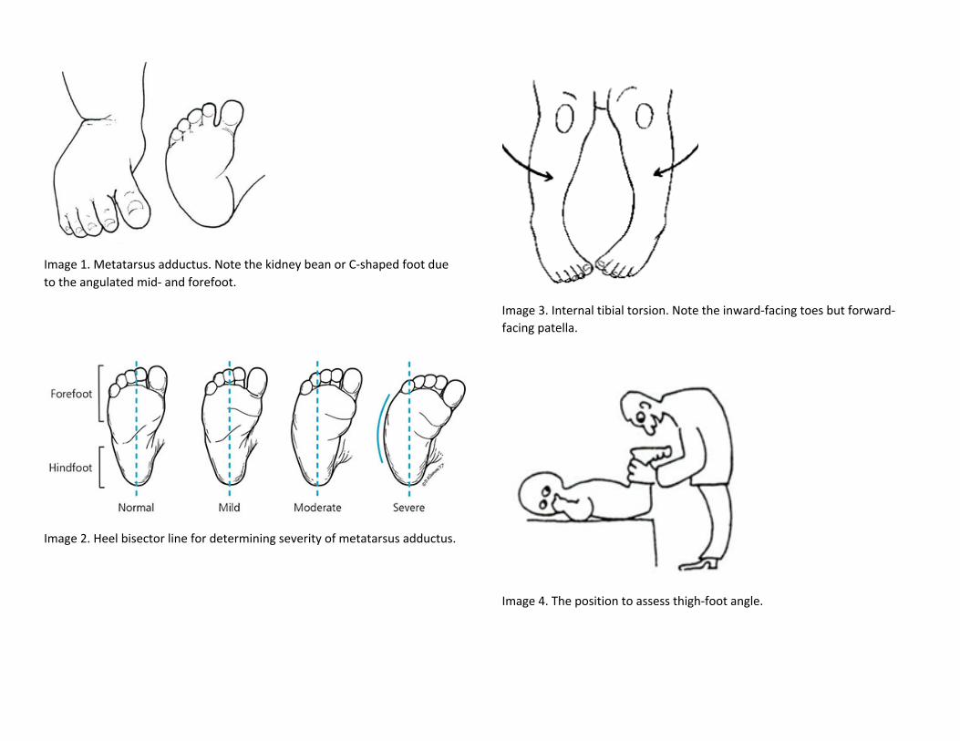
Image 1. Metatarsus adductus. Note the kidney bean or C-shaped foot due to the angulated mid- and forefoot.
Image 2. Heel bisector line for determining severity of metatarsus adductus.
Image 3. Internal tibial torsion. Note the inward-facing toes but forward-facing patella.
Image 4. The position to assess thigh-foot angle.
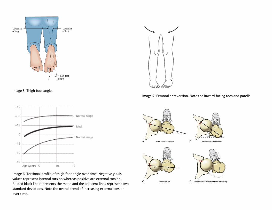
Image 5. Thigh-foot angle.
Image 6. Torsional profile of thigh-foot angle over time. Negative y-axis values represent internal torsion whereas positive are external torsion. Bolded black line represents the mean and the adjacent lines represent two standard deviations. Note the overall trend of increasing external torsion over time.
Image 7. Femoral anteversion. Note the inward-facing toes and patella.

Image 8. Varying severities of femoral anteversion. The solid black line represents the axis of the femoral neck and the hashed line represents the axis of the femoral condyles.
Image 9. The “W” sitting position children with femoral anteversion find comfortable due to the internal torsion of their femurs.
Image 10. The position to assess femoral anteversion and ROM of femoral internal rotation and external rotation.
Image 11. Developmental dysplasia of the hip involving dislocation of the femoral head.
Image 12. Pavlik harness (left) and hip spica cast (right).

Image 13. CAVE deformities of clubfoot.
Image 14. Ponseti method serial manipulation and casting to fix clubfoot.

Image References:
1. https://global-help.org/products/easter_seal_guide_to_childrens_orthopaedics/
2. https://www.aafp.org/afp/2017/0815/p226.html
3. http://www.drdelbello.com/conditions/internal-tibial-torsion-femoral-torsion-metatarsus-adductus
4. https://global-help.org/products/easter_seal_guide_to_childrens_orthopaedics/
5. https://accesspediatrics.mhmedical.com/content.aspx?bookid=2126§ionid=176094147&jumpsectionID=19
1276202
6. https://www.aafp.org/afp/2003/0801/p461.html
7. https://www.orthoseek.com/articles/femtorsion.html
8. https://twitter.com/atcbocstudy/status/869204189687283712
9. https://global-help.org/products/easter_seal_guide_to_childrens_orthopaedics/
10. https://global-help.org/products/easter_seal_guide_to_childrens_orthopaedics/
11. https://orthoinfo.aaos.org/en/diseases--conditions/developmental-dislocation-dysplasia-of-the-hip-ddh/
12. http://www.childrenshospital.org/conditions-and-treatments/conditions/d/developmental-dysplasia-of-the-
hip/treatments
13. https://orthopaedicprinciples.com/2018/07/congenital-talipes-equinovarus/
14. https://www.semanticscholar.org/paper/Idiopathic-congenital-clubfoot%3A-Initial-treatment.-Bergerault-
Fournier/0a2f2204be0ef1b087e11a169318bde1215a43a1/figure/6
Related Documents