Transcranial Magnetic Stimulation in the investigation and treatment of schizophrenia: a review H. Magnus Haraldsson * , Fabio Ferrarelli, Ned H. Kalin, Giulio Tononi Departmen t of Psychiatry , University of Wisco nsin, 6001 Resear ch Park Blvd., Madison, WI 53719, USA Received 31 March 2003; received in revised form 1 October 2003; accepted 29 October 2003 Available online 28 February 2004 Abstract Transcranial Magnetic Stimulation (TMS) is a non-invasive method of stimulating the brain that is increasingly being used in neuropsyc hiatric research and clinical psychiatry . This review examines the role of TMS in schizophren ia research as a diagnostic and a therapeutic resource. After a brief overview of TMS, we describe the application of TMS to schizophrenia in studies of cortical excitability and inhibition, and we discuss the potential confounding role of neuroleptic medications. Based on these studies, it appears that some impairment of cortical inhibition may be present in schizophrenic subjects. We then review attempts to employ TMS for treating different symptoms of schizophrenia. Some encouraging results have been obtained, such as the reduction of auditory hallucinations after slow TMS over auditory cortex and an improvement of psychotic symptoms after high frequency TMS over left prefrontal cortex. However, these results need to be confirmed using better placebo conditions. Future studies are likely to employ TMS in combination with function al brain imaging to examine the effects produced by the stimulated area on activity in other brain regions. Such studies may reveal impaired effective connectivity between specific brain areas, which could identify these regions as targets for selective stimulation with therapeutic doses of TMS. D 2004 Published by Elsevier B.V. Keywor ds: Transcranial Magnetic Stimulation; Schizophrenia; Cortical excitability; Cortical inhibition; Antipsychotic medications; Functional brain imaging 1. Introduction Tra nscrani al Magnetic Stimulation (TMS) , intro- duced almost two decades ago (Barker et al., 1985), is a non-invasive method of stimulating the brain. It is increasingly being used as a tool in basic neurosc ience to study the funct ion of the nervous system, and it has also entered the field of clinical psychiatry as a potentia l treatment option for a variety of mental ill nesses (Burt et al., 2002). Comprehensive reviews of the role of TMS in basic neuroscience and neuropsychiatry have recent- ly been publ ished (Burt et al. , 2002; Fitzgerald et al., 2002a; Geor ge et al., 1999; Hallett, 2000; Lisanby et al., 2000, 2002). In this paper, we focus on TMS as a neur ophysi ological tool in schizo- phrenia research and as a therapeutic resource for the treatment of schizophrenia. Aft er a bri ef intr o- duction about TMS, we describe the applica tion of TMS for studying cortical excitability and assessing 0920-9964/$ - see front matter D 2004 Published by Elsevier B.V. doi:10.1016/j.schres.2003.10.006 * Corresponding author. Tel.: +1-608-263-6063; fax: +1-608- 263-0265. E-mail addr ess: [email protected] (H.M. Haraldsson). www.elsevier.com/locate/schres Schizophrenia Research 71 (2004) 1–16

Welcome message from author
This document is posted to help you gain knowledge. Please leave a comment to let me know what you think about it! Share it to your friends and learn new things together.
Transcript
7/27/2019 TMS in the investigation and treatment of schizophrenia.pdf
http://slidepdf.com/reader/full/tms-in-the-investigation-and-treatment-of-schizophreniapdf 1/16
Transcranial Magnetic Stimulation in the investigation and
treatment of schizophrenia: a review
H. Magnus Haraldsson*, Fabio Ferrarelli, Ned H. Kalin, Giulio Tononi
Department of Psychiatry, University of Wisconsin, 6001 Research Park Blvd., Madison, WI 53719, USA
Received 31 March 2003; received in revised form 1 October 2003; accepted 29 October 2003
Available online 28 February 2004
Abstract
Transcranial Magnetic Stimulation (TMS) is a non-invasive method of stimulating the brain that is increasingly being used in
neuropsychiatric research and clinical psychiatry. This review examines the role of TMS in schizophrenia research as a diagnostic
and a therapeutic resource. After a brief overview of TMS, we describe the application of TMS to schizophrenia in studies of
cortical excitability and inhibition, and we discuss the potential confounding role of neuroleptic medications. Based on these
studies, it appears that some impairment of cortical inhibition may be present in schizophrenic subjects. We then review attempts
to employ TMS for treating different symptoms of schizophrenia. Some encouraging results have been obtained, such as the
reduction of auditory hallucinations after slow TMS over auditory cortex and an improvement of psychotic symptoms after high
frequency TMS over left prefrontal cortex. However, these results need to be confirmed using better placebo conditions. Futurestudies are likely to employ TMS in combination with functional brain imaging to examine the effects produced by the stimulated
area on activity in other brain regions. Such studies may reveal impaired effective connectivity between specific brain areas,
which could identify these regions as targets for selective stimulation with therapeutic doses of TMS.
D 2004 Published by Elsevier B.V.
Keywords: Transcranial Magnetic Stimulation; Schizophrenia; Cortical excitability; Cortical inhibition; Antipsychotic medications; Functional
brain imaging
1. Introduction
Transcranial Magnetic Stimulation (TMS), intro-
duced almost two decades ago (Barker et al., 1985),
is a non-invasive method of stimulating the brain. It
is increasingly being used as a tool in basic
neuroscience to study the function of the nervous
system, and it has also entered the field of clinical
psychiatry as a potential treatment option for a
variety of mental illnesses (Burt et al., 2002).Comprehensive reviews of the role of TMS in
basic neuroscience and neuropsychiatry have recent-
ly been published (Burt et al., 2002; Fitzgerald et
al., 2002a; George et al., 1999; Hallett, 2000;
Lisanby et al., 2000, 2002). In this paper, we focus
on TMS as a neurophysiological tool in schizo-
phrenia research and as a therapeutic resource for
the treatment of schizophrenia. After a brief intro-
duction about TMS, we describe the application of
TMS for studying cortical excitability and assessing
0920-9964/$ - see front matter D 2004 Published by Elsevier B.V.doi:10.1016/j.schres.2003.10.006
* Corresponding author. Tel.: +1-608-263-6063; fax: +1-608-
263-0265.
E-mail address: [email protected] (H.M. Haraldsson).
www.elsevier.com/locate/schres
Schizophrenia Research 71 (2004) 1–16
7/27/2019 TMS in the investigation and treatment of schizophrenia.pdf
http://slidepdf.com/reader/full/tms-in-the-investigation-and-treatment-of-schizophreniapdf 2/16
inhibitory mechanisms. The neurophysiological and
clinical studies using TMS in schizophrenia are
then reviewed.
2. Transcranial Magnetic Stimulation
TMS is based on Faraday’s principle of electro-
magnetic induction. A rapidly changing magnetic
field (2 T) is generated by passing a very brief
(0.2– 0.9 milliseconds (ms)) high-current alternating
electric pulse through an insulated coil made of wire.
When the coil is placed against the scalp the magnetic
field passes readily through the skull and induces a
weak electrical current in the superficial cortex of the
brain lasting exactly as long as the current pulse in the
coil (Barker, 2002). The induced electrical activity can
cause cortical neurons to discharge action potentials.
The strength of the magnetic field decreases exponen-
tially with increasing distance from the coil and
therefore stimulation of nervous tissue deeper than
approximately 2 cm from the scalp is currently not
possible (Lisanby et al., 2000).
TMS can be applied as a single stimulus pulse or
repeated pulses for seconds or minutes (rTMS). Stim-
ulation frequency of more than one pulse per second
(1 Hz) is called rapid TMS. Single pulse TMS isgenerally safe and well tolerated (Wassermann, 1998).
Its most common side effect is mild headache, which
responds readily to analgesics. There is a low risk of
inducing seizures with rTMS and the risk increases
with higher stimulation frequencies and intensities
(Wassermann, 1998). However, at slow rates ( < 1
Hz) and standard intensity the risk for healthy subjects
is virtually nonexistent.
TMS can produce different effects depending on
where and how it is applied. For example, single pulse
TMS applied to the primary motor cortex inducesmuscle twitches that can be recorded with an electro-
myogram (EMG) (Barker et al., 1985), while single
pulse TMS to the occipital cortex can produce sub-
jective light flashes (Kammer, 1999). On the other
hand, high frequency TMS applied to the prefrontal,
left midtemporal and primary visual cortex produces
short-term impairments in working memory (Pascual-
Leone and Hallett, 1994), free recall of verbal material
(Grafman et al., 1994) and visual stimuli identification
(Amassian et al., 1989), respectively. In this way TMS
can provide unique information about the temporal
and topographic organization of various neurophysi-
ological and cognitive processes.
A number of variables influence how accurately aspecific brain area can be stimulated with TMS. These
variables include the intensity of stimulation, the
shape and orientation of the stimulating coil and the
excitability, type and orientation of the neurons in the
area of stimulation (Brasil-Neto et al., 1992a). The
two most common coil shapes are circular and figure-
of-eight. Circular coils are powerful but may stimulate
a large brain area. Figure-of-eight coils are made of a
coil, which is twisted and flipped over onto itself
forming a figure-of-eight. The cross point of the coil is
the site of maximum stimulation intensity (Cohen et
al., 1990). Mapping studies of the motor cortex have
indicated that spatial resolution is a pproximately 0.5 –
1.0 cm with a figure-of-eight coil (Brasil-Neto et al.,
1992b). It is hard to determine the spatial resolution of
TMS in brain areas where no visible response such as
a muscle contraction can be recorded. TMS has a high
temporal specificity and allows for investigating
changes of brain activity that occur over a few ms
(see also for review of TMS: Hallett, 2000; Pascual-
Leone et al., 1998).
3. Studying cortical excitability and inhibitory
mechanisms with TMS
Researchers have utilized TMS of the motor cortex
to study neuronal excitability, and cortical inhibitory
mechanisms, both in patients and healthy subjects
(Fitzgerald et al., 2002a). This has mainly been
achieved by examining EMG recorded motor evoked
potentials (MEPs). Here we briefly review several key
TMS paradigms that are useful for evaluating cortical
excitability and inhibition.
4. Motor threshold and MEP
A MEP is a synchronous muscle response evoked
by a TMS pulse stimulating the motor cortex. It is a
marker of cortical excitability and its size reflects the
number of motor neurons that are activated by a TMS
pulse. The latency from the time of motor cortex TMS
to the onset of a MEP is a measure of corticospinal
H.M. Haraldsson et al. / Schizophrenia Research 71 (2004) 1–16 2

7/27/2019 TMS in the investigation and treatment of schizophrenia.pdf
http://slidepdf.com/reader/full/tms-in-the-investigation-and-treatment-of-schizophreniapdf 3/16
conduction time. The threshold for inducing MEPs
with TMS is called motor threshold. Motor threshold
has been defined as the lowest stimulation intensity
over the motor cortex needed to induce a MEP in anextremity muscle in at least 5 out of 10 consecutive
trials (Rossini et al., 1994). The motor threshold is
well established as an objective and standardized
measure of corticospinal excitability in humans, and
also is widely used to standardize stimulation inten-
sities in various neurocognitive studies (Walsh and
Rushworth, 1999). The motor threshold seems to be
relatively stable within individuals (Mills and Nithi,
1997), although some minor hemispheric differences
have been demonstrated (Cicinelli et al., 1997). A
decrease in motor threshold indicates increased neu-
ronal excitability, whereas an increased motor thresh-
old reflects decreased excitability. TMS pulses also
affect inhibitory neural processes both in the ipsilat-
eral and contralateral motor cortex (Ferbert et al.,
1992). Thus, TMS has been employed to investigate
cortical inhibitory mechanisms by focusing on three
main paradigms: (1) Cortical Silent Period, (2) Re-
sponse to Paired Pulse TMS and (3) Single and Dual
Pulse Transcallosal Inhibition.
Cortical silent period is a period of TMS-induced
EMG suppression during a tonic voluntary contraction
of a muscle. A cortical silent period can be induced by both supra- and sub-threshold stimuli and is therefore
independent of the presence of a MEP (Triggs et al.,
1992). This brief period of EMG silence, which starts
approximately 30– 40 ms after the TMS stimulus,
reflects the effects of inhibitory interneurons in the
motor cortex that are activated by TMS (Hallett, 1995;
Sanger et al., 2001). A decreased level of EMG
suppression and a reduced duration of the silent
period are indications of disturbed cortical inhibition.
Paired pulse TMS involves stimulating the motor
cortex with two successive TMS pulses—a condition-ing pulse followed by a test pulse-delivered at a short
inter-stimulus interval through the same stimulating
coil. The motor response to the test pulse may be
decreased (inhibition) or increased (facilitation)
depending on the length of the inter-stimulus interval.
If a sub-motor threshold conditioning pulse precedes a
supra-motor threshold test pulse, the motor response is
inhibited at inter-stimulus intervals of 1 – 6 ms and
facilitated at intervals of 8 – 30 ms (Kujirai et al.,
1993). Paired pulse inhibition can also be obtained
with a supra-motor threshold conditioning pulse de-
livered 100–200 ms prior to the test pulse (Nakamura
et al., 1997). A decreased level of MEP suppression
by the conditioning pulse is an indication of disturbedcortical inhibition. In both the short and long interval
inhibitory procedures the conditioning pulse is be-
lieved to activate inhibitory interneurons that suppress
the effects of the test pulse (Sanger et al., 2001).
Paired pulse facilitation is probably mediated by
excitatory interneurons (Terao and Ugawa, 2002).
Transcallosal inhibition has been investigated both
with a dual pulse and single pulse technique (Fitzger-
ald et al., 2002a). Dual pulse transcallosal inhibition is
observed when one delivers a conditioning stimulus to
the motor cortex of one hemisphere prior to giving a
test pulse to the motor cortex of the opposite hemi-
sphere, using two TMS coils. The conditioning pulse
induces action potentials that pass through the corpus
callosum to the contralateral motor cortex where they
suppress corticospinal neurons being stimulated by
the TMS test pulse (Ferbert et al., 1992). Inhibition of
the test pulse response (decreased amplitude of the
MEP) is seen when the inter-stimulus interval is
between 5 and 20 ms, which is consistent with trans-
callosal responses recorded with frontal scalp electro-
des f ollowing TMS over the contralateral homologous
area (Cracco et al., 1989).Single pulse transcallosal inhibition is observed
when one stimulates the motor cortex with TMS while
the subject performs a steady contraction of hand
muscles on the same side as the stimulation. The
TMS pulse triggers a volley of action potentials that
pass through the corpus callosum and inhibit the
corticospinal neurons controlling the contralateral
hand muscles, which are voluntarily activated. In this
way, transcallosal inhibition of voluntary muscle con-
traction can be measured. The suppression of EMG
recorded hand muscle activity begins on average 30– 40 ms following the TMS pulse and lasts approxi-
mately 25 ms (Meyer et al., 1995).
A transcallosal inhibition paradigm can thus be
used to measure transcallosal conduction time. That is
calculated by subtracting the time it takes a TMS
pulse to induce a contralateral MEP from the time it
takes the same pulse to generate an EMG inhibition.
The difference reflects the conduction time from ipsi-
to contralateral motor cortex through the corpus
callosum (Fitzgerald et al., 2002a).
H.M. Haraldsson et al. / Schizophrenia Research 71 (2004) 1–16 3
7/27/2019 TMS in the investigation and treatment of schizophrenia.pdf
http://slidepdf.com/reader/full/tms-in-the-investigation-and-treatment-of-schizophreniapdf 4/16
Several studies provide evidence that these inhib-
itory mechanisms are primarily related to cortical but
not peripheral processes (Ferbert et al., 1992; Fuhr et
al., 1991; Inghilleri et al., 1993; Kujirai et al., 1993).For example, the duration of the silent period is
altered in patients with unilateral cortical lesions
(von Giesen et al., 1994). Moreover, the inhibition
of MEP obtained with paired pulse TMS at short inter-
stimulus intervals (2 and 3 ms) is due to a reduced
corticospinal output as indicated by recordings with
electrodes implanted in the cervical epidural space in
awake subjects (Di Lazzaro et al., 2002).
5. Neurophysiological studies using TMS in
patients with schizophrenia
Some histopathological and pharmacological stud-
ies have suggested that the pathophysiology of schizo-
phreni a may in vo lve dy sfun ction of excitatory
(Selemon and Goldman-Rakic, 1999) and/or inhibito-
ry neural function (Olney and Farber, 1995). I n a
number of recent studies, TMS of motor cortex has
been used to evaluate both cortical excitability and
inhibitory mechanisms in patients with schizophrenia.
This research is still in its early days and most of the
studies are limited to small sample sizes. Furthermore,due to methodological differences it is often difficult
to directly compare the results of different studies.
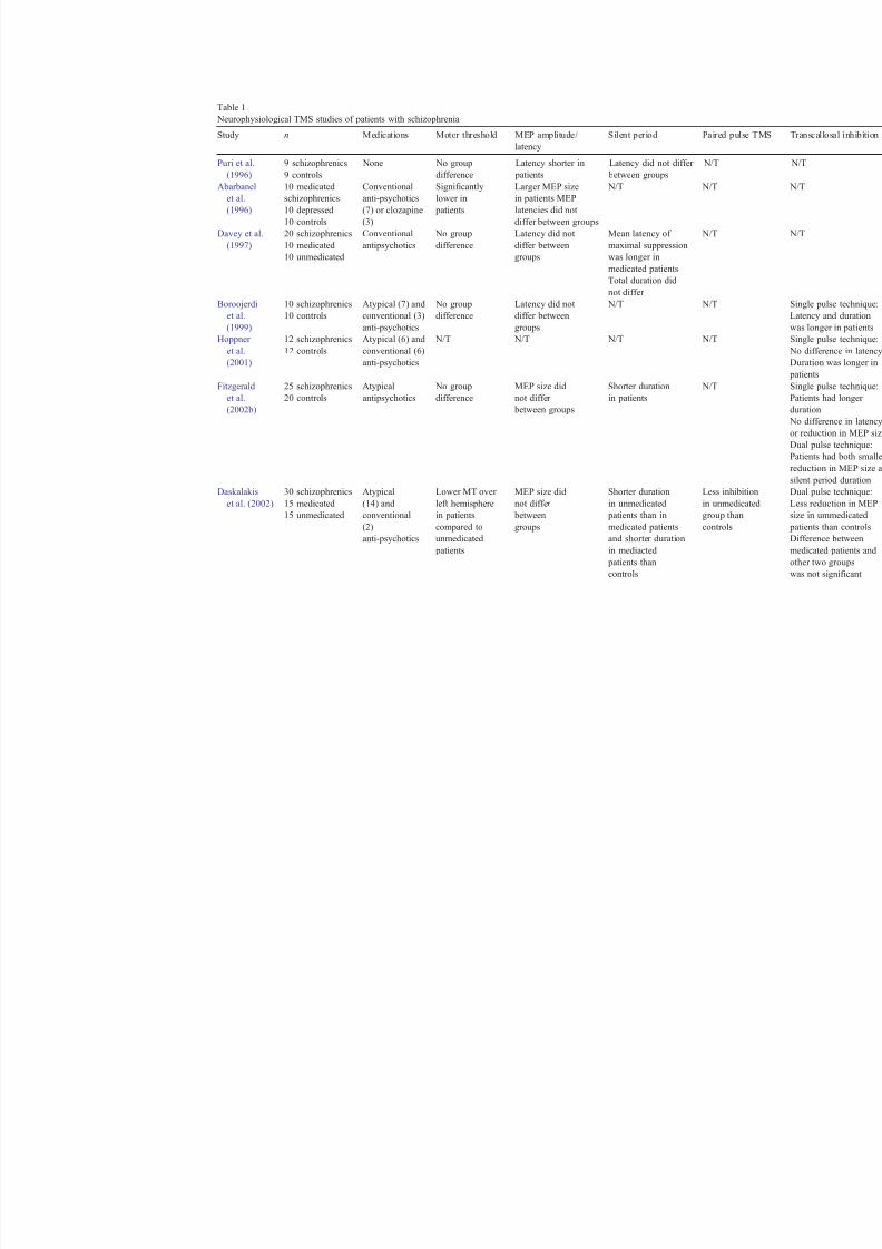
Table 1 summarizes the findings of studies using the
previously described TMS paradigms for evaluating
cortical excitability and inhibition in patients with
schizophrenia.
5.1. Studies of cortical excitatory function
Overall, TMS studies provide little evidence for
significant abnormalities in cortical excitability in patients with schizophrenia. Most investigations have
failed to show any significant difference in motor
threshold, MEP size or paired pulse facilitation be-
tween patients and healthy subjects (Boroojerdi et al.,
1999; Fitzgerald et al., 2002b,c,d; Puri et al., 1996).
One exception is a study by Abarbanel et al. (1996),
which demonstrated larger MEP size and lower motor
thresholds in 10 medicated patients with schizophre-
nia compared to 10 depressed and 10 healthy subjects.
This increased excitability in schizophrenia patients
may have been due to increased muscle tonus sec-
ondary to extrapyramidal side effects from neuroleptic
medications.
A hemispheric difference in corticospinal excitabil-ity between patients with schizophrenia and healthy
subjects was found in a recent study by Pascual-Leone
et al. (2002). These authors found that a group of right
handed patients taking conventional antipsychotic
medications (n = 7) and a group of unmedicated
patients (n = 7) had a 5 – 10% lower motor threshold
in the right compared to the left hemisphere, while the
opposite was found in a group of healthy subjects
(n = 7). Healthy right-handed people generally have a
lower motor threshold in their dominant left hemi-
sphere, which has been linked to facilitation due to
more frequent use of their right hand (Triggs et al.,
1994). The finding of a lower excitability threshold
for the non-dominant hemisphere in schizophrenics
may indicate that, compared to normal subjects,
patients with schizophrenia have reversed asymmetry
in corticospinal excitability.
TMS research has provided inconclusive results
concerning corticospinal conductivity in schizophre-
nia (Abarbanel et al., 1996; Boroojerdi et al., 1999;
Puri et al., 1996). In the first study of motor function
in schizophrenia using TMS, Puri et al. (1996)
detected a significantly shorter latency of MEPs innine unmedicated patients with schizophrenia com-
pared to nine healthy subjects. However, further
studies measuring MEP latency did not find a differ-
ence between medicated schizophrenia patients and
normal controls (Abarbanel et al., 1996; Boroojerdi et
al., 1999).
5.2. Studies of cortical inhibition
Several findings indicate that a lack of cortical
inhibitory control may be involved in the pathophys-iology of schizophrenia (Frith et al., 2000). For
example, studies using auditory evoked potentials
have demonstrated that patients with schizophrenia
lack normal suppression of the P50 auditory evoked
response with a conditioning pre-pulse stimulus
(Freedman et al., 1996; McCarley et al., 1991).
Abnormal motor function such as incoordination,
involuntary movements and impaired fine motor
skills, which are not related to antipsychotic drug
treatment, have been detected in approximately 80%
H.M. Haraldsson et al. / Schizophrenia Research 71 (2004) 1–16 4

7/27/2019 TMS in the investigation and treatment of schizophrenia.pdf
http://slidepdf.com/reader/full/tms-in-the-investigation-and-treatment-of-schizophreniapdf 5/16
of patients with schizophrenia (Yager and Gitlin,
2000). These motor deficits could be explained by
disturbances in central inhibition and fine-tuning of
motor responses (Puri et al., 1996).A number of recent TMS studies indicate that
patients with schizophrenia have impairments of cor-
tical inhibition. The main results of investigations
using the silent period, paired pulse inhibition and
transcallosal inhibition TMS paradigms are summa-
rized in Table 1.
5.2.1. Silent period
Four recent studies have found the silent period
duration to be significantly shorter in medicated
schizophrenic patients compared to healthy controls
(Daskalakis et al., 2002; Fitzgerald et al., 2002b,c,d).
One of these st udies also included a gr oup of unmed-
icated patients (Daskalakis et al., 2002) and found that
these patients had a significantly shorter silent period
duration than the medicated group. Only one smaller
study failed to report a significant difference in silent
period duration between patients on conventional
antipsychotics and healthy controls (Davey et al.,
1997).
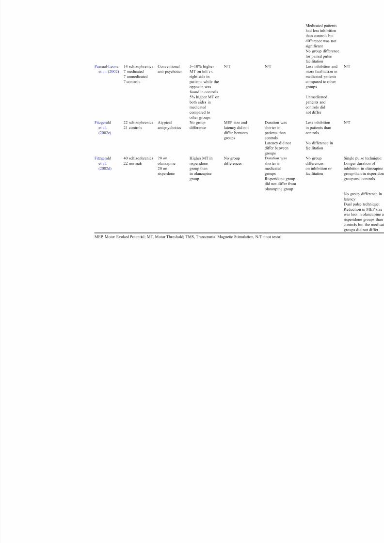
5.2.2. Paired pulse inhibition
A study comparing 40 medicated schizophrenia patients with 22 normal subjects did not find a
significant difference on measurements of paired
pulse inhibition (Fitzgerald et al., 2002d). However,
three smaller studies found less inhibit ion in schizo-
phrenics compared to control subjects (Daskalakis et
al., 2002; Fitzgerald et al., 2002c; Pascual-Leone et
al., 2002). Two of these studies also included groups
of unmedicated patients. In one study, unmedicated
patients did not differ significantly from healthy sub-
jects (Pascual-Leone et al., 2002); in the other one
unmedicated patients had less paired pulse inhibitionthan healthy controls. In the latter study medicated
patients did not differ significantly from either the
unmedicated or the control group (Daskalakis et al.,
2002). Furthermore, in this study a significant corre-
lation (r = 0.5, p = 0.01) was found between the mag-
nitude of paired-pulse inhibition and the severity of
psychotic symptoms in the patient groups such that
patients with higher scores on the Positive and Neg-
ative Symptom Scale (PANSS) exhibited decreased
inhibition.
5.2.3. Transcallosal inhibition
Three studies found a reduction of the magnitude
of transcallosal inhibition in schizophrenic patients
(Daskalakis et al., 2002; Fitzgerald et al., 2002b,d). Inone of these studies, the difference was seen between
unmedicated patients and healthy subjects but a group
of medicated patients did not differ significantly from
the control and unmedicated groups (Daskalakis et al.,
2002). A single pulse technique, used in one study,
did not find significant differences in tr anscallosal
inhibition between patients and controls (Fitzgerald
et al., 2002b). In four studies, the duration of trans-
callosal inhibition was significantly longer in schizo-
phrenics than in healthy subjects (Boroojerdi et al.,
1999; Fitzgerald et al., 2002c,d; Hoppner et al., 2001).
Transcallosal inhibition has also been used to
investigate inter-hemispheric interactions of homolo-
gous brain areas by measuring the latency of the
inhibition. In the first TMS study of transcallosal
inhibition in schizophrenia, Boroojerdi et al. (1999),
using a single pulse paradigm, found a significant
delay in the onset of transcallosal inhibition in 10
medicated schizophrenia patients compared to 10
controls. However, other investigators did not report
a significant delay in the onset of transcallosal inhi-
bition or indications of increased transcallosal con-
duction time in patients with schizophrenia (Fitzgeraldet al., 2002c,d; Hoppner et al., 2001). Altogether,
these reports suggest that stimuli mediating inhibition
travel normally between hemispheres. However, con-
tralateral inhibitory mechanisms activated by the
transcallosal stimuli may be impaired in patients
suffering from schizophrenia.
5.3. Effects of antipsychotic medications on cortical
inhibitory mechanisms
The effects of antipsychotic medications on corti-cal inhibitory mechanisms are not well understood.
However, there are some indications that conventional
antipsychotic medications may disrupt cortical inhibi-
tion (Davey et al., 1997; Pascual-Leone et al., 2002;
Ziemann et al., 1997), while atypical antipsychotic
medications may enhance it (Fitzgerald et al.,
2002c,d).
Ziemann et al. (1997) demonstrated using paired-
pulse TMS that healthy subjects taking haloperidol
had significantly less cortical inhibition while on the
H.M. Haraldsson et al. / Schizophrenia Research 71 (2004) 1–16 5
7/27/2019 TMS in the investigation and treatment of schizophrenia.pdf
http://slidepdf.com/reader/full/tms-in-the-investigation-and-treatment-of-schizophreniapdf 6/16
Table 1
Neurophysiological TMS studies of patients with schizophrenia
Study n Medications Motor threshold MEP amplitude/
latency
Silent period Paired pulse T
Puri et al. 9 schizophrenics None No group Latency shorter in Latency did not differ N/T
(1996) 9 controls difference patients between groups
Abarbanel
et al.
(1996)
10 medicated
schizophrenics
10 depressed
10 controls
Conventional
anti-psychotics
(7) or clozapine
(3)
Significantly
lower in
patients
Larger MEP size
in patients MEP
latencies did not
differ between groups
N/T N/T
Davey et al.
(1997)
20 schizophrenics
10 medicated
10 unmedicated
Conventional
antipsychotics
No group
difference
Latency did not
differ between
groups
Mean latency of
maximal suppression
was longer in
medicated patients
N/T
Total duration did
not differ
Boroojerdi
et al.(1999)
10 schizophrenics
10 controls
Atypical (7) and
conventional (3)anti-psychotics
No group
difference
Latency did not
differ betweengroups
N/T N/T
Hoppner
et al.
(2001)
12 schizophrenics
12 controls
Atypical (6) and
conventional (6)
anti-psychotics
N/T N/T N/T N/T
Fitzgerald
et al.
(2002b)
25 schizophrenics
20 controls
Atypical
antipsychotics
No group
difference
MEP size did
not differ
between groups
Shorter duration
in patients
N/T
Daskalakis
et al. (2002)
30 schizophrenics
15 medicated
15 unmedicated
Atypical
(14) and
conventional
(2)
anti-psychotics
Lower MT over
left hemisphere
in patients
compared to
unmedicated
patients
MEP size did
not differ
between
groups
Shorter duration
in unmedicated
patients than in
medicated patients
and shorter duration
in mediacted
patients than
controls
Less inhibition
in unmedicate
group than
controls
7/27/2019 TMS in the investigation and treatment of schizophrenia.pdf
http://slidepdf.com/reader/full/tms-in-the-investigation-and-treatment-of-schizophreniapdf 7/16
Medicated pat
had less inhib
than controls b
difference was
significant
No group diff
for paired puls
facilitation
Pascual-Leone
et al. (2002)
14 schizophrenics
7 medicated
7 unmedicated
7 controls
Conventional
anti-psychotics
5–10% higher
MT on left vs.
right side in
patients while the
opposite was
found in controls
N/T N/T Less inhibition
more facilitati
medicated pati
compared to o
groups
5% higher MT on
both sides in
medicated
compared to
other groups
Unmedicated
patients and
controls did
not differ
Fitzgerald
et al.
(2002c)
22 schizophrenics
21 controls
Atypical
antipsychotics
No group
difference
MEP size and
latency did not
differ between
groups
Duration was
shorter in
patients than
controls
Less inhibition
in patients tha
controls
Latency did not
differ between
groups
No difference
facilitation
Fitzgerald
et al.
(2002d)
40 schizophrenics
22 normals
20 on
olanzapine
20 on
risperdone
Higher MT in
risperidone
group than
in olanzapine
group
No group
differences
Duration was
shorter in
medicated
groups
Risperidone group
did not differ from
olanzapine group
No group
differences
on inhibition o
facilitation
MEP, Motor Evoked Potential; MT, Motor Threshold; TMS, Transcranial Magnetic Stimulation, N/T = not tested.
7/27/2019 TMS in the investigation and treatment of schizophrenia.pdf
http://slidepdf.com/reader/full/tms-in-the-investigation-and-treatment-of-schizophreniapdf 8/16
drug. Similarly Pascual-Leone et al. (2002) found that
a group of patients with schizophrenia taking conven-
tional antipsychotics had less paired pulse inhibition
than groups of unmedicated patients and healthycontrol subjects.
The effects of conventional antipsychotic medica-
tions on the cortical silent per iod in patients with
schizophrenia were studied by Davey et al. (1997).
They found that in most of the patients taking medi-
cations, the cortical silent period was divided into an
early part with weak suppression of voluntary EMG
and a later component with strong suppression. No
such division was seen in any of the non-medicated
patients in the study, who all had an abrupt onset of
maximum EMG suppression. The delay in maximal
suppression in the medicated patients may be explained
by disruption of basal ganglia inputs to the inhibitory
circuitry in the motor cortex induced by the medica-
tions. Studies of patients with Parkinson’s disease,
where dopamine is depleted, demonstrate a similar
reduction in the strength of EMG suppression in t he
early part of the silent period (Ridding et al., 1995).
The effects of the newer atypical antipsychotic
medications on cortical inhibitory mechanisms may
be different from the effects of typical antipsychotics.
In a recent study by Fitzgerald et al. (2002d), where
the effects of olanzapine and risperidone were com- pared on several measures of cortical inhibition in
patients with schizophrenia, the two medications were
found to differ. Schizophrenia patients taking olanza-
pine (mean dose 12.25 mg) had a significantly higher
level of transcallosal inhibition than patients taking
the risperidone (mean dose 4.1 mg). Olanzapine may
therefore have enhancing effects on cortical inhibitory
mechanisms. Moreover, the length of transcallosal
inhibition was significantly longer in subjects taking
olanzapine and the increased duration correlated with
the dose of olanzapine (Fitzgerald et al., 2002c).Future studies of schizophrenia subjects both on and
off medications will increase the understanding of the
effects of antipsychotic medications on cortical inhib-
itory processes.
6. Treatment of schizophrenia with TMS
Since the mid 1990s, it has been suggested that
TMS may play a role in the treatment of several
neurological and psychiatric disorders (Pridmore and
Belmaker, 1999). Indeed, there is increasing evidence
suggesting that both slow and high frequency TMS
trains applied to the left or right prefrontal cortex haveantidepressant effects, although the effect sizes are
variable between studies and few studies have shown
high rates of strong response or remission (Burt et al.,
2002). There is less data on the effectiveness of TMS
in treating other psychiatric disorders such as mania
(Grisaru et al., 1998b), post-traumatic stress disorder
(Grisaru et al., 1998a), obsessive–compulsive disor-
der (Greenberg et al., 1997) and schizophrenia.
The optimal stimulation parameters for treating any
psychiatric disorder with TMS, such as the frequency,
intensity, duration and location of stimulation, as well
as the total number of stimuli and treatment sessions,
have not yet been determined. Furthermore, various
types and shapes of stimulation coils have been used
and they have been positioned and oriented in differ-
ent ways. A direct comparison of treatment studies is
therefore difficult.
A major concern in controlled TMS trials is the
lack of a reliable placebo (sham) condition. An
optimal sham TMS should induce the same somatic
sensations (scalp twitches) as active TMS without
stimulating the brain. The most commonly used sham
condition today involves tilting the coil 45j or 90j off the head in order to direct the magnetic field away
from the brain. However, it has been found that the
brain may still be affected and subjects may be able to
discriminate between active and sham conditions
(Lisanby et al., 2001). A promising solution to this
problem is the development of a new TMS coil that
induces both active and sham stimulation without
having to be moved or tilted. This coil has a special
sham mode where the intensity of the magnetic field is
below the threshold for activating cortical neurons but
strong enough to induce stimulation of the scalp(Ruohonen et al., 2000).
Another important variable in clinical TMS studies
is the frequency of stimulation. Several studies have
found that after high frequency TMS (>1 Hz) there is
increased excitability in various brain areas, while
after low frequency stimulation ( < 1 Hz) cortical
excitability is decreased (Wu et al., 2000; Chen et
al., 1997; Wassermann et al., 1998). Changes in
cortical excitability also depend on stimulation inten-
sity and duration. Higher intensity is more likely to
H.M. Haraldsson et al. / Schizophrenia Research 71 (2004) 1–16 8
7/27/2019 TMS in the investigation and treatment of schizophrenia.pdf
http://slidepdf.com/reader/full/tms-in-the-investigation-and-treatment-of-schizophreniapdf 9/16
induce activation and long stimulation trains correlate
with longer lasting modification of cortical excitabil-
ity (Pascual-Leone et al., 1998). In principle, such
findings can help designing rational treatment trialsfor various psychiatric symptoms. For example, to the
extent that hypoactivity of prefrontal cortex plays a
role in the pat hophysiology of negative symptoms of
schizophrenia (Andreasen et al., 1997), high frequen-
cy TMS of prefrontal cortex should help reversing
such hypoactivity and related symptoms. Conversely,
positive symptoms such as hallucinations, which are
associated with hyperactivity of temporoparietal areas
(Silbersweig et al., 1995), should benefit from low
frequency TMS to these regions.
Table 2 summarizes the design, stimulation param-
eters and main effects of TMS in therapeutic trials of
schizophrenia. The studies can be separated into two
groups according to the brain region being stimulated.
Left and right dorsolateral prefrontal cortex stimula-
tion has been applied when investigating TMS effects
on positive and negative symptoms of schizoprhenia
and the left temporoparietal cortex was stimulated
when studying the effects of TMS on auditory hallu-
cinations.
7. TMS of prefrontal cortex
The first two studies of TMS aimed at treating
patients with schizophrenia were open trials using
slow repetitive stimulation of the prefrontal cortex
with circular coils (Feinsod et al., 1998; Geller et al.,
1997). Transient improvement in mood was described
in 2 of 10 schizophrenia patients treated with 15 TMS
pulses over each side of the prefrontal cortex (Geller
et al., 1997). Feinsod et al. (1998) treated 10 patients
with right prefrontal TMS at 1 Hz in two 1-min daily
sessions for 10 days. There was a significant reductionin scores on the Brief Psychiatric Rating Scale
(BPRS) in seven patients, but this improvement was
linked to a reduction in symptoms of restlessness,
tension and anxiety and not to an improvement in
psychotic symptoms.
The effects of slow-repetitive TMS on positive and
negative symptoms of schizophrenia were studied in
31 medicated hospitalized patients with schizophrenia
or schizoaffective disorder who had an exacerbation
of psychotic symptoms (Klein et al., 1999). It should
be noted that this was a double-blind sham controlled
study (subjects were randomized to receive either
TMS or sham TMS). In this study, the right prefrontal
cortex was stimulated with a circular coil at a rate of 1Hz in 20-min sessions for 10 days. The patients were
evaluated using the PANSS, the BPRS and the Ham-
ilton Depression Rating Scale (HDRS) at the end of
each treatment week and then 1 and 4 weeks post-
treatment. Both groups displayed a similar mild im-
provement over time on all rating scales but there was
no significant difference between the TMS and sham
treated groups.
Three small trials have demonstrated promising
effects of high frequency prefrontal TMS for treat-
ment of symptoms of schizophrenia. In a study of 12
medicated acutely psychot ic patients with schizophre-
nia, Rollnik et al. (2000) performed high frequency
(20 Hz) pulse trains with a figure-of-eight coil to the
left or dominant dorsolateral prefrontal cortex at 80%
of motor threshold for 10 days. In this study a
crossover design was used in which subjects were
randomized to receive 2 weeks of active TMS and 2
weeks of sham TMS. The patients were rated at the
end of each week using the BPRS, Beck Depression
Inventory (BDI), State-Trait-Anxiety Inventory and a
number connection test to monitor frontal lobe func-
tion. The BPRS values were significantly lower for active treatment than sham treatment at the end of the
second week. This effect was not explained by an
improvement of depressive symptoms, since measures
on the BDI were not significantly decreased with
active TMS. Other ratings did not differ significantly
between active and sham TMS.
The effects of rapid TMS of the prefrontal cortex
on negative symptoms were studied by Cohen et al.
(1999). They found a significant reduction in negative
symptoms, measured with the PANSS, in an open trial
of six patients treated with 20 Hz to the left prefrontalcortex for 2 weeks with a figure-of-eight coil. The
patients in this study were not evaluated for symptoms
of depression. In a double-blind sham controlled trial
of eight schizophrenia patients, Nahas et al. (1999)
reported an improvement of negative symptoms the
day after a single session of 20 Hz TMS to the left
dorsolateral prefrontal cortex.
Finally, high frequency TMS of prefrontal cortex
has been employed to treat two subjects with prom-
inent catatonic symptoms (Grisaru et al., 1998c; Saba
H.M. Haraldsson et al. / Schizophrenia Research 71 (2004) 1–16 9
7/27/2019 TMS in the investigation and treatment of schizophrenia.pdf
http://slidepdf.com/reader/full/tms-in-the-investigation-and-treatment-of-schizophreniapdf 10/16
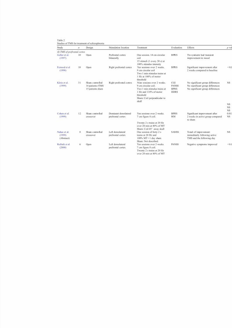
Table 2
Studies of TMS for treatment of schizophrenia
Study n Design Stimulation location Treatment Evaluation Effects
(I) TMS of prefrontal cortex
Geller et al.
(1997)
10 Open Prefrontal cortex
bilaterally
One session. 14 cm circular
coil
BPRS Two patient
improvemen
15 stimuli (1 every 30 s) at
100% stimulus intensity
Feinsod et al.
(1998)
10 Open Right prefrontal cortex Ten sessions over 2 weeks.
9 cm circular coil
BPRS Significant
2 weeks com
Two 1 min stimulus trains at
1 Hz at 100% of motor
threshold
Klein et al.(1999) 31 Sham controlled16 patients rTMS
15 patients sham
Right prefrontal cortex Nine sessions over 2 weeks.9 cm circular coil.
Two 1 min stimulus trains at
1 Hz and 110% of motor
threshold
Sham: Coil perpendicular to
skull
CGIPANSS
BPRS
HDRS
No significa No significa
No significa
Cohen et al.
(1999)
12 Sham controlled
crossover
Dominant dorsolateral
prefrontal cortex
Ten sessions over 2 weeks.
7 cm figure 8 coil.
BPRS
BDI
Significant
2 weeks in
to sham
Twenty 2 s trains at 20 Hz
over 20 min at 80% of MTSham: Coil 45j away skull
Nahas et al.
(1999)
(Abstract)
8 Sham controlled
crossover
Left dorsolateral
prefrontal cortex
One session of forty 2 s
trains at 20 Hz and
100% MT + 1 day sham
SANSS Trend of im
immediately
TMS and th
Sham: Not described.
Rollnik et al.
(2000)
6 Open Left dorsolateral
prefrontal cortex
Ten sessions over 2 weeks.
7 cm figure 8 coil.
PANSS Negative sy
Twenty 2 s trains at 20 Hz
over 20 min at 80% of MT
7/27/2019 TMS in the investigation and treatment of schizophrenia.pdf
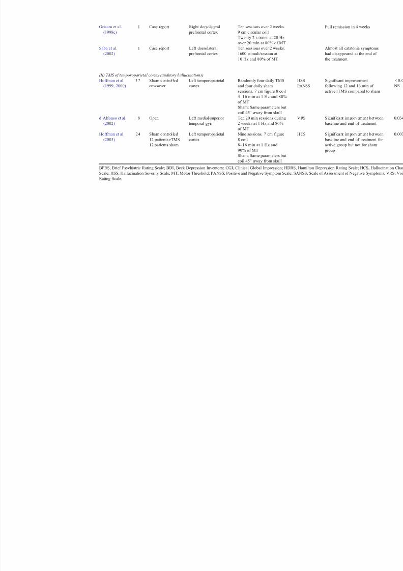
http://slidepdf.com/reader/full/tms-in-the-investigation-and-treatment-of-schizophreniapdf 11/16
Grisaru et al.
(1998c)
1 Case report Right dorsolateral
prefrontal cortex
Ten sessions over 2 weeks.
9 cm circular coil
Full remissi
Twenty 2 s trains at 20 Hz
over 20 min at 80% of MT
Saba et al.
(2002)
1 Case report Left dorsolateral
prefrontal cortex
Ten sessions over 2 weeks.
1600 stimuli/session at
10 Hz and 80% of MT
Almost all c
had disappe
the treatmen
(II) TMS of temporoparietal cortex (auditory hallucinations)
Hoffman et al.
(1999, 2000)
12 Sham controlled
crossover
Left temporoparietal
cortex
Randomly four daily TMS
and four daily sham
sessions. 7 cm figure 8 coil
HSS
PANSS
Significant
following 1
active rTMS
4–16 min at 1 Hz and 80%
of MT
Sham: Same parameters but
coil 45j away from skull
d’Alfonso et al.
(2002)
8 Open Left medial/superior
temporal gyri
Ten 20 min sessions during
2 weeks at 1 Hz and 80%
of MT
VRS Significant
baseline and
Hoffman et al.
(2003)
24 Sham controlled
12 patients rTMS
12 patients sham
Left temporoparietal
cortex
Nine sessions. 7 cm figure
8 coil
8–16 min at 1 Hz and90% of MT
Sham: Same parameters but
coil 45j away from skull
HCS Significant
baseline and
active groupgroup
BPRS, Brief Psychiatric Rating Scale; BDI, Beck Depression Inventory; CGI, Clinical Global Impression; HDRS, Hamilton Depression Rating
Scale; HSS, Hallucination Severity Scale; MT, Motor Threshold; PANSS, Positive and Negative Symptom Scale; SANSS, Scale of Assessment
Rating Scale.
7/27/2019 TMS in the investigation and treatment of schizophrenia.pdf
http://slidepdf.com/reader/full/tms-in-the-investigation-and-treatment-of-schizophreniapdf 12/16
et al., 2002). In this case, the TMS effects may
resemble those induced with electroconvulsive thera-
py, which is known to be an effective treatment for
catatonia. In summary, although these studies indicatethat high frequency TMS to prefrontal cortex may be
effective for treating certain symptoms of schizophre-
nia, larger controlled trials using consistent stimula-
tion parameters are necessary to establish the efficacy
of TMS in the treatment of schizophrenia.
8. TMS of temporoparietal cortex to treat auditory
hallucinations
Recent studies have provided interesting findings
on the effectiveness of TMS applied to one particular
brain area to specifically treat auditory hallucinations
(d’Alfonso et al., 2002; Hoffman et al., 1999, 2000,
2003). A previous study suggested that auditory
hallucinations may stem from abnormalities in brain
areas that are involved in the perception of speech.
Silbersweig et al. (1995) performed PET scans on six
patients with schizophrenia who were hallucinating at
the time and demonstrated increased blood flow in the
left temporoparietal auditory linguistic association
cortex. Based on the finding that long trains (15–30
min) of low frequency (1 Hz) TMS decreases activityin stimulated brain areas (Wassermann et al., 1998),
Hoffman et al. (1999, 2000) treated patients with
schizophrenia that had frequent auditory hallucina-
tions with 1 Hz TMS at 80% of motor threshold to the
left temporo-parietal cortex using a figure-of-eight
coil. In a double-blind crossover designed pilot study,
12 medicated patients underwent TMS and sham
TMS each for 4 days. The stimulation duration was
gradually increased from 4 to 16 min/day. Auditory
hallucinations were rated every day using a scale that
assessed the loudness, frequency, content and level of distress from the hallucinations. Eight of the patients
reported a significant improvement in auditory hallu-
cinations with TMS and the improvement reached
significance following the third and fourth days of
stimulation. Four of the patients had negligible or no
improvement. Other symptoms of schizophrenia did
not significantly change with the treatment. In follow
up assessments, the auditory hallucinations were
found to recur from 1 day to 2 months post-treatment.
It was interesting in this study that five patients taking
anticonvulsive medications had less of a treatment
effect than patients who were not taking these med-
ications. Several previous studies have indicated that
various anticonvulsants can increase cortical inhibi-tion and the threshold for cortical excitation measured
with TMS (Manganotti et al., 1999; Rizzo et al., 2001;
Ziemann et al., 1996).
Hoffman et al. (2003) recently followed their pilot
study with a trial of 24 patients with medication
resistant auditory hallucinations. Again, the left tem-
poroparietal cortex was stimulated at 1 Hz but the
intensity and total number of stimulations was higher
than in the previous studies. Twelve patients received
active TMS at 90% of motor threshold for 8 min on
day 1, 12 min on day 2 and 16 min on days 3 to 9. The
other 12 patients had sham stimulation with the
stimulation parameters but the coil was angled 45j
away from the head. Nine of the sham patients
received a subsequent unblinded trial of active TMS.
The active group had a significant linear decrease in
hallucination frequency during the study and they also
reported a significant decrease in distraction caused by
the hallucinations. These measures did not change
significantly in the sham group. Other hallucination
ratings such as loudness, duration of voices and level
of distress did not differ between the groups. All
patients receiving active TMS who reported morethan 20% improvement in the hallucination rating
scale score were followed by telephone for 1 year.
At 15 weeks, 52% of patients had sustained improve-
ment but at week 52 they were down to approximately
25%. No significant changes were found on measures
of general psychopathologic symptoms (PANSS) or
neuropsychological tests.
A recent open trial of eight schizophrenia patients
with medication resistant auditory hallucinations dem-
onstrated a modest improvement in seven patients
after 2 weeks of daily 20 min TMS of the auditorycortex (middle and superior temporal gyri) at 1 Hz and
80% of motor threshold (d’Alfonso et al., 2002).
9. Conclusions and future directions
The application of TMS in basic neurophysiolog-
ical and neuropsychiatric research has been rapidly
expanding since its introduction in 1985. TMS is a
noninvasive method that can be employed to study
H.M. Haraldsson et al. / Schizophrenia Research 71 (2004) 1–16 12

7/27/2019 TMS in the investigation and treatment of schizophrenia.pdf
http://slidepdf.com/reader/full/tms-in-the-investigation-and-treatment-of-schizophreniapdf 13/16
motor cortex excitability and cortical inhibitory mech-
anisms. A growing number of studies using TMS-
based paradigms support the notion that cortical
inhibition may be deficient in patients with schizo- phrenia. However, the use of TMS as a diagnostic tool
for psychiatric disorders is still in its infancy and
confounding factors related to the variability of stim-
ulation parameters, the severity and duration of the
disease, and the use of medications need to be
resolved before compelling conclusions can be drawn.
On the therapeutic side, initial studies using TMS
on subjects with schizophrenia have provided some
disappointing as well as some encouraging results.
The latter include the reduction of auditory hallucina-
tions with slow TMS over auditory cortex and an
improvement of psychotic symptoms after 2 weeks of
high frequency TMS over left prefrontal cortex (Hoff-
man et al., 2003; Rollnik et al., 2000). It will be
interesting to see whether these studies will be con-
firmed with more patients and longer follow-up peri-
ods. Moreover, it will be important to compare the
therapeutic benefits of TMS with those of standard
treatments, although a truly ideal placebo condition
for TMS remains difficult to envision.
One of the most promising new developments is
the ability to combine TMS with functional brain
imaging. In such paradigms, TMS pulses are deliveredover a cortical region while simultaneously recording
brain activity patterns using PET (Fox et al., 1997;
Paus et al., 1997; Kimbrell et al., 2002), fMRI
(Bohning et al., 1998, 1999, 2000a,b; Nahas et al.,
2001) or high-resolution EEG (Ilmoniemi et al., 1997;
Komssi et al., 2002). These approaches make it
possible to assess not only the cortical activity in-
duced under the TMS coil but also the influence that
the stimulated area exerts onto other brain areas—its
effective connectivity.
The initial studies probing the effective connectiv-ity of cortical regions have so far been performed in
healthy subjects. However, studies of effective con-
nectivity may be especially revealing when applied to
psychiatric disorders to explore the possibility of
disease-related alterations in connectivity between
critical brain regions. For example, a number of recent
neurobiological and neuroimaging studies indicate
that some symptoms of schizophrenia may result from
impaired functional integration of multiple brain areas
rather than from a malfunction of one single area
(Glantz and Lewis, 1997; Karson et al., 1999; Sele-
mon and Goldman-Rakic, 1999; Tononi and Edelman,
2000). If a combination of TMS and neuroimaging
were to uncover a dysfunction in the connections between specific brain areas in patients with schizo-
phrenia, a potential treatment would be to selectively
stimulate these connections with therapeutic doses of
TMS. As suggested by studies of TMS for treating
auditory hallucinations, stimulation of specific brain
sites for targeting different symptoms (cognitive,
positive and negative) may become an effective treat-
ment for this complex disorder.
Acknowledgements
This work was supported by a grant from the
families of Donald and Patricia Cheney and Jack and
Patricia Lane. The authors would also like to thank
two anonymous reviewers for helpful suggestions.
References
Abarbanel, J.M., Lemberg, T., Yaroslavski, U., Grisaru, N., Bel-
maker, R.H., 1996. Electrophysiological responses to transcra-
nial magnetic stimulation in depression and schizophrenia. Biol.
Psychiatry 40 (2), 148–150.
Amassian, V.E., Cracco, R.Q., Maccabee, P.J., Cracco, J.B., Rudell,
A., Eberle, L., 1989. Suppression of visual perception by mag-
netic coil stimulation of human occipital cortex. Electroencepha-
logr. Clin. Neurophysiol. 74 (6), 458– 462.
Andreasen, N.C., O’Leary, D.S., Flaum, M., Nopoulos, P., Watkins,
G.L., Boles Ponto, L.L., Hichwa, R.D., 1997. Hypofrontality in
schizophrenia: distributed dysfunctional circuits in neuroleptic-
naive patients. Lancet 349 (4067), 1730–1734.
Barker, A.T., 2002. The history and basic principles of magnetic
nerve stimulation. In: Pascual-Leone, A., Davey, N., Rothwell,
J., Wassermann, E.M., Puri, B.K. (Eds.), Handbook of Trans-
cranial Magnetic Stimulation, 1st ed. Oxford Univ. Press, New
York, pp. 3 – 17.Barker, A.T., Jalinous, R., Freeston, I.L., 1985. Non-invasive mag-
netic stimulation of human motor cortex. Lancet 1 (8437),
1106–1107.
Bohning, D.E., Shastri, A., Nahas, Z., Lorberbaum, J.P., Andersen,
S.W., Dannels, W.R., Haxthausen, E.U., Vincent, D.J., George,
M.S., 1998. Echoplanar BOLD fMRI of brain activation in-
duced by concurrent transcranial magnetic stimulation. Invest.
Radiol. 33 (6), 336– 340.
Bohning, D.E., Shastri, A., McConnell, K.A., Nahas, Z., Lorber-
baum, J.P., Roberts, D.R., Teneback, C., Vincent, D.J., George,
M.S., 1999. A combined TMS/fMRI study of intensity-depend-
ent TMS over motor cortex. Biol. Psychiatry 45 (4), 385–394.
H.M. Haraldsson et al. / Schizophrenia Research 71 (2004) 1–16 13
7/27/2019 TMS in the investigation and treatment of schizophrenia.pdf
http://slidepdf.com/reader/full/tms-in-the-investigation-and-treatment-of-schizophreniapdf 14/16
Bohning, D.E., Shastri, A., McGavin, L., McConnell, K.A., Nahas,
Z., Lorberbaum, J.P., Roberts, D.R., George, M.S., 2000a. Mo-
tor cortex brain activity induced by 1-Hz transcranial magnetic
stimulation is similar in location and level to that for volitional
movement. Invest. Radiol. 35 (11), 676– 683.Bohning, D.E., Shastri, A., Wassermann, E.M., Ziemann, U., Lor-
berbaum, J.P., Nahas, Z., Lomarev, M.P., George, M.S., 2000b.
BOLD-f MRI response to single-pulse transcranial magnetic
stimulation (TMS). J. Magn. Reson. Imaging 11 (6), 569–574.
Boroojerdi, B., Topper, R., Foltys, H., Meincke, U., 1999. Trans-
callosal inhibition and motor conduction studies in patients with
schizophrenia using transcranial magnetic stimulation. Br. J.
Psychiatry 175, 375–379.
Brasil-Neto, J.P., Cohen, L.G., Panizza, M., Nilsson, J., Roth, B.J.,
Hallett, M., 1992a. Optimal focal transcranial magnetic activa-
tion of the human motor cortex: effects of coil orientation, shape
of the induced current pulse, and stimulus intensity. J. Clin.
Neurophysiol. 9 (1), 132 – 136.
Brasil-Neto, J.P., McShane, L.M., Fuhr, P., Hallett, M., Cohen,
L.G., 1992b. Topographic mapping of the human motor cortex
with magnetic stimulation: factors affecting accuracy and re-
producibility. Electroencephalogr. Clin. Neurophysiol. 85 (1),
9–16.
Burt, T., Lisanby, S.H., Sackeim, H.A., 2002. Neuropsychiatric
applications of transcranial magnetic stimulation: a meta-analy-
sis. Int. J. Neuropsychopharmacol. 5 (1), 73–103.
Chen, R., Classen, J., Gerloff, C., Celnik, P., Wassermann, E.M.,
Hallett, M., Cohen, L.G., 1997. Depression of motor cortex
excitability by low-frequency transcranial magnetic stimulation.
Neurology 48 (5), 1398 – 1403.
Cicinelli, P., Traversa, R., Bassi, A., Scivoletto, G., Rossini, P.M.,
1997. Interhemispheric differences of hand muscle representa-tion in human motor cortex. Muscle Nerve 20 (5), 535–542.
Cohen, L.G., Roth, B.J., Nilsson, J., Dang, N., Panizza, M.,
Bandinelli, S., Friauf, W., Hallett, M., 1990. Effects of coil
design on delivery of focal magnetic stimulation. Technical
considerations. Electroencephalogr. Clin. Neurophysiol. 75
(4), 350–357.
Cohen, E., Bernardo, M., Masana, J., Arrufat, F.J., Navarro, V.,
Valls-Sole, J., Boget, T., Barrantes, N., Catarineu, S., Font,
M., Lomena, F.J., 1999. Repetitive transcranial magnetic stim-
ulation in the treatment of chronic negative schizophrenia: a
pilot study. J. Neurol. Neurosurg. Psychiatry 67 (1), 129 – 130.
Cracco, R.Q., Amassian, V.E., Maccabee, P.J., Cracco, J.B., 1989.
Comparison of human transcallosal responses evoked by mag-
netic coil and electrical stimulation. Electroencephalogr. Clin. Neurophysiol. 74 (6), 417 – 424.
d’Alfonso, A.A., Aleman, A., Kessels, R.P., Schouten, E.A., Post-
ma, A., van Der Linden, J.A., Cahn, W., Greene, Y., de Haan,
E.H., Kahn, R.S., 2002. Transcranial magnetic stimulation of
left auditory cortex in patients with schizophrenia: effects on
hallucinations and neurocognition. J. Neuropsychiatry Clin.
Neurosci. 14 (1), 77 – 79.
Daskalakis, Z.J., Christensen, B.K., Chen, R., Fitzgerald, P.B., Zi-
pursky, R.B., Kapur, S., 2002. Evidence for impaired cortical
inhibition in schizophrenia using transcranial magnetic stimula-
tion. Arch. Gen. Psychiatry 59 (4), 347–354.
Davey, N.J., Puri, B.K., Lewis, H.S., Lewis, S.W., Ellaway, P.H.,
1997. Effects of antipsychotic medication on electromyographic
responses to transcranial magnetic stimulation of the motor cor-
tex in schizophrenia. J. Neurol. Neurosurg. Psychiatry 63 (4),
468–473.Di Lazzaro, V., Oliviero, A., Mazzone, P., Pilato, F., Saturno, E.,
Dileone, M., Insola, A., Tonali, P.A., Rothwell, J.C., 2002.
Short-term reduction of intracortical inhibition in the human
motor cortex induced by repetitive transcranial magnetic stim-
ulation. Exp. Brain Res. 147 (1), 108–113.
Feinsod, M., Kreinin, B., Chistyakov, A., Klein, E., 1998. Prelimi-
nary evidence for a beneficial effect of low-frequency, repetitive
transcranial magnetic stimulation in patients with major depres-
sion and schizophrenia. Depress. Anxiety 7 (2), 65–68.
Ferbert, A., Priori, A., Rothwell, J.C., Day, B.L., Colebatch, J.G.,
Marsden, C.D., 1992. Interhemispheric inhibition of the human
motor cortex. J. Physiol. 453 (1), 525–546.
Fitzgerald, P.B., Brown, T.L., Daskalakis, Z.J., 2002a. The ap-
plication of transcranial magnetic stimulation in psychiatry
and neurosciences research. Acta Psychiatr. Scand. 105 (5),
324–340.
Fitzgerald, P.B., Brown, T.L., Daskalakis, Z.J., deCastella, A., Kul-
karni, J., 2002b. A study of transcallosal inhibition in schizo-
phrenia using transcranial magnetic stimulation. Schizophr. Res.
56 (3), 199–209.
Fitzgerald, P.B., Brown, T.L., Daskalakis, Z.J., Kulkarni, J., 2002c.
A transcranial magnetic stimulation study of inhibitory deficits
in the motor cortex in patients with schizophrenia. Psychiatry
Res. 114 (1), 11–22.
Fitzgerald, P.B., Brown, T.L., Daskalakis, Z.J., Kulkarni, J.,
2002d. A transcranial magnetic stimulation study of the effects
of olanzapine and risperidone on motor cortical excitability in patients with schizophrenia. Psychopharmacology (Berl.) 162
(1), 74 –81.
Fox, P., Ingham, R., George, M.S., Mayberg, H., Ingham, J.,
Roby, J., Martin, C., Jerabek, P., 1997. Imaging human intra-
cerebral connectivity by PET during TMS. NeuroReport 8
(12), 2787 – 2791.
Freedman, R., Adler, L.E., Myles-Worsley, M., Nagamoto, H.T.,
Miller, C., Kisley, M., McRae, K., Cawthra, E., Waldo, M.,
1996. Inhibitory gating of an evoked response to repeated audi-
tory stimuli in schizophrenic and normal subjects. Human re-
cordings, computer simulation, and an animal model. Arch.
Gen. Psychiatry 53 (12), 1114– 1121.
Frith, C.D., Blakemore, S., Wolpert, D.M., 2000. Explaining the
symptoms of schizophrenia: abnormalities in the awareness of action. Brain Res. Rev. 31 (2– 3), 357– 363.
Fuhr, P., Agostino, R., Hallett, M., 1991. Spinal motor neuron ex-
citability during the silent period after cortical stimulation. Elec-
troencephalogr. Clin. Neurophysiol. 81 (4), 257–262.
Geller, V., Grisaru, N., Abarbanel, J.M., Lemberg, T., Belmaker,
R.H., 1997. Slow magnetic stimulation of prefrontal cortex in
depression and schizophrenia. Prog. Neuropsychopharmacol.
Biol. Psychiatry 21 (1), 105–110.
George, M.S., Lisanby, S.H., Sackeim, H.A., 1999. Transcranial
magnetic stimulation: applications in neuropsychiatry. Arch.
Gen. Psychiatry 56 (4), 300–311.
H.M. Haraldsson et al. / Schizophrenia Research 71 (2004) 1–16 14
7/27/2019 TMS in the investigation and treatment of schizophrenia.pdf
http://slidepdf.com/reader/full/tms-in-the-investigation-and-treatment-of-schizophreniapdf 15/16
Glantz, L.A., Lewis, D.A., 1997. Reduction of synaptophysin im-
munoreactivity in the prefrontal cortex of subjects with schizo-
phr eni a. Reg ion al and diagn ost ic spe cif ici ty. Arc h. Gen .
Psychiatry 54 (10), 943–952.
Grafman, J., Pascual-Leone, A., Alway, D., Nichelli, P., Gomez-Tortosa, E., Hallett, M., 1994. Induction of a recall deficit by
rapid-rate transcranial magnetic stimulation. NeuroReport 5 (9),
1157–1160.
Greenberg, B.D., George, M.S., Martin, J.D., Benjamin, J.,
Schlaepfer, T.E., Altemus, M., Wassermann, E.M., Post, R.M.,
Murphy, D.L., 1997. Effect of prefrontal repetitive transcranial
magnetic stimulation in obsessive–compulsive disorder: a pre-
liminary study. Am. J. Psychiatry 154 (6), 867– 869.
Grisaru, N., Amir, M., Cohen, H., Kaplan, Z., 1998a. Effect of
transcranial magnetic stimulation in posttraumatic stress disor-
der: a preliminary study. Biol. Psychiatry 44 (1), 52–55.
Grisaru, N., Chudakov, B., Yaroslavsky, Y., Belmaker, R.H., 1998b.
Transcranial magnetic stimulation in mania: a controlled study.
Am. J. Psychiatry 155 (11), 1608–1610.
Grisaru, N., Chudakov, B., Yaroslavsky, Y., Belmaker, R.H., 1998c.
Catatonia treated with transcranial magnetic stimulation. Am. J.
Psychiatry 155 (11), 1630.
Hallett, M., 1995. Transcranial magnetic stimulation. Negative ef-
fects. Adv. Neurol. 67, 107 – 113.
Hallett, M., 2000. Transcranial magnetic stimulation and the human
brain. Nature 406 (6792), 147 – 150.
Hoffman, R.E., Boutros, N.N., Berman, R.M., Roessler, E.,
Belger, A., Krystal, J.H., Charney, D.S., 1999. Transcranial
magnetic stimulation of left temporoparietal cortex in three
patients reporting hallucinated ‘‘voices’’. Biol. Psychiatry 46
(1), 130–132.
Hoffman, R.E., Boutros, N.N., Hu, S., Berman, R.M., Krystal, J.H.,Charney, D.S., 2000. Transcranial magnetic stimulation and
auditory hallucinations in schizophrenia. Lancet 355 (9209),
1073–1075.
Hoffman, R.E., Hawkins, K.A., Gueorguieva, R., Boutros, N.N.,
Rachid, F., Carroll, K., Krystal, J.H., 2003. Transcranial mag-
netic stimulation of left temporoparietal cortex and medication-
resistant auditory hallucinations. Arch. Gen. Psychiatry 60 (1),
49–56.
Hoppner, J., Kunesch, E., Grossmann, A., Tolzin, C.J., Schulz, M.,
Schlafke, D., Ernst, K., 2001. Dysfunction of transcallosally
mediated motor inhibition and callosal morphology in patients
with schizophrenia. Acta Psychiatr. Scand. 104 (3), 227 – 235.
Ilmoniemi, R.J., Virtanen, J., Ruohonen, J., Karhu, J., Aronen, H.J.,
Naatanen, R., Katila, T., 1997. Neuronal responses to magneticstimulation reveal cortical reactivity and connectivity. NeuroRe-
port 8 (16), 3537 – 3540.
Inghilleri, M., Berardelli, A., Cruccu, G., Manfredi, M., 1993. Si-
lent period evoked by transcranial stimulation of the human
cortex and cervicomedullary junction. J. Physiol. 466 (1),
521–534.
Kammer, T., 1999. Phosphenes and transient scotomas induced by
magnetic stimulation of the occipital lobe: their topographic
relationship. Neuropsychologia 37 (2), 191–198.
Karson, C.N., Mrak, R.E., Schluterman, K.O., Sturner, W.Q.,
Sheng, J.G., Griffin, W.S., 1999. Alterations in synaptic proteins
and their encoding mRNAs in prefrontal cortex in schizophre-
nia: a possible neurochemical basis for ‘hypofrontality’. Mol.
Psychiatry 4 (1), 39– 45.
Kimbrell, T.A., Dunn, R.T., George, M.S., Danielson, A.L., Willis,
M.W., Repella, J.D., Benson, B.E., Herscovitch, P., Post, R.M.,Wassermann, E.M., 2002. Left prefrontal-repetitive transcranial
magnetic stimulation (rTMS) and regional cerebral glucose me-
tabolism in normal volunteers. Psychiatry Res. Neuroimaging
115 (3), 101– 113.
Klein, E., Kreinin, I., Chistyakov, A., Koren, D., Mecz, L., Marmur,
P.D., Ben-Shachar, D., Feinsod, M., 1999. Therapeutic efficacy
of right prefrontal slow repetitive transcranial magnetic stimu-
lation in major depression: a double-blind controlled study.
Arch. Gen. Psychiatry 56 (4), 315–320.
Komssi, S., Aronen, H.J., Huttunen, J., Kesaniemi, M., Soinne, L.,
Nikouline, V.V., Ollikainen, M., Roine, R.O., Karhu, J., Savo-
lainen, S., Ilmoniemi, R.J., 2002. Ipsi- and contralateral EEG
reactions to transcranial magnetic stimulation. Clin. Neurophy-
siol. 113 (2), 175–184.
Kujirai, T., Caramia, M.D., Rothwell, J.C., Day, B.L., Thompson,
A., Ferbert, A., Wroe, S., Asselman, P., Marsden, C.D., 1993.
Corticocortical inhibition in human motor cortex. J. Physiol. 471
(1), 501–519.
Lisanby, S.H., Luber, B., Perera, T., Sackeim, H.A., 2000. Trans-
cranial magnetic stimulation: applications in basic neuroscience
and neuropsychopharmacology. Int. J. Neuropsychopharmacol.
3, 259–273.
Lisanby, S.H., Gutman, D., Luber, B., Schroeder, C., Sackeim,
H.A., 2001. Sham TMS: intracerebral measurement of the in-
duced electrical field and the induction of motor-evoked poten-
tials. Biol. Psychiatry 49 (5), 460–463.
Lisanby, S.H., Kinnunen, L.H., Crupain, M.J., 2002. Applicationsof TMS to therapy in psychiatry. J. Clin. Neurophysiol. 19 (4),
344–360.
Manganotti, P., Bongiovanni, L.G., Zanette, G., Turazzini, M., Fia-
schi, A., 1999. Cortical excitability in patients after loading
doses of lamotrigine: a study with magnetic brain stimulation.
Epilepsia 40 (3), 316–321.
McCarley, R.W., Faux, S.F., Shenton, M.E., Nestor, P.G., Adams,
J., 1991. Event-related potentials in schizophrenia: their biolog-
ical and clinical correlates and a new model of schizophrenic
pathophysiology. Schizophr. Res. 4 (2), 209 – 231.
Meyer, B.U., Roricht, S., Grafin von Einsiedel, H., Kruggel, F.,
Weindl, A., 1995. Inhibitory and excitatory interhemispheric
transfers between motor cortical areas in normal humans and
patients with abnormalities of the corpus callosum. Brain 118(Pt. 2), 429–440.
Mills, K.R., Nithi, K.A., 1997. Corticomotor threshold to magnetic
stimulation: normal values and repeatability. Muscle Nerve 20
(9), 570–576.
Nahas, Z., McConnell, K.C.S., Molloy, M., Oliver, N.C., Risch,
S.C., Christie, S., Arana, G.W., George, M.S., 1999. Could left
prefrontal rTMS modify negative symptoms and attention in
schizophrenia? Biol. Psychiatry 45 (8, Suppl. 1), 37S.
Nahas, Z., Lomarev, M., Roberts, D.R., Shastri, A., Lorberbaum,
J.P., Teneback, C., McConnell, K., Vincent, D.J., Li, X., George,
M.S., Bohning, D.E., 2001. Unilateral left prefrontal transcranial
H.M. Haraldsson et al. / Schizophrenia Research 71 (2004) 1–16 15

7/27/2019 TMS in the investigation and treatment of schizophrenia.pdf
http://slidepdf.com/reader/full/tms-in-the-investigation-and-treatment-of-schizophreniapdf 16/16
magnetic stimulation (TMS) produces intensity-dependent bilat-
eral effects as measured by interleaved BOLD fMRI. Biol. Psy-
chiatry 50 (9), 712 – 720.
Nakamura, H., Kitagawa, H., Kawaguchi, Y., Tsuji, H., 1997. Intra-
cortical facilitation and inhibition after transcranial magneticstimulation in conscious humans. J. Physiol. 498 (3), 817–823.
Olney, J.W., Farber, N.B., 1995. Glutamate receptor dysfunction
and schizophrenia. Arch. Gen. Psychiatry 52 (12), 998–1007.
Pascual-Leone, A., Hallett, M., 1994. Induction of errors in a de-
layed response task by repetitive transcranial magnetic stimula-
tion of the dorsolateral prefrontal cortex. NeuroReport 5 (18),
2517–2520.
Pascual-Leone, A., Tormos, J.M., Keenan, J., Tarazona, F., Canete,
C., Catala, M.D., 1998. Study and modulation of human cortical
excitability with transcranial magnetic stimulation. J. Clin. Neu-
rophysiol. 15 (4), 333–343.
Pascual-Leone, A., Manoach, D.S., Birnbaum, R., Goff, D.C.,
2002. Motor cortical excitability in schizophrenia. Biol. Psy-
chiatry 52 (1), 24–31.
Paus, T., Jech, R., Thompson, C.J., Comeau, R., Peters, T., Evans,
A.C., 1997. Transcranial magnetic stimulation during positron
emission tomography: a new method for studying connectivity
of the human cerebral cortex. J. Neurosci. 17 (9), 3178–3184.
Pridmore, S., Belmaker, R., 1999. Transcranial magnetic stimula-
tion in the treatment of psychiatric disorders. Psychiatry Clin.
Neurosci. 53 (5), 541 – 548.
Puri, B.K., Davey, N.J., Ellaway, P.H., Lewis, S.W., 1996. An in-
vestigation of motor function in schizophrenia using transcranial
magnetic stimulation of the motor cortex. Br. J. Psychiatry 169
(6), 690–695.
Ridding, M.C., Inzelberg, R., Rothwell, J.C., 1995. Changes in
excitability of motor cortical circuitry in patients with Parkin-son’s disease. Ann. Neurol. 37 (2), 181–188.
Rizzo, V., Quartarone, A., Bagnato, S., Battaglia, F., Majorana, G.,
Girlanda, P., 2001. Modification of cortical excitability induced
by gabapentin: a study by transcranial magnetic stimulation.
Neurol. Sci. 22 (3), 229 – 232.
Rollnik, J.D., Huber, T.J., Mogk, H., Siggelkow, S., Kropp, S.,
Dengler, R., Emrich, H.M., Schneider, U., 2000. High frequency
repetitive transcranial magnetic stimulation (rTMS) of the dor-
solateral prefrontal cortex in schizophrenic patients. NeuroRe-
port 11 (18), 4013 – 4015.
Rossini, P.M., Barker, A.T., Berardelli, A., Caramia, M.D., Caruso,
G., Cracco, R.Q., Dimitrijevic, M.R., Hallett, M., Katayama, Y.,
Lucking, C.H., 1994. Non-invasive electrical and magnetic
stimulation of the brain, spinal cord and roots: basic principlesand procedures for routine clinical application. Report of an
IFCN committee. Electroencephalogr. Clin. Neurophysiol. 91
(2), 79–92.
Ruohonen, J., Ollikainen, M., Nikouline, V., Virtanen, J., Ilmonie-
mi, R.J., 2000. Coil design for real and sham transcranial mag-
netic stimulation. IEEE Trans. Biomed. Eng. 47 (2), 145–148.
Saba, G., Rocamora, J.F., Kalalou, K., Benadhira, R., Plaze, M.,
Aubriot-Delmas, B., 2002. Catatonia and transcranial magnetic
stimulation. Am. J. Psychiatry 159 (10), 1794.
Sanger, T.D., Garg, R.R., Chen, R., 2001. Interactions between two
different inhibitory systems in the human motor cortex. J. Phys-
iol. 530 (2), 307–317.
Selemon, L.D., Goldman-Rakic, P.S., 1999. The reduced neuropil
hypothesis: a circuit based model of schizophrenia. Biol. Psy-chiatry 45 (1), 17–25.
Silbersweig, D.A., Stern, E., Frith, C., Cahill, C., Holmes, A.,
Grootoonk, S., Seaward, J., McKenna, P., Chua, S.E., Schnorr,
L., et al., 1995. A functional neuroanatomy of hallucinations in
schizophrenia. Nature 378 (6553), 176 – 179.
Terao, Y., Ugawa, Y., 2002. Basic mechanisms of TMS. J. Clin.
Neurophysiol. 19 (4), 322 – 343.
Tononi, G., Edelman, G.M., 2000. Schizophrenia and the mecha-
nisms of conscious integration. Brain Res. Rev. 31 (2 – 3), 391 –
400.
Triggs, W.J., Macdonell, R.A., Cros, D., Chiappa, K.H., Shahani,
B.T., Day, B.J., 1992. Motor inhibition and excitation are inde-
pendent effects of magnetic cortical stimulation. Ann. Neurol.
32 (3), 345–351.
Triggs, W.J., Calvanio, R., Macdonell, R.A., Cros, D., Chiappa,
K.H., 1994. Physiological motor asymmetry in human handed-
ness: evidence from transcranial magnetic stimulation. Brain
Res. 636 (2), 270–276.
von Giesen, H.J., Roick, H., Benecke, R., 1994. Inhibitory actions
of motor cortex following unilateral brain lesions as studied by
magnetic brain stimulation. Exp. Brain Res. 99 (1), 84–96.
Walsh, V., Rushworth, M., 1999. A primer of magnetic stimula-
tion as a tool for neuropsychology. Neuropsychologia 37 (2),
125–135.
Wassermann, E.M., 1998. Risk and safety of repetitive transcranial
magnetic stimulation: report and suggested guidelines from the
International Workshop on the Safety of Repetitive TranscranialMagnetic Stimulation, June 5 – 7, 1996. Electroencephalogr.
Clin. Neurophysiol. 108 (1), 1–16.
Wassermann, E.M., Wedegaertner, F.M., Ziemann, U., George,
M.R., Chen, R., 1998. Crossed reduction of human motor ex-
citability by 1 Hz transcranial magnetic stimulation. Neurosci.
Lett. 250 (3), 141– 144.
Wu, T., Sommer, M., Tergau, F., Paulus, W., 2000. Lasting influ-
ence of repetitive transcranial magnetic stimulation on intra-
cortical excitability in human subjects. Neurosci. Lett. 287 (1),
37–40.
Yager, J., Gitlin, M.J., 2000. Clinical manifestations of psychiatric
disorders. In: Sadock, B.J., Sadock, V.A. (Eds.), Comprehensive
Textbook of Psychiatry, vol. 1, 7th ed. Lippincott Williams &
Wilkins, Philadelphia, pp. 789–823.Ziemann, U., Lonnecker, S., Steinhoff, B.J., Paulus, W., 1996. Ef-
fects of antiepileptic drugs on motor cortex excitability in hu-
mans: a transcranial magnetic stimulation study. Ann. Neurol.
40 (3), 367–378.
Ziemann, U., Tergau, F., Bruns, D., Baudewig, J., Paulus, W., 1997.
Changes in human motor cortex excitability induced by dopa-
minergic and anti-dopaminergic drugs. Electroencephalogr.
Clin. Neurophysiol. 105 (6), 430–437.
H.M. Haraldsson et al. / Schizophrenia Research 71 (2004) 1–16 16
Related Documents