1 TLR-2 and TLR-9 are sensors of apoptosis in a mouse model of doxorubicin-induced acute inflammation Dmitri V. Krysko, 1,2 # Agnieszka Kaczmarek, 1,2 # Olga Krysko, 3 Liesbeth Heyndrickx, 1,2 Jerzy Woznicki, 1,2 Pieter Bogaert, 1,2 Anje Cauwels, 1,2 Nozomi Takahashi, 1,2 Stefan Magez, 4,5 Claus Bachert 3 , Peter Vandenabeele 1,2 * 1 Department for Molecular Biomedical Research, VIB, 9052 Ghent, Belgium; 2 Department of Biomedical Molecular Biology, Ghent University, Ghent, Belgium; 3 Upper Airway Research Laboratory, Department of Oto-Rhino-Laryngology, Ghent University Hospital, Ghent, Belgium; 4 Laboratory for Cellular and Molecular Immunology, Vrije Universiteit Brussel, Brussels, Belgium; 5 Department of Molecular and Cellular Interactions, VIB, Brussels, Belgium # These authors share first authorship *Communicating author: Department for Molecular Biomedical Research VIB-Ghent University Technologiepark 927, B-9052 Ghent (Zwijnaarde) Belgium Phone: 32 9 3313760; Fax: +32 9 3313609 E-mail: [email protected] peer-00614317, version 1 - 11 Aug 2011 Author manuscript, published in "Cell Death and Differentiation (2011)" DOI : 10.1038/cdd.2011.4

Welcome message from author
This document is posted to help you gain knowledge. Please leave a comment to let me know what you think about it! Share it to your friends and learn new things together.
Transcript
1
TLR-2 and TLR-9 are sensors of apoptosis in a mouse model of doxorubicin-induced acute
inflammation
Dmitri V. Krysko,1,2 # Agnieszka Kaczmarek,1,2 # Olga Krysko,3 Liesbeth Heyndrickx,1,2 Jerzy
Woznicki,1,2 Pieter Bogaert,1,2 Anje Cauwels,1,2 Nozomi Takahashi,1,2 Stefan Magez,4,5 Claus
Bachert3, Peter Vandenabeele1,2 *
1Department for Molecular Biomedical Research, VIB, 9052 Ghent, Belgium;
2Department of Biomedical Molecular Biology, Ghent University, Ghent, Belgium;
3Upper Airway Research Laboratory, Department of Oto-Rhino-Laryngology, Ghent University
Hospital, Ghent, Belgium;
4Laboratory for Cellular and Molecular Immunology, Vrije Universiteit Brussel, Brussels, Belgium;
5Department of Molecular and Cellular Interactions, VIB, Brussels, Belgium
#These authors share first authorship
*Communicating author:
Department for Molecular Biomedical Research
VIB-Ghent University
Technologiepark 927, B-9052 Ghent (Zwijnaarde)
Belgium
Phone: 32 9 3313760; Fax: +32 9 3313609
E-mail: [email protected]
peer
-006
1431
7, v
ersi
on 1
- 11
Aug
201
1Author manuscript, published in "Cell Death and Differentiation (2011)"
DOI : 10.1038/cdd.2011.4
2
Abstract
Anthracycline antibiotics are inducers of an immunogenic form of apoptosis that has
immunostimulatory properties due to the release of damage associated molecular patterns. To study
the mechanisms used by the innate immune system to sense this immunogenic form of cell death,
we established an in vivo model of cell death induced by intraperitoneal injection of doxorubicin, a
prototype of anthracyclines. The acute sterile inflammation in this model is characterized by rapid
influx of neutrophils and increased levels of IL-6 and MCP-1. We demonstrate that acute
inflammation induced by doxorubicin is associated with apoptosis of monocytes/macrophages and
that it is specific for doxorubicin, an immunogenic chemotherapeutic. Further, the inflammatory
response is significantly reduced in mice deficient in MyD88, TLR-2 or TLR-9. Importantly, a
TLR-9 antagonist reduces the recruitment of neutrophils induced by doxorubicin. By contrast, the
acute inflammatory response is not affected in TRIFLps2 mutant mice and in TLR-3, TLR-4 and
caspase-1 knockout mice, which shows that the inflammasome does not play a major role in
doxorubicin-induced acute inflammation. Our findings provide important new insights into how the
innate immune system senses immunogenic apoptotic cells and clearly demonstrate that the TLR-
2/TLR-9-MyD88 signaling pathways play a central role in initiating the acute inflammatory
response to this immunogenic form of apoptosis.
Key words: doxorubicin, TLRs, apoptosis, neutrophils, DAMPs, immunogenic cell death
peer
-006
1431
7, v
ersi
on 1
- 11
Aug
201
1
3
Introduction
Apoptotic cell death is a tightly regulated physiological form of cell death, and one of its important
aspects is targeted elimination of apoptotic cells without induction of inflammation or tissue
scarring. This process serves as an integral part of homeostatic mechanisms. Apoptotic cells exert
anti-inflammatory effects by triggering the release TGF-β, IL-10, platelet-activating factor and
prostaglandin E2 from engulfing cells (1-3). In addition, it has been shown that direct contacts
between apoptotic cells and phagocytes contribute to the immunosuppressive effect of apoptotic
cells (4, 5). Apoptotic cells are cleared via a ‘zipper’-like mechanism of internalization (6). In vivo,
apoptotic cells are rapidly sensed and cleared not by neutrophils but preferentially by monocytes,
which are attracted to the site of cell death by the release of chemoattractants. Elliott et al. (7)
reported that supernatant obtained from apoptotic Jurkat cells, killed by UV or anti-Fas, induces
recruitment of monocytes in a P2Y2-dependent manner by releasing nucleotides such as ATP and
UTP. In fact, apoptotic cells generate negative signals that could prevent neutrophil recruitment and
dampen responsiveness of the innate immune system. Recently, it was shown that apoptotic Burkitt
lymphoma cells specifically inhibit neutrophil chemotaxis by secreting lactoferrin, a pleiotropic
glycoprotein with anti-inflammatory properties, but they do not affect monocyte chemotaxis (8).
The inhibition of neutrophil recruitment by apoptotic cells represents an effective mechanism for
limiting tissue injury and inflammation. It is known that the recruited neutrophils and the release of
their proteolytic enzymes contribute to many pathological inflammatory conditions, such as
ischemic injury of the heart (9), lung (10) and skeletal muscle (11), as well as to toxic insults to the
liver (12) and lung (13).
It was recently shown that chemotherapeutic agents, such as anthracyclines, can induce an
immunogenic form of apoptotic cell death (14, 15), which stimulates the induction of an adaptive
immune response that eradicates tumors. The immunostimulatory properties of these immunogenic
apoptotic cells are explained by the action of damage-associated molecular patterns (DAMPs) or
peer
-006
1431
7, v
ersi
on 1
- 11
Aug
201
1

4
cell death-associated molecules (CDAMs) (16, 17). DAMPs are mostly intracellular molecules that
acquire immunostimulatory activity following their secretion or release by stressed or dying cells.
Thus, immunogenic apoptotic cell death is characterized by surface exposure of calreticulin (CRT)
(18) and release of high mobility group box 1 protein (HMGB1) from the nucleus of dying tumor
cells into the extracellular space (14). Interestingly, DAMPs are recognized by host cells expressing
a set of receptors known as pattern-recognition receptors (PRR), such as Toll-like receptors (TLRs),
the nucleotide-binding oligomerization domain (NOD)-like receptors (NLRs), and RIG-I-like
receptors (RLRs) (19, 20). When these receptors are triggered by cells dying in an immunogenic or
non-physiological way, they rapidly initiate host defense responses. For example, it has been shown
that HMGB1 is required for the induction of a doxorubicin-induced anti-tumor immune response in
a TLR-4/MyD88 dependent pathway in mice (14). Several reports address the interaction of the
adaptive immune system with immunogenic apoptotic cells, but it is not fully understood how the
cells of the innate immune system, such as neutrophils, react to immunogenic apoptotic cells.
Given that doxorubicin induces immunogenic apoptotic cell death and that classical apoptotic cells
preferentially stimulate monocyte recruitment, we sought to determine how cells killed by
doxorubicin are sensed by the innate immune system. We established an in situ murine model of
apoptosis induction by intraperitoneal (i.p.) injection of doxorubicin. Intraperitoneal injection of
doxorubicin resulted in the generation of mostly apoptotic monocytes/macrophages and induced an
acute inflammatory response in the peritoneal cavity characterized by recruitment of neutrophils
and production of IL-6 and MCP-1. This acute inflammatory response was specific to immunogenic
chemotherapeutics because a non-immunogenic drug, mitomycin C, also induced cell death in the
peritoneal cavity but was incapable of eliciting neutrophil attraction. We found that MyD88 is
essential for the doxorubicin-induced acute inflammatory response and that it is required primarily
as an adaptor molecule in the TLR-2 and TLR-9 signaling pathways.
peer
-006
1431
7, v
ersi
on 1
- 11
Aug
201
1

5
Materials and Methods
Mice
For the kinetics and dose response experiments, female C57BL/6 and Balb/c mice (8–10 weeks old)
were purchased from Janvier (Bio Services BV, The Netherlands). The following mutant mice were
used: Myd88–/– (Balb/c background); TLR-2–/–, TLR-4–/– and TLR-9–/– (C57BL/6 background); TLR-
3–/– (B6129 background); and caspase-1-/- (6x back crossed to C57Bl/6). Mutant TRIFLps2 mice
(C57BL/6 background) were generated by random mutagenesis with ENU; these mice have a distal
frame shift error in a TRIF protein that impairs its function (21). For experiments with knockout
mice, wild type mice of appropriate background were used, and they were bred under the same
animal house conditions as the others. Mice were housed in a SPF facility with 12/12h light/dark
cycles and received water and food ad libitum. All experimental procedures were approved by the
local Ethics Committee of Ghent University–VIB. Although some mutants were of Balb/c and
mixed B6129 genetic backgrounds, we found that doxorubicin-induced sterile inflammatory
responses were not different in C57BL/6, Balb/c and B6129 mice (data not shown).
Doxorubicin-induced inflammation in vivo
Doxorubicin hydrochloride (Sigma-Aldricht, Bornem, Belgium) was freshly dissolved in sterile
LPS-free 0.9% NaCl (Braun-N.V.S.A, Diegem, Belgium) at 5 mM. Aliquots were frozen and used
before each experiment to ensure constant quality. Mice were injected i.p. with 10 mg/kg of
doxorubicin (or as otherwise indicated) in 0.2 ml of 0.9% NaCl. Equal volumes of 0.9% NaCl were
injected as negative controls. At different time points, animals were euthanized by CO2 exposure,
and their peritoneal cavities were washed with 8 ml of sterile PBS. The peritoneal lavages were
centrifuged at 450 g for 5 min and supernatants were stored at –20°C until analyzed for cytokines.
peer
-006
1431
7, v
ersi
on 1
- 11
Aug
201
1
6
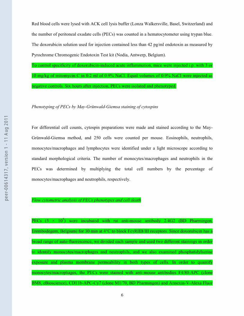
Red blood cells were lysed with ACK cell lysis buffer (Lonza Walkersville, Basel, Switzerland) and
the number of peritoneal exudate cells (PECs) was counted in a hematocytometer using trypan blue.
The doxorubicin solution used for injection contained less than 42 pg/ml endotoxin as measured by
Pyrochrome Chromogenic Endotoxin Test kit (Nodia, Antwerp, Belgium).
To control specificity of doxorubicin-induced acute inflammation, mice were injected i.p. with 3 or
10 mg/kg of mitomycin C in 0.2 ml of 0.9% NaCl. Equal volumes of 0.9% NaCl were injected as
negative controls. Six hours after injection, PECs were isolated and phenotyped.
Phenotyping of PECs by May-Grünwald-Giemsa staining of cytospins
For differential cell counts, cytospin preparations were made and stained according to the May-
Grünwald-Giemsa method, and 250 cells were counted per mouse. Eosinophils, neutrophils,
monocytes/macrophages and lymphocytes were identified under a light microscope according to
standard morphological criteria. The number of monocytes/macrophages and neutrophils in the
PECs was determined by multiplying the total cell numbers by the percentage of
monocytes/macrophages and neutrophils, respectively.
Flow cytometric analysis of PECs phenotypes and cell death
PECs (5 × 10⁵) were incubated with rat anti-mouse antibody 2.4G2 (BD Pharmingen,
Erembodegem, Belgium) for 30 min at 4°C to block FcγRIIB/III receptors. Since doxorubicin has a
broad range of auto-fluorescence, we divided each sample and used two different stainings in order
to identify monocytes/macrophages and neutrophils, and we also examined phosphatidylserine
exposure and plasma membrane permeability in both types of cells. In order to quantify
monocytes/macrophages, the PECs were stained with anti-mouse antibodies F4/80-APC (clone
BM8, eBioscience), CD11b-APC-Cy7 (clone M1/70, BD Pharmingen) and Annexin-V-Alexa Fluor
peer
-006
1431
7, v
ersi
on 1
- 11
Aug
201
1

7
488 (AnV, Invitrogen, Merelbeke, Belgium). To identify neutrophils, the PECs were stained with
anti-mouse antibodies Ly-6G-APC (clone 1A8, BD Pharmingen), CD11b-APC-Cy7 (clone M1/70,
BD Pharmingen) and Ann-V-Alexa Fluor 488 (Invitrogen). All the stainings were done for 30 min
at 4°C in annexin binding buffer (10 mM HEPES-NaOH, 1 mM MgCl2, 2.5 mM CaCl2, 5 mM KCl,
150 mM NaCl, pH 7.4). Just before flow cytometry analysis on BD LSR-II (BD Biosciences), 1.25
nM of Sytox Blue dead cell stain was added (Invitrogen). Data were acquired and analyzed by BD
FACSDiva software (BD Biosciences). Monocytes/macrophages and neutrophil numbers in the
PECs were determined by multiplying the total cell numbers by the percentage of CD11b+F4/80+
and CD11b+Ly6G+ cells, respectively. For all kinetics experiments, the PECs were stained only
with 1.25 nM of Sytox Red dead cell stain (Invitrogen) and analyzed on FACSCalibur (BD
Biosciences). Data were acquired and analyzed by CellQuest software (BD Biosciences). Since
Sytox Red and Sytox Blue stain cells with permeabilized plasma membrane in the same way, we
use the term “Sytox” for both stainings.
Caspase-3/7 enzymatic activity by fluorometry
The fluorogenic substrate assay for caspase-3/7 activity was carried out as described previously (22,
23). Briefly, PECs were washed in cold PBS and lysed in 150 µl of caspase lysis buffer (1% NP-40,
10 mM Tris–HCl, pH 7.4, 10 mM NaCl, 3 mM MgCl2, 1 mM PMSF, 0.3 mM aprotinin, and 1 mM
leupeptin) supplemented with 1 mM oxidized glutathione to block the catalytic cysteine of caspases
and to prevent their activation during lysis. Cell debris was removed by centrifugation and
DEVDase activity was determined by incubating 30 µl (25 ng protein) of the soluble fraction with
20 µM of Ac-DEVD-amc in 150 µl of cell-free system buffer containing 220 mM mannitol, 68 mM
sucrose, 2 mM MgCl2, 2 mM NaCl, 2.5 mM KH2PO4, 0.5 mM EDTA, 0.5 mM sodium pyruvate,
0.5 mM L-glutamine, 10 mM HEPES-NaOH (pH 7.4), and 10 mM dithiothreitol. The release of
fluorescent 7-amino-4-methylcoumarin was measured for 60 min at 2-min intervals by fluorometry
peer
-006
1431
7, v
ersi
on 1
- 11
Aug
201
1

8
(excitation at 360 nm and emission at 480 nm) (Cytofluor; PerSeptive Biosystems, Cambridge, MA,
USA). The maximal rate of increase in fluorescence was calculated ( F/min).
Intraperitoneal injection of MSU
Mice were injected i.p. with 2 mg/mouse of MSU crystals (Enzo Life Sciences, Zandhoven,
Belgium) in 200 µl of PBS. Mice injected with equal volumes of PBS served as negative controls.
Six hours after injection, PECs were isolated. They were analyzed for caspase3/7 activity by
fluorometry and phenotyped by flow cytometry, as described above.
Cytokine and chemokine analysis
Immunoreactive levels of MCP-1 and IL-6 were measured in peritoneal lavage by using a
Cytometric Bead Array (CBA) mouse soluble protein flex set system (BD Biosciences). The
samples were prepared according to the manufacturers' instructions and analyzed on a FACSCalibur
(BD Biosciences). Data were acquired by CellQuest software (BD Biosciences) and analyzed by
FCAP array software (Soft Flow Hungary Ltd.).
Injection of TLR-9 antagonist
Wild type mice were injected i.p. with TLR-9 antagonist (ODN2088, 50 μg) or inactive ODN 2088
(50 μg, Invivogen, Toulouse, France) together with doxorubicin, and six hours later with inactive or
active ODN. PECs were obtained 16 hours after doxorubicin injection for cell phenotyping as
described previously.
HMGB1 analysis
peer
-006
1431
7, v
ersi
on 1
- 11
Aug
201
1
9
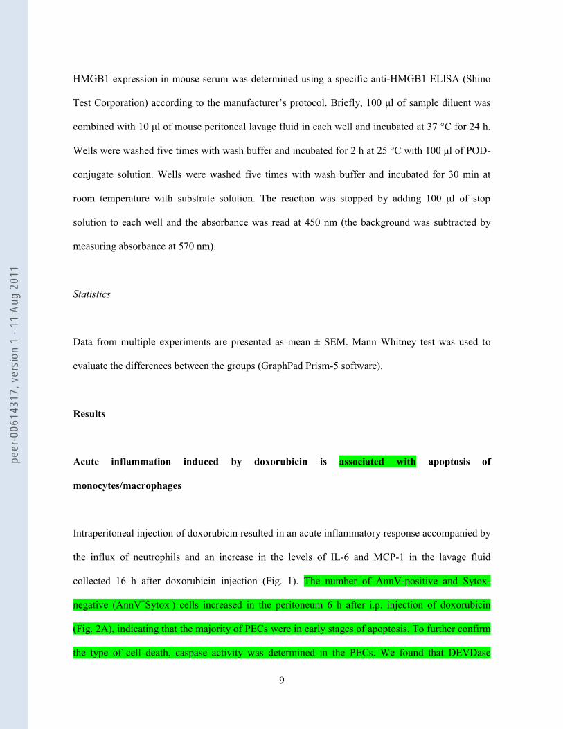
HMGB1 expression in mouse serum was determined using a specific anti-HMGB1 ELISA (Shino
Test Corporation) according to the manufacturer’s protocol. Briefly, 100 μl of sample diluent was
combined with 10 μl of mouse peritoneal lavage fluid in each well and incubated at 37 °C for 24 h.
Wells were washed five times with wash buffer and incubated for 2 h at 25 °C with 100 μl of POD-
conjugate solution. Wells were washed five times with wash buffer and incubated for 30 min at
room temperature with substrate solution. The reaction was stopped by adding 100 μl of stop
solution to each well and the absorbance was read at 450 nm (the background was subtracted by
measuring absorbance at 570 nm).
Statistics
Data from multiple experiments are presented as mean ± SEM. Mann Whitney test was used to
evaluate the differences between the groups (GraphPad Prism-5 software).
Results
Acute inflammation induced by doxorubicin is associated with apoptosis of
monocytes/macrophages
Intraperitoneal injection of doxorubicin resulted in an acute inflammatory response accompanied by
the influx of neutrophils and an increase in the levels of IL-6 and MCP-1 in the lavage fluid
collected 16 h after doxorubicin injection (Fig. 1). The number of AnnV-positive and Sytox-
negative (AnnV+Sytox-) cells increased in the peritoneum 6 h after i.p. injection of doxorubicin
(Fig. 2A), indicating that the majority of PECs were in early stages of apoptosis. To further confirm
the type of cell death, caspase activity was determined in the PECs. We found that DEVDase
peer
-006
1431
7, v
ersi
on 1
- 11
Aug
201
1

10
activity (caspase-3/7) was increased in these PECs at 6 h (Fig. 2C), confirming that they were dying
by apoptosis. By performing multi-color flow cytometry, we found that the majority of cells which
died apoptotically by doxorubicin were mainly monocytes/macrophages with some minor
neutrophils (Figs. 2A and 2B). Moreover, to exclude the possibility that the observed apoptotic cells
were not just regular dying neutrophils, we injected i.p. MSU, which induces strong neutrophil
recruitment (24). MSU induced significantly more neutrophil attraction than doxorubicin, but again
the number of apoptotic neutrophils in the peritoneal cavity was negligible. Also, no caspase3/7
activity was measured in PECs after i.p. injection of MSU (Fig. 2C). All these data indicate that
monocytes/macrophages represent the major cell population that dies by apoptosis after i.p.
injection of doxorubicin.
To determine whether neutrophil attraction is induced by the presence of apoptotic cells in the
peritoneum, we performed a dose titration experiment in which we investigated the relation between
the percent of Sytox+ cells and the number of attracted neutrophils as a function of doxorubicin
dose. We observed a dose-dependent increase in the percent of Sytox+ cells (Figs. 3A and 3C).
Moreover, there was a linear correlation between the percent of Sytox+ cells and neutrophil influx
(Fig. 3B). Notably, the recruitment of neutrophils increased slowly; it was high at 16 h and reached
a maximum at 38 h, which is considerably later than the peak of cell death observed at 6 h (Fig.
3D). This time lag between the peak of dead cells and the maximum number of neutrophils implies
that neutrophils are attracted after the induction of cell death and that it is associated with cell death.
We used this model to investigate how doxorubicin stimulates acute inflammation in the
peritoneum.
Acute inflammation induced by doxorubicin is specific for immunogenic chemotherapeutics
To determine whether an acute inflammatory response, as judged by neutrophil attraction, is
specific to the immunogenic anthracyclines, we injected i.p. mitomycin C, which induces a non-
peer
-006
1431
7, v
ersi
on 1
- 11
Aug
201
1

11
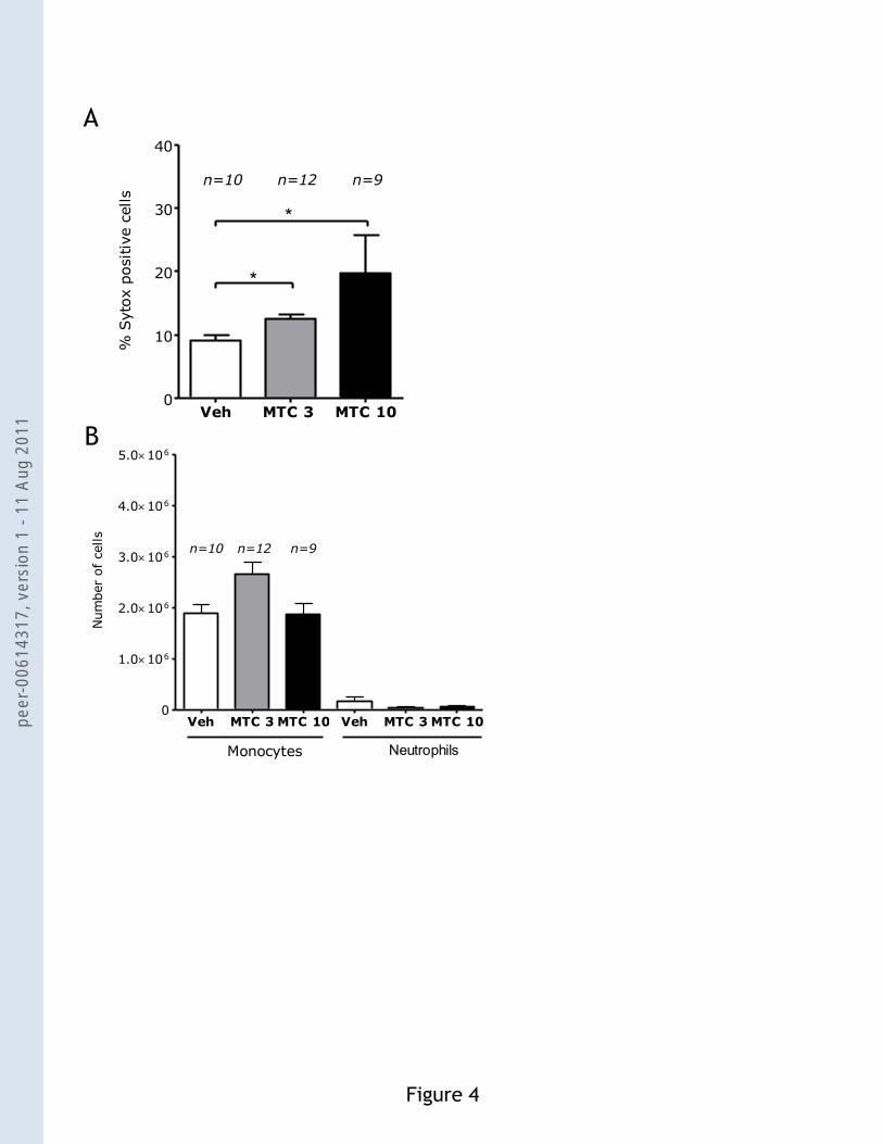
immunogenic cell death (18). Six hours after i.p. injection of mitomcyin C, there was a significant
increase of Sytox+ cells (Fig. 4A), but no increase in neutrophil attraction was detected (Fig. 4B).
This result indicates that the acute inflammatory response induced by doxorubicin is specific for
immunogenic chemotherapeutics.
The role of TLRs in doxorubicin-induced acute inflammation
TLRs recognize signature molecules derived either from pathogens (Pathogen Associated
Molecular Patterns, PAMPs) or from dead or damaged cells (DAMPs or CDAMs). When TLRs
recognize their ligands, they signal through the TIR adaptor protein MyD88, except for TLR-3,
which uses the Toll/IL-1R domain-containing adaptor inducing IFN-α (TRIF). To determine
whether TLRs are involved in the inflammation triggered by doxorubicin, we injected doxorubicin
i.p. in mice deficient in MyD88 (encoded by MyD88) and mice mutant for TRIFLps2 mice (21), and
the acute inflammatory response was evaluated by quantifying the influx of neutrophils. After 16 h,
wild-type and TRIFLps2 mice had abundant neutrophils in their abdominal cavities, but this response
was markedly less in MyD88-/- mice (Figs. 5A and 5B). Remarkably, the acute inflammatory
response in MyD88-deficient mice was reduced by 22 folds at 16 h (Fig. 5A). Consistent with the
reduced neutrophil infiltration in MyD88-deficient mice, the production of IL-6 and MCP-1 was
reduced by 12 and 6.5 folds, respectively (Fig. 6B). The percent of Sytox+ cells in the MyD88
deficient mice was not different from that in wild type mice (Fig. 3E), indicating that the cell death
process itself was not affected.
The requirement for MyD88 in doxorubicin-induced inflammation suggested that TLRs might be
involved. Therefore, we injected doxorubicin into mice deficient in various TLRs (TLR-2, TLR-3,
TLR-4, or TLR-9). Statistical analysis of the results showed no significant reduction in neutrophil
infiltration in mice deficient in TLR-3 or TLR-4 compared with wild-type animals (Figs. 5D and
5E). However, the neutrophil response to doxorubicin-induced acute inflammation in TLR-2 and
peer
-006
1431
7, v
ersi
on 1
- 11
Aug
201
1

12
TLR-9 mutant mice was reduced about twofold (Figs 5C and 5F), implying that these TLR fulfill an
essential role in the recruitment of neutrophils following an apoptotic insult by doxorubicin.
However, we do not know whether TLR-1, TLR-5, TLR-6, TLR-7 and TLR-11 (for which we had
no knockout mice available) are also involved.
The role of NLRs in doxorubicin-induced acute inflammation
Recently, another family of sensors of PAMPs and DAMPs was identified; this family includes
NLRs (25). However, although TLRs mediate recognition at the cell surface and at endosomes,
NLRs induce innate immune responses upon recognition of PAMPs and DAMPs in the cytosol.
Several NLRs form a caspase-1-activating multiprotein complex, termed the inflammasome, which
processes pro-inflammatory cytokines, including IL-1β. It has been shown that dying tumor cells
release ATP, which then acts on P2X7 purinergic receptors from DCs and triggers the NOD-like
receptor family, pyrin domain containing-3 protein (NLRP3)-dependent caspase-1 activation
complex; this allows secretion of interleukin-1β (IL-1β) (26). To investigate whether the
inflammasome is important in the doxorubicin-induced neutrophil influx, we injected doxorubicin
in caspase-1-deficient mice. The acute inflammatory response in caspase-1-deficient mice was not
significantly weaker than in wild-type animals (Fig. 6A). Therefore, NLRs are probably not
involved in the induction of the acute inflammatory response to doxorubicin.
TLR-9 antagonist reduces doxorubicin-induced acute inflammation
Having demonstrated that attraction of neutrophils is reduced in TLR-9-/- mice, we tested whether
acute inflammation can be reduced in wild type mice by the administration of a TLR-9 antagonist.
Wild type mice were injected i.p. with the active TLR-9 antagonist ODN2088 (50 µg) or the
inactive control (50 µg) together with doxorubicin, and 6 h later with inactive or active ODN.
peer
-006
1431
7, v
ersi
on 1
- 11
Aug
201
1
13
Recruited cells were phenotyped 16 h after doxorubicin injection. The active TLR-9 antagonist
significantly reduced the recruitment of neutrophils but the inactive control did not (Fig. 6C). This
further confirms the importance of TLR-9 in doxorubicin-induced acute inflammation and also
identifies a new therapeutic strategy that might be used to limit the inflammatory side effects of
treating peritoneal carcinomatosis by i.p. injection of doxorubicin.
HMGB-1 does not contribute to doxorubicin-induced acute inflammation
Since HMGB1 is one of the DAMPs that could act via TLR-2 (27, 28) and TLR-9 (29, 30), which
are implied in doxorubicin-induced acute inflammation in our model, we measured the
concentration of HMGB1 in the peritoneal lavage. We found that the concentration of HMGB1 in
the peritoneal lavage 3, 6 and 16 h after doxorubicin injection was not significantly different from
that after injection of vehicle (Fig. 6D).
Discussion
Identifying the signaling pathways involved in the sensing of dead or stressed cells that contribute
to the immunomodulatory nature of cell death is critical to our understanding of this fundamental
biological process. Apoptosis and necrosis are two major forms of cell death (31). Classically,
apoptosis was described as an immunologically silent and anti-inflammatory form of cell death
distinct from necrosis (1-3), whereas necrosis was described as causing stimulation of the immune
system (32, 33).
We established a model for inducing apoptotic cell death in situ by i.p. injection of an anthracycline
chemotherapeutic, namely doxorubicin. We show that injection of doxorubicin into the peritoneum
induces a sterile inflammatory response characterized by the induction of apoptosis, recruitment of
peer
-006
1431
7, v
ersi
on 1
- 11
Aug
201
1

14
neutrophils, and production of pro-inflammatory mediators, such as IL-6 and MCP-1.
Monocytes/macrophages represent a major target of doxorubicin. Six hours after i.p. injection of
doxorubicin, most of the monocytes/macrophages were in early apoptosis (AnV+Sytox). Increased
DEVDase activity in the PECs pointed to enhanced activity of caspase-3 and -7, which are crucial
executioners of apoptosis (23). This agrees with a previous report on doxorubicin-induced apoptosis
accompanied by activation of caspase-3 in the human ovarian cancer cell line A2780 (34) and in the
murine colon carcinoma CT26 line (15). The kinetic experiments showed that there was a time lag
between the appearance of the Sytox+ cells (at 6 h) and neutrophil influx (at 16–38 h). Moreover,
influx of neutrophils was dose dependent and linearly correlated (r=0.770; p<0.001) with the
percentage of Sytox+ cells. All these data suggest that the recruitment of neutrophils is associated
with apoptotic cells that are killed in situ by doxorubicin. Of note, it has been recently shown that
doxorubicin does not have a direct immunostimulatory activity on antigen presenting cells (35)
thereby excluding a direct effect of doxorubicin on the host cells.
Only some apoptotic stimuli, including anthracyclines, induce an immunogenic form of apoptosis
characterized by surface exposure of CRT and release of HMGB1 (18). When immunogenic
apoptotic cancer cells are implanted into immunocompetent mice, they can convert the ‘classical’
silent nature of apoptosis into an ‘immunogenically active’ form of apoptosis. To determine
whether only immunogenic chemotherapeutics induce the acute inflammatory response induced by
doxorubicin, we injected i.p. mitomycin C, which induces a non-immunogenic form of apoptosis.
Indeed, we found that this treatment caused substantial cell death but did not induce neutrophil
attraction. These data indicate that the acute inflammatory response induced by doxorubicin is
specific for immunogenic chemotherapeutics.
“Classical” apoptotic cells stimulated with traditional apoptotic stimuli, such as anti-Fas antibodies
or UV, are sensed by monocytes, which are then recruited in a P2Y2-receptor dependent manner due
to the release of ATP and UTP (7). Apoptotic cells secrete signals, such as lactoferrin, that inhibit
the migration of neutrophils but not of monocytes (8). In contrast, we show that apoptotic cells
peer
-006
1431
7, v
ersi
on 1
- 11
Aug
201
1

15
killed by doxorubicin induce an acute inflammatory response characterized by recruitment of
neutrophils but no significant effect on recruitment of monocytes. Thus, apoptotic cells challenged
with doxorubicin behave like necrotic cells in terms of neutrophil recruitment. Indeed, it has been
shown in a model of sterile inflammation that i.p. injection of heat-killed necrotic cells induces a
strong acute inflammation characterized by recruitment of neutrophils (36). However, in contrast to
our results, Chen et al. showed that the IL-1R-MyD88 pathway is important for neutrophil
attraction while TLRs are not. The release of IL-1α from necrotic cells was required for production
of CXCL-1 and IL-6 by mesothelial cells, which was followed by neutrophil recruitment (37). In
another study using a model of bacterial peritonitis induced by cecal ligation and puncture, RNA
from necrotic cells could amplify the inflammatory response in a TLR-3-dependent manner (38).
However, in our study, recruitment of neutrophils was independent of TLR-3, and in contrast to the
Cavassani study, it required the functions of TLR-2 and TLR-9. The discrepancies between our
results and the work of Chen et al. (36) and Cavassani et al. (38) could be explained by differences
in the form of cell death (apoptosis versus primary necrosis) and death stimuli (doxorubicin versus
heat shock or ischemia in combination with bacterial peritonitis). The form of cell death and the
nature of the stimulus might determine which pathway is used in sensing dead cells.
Some NLRs are involved in the recognition of DAMPs released from dead or stressed cells. This
recognition can lead to activation of caspase-1 through the assembly of a cytosolic protein complex
known as the ‘inflammasome” (39). It has been shown that dying tumor cells release ATP, which
then acts on P2X7 purinergic receptors from DCs and triggers the NOD-like receptor family, pyrin
domain containing-3 protein (NLRP3)-dependent caspase-1 activation complex (26). Therefore, we
became interested in the possible involvement of caspase-1 in the doxorubicin induced acute
inflammatory response in the peritoneum. However, unexpectedly, the acute inflammatory response
in caspase-1 deficient mice was comparable to that in wild type mice. Likewise, the level of IL-1β
in the peritoneum was not increased after doxorubicin injection in wild type mice (data not shown).
peer
-006
1431
7, v
ersi
on 1
- 11
Aug
201
1

16
These results indicate that it is unlikely that NLRs are involved in the doxorubicin induced acute
inflammatory response.
The presence of both apoptotic and secondary necrotic cells in our model means that release of
some DAMPs could contribute to the sterile inflammatory response induced by doxorubicin. Since
TLR-3 was not involved in neutrophil recruitment in our model, it is unlikely that RNA acts as a
DAMP. But HSPs, a family of highly conserved chaperone proteins, do act as DAMPs when
exposed on the surface of dead or stressed cells and when they are released (40). HSPs are strong
immunostimulants and induce neutrophil recruitment. However, several studies indicate that HSPs
are recognized mainly by TLR-4 (41-44). Activation of immune cells by HSPs (and possibly by
other DAMPs) via TLR-4 may raise concerns about endotoxin contamination masking the observed
immunostimulatory effects. In our model, neutrophil attraction induced by apoptotic cells was
independent of TLR-4, which excludes the involvement of HSP60, HSP70 or HSP72. The absence
of TLR-4 involvement also excludes the possibility that contaminating LPS-like endotoxins
contributed to the doxorubicin-induced inflammation.
HMGB1 is another potential DAMP that could be released from necrotic (45) and secondary
necrotic cells (14, 28). HMGB1 acts on several receptors, including TLR-2 (27, 28) and TLR-9 (29,
30). Both TLR-2 and TLR-9 are expressed in various cell types, including neutrophils, monocytes,
T cells, B cells, NK cells and dendritic cells (46). In our model, the concentration of HMGB1 in the
peritoneal lavage 3, 6 and 16 h after doxorubicin injection was not significantly different from that
after injection of vehicle. Probably most apoptotic cells are engulfed before going into secondary
necrosis which may exclude the possible release of HMGB1 from secondary apoptotic cells in our
model system. In contrast, previous work has shown that HMGB1 is released from secondary
necrotic (late apoptotic) tumor cells in response to doxorubicin treatment (14). This release was
implicated in the establishment of an anti-tumor adaptive immune response in a TLR-4 dependent
manner. Here, we show that apoptotic/secondary necrotic cells killed in situ by doxorubicin
stimulate neutrophil influx independently of TLR-4.
peer
-006
1431
7, v
ersi
on 1
- 11
Aug
201
1
17
We also show that the sterile neutrophil inflammation induced by doxorubicin can be blocked by a
specific TLR-9 antagonist. Further studies are needed to investigate whether blockade of the TLR-2
and TLR-9 pathways could prevent the sterile inflammation induced by doxorubicin without
markedly affecting its anti-proliferative effects.
Together, our findings provide important new insights into how the innate immune system senses
apoptotic cells killed by immunogenic chemotherapeutic agents. We showed that the acute
inflammatory response induced by doxorubicin is specific to immunogenic chemotherapeutics and
is associated with apoptosis of monocytes/macrophages. Apoptotic cells killed in situ by
doxorubicin are a potential source of DAMPs, which can stimulate TLR-2 and TLR-9 and induce
acute inflammation. Our data clearly demonstrate that the TLR-2/TLR-9-MyD88 signaling
pathways play a central role in initiating the acute inflammatory response to apoptotic cells induced
by doxorubicin administration. We could not identify the DAMPs involved in this process, and it is
possible that new molecules are involved in the induction of the acute inflammatory process
induced by apoptotic/secondary necrotic cells killed in situ.
Figure Legends
Figure 1. Intraperitoneal injection of doxorubicin (10 mg/kg) induces a sterile inflammatory
response. (A) Representative image of May-Grünwald and Giemsa staining of PECs from C57BL/6
wild type mice 16 h after i.p. injection of doxorubicin. White and black arrows point to
monocytes/macrophages and neutrophils, respectively. Bars, 40 µm. (B) Number of neutrophils in
PECs of wild type mice injected with doxorubicin or vehicle. (C) Concentration of IL-6 and MCP-1
in the peritoneal lavage fluids of wild type mice injected with 10 mg/kg doxorubicin or vehicle
(control). Means and SEM are for pooled data, and n is the total number of mice in each group;
***p<0.0001.
peer
-006
1431
7, v
ersi
on 1
- 11
Aug
201
1

18
Figure 2. Intraperitoneal injection of doxorubicin induces apoptosis of
monocytes/macrophages. (A) Identification of macrophages/monocytes (F4/80+CD11b+, total
cells) and (B) neutrophils (CD11b+Ly-6G+, total cells) in combination with phosphatidylserine
staining and detection of plasma membrane permeabilization by Annexin V and Sytox, respectively.
Cells that were AnnV-Sytox-, AnnV+Sytox-, AnnV+Sytox+ were indentified; these cells represent
viable, early apoptotic and secondary necrotic cells, respectively. The experiment was repeated 2
times, and the representative data are shown. (C) Increased DEVDase activity in PECs 6 h after
doxorubicin injection. Of note, the same PECs were used for measurement of DEVDase activity
and for phenotyping by flow cytometry. PECs were isolated 6 h after i.p. injection of either 10
mg/kg of doxorubicin or 2 mg/mouse of MSU. These data represent the values of two pooled
experiments. Means and SEM are for pooled data, and n is the total number of mice in each group;
PC is a positive control, activity of the recombinant caspase-3 (150 ng). RFU/min, relative
fluorescence units per minute. *p<0.01. Negative controls were injected with vehicle alone.
Figure 3. Sterile inflammatory response induced by doxorubicin is associated with apoptosis.
(A) Dead cells were identified by Sytox staining of the recovered PECs 6 h after injection of the
indicated doses of doxorubicin (per kg). (B) Linear correlation of neutrophil counts and percent of
Sytox+ cells. (R=0.770; p<0.001). (C) The number of neutrophils recruited 6 h after injection of the
indicated doses of doxorubicin (per kg of body weight). (D) Kinetics of cell death and neutrophil
(neutr) recruitment at 3, 6, 16 and 38 h. The number of Sytox+ cells was maximal 6 h after
doxorubicin (dox) injection, and then it declined and returned gradually to that seen in control mice
[injected with vehicle (V)]. However, recruitment of neutrophils gradually increased and reached
the maximum at 38 h. There is a clear time lag between the maximum number of dead cells and the
maximum number of attracted neutrophils. (E) Percent of Sytox+ positive cells in PECs 6 h after i.p.
injection of doxorubicin (10 mg/kg) in wild type and MyD88-/- mice. Means and SEM are for
peer
-006
1431
7, v
ersi
on 1
- 11
Aug
201
1

19
pooled data, and n is the total number of mice in each group. *p<0.02; **p<0.004. Negative
controls were injected with vehicle alone.
Figure 4. Intraperitoneal injection of mitomycin C (MTC) does not induce neutrophil
recruitment. (A) Dead cells were identified by Sytox staining of the recovered PECs 6 h after
injection of MTC 3 mg/kg (MTC 3) or 10 mg/kg (MTC 10). (B) The number of monocytes and
neutrophils recruited 6 h after i.p. injection of MTC at 3 mg/kg or 10 mg/kg. Means and SEM are
for pooled data, and n is the total number of mice in each group. *p<0.02. Negative controls were
injected with vehicle alone.
Figure 5. MyD88, TLR-2 and TLR-9 are required for doxorubicin induced neutrophil
recruitment, but TRIF, TLR-3 and TLR-4 are not. Numbers of neutrophils in PECs of wild type
and mutant mice: MyD88-/- (A), TRIFLps2 (B), TLR-2-/- (C), TLR-3-/- (D), TLR-4-/- (E) and TLR-9-/-
(F). PECs were collected 16 h after i.p injection of doxorubicin at 10 mg/kg. Means and SEM are
for pooled data, and n is the total number of mice in each group; ***p<0.0001; **p<0.004.
Negative controls were injected with vehicle alone.
Figure 6. Role of caspase-1 in neutrophil recruitment and the effect of a TLR-9 antagonist.
(A) Caspase-1 is not required for the sterile inflammatory response triggered by doxorubicin.
Numbers of neutrophils in PECs of wild type and caspase-1-/- mice. PECs were collected 16 h after
i.p injection of doxorubicin. (B) Concentration of IL-6 and MCP-1 in the peritoneal lavage fluids of
wild type and MyD88-/- mice injected with doxorubicin or vehicle. (C) Treatment of wild type mice
with a TLR-9 antagonist (ODN2088) significantly reduced the recruitment of neutrophils 16 h after
i.p. injection of doxorubicin. Inactive antagonist used as a control had no effect on neutrophil
recruitment. (D) Concentration of HMGB-1 was measured in the peritoneal lavage fluids of wild
type mice 3, 6 and 16 h after injection of vehicle or doxorubicin. Doxorubicin was injected at 10
peer
-006
1431
7, v
ersi
on 1
- 11
Aug
201
1
20
mg/kg. Means and SEM are for pooled data, and n is a total number of mice in each group;
*p<0.02; **p<0.004. Negative controls were injected with vehicle alone.
Acknowledgments
We thank Prof. S. Akira (for TLR-4-/- mice), Prof. B. Beutler (for TRIFLps2 mice), Prof. G. Lauvau
(for Myd88-/- mice), Prof. R. Beyaert and J. Maelfait (for TLR-3-/- mice). This work was supported
by the Fund for Scientific Research Flanders (FWO-Vlaanderen, 3G072810 to D.V.K.) and by an
individual research grant from FWO-Vlaanderen (31507110 to D.V.K.). D.V.K. is a postdoctoral
fellow and A.K. is a doctoral fellow, both paid by fellowships from FWO-Vlaanderen. Research in
the Vandenabeele unit has been supported by Flanders Institute for Biotechnology (VIB), by
European grants (FP6 ApopTrain, MRTN-CT-035624; FP7 EC RTD Integrated Project, Apo-Sys,
FP7-200767; Euregional PACT II), Belgian grants (Interuniversity Attraction Poles, IAP 6/18) and
Flemish grants (Fonds Wetenschappelijke Onderzoek Vlaanderen, 3G.0218.06), Ghent University
grants (MRP, GROUP-ID). P.V. is holder of a Methusalem grant (BOF09/01M00709) from the
Flemish Government. We thank Dr. Amin Bredan (DMBR-VIB, Ghent) for editing the manuscript.
References
1. Fadok VA, Bratton DL, Konowal A, Freed PW, Westcott JY, Henson PM. Macrophages that have ingested apoptotic cells in vitro inhibit proinflammatory cytokine production through autocrine/paracrine mechanisms involving TGF-beta, PGE2, and PAF. J Clin Invest 1998 Feb 15; 101 (4): 890-898.
2. Stuart LM, Lucas M, Simpson C, Lamb J, Savill J, Lacy-Hulbert A. Inhibitory effects
of apoptotic cell ingestion upon endotoxin-driven myeloid dendritic cell maturation. J Immunol 2002 Feb 15; 168 (4): 1627-1635.
3. Voll RE, Herrmann M, Roth EA, Stach C, Kalden JR, Girkontaite I.
Immunosuppressive effects of apoptotic cells. Nature 1997 Nov 27; 390 (6658): 350-351.
peer
-006
1431
7, v
ersi
on 1
- 11
Aug
201
1

21
4. Cvetanovic M, Mitchell JE, Patel V, Avner BS, Su Y, van der Saag PT, et al. Specific recognition of apoptotic cells reveals a ubiquitous and unconventional innate immunity. J Biol Chem 2006 Jul 21; 281 (29): 20055-20067.
5. Cvetanovic M, Ucker DS. Innate immune discrimination of apoptotic cells: repression
of proinflammatory macrophage transcription is coupled directly to specific recognition. J Immunol 2004 Jan 15; 172 (2): 880-889.
6. Krysko DV, Denecker G, Festjens N, Gabriels S, Parthoens E, D'Herde K, et al.
Macrophages use different internalization mechanisms to clear apoptotic and necrotic cells. Cell Death Differ 2006 Dec; 13 (12): 2011-2022.
7. Elliott MR, Chekeni FB, Trampont PC, Lazarowski ER, Kadl A, Walk SF, et al.
Nucleotides released by apoptotic cells act as a find-me signal to promote phagocytic clearance. Nature 2009 Sep 10; 461 (7261): 282-U165.
8. Bournazou I, Pound JD, Duffin R, Bournazos S, Melville LA, Brown SB, et al.
Apoptotic human cells inhibit migration of granulocytes via release of lactoferrin. J Clin Invest 2009 Jan; 119 (1): 20-32.
9. Romson JL, Hook BG, Kunkel SL, Abrams GD, Schork MA, Lucchesi BR. Reduction
of the extent of ischemic myocardial injury by neutrophil depletion in the dog. Circulation 1983 May; 67 (5): 1016-1023.
10. Hall TS, Breda MA, Baumgartner WA, Borkon AM, Brawn J, Hutchins GM, et al. The
role of leukocyte depletion in reducing injury to the lung after hypothermic ischemia. Curr Surg 1987 Mar-Apr; 44 (2): 137-139.
11. Sadasivan KK, Carden DL, Moore MB, Korthuis RJ. Neutrophil mediated
microvascular injury in acute, experimental compartment syndrome. Clin Orthop Relat Res 1997 Jun (339): 206-215.
12. Liu ZX, Han D, Gunawan B, Kaplowitz N. Neutrophil depletion protects against
murine acetaminophen hepatotoxicity. Hepatology 2006 Jun; 43 (6): 1220-1230. 13. Ghio AJ, Kennedy TP, Hatch GE, Tepper JS. Reduction of neutrophil influx
diminishes lung injury and mortality following phosgene inhalation. J Appl Physiol 1991 Aug; 71 (2): 657-665.
14. Apetoh L, Ghiringhelli F, Tesniere A, Obeid M, Ortiz C, Criollo A, et al. Toll-like
receptor 4-dependent contribution of the immune system to anticancer chemotherapy and radiotherapy. Nat Med 2007 Sep; 13 (9): 1050-1059.
15. Casares N, Pequignot MO, Tesniere A, Ghiringhelli F, Roux S, Chaput N, et al.
Caspase-dependent immunogenicity of doxorubicin-induced tumor cell death. J Exp Med 2005 Dec 19; 202 (12): 1691-1701.
16. Matzinger P. The danger model: a renewed sense of self. Science 2002 Apr 12; 296
(5566): 301-305. 17. Zitvogel L, Kepp O, Kroemer G. Decoding cell death signals in inflammation and
immunity. Cell 2010 Mar 19; 140 (6): 798-804.
peer
-006
1431
7, v
ersi
on 1
- 11
Aug
201
1

22
18. Obeid M, Tesniere A, Ghiringhelli F, Fimia GM, Apetoh L, Perfettini JL, et al.
Calreticulin exposure dictates the immunogenicity of cancer cell death. Nat Med 2007 Jan; 13 (1): 54-61.
19. Rubartelli A, Lotze MT. Inside, outside, upside down: damage-associated molecular-
pattern molecules (DAMPs) and redox. Trends Immunol 2007 Oct; 28 (10): 429-436. 20. Kono H, Rock KL. How dying cells alert the immune system to danger. Nat Rev
Immunol 2008 Apr; 8 (4): 279-289. 21. Hoebe K, Du X, Georgel P, Janssen E, Tabeta K, Kim SO, et al. Identification of Lps2
as a key transducer of MyD88-independent TIR signalling. Nature 2003 Aug 14; 424 (6950): 743-748.
22. Krysko DV, Vanden Berghe T, D'Herde K, Vandenabeele P. Apoptosis and necrosis:
detection, discrimination and phagocytosis. Methods 2008 Mar; 44 (3): 205-221. 23. Krysko DV, Vanden Berghe T, Parthoens E, D'Herde K, Vandenabeele P. Methods for
distinguishing apoptotic from necrotic cells and measuring their clearance. Methods Enzymol 2008; 442: 307-341.
24. Chen CJ, Shi Y, Hearn A, Fitzgerald K, Golenbock D, Reed G, et al. MyD88-
dependent IL-1 receptor signaling is essential for gouty inflammation stimulated by monosodium urate crystals. J Clin Invest 2006 Aug; 116 (8): 2262-2271.
25. Martinon F, Mayor A, Tschopp J. The inflammasomes: guardians of the body. Annu
Rev Immunol 2009; 27: 229-265. 26. Ghiringhelli F, Apetoh L, Tesniere A, Aymeric L, Ma Y, Ortiz C, et al. Activation of
the NLRP3 inflammasome in dendritic cells induces IL-1beta-dependent adaptive immunity against tumors. Nat Med 2009 Oct; 15 (10): 1170-1178.
27. Park JS, Svetkauskaite D, He Q, Kim JY, Strassheim D, Ishizaka A, et al. Involvement
of toll-like receptors 2 and 4 in cellular activation by high mobility group box 1 protein. J Biol Chem 2004 Feb 27; 279 (9): 7370-7377.
28. Urbonaviciute V, Furnrohr BG, Meister S, Munoz L, Heyder P, De Marchis F, et al.
Induction of inflammatory and immune responses by HMGB1-nucleosome complexes: implications for the pathogenesis of SLE. J Exp Med 2008 Dec 22; 205 (13): 3007-3018.
29. Tian J, Avalos AM, Mao SY, Chen B, Senthil K, Wu H, et al. Toll-like receptor 9-
dependent activation by DNA-containing immune complexes is mediated by HMGB1 and RAGE. Nat Immunol 2007 May; 8 (5): 487-496.
30. Ivanov S, Dragoi AM, Wang X, Dallacosta C, Louten J, Musco G, et al. A novel role
for HMGB1 in TLR9-mediated inflammatory responses to CpG-DNA. Blood 2007 Sep 15; 110 (6): 1970-1981.
31. Kroemer G, Galluzzi L, Vandenabeele P, Abrams J, Alnemri ES, Baehrecke EH, et al.
Classification of cell death: recommendations of the Nomenclature Committee on Cell Death 2009. Cell Death Differ 2009 Jan; 16 (1): 3-11.
peer
-006
1431
7, v
ersi
on 1
- 11
Aug
201
1

23
32. Fadok VA, Bratton DL, Guthrie L, Henson PM. Differential effects of apoptotic versus
lysed cells on macrophage production of cytokines: role of proteases. J Immunol 2001 Jun 1; 166 (11): 6847-6854.
33. Krysko DV, D'Herde K, Vandenabeele P. Clearance of apoptotic and necrotic cells and
its immunological consequences. Apoptosis 2006 Oct; 11 (10): 1709-1726. 34. Bellarosa D, Ciucci A, Bullo A, Nardelli F, Manzini S, Maggi CA, et al. Apoptotic
events in a human ovarian cancer cell line exposed to anthracyclines. J Pharmacol Exp Ther 2001 Feb; 296 (2): 276-283.
35. Tanaka H, Matsushima H, Mizumoto N, Takashima A. Classification of
chemotherapeutic agents based on their differential in vitro effects on dendritic cells. Cancer Res 2009 Sep 1; 69 (17): 6978-6986.
36. Chen CJ, Kono H, Golenbock D, Reed G, Akira S, Rock KL. Identification of a key
pathway required for the sterile inflammatory response triggered by dying cells. Nat Med 2007 Jul; 13 (7): 851-856.
37. Eigenbrod T, Park JH, Harder J, Iwakura Y, Nunez G. Cutting edge: critical role for
mesothelial cells in necrosis-induced inflammation through the recognition of IL-1 alpha released from dying cells. J Immunol 2008 Dec 15; 181 (12): 8194-8198.
38. Cavassani KA, Ishii M, Wen H, Schaller MA, Lincoln PM, Lukacs NW, et al. TLR3 is
an endogenous sensor of tissue necrosis during acute inflammatory events. J Exp Med 2008 Oct 27; 205 (11): 2609-2621.
39. Mariathasan S, Monack DM. Inflammasome adaptors and sensors: intracellular
regulators of infection and inflammation. Nat Rev Immunol 2007 Jan; 7 (1): 31-40. 40. Garg AD, Nowis D, Golab J, Vandenabeele P, Krysko DV, Agostinis P. Immunogenic
cell death, DAMPs and anticancer therapeutics: an emerging amalgamation. Biochim Biophys Acta 2010 Jan; 1805 (1): 53-71.
41. Asea A, Kraeft SK, Kurt-Jones EA, Stevenson MA, Chen LB, Finberg RW, et al.
HSP70 stimulates cytokine production through a CD14-dependant pathway, demonstrating its dual role as a chaperone and cytokine. Nat Med 2000 Apr; 6 (4): 435-442.
42. Zou N, Ao L, Cleveland JC, Jr., Yang X, Su X, Cai GY, et al. Critical role of
extracellular heat shock cognate protein 70 in the myocardial inflammatory response and cardiac dysfunction after global ischemia-reperfusion. Am J Physiol Heart Circ Physiol 2008 Jun; 294 (6): H2805-2813.
43. Chase MA, Wheeler DS, Lierl KM, Hughes VS, Wong HR, Page K. Hsp72 induces
inflammation and regulates cytokine production in airway epithelium through a TLR4- and NF-kappaB-dependent mechanism. J Immunol 2007 Nov 1; 179 (9): 6318-6324.
peer
-006
1431
7, v
ersi
on 1
- 11
Aug
201
1

24
44. Wheeler DS, Chase MA, Senft AP, Poynter SE, Wong HR, Page K. Extracellular Hsp72, an endogenous DAMP, is released by virally infected airway epithelial cells and activates neutrophils via Toll-like receptor (TLR)-4. Respir Res 2009; 10: 31.
45. Scaffidi P, Misteli T, Bianchi ME. Release of chromatin protein HMGB1 by necrotic
cells triggers inflammation. Nature 2002 Jul 11; 418 (6894): 191-195. 46. Dasari P, Nicholson IC, Hodge G, Dandie GW, Zola H. Expression of toll-like
receptors on B lymphocytes. Cell Immunol 2005 Jul-Aug; 236 (1-2): 140-145.
peer
-006
1431
7, v
ersi
on 1
- 11
Aug
201
1

40μm
Vehicle
A B
C
Veh Dox Veh Dox0
4.0� 105
8.0� 105
1.2� 106
1.6� 106
2.0� 106
2.4� 106n=8 n=10
Monocytes Neutrophils
***
n=8 n=10
Num
ber
of c
ells
Veh Dox Veh Dox0
20406080
100120140160180200220
IL-6 MCP-1
n=3 n=5 n=3 n=5pg
/ml
40μm
Dox
D
Figure 1
peer
-006
1431
7, v
ersi
on 1
- 11
Aug
201
1

0
2.0� 105
4.0� 105
6.0� 105
8.0� 105
1.0� 106
1.2� 106
Veh Dox MSU
*
n=4 n=5 n=5N
umbe
r of
cel
ls
0
5.0� 104
1.0� 105
1.5� 105
2.0� 1057.0� 105
1.0� 106
1.3� 106
1.6� 106
1.9� 106
Veh Dox MSU
n=4 n=5 n=5
Num
ber
of c
ells
PC Veh Dox MSU0
200
400
600
800
1000***
n=14 n=12n=8
RFU
/min
Figure 2
C
A B
Total cells
AnV+Sytox+
AnV+Sytox-
AnV-Sytox-
peer
-006
1431
7, v
ersi
on 1
- 11
Aug
201
1

Veh Dox Veh Dox0
10
20
30
40
n= 3 n= 5 n= 2 n= 7
WT MyD88-/-
% S
ytox
pos
itive
cel
ls
Veh 0.001 0.01 0.1 100
10
20
30
40
50
60 n=8 n=8n=4 n=4 n=4
*
**
mg/kg
% S
ytox
pos
itive
cel
ls
A B
C
Veh 0.001 0.01 0.1 100
1.0� 105
2.0� 105
3.0� 105
4.0� 105
5.0� 105
6.0� 105
7.0� 105
8.0� 105
*
n=8 n=8n=4 n=4 n=4**
mg/kg
Num
ber
of n
eutr
ophi
ls
D
E
Figure 3
0 400000 800000 1200000
55
50
45
40
35
30
25
20
15
10
Number of neutrophils
% S
ytox
pos
itive
cel
ls
Dox Sytox V SytoxDox Neutr V Neutr
peer
-006
1431
7, v
ersi
on 1
- 11
Aug
201
1
Veh MTC 3 MTC 100
10
20
30
40
n=10 n=12 n=9
*
*
% S
ytox
pos
itive
cel
ls
Veh MTC 3 MTC 10 Veh MTC 3 MTC 100
1.0� 106
2.0� 106
3.0� 106
4.0� 106
5.0� 106
Monocytes Neutrophils
n=10 n=12 n=9
Num
ber
of c
ells
A
B
Figure 4
peer
-006
1431
7, v
ersi
on 1
- 11
Aug
201
1

Veh Dox Veh Dox0
2.5� 105
5.0� 105
7.5� 105
1.0� 106
1.3� 106
1.5� 106 n=8 n=11n=3 n=8
WT TLR3-/-
Num
ber
of n
eutr
ophi
ls
Veh Dox Veh Dox0
2.0� 105
4.0� 105
6.0� 105
8.0� 105
1.0� 106
1.2� 106
1.4� 106
1.6� 106
1.8� 106 n=9 n=10 n=5 n=8
***
WT TLR2-/-
Num
ber
of n
eutr
ophi
ls
Veh Dox Veh Dox0
2.0� 105
4.0� 105
6.0� 105
8.0� 105
1.0� 106
1.2� 106
1.4� 106
1.6� 106
1.8� 106
2.0� 106
2.2� 106n=10 n=13n=9 n=8
WT TLR4-/-
Num
ber
of n
eutr
ophi
ls
E
C
A B
D
F
Figure 5
Veh Dox Veh Dox0
2.5� 105
5.0� 105
7.5� 105
1.0� 106
1.3� 106
1.5� 106
1.8� 106
2.0� 106
2.3� 106
2.5� 106n=8 n=10 n=4 n=11
WT TLR9-/-
**
Num
ber
of n
eutr
ophi
ls
Veh Dox Dox0
2.0� 105
4.0� 105
6.0� 105
8.0� 105
1.0� 106
1.2� 106
1.4� 106
1.6� 106
1.8� 106
n=8 n=10 n=8
WT TRIFLps2-/-
Num
ber
of n
eutr
ophi
ls
Veh Dox Veh Dox0
2.0� 105
4.0� 105
6.0� 105
8.0� 105
1.0� 106
1.2� 106
1.4� 106
1.6� 106
1.8� 106
2.0� 106
2.2� 106n=11 n=11 n=5 n=7
***
WT MyD88-/-
Num
ber
of n
eutr
ophi
ls
peer
-006
1431
7, v
ersi
on 1
- 11
Aug
201
1

Veh Dox Veh Dox0
2.0� 105
4.0� 105
6.0� 105
8.0� 105
1.0� 106
1.2� 106
1.4� 106
1.6� 106
1.8� 106
2.0� 106 n=8 n=8n=13 n=7
WT Casp1-/-
Num
ber
of n
eutr
ophi
ls
Veh Dox Veh Dox Veh Dox Veh Dox0
50100150200250300350400450500
WT Myd88-/- WT Myd88-/-
IL-6 MCP-1
**
**
n=4 n=9n=8n=4 n=4 n=9n=8n=4
pg/m
l
BA
Veh Dox Act+Dox Inact+Dox0
1.5� 105
3.0� 105
4.5� 105
6.0� 105
7.5� 105
9.0� 105
1.1� 106
1.2� 106
1.4� 106
1.5� 106
*
n=4 n=4n=4n=4
*
Num
ber
of
neu
trophils
C
Figure 6
D
Veh Dox Veh Dox Veh Dox0123456789
1011
3h 6h 16h
n=4n=3 n=3
HM
GB-
1 ng
/ml
peer
-006
1431
7, v
ersi
on 1
- 11
Aug
201
1
Related Documents