Single-shot full-field OCT based on four quadrature phase-stepped interferometer Hrebesh Molly Subhash* a , Yuuki Watanabe a , Razvan Dabu b and Manabu Sato a a Graduate School of Human Sensing and Functional Sensor Engineering, Graduate School of Science and Engineering, Yamagata University, 4-3-16 Johnan, Yonezawa 992-8510, Japan b National Institute for Laser, Plasma and Radiation Physics MG-36, Bucharest 077125, Romania ABSTRACT We demonstrate a compact single-shot full-field optical coherence tomography (OCT) system for obtaining real-time high-resolution depth resolved en-face OCT images from weakly scattering specimens. The experimental setup is based on a Linnik type polarization Michelson interferometer and a four-channel compact polarization phase stepper optics. The four-channel phase–stepper optics comprise of a dual channel beam splitter, a Wollaston prism and a pair of wave plate for simultaneously capturing four quadratually phase-stepped images on a single CCD. The interferometer is illuminated using a SLD source with a central wavelength of 842 nm and a bandwidth of 16.2 nm, yielding an axial resolution of 19.8 µm. Using a 10 × (0.25-NA) microscope objective and a CCD camera with 400 × 400 pixels, the system covers an area of 225 µm × 225 µm with a transverse resolution of 4.4 µm. The en-face OCT images of an onion is measured with an exposure time of 7ms and a frame rate of 28 fps. Keywords: optical coherence tomography, medical and biological imaging, interference microscopy, three-dimensional microscopy, phase stepping interferometry 1. INTRODUCTION Optical coherence tomography (OCT) [1] is a non-invasive biomedical imaging modality extensively used for high- resolution subsurface tissue diagnostics. The first generation time-domain OCT (TD-OCT) system is based on a point- by-point scanning approach, in order to build a three-dimensional (3D) image it needs a 3D scanning system. Recently, Fourier-domain OCT (FD-OCT) [2-3] has getting more attentions because of its higher speed and sensitivity. The FD- OCT can provide depth resolved information without any need for the mechanical axial scanning. Using a single detector FD-OCT can be performed by sweeping the source spectrum with a tunable laser, this type of system has been called swept source OCT (SS-OCT). Recently, SS-OCT has been demonstrated with an acquisition speed of 232,000 axial scans/sec, which corresponds to an acquisition rate of 906 fps for 256 transverse pixels [4]. In FD-OCT the spectral domain OCT with the axial scanning measurements at 29,300 axial scans/sec is reported [5]. The two-dimensional (2D) FD-OCT (i.e. “Line-Field FD-OCT ”) based on a parallel spectrometer with CCD has also been demonstrated to measure the longitudinal cross-sectional image with the single-shot exposure [6,7]. In order to increase the imaging speed and to reduce the mechanical complexity of lateral scanning system, the concept of full-field OCT (FF-OCT) [8] has been introduced in conventional TD-OCT system. Moreover, FF-OCT can provide the transverse cross-sectional (en-face) OCT image, which may complement the longitudinal cross-sectional image provided by FD-OCT system. In FF-OCT, the image acquisition relies on a 2D array detector such as CCD camera instead of a single detector. The depth scan is facilitated by scanning the reference mirror or moving the sample axially in the conventional FF-OCT. During the axial scanning the frequency of interference signals is much higher than the frame rate of CCD cameras. Therefore, the direct use of CCD camera for the detection of interference signals is limited by its relatively slow frame rate. Conventionally, the phase-shifting method has been used by means of CCD camera for measurements of the en-face OCT image. The phase-shifting method requires the three or four interference images with the different phase and through the image processing the en-face OCT image is obtained. Coherence Domain Optical Methods and Optical Coherence Tomography in Biomedicine XII, edited by Joseph A. Izatt, James G. Fujimoto, Valery V. Tuchin, Proc. of SPIE Vol. 6847, 684717, (2008) 1605-7422/08/$18 · doi: 10.1117/12.759615 Proc. of SPIE Vol. 6847 684717-1 2008 SPIE Digital Library -- Subscriber Archive Copy

Welcome message from author
This document is posted to help you gain knowledge. Please leave a comment to let me know what you think about it! Share it to your friends and learn new things together.
Transcript

Single-shot full-field OCT based on four quadrature phase-stepped interferometer
Hrebesh Molly Subhash*a, Yuuki Watanabea, Razvan Dabub and Manabu Satoa
a Graduate School of Human Sensing and Functional Sensor Engineering, Graduate School of Science and Engineering, Yamagata University,
4-3-16 Johnan, Yonezawa 992-8510, Japan b National Institute for Laser, Plasma and Radiation Physics
MG-36, Bucharest 077125, Romania
ABSTRACT
We demonstrate a compact single-shot full-field optical coherence tomography (OCT) system for obtaining real-time high-resolution depth resolved en-face OCT images from weakly scattering specimens. The experimental setup is based on a Linnik type polarization Michelson interferometer and a four-channel compact polarization phase stepper optics. The four-channel phase–stepper optics comprise of a dual channel beam splitter, a Wollaston prism and a pair of wave plate for simultaneously capturing four quadratually phase-stepped images on a single CCD. The interferometer is illuminated using a SLD source with a central wavelength of 842 nm and a bandwidth of 16.2 nm, yielding an axial resolution of 19.8 µm. Using a 10 × (0.25-NA) microscope objective and a CCD camera with 400 × 400 pixels, the system covers an area of 225 µm × 225 µm with a transverse resolution of 4.4 µm. The en-face OCT images of an onion is measured with an exposure time of 7ms and a frame rate of 28 fps.
Keywords: optical coherence tomography, medical and biological imaging, interference microscopy, three-dimensional microscopy, phase stepping interferometry
1. INTRODUCTION Optical coherence tomography (OCT) [1] is a non-invasive biomedical imaging modality extensively used for high-
resolution subsurface tissue diagnostics. The first generation time-domain OCT (TD-OCT) system is based on a point-by-point scanning approach, in order to build a three-dimensional (3D) image it needs a 3D scanning system. Recently, Fourier-domain OCT (FD-OCT) [2-3] has getting more attentions because of its higher speed and sensitivity. The FD-OCT can provide depth resolved information without any need for the mechanical axial scanning. Using a single detector FD-OCT can be performed by sweeping the source spectrum with a tunable laser, this type of system has been called swept source OCT (SS-OCT). Recently, SS-OCT has been demonstrated with an acquisition speed of 232,000 axial scans/sec, which corresponds to an acquisition rate of 906 fps for 256 transverse pixels [4]. In FD-OCT the spectral domain OCT with the axial scanning measurements at 29,300 axial scans/sec is reported [5]. The two-dimensional (2D) FD-OCT (i.e. “Line-Field FD-OCT ”) based on a parallel spectrometer with CCD has also been demonstrated to measure the longitudinal cross-sectional image with the single-shot exposure [6,7].
In order to increase the imaging speed and to reduce the mechanical complexity of lateral scanning system, the concept of full-field OCT (FF-OCT) [8] has been introduced in conventional TD-OCT system. Moreover, FF-OCT can provide the transverse cross-sectional (en-face) OCT image, which may complement the longitudinal cross-sectional image provided by FD-OCT system. In FF-OCT, the image acquisition relies on a 2D array detector such as CCD camera instead of a single detector. The depth scan is facilitated by scanning the reference mirror or moving the sample axially in the conventional FF-OCT. During the axial scanning the frequency of interference signals is much higher than the frame rate of CCD cameras. Therefore, the direct use of CCD camera for the detection of interference signals is limited by its relatively slow frame rate. Conventionally, the phase-shifting method has been used by means of CCD camera for measurements of the en-face OCT image. The phase-shifting method requires the three or four interference images with the different phase and through the image processing the en-face OCT image is obtained.
Coherence Domain Optical Methods and Optical Coherence Tomography in Biomedicine XII,edited by Joseph A. Izatt, James G. Fujimoto, Valery V. Tuchin, Proc. of SPIE Vol. 6847, 684717, (2008)
1605-7422/08/$18 · doi: 10.1117/12.759615
Proc. of SPIE Vol. 6847 684717-12008 SPIE Digital Library -- Subscriber Archive Copy

En-face OCT images using conventional sequentional phase shifting of the interferometer’s reference mirror with PZT is realized by several authors [9-11]. Beaurepaire et. al reported the first FF-OCT system implemented in a commercial microscope using an infrared LED light source [8]. They use a fast photoelastic birefringence modulator to introduce a sinusoidal phase shift between sample and reference lights. Four images with the different phase shift are thus recorded successively to reconstruct the en-face OCT image. Akiba et. al used two CCD cameras with fast liquid crystal shutters to realize the 2D heterodyne detection [12]. Using the optical Hilbert transform, Sato et .al reported the quadrature fringes wide field (WF)-OCT to realize the four-steps phase-shifting method with only two image acquisitions [13]. However, basically the main issue with such temporal phase stepping is the degradation of the measurement speed of OCT image compared that of CCD, because the reconstruction needs 3 or 4 interferograms. Moreover, the sample should be stationary through out the measurement time. This significantly compromises the ability to image dynamic samples.
For the rapid measurement of en-face OCT images, Dunsby et. al propose the single-shot FF-OCT that is capable of acquiring four phase stepped images using a single CCD camera in the single-shot exposure [14]. The single-shot FF-OCT does not degrade the measurement speed of OCT and thus reduces the blurring motion artifacts of en-face OCT images in imaging dynamic samples. This OCT is based on the polarization interferometer followed by a four-channel polarization phase stepper that spatially separate the signal and reference lights into four lights and each interference image is quadraturely phase stepped by an ingenious combination of polarizing beam spliters and wave plates. However, the optical setup to introduce four phase-stepped images is quite complex for practical implementation, as well as the OCT system requires the image correction to compensate the difference of image magnifications for four images.
In this paper, we present a single-shot FF-OCT system based on a compact four-quadrature phase-stepper. The main advantages of the system are the simplified optic and the algorithm without the calibration of magnifications for practical implementation. We demonstrated the feasibility of the system for en-face OCT in scattering biological specimen such as onion.
2. EXPERIMENTAL SETUP The schematic of the experimental setup is shown in Fig. 1, which is based on a Linnik type polarizing
interferometer followed by four-channel phase stepper optics. The light from an SLD (GS8C500ALY, Anritsu, central wavelength 842nm, spectral bandwidth 16.2 nm (FWHM) is collimated with lens L1. Using a half wave plate (HWP-1) and a polarizer the light is linearly polarized at +45° and is provided to the polarization Linnik interferometer with the objective lens (10x, NA 0.25). The interferometer is constructed with a polarizing beam splitter, which splits the incoming beam into orthogonally polarized beam and passed through the quarter wave plates QWP-1 and QWP-2 placed at the object and reference arms, respectively. After back reflection the polarization state of both the arms are exchanged and at the output of the interferometer object beam is horizontally-polarized and reference beam is vertically-polarized. A half wave plate (HWP-2) placed at the output of the interferometer converts the orthogonally polarized vertical and horizontal states into +45° and –45°, respectively. A two channel beam separator based on a nonpolarizing beam splitter and 3 mirrors spilt the beam with equal path difference and one of the beam is quadrature shifted by the a quarter wave plate (QWP-3) with fast axis oriented at +45° from the X-axis. A compensation glass plate is placed on the other beam path for balancing the optical path between the two beams of B1 and B2. The lenses L2 (focal length: 125mm) and L3 (focal length: 75mm) form a rely optics, which bring the images on the CCD camera plane (Hamamatsu, C-4880-80, 656×494 pixels, pixel size 9.9×9.9µm2, resolution 12bit) through a Wollaston prism, which splits the orthogonal components of the reference and signal light for obtaining four quadrature phase stepped interferograms. Inset shows the captured four–quadraturely phase stepped interferograms on the CCD plane.
The electric fields of reference arm and signal arm can be written as follows
Signal light, )1(
10
00 SSj
SSj
SS JeEeEE φφ=⎥⎦
⎤⎢⎣
⎡=
Proc. of SPIE Vol. 6847 684717-2

Polarizing Lirniiktiterferorneter
Reference
MO I
4 channel polarization phasestepper optics
QWP-2
Olect
RectangularQWP-3 at 45°
M3 1-
QWP-I
Wollaston
HWP-2 at 22.5°
P-I
Pñsm
HWP-I
MI
L2
LI
ICompensation L3
glass slide
COD
SLD(is 8C500ALY
nfl
Reference light, )2(
01
00 RRj
RRj
RR JeEeEE φφ=⎥⎦
⎤⎢⎣
⎡=
where ES0, ER0, φS0, φR0 are the amplitudes and phases of signal and reference lights, respectively. Finally the four-quadraturely phase stepped interferograms formed at the CCD plane can be written as follows:
( ) )3()0(241
002
0201 °+++== SRRSRSXB CosEEEEIA φ
( ) )4()180(241
002
0201 °+++== SRRSRSYB CosEEEEIB φ
( ) )5()90(241
002
0202 °+++== SRRSRSXB CosEEEEIC φ
( ) )6()270(241
002
0202 °+++== SRRSRSYB CosEEEEID φ
Fig. 1. Experimental setup; L1-3 lenses, HWP half wave plate, P-1 Polarizer, PBS polarizing beam splitter, QWP quarter wave plate, MO1-2 Microscopic objectives (0.25NA/10× or 0.1NA/5×), NPBS nonpolarizing beam splitter, M1-3 Mirrors. Inset shows the captured four-quadrature phase-stepped images of a USAF 1951 test target as objective.
Proc. of SPIE Vol. 6847 684717-3

-40 -20 0 20
lirror Displacew en ((pni)
1-0—M eaured data—Gau fit
Of
OB
04
02
0-0
3. IMAGE RECONSTRUCTION For image reconstructions, the four phase-stepped interference images were simultaneously captured using a 12-bit
cooled progressive scan interline CCD with a frame rate of 28 fps and a 656×494 pixels without any binning. The image was transferred from the camera to a personal computer (Intel P-4, 3.06GHz) via an image acquisition card (IMAQ PCI 1422, National Instruments Corp., USA). In order to obtain the en-face OCT image, the amplitude of the interference signal at a given pixel can be extracted by the standard four phase-stepped algorithm
[ ] )7()()( 2/1221 CDBAOCT −+−=
However, practically there is an influence of dark-current noise and slight intensity unbalances among four images. First, the constant dark-current noise is subtracted from the acquired image. Next, for compensating this slight intensity unbalance, we modified Eq. (7) by introducing four parameters, which is shown in Eq. (8). Unlike previously reported single–shot techniques [28], this doesn’t need any magnification correction.
[ ] )8()()( 2/1222 CcDdBbAaOCT nnnn −+−=
Where an , bn, cn and dn are the intensity calibration parameters, respectively. These coefficients are calculated as the ratio of the smallest mean intensity in four images to the mean intensity of each image, thus all the mean intensity of images are equalized to the smallest mean intensity. Our custom software developed in LabVIEW-IMAQ domain can calculate these intensity coefficients in real time by means of the specific functions in IMAQ. Inputting only positions of four images in 656×494 pixels, image acquisitions, image processing of Eq. (8) and displays of en-face images can be done with a frame rate of 28 fps. For measuring 3D volumetric image, en-face images were sequentially acquired by scanning the reference mirror and microscopic objective together along the optical axis with the constant step using a high-precision motorized translation stage.
4. RESOLUTION AND SENSITIVITY 4.1 Axial resolution
The axial resolution of the OCT system is mainly determined by the coherence length of the optical source, which is inversely proportional to the bandwidth. For measuring the axial resolution, the OCT images were measured by scanning the reference mirror by 1µm steps with fixing the sample mirror at the focus point. The axial resolution was measured at 19.8µm as shown in Fig. 2, which is in close agreement with the calculated value of 19.2µm using the central wavelength at 842 nm and a spectral bandwidth of 16.2nm.
Fig. 2. Measured coherence function by displacing the reference mirror. The red line represents the corresponding
Gaussian fitting curve.
Proc. of SPIE Vol. 6847 684717-4
11Lte'aI Resolulion
1-0-
08-
C
0.4
0-2 -
0-0• II•I•I•I•0.0 0.2 0-4 0.6 08 1.0
Lateni Position
(a) (U)
43Spm
4.2 Sensitivity
We measured the sensitivity using a mirror as an object with an exposure time of 0.7ms. Inserting an OD 2 filter attenuates the signal light with a roundtrip attenuation of 40dB. The reference light was adjusted by a variable attenuator to keep the CCD just close to the saturation level. The signal intensity to a background noise was measured at 21.5dB by translating the reference mirror within the coherence length. Then the measured overall sensitivity was approximately -61.5dB. We estimated the theoretical sensitivity of the system by introducing the photon noise. The sensitivity is calculated at -63dB with the measured reflectivity of 3.0% and the full well capacity of 30,000 neglecting the reflectivity of incoherent light, because the mirror was used as an object. Calculated sensitivity almost corresponds to measured one.
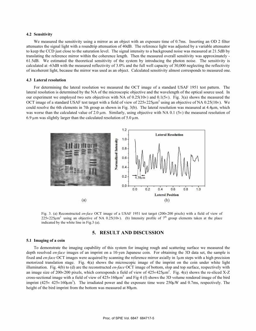
4.3 Lateral resolution
For determining the lateral resolution we measured the OCT image of a standard USAF 1951 test pattern. The lateral resolution is determined by the NA of the microscopic objective and the wavelength of the optical source used. In our experiment we employed two sets objectives with NA of 0.25(10×) and 0.1(5×). Fig. 3(a) shows the measured the OCT image of a standard USAF test target with a field of view of 225×225µm2 using an objective of NA 0.25(10×). We could resolve the 6th elements in 7th group as shown in Fig. 3(b). The lateral resolution was measured at 4.4µm, which was worse than the calculated value of 2.0 µm. Similarly, using objective with NA 0.1 (5×) the measured resolution of 6.9 µm was slightly larger than the calculated resolution of 5.0 µm.
Fig. 3. (a) Reconstructed en-face OCT image of a USAF 1951 test target (200×200 pixels) with a field of view of 225×225µm2 using an objective of NA 0.25(10×). (b) Intensity profile of 7th group elements taken at the place indicated by the white line in Fig.3 (a).
5. RESULT AND DISCUSSION 5.1 Imaging of a coin
To demonstrate the imaging capability of this system for imaging rough and scattering surface we measured the depth resolved en-face images of an imprint on a 10-yen Japanese coin. For obtaining the 3D data set, the sample is fixed and en-face OCT images were acquired by scanning the reference mirror axially in 1µm steps with a high precision motorized translation stage. Fig. 4(a) shows the microscopic image of the imprint on the coin under white light illumination. Fig. 4(b) to (d) are the reconstructed en-face OCT image of bottom, slop and top surface, respectively with an image size of 200×200 pixels, which corresponds a field of view of 425×425µm2. Fig. 4(e) shows the re-sliced X-Z cross-sectional image with a field of view of 425×160µm2 and Fig 4 (f) shows the 3D volume rendered image of the bird imprint (425× 425×160µm3). The irradiated power and the exposure time were 250µW and 0.7ms, respectively. The height of the bird imprint from the bottom was measured at 80µm.
Proc. of SPIE Vol. 6847 684717-5

H(e)
(c)
Fig. 4. (a) Microscopic image of the bird on the 10-yen Japanese coin. (b) to (d) are the reconstructed en-face OCT image of bottom, slop and top surface, respectively. (e) Re-sliced X-Z cross-sectional image with a field of view of 425×160µm2 (indicated by the white dotted line in (b)). (f) 3D volume rendered image of the bird imprint (425× 425×160µm3).
5.2 Imaging of biological specimen
We studied the feasibility of the system for imaging weakly scattering biological specimens. We obtained en-face OCT images from biological samples such an onion slice. The exposure time and irradiation power were 7ms and 224µW, respectively. Fig. 5(a) shows the microscopic image of an onion surface. Figures 5(b) to (f) shows the en-face OCT images the onion at different depth with an image size of 200×200 pixels, which is corresponding to a field of view of 225× 225µm2. Fig. 5(g) shows the re-sliced cross-sectional image in Y-Z direction. A 3D volumetric image of an onion slice with a size of (225× 225×276µm3) is show in Fig. 5(h). We could resolve different tissue organizations with in the sample. The deepest resolvable structure was 276µm beneath the surface with presuming a sample refractive index of 1.3.
Proc. of SPIE Vol. 6847 684717-6

(d) (e) (0
-- *4! 44
—
(g)
Fig. 5(a) Microscopic image of an onion surface. En-face OCT images of onion at different depths: (b) 0µm, (c) 50µm,
(d) 100µm, (e) 150µm, (f) 200µm. (g) Re-sliced cross-sectional image in Y-Z direction acquired from 360 en-face tomographic images (indicated by the white dotted line in (b)). (h) 3D volumetric image of an onion slice with a size of 225× 225×276µm3.
Proc. of SPIE Vol. 6847 684717-7

6. CONCLUSION We have developed a compact single-shot full-field OCT based on Linnik type polarizing Michelson interferometer
and a compact four-channel optics, which can capture four-quadrature phase-stepped interference images to obtain single-shot en-face OCT images in real time. We demonstrate the feasibility of the system for imaging scattering specimens such as an onion. Using objectives of 10× and 5×, the system visualized en-face images with a field of view of 225×225µm2 (200×200 pixels) and 425×425µm2 (200×200 pixels), respectively. The system has an axial resolution of 19.8µm and a lateral resolution of 4.4µm. Single-shot en-face OCT images could be measured and displayed in real-time with an exposure time of 7ms and a frame rate of 28 fps without any averaging and binning. Our proposed single-shot full-filed OCT system is efficient for imaging weakly scattering specimens. We are currently improving the performance of the system for imaging dynamic phenomenon in living specimens.
ACKNOWLEDGEMENTS
The authors are grateful to the Society of Yonezawa Industry (Yonezawa Kougyokai) for their partial supports.
REFERENCES
1. D. Huang, E. A. Swanson, C. P. Lin, J. S. Schuman, W. G. Stinson, W. Chang, M. R. Hee, T. Flotte, K. Gregory, C. A. Puliafito, and J. G. Fujimoto, “Optical Coherence Tomography,” Science 254, 1178–1180 (1991).
2. Gerd H¨ausler and Michael Walter Lindner, “ “Coherence radar” and “spectral radar” —New tools for dermatological diagnosis,” J. Biomed. Opt. 3, 21–31 (1998).
3. A. F. Fercher and C. K. Hitzenberger and G. Kamp and S. Y. El-Zaiat, “Measurement of intraocular distances by backscattering spectral interferometry,” Opt. Commun. 117, 43–48 (1995).
4. R. Huber, M. Wojtkowski, and J. G. Fujimoto, “Fourier Domain Mode Locking (FDML): A new laser operating regime and applications for optical coherence tomography,” Opt. Express 14, 3225-3237 (2006).
5. N. Nassif, B. Cense, B. Park, M. Pierce, S. Yun, B. Bouma, G. Tearney, T. Chen, and J. de Boer, “In vivo high resolution video-rate spectral-domain optical coherence tomography of the human retina and optic nerve,” Opt. Express 12, 367-376 (2004).
6. B. Grajciar, M. Pircher, A. Fercher, and R. Leitgeb, “Parallel Fourier domain optical coherence tomography for in vivo measurement of the human eye,” Opt. Express 13, 1131-1137 (2005).
7. Y. Nakamura, S. Makita, M. Yamanari, M. Itoh, T. Yatagai, and Y. Yasuno, "High-speed three-dimensional human retinal imaging by line-field spectral domain optical coherence tomography," Opt. Express 15, 7103-7116 (2007).
8. E. Beaurepaire, A. C. Boccara, M. Lebec, L. Blanchot, and H. Saint-Jalmes, “Full-field optical coherence microscopy. Opt. Lett. 23(4), 244–246 (1998).
9. L. Vabre, A. Dubois, and A. C. Boccara, “Thermal-light full-field optical coherence tomography”, Opt. Lett. 27(7), 530–532 (2002).
10. A. F. Fercher, R. Leitgeb, C. K. Hitzenberger, and H. Sattmann. Complex Spectral Interferometry OCT. Proc. SPIE 3564, 173–178 (1999).
11. E. Bordenave, E. Abraham, G. Jonusauskas, N. Tsurumachi, J. Oberle, C. Rulliere, P. E. Minot, M. Lassegues, and J. E. S. Bazeille, “Wide-field optical coherence tomography: imaging of biological tissues”, Appl. Opt. 41(10), 2059–2064 (2002).
12. M. Akiba, K. P. Chan, and N. Tanno. Full-field optical coherence tomography by two-dimensional heterodyne detection with a pair of ccd cameras. Opt. Lett. 28(10), 816–818 (2003).
13. M. Sato, T. Nagata, T. Niizuma, L. Neagu, R. Dabu and Y. Watanabe, “Quadrature fringes wide-field optical coherence tomography and its applications to biological tissues,” Opt. Commu. 271, 573-580 (2007).
14. C. Dunsby, Y. Gu, and P. M. W. French, “Single-shot phase-stepped wide-field coherence-gated imaging,” Opt. Express 11(2), 105–115 (2003).
Proc. of SPIE Vol. 6847 684717-8
Related Documents