Titanium alloy mini-implants for orthodontic anchorage: Immediate loading and metal ion release q Liliane S. Morais a,b , Glaucio G. Serra a,b , Carlos A. Muller c , Leonardo R. Andrade d , Elisabete F.A. Palermo d , Carlos N. Elias b , Marc Meyers a, * a Mechanical and Aerospace Engineering Department, University of California-San Diego (UCSD), San Diego, CA, United States b Mechanical Engineering and Material Science Department, Military Institute of Engineering (IME), Rio de Janeiro, RJ, Brazil c Oswaldo Cruz Institute (FIOCRUZ), Rio de Janeiro, RJ, Brazil d Federal University of Rio de Janeiro (UFRJ), Rio de Janeiro, RJ, Brazil Received 23 June 2006; received in revised form 19 October 2006; accepted 24 October 2006 Abstract Removable osseointegrated titanium mini-implants were successfully used as anchorage devices in orthodontics. The early load is nec- essary to simplify the mini-implant methodology, but can lead to failure during osseointegration. The Ti–6Al–4V alloy was used instead of commercially pure Ti due to its superior strength. However, the corrosion resistance is low, allowing for metal ion release. The purpose of this work was to analyze the immediately loaded mini-implant fixation and to gauge the vanadium ion release during the healing pro- cess. Titanium alloy mini-implants were inserted in the tibiae of rabbits. After 1, 4 and 12 weeks, they were submitted to removal torque testing. There was no increase in the removal torque value between 1 and 4 weeks of healing, regardless of the load. Nevertheless, after 12 weeks, a significant improvement was observed in both groups, with the highest removal torque value for the unloaded group. The kid- ney, liver and lung were also extracted and analyzed by atomic absorption spectrometry. In comparison with the control values, the con- tent of vanadium increased slightly after 1 week, significantly increased after 4 weeks and decreased slightly after 12 weeks, without reaching the 1 week values. A stress analysis was carried out which enables both the prediction of the torque at which commercially pure (CP) Ti and Ti–6Al–4V deform plastically and the shear strength of the interface. This analysis reveals that the removal torques for CP Ti dangerously approach the yield stress. The results of this rabbit model study indicate that titanium alloy mini-implants can be loaded immediately with no compromise in their stability. The detected concentration of vanadium did not reach toxic levels in the animal model. Ó 2006 Acta Materialia Inc. Published by Elsevier Ltd. All rights reserved. Keywords: Titanium alloy; Mini-implant; Orthodontic anchorage; Ion release 1. Introduction Anchorage has long been a challenge since the introduc- tion of fixed appliances in orthodontics [1]. Typically, orthodontic movement of a tooth is anchored by a large group of teeth so as to minimize undesired displacements of anchoring teeth. Adequate anchorage becomes difficult when posterior teeth are missing. Intra- and extra-oral aux- iliary devices can be used to assist movement, but the effec- tiveness of these measures is dependent upon the level of patient cooperation [1]. Conventional titanium implants have emerged as an excellent alternative to traditional orthodontic anchorage methodologies, mainly when anchorage dental elements are insufficient in quantity or quality [2]. Unfortunately, conventional dental implants can only be placed in limited sites, such as the retromolar and edentulous areas [2,3]. In 1742-7061/$ - see front matter Ó 2006 Acta Materialia Inc. Published by Elsevier Ltd. All rights reserved. doi:10.1016/j.actbio.2006.10.010 q Research presented at the TMS 2006 Biological Materials Science Symposium. * Corresponding author. Tel.: +1 858 534 4719. E-mail address: [email protected] (M. Meyers). Acta Biomaterialia 3 (2007) 331–339 www.elsevier.com/locate/actabiomat

Welcome message from author
This document is posted to help you gain knowledge. Please leave a comment to let me know what you think about it! Share it to your friends and learn new things together.
Transcript
Titanium alloy mini-implants for orthodontic anchorage:Immediate loading and metal ion release q
Liliane S. Morais a,b, Glaucio G. Serra a,b, Carlos A. Muller c, Leonardo R. Andrade d,Elisabete F.A. Palermo d, Carlos N. Elias b, Marc Meyers a,*
a Mechanical and Aerospace Engineering Department, University of California-San Diego (UCSD), San Diego, CA, United Statesb Mechanical Engineering and Material Science Department, Military Institute of Engineering (IME), Rio de Janeiro, RJ, Brazil
c Oswaldo Cruz Institute (FIOCRUZ), Rio de Janeiro, RJ, Brazild Federal University of Rio de Janeiro (UFRJ), Rio de Janeiro, RJ, Brazil
Received 23 June 2006; received in revised form 19 October 2006; accepted 24 October 2006
Abstract
Removable osseointegrated titanium mini-implants were successfully used as anchorage devices in orthodontics. The early load is nec-essary to simplify the mini-implant methodology, but can lead to failure during osseointegration. The Ti–6Al–4V alloy was used insteadof commercially pure Ti due to its superior strength. However, the corrosion resistance is low, allowing for metal ion release. The purposeof this work was to analyze the immediately loaded mini-implant fixation and to gauge the vanadium ion release during the healing pro-cess. Titanium alloy mini-implants were inserted in the tibiae of rabbits. After 1, 4 and 12 weeks, they were submitted to removal torquetesting. There was no increase in the removal torque value between 1 and 4 weeks of healing, regardless of the load. Nevertheless, after 12weeks, a significant improvement was observed in both groups, with the highest removal torque value for the unloaded group. The kid-ney, liver and lung were also extracted and analyzed by atomic absorption spectrometry. In comparison with the control values, the con-tent of vanadium increased slightly after 1 week, significantly increased after 4 weeks and decreased slightly after 12 weeks, withoutreaching the 1 week values. A stress analysis was carried out which enables both the prediction of the torque at which commercially pure(CP) Ti and Ti–6Al–4V deform plastically and the shear strength of the interface. This analysis reveals that the removal torques for CP Tidangerously approach the yield stress. The results of this rabbit model study indicate that titanium alloy mini-implants can be loadedimmediately with no compromise in their stability. The detected concentration of vanadium did not reach toxic levels in the animalmodel.! 2006 Acta Materialia Inc. Published by Elsevier Ltd. All rights reserved.
Keywords: Titanium alloy; Mini-implant; Orthodontic anchorage; Ion release
1. Introduction
Anchorage has long been a challenge since the introduc-tion of fixed appliances in orthodontics [1]. Typically,orthodontic movement of a tooth is anchored by a largegroup of teeth so as to minimize undesired displacements
of anchoring teeth. Adequate anchorage becomes di!cultwhen posterior teeth are missing. Intra- and extra-oral aux-iliary devices can be used to assist movement, but the e"ec-tiveness of these measures is dependent upon the level ofpatient cooperation [1].
Conventional titanium implants have emerged as anexcellent alternative to traditional orthodontic anchoragemethodologies, mainly when anchorage dental elementsare insu!cient in quantity or quality [2]. Unfortunately,conventional dental implants can only be placed in limitedsites, such as the retromolar and edentulous areas [2,3]. In
1742-7061/$ - see front matter ! 2006 Acta Materialia Inc. Published by Elsevier Ltd. All rights reserved.doi:10.1016/j.actbio.2006.10.010
q Research presented at the TMS 2006 Biological Materials ScienceSymposium.* Corresponding author. Tel.: +1 858 534 4719.E-mail address: [email protected] (M. Meyers).
Acta Biomaterialia 3 (2007) 331–339
www.elsevier.com/locate/actabiomat
addition, conventional dental implants are troublesome forpatients because of the severity of the surgery, the discom-fort of the initial healing and the di!culty of maintainingoral hygiene [4].
Due to these disadvantages, Kanomi [5] proposed tita-nium mini-implants (1.2 mm in diameter and 6.0 mm inlength) for orthodontic anchorage. They are widely usedsince they have few implantation site limitations, a simpleinsertion procedure and easy mechanical force control [6].The methodology for implementation of mini-implantshas been continuously improved. Some complications per-sist, and the sources of failure include the inflammation ofthe soft tissue around the mini-implant and fracture of themini-implant [6].
A period of healing is usually necessary before applyingload to conventional dental implants. This period variesfrom 4 to 6 months in humans [7,8]. When the load isplaced prematurely, histological analyses have suggestedthat there is no uniform intimate bone-implant contactdue to interplayed fibrous tissue [9,10]. This phenomenoncould be favorable for implants for orthodontic anchoragepurposes, since it facilitates the surgical removal of theimplant at the end of the orthodontic treatment. On theother hand, the excess of interplayed fibrous tissue couldlead to implant failure.
Commercially pure titanium (CP Ti) is widely used asimplant material because of its suitable mechanical proper-ties and excellent biocompatibility [11,12]. However, CP Tihas lower fatigue strength than titanium alloys. Ti–6Al–4Vcan be used to overcome this disadvantage [12,13]. How-ever, the corrosion resistance of the mini-implant decreaseswhen the alloy is used, favoring metal ion release, whichhas been associated with clinical implant failure, osteolysis,cutaneous allergic reactions, remote site accumulation [14],kidney lesion [15], cytotoxicity, hypersensitivity and carci-nogenesis [16].
The purpose of this work was to measure the boneanchorage of immediately loaded Ti–6Al–4V mini-implants by removal torque test, and the amount of vana-dium ion release in remote tissues by atomic absorptionspectrometry.
2. Experimental
2.1. Materials
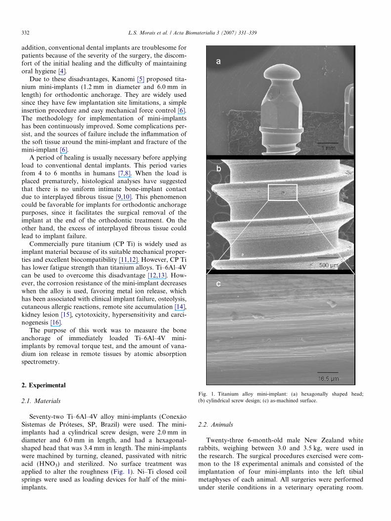
Seventy-two Ti–6Al–4V alloy mini-implants (ConexaoSistemas de Proteses, SP, Brazil) were used. The mini-implants had a cylindrical screw design, were 2.0 mm indiameter and 6.0 mm in length, and had a hexagonal-shaped head that was 3.4 mm in length. The mini-implantswere machined by turning, cleaned, passivated with nitricacid (HNO3) and sterilized. No surface treatment wasapplied to alter the roughness (Fig. 1). Ni–Ti closed coilsprings were used as loading devices for half of the mini-implants.
2.2. Animals
Twenty-three 6-month-old male New Zealand whiterabbits, weighing between 3.0 and 3.5 kg, were used inthe research. The surgical procedures exercised were com-mon to the 18 experimental animals and consisted of theimplantation of four mini-implants into the left tibialmetaphyses of each animal. All surgeries were performedunder sterile conditions in a veterinary operating room.
Fig. 1. Titanium alloy mini-implant: (a) hexagonally shaped head;(b) cylindrical screw design; (c) as-machined surface.
332 L.S. Morais et al. / Acta Biomaterialia 3 (2007) 331–339
Five rabbits were used as controls for the metal ion releasestudy.
The protocol for the animal study was approved by thestanding ethics committee on animal research of OswaldoCruz Institute and all procedures were done based onCanadian Council of Animal Care guidelines. In the pre-surgical period, the rabbits were conditioned in a vivarium,where they remained during an observation period. For theduration of the study, they had free access to pellets andwater, and were maintained at temperature from 22 to26 "C with daily illumination.
2.3. Experimental design
Each animal had four mini-implants inserted, two ofwhich were immediately loaded with a force of 1 N. Thirtymini-implants were used in the removal torque test and theother 42 mini-implants were used in other analysis. Thegroups were formed to investigate three periods of healing:1 week, 4 weeks and 12 weeks. In each assessment period,one group was loaded and another was unloaded, giving atotal of six groups. The removal torque test was carried outto analyze the bone fixation of the mini-implants during thehealing process.
The atomic absorption spectrometry analysis was per-formed on the kidney, liver and lung in order to analyzewhether vanadium ion release occurs and if these metalions accumulate in remote tissues. The three tissues wereextracted from the 18 experimental rabbits at the times pre-viously established (1, 4 and 12 weeks) and from the fivecontrol rabbits, in which no treatment was administered,totaling up to 12 groups (Table 1).
2.4. Surgical procedure
The animals were anesthetized with an intra-muscularinjection of Tiletamine (5 mg/kg) and Zolazepan (5 mg/kg), followed by continuous delivery of 2% Halothaneand Isofluthane throughout the surgery. The hair on themedial surface of the upper portion of the left leg wasremoved and the skin was cleansed with iodinate surgicalsoap. A 70% alcohol solution was used for the local pro-phylaxis. A 50 mm-long incision was made parallel to thelongitudinal axis of the tibia and the periosteum was
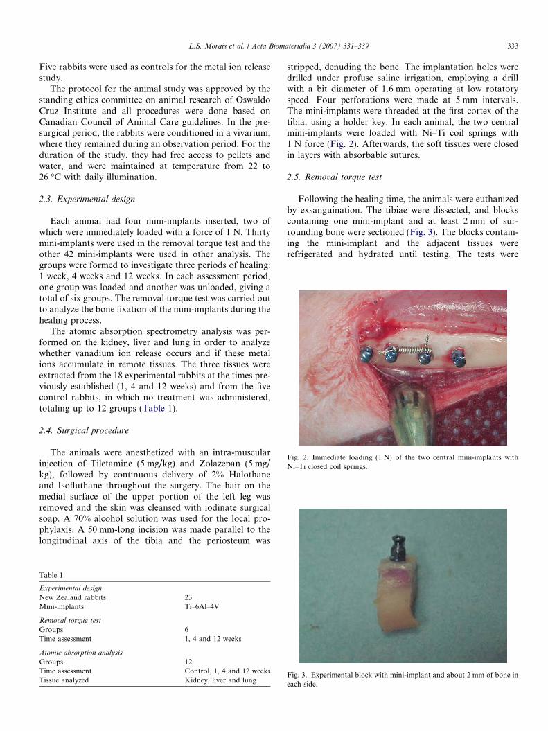
stripped, denuding the bone. The implantation holes weredrilled under profuse saline irrigation, employing a drillwith a bit diameter of 1.6 mm operating at low rotatoryspeed. Four perforations were made at 5 mm intervals.The mini-implants were threaded at the first cortex of thetibia, using a holder key. In each animal, the two centralmini-implants were loaded with Ni–Ti coil springs with1 N force (Fig. 2). Afterwards, the soft tissues were closedin layers with absorbable sutures.
2.5. Removal torque test
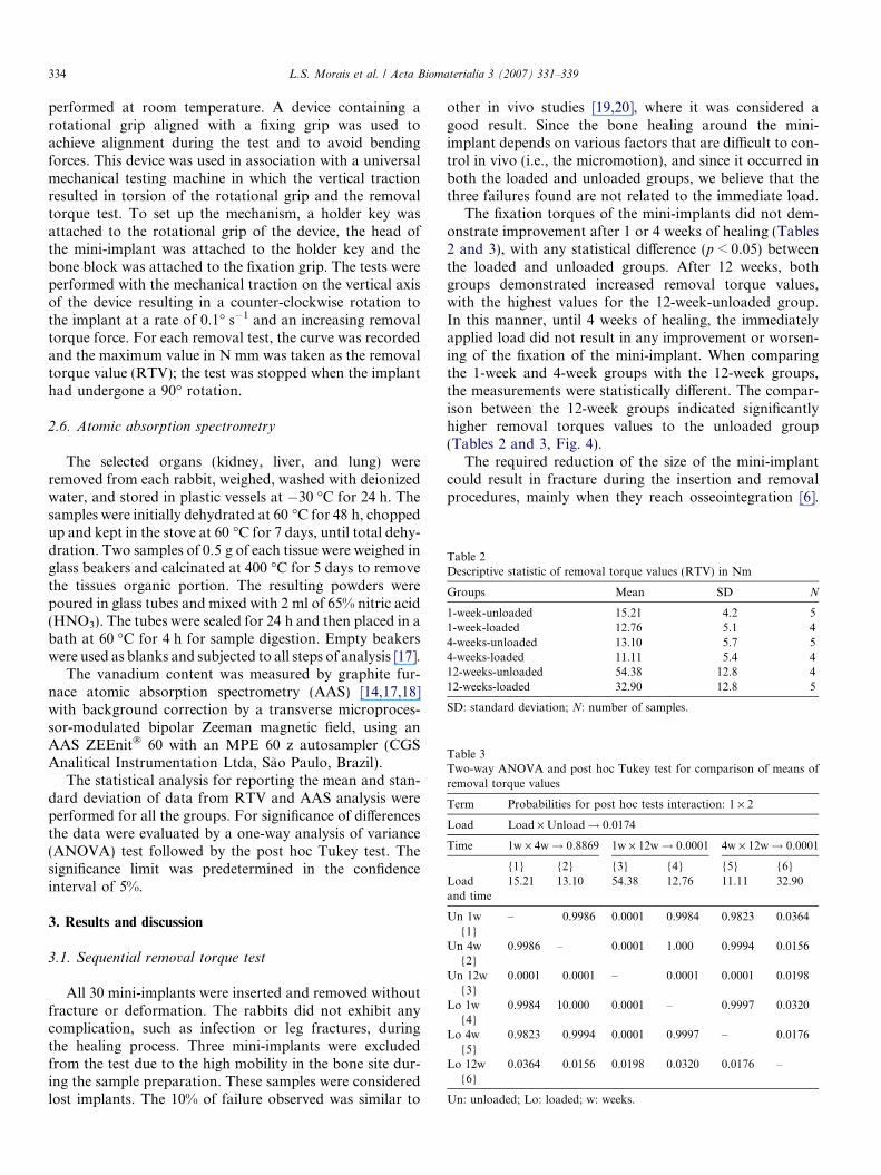
Following the healing time, the animals were euthanizedby exsanguination. The tibiae were dissected, and blockscontaining one mini-implant and at least 2 mm of sur-rounding bone were sectioned (Fig. 3). The blocks contain-ing the mini-implant and the adjacent tissues wererefrigerated and hydrated until testing. The tests were
Table 1
Experimental designNew Zealand rabbits 23Mini-implants Ti–6Al–4V
Removal torque testGroups 6Time assessment 1, 4 and 12 weeks
Atomic absorption analysisGroups 12Time assessment Control, 1, 4 and 12 weeksTissue analyzed Kidney, liver and lung
Fig. 2. Immediate loading (1 N) of the two central mini-implants withNi–Ti closed coil springs.
Fig. 3. Experimental block with mini-implant and about 2 mm of bone ineach side.
L.S. Morais et al. / Acta Biomaterialia 3 (2007) 331–339 333
performed at room temperature. A device containing arotational grip aligned with a fixing grip was used toachieve alignment during the test and to avoid bendingforces. This device was used in association with a universalmechanical testing machine in which the vertical tractionresulted in torsion of the rotational grip and the removaltorque test. To set up the mechanism, a holder key wasattached to the rotational grip of the device, the head ofthe mini-implant was attached to the holder key and thebone block was attached to the fixation grip. The tests wereperformed with the mechanical traction on the vertical axisof the device resulting in a counter-clockwise rotation tothe implant at a rate of 0.1" s!1 and an increasing removaltorque force. For each removal test, the curve was recordedand the maximum value in N mm was taken as the removaltorque value (RTV); the test was stopped when the implanthad undergone a 90" rotation.
2.6. Atomic absorption spectrometry
The selected organs (kidney, liver, and lung) wereremoved from each rabbit, weighed, washed with deionizedwater, and stored in plastic vessels at !30 "C for 24 h. Thesamples were initially dehydrated at 60 "C for 48 h, choppedup and kept in the stove at 60 "C for 7 days, until total dehy-dration. Two samples of 0.5 g of each tissue were weighed inglass beakers and calcinated at 400 "C for 5 days to removethe tissues organic portion. The resulting powders werepoured in glass tubes and mixed with 2 ml of 65% nitric acid(HNO3). The tubes were sealed for 24 h and then placed in abath at 60 "C for 4 h for sample digestion. Empty beakerswere used as blanks and subjected to all steps of analysis [17].
The vanadium content was measured by graphite fur-nace atomic absorption spectrometry (AAS) [14,17,18]with background correction by a transverse microproces-sor-modulated bipolar Zeeman magnetic field, using anAAS ZEEnit# 60 with an MPE 60 z autosampler (CGSAnalitical Instrumentation Ltda, Sao Paulo, Brazil).
The statistical analysis for reporting the mean and stan-dard deviation of data from RTV and AAS analysis wereperformed for all the groups. For significance of di"erencesthe data were evaluated by a one-way analysis of variance(ANOVA) test followed by the post hoc Tukey test. Thesignificance limit was predetermined in the confidenceinterval of 5%.
3. Results and discussion
3.1. Sequential removal torque test
All 30 mini-implants were inserted and removed withoutfracture or deformation. The rabbits did not exhibit anycomplication, such as infection or leg fractures, duringthe healing process. Three mini-implants were excludedfrom the test due to the high mobility in the bone site dur-ing the sample preparation. These samples were consideredlost implants. The 10% of failure observed was similar to
other in vivo studies [19,20], where it was considered agood result. Since the bone healing around the mini-implant depends on various factors that are di!cult to con-trol in vivo (i.e., the micromotion), and since it occurred inboth the loaded and unloaded groups, we believe that thethree failures found are not related to the immediate load.
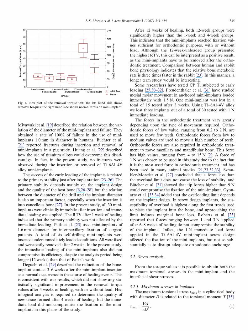
The fixation torques of the mini-implants did not dem-onstrate improvement after 1 or 4 weeks of healing (Tables2 and 3), with any statistical di"erence (p < 0.05) betweenthe loaded and unloaded groups. After 12 weeks, bothgroups demonstrated increased removal torque values,with the highest values for the 12-week-unloaded group.In this manner, until 4 weeks of healing, the immediatelyapplied load did not result in any improvement or worsen-ing of the fixation of the mini-implant. When comparingthe 1-week and 4-week groups with the 12-week groups,the measurements were statistically di"erent. The compar-ison between the 12-week groups indicated significantlyhigher removal torques values to the unloaded group(Tables 2 and 3, Fig. 4).
The required reduction of the size of the mini-implantcould result in fracture during the insertion and removalprocedures, mainly when they reach osseointegration [6].
Table 2Descriptive statistic of removal torque values (RTV) in Nm
Groups Mean SD N
1-week-unloaded 15.21 4.2 51-week-loaded 12.76 5.1 44-weeks-unloaded 13.10 5.7 54-weeks-loaded 11.11 5.4 412-weeks-unloaded 54.38 12.8 412-weeks-loaded 32.90 12.8 5
SD: standard deviation; N: number of samples.
Table 3Two-way ANOVA and post hoc Tukey test for comparison of means ofremoval torque values
Term Probabilities for post hoc tests interaction: 1 · 2
Load Load ·Unload! 0.0174
Time 1w · 4w! 0.8869 1w · 12w! 0.0001 4w · 12w! 0.0001
{1} {2} {3} {4} {5} {6}Loadand time
15.21 13.10 54.38 12.76 11.11 32.90
Un 1w{1}
– 0.9986 0.0001 0.9984 0.9823 0.0364
Un 4w{2}
0.9986 – 0.0001 1.000 0.9994 0.0156
Un 12w{3}
0.0001 0.0001 – 0.0001 0.0001 0.0198
Lo 1w{4}
0.9984 10.000 0.0001 – 0.9997 0.0320
Lo 4w{5}
0.9823 0.9994 0.0001 0.9997 – 0.0176
Lo 12w{6}
0.0364 0.0156 0.0198 0.0320 0.0176 –
Un: unloaded; Lo: loaded; w: weeks.
334 L.S. Morais et al. / Acta Biomaterialia 3 (2007) 331–339
Miyawaki et al. [19] described the relation between the var-iation of the diameter of the mini-implant and failure. Theyobtained a rate of 100% of failure in the use of mini-implants 1.0 mm in diameter in humans. Buchter et al.[21] reported fractures during insertion and removal ofmini-implants in a pig study. Huang et al. [22] describedhow the use of titanium alloys could overcome this disad-vantage. In fact, in the present study, no fractures wereobserved during the insertion or removal of Ti–6Al–4Valloy mini-implants.
The success of the early loading of the implants is relatedto the primary stability just after implantation [23–26]. Theprimary stability depends mainly on the implant designand the quality of the host bone [6,26–28], but the relationbetween the diameter of the drill and the implant diameteris also an important factor, especially when the insertion isinto cancellous bone [27]. In the present study, all 30 mini-implants were clinically immobile after insertion and imme-diate loading was applied. The RTV after 1 week of healingindicated that the primary stability was not a"ected by theimmediate loading. Paik et al. [25] used mini-implants of1.6 mm diameter for intermaxillary fixation of surgicalpatients. A total of six self-drilling mini-implants wereinserted under immediately loaded conditions. All were fixedand were easily removed after 2 weeks. In the present study,the immediate loading of the mini-implants also did notcompromise its e!ciency, despite the analysis period beinglonger (12 weeks) than that of Paiks’s work.
Deguchi et al. [29] described the reduction of the bone-implant contact 3–6 weeks after the mini-implant insertionas a normal occurrence in the course of healing events. Thisis consistent with our results, which did not show any sta-tistically significant improvement in the removal torquevalues after 4 weeks of healing, with or without load. His-tological analysis is required to determine the quality ofnew tissue formed after 4 weeks of healing, but the imme-diate load did not compromise the fixation of the mini-implants in this phase of the study.
After 12 weeks of healing, both 12-week groups weresignificantly higher than the 1-week and 4-week groups.This indicates that the mini-implants reached fixation val-ues su!cient for orthodontic purposes, with or withoutload. Although the 12-week-unloaded group presentedthe highest RTV, this can be interpreted as a positive result,as the mini-implants have to be removed after the ortho-dontic treatment. Comparison between human and rabbitbone physiology indicates that the relative bone metabolicrate is three times faster in the rabbit [23]. In this manner, alonger term study would be interesting.
Some researchers have tested CP Ti subjected to earlyloading [25,30–32]. Freudenthaler et al. [31] have studiedmesial molar movement in anchored mini-implants loadedimmediately with 1.5 N. One mini-implant was lost in atotal of 15 tested after 3 weeks. Using Ti–6Al–4V alloywe lost three implants out of a total of 30 tested with 1 Nimmediate loading.
The forces in the orthodontic treatment vary greatlydepending upon the type of movement required. Ortho-dontic forces of low value, ranging from 0.2 to 2 N, areused to move few teeth. Orthodontic forces from low tomedium values are used to move a high number of teeth.Orthopedic forces are also required in orthodontic treat-ment to move maxillary and mandibular bone. This forcehas high values, ranging from 4 to 15 N [2]. A force of1 N was chosen to be used in this study due to the fact thatit is the most used force in orthodontic treatment and hasbeen used in many animal studies [21,23,32,33]. Szmu-kler-Moncler et al. [27] concluded that a force less thanthe overload limit does not cause the loss of stability, andButcher et al. [21] showed that tip forces higher than 9 Ncould compromise the fixation of the mini-implant. Oyon-arte et al. [33,34] added that the overloading limit dependson the implant design. In screw design implants, the sus-ceptibility of overload is highest along the first treads usedto place the implant into the bone, and approaching thelimit induces marginal bone loss. Roberts et al. [23]reported that forces ranging between 1 and 3 N appliedafter 6–8 weeks of healing do not compromise the stabilityof the implants. Infact, the 1 N immediate load forceapplied in the Ti–6Al–4V mini-implant screw designa"ected the fixation of the mini-implants, but not so sub-stantially as to disrupt adequate orthodontic anchorage.
3.2. Stress analysis
From the torque values it is possible to obtain both themaximum torsional stresses in the mini-implant and theinterfacial shear stresses.
3.2.1. Maximum stresses in implantsThe maximum torsional stress smax in a cylindrical body
with diameter D is related to the torsional moment T [35]:
smax "16TpD3 : #1$
Fig. 4. Box plot of the removal torque test; the left hand side showsremoval torques; the right hand side shows normal stress on mini-implant.
L.S. Morais et al. / Acta Biomaterialia 3 (2007) 331–339 335
The corresponding normal stress is equal to [36]:
rmax "smax!!!3
p " 16T!!!3
ppD3
: #2$
Eq. (2) enables the calculation of the maximum stress act-ing on the mini-implants. At the highest torque observed of5.38 · 10!2 Nm, rmax = 148 MPa. The yield stresses for CPTi and Ti–6Al–4V are:
Grade 2 CP titanium: ry = 250 MPa;Grade 5 Ti–6Al–4V: ry = 880 MPa.If one computes safety factors:(SF) CP Ti = 1.7;(SF) Ti–6Al–4V = 6.0.
Thus, we can conclude that CP Ti is indeed stressed onaverage close to its yield value, which is an undesirable sit-uation, since it is certain some mini-implants will have arelease torque above the mean value used above. On theother hand, the Ti–6Al–4V alloy is stressed significantlybelow its yield stress and is indeed a much safer choice.
3.2.2. Interface stressesThe shear stress acting on the interface, si can also be
calculated from the torsional moment T:
si "2TDAT
: #3$
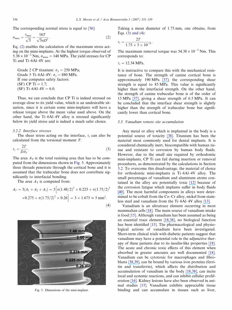
The area AT is the total resisting area that has to be com-puted from the dimensions shown in Fig. 5. Approximatelythree threads penetrate through the cortical bone and it isassumed that the trabecular bone does not contribute sig-nificantly to interfacial bonding.
The area AT is computed from:
AT " 3#A1 % A2 % A3$ " 3 p#1:48=2$2 & 0:225% p#1:75=2$2h
&0:275% p#1:75=2$2 & 0:26i" 3& 1:675 ' 5 mm2:
#4$
Taking a mean diameter of 1.75 mm, one obtains, fromEqs. (3) and (4):
si "2T
1:75& 5& 10!9 :
The maximum removal torque was 54.38 · 10!3 Nm. Thiscorresponds to:
si " 12:34 MPa:
It is instructive to compare this with the mechanical resis-tance of bone. The strength of canine cortical bone isapproximately 190 MPa [37]; the corresponding shearstrength is equal to 85 MPa. This value is significantlyhigher than the interfacial strength. On the other hand,the strength of canine trabecular bone is of the order of13 MPa [37], giving a shear strength of 6.5 MPa. It canbe concluded that the interface shear strength is slightlyhigher than the strength of trabecular bone but signifi-cantly lower than cortical bone.
3.3. Vanadium remote site accumulation
Any metal or alloy which is implanted in the body is apotential source of toxicity [38]. Titanium has been thematerial most commonly used for dental implants. It isconsidered chemically inert, biocompatible with human tis-sue and resistant to corrosion by human body fluids.However, due to the small size required by orthodonticmini-implants, CP Ti can fail during insertion or removalprocedures, as demonstrated by the calculations in Section3.2. To overcome this disadvantage, the material of choicefor orthodontic mini-implants is Ti–6Al–4V alloy. Thesmall percentages of vanadium and aluminum atoms con-tained in the alloy are potentially toxic [12] because ofthe corrosion fatigue which implants su"er in body fluids[40]. The most harmful components in alloys were deter-mined to be cobalt from the Co–Cr alloy, nickel from stain-less steel and vanadium from the Ti–6Al–4V alloy [13].
Vanadium is an ultratrace element occurring in mostmammalian cells [18]. The main source of vanadium intakeis food [15]. Although vanadium has been assumed as beingan essential trace element [18,38], no biological functionhas been identified [15]. The pharmacological and physio-logical actions of vanadium have been investigated.Short-term clinical trials with diabetic patients suggest thatvanadium may have a potential role in the adjunctive ther-apy of these patients due to its insulin-like properties [18].The acute and chronic toxic e"ects of this element whenabsorbed in greater amounts are well documented [18].Vanadium can be cytotoxic for macrophages and fibro-blasts [38,39], can be bound by various iron proteins (ferri-tin and transferrin), which a"ects the distribution andaccumulation of vanadium in the body [18,38], can incitelocal and systemic reactions, and can inhibit cellular prolif-eration [16]. Kidney lesions have also been observed in ani-mal studies [15]. Vanadium exhibits appreciable tissuebinding and can accumulate in tissues such as liver,Fig. 5. Dimensions of the mini-implant.
336 L.S. Morais et al. / Acta Biomaterialia 3 (2007) 331–339
kidneys, bone and spleen [18]. Studies on mice show thatwithin the first day after intra-venous injection of vana-dium the highest levels are found in the kidney [38]. Uri-nary excretion is the main elimination pathway forinjected vanadium in humans [18].
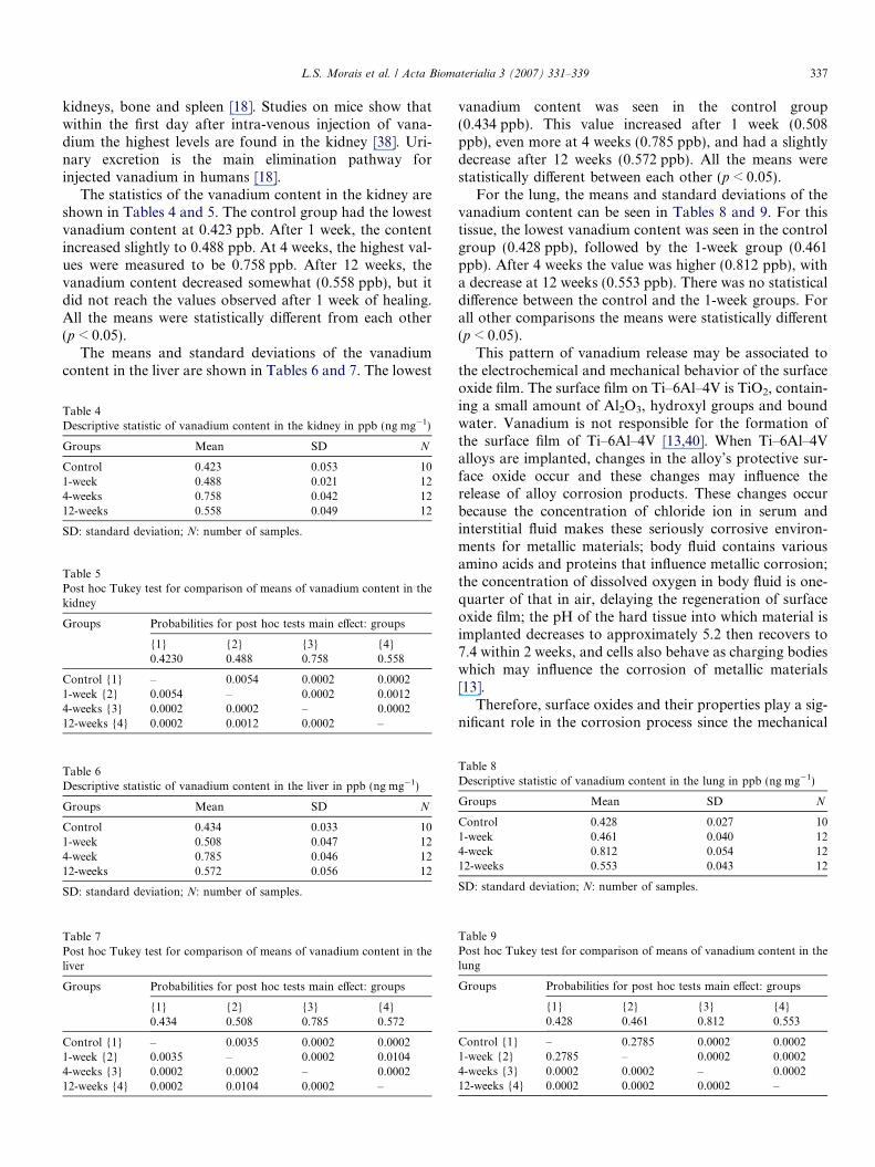
The statistics of the vanadium content in the kidney areshown in Tables 4 and 5. The control group had the lowestvanadium content at 0.423 ppb. After 1 week, the contentincreased slightly to 0.488 ppb. At 4 weeks, the highest val-ues were measured to be 0.758 ppb. After 12 weeks, thevanadium content decreased somewhat (0.558 ppb), but itdid not reach the values observed after 1 week of healing.All the means were statistically di"erent from each other(p < 0.05).
The means and standard deviations of the vanadiumcontent in the liver are shown in Tables 6 and 7. The lowest
vanadium content was seen in the control group(0.434 ppb). This value increased after 1 week (0.508ppb), even more at 4 weeks (0.785 ppb), and had a slightlydecrease after 12 weeks (0.572 ppb). All the means werestatistically di"erent between each other (p < 0.05).
For the lung, the means and standard deviations of thevanadium content can be seen in Tables 8 and 9. For thistissue, the lowest vanadium content was seen in the controlgroup (0.428 ppb), followed by the 1-week group (0.461ppb). After 4 weeks the value was higher (0.812 ppb), witha decrease at 12 weeks (0.553 ppb). There was no statisticaldi"erence between the control and the 1-week groups. Forall other comparisons the means were statistically di"erent(p < 0.05).
This pattern of vanadium release may be associated tothe electrochemical and mechanical behavior of the surfaceoxide film. The surface film on Ti–6Al–4V is TiO2, contain-ing a small amount of Al2O3, hydroxyl groups and boundwater. Vanadium is not responsible for the formation ofthe surface film of Ti–6Al–4V [13,40]. When Ti–6Al–4Valloys are implanted, changes in the alloy’s protective sur-face oxide occur and these changes may influence therelease of alloy corrosion products. These changes occurbecause the concentration of chloride ion in serum andinterstitial fluid makes these seriously corrosive environ-ments for metallic materials; body fluid contains variousamino acids and proteins that influence metallic corrosion;the concentration of dissolved oxygen in body fluid is one-quarter of that in air, delaying the regeneration of surfaceoxide film; the pH of the hard tissue into which material isimplanted decreases to approximately 5.2 then recovers to7.4 within 2 weeks, and cells also behave as charging bodieswhich may influence the corrosion of metallic materials[13].
Therefore, surface oxides and their properties play a sig-nificant role in the corrosion process since the mechanical
Table 4Descriptive statistic of vanadium content in the kidney in ppb (ng mg!1)
Groups Mean SD N
Control 0.423 0.053 101-week 0.488 0.021 124-weeks 0.758 0.042 1212-weeks 0.558 0.049 12
SD: standard deviation; N: number of samples.
Table 5Post hoc Tukey test for comparison of means of vanadium content in thekidney
Groups Probabilities for post hoc tests main e"ect: groups
{1} {2} {3} {4}0.4230 0.488 0.758 0.558
Control {1} – 0.0054 0.0002 0.00021-week {2} 0.0054 – 0.0002 0.00124-weeks {3} 0.0002 0.0002 – 0.000212-weeks {4} 0.0002 0.0012 0.0002 –
Table 6Descriptive statistic of vanadium content in the liver in ppb (ng mg!1)
Groups Mean SD N
Control 0.434 0.033 101-week 0.508 0.047 124-week 0.785 0.046 1212-weeks 0.572 0.056 12
SD: standard deviation; N: number of samples.
Table 7Post hoc Tukey test for comparison of means of vanadium content in theliver
Groups Probabilities for post hoc tests main e"ect: groups
{1} {2} {3} {4}0.434 0.508 0.785 0.572
Control {1} – 0.0035 0.0002 0.00021-week {2} 0.0035 – 0.0002 0.01044-weeks {3} 0.0002 0.0002 – 0.000212-weeks {4} 0.0002 0.0104 0.0002 –
Table 8Descriptive statistic of vanadium content in the lung in ppb (ng mg!1)
Groups Mean SD N
Control 0.428 0.027 101-week 0.461 0.040 124-week 0.812 0.054 1212-weeks 0.553 0.043 12
SD: standard deviation; N: number of samples.
Table 9Post hoc Tukey test for comparison of means of vanadium content in thelung
Groups Probabilities for post hoc tests main e"ect: groups
{1} {2} {3} {4}0.428 0.461 0.812 0.553
Control {1} – 0.2785 0.0002 0.00021-week {2} 0.2785 – 0.0002 0.00024-weeks {3} 0.0002 0.0002 – 0.000212-weeks {4} 0.0002 0.0002 0.0002 –
L.S. Morais et al. / Acta Biomaterialia 3 (2007) 331–339 337
properties and electrochemical behavior of the oxides a"ectthe fracture and repassivation processes. After fracture ofthe Ti–6Al–4V surface oxide, it takes a long time to reform,and metal ions are released during repassivation [13,41].The initial ion release is dominated by ion exchange eventsat the solid–liquid interface, which occur before thermody-namic equilibrium of the surface protein adsorption anddesorption events has been reached. Once the latter isachieved, further ion exchange events may be inhibited[42]. We concluded that at 1 week of implantation therewas not su!cient interaction at the solid–liquid interface.At 4 weeks, large numbers of ion exchange events were tak-ing place at the interface. The thermodynamic equilibriumseems not to be reached at 12 weeks. More refined experi-mental protocols are needed to identify when this eventoccurs.
Dietary intake of several elements has been surveyed byvarious investigators [15,43]. The daily intake of a specificelement may vary considerably according to di"erent eat-ing and drinking habits and geographical location, whilelarge discrepancies may occur between the various compi-lations with regard to intake of a specific element [43].The amounts of vanadium released in rabbits from thepresently surveyed orthodontic Ti–6Al–4V alloy mini-implants were far below the daily intake of this elementthrough food and drink (1.8 mg day!1) [15,43]. Giokaet al. [44] measured in vitro traces of vanadium (2 ppm)released from Ti–6Al–4V orthodontic brackets and it wasconsidered that vanadium release was minimal. Theauthors pointed out that long-term release may be higherthan that occurring within the first weeks. However, in con-trast to the long-term biomedical applications of Ti alloysin orthopedics, the orthodontic use of Ti alloy mini-implants has a limited service life [44]. Thus, the minutelevels of vanadium release may not constitute an alarmingsituation, since they did not reach toxic levels in the animalmodel.
The use of rabbits in implant studies is widely di"used[3,17,23] due to the correlation between rabbit and humanphysiology. The bone turnover in humans is 18 weeks,whereas it is about 6 weeks in rabbits, suggesting that a fac-tor of 3 is a good rule for extrapolating rabbit data to theclinical situation [23]. However, care should be taken inextrapolating the clinical behavior from animal tests sincethe dimensions of the tested material in relation to the bio-logical system of the rabbits could greatly influence theresults [44].
The presence of elements with potential biologicallyhazardous action, especially vanadium, has increased theinterest in adopting other alternatives, such as the devel-opment of new titanium alloys employing Nb as a betastabilizer (Ti–6Al–7Nb) [44], and CP titanium with nano-scale grains, which has greater strength than the conven-tional Ti–6Al–4V alloy. These implant materials arecorrosion resistant and biocompatible with human bodyorgans and fluids, so they can remain in the body foryears [12].
4. Conclusions
The results of this rabbit model study indicate thatimmediately loaded and unloaded titanium alloy mini-implants reached fixation values appropriate for the pur-poses of orthodontic anchorage. The unloaded group hada higher RTV than the loaded group. Since the mini-implants have to be removed at the end of the treatment,immediate loading could be a favorable option intreatment.
The absence of fractures during insertion and removalof the mini-implants indicated that the Ti–6Al–4V alloyhas adequate mechanical properties for this application.Despite the tendency of greater ion release when usingthe titanium alloy, the amount of vanadium detected didnot reach toxic levels in the animal model, even at 4 weeks,when the maximum concentrations were measured.
The normal stresses acting on the mini-implants andthe shear stresses acting at the interface were calculatedfrom the removal torques. The maximum normal stressvalues (148 MPa) approach the yield stress of CP Ti((250 MPa) but are much lower than the yield strengthof Ti–6Al–4V. This analysis suggests that the 2 mm-diam-eter mini-implants of CP Ti are unsafe and proves thatTi–6Al–4V is a safe choice. The shear strength of theinterface was calculated and its value was found to bebetween the shear strengths of cortical and trabecularbone.
Acknowledgements
We thank Sara Bodde for reading and considerablyimproving the manuscript. This research was supportedby CAPES, Ministry of Education and Culture, Brazil.Support of the National Science Foundation CeramicsProgram (Dr. Linnette Madsen, Director) for travel andlaboratory supplies is gratefully acknowledged.
References
[1] Graber TM, Vanarsdall RL. Orthodontics – principles and tech-niques. second ed. Rio de Janeiro: Guanabara Koogan; 1996.
[2] Favero L, Brollo P, Bressan E. Orthodontic anchorage with specificfixtures: related study analysis. Am J Orthod Dentofacial Orthop2002;122:84–94.
[3] Roberts EW, Helm RF, Marshall JK, Gonglo" RK. Rigid endos-seous implants for orthodontic and orthopedic anchorage. AngleOrthod 1989;59(4):247–56.
[4] Ohmae M, Saito S, Morohashi T, Seki K, Qu H, Kanomi R, et al. Aclinical and histological evaluation of titanium mini-implants asanchor for orthodontic intrusion in the beagle dog. Am J OrthodDentofacial Orthop 2001;119:489–97.
[5] Kanomi R. Mini-implant for orthodontic anchorage. JCO1997;36(11):763–7.
[6] Park Y-C, Lee S-Y, Kim D-H, Jee S-H. Intrusion of posterior teethusing mini-screw implants. Am J Orthod Dentofacial Orthop2003;123:690–4.
[7] Roberts EW, Marshall KJ, Mozsary PG. Rigid endosseous implantutilized as anchorage to protract molars and close an atrophicextraction site. Angle Orthod 1990;60(2):135–52.
338 L.S. Morais et al. / Acta Biomaterialia 3 (2007) 331–339
[8] Roberts WE, Nelson CL, Goodacre CJ. Rigid implant anchorage toclose a mandibular first molar extraction site. J Clin Orthod1994;28(12):693–704.
[9] Costa A, Melsen B, Ra"aini M. Miniscrews as orthodontic anchor-age: A preliminary report. Int J Adult Orthod Orthognath Surg1998;13(3):201–9.
[10] Melsen B, Petersen JK, Costa A. Zigoma ligature: an alternative formof maxillary anchorage. J Clin Orthod 1998;32:154–8.
[11] Aparicio C, Gil FJ, Fonseca C, Barbosa M, Planell JA. Corrosionbehavior of commercially pure titanium shot blasted with di"erentmaterials and sizes of shot particles for dental implant applications.Biomaterials 2003;24:263–73.
[12] Latysh V, Krallics G, Alexandrov I, Fodor A. Application of bulknanostructured materials in medicine. Curr Appl Phys 2006;6:262–6.
[13] Hanawa T. Metal ion release from metal implants. Mater Sci Eng C2004;24:745–52.
[14] Okazaki Y, Gotoh E, Manabe T, Kobayashi K. Comparison of metalconcentrations in rat tibia tissues with various metallic implants.Biomaterials 2004;25:5913–20.
[15] Food and Nutrition Board. Dietary reference intakes: elements. 2001.Available from: http://www.iom.edu/Object.File/Master/7/294/0.pdf.
[16] Sedarat C, Harmand MF, Naji A, Nowzari H. In vitro kineticevaluation of titanium alloy biodegradation. J Periodont Res2001;36:269–74.
[17] Liu P, Yao YN, Wu SD, Dong HJ, Feng GC, Yuan XY. The e!cacyof deferiprone on tissues aluminum removal and copper, zinc,manganese level in rabbits. J Inorg Biochem 2005;99:1733–7.
[18] Heinemann G, Fichtl B, Vogt W. Pharmacokinetics of vanadium inhumans after intravenous administration of a vanadium containingalbumin solution. Br J Clin Pharmacol 2003;55:241–5.
[19] Miyawaki S, Koyama I, Inoue M, Mashima K, Sugahara T, Takano-Yamamoto T. Factors associated with the stability of titanium screwsplaced in the posterior region for orthodontic anchorage. Am JOrthod Dentofacial Orthop 2003;124:373–8.
[20] Park H-S, Lee A-K, Kwon O-W. Group distal movement of teethusing microscrew implant anchorage. Angle Orthod 2005;75:602–9.
[21] Buchter A, Wiechmann, Koerdt S, Wiesmann HP, Pi"ko J, Meyer U.Load-related implant reaction of mini-implants used for orthodonticanchorage. Clin Oral Impl Res 2005;16:473–9.
[22] Huang L-H, Shotwell JL, Wang H-L. Dental implants for orthodon-tic anchorage. Am J Orthod Dentofacial Orthop 2005;127:713–22.
[23] Roberts EW, Smith RK, Zilberman Y, Mozsary PG, Smith RS.Osseous adaptation to continuous loading of rigid endosseousimplants. Am J Orthod 1984;86(2):95–111.
[24] Huja SS, Roberts E. Mechanism of osseointegration: characterizationof supporting bone with indentation testing and backscatteredimaging. Semin Orthod 2004;10:162–73.
[25] Paik C-H, Woo YJ, Kim J, Park J-U. Use of miniscrews forintermaxillary fixation of lingual-orthodontic surgical patients. J ClinOrthod 2002;36(3):132–6.
[26] Huja SS, Litsky AS, Beck FM, Johnson KA, Larsen PE. Pull-outstrength of monocortical screws placed in maxillae and mandibles ofdogs. Am J Orthod Dentofacial Orthop 2005;127:307–13.
[27] Szmukler-Moncler S, Salama H, Reingewirtz Y, Dubruille JH.Timing of loading and e"ect of micromotion on bone-implantinterface: review of experimental literature. J Biomed Mater Res1998;43:193–203.
[28] Davis JE. Bone Engineering. first ed. Toronto: University ofToronto Press; 2000.
[29] Deguchi T, Takano-Yamamoto T, Kanomi R, Hartsfield Jr JK,Roberts WE, Garetto LP. The use of small titanium screws fororthodontic anchorage. J Dent Res 2003;82(2):377–81.
[30] Majzoub Z, Finotti M, Miotti F, Giardino R, Aldini NN, Cordioli G.Bone response to orthodontic loading of endosseous implants in therabbit calvaria: early continuous distalizing forces. Eur J Orthod1999;21:223–30.
[31] Freudenthaler JW, Hass R, Bantleon H-P. Bicortical titaniumscrews for critical orthodontic anchorage in the mandible: apreliminary report on clinical applications. Clin Oral Impl Res2001;12:358–63.
[32] Park HS, Kwon TG, Kwon OW. Treatment of open bite withmicroscrew implant anchorage. Am J Orthod Dentofacial Orthop2004;126:627–36.
[33] Oyonarte R, Pilliar R, Deporter D, Woodside DG. Peri-implant boneresponse to orthodontic loading: Part 1. A histomorphometric studyof the e"ects of implant surface design. Am J Orthod DentofacialOrthop 2005;128:173–81.
[34] Oyonarte R, Pilliar R, Deporter D, Woodside DG. Peri-implant boneresponse to orthodontic loading: Part 2. Implant surface geometryand its e"ect on regional bone remodeling. Am J Orthod DentofacialOrthop 2005;128:182–9.
[35] Popov EP. Engineering Mechanics of Solids. Englewood Cli"s,NJ: Prentice-Hall; 1990, p. 178.
[36] Meyers MA, Chawla KK. Mechanical Behavior of Materials. Engle-wood Cli"s, NJ: Prentice-Hall; 1999.
[37] Kaneps AJ, Stover SM, Lane NE. Changes in canine cortical andcancellous bone mechanical properties following immobilization andremobilization with exercise. Bone 1997;21(5):419–23.
[38] Rae T. The biological response to titanium and titanium–aluminium–vanadium alloy particles. Biomaterials 1986;7:30–6.
[39] Tian YS, Chen CZ, Li ST, Huo QH. Research progress on lasersurface modification of titanium alloys. Appl Surf Sci2005;242:177–84.
[40] Hanawa T, Ota M. Calcium phosphate naturally formed on titaniumin electrolyte solution. Biomaterials 1991;12:767–74.
[41] Goldberg JR, Gilbert JL. The electrochemical and mechanicalbehavior of passivated and TiN/AlN-coated CoCrMo and Ti–6Al–4V alloys. Biomaterials 2004;25:851–64.
[42] Lowenberg BF, Lugowski S, Chipman M, Davies JE. ASTM-F86passivation increases trace element release from Ti–6Al–4V intoculture medium. J Mater Sci, Mater Med 1994;5:467–72.
[43] Brune D. Metal release from dental biomaterials. Biomaterials1986;7:163–75.
[44] Gioka C, Bourauel C, Zinelis S, Eliades T, Silikas N, Eliades G.Titanium orthodontic brackets: structure, composition, hardness andionic release. Dent Mater 2004;20:693–700.
L.S. Morais et al. / Acta Biomaterialia 3 (2007) 331–339 339
Related Documents