Tissue is a group of cell with similar shapes and specialized function.

Welcome message from author
This document is posted to help you gain knowledge. Please leave a comment to let me know what you think about it! Share it to your friends and learn new things together.
Transcript
Tissue is a group of cell with similar shapes and specialized function.
TISSUES
Plant Tissue
Animal Tissue
Plant Tissue
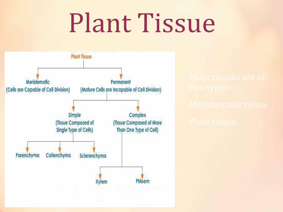
Plant tissues are of two types :-
Meristematic tissue
Plant tissue

Meristematic Tissue
• Meristematic tissues continuously form a number of new cells and helps in growth and are generally made up live cells . Meristematic tissues are the group of cells that have the ability to divide. These tissues in a plant consist of small, densely packed cells that can keep dividing to form new cells. Meristems give rise to permanent tissues and have the following characteristics:
• the cells are small,
• the cells walls are thin,
• cells have large nuclei,
• vacuoles are absent or very small
• there are no intercellular spaces.
Types of Meristematic Tissue
• Apical Meristem:- Apical meristem is present on root apex, stem apex, leaf buds and flower buds. They are responsible for growth in length, i.e. primary growth.
• Lateral Meristem: Lateral meristem is present along the side of the stem. They are responsible for growth in girth, i.e. secondary growth.
• Intercalary Meristem: Intercalary meristem is present at the base of leaf or internodes. They are present on either side of the node.

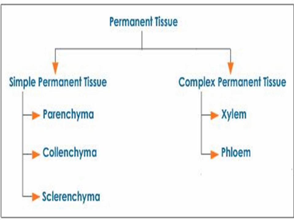
Permanent Tissue [Plant Tissue]Once the cells of meristematic tissue divide to a certain extent, they become specialized for a particular function. This process is called differentiation. Once differentiation is accomplished, the cells lose their capability to divide and the tissue becomes permanent tissue. Permanent tissues are of two types, simple permanent tissue and complex permanent tissue.
Permanent tissue gives support and are generally made up of dead cells . The cells of permanent tissues do not have the ability to divide. These cells are already differentiated in different tissue types and is now specialized to perform specific functions. They are subdivided into two groups, simple tissues consisting of cells which are more or less similar, e.g. epidermis, parenchyma, chlorenchyma, collenchyma, sclerenchyma and complex tissues consisting of different kinds of cells, e.g. xylem and phloem.
Parenchyma tissue
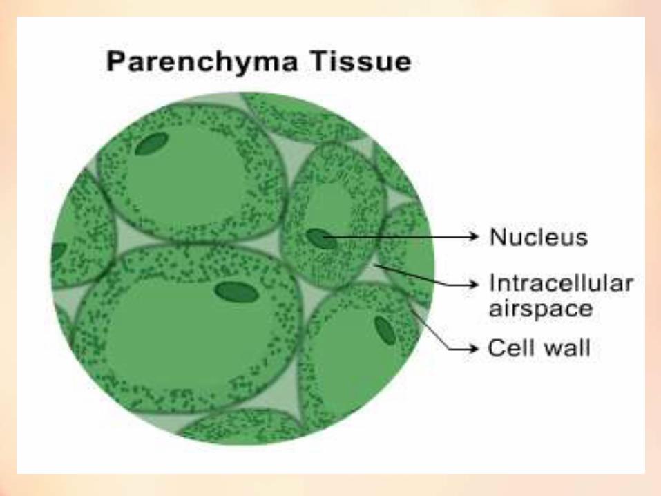
• The cells of parenchyma have thin cell wall. They are loosely packed; with lot of intercellular spaces between them. Parenchyma makes the largest portion of a plant body. Parenchyma mainly works are packing material in plant parts. The main function of parenchyma is to provide support and to store food.
• It is loosely packed and inter cellular spaces are there .
• In aquatic plants , air is filled in parenchyma tissue , so they are called Arenchyma .
• Parenchyma in which chlorophyll is present is called chlorenchyma .
Collenchyma tissue
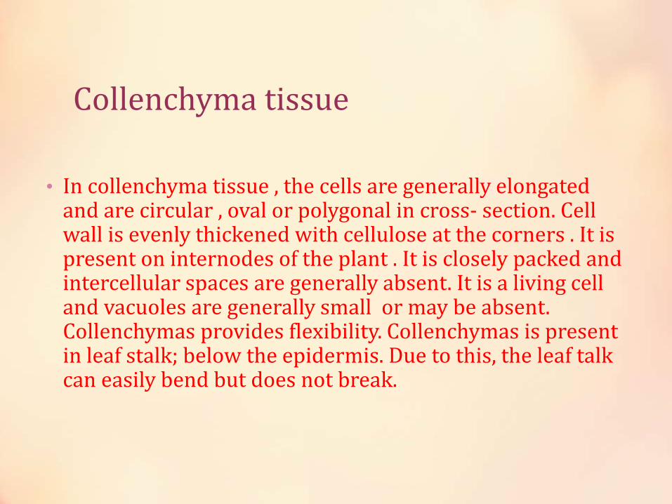
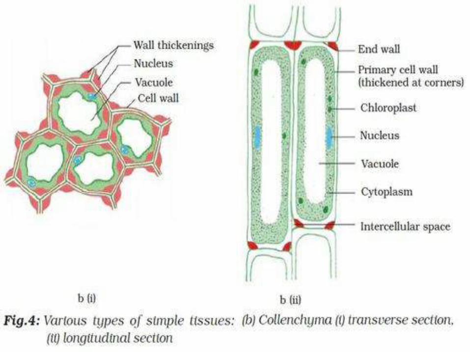
• In collenchyma tissue , the cells are generally elongated and are circular , oval or polygonal in cross- section. Cell wall is evenly thickened with cellulose at the corners . It is present on internodes of the plant . It is closely packed and intercellular spaces are generally absent. It is a living cell and vacuoles are generally small or may be absent. Collenchymas provides flexibility. Collenchymas is present in leaf stalk; below the epidermis. Due to this, the leaf talk can easily bend but does not break.
Sclerenchyma tissue
The cell wall in sclerenchyma is highly thickened all around. The cells are dead and intercellular space is absent. In sclerenchyma lignin is present by which Sclerenchyma provides structural rigidity to plant . These are the hardest tissue. For example :- Bark is composed of sclerenchyma. Another example of sclerenchyma can be seen in the coconut husk .

Sclerenchyma
Epidermis
The epidermis is a protective outer covering of plants. It may be comprised of a single layer in plants. The primary role of the epidermis is to protect the more susceptible layers of the skin. In plants, the epidermal cells secrete further protective substance(called cuticle) to prevent desiccation (water loss). A plant leaf is lined with two layers of epidermis (i.e. upper and lower): one on top of and another one below the mesophyll layer. It protects the plant from any harmful things , like :- fungi , parasites .
Stomata
The epidermis of leaves has small pores called stomata. A stoma is a composed of two guard cells which regulate the opening and closing of stoma. Stomata facilitates exchange of gases and transpiration.


Complex Permanent Tissue
• Variety of cells performing same or different function.
• Complex permanent tissue is composed of different types of cells. Complex permanent tissues are of two types, viz. xylem and phloem. Xylem and phloem together make the vascular bundle in plants.
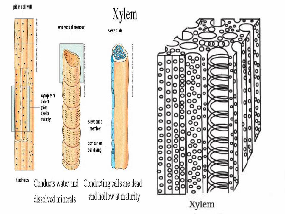
XylemXylem tissue helps in conduction of water. Transportation in xylem is only a way movement , from downwards to upwards . It transport water from roots to the whole parts of the plant , in the upwards direction . Xylem is composed of – trachieds , vessels , xylem fibre and xylem parenchyma . It is made up of dead tissues except xylem parenchyma . Xylem fibre gives support and strength . Trachieds helps in passage of water. Vessels helps in conduction of water and are present as the tubulls in the stem .

PhloemPhloem helps in conduction of food material .Phloem is composed of sieve tubes, companion cells, phloem fibre and phloem parenchyma. Made up of live cells except phloem fibre. Sieve tubes are tubular cells with perforated walls. Sieve tubes are the conducting elements of phloem. Phloem is responsible for translocation of food in plants. It transport food from leaves to the whole plant .The transport of food in phloem is a two way movement.

Vascular Tissue
• Vascular tissue is a complex conducting tissue, formed of more than one cell type, found in vascular plants. The primary components of vascular tissue are the xylem and phloem. These two tissues transport fluid and nutrients internally. There are also two meristems associated with vascular tissue: the vascular cambium and the cork cambium. All the vascular tissues within a particular plant together constitute the vascular tissue system of that plant.

Animal Tissues
• Animal tissues are of four types, viz. epithelial tissue, connective tissue, muscular tissue and nervous tissue.

Epithelial Tissue
• The epithelial tissue forms the covering or lining of most of the organs. The cells of epithelial tissue are tightly packed and form a continuous sheet. There is small amount of cementing materials between the cells and no intercellular space is present. Permeability of the epithelial tissue plays a great role in exchange of materials among various organs it also plays an important role in osmoregulation. All epithelial tissues are separated by the underlying tissue by an extracellular fibrous basement membrane.

Epithelial Tissue
• Epithelial tissue covers external surfaces and internal cavities and organs. Glands are also composed of epithelial tissue.
• Epithelia forms boundaries. Most substances that move into or out of the body must pass through epithelial tissue.
• One surface of the tissue is free and the other adheres to a basement membrane.

Function of Epithelial Tissue• Protection
• Epithelial tissue forms the skin of many animals.
• Terrestrial vertebrates have keratin in their skin cells making them resistant to water loss.
• Ciliated epithelium lines the respiratory tract. Numerous cilia on these cells sweep impurities toward the throat.
• Absorption
• Absorption is an important function of epithelial tissue. For example, the gut is lined with epithelial tissue and it functions to absorb nutrients from food. The lungs are also lined with epithelial tissue and it functions to absorb oxygen.
• Secretion
• Glandular epithelium secretes chemicals.
• Endocrine glands secrete hormones directly into the extracellular space.
• Exocrine glands often secrete through DUCTS; they secrete mucus, saliva, wax, milk, etc.

Squamous Epithelial Tissue
• A squamous epithelium tissue is a single layer of flat cells in contact with the basal lamina (basement membrane) of the epithelium.[1] This type of epithelium is often permeable and occurs where small molecules pass quickly through membranes via filtration or diffusion. Simple squamous epithelia are found in capillaries, alveoli, glomeruli, and other tissues where rapid diffusion is required . It is a protective tissue . It is tightly packed and have no in cellular spaces .
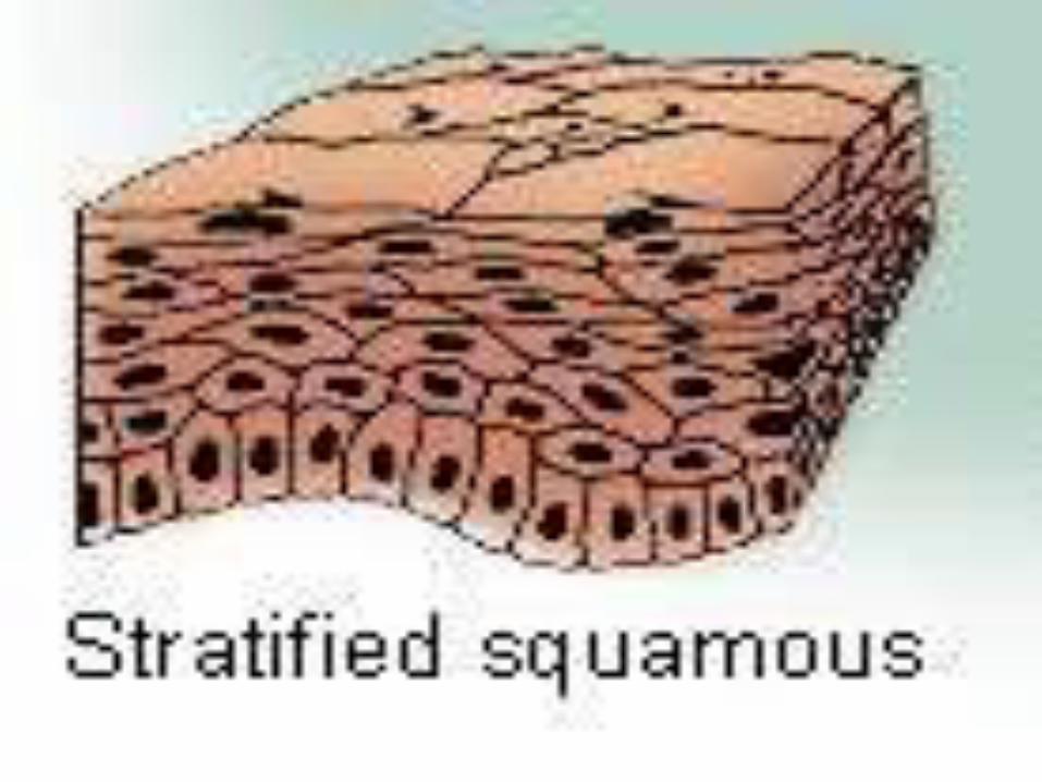
Stratified Squamous Epithelial Tissue
• A stratified squamous epithelium consists of more than one (typically many) layers, or strata, of epithelial cells (see figure). The basal layer of epithelial cells are small and cuboidal-to-columnar; the cells gradually become larger and more squamous as the cells migrate from the basal layer to the apical layer.
• Stratified squamous epithelia are specialized to withstand the mechanical stresses of abrasion. The apical layers of epithelial are designed to give way to abrasive forces, protecting the deeper tissues from the mechanical stress. As the apical layers of cells give way, they are continuously replaced by the deeper layers of epithelial cells, all of which are derived from the highly mitotic cuboidal cells of the basal layer. Example :- skin

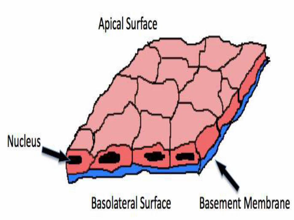
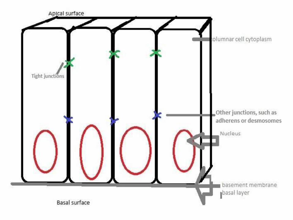
Columnar Epithelial Tissue
• Columnar epithelial cells are longer than they are wide. Characteristically, their nuclei are found at the base of the cell. The cells are connected by tight junctions. The cells receive nutrients through the basement membrane, which Simple columnar epithelial cells are some of the most prolific cells in the body, mainly because they can fulfill so many functions. They are found throughout the body's organ system, including the digestive track . They are found in the respiratory system, including the nasal passage. Because epithelium can be innervated, simple columnar epithelial cells can also serve a sensory function: simple columnar epithelium line the ears and buccal cavity and are found in the eye. separates the cells from the capillary basal layer. It protects or stop the unwanted particles to enter our body .
Cuboidal Epithelial Tissue
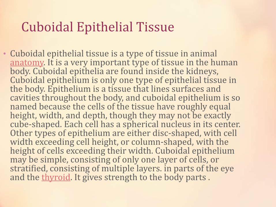
• Cuboidal epithelial tissue is a type of tissue in animal anatomy. It is a very important type of tissue in the human body. Cuboidal epithelia are found inside the kidneys, Cuboidal epithelium is only one type of epithelial tissue in the body. Epithelium is a tissue that lines surfaces and cavities throughout the body, and cuboidal epithelium is so named because the cells of the tissue have roughly equal height, width, and depth, though they may not be exactly cube-shaped. Each cell has a spherical nucleus in its center. Other types of epithelium are either disc-shaped, with cell width exceeding cell height, or column-shaped, with the height of cells exceeding their width. Cuboidal epithelium may be simple, consisting of only one layer of cells, or stratified, consisting of multiple layers. in parts of the eye and the thyroid. It gives strength to the body parts .
Connective Tissue
• The cells of connective tissue are separated by non-living material.
• Connective tissue binds and supports body parts, protects, fills spaces, stores fat (for energy), and transports materials.
• Structure of Loose and Dense Connective Tissue
• Loose connective tissue and dense connective tissue contain three kinds of fibers. Collagen fibers provide strength and flexibility. Collagen is the most abundant protein in animal bodies. Elastic fibers provide elasticity. When stretched, they return to their original shape. Reticular fibers are small and branched. They provide a support framework for organs such as the liver and lymph nodes.
• The cells of loose and dense connective tissue are called fibroblasts. They produce the fibers and nonliving matrix material. Macrophages are cells specialized for phagocytizing foreign materials, bacteria, and cleaning up debris. Macrophages will be discussed in the chapter on the immune system.
• Loose Connective Tissue
• Loose connective tissue includes areolar, adipose, and reticular connective tissue.
• Areolar Connective Tissue
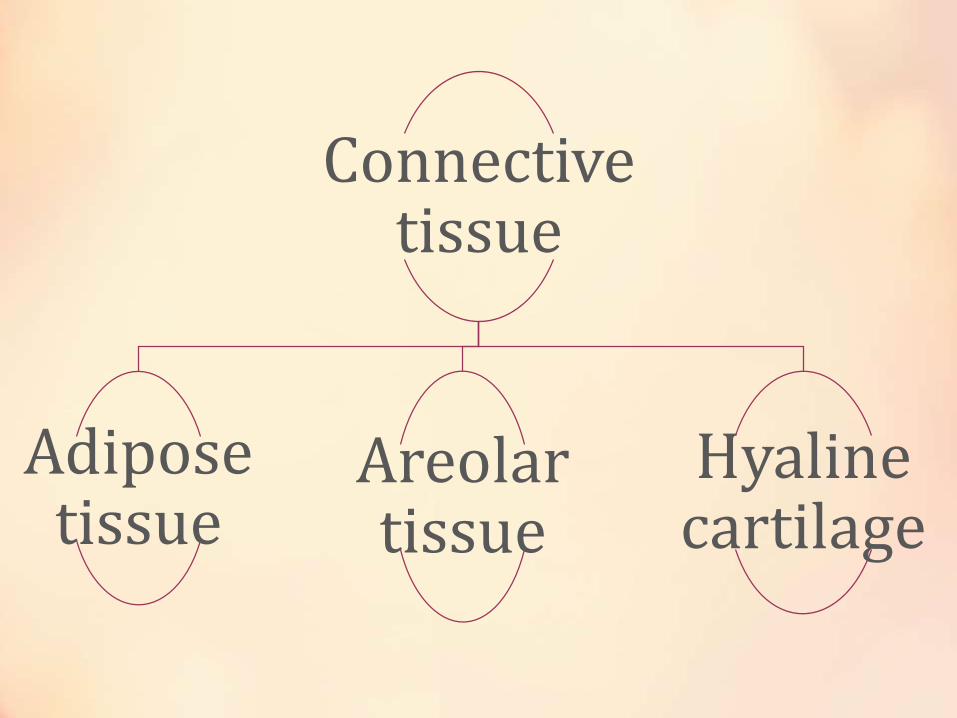
Connective tissue
Adipose tissue
Areolar tissue
Hyaline cartilage
Adipose Tissue
• Adipose tissue is one of the main types of connective tissue. Its main role is to store energy in the form of lipids, although it also cushions and insulates the body. In humans, adipose tissue is located beneath the skin (subcutaneous fat), around internal organs (visceral fat), in bone marrow (yellow bone marrow) and in the breast tissue. Adipose tissue is found in specific locations, which are referred to as adipose depots. Around organs, it provides protective padding. However, its main function is to be a reserve of lipids, which can be burned to meet the energy needs of the body and to protect it from excess glucose by storing triglycerides produced by the liver from sugars, although some evidence suggests that most lipid synthesis from carbohydrates occurs in the adipose tissue itself.[3] Adipose depots in different parts of the body have different biochemical profiles. Under normal conditions, it provides feedback for hunger and diet to the brain.

Areolar Tissue
• Found throughout the body, areolar tissue is a type of connective tissue. It consists of cells and various properties of the tissue’s intercellular matrix. Cushioning surrounding organs, connecting different tissues, and supporting blood vesselsrepresent just a few of the functions of this specific connective tissue.
• Areolar tissue is composed of several cell types. Among the mix of cells are adipose cells, better known as fat cells, mast cells and macrophages. The remaining composition includes leukocytes and plasma cells.
• Like other loose connective tissues, areolar connective tissue consists of three different types of fibers. These fibers include collagenous fibers, elastic fibers and reticular fibers. Together these fibers make up the traditional weaved appearance of areolar, and other loose connective tissues.
• The intercellular matrix of areolar tissue is composed of the matrix of collagenous, elastic and reticular fibers and ground substance. Proteins and proteoglycans contribute to the makeup of ground substance. Ground substance works to hold together the various properties of this tissue.
• Various cells and properties of the intercellular matrix of this tissue can be seen under a microscope. Under the microscope collagenous, elastic, and reticular fibers appear as pink bands of varying widths arranged in no distinct pattern. Fibroblasts may appear as dark dots with finger-like extensions. The surrounding substance, referred to as the ground substance, appears as a light stain on microscope slides but may be difficult to see.
Hyaline Cartilage
• Hyaline cartilage is semi-transparent and appears bluish-white in color. It is extremely strong, but very flexible and elastic. Hyaline cartilage consists of living cells, chondrocytes, which are situated far apart in fluid-filled spaces, the lacunae. There is an extensive amount of rubbery matrix between the cells and the matrix contains a number of collagenous fibres. Hyaline cartilage occurs in trachea, the larynx, the tip of the nose, in the connection between the ribs and the breastbone and also the ends of bone where they form joints. Temporary cartilage in mammalian embryos also consists of hyaline cartilage.
Functions :-
• Reduces friction at joints. By virtue of the smooth surface of hyaline cartilage, it provides a sliding area which reduces friction, thus facilitating bone movement.
• Movement Hyaline cartilage joins bones firmly together in such a way that a certain amount of movement is still possible between them.
• Support The c-shaped cartilaginous rings in the windpipes (trachea and bronchi) assist in keeping those tubes open.
• Growth Hyaline cartilage is responsible for the longitudinal growth of bone in the neck regions of the long bones.
Muscle Tissue
• Muscular tissue is composed of muscle cells. Muscle cells are specialized cells which have the capability to contract and expand. Due to contraction and expansion, muscles facilitate various kinds of movements in the body. Muscles contain special proteins called contractile proteins , which contract and relax to cause movement. Muscular tissues are of three types:
• Striated muscle
• Smooth muscle
• Cardiac muscle
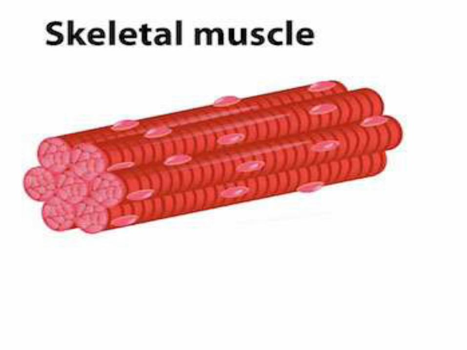
Striated Muscle Tissue
• striated muscle, also called voluntary muscle, striped muscle, or skeletal muscle , most common of the three types of muscle in the body. Striated muscle is attached to bone and produces all the movements of body parts in relation to each other; unlike smooth muscle and cardiac muscle, striated muscle is under voluntary control. Its multinucleated fibres are long and thin and are crossed with a regular pattern of fine red and white lines, giving the muscle its distinctive appearance and its name.

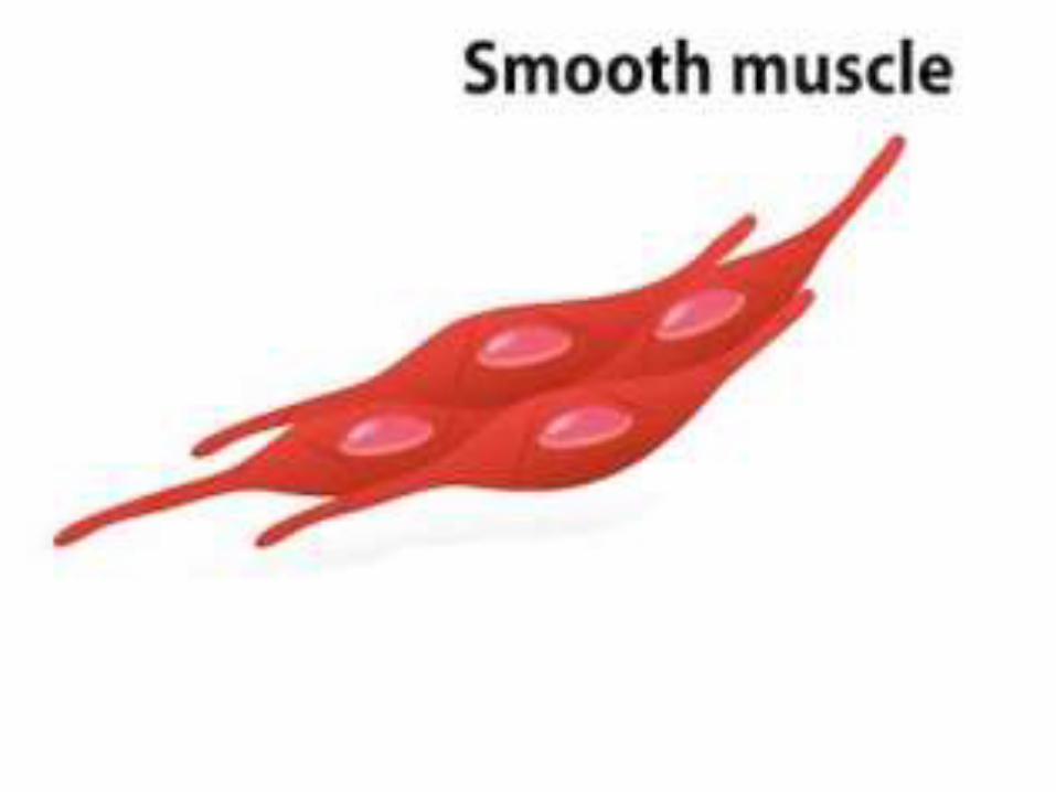
Smooth muscle tissue
• Smooth muscle is an involuntary non-striated muscle. Smooth muscle is found within the walls of blood vessels (such smooth muscle specifically being termed vascular smooth muscle) such as in the tunica media layer of large (aorta) and small arteries, arterioles and veins. Smooth muscle is also found in lymphatic vessels, the respiratory tract, erector pili of skin, the ciliary muscle, and iris of the eye. The structure and function is basically the same in smooth muscle cells in different organs, but the inducing stimuli differ substantially, in order to perform individual effects in the body at individual times. In addition, the glomeruli of the kidneys contain smooth muscle-like cells called mesangial cells. Smooth muscle is found in the walls of hollow organs like your intestines and stomach. They work automatically without you being aware of them. Smooth muscles are involved in many 'housekeeping' functions of the body. The muscular walls of your intestines contract to push food through your body
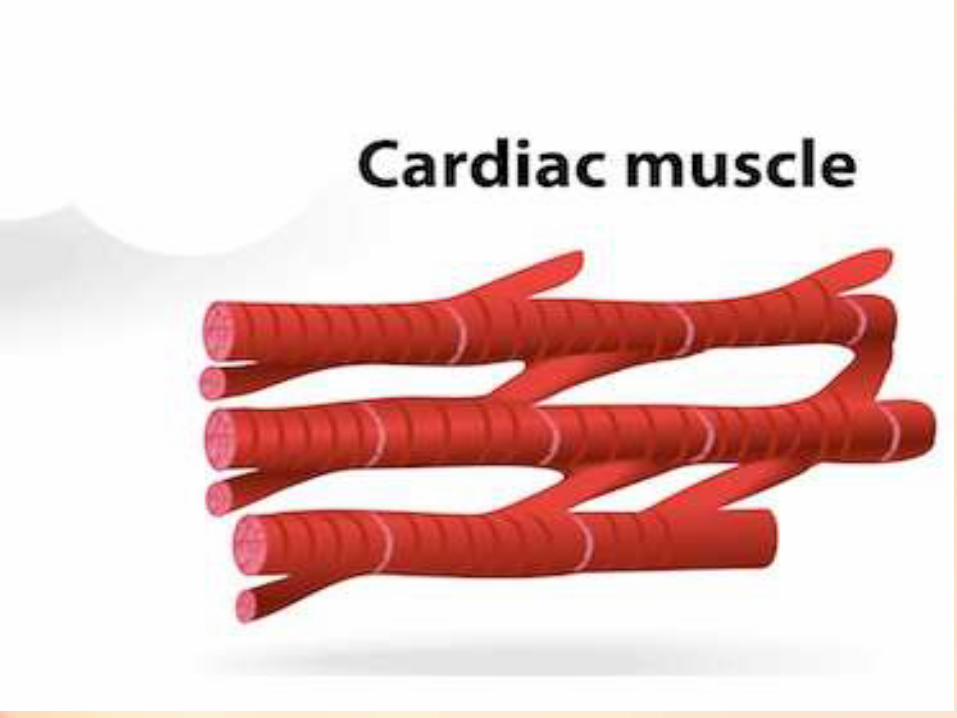
Cardiac muscle tissue
• Cardiac muscle tissue is an extremely specialized form of muscle tissue that has evolved to pump blood throughout the body. In fact, cardiac muscle is only found in the heart and makes up the bulk of the heart’s mass. The heart beats powerfully and continuously throughout an entire lifetime without any rest, so cardiac muscle has evolved to have incredibly high contractile strength and endurance. And because the heart maintains its own rhythm, cardiac muscle has developed the ability to quickly spread electrochemical signals so that all of the cells in the heart can contract together as a team. Cardiac muscle tissue is made up of many interlocking cardiac muscle cells, or fibers, that give the tissue its properties. Each cardiac muscle fiber contains a single nucleus and is striated, or striped, because it appears to have light and dark bands when seen through a microscope. The dark bands represent areas of thick protein filaments made of myosin proteins that block light passing through the cell and appear dark. Between the dark bands are thin filaments made of actin protein that allow light to pass through and appear light. When the muscle fibers contract, myosin pulls the actin filaments together like an accordion to shrink the muscle cell and make it contract. While each cell is not very strong by itself, millions of cardiac muscle cells working together are easily able to pump all of the blood in the body through the heart in less than a minute.

Nervous Tissue
• Nervous tissue makes the nervous system and is composed of specialized cells called neuron. A neuron can be divided into two distinct parts, viz. head and tail. The head is somewhat star-shaped and contains nucleus and some other cell organelles. This is called cyton. There are numerous hair-like outgrowths coming out of the cyton. These are called dendrites. The tail ends in axon terminals. Dendrites receive the nerve impulse, while axon relays the nerve signals.


Presented By :-
Prakriti AnjaliShivani JoshiaYamini
Related Documents