JOURNAL OF VIROLOGY, 0022-538X/00/$04.0010 Apr. 2000, p. 3112–3121 Vol. 74, No. 7 Copyright © 2000, American Society for Microbiology. All Rights Reserved. Tissue Sites of Persistent Infection and Active Replication of Equine Infectious Anemia Virus during Acute Disease and Asymptomatic Infection in Experimentally Infected Equids SHARON M. HARROLD, 1 SHEILA J. COOK, 2 R. FRANK COOK, 2 KEITH E. RUSHLOW, 1 ² CHARLES J. ISSEL, 2 AND RONALD C. MONTELARO 1 * Department of Molecular Genetics and Biochemistry, School of Medicine, University of Pittsburgh, Pittsburgh, Pennsylvania 15261, 1 and Gluck Equine Research Center, Department of Veterinary Science, University of Kentucky, Lexington, Kentucky 40546 2 Received 9 August 1999/Accepted 20 December 1999 Equine infectious anemia virus (EIAV) infection of horses is characterized by recurring cycles of disease and viremia that typically progress to an inapparent infection in which clinical symptoms are absent as host immune responses maintain control of virus replication indefinitely. The dynamics of EIAV viremia and its association with disease cycles have been well characterized, but there has been to date no comprehensive quantitative analyses of the specific tissue sites of EIAV infection and replication in experimentally infected equids during acute disease episodes and during asymptomatic infections in long-term inapparent carriers. To characterize the in vivo site(s) of viral infection and replication, we developed a quantitative competitive PCR assay capable of detecting 10 copies of viral DNA and a quantitative competitive reverse transcription-PCR assay with a sensitivity of about 30 copies of viral singly spliced mRNA. Animals were experimentally infected with one of two reference viruses: the animal-passaged field isolate designated EIAV Wyo and the virulent cell-adapted strain designated EIAV PV . Tissues and blood cells were isolated during the initial acute disease or from asymptomatic animals and analyzed for viral DNA and RNA levels by the respective quantitative assays. The results of these experiments demonstrated that the appearance of clinical symptoms in experi- mentally infected equids coincided with rapid widespread seeding of viral infection and replication in a variety of tissues. During acute disease, the predominant cellular site of viral infection and replication was the spleen, which typically accounted for over 90% of the cellular viral burden. In asymptomatic animals, viral DNA and RNA persisted in virtually all tissues tested, but at extremely low levels, a finding indicative of tight but incomplete immune control of EIAV replication. During all disease states, peripheral blood mononuclear cells (PBMC) were found to harbor less than 1% of the cellular viral burden. These quantitative studies demonstrate that tissues, rather than PBMC, constitute the predominant sites of virus replication during acute disease in infected equids and serve as resilient reservoirs of virus infection, even in the presence of highly effective immune responses that maintain a stringent control of virus replication in long-term inapparent carriers. Thus, these observations with EIAV, a predominantly macrophage-tropic lentivirus, highlight the role of tissues in sequestering lentiviral infections from host immune surveillance. Equine infectious anemia virus (EIAV) is unique among lentiviruses in that the clinical course of infection in equids results initially in a rapid and dynamic series of clearly demar- cated cycles of disease and associated viremia that begin by 3 weeks postinfection and continue at irregular intervals sepa- rated by weeks or months (reviewed in reference 25). Disease cycles last 3 to 5 days and are characterized by fever, diarrhea, lethargy, edema, anemia, and thrombocytopenia. This stage of disease, defined as chronic equine infectious anemia (EIA), typically lasts about 8 to 12 months postinfection, with the frequency and severity of clinical episodes decreasing with time. In contrast to the progressive degenerative disease asso- ciated with most lentiviral infections, horses infected with EIAV typically make a transition during the first year postin- fection from chronic EIA to an inapparent infection in which clinical symptoms are absent and viremia is usually undetect- able for the remainder of the animal’s life span of up to about 20 years. Thus, the EIAV systems provides a novel model in which to examine the dynamics of lentivirus replication during clearly defined cycles of disease and during long-term asymp- tomatic infections. Several lines of evidence indicate that the control of EIAV replication and disease in long-term inapparent carriers is me- diated by virus-specific host immune responses that evolve during the first year postinfection to achieve an enduring ef- fective suppression of virus replication. For example, experi- mental infection of foals with severe combined immunodefi- ciency results in a progressive infection leading to death, demonstrating the necessity of the host immune system in accomplishing the temporal control of virus replication asso- ciated with infection of immunocompetent horses (29). In ad- dition, it has been shown that severe stress or treatment of long-term inapparent carriers with immunosuppressive drugs can cause recrudescence of viremia and disease, even after decades of clinical quiescence (16, 48). Finally, it has been demonstrated that transfer of whole blood from long-term inapparent carriers to naive horses results in EIAV infection and disease in the recipient horses (12). Taken together, these observations demonstrate the lack of attenuation of the infect- * Corresponding author. Mailing address: Department of Molecular Genetics and Biochemistry, School of Medicine, University of Pitts- burgh, Pittsburgh, PA 15261. Phone: (412) 648-8869. Fax: (412) 383- 8859. E-mail: [email protected]. ² Present address: Ross Products Division, Abbott Laboratories, Columbus, OH 43215. 3112 on November 10, 2015 by guest http://jvi.asm.org/ Downloaded from

Welcome message from author
This document is posted to help you gain knowledge. Please leave a comment to let me know what you think about it! Share it to your friends and learn new things together.
Transcript

JOURNAL OF VIROLOGY,0022-538X/00/$04.0010
Apr. 2000, p. 3112–3121 Vol. 74, No. 7
Copyright © 2000, American Society for Microbiology. All Rights Reserved.
Tissue Sites of Persistent Infection and Active Replication of EquineInfectious Anemia Virus during Acute Disease and Asymptomatic
Infection in Experimentally Infected EquidsSHARON M. HARROLD,1 SHEILA J. COOK,2 R. FRANK COOK,2 KEITH E. RUSHLOW,1†
CHARLES J. ISSEL,2 AND RONALD C. MONTELARO1*
Department of Molecular Genetics and Biochemistry, School of Medicine, University of Pittsburgh,Pittsburgh, Pennsylvania 15261,1 and Gluck Equine Research Center, Department of Veterinary Science,
University of Kentucky, Lexington, Kentucky 405462
Received 9 August 1999/Accepted 20 December 1999
Equine infectious anemia virus (EIAV) infection of horses is characterized by recurring cycles of disease andviremia that typically progress to an inapparent infection in which clinical symptoms are absent as hostimmune responses maintain control of virus replication indefinitely. The dynamics of EIAV viremia and itsassociation with disease cycles have been well characterized, but there has been to date no comprehensivequantitative analyses of the specific tissue sites of EIAV infection and replication in experimentally infectedequids during acute disease episodes and during asymptomatic infections in long-term inapparent carriers. Tocharacterize the in vivo site(s) of viral infection and replication, we developed a quantitative competitive PCRassay capable of detecting 10 copies of viral DNA and a quantitative competitive reverse transcription-PCRassay with a sensitivity of about 30 copies of viral singly spliced mRNA. Animals were experimentally infectedwith one of two reference viruses: the animal-passaged field isolate designated EIAVWyo and the virulentcell-adapted strain designated EIAVPV. Tissues and blood cells were isolated during the initial acute diseaseor from asymptomatic animals and analyzed for viral DNA and RNA levels by the respective quantitativeassays. The results of these experiments demonstrated that the appearance of clinical symptoms in experi-mentally infected equids coincided with rapid widespread seeding of viral infection and replication in a varietyof tissues. During acute disease, the predominant cellular site of viral infection and replication was the spleen,which typically accounted for over 90% of the cellular viral burden. In asymptomatic animals, viral DNA andRNA persisted in virtually all tissues tested, but at extremely low levels, a finding indicative of tight butincomplete immune control of EIAV replication. During all disease states, peripheral blood mononuclear cells(PBMC) were found to harbor less than 1% of the cellular viral burden. These quantitative studies demonstratethat tissues, rather than PBMC, constitute the predominant sites of virus replication during acute disease ininfected equids and serve as resilient reservoirs of virus infection, even in the presence of highly effectiveimmune responses that maintain a stringent control of virus replication in long-term inapparent carriers.Thus, these observations with EIAV, a predominantly macrophage-tropic lentivirus, highlight the role oftissues in sequestering lentiviral infections from host immune surveillance.
Equine infectious anemia virus (EIAV) is unique amonglentiviruses in that the clinical course of infection in equidsresults initially in a rapid and dynamic series of clearly demar-cated cycles of disease and associated viremia that begin by 3weeks postinfection and continue at irregular intervals sepa-rated by weeks or months (reviewed in reference 25). Diseasecycles last 3 to 5 days and are characterized by fever, diarrhea,lethargy, edema, anemia, and thrombocytopenia. This stage ofdisease, defined as chronic equine infectious anemia (EIA),typically lasts about 8 to 12 months postinfection, with thefrequency and severity of clinical episodes decreasing withtime. In contrast to the progressive degenerative disease asso-ciated with most lentiviral infections, horses infected withEIAV typically make a transition during the first year postin-fection from chronic EIA to an inapparent infection in whichclinical symptoms are absent and viremia is usually undetect-
able for the remainder of the animal’s life span of up to about20 years. Thus, the EIAV systems provides a novel model inwhich to examine the dynamics of lentivirus replication duringclearly defined cycles of disease and during long-term asymp-tomatic infections.
Several lines of evidence indicate that the control of EIAVreplication and disease in long-term inapparent carriers is me-diated by virus-specific host immune responses that evolveduring the first year postinfection to achieve an enduring ef-fective suppression of virus replication. For example, experi-mental infection of foals with severe combined immunodefi-ciency results in a progressive infection leading to death,demonstrating the necessity of the host immune system inaccomplishing the temporal control of virus replication asso-ciated with infection of immunocompetent horses (29). In ad-dition, it has been shown that severe stress or treatment oflong-term inapparent carriers with immunosuppressive drugscan cause recrudescence of viremia and disease, even afterdecades of clinical quiescence (16, 48). Finally, it has beendemonstrated that transfer of whole blood from long-terminapparent carriers to naive horses results in EIAV infectionand disease in the recipient horses (12). Taken together, theseobservations demonstrate the lack of attenuation of the infect-
* Corresponding author. Mailing address: Department of MolecularGenetics and Biochemistry, School of Medicine, University of Pitts-burgh, Pittsburgh, PA 15261. Phone: (412) 648-8869. Fax: (412) 383-8859. E-mail: [email protected].
† Present address: Ross Products Division, Abbott Laboratories,Columbus, OH 43215.
3112
on Novem
ber 10, 2015 by guesthttp://jvi.asm
.org/D
ownloaded from
ing virus during persistent infection and the importance of hostimmune responses in establishing and maintaining control ofEIAV replication and disease. The same experiments, how-ever, also clearly demonstrate the persistence of virulent EIAVinfection in infected horses during asymptomatic and nonvire-mic periods, either during chronic EIA or in long-term inap-parent infections. This persistence of EIAV infection in thepresence of enduring, robust, and highly suppressive host im-munity raises a number of interesting questions about thereservoirs of EIAV infection that are able to escape host im-mune surveillance.
The primary target of EIAV in vivo are cells of the mono-cyte/macrophage lineage (25), although there has recentlybeen a report of limited infection of macrovascular endothelialcells in the renal tissues of a long-term inapparent carrier horse(22). Infection of blood monocytes with EIAV results in anonproductive infection, and differentiation of virus-infectedmonocytes to macrophages is required to activate virus repli-cation (21, 43). This pattern of infection has led to the modelthat virus-infected monocytes may serve as “Trojan horses”that can widely disseminate EIAV infection to tissues withoutdetection by the host immune system, suggesting that mono-cytes may sequester EIAV during asymptomatic infection. Theidentification of EIAV infection of macrovascular endothelialcells suggests that this cell type may also serve as a reservoir forvirus infection, at least in long-term inapparent infections. Therole, if any, of endothelial cell infection in acute disease re-mains to be defined.
The primary sites of EIAV replication in persistently in-fected horses has been examined by immunofluorescent tech-niques to detect viral antigens (23), by Southern blot analysesto detect proviral DNA (35), and more recently by qualitativePCR techniques to detect viral DNA or RNA (14, 27, 42). Theresults of these studies uniformly demonstrate that the highlevels of viremia observed during acute EIA are associatedwith high levels of virus replication in macrophage-rich tissues,including liver, spleen, and kidney; peripheral blood mononu-clear cells (PBMC) and other tissues appeared to contain onlyminor levels of virus infection, despite the high levels of virusin the blood. In contrast to the abundance of viral antigens andDNA observed in tissues from EIAV-infected horses duringacute disease, qualitative analyses of EIAV infection sites bySouthern blot (35) and PCR (14, 42) in a limited number ofinapparent carrier horses indicate low levels of EIAV infectionassociated with tissue macrophages and typically undetectablelevels of EIAV DNA in PBM. These general patterns of EIAVinfection were confirmed recently in an informative study byOaks et al. (26) using a combination of PCR and in situ hy-bridization techniques to elucidate tissue macrophages as thepredominant site of EIAV infection during clinical and sub-clinical infections.
The previous studies of the sites of EIAV infection in ex-perimentally infected horses provide an important qualitativedescription of virus infection in vivo, but there has been to dateno comprehensive quantitative analyses of EIAV DNA andRNA in experimentally infected horses to ascertain the relativeimportance of different tissues in virus production during acutedisease or in persistence during inapparent infections. In ad-dition, the majority of these published studies of EIAV infec-tion during acute disease are based on experimental infectionswith the highly virulent EIAVWyo strain of virus that usuallycauses death within several weeks postinfection, in markedcontrast to the chronic EIA observed in most EIAV infectionsin the field. The highly virulent nature of the EIAVWyo raisessome concerns about the relevance of this experimental infec-tion model to the more typical infections that result in chronicEIAV that progresses to a long-term inapparent infection.Finally, the characterization of EIAV replication in vivo hasbeen limited by the use of only one or two animals in manypublished studies.
Therefore, we sought in this current study to characterizeviremia levels and the tissue sites of EIAV infection and rep-lication during symptomatic and asymptomatic infections byusing newly developed sensitive and quantitative PCR andreverse transcriptase PCR (RT-PCR) techniques to measureviral DNA and RNA, respectively. For these studies we em-ployed the well-characterized EIAVPV reference strain (25,38), which has been used extensively in our laboratory in ex-perimental infections to reliably produce in equids a clinicalprogression from chronic EIA to a long-term inapparent in-fection. For comparison to previous studies, we also examinedvirus infection and replication during acute disease in horsesinoculated with the highly virulent EIAVWyo. The results ofthese studies provide for the first time a comprehensive quan-titative analysis of the in vivo sites of viral infection and rep-lication in various tissues from experimentally infected animalsduring the initial acute disease and during long-term inappar-ent infection.
MATERIALS AND METHODS
Virus strains and experimental animals. The highly virulent wild-typeEIAVWyo strain, an animal-passaged isolate, was produced as serum taken dur-ing acute disease in an experimentally infected horse, as described earlier (35).The titer of the EIAVWyo stock was 106 horse infectious doses per ml. Thecell-adapted pathogenic EIAVPV strain was derived, and the titers were deter-mined as described elsewhere (49). Infectious stocks of EIAVPV were grown, andtiters were determined in primary fetal equine kidney cell cultures. The titer ofEIAVPV stock was 106.5 50% tissue culture infectious doses (TCID50) per ml.
Two thoroughbred horses (298 and 94-07) and four mixed-breed ponies(112, 123, 18, and 72) were used in these studies. Prior to infection, animalswere shown to be seronegative for EIAV-reactive antibodies by the agar immu-nodiffusion test (IDEXX Laboratories, Westbrook, Mass.), and viral RNA wasnot detected in the plasma by RT-PCR analyses (see below). The two horseswere intravenously inoculated with 1 ml of EIAVWyo serum (Table 1). Ponies 112and 123 were inoculated intravenously with 106 TCID50 of the EIAVPV strain,
TABLE 1. Clinical profiles and levels of EIAV in the plasma of experimentally infected animals
Animalno.
Virusstrain
Infectiousdosea
Euthanized(days postinfection) Clinical status Plasma viremia
(TCID50/ml)bPlasmaRNAc
298 EIAVWyo 1 3 106 12 Acute 105.0 3.1 3 108
94-07 EIAVWyo 1 3 106 11 Acute 103.9 3.6 3 107
112 EIAVPV 1 3 106 9 Acute 103.6 2.1 3 107
123 EIAVPV 1 3 106 13 Acute 104.7 4.2 3 107
18 EIAVPV 1 3 104 538 Asymptomatic Negd Neg72 EIAVPV 3 3 102 506 Asymptomatic Neg 1.2 3 102
a EIAVWyo, horse infectious doses; EIAVPV, TCID50.b TCID50 per ml as determined in primary MDM cultures from plasma obtained the day of euthanization.c Copies of virion genomic RNA per ml of plasma as determined by RT-PCR from samples obtained the day of euthanization.d Neg, negative for detectable infectious virus or viral RNA.
VOL. 74, 2000 EIAV INFECTION AND REPLICATION IN VIVO 3113
on Novem
ber 10, 2015 by guesthttp://jvi.asm
.org/D
ownloaded from

while ponies 18 and 72 were inoculated intravenously with 300 and 104 TCID50of the EIAVPV strain, respectively. All animals were monitored daily via physicalexams and rectal temperatures for clinical signs of EIA.
Clinical samples. Heparinized whole blood was collected by jugular venipunc-ture from experimentally infected animals during defined febrile episodes andfrom asymptomatic animals. Plasma and PBMC were isolated by discontinuousdensity gradient centrifugation. Serum samples were collected. Six animals wereeuthanized by administration of a lethal intravenous injection of Beuthanasia, atwhich time replicate tissues samples were collected and flash frozen immediatelyin liquid nitrogen. Tissue samples from the liver, spleen, medulla and cortex ofthe kidney, peripheral lymph nodes including the sublingual and mandibularnodes, and bone marrow were collected from all animals. The cerebrum, me-dulla, and cerebellum from the brain were collected from three animals (horse94-07 and ponies 112 and 123). Tissue, cellular, plasma, and serum samples werestored at 280°C until isolation of nucleic acids.
Nucleic acid purification from tissue samples. Total cellular DNA was puri-fied by standard techniques (40). Briefly, tissues were immersed in liquid nitro-gen and immediately pulverized. Samples were then suspended in 1 ml of 0.1%sodium dodecyl sulfate (SDS)–10 mM EDTA per 100 mg of tissue or per 106
PBMC, digested with 100 mg of proteinase K per ml at 37°C for a minimum of5 h, followed by phenol-chloroform extraction and ethanol precipitation. Pel-leted DNA was resuspended in 10 mM Tris-HCl–1 mM EDTA by gentle rockingat room temperature overnight. The concentration of the DNA was determinedspectrophotometrically, and the quality of the DNA was assayed by gel electro-phoresis. Samples were stored at 220°C until quantification of the cellulargenomic and EIAV viral DNA content.
RNA was extracted from pulverized tissue samples and PBMC using either 1ml of RNAzol (Biotecz) or TRIzol (BRL) per 100 mg of sample or per 106
PBMC according to the manufacturer’s protocol. RNA was resuspended indiethyl pyrocarbonate (DEPC)-treated water. The concentration of the RNAwas determined by spectrophotometry, and the integrity of the RNA was mon-itored by formaldehyde denaturing gel electrophoresis. Samples were stored at270°C until assayed for viral and cellular RNAs.
Virion-associated genomic RNA was recovered from plasma samples by ul-tracentrifugation to pellet virus and extracted as described earlier (19). Briefly, aminimum of 1 ml of each plasma sample was centrifuged at 100,000 3 g for 45min in a type 55.1 rotor (Beckman Instruments, Palo Alto, Calif.) at 4°C. Thesupernatant was aspirated, and the pellet resuspended in 0.1% SDS. RNA wasextracted using 1 ml of TRIzol as described above. The RNA was precipitated inthe presence of 20 mg of glycogen as carrier. RNA was pelleted by centrifugationat 12,000 3 g for 15 min at 4°C and resuspended in 10 to 20 ml of DEPC-treatedwater. All samples were stored at 270°C until quantification of the viral RNA.
Quantitative PCR analyses. The viral DNA burden was determined by usinga competitive PCR (QC-PCR) assay to quantify viral DNA (30). To optimizedetection of variant quasispecies, highly conserved gag-specific primers werechosen based on alignment of all of the EIAV sequences in the database usingthe Genetics Computer Group (Madison, Wis.) package analyses software (10).A SmaI-XbaI fragment of the gag gene was cloned into a trpLE vector (3). Thecompetitor template containing a 55-bp deletion at the unique ApaI site wasgenerated by Bal31 exonuclease digestion. The competitor template was quan-tified by spectrophotometry and limiting-dilution analyses (data not shown).Amplification of wild-type and competitor templates by using the sense gag11(59-ATGTATGCTTGCAGAGACATTG-39) and antisense gag34 (59-GCTGACTCTTCTGTTGTATCG-39) primers yielded 310- and 255-bp products, respec-tively. The PCR reaction contained 10 mM Tris-HCl (pH 8.3), 50 mM KCl, 1.5mM MgCl, 0.001% gelatin, 0.05 mM concentrations of each deoxyribonucleosidetriphosphate, 12.5 pmol of each primer, 1 mCi of [a-32P]dATP (3,000 Ci/mmol;NEN, Boston, Mass.), and 2.5 U of AmpliTaq DNA polymerase (Perkin-Elmer,Norwalk, Conn.) in a final volume of 25 ml. The thermal cycling conditionsincluded an initial hot start at 99°C, followed by 94°C for 1 min, 55°C for 1 min,and 72°C for 1 min for 30 cycles, with a final 10-min extension at 72°C. PCRproducts were separated by electrophoresis on 6% acrylamide gels containing 8M urea. Gels were fixed in a 10% methanol–10% acetic acid solution and dried.The radioactivity incorporated in each band was quantified by using phosphor-imaging technologies (Molecular Dynamics, Sunnyvale, Calif.). Preliminary ex-periments were performed to determine the amount of sample DNA that waswithin the linear range of the amplification reaction. Addition of carrier DNA tomaintain a constant mass had a minimal effect on the amplification efficiency;therefore, carrier DNA was not included in the reactions.
Independent amplification of a single-copy cellular genomic sequence wasperformed to control for sample loss or DNA degradation and to normalize thecellular DNA content. A 94-bp fragment of the equine sodium channel gene wasamplified by using the sense (59-GGGAGTGTGTGCTCAAGATGT-39) andantisense (59-AATGGACAGGATGACAACCAC-39) primers, as described ear-lier (37). The concentrations of the PCR reagents were the same as thosedescribed above, except that 6.25 pmol of each primer was used. Half-log dilu-tions from 10 to 100 ng of the sample DNA were used. The thermal cyclingconditions were 94°C for 45 s and 65°C for 4 min for 30 cycles, followed by a10-min extension at 72°C. PCR products were electrophoretically separated andquantified as described above. A standard curve was generated by linear regres-sion analysis of known amounts of cloned DNA. The cellular equivalents (CE) of
all samples were calculated by using the equation of the line, as describedpreviously (44).
Quantitative RT-PCR analyses. To determine the levels of EIAV replicationin various tissues, the amount of singly spliced viral mRNA was quantified bycompetitive RT-PCR (QC-RT-PCR [30]). A 910-bp cDNA fragment from singlyspliced EIAV mRNA (33) was subcloned into a low-copy vector, pLG338 (6),modified to contain a T7 RNA promoter site. A 104-bp deletion was generatedby ligation-mediated PCR techniques. Wild-type and competitor transcripts weregenerated by in vitro transcription using a commercially available T7 RNApolymerase kit (Ambion) according to the manufacturer’s directions. Transcriptswere subjected to a second RNase-free DNase (Promega) digestion, organicextraction, and ethanol precipitation. RNA was resuspended in DEPC-treatedwater and quantified by spectrophotometry. Reverse transcription of definedquantities of sample RNA and titered quantities of competitor transcripts wasperformed with Superscript II (BRL) according to the manufacturer’s recom-mendations. cDNA was synthesized by using 12.5 pmol of an EIAV env-specificprimer (59-GTATTCCTCCAGTAGTTC-39). Radiolabeled amplification of 2 mlof the RT reaction was performed by using the sense (59-TGAACCTGGCTGATCGTAGGATCC-39) and antisense (59-GTATTCCTCCAGTAGTTCCTGCTAAGC-39) primers. The concentration of the PCR reagents was as stated abovefor amplification of viral DNA. A 510-bp product and a 406-bp product wereproduced from the wild-type and competitor templates, respectively. PCR prod-ucts were separated by polyacrylamide gel electrophoresis and quantified asdescribed for viral DNA. Preliminary RT-PCR reactions were performed todetermine the amount of sample RNA that fell within the linear range of theassay.
An equine glyceraldehyde-3-phosphate dehydrogenase (GAPDH) cDNAclone was produced to provide a cellular marker gene. RT-PCR was performedusing commercially available primers (Clontech) complementing highly homol-ogous cross-species GAPDH-specific sequences. RT reactions were carried outas described above, except that 10 pmol of the antisense primer (59-TCCACCACCCTGTTGCTGTA-39) and 1 mg of cellular RNA from an EIAVWyo-infectedanimal was used. Then, 2 ml of the RT product was amplified with 10 pmol ofeach of the sense (59-ACCACAGTCCATGCCATCAC-39) and the above-de-scribed antisense primers and reagent concentrations analogous to the viralDNA reactions. The thermal cycling conditions were 94°C for 45 s, 45°C for 45 s,and 72°C for 2 min for 35 cycles, followed by a 10-min extension at 72°C. Theresultant 452-bp product was purified by agarose gel electrophoresis and clonedinto the pGEM T vector (Promega). The identity of an equine GAPDH cDNAwas confirmed by gene sequencing. Tissue RNA samples were subjected toindependent RT-PCR analyses for equine GAPDH transcripts to assess if thesamples were amenable to RT-PCR analyses. The cDNAs were subjected toradiolabeled amplification and analyzed by denaturing acrylamide electrophore-sis. The integrity of the tissue-derived RNAs was assayed by Northern blothybridization with the equine GAPDH-specific probe (40).
The viral burden in the plasma was determined by quantitative RT-PCR. Asynthetic gag-specific RNA was employed as the external standard. The reactionconditions were as described elsewhere (19). Briefly, 1 to 4 ml of virion RNA wasreverse transcribed with 10 pmol of gag34 primer as described above. Radiola-beled hot start PCR was performed as described for viral DNA analyses by using10 pmol of each of the gag11 and gag34 primers. PCR products were separatedby polyacrylamide gel electrophoresis, and the amount of radiolabel incorpo-rated was quantified as described above. The copies of virion RNA per milliliterof plasma were calculated by using the equation of the line. At least two inde-pendent assays were performed for each sample.
Measurement of infectious EIAV in plasma. To complement the measurementof viral RNA levels in plasma, we also determined the levels of infectious EIAVby using primary equine monocyte-derived macrophage (MDM) cultures. Mono-cytes were isolated by overnight adherence, cultured, and frozen as previouslydescribed (32). Thawed monocytes were plated at 105 cells per well in 48-wellplates in minimal essential medium-alpha medium (Gibco BRL) supplementedwith 50% heat-inactivated adult bovine serum (HyClone), 2 mM glutamine, 100U of penicillin per ml, and 100 mg of streptomycin per ml. Two days afterthawing, the MDM cells were infected for 2 h at 37°C in Mg21- and Ca21-freeHanks balanced salt solution (Gibco BRL). Six wells of MDM cells per dilutionof plasma were infected. Tenfold serial dilutions of plasma samples, starting at a1:10 dilution, were used. Serum-free medium was added after 0.5 h to permitreadherence of cells. Cells were washed once with serum-free medium. Cultureswere maintained in minimal essential medium-alpha supplemented with 10%heat inactivated horse serum (HyClone), penicillin, streptomycin, and glutamine.Supernatants were removed weekly for micro-RT assays (19), and the TCID50was calculated 14 days postinfection by using the Karber formula. At least twoindependent infections of MDM cells were performed for each plasma sample.
RESULTS
Clinical profiles of EIAV experimentally infected animals.Six animals were infected intravenously with either the EIAVWyoor EIAVPV strain (Table 1). All infected animals developedclinical disease symptoms, including febrile episodes, depres-
3114 HARROLD ET AL. J. VIROL.
on Novem
ber 10, 2015 by guesthttp://jvi.asm
.org/D
ownloaded from

sion, and anorexia, within 1 month postinfection. Horses 298and 94-07 infected with the highly virulent EIAVWyo strain be-came moribund and were euthanized 12 and 11 days postinfec-tion, respectively. Four ponies were infected with the EIAVPVisolate. Ponies 112 and 123 were inoculated with EIAVPV anddeveloped a fever associated with acute EIA at 9 and 13 dayspostinfection, respectively. The ponies were sacrificed at thistime. Ponies 18 and 72 experienced multiple, irregularly spaceddisease cycles typical of chronic EIA within 6 months of infec-tion. Pony 18 resolved the chronic disease and remained clin-ically quiescent for more than 1 year. In contrast, pony 72 wasclinically quiescent for 6 months and subsequently experiencedtwo additional late disease cycles. These recrudescent febrileepisodes were at 5 months and at 7 days prior to being eutha-nized, respectively. Ponies 18 and 72 were clinically normal(asymptomatic) at the time of sacrifice. Tissue samples andPBMC were isolated at the time of necropsy from all experi-mentally infected animals.
Quantification of EIAV in the plasma. As an initial measureof the systemic levels of virus replication associated with thevarious experimental infections, we assayed the levels of plas-ma viremia by using infectivity assays in cultured MDM cells.The results of these assays (Table 1) indicated similar plasmaviremia levels ranging from 103.6 to 105.0 TCID50 per ml duringacute disease caused by either the EIAVWyo (animals 298 and94-07) or EIAVPV (animals 112 and 123) infections. In markedcontrast to the levels of infectious EIAV in plasma duringacute disease, no infectious virus was detected in repeatedassays of plasma taken during the asymptomatic stage of EIA(animals 18 and 72). The levels of infectious virus associatedwith acute and asymptomatic EIA were in general agreementwith previous studies from this laboratory and others usinginfectivity assays in FEK cells (11, 18, 31). However, theseexperiments represented the first direct comparison of plasmasamples from EIAVWyo- and EIAVPV-infected animals madepossible by the use of MDM cells.
As noted above, EIAV plasma viremia has to date beenmeasured using virus infectivity assays in cell culture. With thedevelopment of a quantitative RT-PCR technique (19), we wereable to also compare plasma viral RNA levels in EIAVWyo andEIAVPV infections in parallel (Table 1). The results of theseassays indicated high levels of plasma viral RNA in the twoEIAVWyo-infected horses with 3.1 3 108 and 3.6 3 107 copiesper ml in animals 298 and 94-07, respectively. The EIAVPVacutely infected ponies were found to have 2.1 3 107 and 4.2 3107 copies per ml for ponies 112 and 123, respectively. Theamounts of viral RNA detected in these experimentally in-fected animals were within the range of RNA levels observedduring acute EIA in our historical panel of experimental in-fections (32). Using both the measurements of infectious virusand plasma RNA levels, it can be calculated for the first timethat one infectious unit of either the EIAVWyo or EIAVPV inblood corresponded to about 3,000 copies of viral RNA asmeasured by RT-PCR.
In contrast to acute EIA, the asymptomatic animals hadeither undetectable or barely detectable levels of plasma viralRNA (Table 1). Interestingly, the level of suppression of EIAVreplication was apparently similar in pony 18, which remainedasymptomatic for over a year, and in pony 72, which had justrecovered from a recrudescence of disease. One week prior tothe euthanization of pony 72, the amount of viral RNA wasdetermined to be about 4.2 3 107 copies of per ml of plasma.The precipitous drop of over 5 orders of magnitude in plasmaRNA levels over the course of 1 week in an animal that hadbeen infected for more than 1 year indicated the maintenanceof rapid, potent host clearance mechanisms.
Taken together, these quantitative viremia studies demon-strate that acute disease was associated with very high plasmavirus levels as measured by plasma viremia in MDM cells andby quantitation of viral RNA. While the sample size was small,no association between disease severity and virus levels wereobserved, confirming previous observations (31). Additionally,the lack of detectable infectious virus and the low levels of viralRNA indicated that production of EIAV is tightly controlled inasymptomatic animals.
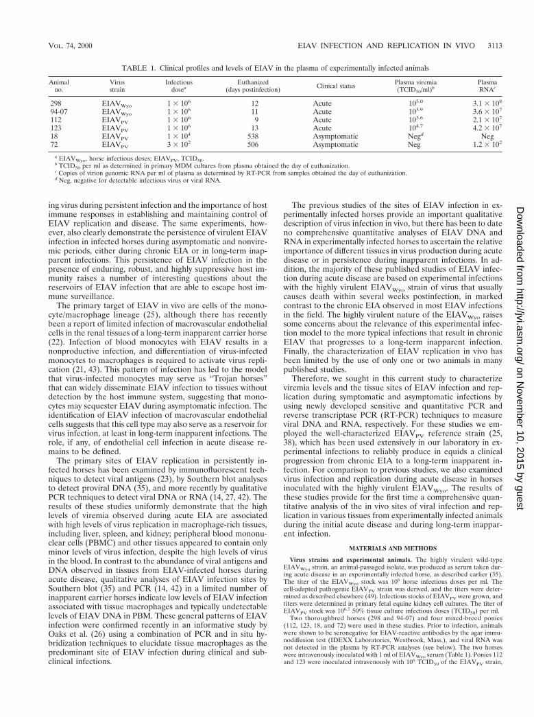
Identification of the in vivo sites of EIAV infection. To de-termine the viral DNA burden in tissues and thus identify thein vivo sites of viral infection, we developed a sensitive and spe-cific assay for quantifying viral DNA. To ascertain the speci-ficity of the QC-PCR, DNA from infected and uninfectedsamples were amplified with viral gag-specific primers. A 310-bp product was detected only in samples from infected ani-mals, indicating that the amplified DNA was of viral origin(data not shown). Amplification of viral sequences was addi-tionally confirmed by restriction enzyme digestion, by South-ern blot hybridization using EIAV gag-specific probes, and bysequencing (data not shown). To determine the sensitivity ofthe quantitative reactions, competitive amplification experi-ments were performed using specific known quantities of clonedviral DNA. The calculated copies were in close agreement withthe expected values (Fig. 1, R 5 0.99), and the sensitivity of theassay was determined to be 10 copies of viral DNA.
The levels of EIAV DNA were quantified from a panel oftissue samples isolated from the experimentally infected ani-mals described in Table 1. Figure 2 summarizes representativedata from a QC-PCR analysis using tissues taken during acutedisease the EIAVPV-infected pony 123. These data qualita-tively demonstrate markedly different levels of viral DNA inthe various tissues.
Quantification of QC-PCR data for each of the six experi-mentally infected animals is summarized in Table 2. The re-sults of the QC-PCR analyses revealed that the two EIAVWyo-infected animals had high cellular levels of viral DNA in mosttissues, up to 290,000 copies/100,000 CE, indicating extensivedisseminated infection. A broad range of copies of viral DNAthat varied by up to a 100-fold (range, 2,700 to 290,000 and1,200 to 120,000 copies/100,000 CE for horses 298 and 94-07,respectively) was observed, indicating preferential sites of in-fection. Interestingly, a different pattern of infection in thetissues of both animals was observed. Among the tissues ex-amined from both animals, the spleen contained the highestconcentration of cellular viral DNA at 290,000 and 120,000copies/100,000 CE in horses 298 and 94-07, respectively. Inhorse 298, the burden was reduced to 45,000 copies/100,000CE in the liver and to less than 8,000 copies/100,000 CE in theremaining samples. In contrast, the liver, lymph node, bonemarrow, and PBMC from horse 94-07 harbored from 20,000 to60,000 copies of viral DNA/100,000 CE, while the brain andkidney contained less than 2,000 copies.
A decreased amount of cellular viral DNA in all tissues inthe EIAVPV acutely infected animals was observed comparedto the EIAVWyo acutely infected animals (Table 2). Viral DNAwas detectable in all samples in the EIAVPV acutely infectedanimals, once again indicating a rapid dissemination of viralinfection. The cellular viral DNA burden in the two EIAVPVacutely infected ponies had viral DNA loads that varied by upto a 1,000-fold between samples within an animal (range, ,10to 8,000 and 70 to 53,000 copies/100,000 CE for ponies 112 and123, respectively), revealing preferential sites of infection.Among the tissues, the spleen was the primary site of infectedcells in both animals with 8,000 and 53,000 copies/100,000 CEin ponies 112 and 123, respectively. For pony 112, all of the
VOL. 74, 2000 EIAV INFECTION AND REPLICATION IN VIVO 3115
on Novem
ber 10, 2015 by guesthttp://jvi.asm
.org/D
ownloaded from
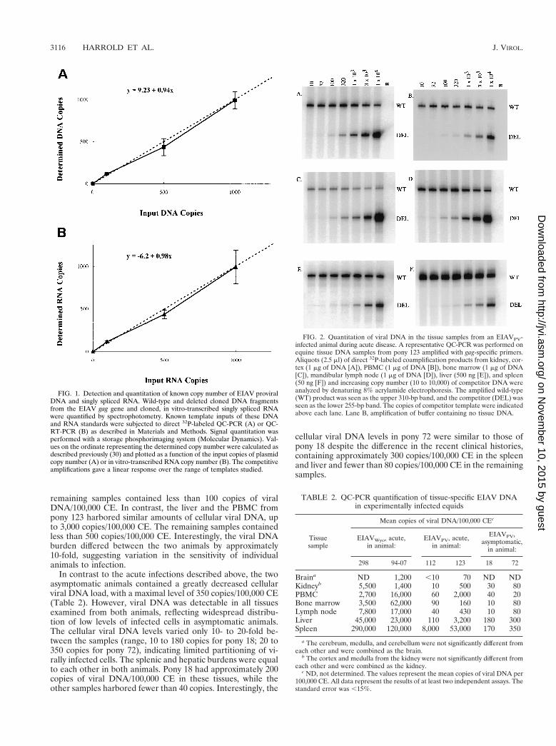
remaining samples contained less than 100 copies of viralDNA/100,000 CE. In contrast, the liver and the PBMC frompony 123 harbored similar amounts of cellular viral DNA, upto 3,000 copies/100,000 CE. The remaining samples containedless than 500 copies/100,000 CE. Interestingly, the viral DNAburden differed between the two animals by approximately10-fold, suggesting variation in the sensitivity of individualanimals to infection.
In contrast to the acute infections described above, the twoasymptomatic animals contained a greatly decreased cellularviral DNA load, with a maximal level of 350 copies/100,000 CE(Table 2). However, viral DNA was detectable in all tissuesexamined from both animals, reflecting widespread distribu-tion of low levels of infected cells in asymptomatic animals.The cellular viral DNA levels varied only 10- to 20-fold be-tween the samples (range, 10 to 180 copies for pony 18; 20 to350 copies for pony 72), indicating limited partitioning of vi-rally infected cells. The splenic and hepatic burdens were equalto each other in both animals. Pony 18 had approximately 200copies of viral DNA/100,000 CE in these tissues, while theother samples harbored fewer than 40 copies. Interestingly, the
cellular viral DNA levels in pony 72 were similar to those ofpony 18 despite the difference in the recent clinical histories,containing approximately 300 copies/100,000 CE in the spleenand liver and fewer than 80 copies/100,000 CE in the remainingsamples.
FIG. 1. Detection and quantitation of known copy number of EIAV proviralDNA and singly spliced RNA. Wild-type and deleted cloned DNA fragmentsfrom the EIAV gag gene and cloned, in vitro-transcribed singly spliced RNAwere quantified by spectrophotometry. Known template inputs of these DNAand RNA standards were subjected to direct 32P-labeled QC-PCR (A) or QC-RT-PCR (B) as described in Materials and Methods. Signal quantitation wasperformed with a storage phosphorimaging system (Molecular Dynamics). Val-ues on the ordinate representing the determined copy number were calculated asdescribed previously (30) and plotted as a function of the input copies of plasmidcopy number (A) or in vitro-transcribed RNA copy number (B). The competitiveamplifications gave a linear response over the range of templates studied.
FIG. 2. Quantitation of viral DNA in the tissue samples from an EIAVPV-infected animal during acute disease. A representative QC-PCR was performed onequine tissue DNA samples from pony 123 amplified with gag-specific primers.Aliquots (2.5 ml) of direct 32P-labeled coamplification products from kidney, cor-tex (1 mg of DNA [A]), PBMC (1 mg of DNA [B]), bone marrow (1 mg of DNA[C]), mandibular lymph node (1 mg of DNA [D]), liver (500 ng [E]), and spleen(50 ng [F]) and increasing copy number (10 to 10,000) of competitor DNA wereanalyzed by denaturing 8% acrylamide electrophoresis. The amplified wild-type(WT) product was seen as the upper 310-bp band, and the competitor (DEL) wasseen as the lower 255-bp band. The copies of competitor template were indicatedabove each lane. Lane B, amplification of buffer containing no tissue DNA.
TABLE 2. QC-PCR quantification of tissue-specific EIAV DNAin experimentally infected equids
Tissuesample
Mean copies of viral DNA/100,000 CEc
EIAVWyo, acute,in animal:
EIAVPV, acute,in animal:
EIAVPV,asymptomatic,
in animal:
298 94-07 112 123 18 72
Braina ND 1,200 ,10 70 ND NDKidneyb 5,500 1,400 10 500 30 80PBMC 2,700 16,000 60 2,000 40 20Bone marrow 3,500 62,000 90 160 10 80Lymph node 7,800 17,000 40 430 10 80Liver 45,000 23,000 110 3,200 180 300Spleen 290,000 120,000 8,000 53,000 170 350
a The cerebrum, medulla, and cerebellum were not significantly different fromeach other and were combined as the brain.
b The cortex and medulla from the kidney were not significantly different fromeach other and were combined as the kidney.
c ND, not determined. The values represent the mean copies of viral DNA per100,000 CE. All data represent the results of at least two independent assays. Thestandard error was ,15%.
3116 HARROLD ET AL. J. VIROL.
on Novem
ber 10, 2015 by guesthttp://jvi.asm
.org/D
ownloaded from

Comparison of the viral burden in particular tissues amongthe different infections and disease states revealed that thenumber of viral DNA copies in the EIAVWyo-infected animalswas approximately 10-fold higher than in the EIAVPV acutelyinfected ponies, which in turn was in general 10-fold greaterthan the asymptomatic animals. These results suggested therewas an association between the infecting viral strain and theviral DNA burden. However, regardless of the infecting strain,the appearance of clinical symptoms coincided with rapidwidespread seeding of infection, although preferential sites ofinfection could be identified. During clinical quiescence, bothlong term and recent, persistent infection and widespread ac-tive viral replication was observed, suggesting reservoirs ofinfected cells apparently resistant to immune clearance.
Determination of the tissue sites of viral replication. Todetermine sites of active viral replication during acute diseaseand of continuous virus production in acutely infected andasymptomatic animals, the amount of singly spliced EIAVmRNA was quantified by directly radiolabeled QC-RT-PCR.The active sites of lentivirus replication have been previouslyidentified by quantification of either genomic or singly splicedmRNA species (2, 24, 34, 39). Studies performed by our lab-oratory observed high levels of cell-free virus, up to 108 parti-cles per ml of plasma, during disease episodes (Table 1 andreference 32). Thus, levels of EIAV replication were assayed
based on quantification of a singly spliced mRNA species toavoid any contribution of viral genomic RNA to these mea-surements. Amplification of viral singly spliced mRNA se-quences was detected only in infected samples containing re-verse transcriptase, indicating that the reaction was specific forthis viral RNA (Fig. 3). No amplified product was detected inplasma RNA samples from febrile animals, indicating thatvirions do not contain viral singly spliced mRNA (data notshown). Restriction enzyme digestion and sequencing of theamplified products confirmed the specificity of this assay (datanot shown). To determine the sensitivity of the QC-RT-PCR,experiments analogous to those described for viral DNA wereperformed. There was excellent agreement between the calcu-lated copies and the determined copies of in vitro-transcribedRNAs (Fig. 1, R 5 100). The assay was sensitive (limit of de-tection, 30 copies of viral singly spliced mRNA) and reproduc-ible (coefficient of variation, ,30%).
Quantification of the amount of viral singly spliced mRNAwas determined on the same panel of tissues as that used forthe viral DNA analyses. A radiolabeled QC-RT-PCR experi-ment from pony 123, acutely infected with EIAVPV, is shownin Fig. 3. The abundance of viral singly spliced mRNA from theexperimentally infected animals was measured (Table 3). Sim-ilar to the DNA results, there were various levels of replicatingvirus throughout these animals. A pattern reminiscent of theviral infection emerged when the distribution of viral replica-tion was examined.
The EIAVWyo-infected animals harbored high levels, up toseveral million copies, of viral singly spliced mRNA in mostof the samples, indicating abundant, rapid disseminated viralreplication (Table 3). A broad range of copies of viral singlyspliced mRNA in various tissues was observed (700 to7,300,000 and 6,000 to 92,000 copies/mg of total RNA forhorses 298 and 94-07, respectively), reflecting that virus repli-cated in preferential sites. The spleen of horse 298 containedthe highest amount of replicating virus with 7,300,000 cop-ies/mg of total RNA. The liver and the lymph nodes had com-parable cellular viral RNA loads, harboring approximately100,000 copies/mg of total RNA. The remaining samples hadless than 1,500 copies/mg of total RNA. By comparison, similarlevels of cellular viral singly spliced mRNA were observed inthe spleen, liver, and lymph node from horse 94-07, with ap-proximately 90,000 copies/mg of total RNA in each tissue. Theother samples contained less than 30,000 copies/mg of total
FIG. 3. Quantitation of viral singly spliced mRNA in the tissue samples froman EIAVPV-infected animal during acute disease. A representative QC-RT-PCRwas performed on equine tissue RNA samples from pony 123 amplified withprimers specific for EIAV singly spliced RNA species. Aliquots (2.5 ml) of direct32P-labeled coamplification products from the kidney, cortex (1 mg of RNA [A]),kidney, medulla (1 mg of RNA [B]), mandibular lymph node (1 mg of RNA [C]),bone marrow (1 mg of RNA [D]), liver (500 ng of RNA [E]), and spleen (50 ngof RNA [F]) and increasing copy number (320 to 32,000) of in vitro-synthesizedcompetitor RNA were analyzed by denaturing 8% acrylamide electrophoresis.The amplified wild-type (WT) product was seen as the upper 510-bp band, andthe deleted competitor (DEL) was seen as the lower 406-bp band. The copies ofcompetitor RNA were indicated above each lane. Lane B, RT-PCR of buffer con-taining no tissue RNA; lane 2, RT-PCR of RNA without reverse transcriptase.
TABLE 3. QC-RT-PCR quantification of tissue-specific EIAVsingly spliced mRNA in experimentally infected equids
Tissuesamplea
Mean copies of viral singly splicedmRNA/mg of total RNAb
EIAVWyo, acute,in animal:
EIAVPV, acute,in animal:
EIAVPV,asymptomatic,
in animal:
298 94-07 112 123 18 72
Brain ND 7,000 ,30 100 ND NDKidney 650 27,000 ,30 650 ,30 ,30PBMC 700 6,000 ,30 17,000 ,30 190Bone marrow 1,500 6,000 130 700 ,30 ,30Lymph node 130,000 90,000 90 1,600 ,30 30Liver 110,000 76,000 3,600 23,000 740 140Spleen 7,300,000 92,000 185,000 510,000 50 750
a The brain and the kidney are as described in Table 2.b ND, not determined. The values represent the mean copies of viral singly
spliced mRNA per mg of total RNA. All data represent the results of at least twoindependent assays. The standard error was ,25%.
VOL. 74, 2000 EIAV INFECTION AND REPLICATION IN VIVO 3117
on Novem
ber 10, 2015 by guesthttp://jvi.asm
.org/D
ownloaded from
RNA. Interestingly, for both animals the lymph nodes con-tained a relatively high amount of viral singly spliced (Table 3)mRNA, despite containing low levels of viral DNA (Table 2).
The level of cellular viral singly spliced mRNA in mostsamples in the EIAVPV acutely infected animals was similar or10-fold less than in the EIAVWyo infections (Table 3). A widerange of copies of viral singly spliced mRNA was determinedamong the different tissues (undetectable to 200,000 and 100 to500,000 copies/mg of total RNA for ponies 112 and 123, re-spectively), again indicating the preferred sites of virus repli-cation. In both ponies, the spleen contained the highest cellu-lar levels of replicating virus, up to 500,000 copies/mg of totalRNA. In pony 112, the cellular viral singly spliced mRNA loadwas reduced to 4,000 copies and less than 100 copies/mg oftotal RNA in the liver and the rest of the samples, respectively.By comparison, the liver and the PBMC in pony 123 containedequivalent levels of cellular viral singly spliced mRNA (20,000copies/mg of total RNA), which was approximately 10-fold lessthan the spleen. Interestingly, relatively high levels of EIAVsingly spliced mRNA were also detected in the PBMC fromone of the EIAVWyo acutely infected animals (horse 94-07),suggesting that viral replication in this case was not confined tothe tissues. This observation was in contrast to previous anal-yses, which report a lack of productive EIAV infection of bloodmonocytes (21, 43). The remaining samples in pony 123 har-bored fewer than 2,000 copies of viral singly spliced mRNA/mgof total RNA, which was in general 100- to 1,000-fold less thanthe spleen. The viral singly spliced mRNA load in the twoacute EIAVPV infections varied by approximately 10-fold be-tween the two animals, indicating variation between animals.
In asymptomatic animals, the levels of viral singly splicedmRNA were dramatically reduced compared to the EIAVWyo-infected animals, with a maximal load of 750 copies/mg of totalRNA (Table 3). In some tissue samples, viral singly splicedRNA was undetectable in repeated analyses. Within an animal,the difference in the number of copies of viral singly splicedmRNA between samples was limited compared to the acutelyinfected animals (range, undetectable to 750 copies/mg of totalRNA for both animals). A predominant site of viral replicationappeared to be either the hepatic or splenic compartments forponies 18 and 72, respectively. The remaining samples frompony 18 harbored fewer than 50 copies and from pony 72 fewerthan 150 copies of viral singly spliced mRNA/mg of total RNA,respectively. Together, these results indicated that viral replica-tion was suppressed and restricted during clinical quiescence.
In comparison to the viral DNA analyses, the levels of singlyspliced viral RNA failed to show a clear gradient when thedifferent disease states and the infecting strains were compared
(Table 3). The EIAVWyo-infected animals had similar or great-er levels of viral replication relative to the EIAVPV acutelyinfected animals. In asymptomatic animals, the level of viralRNA expression was clearly reduced. These data suggestedthat during acute disease the amount of viral replication wassimilar in some samples between EIAV strains. During acutedisease, replication was in general detected throughout in-fected animals, although the spleen was a predominant cellularsite of highly active replication. During clinical quiescence,tight but not absolute control of viral replication was evident,indicating that a continuous low level of replication persisted.
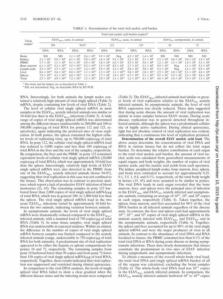
Determination of the overall EIAV nucleic acid load. Theabove assays determine the concentration of viral DNA andRNA in various tissues but do not reflect the total organburden. To determine the relative contribution of each organto the total viral nucleic acid burden, the amount of viral nu-cleic acids was calculated from generalized measurements ofequid organs and body weights, the number of copies of viralnucleic acids, and the nucleic acid yields from each specimen.The kidney, peripheral lymph nodes, bone marrow, liver, spleen,and brain were estimated to account for approximately 0.25,0.1, 3.5, 1, 0.4, and 0.1%, respectively, of the total body weightin horses (15, 45) and were assumed to be similar in ponies.The viral DNA loads in each organ revealed that the bonemarrow, liver, and spleen were the principal sites of infectionin the EIAVWyo and EIAVPV acutely infected and asymptom-atic animals, containing an average of 1011, 109, and 108 copiesin each organ, respectively (Table 4). Taken together, thespleen, bone marrow, and liver accounted for 98% of the viralDNA burden in all infected animals regardless of the diseasestate. In contrast, the liver and spleen each had approximately1012, 1011, and 109 copies of viral singly spliced mRNA in theanimals acutely infected with EIAVWyo and EIAVPV and inthe asymptomatic animals, respectively (Table 4). Together,the spleen and liver accounted for up to 98% of the viral singlyspliced mRNA and were the major producers of virus in allanimals. In contrast to the abundance of viral DNA and RNAlocalized to tissues, the PBMC contained less than 0.1% of thetotal viral DNA or RNA during acute disease or during asymp-tomatic infections. These data clearly demonstrate that tissuesconstitute the predominant in vivo sites of EIAV infectionduring symptomatic and asymptomatic infections.
To obtain a measure of the overall whole-body viral load,the total viral DNA and singly spliced mRNA burden of allof the organs was calculated, excluding the brain samples(Table 4). The whole-body viral DNA load was 1011 copiesin the EIAVWyo acutely infected animals. In comparison, theEIAVPV acutely infected animals harbored 109 to 1010 copies
TABLE 4. Determination of the total viral nucleic acid burden
Tissuesamplea
Total viral nucleic acid burden (copies)b
EIAVWyo, acute, in animal: EIAVPV, acute, in animal: EIAVPV, asymptomatic, in animal:
298 94-07 112 123 18 72
DNA RNA DNA RNA DNA RNA DNA RNA DNA RNA DNA RNA
Brain ND ND 1.1 3 108 2.6 3 109 9.5 3 104 Neg 4.5 3 106 1.4 3 107 ND ND ND NDKidney 2.1 3 109 5.8 3 108 6.1 3 108 5.0 3 1010 1.6 3 106 9.1 3 106 5.2 3 107 2.1 3 108 5.5 3 106 4.6 3 106 3.0 3 107 1.9 3 106
PBMC 1.7 3 107 1.1 3 108 9.1 3 108 4.9 3 108 1.8 3 106 6.5 3 105 4.3 3 107 3.6 3 108 1.2 3 106 1.3 3 105 1.4 3 105 3.3 3 106
Bone marrow 6.1 3 1010 3.2 3 1010 5.5 3 1011 9.2 3 1010 1.5 3 108 8.3 3 108 2.7 3 107 2.1 3 108 9.9 3 107 Neg 1.1 3 109 3.2 3 108
Lymph node 2.9 3 1010 1.5 3 1011 1.1 3 1010 1.2 3 1011 2.9 3 106 2.4 3 107 3.0 3 107 3.0 3 108 5.3 3 106 Neg 1.6 3 107 8.9 3 106
Liver 8.6 3 1010 5.6 3 1011 8.8 3 1010 1.1 3 1012 1.3 3 108 1.1 3 1010 5.0 3 109 1.0 3 1011 2.5 3 108 1.8 3 109 5.1 3 108 4.8 3 108
Spleen 7.6 3 1010 4.2 3 1012 6.8 3 1010 1.1 3 1011 1.7 3 109 1.0 3 1011 6.7 3 109 3.1 3 1011 9.5 3 107 3.3 3 107 2.2 3 108 4.8 3 108
Total 2.3 3 1011 4.9 3 1012 7.2 3 1011 1.5 3 1012 2.0 3 109 1.1 3 1011 1.2 3 1010 4.1 3 1011 4.6 3 108 1.8 3 109 1.9 3 109 1.3 3 109
a The brain and the kidney are as described in Table 2.b ND, not determined; Neg, no detectable RNA by RT-PCR.
3118 HARROLD ET AL. J. VIROL.
on Novem
ber 10, 2015 by guesthttp://jvi.asm
.org/D
ownloaded from
of viral DNA, representing a 10- to 100-fold decrease. The viralDNA burden was decreased by up to an additional 10-fold inthe asymptomatic animals, containing 108 copies of viral DNA.In contrast, the whole-body viral singly spliced RNA level was1012 copies in the EIAVWyo acutely infected animals. In com-parison, the overall viral singly spliced mRNA burden in theEIAVPV acutely infected animals was reduced approximately10-fold to 1011 copies. The asymptomatic animals harbored109 copies of viral singly spliced mRNA, which representedan additional 100-fold decrease. These data suggested that thevirulence of the EIAVWyo strain was related to an increasedrate of infection and replication in the tissues.
Calculation of the mean transcriptional level (the ratio ofthe total viral singly spliced RNA load to the total viral DNAload) has been used to compare human immunodeficiencyvirus type 1 (HIV-1) expression levels in various organs (1, 24).In a similar manner, the mean transcriptional level (viral tran-scripts per proviral genome) for the various organs in theEIAV-infected animals was calculated from the quantitativeDNA and RNA data summarized in Table 4. In the EIAVWyoand EIAVPV acutely infected animals the spleen, liver, andlymph nodes consistently had transcriptional indices up to 85,indicating the highest levels of virus expression per proviralgenome. While transcription was not limited to these sites, thelevels in the remaining samples were typically less than 10.Notable exceptions were the brain and kidney in horse 94-07,which had indices of 25 and 80, respectively. In contrast to theacute infections, the highest transcriptional levels from theclinically quiescent animals were in the liver and the spleen;however, the indices were only 7 and 2. These results providedadditional support for the suppression of viral transcription intissues of inapparent carriers. Interestingly, the PBMC in threeanimals (298, 123, and 72) had transcriptional indices of 6, 8,and 24, respectively, suggesting low levels of EIAV proviralexpression in blood monocytes during both acute and chronicinfection.
DISCUSSION
The clearly demarcated disease cycles associated with EIAVinfection provide a unique in vivo model for characterizationof the tissue reservoirs of productive and latent lentiviral in-fection during clinical episodes and during long-term asymp-tomatic infections. The experiments described here present forthe first time a rigorous quantitative analysis of the levels ofboth EIAV viral DNA as a measure of infection and singlyspliced viral RNA as a measure of virus replication in varioustissue compartments of experimentally infected equids. The re-sults of these studies identified preferential sites of virus infec-tion, replication, and latency associated with persistent EIAVinfections.
The levels of EIAV DNA and singly spliced viral RNAmeasured in the experimentally infected equids differed mark-edly depending on the clinical status of the infected animalsand the strain of virus used for the infection. In terms ofplasma viremia, the current studies clearly demonstrate thatacute EIAV infection is associated with very high levels of virusin the blood, as measured by infectivity in a recently developedquantitative equine macrophage assay (32). In marked con-trast, infectious virus was undetectable in blood during asymp-tomatic EIAV infection. The current experiments for the firsttime extend the measurements of blood viral burden to includequantification of plasma viral RNA by RT-PCR. These datarevealed plasma RNA levels of 107 to 108 copies of virion RNAper ml of plasma during acute disease and fewer than 100copies per ml during asymptomatic stages of infection. Thus,
the new combination of in vitro infectivity and PCR assaysclearly illustrate the characteristic wave of viremia associatedwith EIAV disease episodes and the rigorous control of virusreplication during clinical latency.
While these blood virus load measurements in general con-firm previously reported viremia data derived from infectivityassays in equine fibroblastic cells, the new assays also providenew insights into the pattern of viremia during persistentEIAVPV infection. First, the viremia levels measured in equinemacrophages are similar to viremia levels reported previouslyby infectivity assays in equine fibroblastic cells (11, 18, 31).This agreement indicates that the latter assays in fact providea relevant measure of EIAVPV viremia, eliminating a lingeringconcern about the validity of using equine fibroblastic cells tomeasure EIAVPV infectivity. Second, the quantitative measure-ments of plasma viral RNA closely parallel viremia measure-ments in assessing viral load during acute disease and clinicallatency. However, the viral RNA measurements demonstratefor the first time that the negligible levels of infectious EIAVassociated with clinical latency is in fact due to an absence ofviral particles in the blood and not to an inactivation of virus byneutralizing serum antibodies. This observation appears to in-dicate the efficacy of the host immune control of viral replica-tion, presumably by the reduction of infected target cells. Fi-nally, the viremia data summarized here indicates that thetypically fatal outcome of EIAVWyo infection compared to thechronic EIA induced by EIAVPV is not reflected in substantialdifferences in plasma viremia levels, since the two infectionsproduced similar plasma virus loads during the initial acutedisease. As discussed below, however, the differences in dis-ease severity may be more accurately associated with the tissueviral load rather than the levels of virus in blood.
During the initial stages of acute disease within 2 weekspostinfection, viral infection and replication were shown to bewidely disseminated among all of the tissues examined in boththe EIAVWyo- and EIAVPV-infected animals (Tables 2 to 4).In this regard, the results of the current studies are in agree-ment with previous analyses of EIAV replication in infectedhorses during acute disease using cultured cells to detect virus(17), fluorescent antibody (23), Southern blot (36), in situhybridization (41), or PCR (13, 14, 26) procedures and withthe pathological findings characteristic of EIAV infection (41).The results of these earlier qualitative analyses established anassociation between sites of virus infection and host tissues richin macrophages. However, the quantitative assays employedhere extend these studies to clearly identify the spleen as thepredominant cellular site of EIAV infection and replicationduring acute disease. The concentration of proviral DNA andsingly spliced viral RNA in the splenic tissues was typically 10to 20 times greater than the concentration of EIAV DNA orRNA measured in any other tissue. In contrast, the asymptom-atic animals had equivalent cellular loads of viral DNA andmRNA in the spleen and the liver (Tables 2 and 3). In allanimals, estimation of total organ viral loads (Table 4) indi-cates further that the spleen and liver tissues contain up to85% of the total viral DNA or mRNA that was detected in thetissues examined. It is assumed that the high levels of virusinfection and replication in the splenic and hepatic tissuesreflect the abundance of target macrophages in these organs.In addition, these quantitative analyses revealed that duringthe initial acute disease the EIAVWyo-infected animals con-tained 10- to 100-fold more viral DNA and singly spliced RNAin tissues compared to the levels measured in the EIAVPV-infected animals. This differential in tissue levels of viral DNAand singly spliced viral RNA is in contrast to the similar levelsof plasma viral genomic RNA measured during acute disease
VOL. 74, 2000 EIAV INFECTION AND REPLICATION IN VIVO 3119
on Novem
ber 10, 2015 by guesthttp://jvi.asm
.org/D
ownloaded from

in animals infected by the two viral strains. These observationsindicate that the levels of tissue-associated viral DNA andsingly spliced viral RNA, especially in the spleen and liver,constitute a more accurate correlate of disease severity thanplasma viral RNA load.
Although the quantitative viral DNA and singly spliced viralRNA analyses in general identified macrophage-rich tissues asthe predominant sites of viral infection during acute EIA, inagreement with pathological findings, these detailed analysesalso revealed some unexpected aspects of EIAV infections.For example, a number of studies have reported that EIAVDNA and RNA is either very low or undetectable in PBMCisolated from horses infected with EIAV (14, 21, 42, 43). Theseobservations support the widely accepted concept that infec-tion of blood monocytes with EIAV results in latent infectionand that cellular differentiation to macrophages is necessaryfor the production of virus, as proposed for other lentiviruses(8). However, in the current studies we observed substantiallevels of viral DNA (2,000 to 16,000 copies per 100,000 CE)and viral singly spliced RNA (700 to 17,000 copies per mg ofRNA) in PBMC in three of the four infected animals duringacute disease. These findings suggest that blood monocytes canin fact serve as targets of EIAV infection and of virus produc-tion during waves of viremia associated with disease episodes,perhaps reflecting widespread activation of blood monocytesduring cycles of EIAV disease.
A second unexpected observation is the relatively high levelof viral DNA (Table 2) accompanied by a low level of viralsingly spliced RNA (Table 3) in the bone marrow of theEIAVWyo acutely infected and the asymptomatic EIAVPV-infected animals. These observations extend previous findingsfrom visna virus (9), simian immunodeficiency virus (4), andHIV-1 (7, 28) indicating that the less-differentiated cells of thebone marrow can serve as a reservoir of latently infected cells.Recent studies detected increased levels of inflammatory cy-tokines in the bone marrow during EIAV infection in vivo (47)and in bone marrow derived macrophages in vitro (46), whichare hypothesized to suppress platelet production (5). Thus,cytokine dysregulation could result in thrombocytopenia, aclinical finding consistently associated with recrudescence ofdisease. Additionally, severe hemorrhaging is the principal con-tributing pathology in lethal EIAVWyo infection. Therefore,the organ-specific viral loads may directly or indirectly accountfor the pathological features manifest during disease cycles.
In striking contrast to the high levels of viral DNA and viralsingly spliced RNA found in tissues from animals during acutedisease, asymptomatic EIAV infections were characterized byextremely low levels of viral DNA (150 to 350 copies per100,000 CE) and viral singly spliced RNA (50 to 750 copies permg of RNA) concentrated predominantly in the liver andspleen. Other tissues examined contained fewer than 100 cop-ies of DNA and usually ,30 copies of singly spliced viral RNA.These observations demonstrate a rigorous control of EIAVreplication that severely limits the number of EIAV-infectedcells and the levels of virus production. However, it is impor-tant to note that despite the lifelong asymptomatic state usu-ally maintained by EIAV-infected animals following 6 to 12months of chronic EIA, the viral infection is never completelyeliminated since tissue reservoirs of productively infected cellsare sustained despite the high levels of immune control estab-lished by the host.
The ability of animals to routinely establish immunologicalcontrol of EIAV infection and disease is considered the bestexample of natural immune control of a virulent lentivirus in-fection (25). The concept of immunologic control of EIAVreplication in infected asymptomatic animals has been devel-
oped mostly based on measurements of viremia that indicatethe absence of detectable virus in the blood of asymptomaticanimals in marked contrast to the viremia levels of 105 to 106
TCID50 per ml of blood during clinical episodes (25). How-ever, it has been shown that transfer of as little of 7 ml of wholeblood from asymptomatic animals to naive equids producesinfection of as least 80% of the recipient animals (12). There-fore, it has not been clear from these studies whether the lackof infectious virus in the blood actually reflected a lack of virusparticles or inactivation of plasma virus by the high levels ofEIAV-specific neutralizing antibodies present in asymptomaticanimals (11). The RT-PCR assays described here demonstratefor the first time that the lack of infectious virus in the blood inasymptomatic animals is indeed reflective of a virtual absenceof detectable viral genomic RNA in the plasma from theseanimals. Thus, the infectivity of blood from asymptomatic an-imals is most likely associated with the approximate 0.04% ofPBMC containing EIAV proviral DNA.
The results described in the current study define the tissuesites of EIAV infection but do not specifically define the targetcells within these tissues. In agreement with the well-estab-lished macrophage tropism of EIAV, the predominant sitesof EIAV infection and replication during symptomatic andasymptomatic infections were determined to be tissues rich inmacrophages, such as the spleen and liver. While a recentstudy (22) indicates that renal macrovascular endothelial cellsmay also serve as a reservoir of EIAV infection in a long-terminapparent carrier, the relative contributions of macrophagesand endothelial cells in different tissues to viral persistencewere not determined in this study. In addition, there has beenno characterization of endothelial cell infection in experimen-tally infected horses during acute disease to assess the poten-tial role of these cells in pathogenesis. The results of thecurrent study seem to support the predominant importance ofmonocytes and macrophages in viral pathogenesis and persis-tence, since EIAV infection and replication correlated with theabundance of tissue macrophages in the respective organs.
These quantitative studies of EIAV infection and replicationin vivo provide a detailed description of the patterns of virusreplication associated with clinical and asymptomatic infec-tions. While the data are specific for EIAV, the results haveimplications for broader issues related to the role of tissuereservoirs in lentiviral persistence, the potential for immunecontrol or ultimate clearance of lentiviral infections, and forunderstanding the mechanisms of lentiviral pathogenesis.Based on the studies with EIAV, a predominantly macro-phage-tropic lentivirus, it is clear that tissues can provide awidespread and resilient reservoir for lentiviral infection, evenin the presence of potent host immune clearance mechanisms.Moreover, it appears that the extent of viral pathogenesis canbe most closely associated with the levels of virus replication intissues rather than plasma virus burden. Thus, it will be im-portant to examine in more detail the potential role of tissuemacrophages in the persistence of lentiviruses that are alsolymphotropic and to define the mechanisms by which tissuemacrophage infections lead to characteristic lentiviral pathol-ogies such as fever, diarrhea, anemia, thrombocytopenia,and encephalitis.
ACKNOWLEDGMENTS
We thank Drew Lichtenstein for stimulating discussions, CarolineLeroux for thoughtful review of the manuscript, and Gary Thomas andBrian Meade for excellent animal care.
This work was supported by National Institutes of Health grant R01CA49296, by funds from the Lucille P. Markey Charitable Trust andthe Kentucky Agricultural Experimental Station, and in part by a grant
3120 HARROLD ET AL. J. VIROL.
on Novem
ber 10, 2015 by guesthttp://jvi.asm
.org/D
ownloaded from
from the Pittsburgh Supercomputer Center through the NIH NationalCenter for Research Resources (resource grant 2 P41 RR06009).S.M.H. was supported by NIH AIDS training grant 5T32 AI07487.
REFERENCES1. Bagnarelli, P., S. Menzo, A. Valenza, A. Manzin, M. Giacca, F. Ancarani, G.
Scalise, P. E. Varaldo, and M. Clementi. 1992. Molecular profile of humanimmunodeficiency virus type 1 infection in symptomless patients and inpatients with AIDS. J. Virol. 66:7328–7335.
2. Bagnarelli, P., A. Valenza, S. Menzo, R. Sampaolesi, P. E. Varaldo, L. Butini,M. Montroni, C.-F. Perno, C. Balotta, and M. Clementi. 1996. Dynamics andmodulation of human immunodeficiency virus type 1 transcripts in vitro andin vivo. J. Virol. 70:7603–7613.
3. Chong, Y.-H., S. L. Payne, C. J. Issel, R. C. Montelaro, and K. E. Rushlow.1991. Characterization of the antigenic domains of the major core protein(p26) of equine infectious anemia virus. J. Virol. 65:1007–1012.
4. Clements, J. E., and M. C. Zink. 1996. Molecular biology and pathogenesisof animal lentivirus infections. Clin. Microbiol. Rev. 9:100–117.
5. Crawford, T. B., K. J. Wardrop, S. J. Tornquist, E. Reilich, K. M. Meyers,and T. C. McGuire. 1996. A primary production deficit in the thrombocyto-penia of equine infectious anemia virus. J. Virol. 70:7842–7850.
6. Cunningham, T. P., R. C. Montelaro, and K. E. Rushlow. 1993. Lentivirusenvelope sequences and proviral genomes are stabilized in Escherichia coliwhen cloned in low-copy-number plasmid vectors. Gene. 124:93–98.
7. Embretson, J., M. Zupancic, J. L. Ribas, A. Burke, P. Racz, K. Tenner-Racz,and A. T. Haase. 1993. Massive covert infection of helper T lymphocytes andmacrophages by HIV during the incubation period of AIDS. Nature 362:359–362.
8. Gendelman, H. E., O. Narayan, S. Kennedy-Stoskopf, P. G. E. Kennedy, Z.Ghoti, J. E. Clements, J. Stanley, and G. Pezeshkpour. 1986. Tropism ofsheep lentiviruses for monocytes: susceptibility to infection and virus geneexpression increase during maturation to macrophages. J. Virol. 58:67–74.
9. Gendelman, H. E., O. Narayan, S. Moplineaux, J. E. Clements, and Z.Ghotbi. 1985. Slow, persistent replication of lentiviruses: role of tissue mac-rophages and macrophage precursors in bone marrow. Proc. Natl. Acad. Sci.USA 82:7086–7090.
10. Genetics Computer Group. 1994. Program manual for the Wisconsin Pack-age. Genetics Computer Group, Madison, Wis.
11. Hammond, S. A., S. J. Cook, D. L. Lichtenstein, C. J. Issel, and R. C.Montelaro. 1997. Maturation of the cellular and humoral immune responsesto persistent infection in horses by equine infectious anemia virus is acomplex and lengthy process. J. Virol. 71:3840–3852.
12. Issel, C. J., J. W. V. Adams, L. Meek, and R. Ochoa. 1982. Transmission ofequine infectious anemia virus from horses without clinical signs of disease.J. Am. Vet. Med. Assoc. 180:272–275.
13. Kim, C. H., and J. W. Casey. 1992. Genomic variation and segregation ofequine infectious anemia virus during acute infection. J. Virol. 66:3879–3882.
14. Kim, C. H., and J. W. Casey. 1994. In vivo replication status and envelopeheterogeneity of equine infectious anemia virus in an inapparent carrier.J. Virol. 68:2777–2780.
15. Klaus, G. G. B. (ed.). 1987. Lymphocytes: a practical approach. IRL Press,Ltd., Oxford, England.
16. Kono, Y., K. Hirasawa, Y. Fukunaga, and T. Taniguchi. 1976. Recrudes-cence of equine infectious anemia by treatment with immunosuppressivedrugs. Natl. Inst. Anim. Health Q. 16:8–15.
17. Kono, Y., K. Kobayashi, and Y. Fukunaga. 1971. Distribution of equineinfectious anemia virus in horses infected with the virus. Natl. Inst. Anim.Health Q. 11:11–20.
18. Langemeier, J. L., S. J. Cook, R. F. Cook, K. E. Rushlow, R. C. Montelaro,and C. J. Issel. 1996. Detection of equine infectious anemia viral RNA inplasma samples from recently infected and long-term inapparent carrieranimals by PCR. J. Clin. Microbiol. 34:1481–1487.
19. Lichtenstein, D. L., K. E. Rushlow, R. F. Cook, M. L. Raabe, C. J. Swardson,G. J. Kociba, C. J. Issel, and R. C. Montelaro. 1995. Replication in vitro andin vivo of an equine infectious anemia virus mutant deficient in dUTPaseactivity. J. Virol. 69:2881–2888.
20. Lifson, J. D., M. A. Nowak, S. Goldstein, J. L. Rossio, A. Kinter, G. Vasquez,T. A. Wiltrout, C. Brown, D. Schneider, L. Wahl, A. L. Lloyd, J. Williams,W. R. Elkins, A. S. Fauci, and V. M. Hirsch. 1997. The extent of early viralreplication is a critical determinant of the natural history of simian immu-nodeficiency virus infection. J. Virol. 71:9508–9514.
21. Maury, W. 1994. Monocyte maturation controls expression of equine infec-tious anemia virus. J. Virol. 68:6270–6279.
22. Maury, W., J. L. Oaks, and S. Bradley. 1998. Equine endothelial cellssupport productive infection of equine infectious anemia virus. J. Virol. 72:9291–9297.
23. McGuire, T. C., T. B. Crawford, and J. B. Henson. 1971. Immunofluorescentlocalization of equine infectious anemia virus in tissue. Am. J. Pathol. 62:283–294.
24. Michael, N. L., M. Vahey, D. S. Burke, and R. R. Redfield. 1992. Viral DNAand mRNA expression correlate with the stage of human immunodeficiencyvirus (HIV) type 1 infection in humans: evidence for viral replication in all
stages of HIV disease. J. Virol. 66:310–316.25. Montelaro, R. C., J. M. Ball, and K. E. Rushlow. 1993. Equine retroviruses,
p. 257–360. In J. A. Levy (ed.), The Retroviridae, vol. 2. Plenum Press, NewYork, N.Y.
26. Oaks, J. L., T. C. McGuire, C. Ulibarri, and T. C. Crawford. 1998. Equineinfectious anemia virus is found in tissue macrophages during subclinicalinfection. J. Virol. 72:7263–7269.
27. O’Rourke, K. I., M. L. Besola, and T. C. McGuire. 1991. Proviral sequencesdetected by polymerase chain reaction in peripheral blood cells of horseswith equine infectious anemia virus. Arch. Virol. 117:109–119.
28. Pantaleo, G., C. Graziosi, J. F. Demarest, L. Butini, M. Montroni, C. H. Fox,J. M. Orenstein, D. P. Kotler, and A. S. Fauci. 1993. HIV infection is activeand progressive in lymphoid tissue during the clinically latent stage of dis-ease. Nature 362:355–358.
29. Perryman, L. E., K. I. O’Rourke, and T. C. McGuire. 1988. Immune re-sponses are required to terminate viremia in equine infectious anemia len-tivirus infection. J. Virol. 62:3073–3076.
30. Piatak, M., M. S. Saag, L. C. Yang, S. J. Clark, J. C. Knappes, K.-C. Luk,B. H. Hahn, G. M. Shaw, and J. D. Lifson. 1993. High levels of HIV-1 inplasma during all stages of infection determined by competitive PCR. Sci-ence 259:1749–1754.
31. Raabe, M. L., C. J. Issel, S. J. Cook, R. F. Cook, B. Woodson, and R. C.Montelaro. 1998. Immunization with a recombinant envelope protein(rgp90) of EIAV produces a spectrum of vaccine efficacy ranging from lackof clinical disease to severe enhancement. Virology 245:151–162.
32. Raabe, M. L., C. J. Issel, and R. C. Montelaro. 1998. Equine monocytederived macrophage cultures and their application for infectivity and neu-tralization studies of equine infectious anemia virus. J. Virol. Methods 71:87–104.
33. Rasty, S., B. R. Dhruva, R. L. Schlitz, D. S. Shih, C. J. Issel, and R. C.Montelaro. 1990. Proviral DNA integration and transcriptional patterns ofequine infectious anemia virus during persistent and cytopathic infections.J. Virol. 64:86–95.
34. Reinhart, T. A., M. J. Rogan, D. Huddleston, D. M. Rausch, L. E. Eiden, andA. T. Haase. 1997. Simian immunodeficiency virus burden in tissues andcellular compartments during clinical latency and AIDS. J. Infect. Dis. 176:1198–1208.
35. Rice, N. R., A.-S. Lequarre, J. W. Casey, S. Lahn, R. M. Stephens, and J.Edwards. 1989. Viral DNA in horses infected with equine infectious anemiavirus. J. Virol. 63:5194–5200.
36. Rice, N. R., S. Simek, O. A. Ryder, and L. Coggins. 1978. Detection ofproviral DNA in horse cells infected with equine infectious anemia virus.J. Virol. 16:577.
37. Rudolph, J. A., S. J. Spier, G. Byrns, C. V. Rojas, D. Bernoco, and E. P.Hoffman. 1992. Periodic paralysis in quarter horses: a sodium channel mu-tation disseminated by selective breeding. Nat. Genet. 2:144–147.
38. Rwambo, P. M., C. J. Issel, K. A. Hussain, and R. C. Montelaro. 1990. Invitro isolation of a neutralization escape mutant of equine infectious anemiavirus (EIAV). Arch. Virol. 111:275–280.
39. Saltarelli, M. J., E. Hadziyannis, C. E. Hart, J. V. Harrison, B. K. Felber,T. J. Spira, and G. N. Pavlakis. 1996. Analysis of human immunodeficiencyvirus type 1 mRNA splicing patterns during disease progression in peripheralblood mononuclear cells from individuals. AIDS Res. Hum. Retrovir. 12:1443–1456.
40. Sambrook, J., E. F. Fritsch, and T. Maniatis. 1989. Molecular cloning: alaboratory manual, 2nd ed. Cold Spring Harbor Laboratory Press, ColdSpring Harbor, N.Y.
41. Sellon, D. C., F. J. Fuller, and T. C. McGuire. 1994. The immunopathogen-esis of equine infectious anemia virus. Virus Res. 32:111–138.
42. Sellon, D. C., S. T. Perry, L. Coggins, and F. J. Fuller. 1992. Wild-typeequine infectious anemia virus replicates in vivo predominantly in tissuemacrophages, not in peripheral blood monocytes. J. Virol. 66:5906–5913.
43. Sellon, D. C., K. M. Walker, K. E. Russell, S. T. Perry, P. Covington, andF. J. Fuller. 1996. Equine infectious anemia virus replication is upregulatedduring differentiation of blood monocytes from acutely infected horses. J. Vi-rol. 70:590–594.
44. Simmonds, P., P. Balfe, J. Peutherer, C. A. Ludlam, J. O. Bishop, and A. J. L.Brown. 1990. Human immunodeficiency virus-infected individuals containprovirus in small numbers of peripheral mononuclear cells and at low copynumbers. J. Virol. 64:864–872.
45. Smith, B. P. 1990. Large animal internal medicine. Diseases of horses, cattle,sheep, and goats. C.V. Mosby Co., St. Louis, Mo.
46. Swardson, C. J., D. L. Lichtenstein, S. Wang, R. C. Montelaro, and G. J.Kociba. 1997. Infection of bone marrow macrophages by equine infectiousanemia virus. Am. J. Vet. Res. 58:1402–1407.
47. Tornquist, S. J., J. L. Oakes, and T. B. Crawford. 1997. Elevation of cyto-kines associated with the thrombocytopenia of equine infectious anemiavirus. J. Gen. Virol. 78:2541–2548.
48. Tumas, D. B., M. T. Hines, L. E. Perryman, W. C. Davis, and T. C. McGuire.1994. Corticosteroid immunosuppression and monoclonal antibody-medi-ated CD51 T lymphocyte depletion in normal and equine infectious anemiavirus-carrier horses. J. Gen. Virol. 75:959–968.
VOL. 74, 2000 EIAV INFECTION AND REPLICATION IN VIVO 3121
on Novem
ber 10, 2015 by guesthttp://jvi.asm
.org/D
ownloaded from
Related Documents