ix INTRODUCTION

Welcome message from author
This document is posted to help you gain knowledge. Please leave a comment to let me know what you think about it! Share it to your friends and learn new things together.
Transcript

ix
INTRODUCTION

1
1.1 Introduction
Plants support all other life forms and maintain the oxygen content of the air,
ecosystems and control the climate. Plants are primary source of vitamins and provide
medicine, clothes, shelter and raw materials from which innumerable products are
made. These benefits are widely recognized. Therefore, they are essential parts of the
world‟s biological diversity, and human beings are very dependent on plant.
During the past decade, research and investigation on plant potential abilities
have been valuable topics in the world. Tissue culture is an experimental technique in
which mass of cells are produced from the explants. Tissue cultures raised the
knowledge in some areas including differentiation, cell division, and nutrition and cell
preservation. Nowadays, cells are cultivated in vitro in bulk or as clone from single
cells to grow whole plants from isolated meristem, produce callus and develop com-
plete plantlets by organogenesis or by embryogenesis.
Biotechnology offers an opportunity to use the cell, tissue, organ or entire or-
ganism by growing them in vitro and to genetically manipulate them to get desired
compounds.
Since the world population is increasing rapidly, there is an extreme pressure on the
available cultivable land to produce food. For other uses such as production of phar-
maceuticals and chemicals from plants, the available land should be used effectively.
During the past decade, a considerable progress has been made to stimulate formation
and accumulation of secondary metabolites using plant cell cultures (Ravishankar &
Rao, 2000).

2
The callus formed can be utilized directly to regenerate plantlets, or be a
source of primary and secondary metabolites. The adopted methods for enhancing the
secondary metabolites include obtaining efficient cell lines for growth, monitoring of
high-growth cell line that produces metabolites of interest, immobilization of cells to
enhance yields of extra-cellular metabolites and to facilitate biotransformation, use of
elicitors to enhance productivity in a short period of time, permeation of metabolites
to facilitate downstream processing, adsorption of the metabolites to partition the
products from the medium and to overcome feedback inhibition and scale-up of cell
cultures in suitable bioreactors.
Medicinal plants are the earliest known to have various benefits and usage.
Based on clay written tablet excavated in Iraq, their historical record was about six
thousand years ago which proves that human beings have been depending on plants
mainly for food and medicine, apart from other uses. Crude extracts, and whole plants
have been used as medicine without knowledge of their active components (Judith,
2000; Endress, 1994).
Plant secondary metabolites are sources of phytochemicals that can be used
directly or as intermediates for the production of pharmaceuticals, as additives in
cosmetic, food or drink supplements. Consumers preferred to use plants as producers
of secondary metabolites (Stafford, 2003).
The use of traditional medicinal plants is popular in China, India, Japan, Paki-
stan, Sri Lanka, and Thailand. These countries and the African continent are rich
sources of medicinal plants (Lemma, 1991).
The range of species and their scope used for healing is vast and many undiscovered
existing plants are yet to be described. Currently, it is estimated that more than 50,000
plant species are used worldwide for medicinal purposes (Schippmann et al., 2002).

3
This equates to approximately 20% of the world‟s vascular flora and constitutes the
biggest spectrum of biodiversity used by people for a specific purpose (Hamilton et
al., 2006).
Medicinal plants are obviously an important global resource in terms of health
care, and an important economic resource, traded widely on scales ranging from local
to the international places. The trade in medicinal plants is estimated to be 60 billion
U.S. dollars per annum (Bank, 2004) with increasing rate of 7% in year (Koul, 2004).
Malaysia is one of the 12 mega-diversity centers in the world with 1200 plant
species reported to have medicinal properties. This country is rich in plant genetic
diversity, and most of them are used for medicinal purposes (Jamal et al., 2010;
Shyun & Rasadah, 2004). Setefarzi (2001) predicted that the market of plant-based
medicine and herbs in Malaysia would increase to 1.37 billion U.S. dollars in 2010.
The worth of this market was about 527 million U.S. dollars in 2000 (Shyun &
Rasadah, 2004).
The history of research on medicinal plants in Malaysia is almost half century
but most of these researches focused on natural plants in the last 20 years. Govern-
ment researches, Institutions of higher learning, other government agencies and pri-
vate companies manufacturing herbal products are involved in various aspects of me-
dicinal plant research. Focus on the bio prospecting study of medical plant was the
first goal of the earlier researches and afterwards, screening of phytochemical begun
at the University of Malaya and followed by others universities working on taxonom-
ic, ethno botanical, and bioassay-guided studies (Shyun et al., 2004).
Gardenia jasminoides or commonly Gardenia, Cape jasmine or Cape jessa-
mine is a fragrant flowering evergreen tropical plant. This plant originated in the trop-

4
ical and subtropical regions of Africa, Southern Asia, Australia, and Oceania with its
shiny green leaves and fragrant white summer flowers. It has been cultivated in China
for at least ten centuries (Keswick et al., 2003) and was introduced to English gardens
in the middle of 18th
century. Some varieties have been bred for horticulture, with low
growing, and large and long flowering forms.
The genus Gardenia belongs to Rubiaceae family and there are about 200 spe-
cies of this genus. The name Gardenia was given to commemorate Dr. Alexander
Garden 1780-1791, (Green, 1965). This species is an evergreen shrub with dark green,
glossy and oval. The blooms are waxy and the color ranges from pale yellow to
creamy white. They are native to the tropical and subtropical regions of Africa,
Southern Asia, Australia, and Oceania (Neal, 1965).
Suffix of “oides” means“-like “and G. jasminoides is “jasmine-like” flower.
This genus was registered in 1725s by Edwards‟s Bot (Roxburgh, 1975), which is a
native of South China and there are more than 15 cultivars of this plant in China
(Chen et al., 2010).
This plant is an evergreen shrub, growing up to 1-2 meter tall, with sweetly
fragrant flower can be used as a cut flower and landscape shrub, also is one of the
most popular plants in the USA and many of the European countries (Green, 1965) .
The other names of this plant include:
English: Cape jasmine, Malaysia: Bunga Cina, Indonesia: Ceplok Piring, Phil-
ippines: Rosal, Thailand: Phut Cheen, and Vietnamese: Donh Donh.
This plant was chosen for this research due to its medical uses as well as
„magical‟ uses for treating jaundice, hemorrhage, hepatitis, toothaches, wounds,

5
sprains, and skin conditions (Choi et al., 2007; Lelono et al., 2009; George et al.,
1993).
Gardenia is considered very effective as a haemostatic agent and effective in treating
injuries to the muscles, joints, and tendons. A yellow silk dye has been made for cen-
turies from the chemical compound „crocetin‟, which is extracted from the gardenia
berry. Gardenias are widely used as exotic ornamental flowers in corsages, as house-
plants, and outdoor plants.

6
1.2 Objectives of Study
The present study was conducted to investigate the potential of WPM media
on plant regeneration and callus induction using leaf explants of Gardenia jas-
minoides Ellis and to determine the anti-bacterial and anti-oxidant activities from in
vivo leaf extract and in vitro callus extract G.jasminoides Ellis. MS (Murashige &
Skoog, 1962) and LS (Linsmaier & Skoog, 1965) media have been attempted for mi-
cro propagation of G.jasminoides. However, there is still no report available about in
vitro culture of this species on WPM media (Lloyd & Mc Cown, 1980). In this study
callus induction of G.jasminoides on different media supplemented with various types
and concentrations of auxin and cytokinin for investigation of secondary metabolite
activities were evaluated.

7
Literature review

8
2.1 Botanical Characteristics of Gardenia jasminoides
G. jasminoides is native to the south of Japan and China. The leaves are oppo-
site, thick, dark green with lanceolate shape to ovate and can reach 10 cm in length
(Fig 2.1). The sweet fragrant, terminal flower, 8-10 cm across, consists of a calyx
(with five green fascinated teeth) and a corolla (with six whorls and five to nine white
waxy petals). Frequently stamens and pistil(s) are transformed, resulting in sterile
flowers. Flower induction and development are influenced by different factors. Stem
cutting after flowering is the conventional method for propagation (Hutchinson,
1980).
Figure 2. 1: Leaf and flower of Gardenia jasminoides Ellis

9
2.1.1 Benefits of Gardenia jasminoides Ellis
Aside from being used a cut flower, in landscape designs oil & scent (flower)
for Traditional Chinese Medicine, Gardenia is also used as: fried and charred to
stanch bleeding and the husk (fruit without seeds) or flower clears heat from the
lungs. The simple dried fruit i.e. (Fig 2.2), commonly is used in heat/fire signs such as
Irritability, restlessness, insomnia, delirious speech and a stifling sensation in the
chest.
Figure 2.2: Dry and fresh fruit of Gardenia jasminoides Ellis
According to Traditional Chinese Medicine (TCM), some other studies have been
investigated on this plant:
Gardenia seed clears internal heat and eliminates heart vexation, removing the patho-
genic fire, relieving restlessness and inducing diuresis (Dharmananda, 2003; Xinrong,
2003). The effect of ethanol extract of Cape jasmine could be useful in preventing
vascular disease (Hwang et al., 2010).

10
The importance pigments of Gardenia fruit as natural colorants in food science
was studied by Mortensen in 2006. G. jasminoides extract could be used as chemo-
preventive agent in Alzheimer‟s disease (Choi et al., 2007). G. jasminoides has
antifungal activity against agricultural pathogens with no environmental side effects
(Lelono et al., 2009). The fleshy fruit of this plant is a diuretic, stimulant, an emetic
and used in lung, jaundice and kidney disorders (George et al., 1993).
Meanwhile, the major components of the fruits of Gardenia are iridoid glycosides
(geniposide and related constituents). This colourless iridoids can be converted into
pigments of a variety of colors of which blue and red pigments are the most important
(Fig 2.3), while polar crocetin derivative is a major factor in the color of medicine.
These crocetin derivatives are known for their coloring properties owing to their
particular water-soluble behavior, which is the reason for its great application as food,
in contrast to most plant families of carotenoids (Van-Calsteren et al., 1997; Giovanni
et al., 2003; Mortensen, 2006; Wenhao et al., 2010).
Figure 2.3: Structure of genioposide (R-Glucose) and genipin (R-H)

11
The carotenoids are considered as the main contributors to the antioxidant ca-
pacity of the plant and responsible for a variety of pharmacological effects, such as
preventing cardiovascular diseases (Shu-Ying et al., 2005; Xiang et al., 2006).
Crocetin was able to improve sleeping problem (Kuratsune et al., 2010), and
could be used as remedies for the treatment of liver disease (Kotoky & Das, 2008). It
can also, inhibit tumor cell proliferation (Magesh et al., 2006), Nero and protect cells
(Ochiai et al., 2004; Ahmad et al., 2005), and hepatocytes (Tseng et al., 1995).
2.2 Biotechnology
There are various definitions of biotechnology, but Ereky in 1919 as the first
person involved in biotechnology stated:” Any process where a biological organism is
used to make a product for human source.” However, this definition could not include
modern biotechnology, moreover does not recognize the incremental use of genetical-
ly modified organism.
Modern biotechnology is a mixture of many different sciences such as Biolo-
gy, Chemistry, Medicine, Computer science, and Mathematics (Manning & Hugh,
2000; Shmaefsky, 2006). It begun in 1980, and at the meantime, the Supreme Court in
U.S.A ruled a privilege of genetically modified microorganism to Chakrabarty, whom
developed a bacterium capable to collapse of crude oil with proposed to use in treat-
ing oil (Supreme court, 1980). Biotechnology is divided into various fields such as
bioenergy, bioethical, bioinformatics, bio nanotechnology and agricultural technolo-
gy. Agriculture is one of the sciences that is clearly suitable to the definition of “using
a biotechnological system to make product”.

12
Some of agricultural biotechnology field consists of plant tissue culture, forest bio-
technology, marine biotechnology and food technology. Traditional farmers began the
earlier biotechnology by domesticating crops, animals and selecting the plant material
for propagation and animals for breeding about 10 000 years BC. The result of that
exploitation was different from their early forerunners. However, the main goal of
using modern and traditional biotechnology is producing superior animals or crops.
Nowadays, modern plant biotechnology is increasing the quality of crop
yields. Genes transfer and use of DNA molecular markers and in vitro micropropaga-
tion are some areas of biotechnologies that are used in reproducing and improving the
breed of trees and crops.
2.3 Tissue Culture Technique
According to estimations of the World Health Organization (WHO), more
than 80% of the world‟s population in developing countries relies primarily on herbal
medicine for basic health care needs. However, difficulty in cultivation of some of the
plants due to specific ecological requirements or low germination rates justified. Plant
tissue culture as an alternative method for the preservation of our medicinal and
aromatic plants.
Micropropagation of some of the medicinal and aromatic plants has been
performed via shoot-tips and auxiliary buds. Stem cutting is the conventional method
of propagation of G. jasminoides, but this way is slow,therefore, micropropagation
could be an alternative method for rapid regeneration. Clonal multiplication has been
successfully carried out from auxiliary buds (Pontikis, 1983; George, et al., 1993) and
shoot tips (Economou & Spanoudaki, 1986b).

13
2.3.1 Review of Tissue Culture
Gautheret (1985) believed that experiment of Moneceau′s in 1756 could be
considered as a preface for the discovery of plant tissue culture. He proved wounds of
the Elm plants heals naturally by callus formation (Radzan, 2002). In addition,
Schwan and Schleiden in 1839, the founders of cell theory stated that living cell is
capable of developing into a multicellular organism if put in the proper media and
condition.
Tissue culture is possible because of Morgan in 1901 stated that totipotency is the
ability of the cell to develop and regenerate into a whole organism” (Dodds & Lorin,
1995; Pierik, 1997).
Sterile small pieces of a whole plant can be used in tissue culture. These
pieces are known as explant, may consist of seeds, leaves , roots as pieces of organs,
pollen or endosperm. The type of many affect the efficiency of culture initiation.
Generally, younger explant (at an early stage of development) with more rapidly
growing tissue is most effective. Micro propagation often called tissue culture is an-
other type of asexual propagation where a very small piece of tissue (shoot apex, leaf
or even cell) is excised and placed aseptically on a sterile container containing a spe-
cial culture medium.
The media contain the proper ratio of nutrient, sugar, vitamins with or without
jelly agent, and plant growth regulators such as Auxin, Cytokinin, which causes the
plant part to grow at very rapid rates to produce new plantlets. All the procedures are
done in an aseptic operating room in a laboratory with special ventilated cabinet.
In vitro micro propagation or tissue culture could be divided into two main
types of cell growth and differentiation systems: Unorganized tissue that is later led to
organized tissue formation (callus) and maintenance of organized tissue (bud tissue

14
culture) which has a limited and very specific uses such as germplasm maintenance
and rapid increase of rare genotypes in colonial propagated materials (woody plant
and flower).
Organogenesis or embryogenesis could change unorganized tissue to an orga-
nized one. Plenty of information on the best method for taking care of plant material
of various species and in vitro micro propagation have been collected for many years.
2.3.2 In Vitro Microropagation of Medicinal and Aromatic Plants
Clonal mass multiplication is necessary for those groups of medicinal plants,
which yield costly active principles present in small quantities, but are required in
enormous amounts, like, Catharanthus roseus (L.) or (Madagascar periwinkle, Sada-
bahar). Two tons of the leaves which yield only one gram of alkaloid is required to
treat a leukemia patient for six weeks. For Taxus brevifolia Nutt (Pacific yew), bark of
one full mature tree which is two hundred years of age is needed to treat one patient
with ovarian cancer (Chaturvedi et al., 2007). Rapid propagation is also essential for
endangered plant. Dioscorea deltoidea (Medicinal yam), an indigenous species
having the highest diosgenin among of Dioscorea species, has very long regeneration
cycle of 10 years (Chaturvedi et al., 2007; Martin & Gaskins, 1968).
Plants can be regenerated in vitro either by somatic embryogenesis or by shoot
morphogenesis. Plenty of important Chinese traditional medicinal plants have been
successfully regenerated in vitro. Each plant has a special group of bioactive
compounds.
Taxus tree is one of the anticancer factors known due to its unique mode of
action on microtubular cell system. Taxol or plaxitaxol, which is a complex diterpene

15
alkaloid, is found in the bark of Taxus tree. Another example of medicinal usage of
plants is latex from Papaver somniferum or Opium poppy, which is a commercial
source of the Codeine, Analgesics and Morphine (Tam et al., 1980; Yoshikawa, 1985;
Siah & Doran, 1991).
Ginsenosides which are primary bioactive components of ginseng are a group
of triterpenoid saponins (Proctor, 1996; Sticher, 1998) and Berberine in the roots of
Coptis japonica is an isoquinoline alkaloid (Nakagawa et al., 1982). D. deltoidea
contained diosgenin which is a forerunner for the chemical synthesis of steroidal
drugs and possess tremendous importance to the pharmaceutical industry (Zenk,
1978; Yeh et al., 1994).
There are numerous reports and experiments about micropropagation of
different species of ornamental and medicinal plants. For example, camptothecin is a
powerful antitumor alkaloid, isolated in vitro from Camptotheca acuminata (Liu &
Li, 2001). In vitro flowering of Withania somnifera as an antitumor medicinal plant
(Saritha & Naidu, 2007) and embryogenic tissues of Ginseng (Panax ginseng) has
been reported by Asaka et al (1993).
There are many reports about production of virus-free-plant via meristem
culture method (Tyagi et al., 2010; Alam et al., 2009; Arora & Bhojwani, 1989;
Hunter, 1988; Hunter, 1979). Seed and hypocotyl culture of Ruta graveolens L. are
sources of pharmacologically active molecules (Lièvrea et al., 2005).
Mass propagation of Paederia foetida L. as an important medicinal Asian
plant reported by Kumar in 1995 (Amin et al., 2003), in vitro culture of Crocus
sativus or Saffron studied by Ajinomoto (Karl et al., 2009a), cell culture of Gingko
biloba was carried out by Wilson, 1995, rapid micropropagation of Clitoria ternatea
L. or „Aparajita‟ which is Indian medicinal herb, was studied by Pandeya (2010) and

16
tissue culture of Jasminum officinale L. as aromatic plant carried out by Bhattacharya
in 2010.
Various experiments on in vitro propagation of medicinal and ornamental
plant by root culture have been reported (Kubota et al., 1995; Pradel et al., 1997;
Beruto, 2010; Bhojwani & Razdan, 1996b) .
Leaf is one of the most suitable part in isolation of cell. Leaf culture in Arachis
hypogaea have been reported by Ball (1965) and Joshi (1968). Similar attempt has
been done by Edwards and Black (1971) on spinach and crabgrass (Bhojwani &
Razdan, 1996a), Geier (1986) used long leaves for in vitro propagation of Anthorium
scherzerianum (Karl et al., 2009b), Hippeastrum, Amaryllis also was cultured via leaf
(Kyte & Kleyn, 1996). Young leaves of G. jasminoides Ellis have been studied for
callus induction (Al-Juboory et al., 1998; Mizukami et al., 1987).
shoot tip culture was successfully used in many of medicinal and aromatic
plants; like Aconitum coreanum reported by Xu et al (2004), multiple buds of
Hypericum patulum Thunb from in vitro shoot tip culture was carried out by Baruah et
al (2001) and Ananthi et al (2011) used shoot tip as explants in micropropagation of
Rorippa indica L.
Micropropagation of G. jasminoides Ellis by shoot tips (Economou &
Spanoudaki, 1986a; Serret et al., 1996; Sayd et al., 2010) and microshooting
(Hatzilazarou et al., 2006) were reported.
Citrullus colocynthis L. was micropropogated via shoot tip and direct shoot
regeneration from auxiliary bud (Meena et al., 2010). However, shoots tip explants of
Solanum nigrum was reported by Seridhar & Naidu (2011), micropropagation of Aloe
barbadensis Mill. through in vitro culture of shoot tip explants was reported by

17
Baksha et al (2005) and shoot-tip culture of Limonium wrightii (Hance), an
endangered medicinal plant, was achieved (Huang et al., 2000).
Shoot regeneration in vitro from root pieces was reported from some
medicinal and aromatic plants such as Zingiberaceae, Vernonia amygdalina, Inula
helenium L. (Khalafalla et al., 2009; Tripathi & Tripathi, 2003; Stojakowska et al.,
2004). It is a method of propagation that is potentially applicable to a wide range of
species.
Shoot regeneration from root pieces does not offer a continuous method of
micropropagation unless there is a ready supply of aseptic root material from isolated
root cultures (George et al., 2008a). Chuenboonngarm (2001) used the young shoots
of G. jasminoides Ellis as explant. In addition, ovary culture for callus initiation via
immature ovary portion of flower of G. jasminoides (George et al., 1993), and in
vitro culture of single nodes and shoot tip from G. jasminoides by Duhoky and
Rasheed (2010) have been reported.

18
2.3.3 Review of PGR’sEffect on Plant
The effects of auxin and cytokinin on shoot multiplication of various
medicinal plants were reported (Rout et al., 2000; Ahuja et al., 1982; Arora &
Bhojwani, 1989; Faria & Illg, 1995; Sahoo et al., 1997). Although, cytokinin levels
was shown to be the most essential for multiplication of many medicinal plants
(Bhojwani & Razdan, 1996a; Mao et al., 1995; Sharma et al., 1993; Chen et al.,
1995).
The development of axillary meristems and shoot tips of Atropa belladonna.
L. was stimulated through BA with 0.001-0.05 mg l-1
concentration (Benjamin et al.,
1987) and kinetin 1.0–5.0 mg l-1
increased rapid proliferation rate in Picrorhiza
kurroa (Lal et al., 1988). In addition, Barna and Wakhlu (1988) reported that a
medium containing a combination of 0.9-1.3 mg l-1
kinetin and 0.01 mg l-1
NAA gave
a higher production of multiple shoots in Plantago ovate. In many genotypes for
optimal quantity of shoot proliferation, cytokinin with low concentration of auxin is
required (Shasany et al., 1998; Sharma et al., 1993; Rout & Das, 1997; Roja et al.,
1990). Morever, a single cytokinin could induced embryogenesis in coffee (Yasuda &
Fujii, 1985; Bhojwani & Razdan, 1996a).
Benzylaminopurine (BAP) showed high proliferation of G. jasminoides
compared to 2ip and kinetin Sayd et al (2010) using indole-3-butyric acid (IBA) in
micro cuttings of G. jasminoides, high percentages of root in vitro and ex vitro were
obtained (Pontikis, 1983; Hatzilazarou et al., 2006).
Dumitrescu (2002) carried out a successful combination of 0.1 mg l-1 IAA and 1
mg l-1 BAP for chlorophyll extract on G.jasminoides Ellis, and a higher range of shoot
proliferation of this plant via BAP reported by Chuenboonngarm, et al (2001).

19
Furthermore, gibberellin increased numbers and length of shoots in G. jasminoides
and improved the shoot quality rating (Economou & Spanoudaki, 1986b).
Pontikis (1983) revealed long and high-quality of shoot in cape jasmine via 2iP, but in
2011 Chuenboonngarm reported, 2iP made 100% chimeric plants of this species.
Based on an Economou‟s report, BA induced axillary buds in G. jasminoides
(Economou & Spanoudaki, 1986a).
Using 1.7 mg l-1
TDZ with IAA 1 mg l-1
produced adventitious shoot on
Gardenia (Al-Juboory et al., 1998). However, BAP and NAA showed high
proliferation compared to 2ip and kinetin on this crop (Sayd et al., 2010). In addition,
indolic-3-butyric acid (IBA) in micro cuttings of G. jasminoides produced high
percentages of roots in vitro and ex vitro (Pontikis, 1983; Hatzilazarou et al., 2006).
Combination of 0.2 mg l-1
2, 4- D and Kinetin on suspension culture of G. jasminoides
Ellis produces salicin from salicyl alcohol (Mizukami et al., 1987; Kubota et al.,
1995).

20
2.3.4 Explant Sterilization
Purnima and Sabita (2010) sterilized the surface shoot and bud of Crataeva
adansonii and Jasminum officinale L. an ornamental and medicinal plant as explant
with 0.1% (w/v) mercuric chloride solution for 5 min followed by 5–6 rinses with
autoclaved distilled water followed by detergent (0.5 ml 20% Extran) and 0.1% (v/v)
tween-80 in 250 ml sterile conical flasks by continuous shaking for 20 min. In tissue
culture of Myrtus, Barbara et al (2010) used a few drops of liquid dish soap, followed
by 70% ethanol for 30 seconds and sterilized with NaOCl solution, (1.2% of active
chlorine) for 20 minute and rinsed twice by autoclaved distilled water.
For G. jasminoides Ellis Al-Juboory et al, (1998) sterilized the explants using
1% (v/v) sodium hypochlorite (NaOCl) solution containing 0.1% tween-20 for 10
min, and three separate rinses with sterile distilled water for five min each. However,
Chuenboonngarm et al, (2001) sterilized the shoot of G. jasminoides using of two
time clorox 15% and 10% (v/v), respectively for 10 minute, both supplemented with
0.25% (v/v) Tween-20. Sayd et al., 2010 followed Chuenboonngarm et al (2001)
method but he used 0.1% mercuric chloride for second step instead of clorox 10%
Economou & Spanoudaki, (1986a), sterilized vegetative shoot tip explants of
G. jasminoides by immersing it in 0.1% captan solution (w/v) for 10 min followed by
a soaking in 1% sodium hypochlorite (v/v) solution that had been supplemented with
5 drops Tween 20 for 15 minutes.

21
2.4 Secondary Metabolites
Secondary metabolism in plant was used by one of the great pioneers of plant
physiology, Julius Sachs in 1873. In his published textbook he wrote:
“We can designate as by-products of metabolism such compounds that are formed by
metabolism, but are no longer used for the formation of new cells. Any importance of
these compounds for the inner economy of the plant is as yet unknown” (Karl et al.,
2009b).
Kossel in 1891 introduced the term “secondary”, which implies, the secondary
metabolites do not have an important effect for plant life and are present only
incidentally, but primary metabolites are present in every living cell capable of
dividing (Edreva et al., 2008). In last decade, secondary metabolites, have been
changed to a subject of dramatically increasing interest relevant to their important
practical application for medicinal, nutritive and cosmetic purposes (Kumar &
Shekhawat, 2009).
In the first of the 1970s, plant tissue culture had achieved a developmental
status employing methods of microbial fermentation techniques and antibiotic
production to be used for large-scale cultures from plants, in order to avoid the above
mentioned problems of imports of raw materials.
Nowadays, traditional medicinal systems utilize plant-based medicines, and
are experiencing a revival worldwide. This has resulted in enormous pressures on
biodiversity, and the destruction of valuable biotopes particularly in developing
countries involved in meeting the demands of global markets. Tissue culture could
provide alternatives.

22
2.4.1 Secondary Metabolites in Plants
Plants in natural environment produce a diversity of compounds from a single
highly purified molecule to highly complex molecule. Furthermore, some of the
metabolites are produced by certain stereo specific reactions, which are carried out
only by plant system. e. g. Digicoxin (Karl et al., 2009b; George 1995; Seong et al.,
2010).
In recent years, there has been a sudden rise in consumer demand for the
natural plant derived products. This has led to increased use in the development of
biotechnological methods and plant products for the production of such compounds
(Seong et al., 2010). Ever since Routien and Nickell (1956) suggested to use of plant
tissue culture for commercial exploitation, many plants have been screened for
potential metabolites (George, 1995).
The low yield of the metabolites from plant tissue culture, as compared to
intact plant, has been the main factor in the commercial development of many of the
compounds (Tab 2.1). In addition, some results equal or higher than intact plant in
secondary metabolites have been reported (Knorr, 1989; Xinrong, 2003).
Among of various in vitro produced plant metabolites, only a small number
have been found to have the requirements of a market‟s price and size, which are the
two major factors in the commercialization of any compound (Endress, 1994).
The product cost of vanillin production from cell culture has been brought
down by manipulating the culture conditions. The use of immobilization technique
has the potential to bring down the price of vanilla (Dziezak, 1986; Prosper-Cabral et
al., 2007).

23
2.4.2 Antioxidant Compounds
Antioxidant compounds reduce the risk for chronic diseases including cancer
and heart disease and play an important role as a health-protecting factor. They
consist of a group of molecule capable of inhibiting the oxidation of other molecules
that have health enhancing effects in our bodies such as vitamins, minerals and
enzymes. Most of these compounds in a typical diet are derived from plant sources
and belong to various classes of compounds with a wide variety of physical and
chemical properties.
Some compounds, such as gallates, have strong antioxidant activity, while
others, such as the mono-phenols are weak antioxidants. Free radicals harm our
immune system leading to many degenerative diseases. They are atoms that cause
damage to our cells. These atoms are formed by our cells being exposed to a variety
of substances such as smoke, pollutions, radiations, chemicals, drugs, alcohol, and
pesticides.
Antioxidants works by donating an electron to the free radicals to convert
them to harmless molecules. This protects cells from oxidative damage that leads to
aging and various diseases.
Many types of minerals and vitamins are classified as antioxidants but they are
not the same. Some antioxidants, including enzymes and other molecules are made in
our cells and some other essential antioxidants such as vitamins C, E, and selenium
must come from our diets (Aruna et al., 2001; Ramamoorthy & Awang, 2007).
Various plants and spices such as Ocimum sanctum, Piper cubeba L., Allium sativum
L., Terminalia bellerica, Zingiber officinale Roscoe and several Indian and Chinese
plants have been reported to possess antioxidant activity. Majority of this activity is

24
due to the flavones, isoflavones, anthocyanin, flavonoids, coumarin lignans, catechins
and isocatechins (Aqil et al.,, 2006; Khalaf et al., 2008).
Antioxidant-based drug formulations are used for the prevention and treatment
of complex diseases like Atherosclerosis, Stroke, Diabetes, Alzheimer‟s disease and
Cancer (Devasagayam et al., 2004; Edreva et al., 2008; Hsin-Sheng, 2004; Choi et al.,
2007; Aruna et al., 2001; Karl et al., 2009a).
2.4.3 Antioxidant Activity Screening Methods
These screening methods are popular due to their high speed and sensitivity:
a. Total Phenolic Content (TPC)
Polyphenols in plants possess an ideal structural chemistry for free radical
scavenging activity. These diverse group of phenolic compounds include flavanols,
flavonols, anthocyanins, phenolic acidsand many others. Antioxidative properties of
this group arise from their high reactivity as electron donors or hydrogen from the
ability of the polyphenol derived radical to stabilize and delocalize the unpaired
electron and from their potential to chelate metal ions. The amount of total phenol
content can be determined by Folin-Ciocalteu Reagent (FCR) method (Chanda &
Dave, 2009; Riceevans et al., 1997).
b. Total Flavonoid (TF)
The amount of total flavonoid content can be determined by aluminum
chloride method. Quercetin or catechin can be used as a positive control. The

25
flavonoid content is expressed in terms of (mg 1-1
of extracted compound) standard
equivalent (Chanda & Dave, 2009).
c. Free Radical Scavening Assay
1,1-diphenyl-2-picrylhydrazyl free radical scavenging (DPPH) assay, this
method is a stable free radical and is widely used to assess the radical scavenging
activity of antioxidant compounds. DPPH is based on the reduction of DPPH in
methanol solution in the presence of a hydrogen–donating antioxidant due to the
formation of the non radical form DPPH-H (Khalaf et al., 2008).
d. Superoxide Anion Radical Scavenging (SO) Assay
The superoxide anion is a weak oxidant. It gives rise to generation of powerful
and dangerous hydroxyl radicals as well as singlet oxygen, both of which contribute
to oxidative stress. Numerous biological reactions generate superoxide anions that are
highly toxic species. Measurement of the superoxide anion scavenging activity of the
extracts was based on the method described by (Liu et al., 1997) with slight
modification of (Oktay et al., 2003). Superoxide radicals are generated non-
enzymatically in PMS–NADH systems by the oxidation of NADH and assayed by the
reduction of Nitro Blue Tetrazolium (NBT).

26
e. Xanthine Oxidase Method
Xanthine oxidase (XO) is one of the important biological sources of oxygen-
derived free radicals that contribute to oxidative damage to living tissues that are
involved in many pathological processes such as inflammation, atherosclerosis, cancer
and aging (Chiang et al., 1994; Cos et al., 1998).
In vitro bioassays are used to examine test materials for xanthine oxidase
inhibition, as inhibitors of xanthine oxidase may be potentially useful for the
treatment of gout or other XO-induced diseases .
Two different assays can be used to determine superoxide anion-scavenging
activity: the enzymatic method with cytochrome C and the no enzymatic method with
nitroblue tetrazolium (NBT).
that enzymatic method, superoxide anions can be generated by xanthine and xanthine
oxidase system (Sweeney et al., 2001 ).
2.4.4 Review of previous experiments
Ramamoorthy and Awang (2007) tested antioxidant activity of Morinda
citrifolia (as a very old folk medicinal plant) fruit extracts. They tried various solvent
such as butylated hydroxyl toluene and tannic acid and were analyzed for their
antioxidant activity by peroxide value method and diphenyl picryl hydrazyl (DPPH)
radical scavenging method. Khalaf et al (2009) and Edreva et al (2008) investigated
antioxidant activity of methanolic extract of Camellia sinensis L. (green and black
tea) leaves powdered, rhizomes of Zingiber officinale Roscoe (Gingers), seeds of
Trigonella foenum-graecum L. (Fenugreek), cloves buds Eugenia caryophyllus
(Spreng), Piper nigrum L. (Black Pepper), Elettaria cardamomum L. (Cardamom)

27
and Piper cubeba L. (Sweet Pepper) by free radical scavenger activity method
(DPPH).
Antioxidant activity of Ginkgo biloba and Panax ginsen were measured by
Mantle et al (2000). They used in vitro extract and methanol as a solvent. The SOD
assay has been used in this experiment, O‟Sullivan et al (2011) also used this method
for screening antioxidant activity in different medicinal plants. His essay has been
evaluated via methanol extract (in vitro) and SOD kit .
A comparison between antioxidant activity of callus extract and in vivo grown
extracts of Asparagus officinalis was reported by Khorasani et al (2010). The
experiment was carried out via SOD assay and DPPH method, they used ethanol as a
solvent.
In vivo and in vitro methanol extracts of Gardenia jasminoide Ellis was tested
for antioxidant activity by DPPH method. All in vitro extracts growth on MS medium
supplemented with different concentrations of various auxin compared to other
hormones and intact plants showed higher levels of antioxidant content (Sayd et al.,
2010).
High potential antioxidant activity of methanol fruit extract via DPPH method
(Chen et al., 2010; Chen et al., 2008) and water fruit extract of G. jasminoide by
superoxide dismutase like (SOD-like) and DPPH assay (Debnath et al., 2011) have
been reported.

28
Table 2.1: Secondary Metabolites by Plant Cell Cultures (George, 1995; Edreva et al., 2008; Khalfalla
et al., 2009)
Metabolite Obtained form Application
Shikonin Lithospermum
Pharmaceutical, cosmetic Erythrorhyzon
Indole alkaloids Rauwolfia serpentina Pharmaceutical
Indole alkaloids Catharanthus roseus Pharmaceutical
Berberine Coptis japonica Pharmaceutical
Rosmarinic acid Coleus blumei Flavour
Artemesin Artemesia annua Artimalarial
Carotenoids Many sources Food colourant
Diosgenin Dioscorea spp Contraceptive
Stevioside Rebaudioside Stevia rebaudiana Sweetener
Vanillin Vanila planifolia Flavour
Glycyrrhizin Glycyrrhyza glabra Sweetener
Capsaicin Capsicum annum
Pungent food additive
Capsicum frutescens
Betaxanthins Beta vulgaris Food colourant
Digitoxin Digitalis Lanata Pharmaceutical
Nicotine Nicotiana tabaccum Insecticides

29
2.5 Antibacterial Activity
Waksman (1942) stated the term “antibiotic” to describe any substance
produced by a microorganism that is antagonistic to the growth of other
microorganisms in high dilution. This definition excluded synthetic antibacterial
substances and compounds that kill bacteria but are not produced by microorganisms.
Many antibacterial compounds are relatively small molecules with a molecular weight
of less than 2000 atomic mass units.
Despite the fact that pharmacological industries have produced a number of
new antibiotics in the last three decades, resistance to these drugs by microorganisms
has increased. In general, bacteria have the genetic ability to transmit and obtain
resistance to drugs, which are used as therapeutic agents (Cohen, 1992; Nascimento et
al., 2000).
Such a fact is cause for concern, because of the numbers of patients in
hospitals who have suppressed immunity, and have new bacterial strains, which are
multi-resistant. Consequently, new infections can occur in hospitals resulting in high
mortality. Using extracts from phytochemicals obtained from plants, with known
antibacterial properties, can be of great importance in therapeutic treatments. In the
last few years, a number of studies have been conducted in different countries to
prove such efficiency (Sabahat & Tariq, 2009). Natural products as medicinal agents
has become progressively popular. However, The lack of standardized methods also
makes direct comparison of results between studies impossible. The various methods
used are disc diffusion, well diffusion, agar dilution and broth dilution. (Pati &
Kurade, 2005). The diffusion and dilution methods are routinely used in antibacterial
susceptibility testing and have been widely used for many years to accurately measure
antibacterial activity (Janssen et al., 1987).

30
Antibacterial activity based on broth dilution technique show enormous
variations in methodology and the choice of surfactants and solvents such as Tween
20, Tween 80, Dimethyl sulphoxide (DMSO) and Ethanol (Flamini et al., 1999;
Hammer et al., 1999; Pati & Kurade, 2005).
Tween 80 has various effects on bacteria at concentrations as low as 0.05%,
0.5% and 1%. These effects observed include a bacteriostatic action, inhibition of
nucleic acid synthesis and alteration of fatty acid composition, respectively.
Furthermore, tween 80 has been showed most accurate results as an emulsify oil for
testing the antimicrobial activity of the hydrophobic and viscous essential oils in
Broth dilution method (Hood et al., 2003).
2.5.1 Methods and Review on Antibacterial Activity Assay
Some of the common methods for screening antimicrobial activity test
include: Agar Absorption Assay, Agar Dilution Assay, Disc Diffusion Assay, Well
Diffusion Assay and Broth dilution assay.
Sabahat and Tariq (2009) has used disc diffusion method for screening
antibacterial activity in Origanum vulgare (oregano) against 111 gram-positive
bacterial isolates belonging to 23 different species related to three genera. The
inhibitory activity of Vatairea macrocarpa on Klebsiella spp and Staphylococcus
aureus were reported (Matos et al., 1988). Another study by Lemo (1992), showed
antibacterial and antifungal (C. albicans) activity of essential oils from leaves of
Croton triangularis.

31
A study of five bacteria species proved that ethanol extracts from 70 % of the
plants were toxic to cells and only one of the species of Combretum duarteanum
showed antibacterial activity (Nascimento et al., 1990).
In 1988 the toxicity of extract from Arthemus sativa, which is known to have
antibacterial activity, was reported (Carvalho et al., 1988; Nascimento et al., 2000).
Antibacterial activity from Mikania triangularis, known as “Thin leaf guaco”, was
tested against five genera of bacteria and three genera of yeast, and its activity against
Bacillus cereus, Escherichia coli, Pseudomonas aeruginosa, Staphylococcu aureus
and Staphylococcus epidermidis has been proven (Cruz et al., 1996; Choudhar et al.,
2011).
Effects of phytochemical and the antimicrobial activity of anacardic acid on
S.aureus, Brevibacterium ammoniagenes, Streptococcus mutans and
Propionibacterium acnes were observed (Izzo et al., 1995).
Antibacterial effect of Thyme (Geraniol) , Lavender and Rosemary via disc
diffusion method with Mueller–Hinton agar (MHA) as basal medium against
Haemophilus influenzae, Streptococcus pyogenes, S.aureus and E. coli has been
reported (Shigeharu et al., 2001). In vitro dried extracts of Pimpinella anisum,
Cinnamomum cassia, Coriandrum sativum, Juniperus oxycedrus, Glycyrrhiza glabra
(Ates & Erdorul, 2003) and crude petroleum ether extract obtained from leaf callus
tissue of Decalepis hamiltoni against various bacterial species for antibacterial
potential by agar diffusion methods and (MHA) were studied (Thangavela et al.,
2011).
In this study, antibacterial activity of in vitro and in vivo extracts of Gardenia
jasminoides Ellis by disk diffusion method with MHA as basal medium against
selected bacteria was evaluated.

32
2.6 Cell Suspension Cultures and Somatic Embryogenesis
Callus cultures, has been known in two categories: compact or friable. In
compact callus, the cells are seen in densely aggregated, however in friable callus the
cells are not fitting tightly to each other and the callus becomes soft and easily could
be separated.
Friable callus provides the substance to form cell-suspension cultures. Explants from
particular cell types or some plant species tend not to form friable callus, making cell-
suspension initiation a difficult task.
Sometimes, friability callus can be improved by repeated subculturing or by
manipulating the medium components and even by culturing it on medium with a low
concentration of agar or semi-solid medium. Friable callus into a liquid medium that
is usually the same compound as the solid medium used to the callus culture and then
agitated, single cells and/or small cluster of cells is released into the media under
proper conditions, the released cells continue to grow and divide and finally
producing a cell-suspension culture. For quick build up the cell numbers should be
used relatively large inoculums when commencing cell suspensions. However, some
toxic products released from stressed or damaged cells, and could build up to harmful
and deadly levels and shall be removed the large cell cluster during subculturing.
Maintaining of cell suspensions is same as a culture in conical flasks.
Repeated subculturing into fresh media sequentially cultures them, the results in
dilution of the suspension and the beginning of another growth cycle. The dilutions
degree during subculture is very important and should be determined scientifically for
each culture.

33
The other method to direct tissue extraction for products that cannot be
chemically synthesized is by using of cell suspension cultures and callus for the
production of a known secondary metabolite (González-Rábade et al., 2011).
Basic of somatic embryogenesis is the development of somatic cells into
somatic embryos (Arnold et al., 2002) through characteristic embryological
developments without gametic fertilization (Schumann et al., 1995). Somatic
embryogenesis due to high production of regenerates, lower frequency of chimeras
and incidence of somaclonal variation is a reliable micropropagation method
(Ahloowalia, 1991) and also can be induced to occur directly or indirectly by
modulating tissue culture conditions in vitro (Sharp et al., 1980; Namasivayam,
2007).
Embryos directly develop on the surface of explants in direct somatic
embryogenesis but there is an intermediary step of cell suspension culture or callus
formation in indirect somatic embryogenesis (Williams & Maheswaran, 1986). Direct
or indirect somatic embryogenesis can be achieved in a plant species by manipulating
the plant growth regulators and explant types (Siong et al., 2011; Ali, et al., 2007).
2.7 Double Staining Test
Double staining procedure described by Gupta & Durzan (1987) allows dense-
ly cytoplasmic cells and highly vacuolated cells (Emons, 1994) be distinguished,
which constitute most multicellular aggregates (Filonova et al., 2000).
This method, because of two stain, acetocarmine and Evan‟s blue for staining cells,
has been called double staining.

34
Based on Gupta and Durzan (1987) in this method:
1) Embryogenic cells have large nuclei and dense cytoplasm. The-
se nuclei stain an intense, bright red with acetocarmine. Strands in the cyto-
plasm also show an affinity for acetocarmine and stain bright red. Acetocar-
mine is used to detect Glycoproteins, Chromatin, and DNA in cytochemical
studies (Sharma & Sharma, 1980).
2) Smaller nuclei, which are associated with formation of suspen-
sions derived from embryonal cells, react with Evan‟s blue to further differen-
tiate the embryogenic mass.
This method easily distinguished embryogenic masses from non-embryogenic
cells (Bozhkov et al., 2002; Dos et al., 2002; Steiner et al., 2005; Hiraoka et al., 2004;
Bhansali & Singh, 2000; Jain & Gupta, 2005; Gupta & Durzan, 1987).

35
Materials
and
Methods

36
3.1 Incubation Conditions
Temperature, light and humidity are important parameters in culture room.
Temperature usually applied in the culture incubation room is approximately 25 ºC
while some of the tropical species usually require higher temperatures.
Light is another essential parameter for morphogenetic processes like shoot
and root initiation and somatic embryogenesis. Quality, intensity and photoperiod are
very critical to the success of certain culture experiments (Murashige, 1977).
Exposure to light for 12–16 hours per day under 35–112 mmol m-2
s -1
provided by
cool, white fluorescent lamps is usually recommended.
3.2 Basic Processes in Tissue Culture
The major procedures normally performed in a tissue culture laboratory are:
A. Glassware washing
B. The media preparation
C. Sterilization (equipment and media)
D. Explants preparation and aseptic for transferetion
E. Culturing and growth explants

37
3.3 Basic Organization of Laboratory
Each plant tissue culture laboratory should be having three areas:
1) General laboratory (This area provides enough space for common
task doing individually or in-group working).
2) Aseptic area for explants transfer
3) Culture rooms (The conditions of these rooms such as light,
temperature and humidity must be controlled).
3.4 General Laboratory Area
This place has arranged with most of the equipment:
Washing area: should be having a big washbasin (alkaline and acid
resistance is preferable) taps water; at least two sinks and table and
racks for the proper drying glassware place.
Refrigerator: For keeping some chemicals, prepared media, PGRs.
Autoclave: This is one of the vital equipment in most of biology
laboratory which using for sterilization media, glassware, water and
instrument. High pressure and temperature during the specific time
(121ºC and 15 Psi between 15- 21 minutes) is a proper sterilization
method even for hardy fungus and spores.
Hotplate, Stirrer: For preparation media or hormone or making solid
or semi-solid and measuring the pH during media stirring.
pH meter: Required for measuring pH.
Water distiller: To provide high quality water.

38
Balance: A triple beam could be useful, measuring of material and
chemical is essential in plant tissue culture laboratory.
3.5 Culture Area
In this area temperature, light quality, relative humidity and photoperiod
should be taken into consideration. Some of the varieties need more than 5,000 Lux,
but most of them require an illumination between 500 to 3,000 Lux and others just
need darkness as in the case of in vitro tube induction.
Culture shelves can be metallic or wooden, and should be painted white,
otherwise arrangement, and number of the shelves, where containers and tubes with
the cultures are placed, will vary according to the room‟s dimensions.
3.6 Aseptic Transfer Area
Preferable a separate room and as clean as possible, Still-air boxes or Laminar
flow hood with ultraviolet (UV) is installed. Aseptic procedure will be doing inside
the chamber.
3.6.1 Chamber Sterilization Steps
Spraying 70% (v/v) ethanol
Dry with paper towel‟s
Turning on the air flow 45 minute before
Turning on the (UV) for 15-20 minutes before startting work

39
3.6.2 Preparing Ethanol Solution
Ethanol solution was prepared by:
1) Ethanol 100%
2) Distilled water
3) Graduated cylinder 100 ml
70 ml of ethanol was measured by graduated cylinder and the volume was
adjusted to 100 ml with distilled water.
3.7 In vitro Culture Establishment Stage
For this step, a clean place that guarantees the quality, uniformity, and strength
of the material for marketing and research at the final stage is selected. The selected
plants can develop and grow through the process of thermotherapy and meristem
culture. These types of plants will be used as a source of explants for the production
process. In some infection cases, antibiotics threat until the complete elimination of
infection symptoms is necessary. Otherwise, if pathogen free cases, entire buds are
taken and placed in a temporary culture medium where they will be observed for one
or two weeks.

40
3.8 Production Stage
The propagation range depends on the species:
These ranges commonly present as a reference in most of the micropropagated plants
that have been taken. In the same crops, propagation range may vary according to the
phytohormones in the culture medium. The average time of each propagation cycle is
between three or four weeks for each step , depends on these three causes:
I. The environmental conditions
II. Species behaviour
III. Culture medium
3.8.1 Preparation MS Media
To prepare one litter MS medium, these items were used:
MS (Murashige & Skoog, 1962) powder with Gamborg
vitamins
Gelling agent (gellan gum)
Carbon source (sucrose)
Distilled water
MS powder (4.43 g) and 30 g of sucrose were weighed out and were added
into 1000 ml beaker filled with distilled water (800 ml) on a magnetic stirrer, and then
stirred until fully dissolved.
The volume was adjusted to 1000 ml with distilled water (removed the beaker
from the stir plate and the medium was poured into a graduated cylinder, the volume

41
was brought up to 1000 ml and the medium was poured back into the beaker and was
stirred.).
The pH was adjusted to 5.8 using one or two drops of 0.1N NaOH or HCI.
In the final step, 5 g of gellan gum was added and media were stirred and the solution
was heated until agar was completely dissolved and the media were cleared.
If adding hormones to the media was required, pH adjusting should be done after
adding the appropriate concentration of hormones by using prepared stock hormones.
MS medium were dispensed into proper container sealed with aluminium foil
and were autoclaved for sterilization (21 minutes under 121ºC and 15 psi).
3.8.2 WPM Medium Preparation Method
To prepare one litter WPM medium (Lloyd & Mc Cown, 1980), these items
were used:
WPM powder with vitamins
Gelling agent (gellan gum)
Carbon source (sucrose)
Distilled water
WPM powder (2.41 g) and 30 g of sucrose were weighed out and were added
into 1000 ml beaker filled with distilled water (800 ml) on a magnetic stirrer, and then
stirred until fully dissolved.
The volume was adjusted to 1000 ml with distilled water (removed the beaker from
the stir plate and the medium was poured into a graduated cylinder, the volume was

42
brought up to 1000 ml and the medium was poured back into the beaker and was
stirred.). The pH was adjusted to 5.8 using one or two drops of 0.1N, NaOH or HCI.
In the final step, 5 g gellan gum was added and media were stirred and the
solution was heated until agar was completely dissolved and the media were cleared.
If adding hormones to the media was required, pH adjusting, should be done after
adding the appropriate concentration of hormones by using prepared stock hormones.
WPM medium were dispensed into proper containers, sealed with aluminium
foil and were autoclaved for sterilization(21 minutes under 121ºC and 15 psi).
3.8.3 Hormone Stock Preparation
Most plant tissue culture laboratories prepared their plant growth regulators as
stock solutions. The stock solution is a concentrated solution of a desired chemical.
When the chemical is needed, a small amount of stock solution is added to a medium.
This avoids having to weigh out frequent and small amounts of plant growth regula-
tors.
To prepare, 1 g l-1
hormone stock solutions for tissue culture these chemicals
were used:
Plant growth regulators
Desired solvent
Distilled water
Plant growth regulator (100 mg) was added to a 100 ml volumetric flask and
3-5 ml of solvent was added to dissolve the powder. Once completely dissolved, vol-
ume topped up with distilled/ deionized water. One ml of the stock solution in one
litter of medium will yield a final concentration of 1.0 g l-1
of the plant growth.

43
In this experiment different types of auxin such as NAA, IBA, IAA, 2, 4- D and two
types of cytokinin (Kn, TDZ) were used. However, NaOH 1Normal, was used as a
solvent for all PGR‟s, except TDZ (DMSO or Dimethyl Sulfoxide was used as a sol-
vent for TDZ).
Table 3.1: Plant Growth Regulators Concentration Conversions and Chemical Specifications (phy-
totechlab, 2011)
CA= Co autoclave with other media components
F= Filter Sterilize
CA/F= Co autoclave with media components, however, some loss of activity may
occur
RT = Room temperature
Plant Growth
Regulator ABA BAP 2,4-D IAA IBA Kinetin NAA TDZ
Mol. Weight 264.3 225.3 221.0 175.2 203.2 215.2 186.2 220.2
Preparation and Storage
Solvent NaOH/KoH 1 N
NaOH/KoH 1 N
NaOH/KoH 1 N
NaOH/KoH 1 N
NaOH/KoH 1 N
NaOH/KoH
1 N
NaOH/KoH 1 N
DMSO
Diluents Water Water Water Water Water Water Water Water
Powder Storage -20 to 0 C RT RT - 0 C 0-5 C - 0 C RT RT
Liquid Storage -20 to 0 C 0-5 C 0-5 C - 0 C - 0 C - 0 C 0-5 C 0-5 C
Sterilization
CA/F CA/F CA CA/F CA/F CA/F CA CA/F

44
3.9 Plant Materials
For initiation of callus cultures, young leaves were collected from 7-year-old
field grown plants, from UM botanical garden and nurseries. The young, healthy and
without infection or disease leaves were washed in running tap water for 30 minutes,
followed by washing in liquid detergent (Teepol, Sigma Aldrich brand ).
3.10 Sterilization Techniques
The young leaves were washed using tap water with some drops of teepol for
45 minutes, according to the reviewed literatures (Chuenboonngarm et al., 2001; Sayd
et al., 2010) next steps were carried out inside the laminar flow.
Explants were immersed in 70 % (w/v) ethanol for 1 minute, were soaked with 70%
(w/v) Clorox for 15 minutes, were rinsed one time with sterile distilled water, and
were soaked 3 minutes in 0.1 g l-1
Mercuric Chloride. In the final step, explants were
rinsed for five times with sterile distilled water each for 3 minutes.
The scalpel, forceps, and petri dishes were wrapped in thin aluminium foil and
were sterilized by autoclaved at 121 ºC for 20 minutes. Distilled water was sterilized
by autoclave. Laminar flow and surface of all equipment were scrubbed with cotton
dipped in ethanol 70% (v/v). The manipulation was carried out under strict aseptic
conditions inside the laminar airflow bench fitted with a bactericidal UV.
The laminar airflow was sterilized by spraying 70% (v/v) ethanol and UV rays con-
tinuously for 20 minutes. Gloves were sterilized with 70% alcohol before inoculation.
45
3.11 Inoculation
The sterilized leaf explants were cut (50 mm X 50 mm) using a sterile blade
and were cultured in sterile containers, containing 30 ml MS (Murashige & Skoog,
1962) and WPM (Lloyd & Mc Cown, 1980) basal media supplemented with various
concentrations of plant growth substances consisted of: 2,4-D, NAA, IBA, IAA, TDZ
and Kn. The concentrations of PGRs were (0, 0.5, 1, 1.5, 2, 2.5, 3, 3.5, 4, 4.5, 5 mg l-
1) and hormones added prior to autoclaving. Each flask contained three explants. The
cultures were maintained in air-conditioned culture room at 24 ºC, with 16 hours light
and 6 hours dark conditions.
The light source was fluorescent tube (40 watt) and the intensity of each light
was 1000 Lux at the level of cultures. This experiment was carried out with five repli-
cations. MS and WPM without hormones were considered as controls media. After
every one month, sub-culturing was performed in the same media and hormone con-
centrations and after six months callus were weighed, and shoot numbers and root
length were measured.

46
3.12 Secondary Metabolites Assay
Extract Preparation
Six-month-old callus obtained from the different treatments. The callus were
weighed and dried at culture room temperature. At the same time, some young and
healthy leaves from explants source were obtained and dried in the oven. The dried
plant materials were ground using an electric grinder. The extraction was done at
room temperature.
For preparation in vivo extract, 100 g of dried and ground leaf were soaked in metha-
nol (99%) for 3-5 days separately.
Extracts from callus were also obtained. Depending on the weight of the callus, be-
tween 3 to 10 g of dried and ground callus were soaked in methanol (99%) for 3-5
days separately.
The soaked material was stirred every 18 hours using a sterilized glass rod. The final
extracts were passed through Whatman filter paper No.1 (Whatman Ltd., England).
The filtrates obtained were concentrated under vacuum on a rotary evaporator at 40 ºC
and stored at 4 ºC for further use.
The stock solution of callus extracts (100 g l-1
) was prepared by dissolving a known
amount of dry extract in 5% tween 80. Extracts were kept inside the refrigerator for
further study.
47
3.12.1 Antibacterial Activity
A. Media Preparation
Disk diffusion method was used in this experiment. Mueller-Hinton or MH
was purchased as prepared agar plates.
Mueller-Hinton preparation method:
Mueller-Hinton or MH powder (2.43 g) and of bacto agar (17 g) were weighed out.
One-liter beaker was filled with 500 ml of distilled water after a magnetic stir bar was
added and stir at medium speed. The MH powder and bacto agar were added to the
beaker and then removed the beaker from the stir plate and the medium was poured
into a graduated cylinder. The volume was brought up to 1000 ml. The medium was
poured back into the beaker and stirred for three minutes.
The solution was autoclaved at 121°C for 15 minutes, dispensed to a depth of
4 mm (approximately 25 ml) in 100 mm petri dishes under laminar flow and allowed
to solidify at room temperature. The petri dishes were sealed by using of para film and
were stored at 4 to 8 °C for further experiment. Mueller-Hinton agar is stable for ap-
proximately 70 days from the date of preparation.

48
B. Equipment Sterilization
Paper disks (10 mm) were made by punching of five-layer Whatman filter pa-
per No.1 (Whatman Ltd., England). A 50 ml distilled water, 50 ml of extract solvent
(5% tween 80), in either 50 mM phosphate-buffered saline (PBS; pH 7.2, containing
0.8% NaCl), and two wrapped forceps with aluminum foil were autoclaved for
sterilization(21 minutes under 121ºC and 15 psi).
C. Preparation of bacteria
Four common bacteria (Escherichia coli, Pseudomonas aeruginosa, Bacillus
cereus, and Staphylococcus aureus) were collected from Microbiology Division of
Institute of Biological Sciences, University of Malaya and then were grown in nutri-
ent broth medium to yield a final concentration of 107 colonies-forming units (CFU)
ml-1
, and were kept in a refrigerator.
D. Nutrient Broth or NB medium Preparation
An 80 mg of the Nutrient Broth or NB medium was weighed and dissolved in
100 ml of purified water inside a conical flask. The solution was mixed magnetic stir-
rer bar and after wrapping by aluminum foil was autoclaved at 121ºC for 15 minutes.
The media (10 ml) were dispensed under laminar flow into sterile test tubes
and each species of bacteria was incubated by sterile cotton swab on two test tubes.

49
E. Antibacterial Assay
The test bacteria (0.1 ml) were streaked on Mueller Hinton medium plates us-
ing sterile cotton swabs. Sterilized filter paper discs were soaked in tween 80 extracts
(100 g l-1
) and were placed in the center of test bacteria plates. The plates were incu-
bated 24 hours and were kept for another 24 hours at room temperatures. The diame-
ters of the inhibition zones were measured after 48 hours of inoculation. Tetracycline
disc (30 μg) and PBS were used as the positive and negative controls, respectively.
3.12.2 Antioxidant assay
Superoxide dismutase (SODs) has been evaluated by SOD kit (Cayman Chem-
ical Company, USA) and according to kit‟s manual, these steps were followed:
A. Reagent Preparation
1) Assay Buffer (10X)
A 3 ml of Assay Buffer concentrate with 27 ml of HPLC-grade water for as-
saying 96 wells were diluted. This final Assay Buffer (50 mM Tris-HCl, pH 8.0) con-
taining 0.1 mM Di ethyl enetriamine pentaacetic acid (DTPA) and 0.1 mM Hypoxan-
thine was used to dilute the radical detector. The solutions were stored at 4 ºC, this
diluted assay buffer was stable for at least two months.

50
2) Sample Buffer (10X)
A 2 ml of Sample Buffer concentrate with 18 ml of HPLC-grade water for as-
saying 96 wells were diluted. This final sample buffer (50 mM Tris-HCl, pH 8.0) was
used for preparing the SOD standards, diluting the xanthine oxidase, and SOD sam-
ples prior to assaying. The solutions were stored at 4º C. This diluted sample buffer
was stable for at least six months.
3) Radical Detector
The factory prepared vials contained 250 μl of a Tetrazolium salt solution.
Prior to use, 50 μl of supplied solution was transferred to another vials, was diluted
with 19.95 ml of diluted assay buffer, and was covered with a thin aluminum foil. The
diluted radical detector was stable for two hours. This volume of radical detector was
enough for 96 wells.
4) SOD Standard
The prepared vials contained 100 μl of bovine Erythrocyte SOD (Cu/Zn). The
enzyme was ready to use as supplied. The thawed enzyme was stored in ice.
5) Xanthine Oxidase
These prepared vials contained 150 μl of xanthine oxidase. Prior to use, 50 μl
of the supplied enzyme was transferred to another vial, and was diluted with 1.95 ml
of Sample Buffer. The Thawed and diluted xanthine oxidase were stored in ice. The
diluted enzyme was stable for one hour.
51
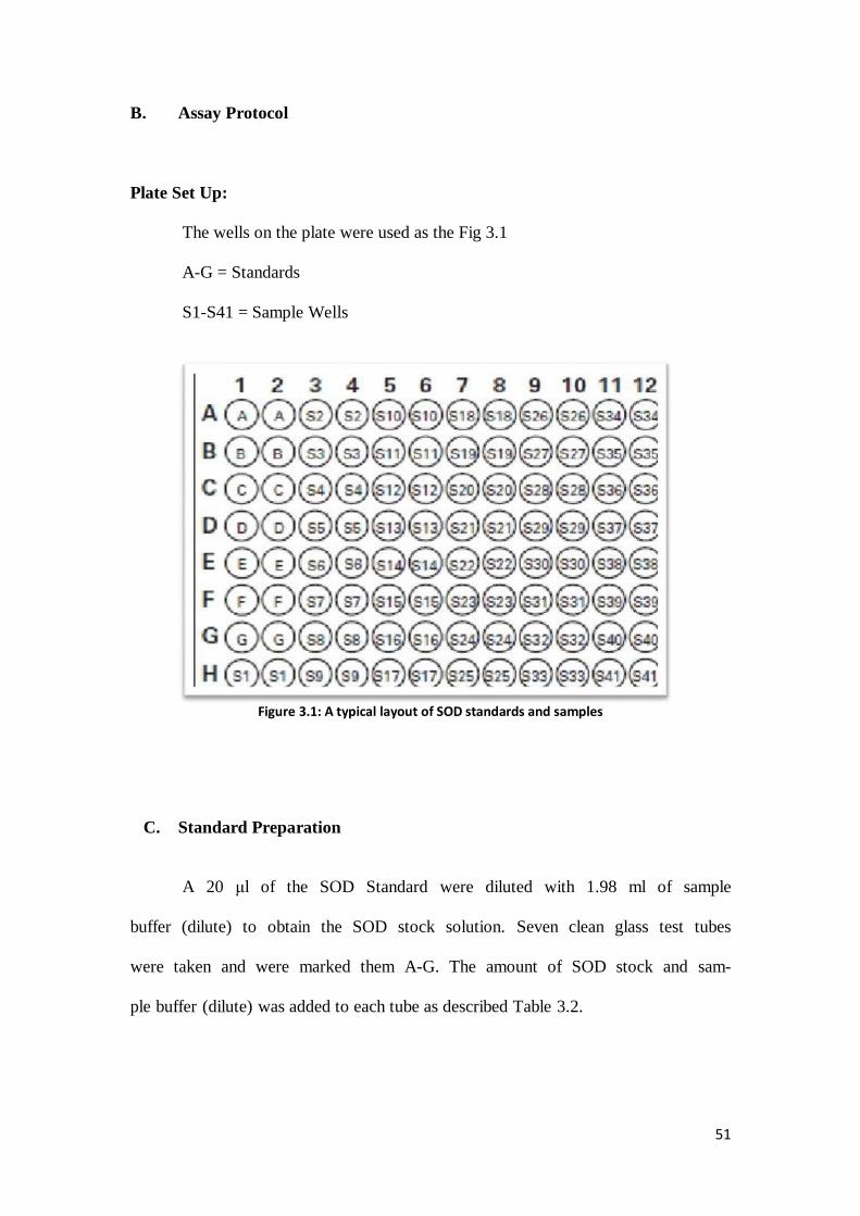
B. Assay Protocol
Plate Set Up:
The wells on the plate were used as the Fig 3.1
A-G = Standards
S1-S41 = Sample Wells
Figure 3.1: A typical layout of SOD standards and samples
C. Standard Preparation
A 20 μl of the SOD Standard were diluted with 1.98 ml of sample
buffer (dilute) to obtain the SOD stock solution. Seven clean glass test tubes
were taken and were marked them A-G. The amount of SOD stock and sam-
ple buffer (dilute) was added to each tube as described Table 3.2.

52
Table 3.2: Superoxide Dismutase standards
Tube SOD Stock (μl)
Sample Buffer (μl) Final SOD Activity
(U/ml)
A 0 1000 0
B 20 980 0.025
C 40 960 0.05
D 80 920 0.1
E 120 880 0.15
F 160 840 0.2
G 200 800 0.25
D. Performing the Assay
a) SOD Standard Wells: 200 μl of the diluted radical detector and
10 μl of standard ,(Tubes A-G) per well were added to the designated wells on
the plate.
b) Sample Wells: 200 μl of the diluted radical detector and 10 μl
of in vitro and in vivo extracts (100 g l-1
) were added to the wells.
c) A 20 μl of diluted xanthine oxidase to all the wells as quickly
as possible was added.
d) Carefully were shook the 96-well plates for a few second to
mix and covered with the plate cover.
e) The plate was incubated the plate on a shaker for 20 minutes at
room temperature.
f) The absorbance was read at 440-460 nm using a plate reader.

53
E. Calculations
i. The average absorbance of each standard, in vivo, and in vitro extracts were
calculated.
ii. Standard A‟s absorbance was divided by itself and the other standards and
samples extract absorbance to yield the liberalized rate.
iii. Plot the liberalized SOD standard rate (LR) (from step 2 above) as a function
of final SOD activity (U ml-1
) from superoxide dismutase standard table.
iv. The SOD activity of the samples using the equation obtained from the linear
regression of the standard curve substituting the liberalized rate (LR) for each
sample was calculated.
One unit was defined as the amount of enzyme needed to exhibit 50% dismutation of
the superoxide radical. SOD activity was standardized using the cytochrome „C‟ and
xanthine oxidase coupled assay.
According to factory manual, SOD was calculated by this formula:
SOD (U/ml) = [(sample LR - y-intercept / slope) x 0.23 ml / 0.01 ml] x sample dilu-
tion

54
RESULTS
AND
DISCUSSION

55
4.1 Initiation of Callus and Maintenance
Initiation of callus was observed from the young leaf explants after two weeks
of culture on the MS and WPM medium supplemented with different plant growth
regulators such as NAA and IAA. For maintenance of good growth, callus was sub
cultured every month, onto MS and WPM medium supplemented with the same hor-
mone and concentration (e.g. from MS supplemented with 1mg l-1
to fresh MS with 1
mg l-1
of NAA).
Callus with different colour were noted (Fig 4.1), when the media was sup-
plemented with TDZ 1, 1.5 mg l-1
and IBA 1 mg l-1
. The same was reported by Eeck-
haut, et al in 2010.
Figure 4.1: Greenish and yellowish callus from 1 mg l-1
TDZ and 1.5 mg l-1
IBA after 5 weeks

56
In order to check whether the callus formation were embryogenic or not, dou-
ble staining test was done. The results showed that embryogenic cells were formed on
callus grown with various auxin (Fig 4.2a) and non-embryogenic on callus formed on
MS supplemented with TDZ and Kn (Fig 4.2b).
a) embryogeniccells b) Non-embryogenic cells
Figure 4.2: Early stage embryos after double staining, embryonal heads stained red (acetocarmine)
and tstlurtentts srusepsu (Ev r’tepsu) .

57
4.1.1 Callus Formation in MS Medium
Callus formation from this species was observed after two weeks of cultures.
The types of callus obtained were type one from leaf explant.
The α-Naphthalene acetic acid (NAA) is synthetic auxin and is commonly
used in tissue culture media (Bhojwani & Razdan, 1996a). Based on the obtained da-
ta, the explants responded to various concentrations of NAA after 14 days of culture.
As shown in Fig 4.3, leaf explants cultured on MS medium supplemented
with NAA (1, 1.5, 2, 2.5, and 3 mg l-1
) showed higher percentage of callus formation
compared to other concentrations. Callus formation was 98 % using 1 and 1.5 mg l-1
and 100 % using 2, 2.5, and 3 mg l-1
concentration of NAA. Callus induction dropped
when concentration of NAA increased from 3.5 to 5 mg l-1
.
2, 4-D or 2, 4-Dichlorophenoxyacetic acid is very effective for the induction
and callus growth and for somatic embryogenesis in vitro conditions (Bhojwani &
Razdan, 1996c; Philip, 1984; King, 1984). Callus formation of leaf explants on MS
medium supplemented with 2 and 2.5 mg l-1
2, 4-D was 100% and using 1.5 and 3 mg
l-1
showed 99 and 98 % , respectively. Other concentrations of this hormone gave less
than 80 % callus formation.
Indole-3-acetic acid (IAA) is one of the most commonly detected natural aux-
in, used to induce callus, meristem and shoot culture (Moshkov et al., 2008). In many
of plant species, the effect of IAA was similar to indole-3-butyric acid (IBA) but IAA
is the least stable in the medium (Bhojwani & Razdan, 1996c).
Different concentrations of IAA demonstrated various percentages of callus
formation (from 17% until 99%), but the highest percentage (99% and 84%) were

58
observed on 3.5 and 3 mg l-1
of IAA. In contrast, callus from leaf explants were
formed 100% on 2.5 and 3 mg l-1
concentration of IBA and formation was observed
more than 80 % between 3.5 until 5 mg l-1
concentration of IBA.
Figure 4.3: Percentage of callus formation of G. jasminoides in MS medium supplemented with
different type of auxin at various concentrations
Since 1982, N-phenyl-N′-1, 2, 3-thiadiazol-5-ylurea or Thidiazuron which is known
as TDZ, has been used as a cytokinin in several studies on shoot multiplication and
especially more effective than the other cytokinin with recalcitrant woody species
(Lu, 1993). Moreover, this cytokinin in some species is used to obtain higher rate of
somatic embryogenesis than other hormones, also cytokinin alone has been found to
substitute for both auxin and cytokinin in many species. In some plants, a higher rate
of somatic embryogenesis is obtained with TDZ than with other PGRs. (Von, 2007).
Based on Figure 4.4, callus formation started (73%) from 0.5 mg l-1
and in-
creased to 84% when MS medium supplemented with 1 mg l-1
of TDZ, this percent-
age fluctuated slightly (between 78% until 70%) on 0.5 to 3 mg l-1
and plunged to
0%
20%
40%
60%
80%
100%
120%
0 0.5 1 1.5 2 2.5 3 3.5 4 4.5 5
Cal
lus
Pe
rce
nta
ge
Auxin (mg l-1)
IAA
IBA
NAA
2,4-D

59
45% on 5 mg l-1
. Kinetin (Kn) or 6-furfurylaminopurine is often used in culture media
for cell suspensions, callus induction, growth and induction of morphogenesis while
higher concentrations can be used to induce rapid multiplication of meristems and
shoots (Harisha, 2007). However, MS medium supplemented with 1, 1.5, and 2 mg l-1
of Kn showed 69, 65 and 60% callus formation, respectively. Percentage of callus
formation rapidly dropped to 32% on 3 mg l-1
until 11% on 5 mg l-1
of kinetin.
Data analyzed showed no significant differences in callus formation between
various used auxin on WPM medium, in contrast, MS medium showed significant
differences between IAA and other auxin. In addition, Kinetin and TDZ added to MS
medium showed statistical differences compare to WPM. There was a little difference
between these two hormones on WPM medium but TDZ showed more variation on
callus formation compared to Kn.
Shoot formation from leaf explants of G. jasminoides Ellis was reported by
Duhoky & Rasheed, (2010) and Sayd et al., (2010), but the present results contradict
their results. When leaf explant was cultured on MS and WPM media supplemented
with a combination used auxin with TDZ and Kn, no shoot formation was observed.

60
Figure 4.4: Percentage of callus formation on MS medium supplemented with different types of
cytokinin at various concentrations
Figure 4.5: Callus induction and root formation from leaf explant of G. jasminoides Ellis on MS me-
dium supplemented with 1.5 mg l-1 NAA after 2.5 months
0%
10%
20%
30%
40%
50%
60%
70%
80%
90%
100%
0 0.5 1 1.5 2 2.5 3 3.5 4 4.5 5
Cal
lus
Pe
rce
nta
ge
Cytokinin (mg l-1)
TDZ
Kn

61
4.1.2 Callus Formation in WPM Media
Leaf explants cultured on WPM medium supplemented with different concen-
trations of NAA except in media without hormone and 0.5 mg l-1
of NAA produced
more than 80% callus. Based on the figure 4.6, callus formation was observed (73%)
on 0.5 mg l-1
and 100% on 2, 2.5, and 3 mg l-1
concentrations of NAA. No significant
difference was observed in the callus percentage between 3.5 to 5 mg l-1
of NAA.
Callus (29%) on WPM medium supplemented with different concentrations of 2, 4-D
was formed on 0.5 mg l-1
of hormone and increased to 88% on the 2 mg l-1
. The callus
formation was maintained at 90% on 2.5 and 3 mg l-1
of 2, 4-D and dropped to 76%
on higher concentration of hormone (5 mg l-1
).
Different concentrations of IAA showed various percentages of callus for-
mation, but 100 % was formed in 2.5, and 3 mg l-1
of IAA. WPM medium showed
more than 80% of callus formation on 3.5 and 4 mg l-1
of IAA; however, on 0.5 mg l-1
concentration of this hormone callus was not formed.
Indole-3-butyric acid (IBA) is another synthetic and common auxin for plant
tissue culture and for obtaining root initiation in conventional cuttings (Bhojwani &
Razdan, 1996 b; Machakova et al., 2008).
The percentage of callus formation on IBA was very close to callus formation on
NAA with the same concentrations. A hundred percent callus formation was noted in
2, 2.5, and 3 mg l-1
of the IBA and callus percentage was more than 95% between 3.5
until 5 mg l-1
of the IBA from leaf explants.

62
Figure 4.6: Percentage of callus formation in WPM medium supplemented with different type of
auxin at various concentrations
Figure 4.7: Callus induction and root formation from leaf explant of G. jasminoides Ellis on MS me-
dium supplemented with 1.5 mg l-1 NAA after 2.5 months
0%
20%
40%
60%
80%
100%
120%
0 0.5 1 1.5 2 2.5 3 3.5 4 4.5 5
Cal
lus
Pe
rce
nta
ge
Auxin (mg l-1)
IAA
IBA
NAA
2,4-D

63
Figure 4.8 shows WPM medium supplemented with 1 mg l-1
of TDZ gave
higher percentage of callus (58%). However, equal percentages (51%) were observed
at concentrations of 1.5 and 2 mg l-1
of TDZ. In addition, callus percentage suddenly
declined from 34% to 7% on concentration of 2.5 to 5 mg l-1
of TDZ. Percentage of
callus on WPM supplemented with kinetin started (12%) from 0.5 mg l-1
and reached
to the highest amount (24 and 25%) on 2 and 2.5 mg l-1
and fell to lowest percentage
(9%) on 4.5 mg l-1
.
Figure 4.8: Percentage of callus formation on WPM medium supplemented with different types of cytokinin at various concentrations
0%
10%
20%
30%
40%
50%
60%
70%
0 0.5 1 1.5 2 2.5 3 3.5 4 4.5 5
Cal
lus
Pec
anta
ge
Cytokinin (mg l-1)
TDZ
Kn

64
4.2.1 Comparison of WPM and MS Media Supplemented with Various Con-
centrations of NAA for Callus Formation
As data shown in fig 4.9, the horizontal axis represents the concentration of
NAA with fixed increment of 0.5 mg l-1
and vertical axis represent the percentage of
callus formation. Based on observations and collected data, callus formation from leaf
explants on MS medium started (51%) on 0.5 mg l-1
and rose slightly to 98% on 1 mg
l-1
, a peak (100%) of callus was observed in 2, 2.5, and 3 mg l-1
of NAA.
Figure 4.9: A comparison between WPM and MS media supplemented with NAA for callus for-
mation
Formation of callus gradually declined to 83% on 3.5 mg l-1
until 73% on 5
mg l-1
. Based on fig 4.9, callus was formed (73%) on WPM medium supplemented
with 0.5 mg l-1
NAA from leaf explants and increased to 88% and 98% in concentra-
tions of 1 and 1.5 mg l-1
NAA. Maximum percentage of callus formation (100%) was
observed in WPM medium with 2, 2.5, and 3 mg l-1
of NAA. However, the callus
formation maintained between 98 and 99% on 3.5 and 4 mg l-1
. Behbahani et al
0%
20%
40%
60%
80%
100%
120%
0 0.5 1 1.5 2 2.5 3 3.5 4 4.5 5
Cal
lus
Per
cen
tage
Concentrations of NAA (mg l-1)
WPM
MS

65
(2011) compared these media and reported that WPM medium was better than B5 and
MS medium in Lecythidaceae family.
A comparison between callus formation of leaf explants of G. jasminoides Ellis on
MS and WPM media showed the optimum callus (100%) was formed on 2, 2.5, and 3
mg l-1
NAA when added to MS and WPM media.
Raising concentration of hormone (3 to 5 mg l-1
) caused the callus formation to de-
crease (100% to 70%) on MS medium, but percentage of callus maintained (100% to
95%) between 3 to 5 mg l-1
on WPM medium.
The results of MS medium supplemented with NAA are in agreement with previous
reports about in vitro culture of G. jasminoides (Sayd et al., 2010; Duhoky &
Rasheed, 2010) , but there are no reports available about in vitro culture of G. jas-
minoides Ellis on WPM medium.

66
4.2.2 Comparison of WPM and MS Media Supplemented with Various Con-
centrations of IBA for Callus Formation
There was a steep rise on percentage of callus formation when WPM and MS
media supplemented with IBA at various concentrations.
Based on fig 4.10, leaf explants of this species formed 53% callus on WPM medium
with 0.5 mg l-1
of IBA and increased (76% to 92%) on 1 and 1.5 mg l-1
of IBA.
Figure 4.10: A comparison between WPM and MS media supplemented with IBA for callus for-
mation
One hundred percent (100%) of callus were formed on WPM with concentra-
tions of 2, 2.5, and 3 mg l-1
of the IBA (Fig 4.10), however, callus maintained be-
tween 95 until 98% on higher concentrations (4, 4.5 and 5 mg l-1
).
Callus formed (21%) on MS medium with 0.5 mg l-1
of IBA and increased
(36, 54, and 81%) on 1, 1.5, and 2 mg l-1
concentrations of this hormone, respectively.
0%
20%
40%
60%
80%
100%
120%
0 0.5 1 1.5 2 2.5 3 3.5 4 4.5 5
Cal
lus
Per
cen
tage
Concentrations of IBA (mg l-1)
WPM
MS

67
MS medium with 2.5 and 3 mg l-1
of IBA showed 100% callus formation and main-
tained at 80% on higher concentrations of this hormone (4, 4.5, and 5 mg l-1
).
These results are similar with those reported by Sayd et al (2010) and George
(1995). They reported with optimum concentration of IBA (2-3 mg l-1
) for callus in-
duction of Gardenia on MS medium.
Figure 4.11: Callus formation from leaf explant in WPM supplemented with 2.5 mg l-1 IBA after 4
months

68
4.2.3 Comparison of WPM and MS Media Supplemented with Various Con-
centrations of IAA for Callus Formation
According to figure 4.12, collected data and observation of callus formation,
MS medium with 3 mg l-1
showed 99% callus formation on the peak of this curve and
at 3.5 mg l-1
showed 84% callus formation. These results proved former report by
Duhoky & Rasheed (2010) about MS medium with the optimum concentration of
IAA at 3- 4 mg l-1
.
Figure 4.12: A comparison between WPM and MS media supplemented with IAA for callus for-
mation
However, callus was induced (19%) on 0.5 mg l-1
IAA and gradually increased
to 35% and 71% when MS medium was supplemented with 1.5 and 2.5 mg l-1
of this
type of auxin, and dropped to 60% and 43% with higher concentrations (4 and 5 mg l-
1) of IAA.
WPM medium showed callus formation (51%) from 1 mg l-1
concentration of IAA
and grew (78 and 98%) on 1.5 and 2 mg l-1
of IAA. Callus percentage reached to the
highest percentage (100%) on 2.5 mg l-1
and was maintained at this level when con-
0%
20%
40%
60%
80%
100%
120%
0 0.5 1 1.5 2 2.5 3 3.5 4 4.5 5
Cal
lus
Per
cen
tage
Concentrations of IAA (mg l-1)
WPM
MS

69
centration rose to 3 mg l-1
. Callus induction dropped to 91% on 3.5 mg l-1
until 83%
on 5 mg l-1
IAA.

70
4.2.4 Comparison of WPM and MS Media Supplemented with Various Con-
centrations of 2, 4-D for Callus Formation
Based on previous reports, 2, 4-D is one of the more effective auxin for callus
formation. Figure 4.13 represents the effect of this hormone on callus formation in
MS and WPM media. Induction of callus started from a concentration of 0.5 mg l-1
2,
4-D on MS and WPM media with 45% and 29%, respectively.
Figure 4.13: A comparison between WPM and MS media supplemented with 2, 4-D for callus for-
mation
Percentage of callus surged to 100% on 1.5, 2, and 2.5 mg l-1
and 98% on 3
mg l-1
concentration of hormone and fell to 42% on 5 mg l-1
on MS medium.
This result is in agreement with that reported by George (1995) and Sayd et al
(2010). They reported optimum callus formation (100%) of G. jasminoides Ellis on
MS medium supplemented with concentration of 2 - 3 mg l-1
of 2, 4-D from leaf ex-
plants. Formation of callus slowly increased in WPM medium (45 and 72%) on 1 and
0%
20%
40%
60%
80%
100%
120%
0 0.5 1 1.5 2 2.5 3 3.5 4 4.5 5
Cal
lus
Per
cen
tage
Concentrations of 2, 4-D (mg l-1)
WPM
MS

71
1.5 mg l-1
and reached to higher amount (88 until 90%) between 2 – 3.5 mg l-1
and
gradually dropped to 76% on 5 mg l-1
of 2, 4-D.
4.3 Root Elongation Study
A. MS medium
Based on collected data and observed results, MS and WPM media supple-
mented with various concentrations of auxin demonstrated different effect on root
formation and root elongation from leaf explants of Gardenia jasminoides Ellis after
six months.
Figure 4.14: Root elongation on MS medium
Rooting started on both media supplemented with various auxin after the fifth
weeks. Figure 4.14 shows, among different types and concentrations of used auxin in
MS medium, NAA (1.5 and 2 mg l-1
) showed higher response for root length (14.8
and 13.4 cm). Roots formation started 0.4 and 3.4 cm from lower concentrations (0.5
-2
0
2
4
6
8
10
12
14
16
18
0 0.5 1 1.5 2 2.5 3 3.5 4 4.5 5
Ro
ot
len
gth
(cm
)
PGR'S (mg l-1)
IAA
IBA
NAA
2,4-D

72
and 1.0 mg l-1
) of NAA and rose to (10.7 and 10.9 cm) on 2.5 and 3 mg l-1
and de-
creased to (3.9 cm) 5 mg l-1
of this hormone (Fig 4.17).
Roots formed (0.2 and 0.3 cm) on MS medium supplemented with 0.5 and 1
mg l-1
IAA and rose (8.8 and 7.8 cm) on 2.5 and 3 mg l-1
of IAA. The root elongation
fell (5.3 and 4.7 cm) at 4.5 and 5 mg l-1
. In addition, root formation started on MS
medium (0.7 and 5.4 cm) supplemented with 1.0 and 1.5 mg l-1
of IBA after four
weeks. Root elongation gradually increased (7.9 and 7.6 cm) on 2 and 2.5 mg l-1
of
IBA. However, the level of elongation showed between 6.2 to 6.9 cm on higher con-
centrations of IAA (3.0, 3.5, 4.0, 4.5, and 5 mg l-1
).
MS medium supplemented with 0.5 mg l-1
2, 4-D showed 0.5 cm root length.
This rate increased at the higher levels (4.8 and 4.1 cm) on 2 and 2.5 mg l-1
of 2, 4-D.
The level of root growth declined from 3 mg l-1
(3.5 cm) to 5 mg l-1
(2.2 cm). Howev-
er, MS medium showed statistically difference when supplemented with NAA com-
paring to IAA and IBA.
Figure 4.15: Rooting formation in MS medium supplemented with 2 mg l-1 NAA after 6 months

73
B. WPM Medium
WPM medium supplemented with various auxin showed different results of
root formation. Figure 4.16, represents longest root length (18.3 and 18.7 cm) on two
concentrations of IAA (4.5 and 5 mg l-1
), respectively. Rooting formation started from
0.7 cm in a concentration of 0.5 mg l-1
and gradually increased to 13.5 cm in 4 mg l-1
.
Figure 4.16: Root elongation on WPM medium
In addition, higher root length was observed in WPM media supplemented
with 2, 2.5, and 3 mg l-1
NAA (17.4, 15.7, and 13.9 cm). Roots formed from 0.5 mg l-1
(0.8 cm) and increased to 12.8 cm (1.5 mg l-1
). Decreasing of root growth started
(10.6 cm) from 3.5 mg l-1
until (5.9 cm) 5 mg l-1
of NAA (Fig 4.11).
-5
0
5
10
15
20
25
0 0.5 1 1.5 2 2.5 3 3.5 4 4.5 5
Ro
ot
len
gth
(cm
)
PGR'S (mg l-1)
IAA
IBA
NAA
2,4-D

74
Figure 4.17: Rooting in WPM supplemented with 1 mg l-1 IAA one week after the second subculture
Roots formed (0.3 cm) on 0.5 mg l-1
of IBA on WPM medium and slightly
elongated (0.9 cm) on 1 mg l-1
of this hormone. The higher length of roots (8.4, 8.9,
and 8.6 cm) was observed in WPM medium supplemented with 1.5, 2, and 2.5 mg l-1
of IBA (Fig 4.10), respectively. Level of root length almost stabilized (7.9 to 6.5 cm)
between 3.0, 3.5, 4.0, 4.5, and 5.0 mg l-1
of the IBA.
Roots were formed (0.1 cm) on WPM medium supplemented with 0.5 mg l-1
of 2, 4-D. In addition, concentrations of 2 and 2.5 mg l-1
of 2, 4-D showed maximum
growth of root length (2.7 and 2.9 cm) on WPM medium.
2, 4- Dichloro phenoxy acetic acid (2, 4-D) is one of the synthetic auxin. There
are some successful reports of using this auxin for in vitro propagation (Tyagi et al.,
2010; Godo et al., 2010) and also on Gardenia jasminoides Ellis (Mizukami et al.,
1987; George & Ravishankar, 1995). On root elongation study, data analyses showed
significant differences in WPM medium supplemented with IAA, NAA, and IBA,
respectively.

75
4.3.1 Comparison of MS and WPM Media Supplemented With NAA on Root
Formation
NAA or α-Naphthalene acetic acid was reported as one of the effective auxin
for in vitro root formation when applied singly (Ładyżyński & Rybczyński, 2009;
Sujana & Naidu, 2011) or supplemented with other hormones (Tripathi & Tripathi,
2003; Sanavy & Jami, 2003).
Figure 4.18: heesupere ssersridauss tsllpuaursustssR 2 mg l-1
NAA after 4 months from leaf
explant of G. jasminoides Ellis
Al-Juboory (1998) reported that 1 mg l-1
NAA was effective for optimum root
elongation for micro cutting explants of gardenia and the same hormone with 2 to 5
mg l-1
could give root formation. In addition, MS medium supplemented with 4 mg l-1
of NAA showed the highest average number for root length from leaf explants in G.
jasminoides Ellis (Duhoky & Rasheed, 2010).

76
Figure 4.19: A Comparison between MS and WPM media supplemented with various concentra-
tions of NAA for root elongation
In the present study, the data were analyzed by ANOVA and length of roots on MS
and WPM medium supplemented with various concentrations of NAA were compared
using Duncan‟s multiple comparison test (DMCT).
Based on the data analyzed, there are significant differences between concentrations
of 2, 2.5, 3 and 4 mg l-1
of NAA supplemented to MS and WPM media (p < 0.05).
The sudden growths (14.8 and 13.4 cm) were observed on MS medium supplemented
with 1.5 and 2 mg l-1
NAA and maintained (10.7 and 10.9 cm) on 2.5 and 3 mg l-1
of
hormone (Fig 4.18), root length dropped (8.6 to 3.9 cm) from 3.5 to 5 mg l-1
NAA on
MS medium. On the other hand, abrupt growth in root length (12 cm) on WPM start-
ed from 1.5 mg l-1
and grew (17.4, 15.7, and 13.9 cm) on 2, 2.5, and 3 mg l-1
of NAA.
Level of root length, gradually decreased (10.6 cm) on 3.5 mg l-1
of NAA added to
WPM and dropped (8.7, 6.2, and 5.9 cm) on 4, 4.5, and 5 mg l-1
of the hormone (Fig-
ure 4.19).
-5
0
5
10
15
20
25
0 0.5 1 1.5 2 2.5 3 3.5 4 4.5 5
Ro
ot
Len
gth
(cm
)
NAA (mg l-1)
WPM
MS

77
4.3.2 Comparison of MS and WPM Media Supplemented With IAA for Root
Formation
Indole-3-acetic acid (IAA) is a known as auxin for promoting roots in vitro
(Al-Amin et al., 2009; Kaladhar et al., 2011; Komal, 2011) and also in vivo (George
et al., 2008b; Siegel & Galston, 1953). However, Duhoky & Rasheed (2010) reported
(8 mg l-1
) that IAA has formed the highest number and length of roots in G. jas-
minoides Ellis (3.40 and 3.50 cm roots respectively).
The data were analyzed by ANOVA and length of roots on MS and WPM me-
dia supplemented with various concentrations of IAA were compared using Duncan‟s
multiple comparison test (DMCT).
Statistically differences were noted between concentrations 3, 3.5, 4 and 5 mg l-1
of
IAA supplemented to MS and WPM media respectively (P < 0.05). The concentra-
tions between 3.5 to 5 mg l-1
of the IAA are strongly significant.
Furthermore, root formation on WPM medium supplemented with IAA grown rapidly
with increased hormone concentration. However, differential growth was observed in
MS medium. Gradual growth (0.2 and 3.0 cm) started from low concentrations (0.5
and 1.0 mg l-1
) of the IAA and increased at 2.0 and 3.0 mg l-1
showed the peak of root
length growth (8.8 and 7.8 cm).
Root elongation dropped (5.3 to 4.7 cm) on higher concentration of IAA (from 3.5 to
5 mg l-1
) on MS medium (Figure 4. 20).

78
Figure 4.20: A Comparison between MS and WPM media supplemented with various concentra-
tions of IAA for root elongation
Figure 4.21: Rooting in WPM supplemented with 2 mg l
-1 IAA 4 weeks after the second subculture of
leaf explant of G. jasminoides Ellis
-5
0
5
10
15
20
25
0 0.5 1 1.5 2 2.5 3 3.5 4 4.5 5
Ro
ot
Len
gth
(cm
)
IAA (mg l-1)
WPM
MS

79
4.3.3 Comparison of MS and WPM Media Supplemented with IBA On Root
Formation
Applying IBA showed a higher rooting rate in WPM medium over time. The
optimum rooting observed between 1.5 until 2.5 mg l-1
of the IBA in WPM and
dropped on higher concentration, but this value in MS medium demonstrated in 2 mg
l-1
with the highest rate and minimum and maximum difference between rooting of
both media was observed in 1 and 1.5 mg l-1
respectively (Figure 4.24).
Indole-3-butyric acid or IBA is used in the same manner as IAA and is accept-
ed around the world as a propagating and rooting hormone for in vitro ornamental and
fruit grafting and cuttings. There are some reports of optimum rooting from explants
between, 1-2 mg l-1
of IAA or IBA (Meyer, 1982; Eeckhaut et al., 2010; George et al.,
2008a; Bhojwani & Razdan, 1996a). However, the results of IBA are in agreement
with former reports (George et al., 1993; Al-Juboory et al., 1998; Hatzilazarou et al.,
2006).
Figure 4.22: Root elongation in WPM medium supplemented with 2 mg l
-1 IBA after 4 months
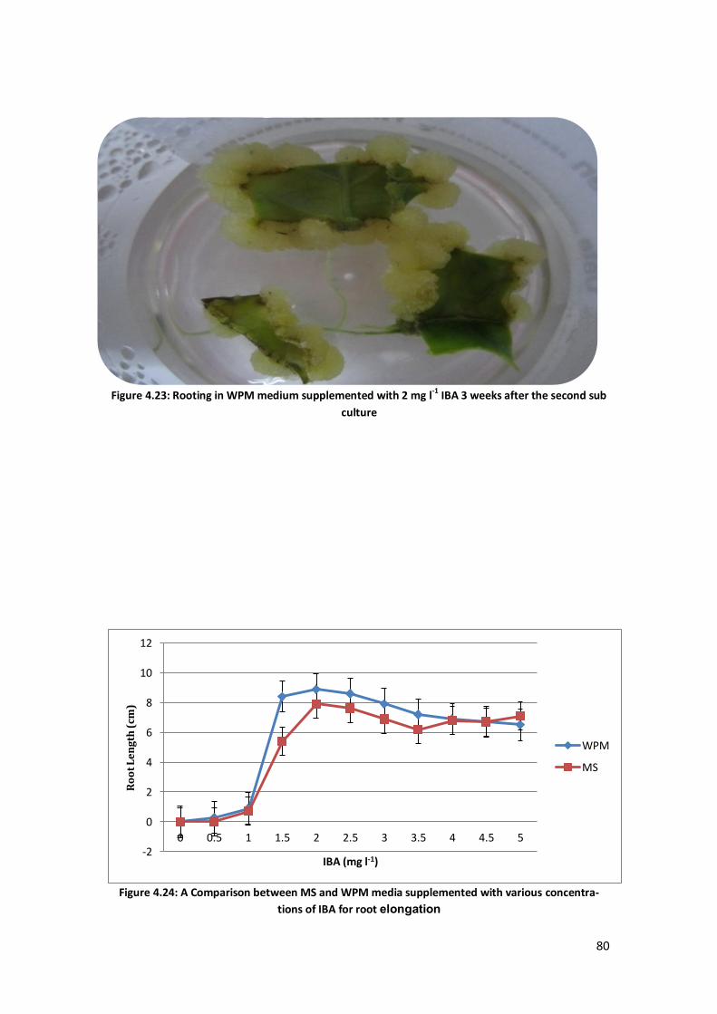
80
Figure 4.23: Rooting in WPM medium supplemented with 2 mg l-1 IBA 3 weeks after the second sub
culture
Figure 4.24: A Comparison between MS and WPM media supplemented with various concentra-
tions of IBA for root noleagnole
-2
0
2
4
6
8
10
12
0 0.5 1 1.5 2 2.5 3 3.5 4 4.5 5
Ro
ot
Le
ng
th (
cm)
IBA (mg l-1)
WPM
MS

81
4.4 Fresh and Dry Weight of Callus
In many cases, the evaluation of experimental results is by determination of
fresh and dry weight of callus (Karl et al., 2009a). Figures 4.25 and 4.27, revealed the
minimum and maximum weight of callus in both media. Fresh grown callus on MS
medium supplemented with NAA (3 mg l-1
) and Kn (5 mg l-1
) were 34.23 g and 3.39
g; respectively. In addition, 30.04 g and 3.78 g were measured as fresh weight callus,
cultured on WPM medium supplemented with (2.5 mg l-1
) NAA and (4 mg l
-1)
Kn.
Figure 4.25: Fresh weight of callus in MS medium supplemented with various types of hormones
and oeenenendd concentrations
Callus fresh weight on MS medium was measured (14.04 g) on 0.5 mg l-1
of
NAA and a high growth was observed (29.94 g) on 1.0 mg l-1
and rose to 34.13 g on
1.5 mg l-1
. Callus Fresh weight gradually increased to 34.23 g on 3.0 mg l
-1 of NAA
and dropped (24.26 g) on higher concentrations of this auxin from 3.0 to 5.0 mg l-1
.
NAA and IBA at 1.5 to 4 mg l-1
produced same amount of fresh weight on
WPM medium. The minimum volume of fresh callus was weighed to be 21.2 and
0
5
10
15
20
25
30
35
40
0.5 1 1.5 2 2.5 3 3.5 4 4.5 5
Cal
lus
Wei
ght
(g)
PGR'S (mg l-1)
IAA
IBA
Naa
2, 4-D
TDZ
Kn

82
22.1 g on 0.5 and 1.0 mg l-1
and these weights were kept in 28.7-30.0 g between 1.5 to
4.0 mg l-1
of NAA. Fresh callus on MS medium supplemented with IBA (0.5 mg l-1
)
weighed 6.08 g. However, this weight was increased (9.17 to 20.0 g) on higher con-
centration of hormone (1 to 2 mg l-1
) and maximum weight of callus (30.5 and 32.4 g)
was measured on 2.5 and 3 mg l-1
of IBA , this amount was kept on 20±2 g on other
concentrations. Increasing weight of 10.0 and 16.64 g callus started on WPM medium
from 0.5 and 1.0 mg l-1
of the IBA and showed slight changes on the weight (21.6 to
20.8) between 2.0 to 4.5 mg l-1
of the IBA. Based on reviewed literature, 2, 4-D is
used for callus induction and embryogenesis, but based on data as shown in figures
4.24 and 4.26, this auxin gave lower result on fresh weight of callus on both media
compared to others auxin. Minimum weight of fresh callus (3.9 g) was observed on
0.5 mg l-1
with a slight growth and maximum weights were demonstrated between
15.36, 16.76, and 14.10 g on 2.0, 2.5 and 3.0 mg l-1
concentrations of 2, 4- D supple-
mented to MS medium. Weight of callus on WPM medium supplemented with 2, 4-D
from 9.4 g to 10.59 g at concentrations of 0.5 to 5.0 mg l-1
were varied. Maximum
weight (14 g ± 0.2) of fresh callus was recorded between 2 until 3 mg l-1
of 2,4-D
supplemented to WPM medium.
Two cytokinin (TDZ and Kn) gave the lowest weight of callus compared to
other hormones on both MS and WPM media. The maximum weight of induced cal-
lus from MS (17.5 and 15.3 g) and WPM medium (9.7 and 9.08 g) were recorded be-
tween 1.5 – 2.0 mg l-1
TDZ. The weight of callus dropped with high concentrations of
TDZ in MS (4.99 g) and WPM medium (4.65 g).
In addition, MS medium supplemented with 1.5, 2.0, and 2.5 mg l-1
kinetin
showed optimum weight (11.46, 12.8, and 12.98 g) among another cytokinin and de-
creased (5.56 and 3.39) on higher concentrations of Kn (4.5 and 5.0 mg l-1
) on MS

83
medium. In contrast, this hormone supplemented to WPM medium showed similar
weights (3.93-3.98 g) from 0.5 to 5 mg l
-1.
Figure 4.29 shows there are a close relationship between callus dry and fresh
weight grown on both media supplemented with different auxin and cytokinin with
various concentrations. However, the relationship between dry and fresh weight of
callus is approximately linear.
Effect of NAA and IBA on promoting fresh and dry weight of callus supplemented in
MS medium was reported by Chinnamadasamy et al (2010), Pant & Joshi (2009),
Helgeson & Upper (1970) and one study on comparing of WPM and MS media was
carried out by Behbahani et al (2011). These results were in agreement with the find-
ings of Kende, (1989); Sayd et al (2010) and George et al (1993). Using NAA or IBA
in the media, promoted fresh and dry weights values. The increasing effect of IBA,
NAA on growth and callus formation might be attributed to auxin as it encourages the
biosynthesis of ethylene by promoting the activity of 1-amino cylopropane-1-
carboxylic acid (ACC) syntheses. 2, 4-D was the auxin choice for callus induction,
but it gave the lowest values of both fresh and dry weights of callus. These results
were in agreement with those obtained before (Sayd et al., 2010; Abdel-Rahim et al.,
1998). Consequently, the higher fresh and dry weight of callus was recorded on both
media supplemented with NAA as auxin and TDZ as cytokinin.

84
Figure 4.26: Dry weight of callus in MS medium supplemented with various type of hormones
in sseeunurs concentrations
Figure 4.27: Fresh weight of callus in WPM medium supplemented with various type of hormones
and sseeunurs concentrations
0
1
2
3
4
5
6
7
0.5 1 1.5 2 2.5 3 3.5 4 4.5 5
Cal
lus
Wei
ght
(g)
PGR'S (mg l-1)
IAA
IBA
NAA
2,4-D
TDZ
KN
-5
0
5
10
15
20
25
30
35
0.5 1 1.5 2 2.5 3 3.5 4 4.5 5
Cal
lus
Wei
ght
(g)
PGR'S (mg l-1)
IAA
IBA
Naa
2, 4-D
TDZ
Kn

85
Figure 4.28: Dry weight of callus in WPM medium supplemented with various type of hormones
in sseeunurscercursn ssert
Figure 4.29: A comparison between dry and fresh weight of callus in WPM and MS media supple-
mented tssRsseeunurs hormones
-1
0
1
2
3
4
5
6
0.5 1 1.5 2 2.5 3 3.5 4 4.5 5
Cal
lus
Wei
ght
(g)
PGR'S (mg l-1)
IAA
IBA
NAA
2, 4-D
TDZ
Kn
0
50
100
150
200
250
300
IAAFresh
IAADry
IBAFresh
IBADry
NAAFresh
NAADry
2,4-DFresh
2,4-DDry
TDZFresh
TDZDry
KNFresh
KNDry
Tota
l wei
ght
of
callu
s p
er (
g)
MS
WPM
Linear (MS)
Linear (WPM)

86
4.5 Antibacterial Assay
The results of different studies provide evidence that some medicinal plants
might indeed be potential sources of new antibacterial agents even against some anti-
biotic-resistant strains (Kone et al., 2004). In this study, the disk diffusion method
showed that extracts of G. jasminoides Ellis produced antibacterial activity against
pathogenic bacteria. Among all in vitro and in vivo tested extracts, only callus on MS
and WPM media supplemented with NAA showed inhibition zone to Escherichia coli
and Bacillus cereus. However, the rest of the extracts showed no inhibition zone in
the concentration of 100 mg/ml against tested pathogenic bacteria (Fig 4.30 and Fig
4.31).
Figure 4.30: Inhibition zone in tetracycline as a control

87
Figure 4.31: Inhibition zone of in vitro extract grown on MS medium supplemented with NAA
against E. coli
Only few reports on (Ragasa et al., 2007 ; Ali et al., 1995) a slightly antibacte-
rial activity of fruit extracts of G. jasminoides against Escherichia coli, Pseudomonas
aeruginosa, Staphylococcus aureus, and Trichophyton mentagrophytes; and inactive
against Bacillus subtilis and Aspergillus niger.
Generally, plant extracts are usually more active against gram-positive bacteria than
gram-negative bacteria (Basri & Fan, 2005). Abu-Shanab et al (2005) reported gram-
negative bacteria are more resistant to plant extract compared to gram-positive bacte-
ria. This may be due to the permeability barrier provided by the cell wall or to the
membrane accumulation mechanism (Wei et al., 2008).
Callus grown on both MS and WPM media supplemented with NAA showed
antibacterial activity against E. coli and B. cereus as gram-negative and gram-positive

88
bacteria. The others in vivo and in vitro extracts showed no antibacterial zone against
E. coli, S. aureus, P. aeruginosa, and B. cereus as pathogenic bacteria. This result is
in agreement with those found previously (Ali et al., 1995; Ragasa et al., 2007). Mør-
etrø et al., (2006) and Wei et al., (2008), also reported similar results.
Table 4.1: Inhibition zone from 100 g l-1 in vivo and in vitro leaf extracts of Gardenia jasminoides
Ellis against the four pathogenic bacteria
Bacteria Inhibition zone (mm)
In vitro (MS with
NAA)
In vitro (MS with
NAA) Tetracycline (30
μg)
Escherichia coli + + 39 ± 3.04
Staphylococcus aureus - - 21 ± 1.98
Pseudomonas aeruginosa - - 11 ± 1.99
Bacillus cereus + + 41±3
Inhibition zone = +
None inhibition zone = -

89
4.6 Antioxidant Assay
In this study, the antioxidant activity of G. jasminoides Ellis was evaluated
(Fig 4.32). The data were analyzed by two-way ANOVA and there are significant
differences between in vivo and in vitro extracts. Leaf extract from intact plants as in
vivo, showed the minimum rate of SOD-like in MS (10.84 Uml-1
) and WPM (10.93
Uml-1
) media. In contrast, there are no statistical differences between the two types of
media supplemented with various hormones in antioxidant content (Debnath et al.,
2011). Screening of antioxidant compounds from callus extract of G. jasminoides was
confirmed, that this plant could be considered as a good source of antioxidant activity.
Previous reports from fruit extracts by free radical scavenging activity method
(Chen et al., 2010; Chen et al., 2008; Choi et al., 2007) and Superoxide dismutase-
like (SOD-like) activity and catalase activity of the extracts (Debnath et al., 2011)
proved that G. jasminoides Ellis is a plant with high antioxidant activity.
Furthermore, in vivo and in vitro extract of G. jasminoides Ellis by free radical
scavenging activity method revealed high antioxidant properties as reported by Sayd
et al in 2010. Superoxide dismutase (SOD) is a class of enzymes that catalyze the
dismutation of superoxide into oxygen and hydrogen peroxide. As such, they are an
important antioxidant defense in nearly all cells exposed to oxygen. SOD is also a part
of the defense system against oxidative stress in aerobic organisms. It catalyses su-
peroxide anion (O2) and hydrogen peroxide, which is then reduced to water (by hy-
drogen peroxide scavenging enzyme- Catalase). Therefore, Catalase and SOD are
thought to limit the accumulation of reactive oxygen species.
Miszalski et al., (2007), Dos Santos et al., (2000), Shilpashree & Kumar,
(2011) used SOD-kit like enzyme in a different study. However, there are other re-

90
ports of evaluating antioxidant from fruit extract of G. jasminoides Ellis by Fan et al.,
(2011), Wei et al., (2009), Lee et al., (2006) and Pham et al., (2000).
Figure 4.32: A comparison of SOD activity between in vivo (leaf of G. jasminoides Ellis) and in vitro
from oeenenend n ogdge dd v nsestPGh’t d
0
5
10
15
20
25
30
35
40
45
50
Control IAA IBA NAA 2,4-D Kn TDZ Leaf
Um
l-1
MS
WPM

91
CONCLUSION
92
The present study evaluated in vitro propagation and callus indication of Gardenia
jasminoides Ellis by using two different media (MS and WPM) and various types of
auxin and cytokinin. Callus extract were tested for broad antibacterial and antioxidant
activities.
1. Investigation of media effects on callus formation
WPM medium was observed as a better medium for callus formation com-
pared to MS medium this could possibly be due to the various nutrients such
as calcium or phosphate the major reason for this diffrence.
2. Investigation of hormones effects on root induction
WPM medium supplemented with IAA, NAA, and IBA, respectively showed
statistical differences on root induction. Furthermore, these differences were
observed on MS medium when supplemented with NAA compare to IAA and
IBA in terms of root elongation.
3. Screening of antioxidant activity between intact plant and different callus
grown on MS and WPM
The data analyzed indicated that the calluses from in vitro-grown tissues using
NAA, IBA, TDZ, and Kn have antioxidant activities that are significantly dif-
ferent with intact plant. G. jasminoides Ellis is indeed a rich source of antioxi-
dant.

93
4. Screening of antibacterial activity different callus grown on MS and
WPM and intact plant
Only callus grown on both media supplemented with NAA showed antibacte-
rial activity against E. coli and B. cereus. The other in vivo and in vitro ex-
tracts showed no antibacterial zone.
This is the first study to do micropropagation of Gardenia jasminoides Ellis on woody
plant media (WPM) and screen its callus for antibacterial activity.
Recommendations for future work
Alternative media for micropropagation of Gardenia jasminoides Ellis can be further
carried out, which include SH, B5 and LS.
Anti-tumor and anti-microbial activities from the plant and callus can be investigated.
A study can be conducted to find the best organ with maximum antioxidant content.
Related Documents