4/5/2018 1 Tiny People, Giant Problems: Commonly Seen and Treated Neonatal Skin Injuries Sara Anderson BSN, RN, CWON, IBCLC, CCRN Media Esser NNP‐BC, APNP, CCRN, CWN Disclosure • Sara Anderson • Nothing to disclose • Media Esser • Member of the Kimberly Clark‐ Huggies Nursing Advisory Council • Consultant for Neotech Objectives • Discuss two or more ways to manage neonatal skin injuries • List the three types of neonatal skin injuries • List at least one characteristic of neonatal skin that places this population at risk for skin injury • Provide at least one example of how consistent care can assist in the healing of neonatal skin injuries

Welcome message from author
This document is posted to help you gain knowledge. Please leave a comment to let me know what you think about it! Share it to your friends and learn new things together.
Transcript
4/5/2018
1
Tiny People, Giant Problems:
Commonly Seen and Treated Neonatal Skin Injuries
Sara Anderson BSN, RN, CWON, IBCLC, CCRN
Media Esser NNP‐BC, APNP, CCRN, CWN
Disclosure
• Sara Anderson• Nothing to disclose
• Media Esser
• Member of the Kimberly Clark‐ Huggies Nursing Advisory Council
• Consultant for Neotech
Objectives
• Discuss two or more ways to manage neonatal skin injuries
• List the three types of neonatal skin injuries
• List at least one characteristic of neonatal skin that places this population at risk for skin injury
• Provide at least one example of how consistent care can assist in the healing of neonatal skin injuries
4/5/2018
2
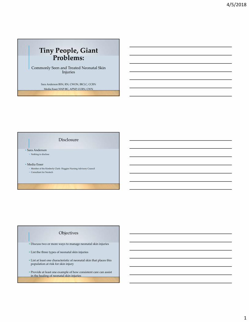
Overview
• Common Types of Skin Injuries
• Overview of Infant Skin
• Phases of Wound Healing
• Products Available for NICU
• Cases of Skin Injury
Neonatal Skin Injuries
• Neonatal Skin injuries occur in up to 57% of infants <32 weeks
• Pressure injuries occur in 5.6‐33% of infants in level III NICUs
• As many as 4% of patients are discharged from the NICU with
significant scarring.
• Technological advances in medicine have not reduced or eliminated the
occurrence of skin injuries
Migoto, M. T. et al. (2013); Sardesai, S. et al. (2011); Razmus, I. et al. (2017); Cousins, (2014)
Types of Neonatal Skin Injuries
Skin Injuries
Diaper dermatitis
Ecchymosis
Skin tears
Pressure injuries
IV injuries
Adhesive Injuries

4/5/2018
3
Neonatal Skin Is Different
• Much fewer layers than term
counterparts ‐30% thinner than adults!
• Stratum Corneum forms at 18‐19 weeks
gestation
• By 23 weeks, only a few layers
• Fully keratinized by 26 weeks, several
layers under the 5‐6 layers of stratum
corneum
• Thicker and more formed by 34‐35
weeks
• Time of well formed skin still unknown
Premature Skin
• Prematurity of epidermis and dermis may contribute to infant skin injury
• Incomplete barrier function leaves infants susceptible to breakdown
• Skin breakdown creates a portal for pathogenic bacteria
• Superficial wounds in neonates are considered full thickness due to the thin and underdeveloped nature of the skin
Kalia, Y., et al. (1998). The Society of Investigative Dermatology, Inc., 111(2), 320‐326. Visscher, M. et. al. (2013). Journal of the European Academy of Dermatology and Venereology, 27(4), 486‐493.
Photo: http://sciencenetlinks.com/media/filer/2011/10/25/si_skin_book.pdf
Premature Skin
• Could be considered “wounded” skin
• At risk for increased permeability, infection, delayed maturation
• Few cornified layers, thin, not fully formed
• Dermis is deficient
• SC formation accelerates with exposure to dry environment with full maturation
developing about 9 wks
• Poor mechanical properties
• Connections between epidermis and dermis are weak‐ tape and adhesives have better
hold to epidermis than the dermis does

4/5/2018
4
Term Skin
• Have highly effective barrier
• TEWL is equal to or less than adult levels
• Acidity neutral initially then decreasing over 1‐4 days and continues over the first 3
months to continue to form the acid mantle
Similarities of Preterm Skin and Wounds
• 23 week skin has similar properties to a wound
• SC extremely thin
• TEWL more than 75g/m2/hr (term 5‐6g/m2/hr, ~29 wks ~17g/m2/hr)
• TEWL continues elevated for up to a month
• Continued abnormal desquamation may indicate hyperproliferation
Top‐Down Injuries
• More common term for superficial skin injuries
• Common types include:
• Skin tears
• Medical adhesive related skin injury (MARSI)
• Tension blisters
• Skin stripping and tears
• Moisture associated skin damage (MASD)
4/5/2018
5
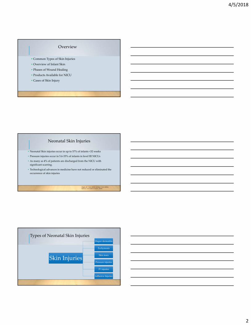
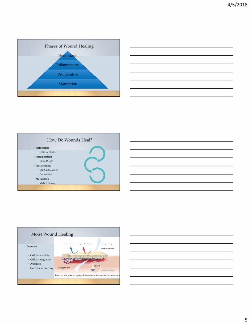
Phases of Wound Healing
Hemostasis
Inflammatory
Proliferative
Maturation
How Do Wounds Heal?
• Hemostasis
• Lets Get Started!
• Inflammation
• Clean It Up!
• Proliferation
• Start Rebuilding!
• Granulation
• Maturation
• Make It Strong!
Moist Wound Healing
• Promotes:
• Cellular viability
• Cellular migration
• Autolysis
• Decrease in scarring
Taken from:http://en.matopat‐global.com/our‐solutions‐view/wound‐treatme
4/5/2018
6
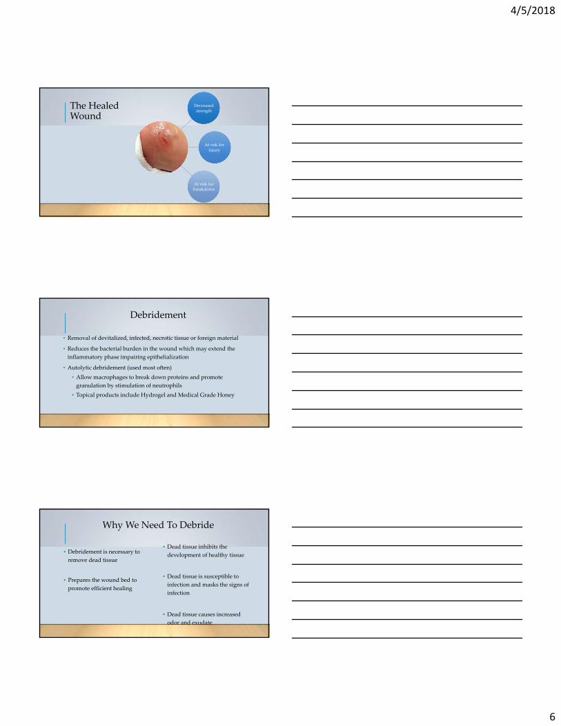
The Healed Wound
Decreased strength
At risk for injury
At risk for breakdown
Debridement
• Removal of devitalized, infected, necrotic tissue or foreign material
• Reduces the bacterial burden in the wound which may extend the
inflammatory phase impairing epithelialization
• Autolytic debridement (used most often)
• Allow macrophages to break down proteins and promote
granulation by stimulation of neutrophils
• Topical products include Hydrogel and Medical Grade Honey
Why We Need To Debride
• Debridement is necessary to
remove dead tissue
• Prepares the wound bed to
promote efficient healing
• Dead tissue inhibits the
development of healthy tissue
• Dead tissue is susceptible to
infection and masks the signs of
infection
• Dead tissue causes increased
odor and exudate
4/5/2018
7
Autolytic Debridement
• Enhance moisture in the wound bed
to degrade dead tissue
• Body will use its own natural
enzymes to breakdown and
deslough the dead tissue
• Won’t work in the severely
immunocompromised and
neutropenic populations
Enzymatic Debridement
• Application of an enzymatic
substance to facilitate the
breakdown of devitalized tissue
• Breaks down the collagen
which provides framework that
adheres the necrotic/slough
tissue to the wound bed
Mechanical Debridement
• Non‐selective
• Sacrifice some healthy tissue
with the removal of necrotic
or devitalized tissue
• Will have some bleeding
• Wet‐to‐dry dressing
• Can be painful – pre‐medicate
• Wound Scrubbing
• Lavage/Irrigation
4/5/2018
8
Sharp Debridement
• Removal of necrotic or
devitalized tissue quickly
• Selective when done correctly
• Must be trained
• Certified in Advanced Sharp
Debridement
• Advanced Practice
• Will have some bleeding
• Can be painful – pre‐medicate
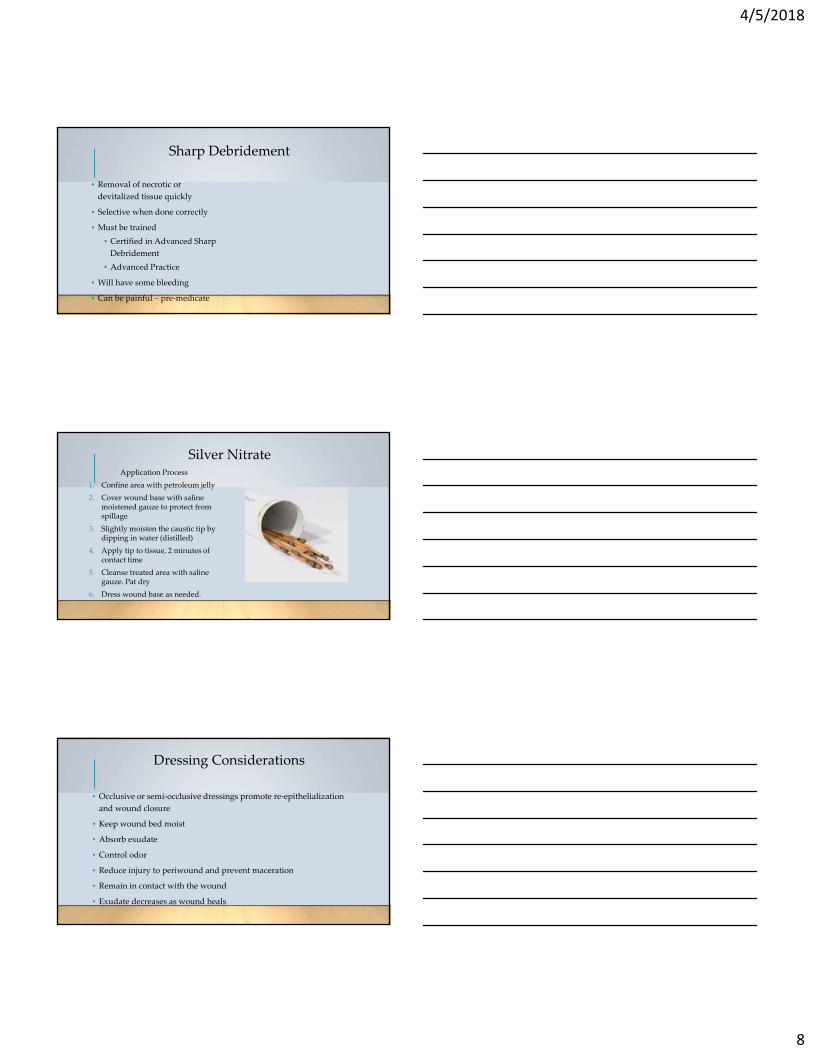
Silver NitrateApplication Process
1. Confine area with petroleum jelly
2. Cover wound base with saline moistened gauze to protect from spillage
3. Slightly moisten the caustic tip by dipping in water (distilled)
4. Apply tip to tissue, 2 minutes of contact time
5. Cleanse treated area with saline gauze. Pat dry
6. Dress wound base as needed.
Dressing Considerations
• Occlusive or semi‐occlusive dressings promote re‐epithelialization
and wound closure
• Keep wound bed moist
• Absorb exudate
• Control odor
• Reduce injury to periwound and prevent maceration
• Remain in contact with the wound
• Exudate decreases as wound heals

4/5/2018
9
Types of Dressings/Treatments
• Liquid barrier films (Cavilon, No‐Sting Barrier Film)
• Semipermeable films (Tegaderm)
• Hydrocolloid dressings (Duoderm, Replicare)
• Soft silicone dressings (Mepilex lite, Mepilex border,
Allyven)
• Hydrofiber dressings (Aquacel ropes, Caltostat,
Angel hair)
• Alginates (Aquacel sheets)
• Medical Grade Honey (MediHoney, Therahoney)
• Hydrogel (Intrasite, Restore, Wound Gel, Vigalon,
Spandgel)
Silver Dressings
• Contraindicated in neonates due to concern of
absorption
• Use when wound at high risk of infection, infected,
highly colonized
• Generally avoid prolonged use and discontinue when
wound infection controlled
Gauze Dressing
• May cause pain when removed
• May lead to desiccation of viable
tissue
• Highly evaporative when used
alone
• Loosely pack
4/5/2018
10
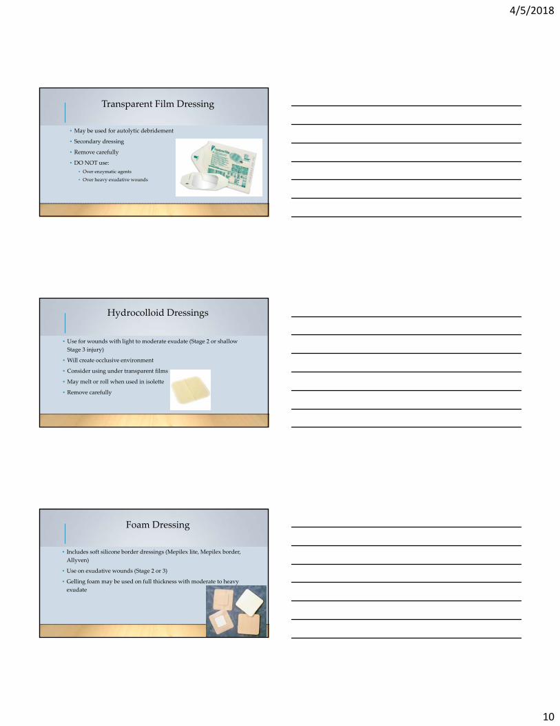
Transparent Film Dressing
• May be used for autolytic debridement
• Secondary dressing
• Remove carefully
• DO NOT use:
• Over enzymatic agents
• Over heavy exudative wounds
Hydrocolloid Dressings
• Use for wounds with light to moderate exudate (Stage 2 or shallow
Stage 3 injury)
• Will create occlusive environment
• Consider using under transparent films
• May melt or roll when used in isolette
• Remove carefully
Foam Dressing
• Includes soft silicone border dressings (Mepilex lite, Mepilex border,
Allyven)
• Use on exudative wounds (Stage 2 or 3)
• Gelling foam may be used on full thickness with moderate to heavy
exudate

4/5/2018
11
Alginate Dressing
• Hydrofiber dressings (Aquacel ropes, Caltostat, Angel hair, Aquacel
sheets)
• Useful in heavily exudating wounds
• Irrigate prior to removal
• Not useful in dry wounds
Silicone Dressing
• Promotes atraumatic dressing
change
• Preventative in periwound tissue
injury
Medical Grade Honey Dressing
• Available in gel and sheets
• Promotes moist wound healing
• Decreased periwound maceration

4/5/2018
12
Hydrogel
• Hydrogel is made up of humectants and water.
• The main ingredients:
• Humectants‐ attracts, holds, and binds water to itself or the product
• Propylene glycol‐ usually represents the most common polymer available in the
general market
• The higher the concentration the more exudate it will pull from the
wound
Hydrogel Dressing
• Use on shallow minimally exudating wounds
• Use for dry wound beds
• Consider using sheets
HydrogelDressingSpecifics
• Cleanse wound
• Apply Hydrogel gel to area
• Place Hydrogel sheet over hydrogel gel
• Place Vaseline gauze
• Place Non‐adhesive gauze over Vaseline
gauze
• Wrap with rolled gauze
• Change dressing Daily then Monday,
Wednesday, Friday with Physical Therapy,
NNP/MD/Plastics (when able), RN

4/5/2018
13
Cases• Full Thickness Wound
• IV Infiltrate
• Diaper Dermatitis
Full Thickness Wound
• 26 week gestational age
• Presumed amniotic banding syndrome
IV Infiltrate

4/5/2018
14
IV infiltrates/Extravasations
• Infiltration rate as high as 57‐70%
• Extravasation rates as high as 11‐23%
• Have the potential to cause peripheral tissue injury (extravasation) or
compartment syndrome (infiltration)
• 95% of PIVs are removed due to complicationsBeall, V., Hall, B., Mulholland, J.T., & Gephart, S.M. (2013). Neonatal Extravasation: An overview and algorithm for evidence‐based treatment. Newborn & Infant Nursing Reviews, 13, 189‐195.
Hyaluronidase
• 5 injections of 0.2mL
• each containing 15 units/mL or 150 units/mL
• Inject subcutaneously in a circle around the leading edge of the
infiltrate/extravasation.
• Administer within 1 hour
• Use hydrogel or honey dressings
Diaper Dermatitis

4/5/2018
15
Diaper Dermatitis
• Diaper dermatitis (DD) is the most common skin injury
• Diaper dermatitis rates of 11‐29%
• Feeding status is speculated to impact diaper dermatitis
• Gestational age and birth weights have not been correlated with DD
rates Migoto, M. T. et al. (2013). OBJN 12(2): 377‐392Sardesai, S. et al. (2011). J Mat Chld Health Med 24(2): 197‐203Theone, M. et al. (2014). Nutrients 6: 261‐275
Causes of Diaper Dermatitis
Over hydration of skin = increased
pH
Elevated pH affects flora of skin
Stool and urine cause pH to
become alkaline
Acidic barrier of skin compromised
Unable to protect from invasion
Friction from diapers and wiping
Redness and breakdown occurs
Esser, M.. (2016). ANC 16: s21‐25
Keep in mind
• Utilize your certified wound nurses
• Create skin care teams/champions
• Build skin care formulary

4/5/2018
16
Key Takeaways
• Moist wound healing is best practice.
• Infection eliminates the ability to do most types of moist wound healing.
• Neonates have faster wound healing due to increased fibroblasts.
• Wound depth is challenging to clearly identify in premature infants.
• More research needs to be published on wound care methods in NICU
patients.
Questions????
References
Bauer, J. (2012). Management of incontinent associated dermatitis (IAD) in the neonatal population.
De Raeve, L. (2008). Diaper dermatitis: Differential diagnosis and treatment. Expert Review of Dermatology, 3(6), 701‐709. doi:http://dx.doi.org.ezproxy.apollolibrary.com/10.1586/17469872.3.6.701
Esser, MS. (2014). The full term infant without congenital defects: Hospitalized infant diaper dermatitis care. The Medical College of Wisconsin Handbook for Care of the Ill Newborn and the Infant in the NICU, 7th ed., 127.
Heimall, L., Storey, B., Stellar, J., & Davis, K. (2012). Beginning at the bottom: Evidence‐based care of diaper dermatitis. MCN, 37(1), 10‐16.
Pasek, et al. (2008). Skin care team in the pediatric intensive care unit: A model for excellence. Critical Care Nurse, 28(2), 125‐135.
Catherine Ratliff, M. D. (2007). Treatment of incontinence‐associated dermatitis (diaper rash) in a neonatal unit. Journal of Wound, Ostomy, Continence Nursing, 34(2), 158.
Shin, H. T. (2005). Diaper dermatitis that does not quit. Dermatologic Therapy, 18(2), 124‐135. Retrieved from http://search.ebscohost.com.ezproxy.apollolibrary.com/login.aspx?direct=true&db=mdc&AN=15953142&site=ehost‐live
Visscher, M. O. (2009). Recent advances in diaper dermatitis: Etiology and treatment. Pediatric Health, 3(1), 81‐98. doi:http://dx.doi.org.ezproxy.apollolibrary.com/10.2217/17455111.3.1.81

4/5/2018
17
References Cont.
Cisler‐Cahill, L. (2006). A protocol for the use of amorphous hydrogel to support wound healing in neonatal patients: an adjunct to nursing skin care. Neonatal Nursing, 25(4), 267‐273.
Taquino, L. (2000). Promoting wound healing in the neonatal setting.: Process versus protocol. Journal of Perinatal and Neonatal Nursing, 14(1), 104‐118.
Visscher, M. & Narendran, V. (2014). The ontogeny of skin. Advances in Wound Care. 3(4), 291‐303.
Hoath, S. B. (2001). Development of the epidermal barrier. NeoReviews. 2(12), c269‐281.
Kalia, Y.N., Lourdes, L.B., Lund, C.H., & Guy, R.H. (1998). Development of skin barrier function in premature infants. The Journal of Investigative Dermatology, 3(2), 320‐326.
Visscher, M. O. (2009). Recent advances in diaper dermatitis: Etiology and treatment. Pediatric Health, 3(1), 81‐98. doi:http://dx.doi.org.ezproxy.apollolibrary.com/10.2217/17455111.3.1.81
Cousins, Y. (2014). Wound care considerations in neonates. Nursing Standard, 28 (46), 61‐70.
Beall, V., Hall, B., Mulholland, J.T., & Gephart, S.M. (2013). Neonatal Extravasation: An overview and algorithm for evidence‐based treatment. Newborn & Infant Nursing Reviews, 13, 189‐195.
Food and Drug Administration. (2008). 510 (k) Summary for Derma Sciences Medihoney Dressings with Active Manuka Honey. Retrieved from https://www.accessdata.fda.gov/cdrh_docs/pdf8/K080315.pdf
References Cont.
Cooper, R.A., & Jenkins, L. (2009). A comparison between medical grade honey and table honeys in relation to antimicrobial efficacy. Wounds, 21(2), 29‐36.
Baghel, P.S., Shukla, S., Mathur, R.K., & Randa, R. (2009). A comparative study to evaluate the effect of honey dressing and silver sulfadiazene dressing on wound in healing in burn patients. Indian Journal of Plastic Surgery, 42(2), 176‐181. doi: 10.4103/0970‐0358.59276.
Amaya, R. (2015). Safety and efficacy of active leptospermum honey in neonatal and paediatric wound debridement. Journal of Wound Care, 24(3), 95‐103.
Ovington, L.G. (2007). Advances in wound dressings. Clinics in Dermatology, 25, 33‐38. doi: 10.1016/j.clindermatol.2006.09.003.
Fox, M.D. (2011). Wound care in the neonatal intensive care unit. Neonatal Network, 30(5), 291‐303.
Beitz, J. (2015). Wound Healing In Doughty, D., McNichol, L. (Eds.), WOCN Core Curriculum: Wound Management.Philadelphia, PA. Wolters Kluwer.
Migoto, M. T., de Souza, S. N. D. H., & Rossetto, E. G. (2013). Skin lesions of newborns in a neonatal unit: observational study. Online Brazilian Journal of Nursing, 12(2), 377‐92.
August, D.L., Edmonds, L., Brown, D.K., Murphy, M., & Kandasamy, Y. (2014). Pressure injuries to the skin in a neonatal unit: Fact or fiction. Journal of Neonatal Nursing, 20, 129‐137.
Sardesai, S., Kornacka, M., Walas, W., & Ramanathan, R. (2011). Iatrogenic skin injury in the neonatal intensive care unit. The Journal of Maternal‐Fetal and Neonatal Medicine,24(2), 197‐203.

4/5/2018
18
Related Documents