Review Timing matters: error-prone gap filling and translesion synthesis in immunoglobulin gene hypermutation Julian E. Sale * , Christopher Batters, Charlotte E. Edmunds, Lara G. Phillips, Laura J. Simpson and Da ´vid Szu ¨ ts Medical Research Council Laboratory of Molecular Biology, Hills Road, Cambridge CB2 0QH, UK By temporarily deferring the repair of DNA lesions encountered during replication, the bypass of DNA damage is critical to the ability of cells to withstand genomic insults. Damage bypass can be achieved either by recombinational mechanisms that are generally accurate or by a process called translesion synthesis. Translesion synthesis involves replacing the stalled replicative polymerase with one of a number of specialized DNA polymerases whose active sites are able to tolerate a distorted or damaged DNA template. While this property allows the translesion polymerases to synthesize across damaged bases, it does so with the trade-off of an increased mutation rate. The deployment of these enzymes must therefore be carefully regulated. In addition to their important role in general DNA damage tolerance and mutagenesis, the translesion polymerases play a crucial role in converting the products of activation induced deaminase-catalysed cytidine deamination to mutations during immunoglobulin gene somatic hypermutation. In this paper, we specifically consider the control of translesion synthesis in the context of the timing of lesion bypass relative to replication fork progression and arrest at sites of DNA damage. We then examine how recent observations concerning the control of translesion synthesis might help refine our view of the mechanisms of immunoglobulin gene somatic hypermutation. Keywords: translesion synthesis; immunoglobulin hypermutation; DNA polymerases; post-replication repair; mismatch repair 1. INTRODUCTION The majority of mutations in DNA arise in consequence of errors introduced during replication, and are frequently precipitated by DNA damage. It is just over 40 years since Evelyn Witkin showed that mutation in Escherichia coli is not an inevitable consequence of DNA damage, such as UV light. Instead, mutagenesis is determined by specific genetic loci and is, along with a number of other phenotypes, induced following exposure to UV light (Witkin 1967a,b). At about the same time, Dean Rupp and Paul Howard-Flanders demonstrated that E. coli defective in the ability to excise UV light-induced pyrimidine dimers could replicate with normal kinetics even when their genome contains up to 50 lesions. However, replication in these circumstances is associated with the formation of gaps in the newly synthesized DNA, at approximately the same spacing as the dimers, which are subsequently filled by a recombinational mechanism that gives rise to sister strand exchanges (Rupp & Howard-Flanders 1968; Rupp et al. 1971). Integration of these obser- vations led to the first models for DNA damage bypass. These proposed that replication blocked at a site of UV damage restarted downstream of the lesion leaving a gap that is filled either by recombination or by an error-prone form of DNA synthesis across the lesion, now termed translesion synthesis (Radman 1970; figure 1). Although this early evidence suggested that lesion bypass was a post-replicative phenomenon, many subsequent models for lesion bypass in higher organisms have considered it as operating at stalled replication forks themselves as a mechanism to preserve fork integrity and progression (reviewed in Lehmann & Fuchs 2006). While mutagenic DNA damage bypass makes some kind of teleological sense in single-celled organisms in which it can be used as a mechanism to escape environmental bottlenecks, its preservation in higher organisms is more puzzling. Indeed, far from being lost, there has been a huge expansion in the number of specialized polymerases in vertebrates and there is clear evidence that these polymerases play a central role in DNA damage tolerance (figure 2). The specialized translesion polymerases (reviewed in Prakash et al. 2005) are characterized by active sites that are tolerant of distortions in the DNA helix introduced Phil. Trans. R. Soc. B (2009) 364, 595–603 doi:10.1098/rstb.2008.0197 Published online 13 November 2008 One contribution of 17 to a Discussion Meeting Issue ‘DNA deamination in immunity, virology and cancer’. * Author and address for correspondence: Division of Protein & Nucleic Acid Chemistry, Medical Research Council Laboratory of Molecular Biology, Hills Road, Cambridge CB2 0QH, UK. ([email protected]). 595 This journal is q 2008 The Royal Society

Welcome message from author
This document is posted to help you gain knowledge. Please leave a comment to let me know what you think about it! Share it to your friends and learn new things together.
Transcript
Phil. Trans. R. Soc. B (2009) 364, 595–603
doi:10.1098/rstb.2008.0197
Published online 13 November 2008
Review
Timing matters: error-prone gap filling andtranslesion synthesis in immunoglobulin
gene hypermutation
Julian E. Sale*, Christopher Batters, Charlotte E. Edmunds, Lara G. Phillips,Laura J. Simpson and David Szuts
One codeamina
*AuthoNucleicMolecu(jes@m
Medical Research Council Laboratory of Molecular Biology, Hills Road, Cambridge CB2 0QH, UK
By temporarily deferring the repair of DNA lesions encountered during replication, the bypass ofDNA damage is critical to the ability of cells to withstand genomic insults. Damage bypass can beachieved either by recombinational mechanisms that are generally accurate or by a process calledtranslesion synthesis. Translesion synthesis involves replacing the stalled replicative polymerase withone of a number of specialized DNA polymerases whose active sites are able to tolerate a distorted ordamaged DNA template. While this property allows the translesion polymerases to synthesize acrossdamaged bases, it does so with the trade-off of an increased mutation rate. The deployment of theseenzymes must therefore be carefully regulated. In addition to their important role in general DNAdamage tolerance and mutagenesis, the translesion polymerases play a crucial role in converting theproducts of activation induced deaminase-catalysed cytidine deamination to mutations duringimmunoglobulin gene somatic hypermutation. In this paper, we specifically consider the control oftranslesion synthesis in the context of the timing of lesion bypass relative to replication forkprogression and arrest at sites of DNA damage. We then examine how recent observationsconcerning the control of translesion synthesis might help refine our view of the mechanisms ofimmunoglobulin gene somatic hypermutation.
Keywords: translesion synthesis; immunoglobulin hypermutation; DNA polymerases;post-replication repair; mismatch repair
1. INTRODUCTIONThe majority of mutations in DNA arise in consequence
of errors introduced during replication, and are
frequently precipitated by DNA damage. It is just over
40 years since Evelyn Witkin showed that mutation in
Escherichia coli is not an inevitable consequence of DNA
damage, such as UV light. Instead, mutagenesis is
determined by specific genetic loci and is, along with a
number of other phenotypes, induced following
exposure to UV light (Witkin 1967a,b). At about the
same time, Dean Rupp and Paul Howard-Flanders
demonstrated that E. coli defective in the ability to excise
UV light-induced pyrimidine dimers could replicate
with normal kinetics even when their genome contains
up to 50 lesions. However, replication in these
circumstances is associated with the formation of gaps
in the newly synthesized DNA, at approximately the
same spacing as the dimers, which are subsequently
filled by a recombinational mechanism that gives rise to
ntribution of 17 to a Discussion Meeting Issue ‘DNAtion in immunity, virology and cancer’.
r and address for correspondence: Division of Protein &Acid Chemistry, Medical Research Council Laboratory of
lar Biology, Hills Road, Cambridge CB2 0QH, UK.rc-lmb.cam.ac.uk).
595
sister strand exchanges (Rupp & Howard-Flanders
1968; Rupp et al. 1971). Integration of these obser-
vations led to the first models for DNA damage bypass.
These proposed that replication blocked at a site of UV
damage restarted downstream of the lesion leaving a gap
that is filled either by recombination or by an error-prone
form of DNA synthesis across the lesion, now termed
translesion synthesis (Radman 1970; figure 1). Although
this early evidence suggested that lesion bypass was a
post-replicative phenomenon, many subsequent models
for lesion bypass in higher organisms have considered it
as operating at stalled replication forks themselves as a
mechanism to preserve fork integrity and progression
(reviewed in Lehmann & Fuchs 2006).
While mutagenic DNA damage bypass makes some
kind of teleological sense in single-celled organisms in
which it can be used as a mechanism to escape
environmental bottlenecks, its preservation in higher
organisms is more puzzling. Indeed, far from being lost,
there has been a huge expansion in the number of
specialized polymerases in vertebrates and there is clear
evidence that these polymerases play a central role in
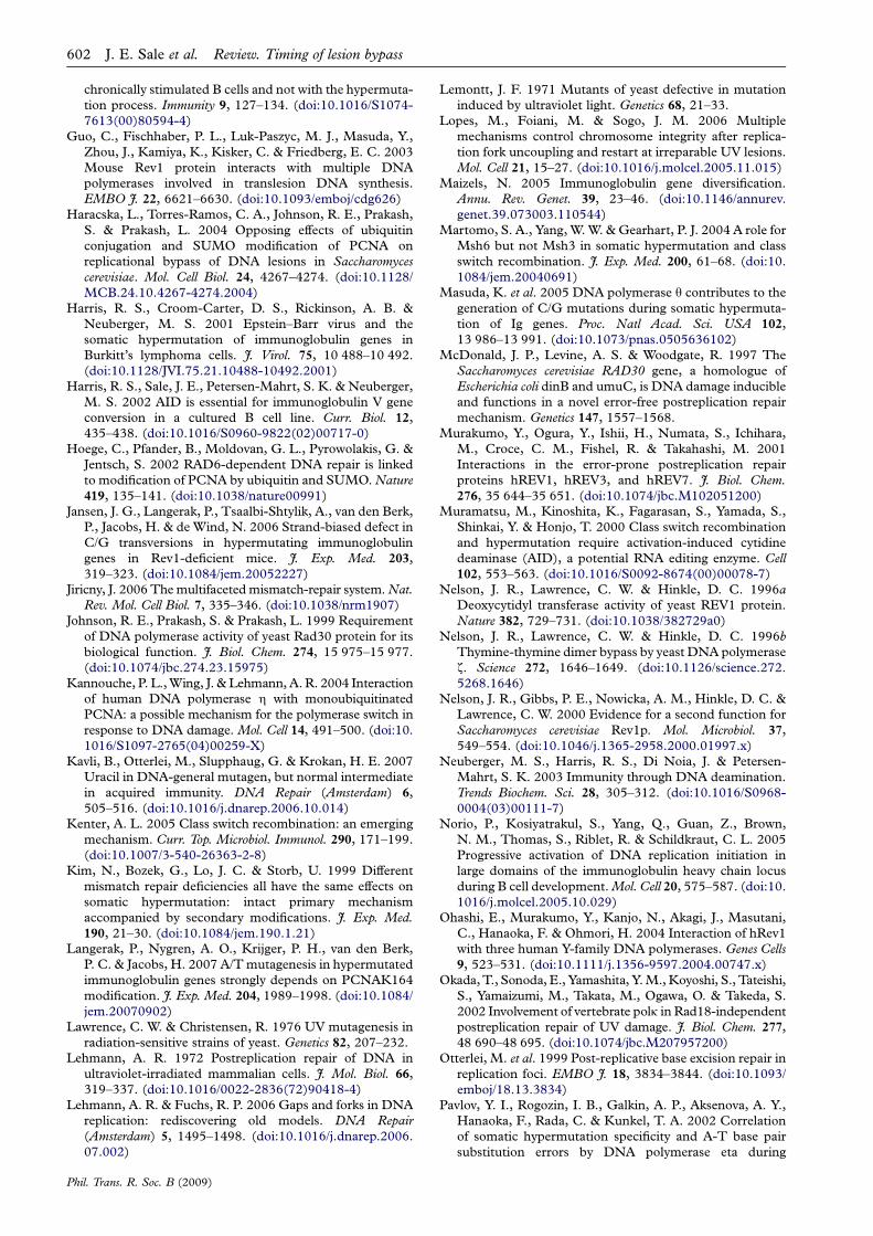
DNA damage tolerance (figure 2).
The specialized translesion polymerases (reviewed in
Prakash et al. 2005) are characterized by active sites that
are tolerant of distortions in the DNA helix introduced
This journal is q 2008 The Royal Society
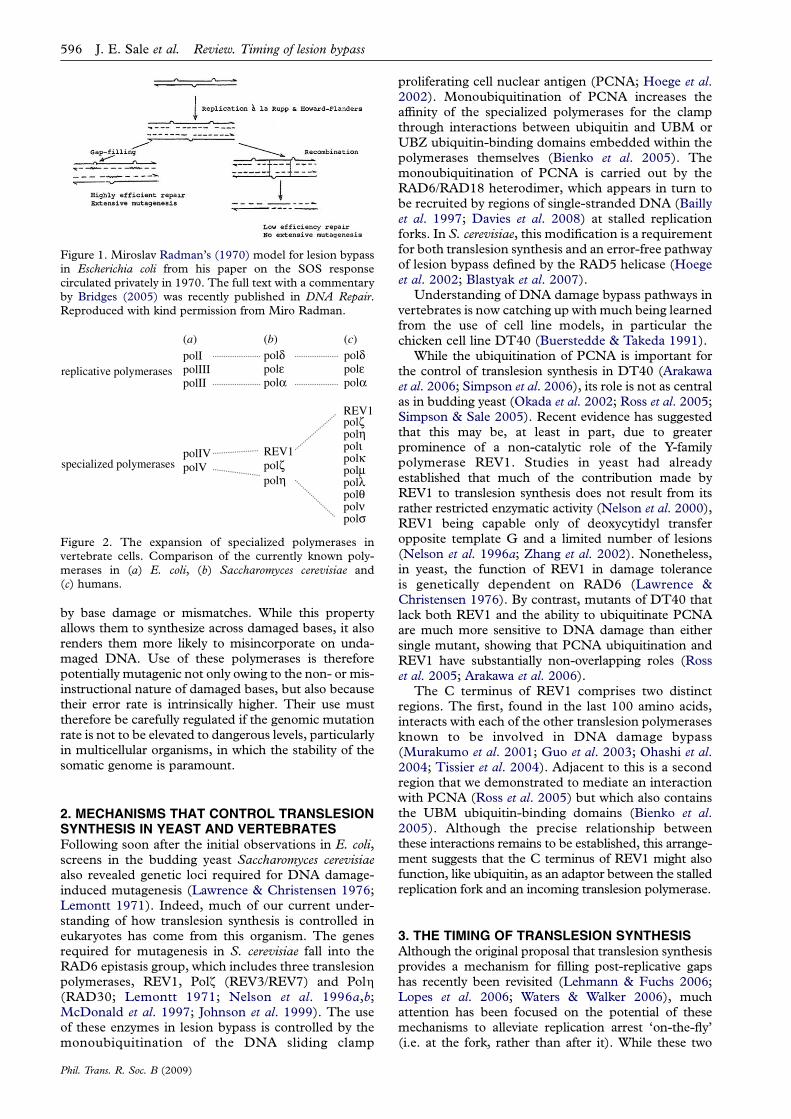
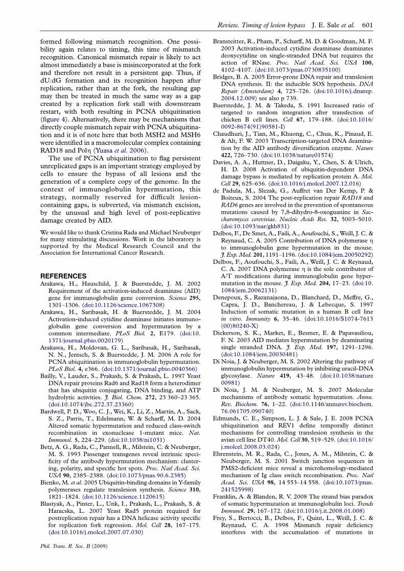
Figure 1. Miroslav Radman’s (1970) model for lesion bypassin Escherichia coli from his paper on the SOS responsecirculated privately in 1970. The full text with a commentaryby Bridges (2005) was recently published in DNA Repair.Reproduced with kind permission from Miro Radman.
replicative polymerases
specialized polymerases
polIpolIIIpolII
polIVpolV
polδpolεpolα
polδpolεpolα
REV1
REV1
polζpolη
polζpolη
polιpolκpolµpolλpolθpolνpolσ
(a) (b) (c)
Figure 2. The expansion of specialized polymerases invertebrate cells. Comparison of the currently known poly-merases in (a) E. coli, (b) Saccharomyces cerevisiae and(c) humans.
596 J. E. Sale et al. Review. Timing of lesion bypass
by base damage or mismatches. While this propertyallows them to synthesize across damaged bases, it alsorenders them more likely to misincorporate on unda-maged DNA. Use of these polymerases is thereforepotentially mutagenic not only owing to the non- or mis-instructional nature of damaged bases, but also becausetheir error rate is intrinsically higher. Their use musttherefore be carefully regulated if the genomic mutationrate is not to be elevated to dangerous levels, particularlyin multicellular organisms, in which the stability of thesomatic genome is paramount.
2. MECHANISMS THAT CONTROL TRANSLESIONSYNTHESIS IN YEAST AND VERTEBRATESFollowing soon after the initial observations in E. coli,screens in the budding yeast Saccharomyces cerevisiaealso revealed genetic loci required for DNA damage-induced mutagenesis (Lawrence & Christensen 1976;Lemontt 1971). Indeed, much of our current under-standing of how translesion synthesis is controlled ineukaryotes has come from this organism. The genesrequired for mutagenesis in S. cerevisiae fall into theRAD6 epistasis group, which includes three translesionpolymerases, REV1, Polz (REV3/REV7) and Polh(RAD30; Lemontt 1971; Nelson et al. 1996a,b;McDonald et al. 1997; Johnson et al. 1999). The useof these enzymes in lesion bypass is controlled by themonoubiquitination of the DNA sliding clamp
Phil. Trans. R. Soc. B (2009)
proliferating cell nuclear antigen (PCNA; Hoege et al.2002). Monoubiquitination of PCNA increases theaffinity of the specialized polymerases for the clampthrough interactions between ubiquitin and UBM orUBZ ubiquitin-binding domains embedded within thepolymerases themselves (Bienko et al. 2005). Themonoubiquitination of PCNA is carried out by theRAD6/RAD18 heterodimer, which appears in turn tobe recruited by regions of single-stranded DNA (Baillyet al. 1997; Davies et al. 2008) at stalled replicationforks. In S. cerevisiae, this modification is a requirementfor both translesion synthesis and an error-free pathwayof lesion bypass defined by the RAD5 helicase (Hoegeet al. 2002; Blastyak et al. 2007).
Understanding of DNA damage bypass pathways invertebrates is now catching up with much being learnedfrom the use of cell line models, in particular thechicken cell line DT40 (Buerstedde & Takeda 1991).
While the ubiquitination of PCNA is important forthe control of translesion synthesis in DT40 (Arakawaet al. 2006; Simpson et al. 2006), its role is not as centralas in budding yeast (Okada et al. 2002; Ross et al. 2005;Simpson & Sale 2005). Recent evidence has suggestedthat this may be, at least in part, due to greaterprominence of a non-catalytic role of the Y-familypolymerase REV1. Studies in yeast had alreadyestablished that much of the contribution made byREV1 to translesion synthesis does not result from itsrather restricted enzymatic activity (Nelson et al. 2000),REV1 being capable only of deoxycytidyl transferopposite template G and a limited number of lesions(Nelson et al. 1996a; Zhang et al. 2002). Nonetheless,in yeast, the function of REV1 in damage toleranceis genetically dependent on RAD6 (Lawrence &Christensen 1976). By contrast, mutants of DT40 thatlack both REV1 and the ability to ubiquitinate PCNAare much more sensitive to DNA damage than eithersingle mutant, showing that PCNA ubiquitination andREV1 have substantially non-overlapping roles (Rosset al. 2005; Arakawa et al. 2006).
The C terminus of REV1 comprises two distinctregions. The first, found in the last 100 amino acids,interacts with each of the other translesion polymerasesknown to be involved in DNA damage bypass(Murakumo et al. 2001; Guo et al. 2003; Ohashi et al.2004; Tissier et al. 2004). Adjacent to this is a secondregion that we demonstrated to mediate an interactionwith PCNA (Ross et al. 2005) but which also containsthe UBM ubiquitin-binding domains (Bienko et al.2005). Although the precise relationship betweenthese interactions remains to be established, this arrange-ment suggests that the C terminus of REV1 might alsofunction, like ubiquitin, as an adaptor between the stalledreplication fork and an incoming translesion polymerase.
3. THE TIMING OF TRANSLESION SYNTHESISAlthough the original proposal that translesion synthesisprovides a mechanism for filling post-replicative gapshas recently been revisited (Lehmann & Fuchs 2006;Lopes et al. 2006; Waters & Walker 2006), muchattention has been focused on the potential of thesemechanisms to alleviate replication arrest ‘on-the-fly’(i.e. at the fork, rather than after it). While these two
replication fork stall
translesion synthesis
post-replicative gap
translesion synthesis
REV1 UbPCNA
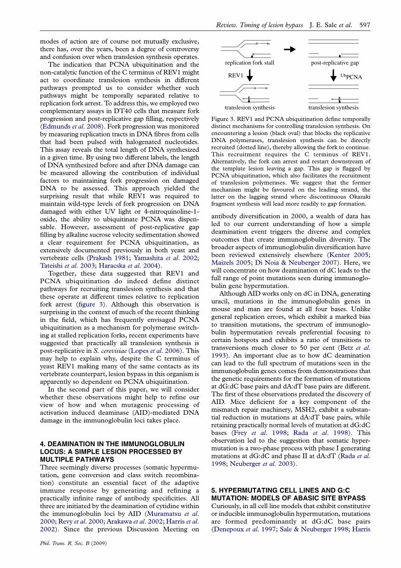
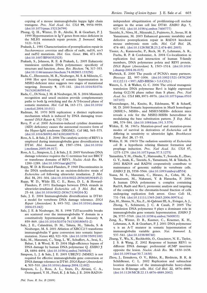
Figure 3. REV1 and PCNA ubiquitination define temporallydistinct mechanisms for controlling translesion synthesis. Onencountering a lesion (black oval) that blocks the replicativeDNA polymerases, translesion synthesis can be directlyrecruited (dotted line), thereby allowing the fork to continue.This recruitment requires the C terminus of REV1.Alternatively, the fork can arrest and restart downstream ofthe template lesion leaving a gap. This gap is flagged byPCNA ubiquitination, which also facilitates the recruitmentof translesion polymerases. We suggest that the formermechanism might be favoured on the leading strand, thelatter on the lagging strand where discontinuous Okazakifragment synthesis will lead more readily to gap formation.
Review. Timing of lesion bypass J. E. Sale et al. 597
modes of action are of course not mutually exclusive,there has, over the years, been a degree of controversyand confusion over when translesion synthesis operates.
The indication that PCNA ubiquitination and thenon-catalytic function of the C terminus of REV1 mightact to coordinate translesion synthesis in differentpathways prompted us to consider whether suchpathways might be temporally separated relative toreplication fork arrest. To address this, we employed twocomplementary assays in DT40 cells that measure forkprogression and post-replicative gap filling, respectively(Edmunds et al. 2008). Fork progression was monitoredby measuring replication tracts in DNA fibres from cellsthat had been pulsed with halogenated nucleotides.This assay reveals the total length of DNA synthesizedin a given time. By using two different labels, the lengthof DNA synthesized before and after DNA damage canbe measured allowing the contribution of individualfactors to maintaining fork progression on damagedDNA to be assessed. This approach yielded thesurprising result that while REV1 was required tomaintain wild-type levels of fork progression on DNAdamaged with either UV light or 4-nitroquinoline-1-oxide, the ability to ubiquitinate PCNA was dispen-sable. However, assessment of post-replicative gapfilling by alkaline sucrose velocity sedimentation showeda clear requirement for PCNA ubiquitination, asextensively documented previously in both yeast andvertebrate cells (Prakash 1981; Yamashita et al. 2002;Tateishi et al. 2003; Haracska et al. 2004).
Together, these data suggested that REV1 andPCNA ubiquitination do indeed define distinctpathways for recruiting translesion synthesis and thatthese operate at different times relative to replicationfork arrest (figure 3). Although this observation issurprising in the context of much of the recent thinkingin the field, which has frequently envisaged PCNAubiquitination as a mechanism for polymerase switch-ing at stalled replication forks, recent experiments havesuggested that practically all translesion synthesis ispost-replicative in S. cerevisiae (Lopes et al. 2006). Thismay help to explain why, despite the C terminus ofyeast REV1 making many of the same contacts as itsvertebrate counterpart, lesion bypass in this organism isapparently so dependent on PCNA ubiquitination.
In the second part of this paper, we will considerwhether these observations might help to refine ourview of how and when mutagenic processing ofactivation induced deaminase (AID)-mediated DNAdamage in the immunoglobulin loci takes place.
4. DEAMINATION IN THE IMMUNOGLOBULINLOCUS: A SIMPLE LESION PROCESSED BYMULTIPLE PATHWAYSThree seemingly diverse processes (somatic hypermu-tation, gene conversion and class switch recombina-tion) constitute an essential facet of the adaptiveimmune response by generating and refining apractically infinite range of antibody specificities. Allthree are initiated by the deamination of cytidine withinthe immunoglobulin loci by AID (Muramatsu et al.2000; Revy et al. 2000; Arakawa et al. 2002; Harris et al.2002). Since the previous Discussion Meeting on
Phil. Trans. R. Soc. B (2009)
antibody diversification in 2000, a wealth of data hasled to our current understanding of how a simpledeamination event triggers the diverse and complexoutcomes that create immunoglobulin diversity. Thebroader aspects of immunoglobulin diversification havebeen reviewed extensively elsewhere (Kenter 2005;Maizels 2005; Di Noia & Neuberger 2007). Here, wewill concentrate on how deamination of dC leads to thefull range of point mutations seen during immunoglo-bulin gene hypermutation.
Although AID works only on dC in DNA, generatinguracil, mutations in the immunoglobulin genes inmouse and man are found at all four bases. Unlikegeneral replication errors, which exhibit a marked biasto transition mutations, the spectrum of immunoglo-bulin hypermutation reveals preferential focusing tocertain hotspots and exhibits a ratio of transitions totransversions much closer to 50 per cent (Betz et al.1993). An important clue as to how dC deaminationcan lead to the full spectrum of mutations seen in theimmunoglobulin genes comes from demonstrations thatthe genetic requirements for the formation of mutationsat dG:dC base pairs and dA:dT base pairs are different.The first of these observations predated the discovery ofAID. Mice deficient for a key component of themismatch repair machinery, MSH2, exhibit a substan-tial reduction in mutations at dA:dT base pairs, whileretaining practically normal levels of mutation at dG:dCbases (Frey et al. 1998; Rada et al. 1998). Thisobservation led to the suggestion that somatic hyper-mutation is a two-phase process with phase I generatingmutations at dG:dC and phase II at dA:dT (Rada et al.1998; Neuberger et al. 2003).
5. HYPERMUTATING CELL LINES AND G:CMUTATION: MODELS OF ABASIC SITE BYPASSCuriously, in all cell line models that exhibit constitutiveor inducible immunoglobulin hypermutation, mutationsare formed predominantly at dG:dC base pairs(Denepoux et al. 1997; Sale & Neuberger 1998; Harris
598 J. E. Sale et al. Review. Timing of lesion bypass
et al. 2001). For example, in the Burkitt lymphoma lineRamos over 80 per cent of mutations are found atdG:dC base pairs (Sale & Neuberger 1998). In DT40cells, which normally diversify their immunoglobulingenes predominantly by gene conversion, disruption ofhomologous recombination or removal of the Vpseudogene donors results in almost exclusive (approx.95%) mutations at dG:dC (Arakawa et al. 2004; Saleet al. 2001). Thus, while hypermutating variants ofDT40 provide a genetically most attractive modelsystem, the immunoglobulin mutagenesis in these linesprobably reflects simply the direct consequence ofreplication across either dU, generating dC/dTtransition mutations, or across abasic sites formedfollowing excision of uracil by uracil DNA glycosylase(UNG; Di Noia & Neuberger 2002; Sale 2004).Nevertheless, hypermutation in cell lines appears toaccurately reflect the ‘phase I’ mutagenesis seen in miceand humans. The reasons for this dG:dC bias, when theparental organism is perfectly capable of mutagenesis atdA:dT base pairs as well, remain unclear. However, it istempting to speculate that it is related to a commonfeature or combination of features of transformedgerminal centre B cells such as deregulated MYC orBCL-6 expression and loss of p53.
The contributions of PCNA ubiquitination and ofREV1 to bypass of abasic sites in the immunoglobulinloci of DT40 broadly mirror their roles in DNAdamage tolerance. Efficient mutagenesis requires bothREV1 and the ubiquitination of PCNA but loss of bothresults in almost total abrogation of mutation (Arakawaet al. 2006). However, part of the reduction in mutationin rev1 DT40 cells may be due to loss of thedeoxycytidyl transferase activity of REV1 since abasicsites are good substrates for this enzyme. Reconstitu-tion of rev1 DT40 cells with human REV1 carryingcatalytically inactivating point mutations demonstratedthat REV1 was required for dC/dG transversionmutations on both strands of the immunoglobulin lightchain locus in DT40. However, the decrease in dC/dG transversions was fully compensated for by anincrease in dC/dA and dC/dT mutations (Ross &Sale 2006). Surprisingly, although disruption of thewhole of REV1 results in decreased levels of pointmutation, deletion of the C terminus does not.Furthermore, the frequency of dC/dG transversionsis unaffected suggesting that the C terminus is notrequired to recruit the catalytic activity of REV1.However, an increase in dC/dA transversions oneither strand, at the expense of dC/dT transitions,suggested that the C terminus of REV1 can nonethelessinfluence polymerase selection at abasic sites evenwhen the catalytic activity of the protein isnot employed.
6. GENETIC REQUIREMENTS FOR IMMUNOGLO-BULIN HYPERMUTATION IN MICE AND HUMANSHypermutation in mice and humans is more complexthan the simple bypass of AID-induced damage seen inDT40 as mutations at dA:dT base pairs are generallymore numerous than those at dG:dC. Furthermore,genetic analyses in mice have so far revealed a fargreater degree of redundancy between the different
Phil. Trans. R. Soc. B (2009)
pathways that process dU than is evident in DT40,making interpretation of the contribution of individualcomponents more problematic.
Analysis of hypermutation in rev1K/K mice alsoreveals a clear contribution of the catalytic activity ofthe protein in bypassing abasic sites. However, incontrast to DT40, this activity appears to be stronglystrand biased with the decrease in dC/dG transver-sions being seen only on the coding (non-transcribed)strand (Jansen et al. 2006). The overall mutationburden was the same as in control animals, the loss ofdC/dG transversions being compensated for by anincrease in dC/dA transversions and in mutations atdA:dT base pairs. One possible source of theredundancy in mutagenesis at dG:dC is DNA poly-merase q, a member of the A-family of DNApolymerases, which is also capable of direct catalyticbypass of abasic sites, strongly favouring A anddisfavouring C in vitro (Seki et al. 2004). Polq hasbeen shown to play a role in the formation of mutationsat dG:dC during immunoglobulin hypermutation inmouse (Masuda et al. 2005; Zan et al. 2005), and whileit is tempting to speculate, based on their in vitroactivities, that Polq and REV1 play compensatory rolesto each other, this remains to be tested. Furthermore,very little is yet known about the mechanisms thatcontrol Polq.
Mutagenesis at dA:dT cannot, of course, beexplained by direct misincorporation opposite dU oran abasic site. Unlike mutagenesis at dG:dC, whichrequires uracil excision, mutagenesis at dA:dT dependsheavily on mismatch recognition of dU:dG mispairs.First noted in Msh2K/K mice (Frey et al. 1998; Radaet al. 1998), a similar loss of dA:dT mutagenesis hasbeen documented in mice deficient for MSH6(Wiesendanger et al. 2000; Martomo et al. 2004) andEXO1 (Bardwell et al. 2004). Curiously, mice deficientin PMS2 and MLH1, proteins required to couplemismatch recognition to excision (reviewed in Jiricny2006), do not exhibit as striking a phenotype in termsof hypermutation (Frey et al. 1998; Kim et al. 1999;Phung et al. 1999; Ehrenstein et al. 2001). Thissuggests that either other factors can substitute forPMS2 and MLH1 in this context or the requirementfor MSH2/6 and EXO1 in dA:dT mutagenesis does notreflect their canonical role in mismatch repair.Importantly, the dependence of dA:dT mutagenesison mismatch repair factors is not complete and it isclear that a proportion of dA:dT mutations are alsotriggered by excision of dU by UNG (Rada et al. 2004).
Two further genetic requirements help provide avery strong framework for understanding mutagenesisat dA:dT base pairs. Numerous lines of evidence pointto a central role for DNA polymerase h (Zeng et al.2001; Delbos et al. 2005). Indeed, recent data havesuggested that it is the sole polymerase responsible(Delbos et al. 2007). Additionally, Langerak et al.(2007) have also very recently demonstrated a pivotalrequirement for PCNA ubiquitination. Polh interactswith ubiquitinated PCNA via its C terminal UBZdomain (Bienko et al. 2005) and the mutationspectrum of Polh on undamaged DNA is similar tothe spectrum of dA:dT mutations seen duringimmunoglobulin hypermutation (Pavlov et al. 2002)
Review. Timing of lesion bypass J. E. Sale et al. 599
suggesting that, unlike mutations at dG:dC (whicharise due to bypass of a non-instructional lesion),mutations at dA:dT arise in consequence of misincor-poration during DNA patch synthesis by Polh.Together, these observations support a model inwhich PCNA ubiquitination recruits Polh to theimmunoglobulin loci. As in the rev1K/K mice,redundancy of mutagenic mechanisms exists and acompensatory increase in mutation at dG:dC is seen inboth the pcnaK164R and polhK/K animals. Interest-ingly, the frequency of dC/dG transversions on thecoding strand, which as discussed above are highlydependent on the catalytic activity of REV1, wasunaffected by disruption of PCNA ubiquitination,confirming that the recruitment of the catalytic activityof REV1 is independent of the ability of REV1 to bindubiquitin on PCNA.
7. CAN IMMUNOGLOBULIN HYPERMUTATION BEUNDERSTOOD IN TERMS OF THE TIMINGOF TRANSLESION SYNTHESIS?In this last section we examine how, despite someimportant differences between studies of damagetolerance in DT40 and hypermutation in mice,some of the concepts uncovered in DT40 might beuseful in improving our understanding of immuno-globulin hypermutation.
Substantial evidence points to AID activity beinglinked to transcription (reviewed in Di Noia &Neuberger 2007), the transcription bubble providingthe single-stranded DNA upon which AID hasbiochemically been shown to act (Bransteitter et al.2003; Chaudhuri et al. 2003; Dickerson et al. 2003).Transcription of highly active loci such as theimmunoglobulin genes in germinal centre B cells islikely to be only transiently interrupted by the passageof a replication fork. Thus, the deamination of dC willtake place almost continuously both before and afterreplication of the immunoglobulin V genes.
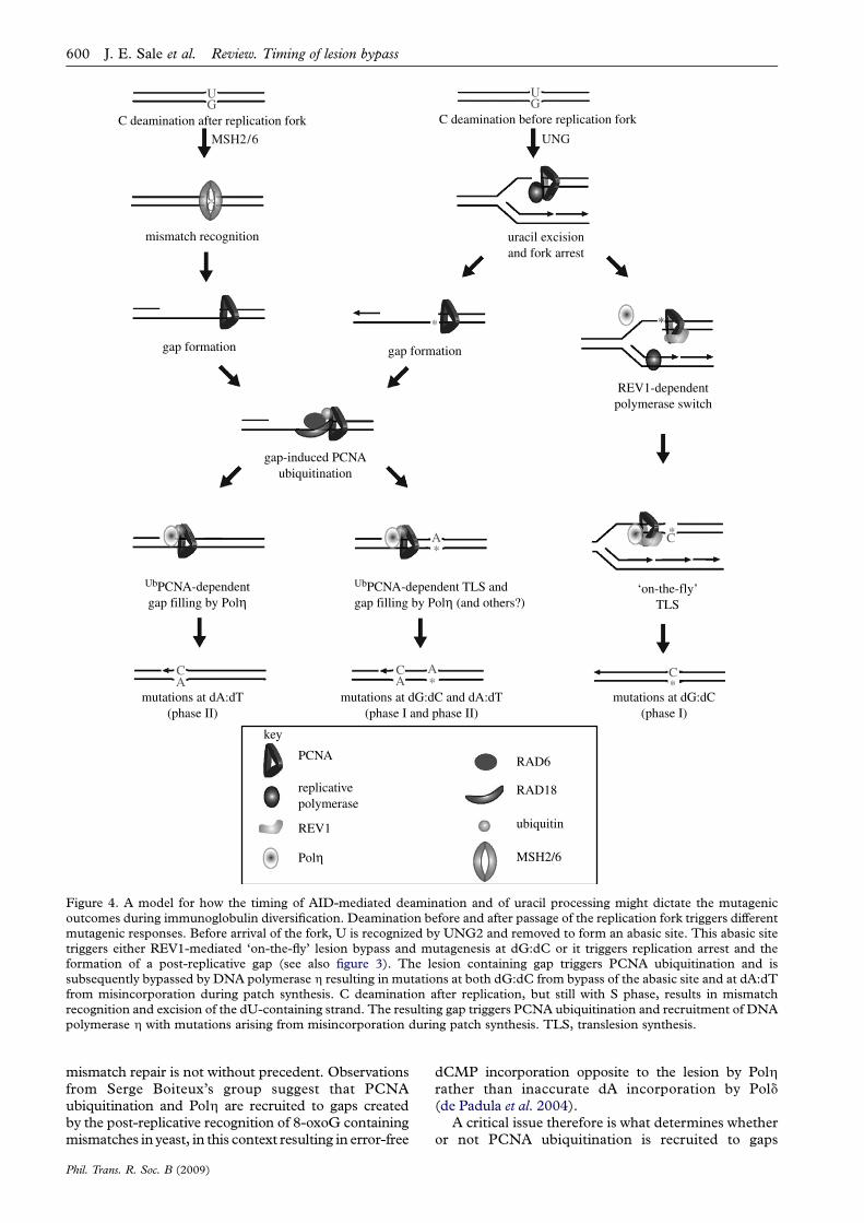
Uracil excision in the context of base excision repairis rapidly followed by elimination of the resulting abasicsite by AP endonuclease and patch repair (reviewed inKavli et al. 2007). However, for mutagenesis, the abasicsite must be replicated before it is acted on by APendonuclease. This is most likely to be achieved whenU is encountered by a replication fork and is removedby UNG2 physically recruited to PCNA (Otterlei et al.1999; Warbrick 2000). The resulting abasic site poses ablock to the replicative polymerases leaving thereplication machinery with two choices, to directlybypass the lesion, maintaining fork progression, or toarrest leaving a gap and deferring bypass of the lesionuntil later (figure 4).
By analogy with bypass of UV damage in DT40,bypass of the abasic site at the fork should requireREV1, while bypass at a post-replicative gap shouldrequire ubiquitination of PCNA (figure 4). It is notclear what would determine the division of labourbetween these two mechanisms, but an obviouspossibility is whether the deamination has occurredon the leading or lagging strand. Owing to theinherently discontinuous nature of lagging strandsynthesis, gap formation may be favoured on the
Phil. Trans. R. Soc. B (2009)
lagging strand, while a mechanism to maintain forkprogression would be favoured on the leading strand.Current evidence suggests that replication in theimmunoglobulin loci of mature B cells proceeds fromdownstream of the C region, back towards the V region(Zhou et al. 2002; Norio et al. 2005). Thus, the(coding) non-transcribed strand of the V gene wouldalso be the leading strand. Our model suggests that theuse of REV1 for abasic site bypass might be favoured onthis strand, a notion consistent with the observedstrand bias of dC/dG mutagenesis seen in rev1K/K
mice (Jansen et al. 2006). The analysis of hypermuta-tion in rev1K/K/pcnaK164R doubly mutant micewill certainly be illuminating for testing the extent towhich these two mechanisms compensate for eachother and for uncovering other modes of translesionsynthesis control.
The strong dependence of mutagenesis at dA:dT onPCNA ubiquitination places it as a post-replicativeevent in our model. Although it has recently beenproposed that dA:dT mutagenesis takes place in G1(Franklin & Blanden 2008), this is not consistent withthe observations that show a requirement for replica-tion forks in order for PCNA ubiquitination to bedetectable in both budding yeast and human cells(Kannouche et al. 2004; Davies et al. 2008). Further-more, we propose that the two pathways that result indA:dT mutations (dU excision and recognition ofdU:dG by MSH2/6; Rada et al. 2004) converge on asingle-strand gap formed or remaining after bulkreplication has been completed (figure 4). This modelsuggests that a key difference between the gaps createdin each pathway is whether or not they contain anabasic site and, therefore, whether or not theypotentially give rise to linked mutations at bothdG:dC and dA:dT. However, it is debateable whethersuch linkage would be convincingly detectable as theproportion of mutations generated by this pathway isprobably too small (Rada et al. 2004) and the range ofgap lengths, and hence the range over which linkagemight occur, is potentially quite large. Originalexperimental estimates for post-replicative gaps formedafter UV irradiation suggested a length of approxi-mately 800 bp (Lehmann 1972). However, the limit ofthe hypermutation domain in the immunoglobulin locisuggests rather shorter distances of the order of 30–100nucleotides (Di Noia & Neuberger 2007).
Although dU excision can trigger mutation at dA:dTpairs, most of these mutations occur independently ofUNG, instead relying on recognition of dU:dG mis-matches by the MSH2/MSH6 heterodimer (Rada et al.2004). This involvement of mismatch repair raises aparadox. Mismatch repair usually functions to eliminatebase misincorporations during replication and is there-fore antimutagenic. However, in the context of AID-induced dU:dG mismatches, recognition by MSH2/6 isclearly mutagenic. Why this is the case remains unclearbut is likely to be related to the recruitment of PCNAubiquitination. The involvement of EXO1 in thegeneration of mutations at dA:dT suggests that mis-match recognition is likely to result in the exonucleolyticdegradation of one strand to create a gap. Why mightgaps created by recognition of dU:dG trigger PCNAubiquitination? Recruitment of PCNA ubiquitination by
mutations at dG:dC(phase I)
mutations at dG:dC and dA:dT(phase I and phase II)
mutations at dA:dT(phase II)
*
*
mismatch recognition
gap-induced PCNAubiquitination
UbPCNA-dependentgap filling by Polη
uracil excisionand fork arrest
‘on-the-fly’TLS
UbPCNA-dependent TLS and gap filling by Polη (and others?)
GU
GU
*
AC
*A
AC
*C
*
MSH2/6 UNG
*
C deamination before replication forkC deamination after replication fork
GU
gap formationgap formation
PCNA
replicativepolymerase
REV1
RAD6
RAD18
ubiquitin
MSH2/6Polη
REV1-dependentpolymerase switch
key
CA
Figure 4. A model for how the timing of AID-mediated deamination and of uracil processing might dictate the mutagenicoutcomes during immunoglobulin diversification. Deamination before and after passage of the replication fork triggers differentmutagenic responses. Before arrival of the fork, U is recognized by UNG2 and removed to form an abasic site. This abasic sitetriggers either REV1-mediated ‘on-the-fly’ lesion bypass and mutagenesis at dG:dC or it triggers replication arrest and theformation of a post-replicative gap (see also figure 3). The lesion containing gap triggers PCNA ubiquitination and issubsequently bypassed by DNA polymerase h resulting in mutations at both dG:dC from bypass of the abasic site and at dA:dTfrom misincorporation during patch synthesis. C deamination after replication, but still with S phase, results in mismatchrecognition and excision of the dU-containing strand. The resulting gap triggers PCNA ubiquitination and recruitment of DNApolymerase h with mutations arising from misincorporation during patch synthesis. TLS, translesion synthesis.
600 J. E. Sale et al. Review. Timing of lesion bypass
mismatch repair is not without precedent. Observationsfrom Serge Boiteux’s group suggest that PCNAubiquitination and Polh are recruited to gaps createdby the post-replicative recognition of 8-oxoG containingmismatches in yeast, in this context resulting in error-free
Phil. Trans. R. Soc. B (2009)
dCMP incorporation opposite to the lesion by Polhrather than inaccurate dA incorporation by Pold(de Padula et al. 2004).
A critical issue therefore is what determines whetheror not PCNA ubiquitination is recruited to gaps
Review. Timing of lesion bypass J. E. Sale et al. 601
formed following mismatch recognition. One possi-bility again relates to timing, this time of mismatchrecognition. Canonical mismatch repair is likely to actalmost immediately a base is misincorporated at the forkand therefore not result in a persistent gap. Thus, ifdU:dG formation and its recognition happen afterreplication, rather than at the fork, the resulting gapmay then be treated in much the same way as a gapcreated by a replication fork stall with downstreamrestart, with both resulting in PCNA ubiquitination(figure 4). Alternatively, there may be mechanisms thatdirectly couple mismatch repair with PCNA ubiquitina-tion and it is of note here that both MSH2 and MSH6were identified in a macromolecular complex containingRAD18 and Polh (Yuasa et al. 2006).
The use of PCNA ubiquitination to flag persistentunreplicated gaps is an important strategy employed bycells to ensure the bypass of all lesions and thegeneration of a complete copy of the genome. In thecontext of immunoglobulin hypermutation, thisstrategy, normally reserved for difficult lesion-containing gaps, is subverted, via mismatch excision,by the unusual and high level of post-replicativedamage created by AID.
We would like to thank Cristina Rada and Michael Neubergerfor many stimulating discussions. Work in the laboratory issupported by the Medical Research Council and theAssociation for International Cancer Research.
REFERENCESArakawa, H., Hauschild, J. & Buerstedde, J. M. 2002
Requirement of the activation-induced deaminase (AID)gene for immunoglobulin gene conversion. Science 295,1301–1306. (doi:10.1126/science.1067308)
Arakawa, H., Saribasak, H. & Buerstedde, J. M. 2004Activation-induced cytidine deaminase initiates immuno-globulin gene conversion and hypermutation by acommon intermediate. PLoS Biol. 2, E179. (doi:10.1371/journal.pbio.0020179)
Arakawa, H., Moldovan, G. L., Saribasak, H., Saribasak,N. N., Jentsch, S. & Buerstedde, J. M. 2006 A role forPCNA ubiquitination in immunoglobulin hypermutation.PLoS Biol. 4, e366. (doi:10.1371/journal.pbio.0040366)
Bailly, V., Lauder, S., Prakash, S. & Prakash, L. 1997 YeastDNA repair proteins Rad6 and Rad18 form a heterodimerthat has ubiquitin conjugating, DNA binding, and ATPhydrolytic activities. J. Biol. Chem. 272, 23 360–23 365.(doi:10.1074/jbc.272.37.23360)
Bardwell, P. D., Woo, C. J., Wei, K., Li, Z., Martin, A., Sack,S. Z., Parris, T., Edelmann, W. & Scharff, M. D. 2004Altered somatic hypermutation and reduced class-switchrecombination in exonuclease 1-mutant mice. Nat.Immunol. 5, 224–229. (doi:10.1038/ni1031)
Betz, A. G., Rada, C., Pannell, R., Milstein, C. & Neuberger,M. S. 1993 Passenger transgenes reveal intrinsic speci-ficity of the antibody hypermutation mechanism: cluster-ing, polarity, and specific hot spots. Proc. Natl Acad. Sci.USA 90, 2385–2388. (doi:10.1073/pnas.90.6.2385)
Bienko, M. et al. 2005 Ubiquitin-binding domains in Y-familypolymerases regulate translesion synthesis. Science 310,1821–1824. (doi:10.1126/science.1120615)
Blastyak, A., Pinter, L., Unk, I., Prakash, L., Prakash, S. &Haracska, L. 2007 Yeast Rad5 protein required forpostreplication repair has a DNA helicase activity specificfor replication fork regression. Mol. Cell 28, 167–175.(doi:10.1016/j.molcel.2007.07.030)
Phil. Trans. R. Soc. B (2009)
Bransteitter, R., Pham, P., Scharff, M. D. & Goodman, M. F.2003 Activation-induced cytidine deaminase deaminatesdeoxycytidine on single-stranded DNA but requires theaction of RNase. Proc. Natl Acad. Sci. USA 100,4102–4107. (doi:10.1073/pnas.0730835100)
Bridges, B. A. 2005 Error-prone DNA repair and translesionDNA synthesis. II: the inducible SOS hypothesis. DNARepair (Amsterdam) 4, 725–726. (doi:10.1016/j.dnarep.2004.12.009) see also p 739.
Buerstedde, J. M. & Takeda, S. 1991 Increased ratio oftargeted to random integration after transfection ofchicken B cell lines. Cell 67, 179–188. (doi:10.1016/0092-8674(91)90581-I)
Chaudhuri, J., Tian, M., Khuong, C., Chua, K., Pinaud, E.& Alt, F. W. 2003 Transcription-targeted DNA deamina-tion by the AID antibody diversification enzyme. Nature422, 726–730. (doi:10.1038/nature01574)
Davies, A. A., Huttner, D., Daigaku, Y., Chen, S. & Ulrich,H. D. 2008 Activation of ubiquitin-dependent DNAdamage bypass is mediated by replication protein A. Mol.Cell 29, 625–636. (doi:10.1016/j.molcel.2007.12.016)
de Padula, M., Slezak, G., Auffret van Der Kemp, P. &Boiteux, S. 2004 The post-replication repair RAD18 andRAD6 genes are involved in the prevention of spontaneousmutations caused by 7,8-dihydro-8-oxoguanine in Sac-charomyces cerevisiae. Nucleic Acids Res. 32, 5003–5010.(doi:10.1093/nar/gkh831)
Delbos, F., De Smet, A., Faili, A., Aoufouchi, S., Weill, J. C. &Reynaud, C. A. 2005 Contribution of DNA polymerase hto immunoglobulin gene hypermutation in the mouse.J. Exp.Med. 201, 1191–1196. (doi:10.1084/jem.20050292)
Delbos, F., Aoufouchi, S., Faili, A., Weill, J. C. & Reynaud,C. A. 2007 DNA polymerase h is the sole contributor ofA/T modifications during immunoglobulin gene hyper-mutation in the mouse. J. Exp. Med. 204, 17–23. (doi:10.1084/jem.20062131)
Denepoux, S., Razanajaona, D., Blanchard, D., Meffre, G.,Capra, J. D., Banchereau, J. & Lebecque, S. 1997Induction of somatic mutation in a human B cell linein vitro. Immunity 6, 35–46. (doi:10.1016/S1074-7613(00)80240-X)
Dickerson, S. K., Market, E., Besmer, E. & Papavasiliou,F. N. 2003 AID mediates hypermutation by deaminatingsingle stranded DNA. J. Exp. Med. 197, 1291–1296.(doi:10.1084/jem.20030481)
Di Noia, J. & Neuberger, M. S. 2002 Altering the pathway ofimmunoglobulin hypermutation by inhibiting uracil-DNAglycosylase. Nature 419, 43–48. (doi:10.1038/nature00981)
Di Noia, J. M. & Neuberger, M. S. 2007 Molecularmechanisms of antibody somatic hypermutation. Annu.Rev. Biochem. 76, 1–22. (doi:10.1146/annurev.biochem.76.061705.090740)
Edmunds, C. E., Simpson, L. J. & Sale, J. E. 2008 PCNAubiquitination and REV1 define temporally distinctmechanisms for controlling translesion synthesis in theavian cell line DT40. Mol. Cell 30, 519–529. (doi:10.1016/j.molcel.2008.03.024)
Ehrenstein, M. R., Rada, C., Jones, A. M., Milstein, C. &Neuberger, M. S. 2001 Switch junction sequences inPMS2-deficient mice reveal a microhomology-mediatedmechanism of Ig class switch recombination. Proc. NatlAcad. Sci. USA 98, 14 553–14 558. (doi:10.1073/pnas.241525998)
Franklin, A. & Blanden, R. V. 2008 The strand bias paradoxof somatic hypermutation at immunoglobulin loci. TrendsImmunol. 29, 167–172. (doi:10.1016/j.it.2008.01.008)
Frey, S., Bertocci, B., Delbos, F., Quint, L., Weill, J. C. &Reynaud, C. A. 1998 Mismatch repair deficiencyinterferes with the accumulation of mutations in
602 J. E. Sale et al. Review. Timing of lesion bypass
chronically stimulated B cells and not with the hypermuta-
tion process. Immunity 9, 127–134. (doi:10.1016/S1074-
7613(00)80594-4)
Guo, C., Fischhaber, P. L., Luk-Paszyc, M. J., Masuda, Y.,
Zhou, J., Kamiya, K., Kisker, C. & Friedberg, E. C. 2003
Mouse Rev1 protein interacts with multiple DNA
polymerases involved in translesion DNA synthesis.
EMBO J. 22, 6621–6630. (doi:10.1093/emboj/cdg626)
Haracska, L., Torres-Ramos, C. A., Johnson, R. E., Prakash,
S. & Prakash, L. 2004 Opposing effects of ubiquitin
conjugation and SUMO modification of PCNA on
replicational bypass of DNA lesions in Saccharomyces
cerevisiae. Mol. Cell Biol. 24, 4267–4274. (doi:10.1128/
MCB.24.10.4267-4274.2004)
Harris, R. S., Croom-Carter, D. S., Rickinson, A. B. &
Neuberger, M. S. 2001 Epstein–Barr virus and the
somatic hypermutation of immunoglobulin genes in
Burkitt’s lymphoma cells. J. Virol. 75, 10 488–10 492.
(doi:10.1128/JVI.75.21.10488-10492.2001)
Harris, R. S., Sale, J. E., Petersen-Mahrt, S. K. & Neuberger,
M. S. 2002 AID is essential for immunoglobulin V gene
conversion in a cultured B cell line. Curr. Biol. 12,
435–438. (doi:10.1016/S0960-9822(02)00717-0)
Hoege, C., Pfander, B., Moldovan, G. L., Pyrowolakis, G. &
Jentsch, S. 2002 RAD6-dependent DNA repair is linked
to modification of PCNA by ubiquitin and SUMO. Nature
419, 135–141. (doi:10.1038/nature00991)
Jansen, J. G., Langerak, P., Tsaalbi-Shtylik, A., van den Berk,
P., Jacobs, H. & de Wind, N. 2006 Strand-biased defect in
C/G transversions in hypermutating immunoglobulin
genes in Rev1-deficient mice. J. Exp. Med. 203,
319–323. (doi:10.1084/jem.20052227)
Jiricny, J. 2006 The multifaceted mismatch-repair system. Nat.Rev. Mol. Cell Biol. 7, 335–346. (doi:10.1038/nrm1907)
Johnson, R. E., Prakash, S. & Prakash, L. 1999 Requirement
of DNA polymerase activity of yeast Rad30 protein for its
biological function. J. Biol. Chem. 274, 15 975–15 977.
(doi:10.1074/jbc.274.23.15975)
Kannouche, P. L., Wing, J. & Lehmann, A. R. 2004 Interaction
of human DNA polymerase h with monoubiquitinated
PCNA: a possible mechanism for the polymerase switch in
response to DNA damage. Mol. Cell 14, 491–500. (doi:10.
1016/S1097-2765(04)00259-X)
Kavli, B., Otterlei, M., Slupphaug, G. & Krokan, H. E. 2007
Uracil in DNA-general mutagen, but normal intermediate
in acquired immunity. DNA Repair (Amsterdam) 6,
505–516. (doi:10.1016/j.dnarep.2006.10.014)
Kenter, A. L. 2005 Class switch recombination: an emerging
mechanism. Curr. Top. Microbiol. Immunol. 290, 171–199.
(doi:10.1007/3-540-26363-2-8)
Kim, N., Bozek, G., Lo, J. C. & Storb, U. 1999 Different
mismatch repair deficiencies all have the same effects on
somatic hypermutation: intact primary mechanism
accompanied by secondary modifications. J. Exp. Med.
190, 21–30. (doi:10.1084/jem.190.1.21)
Langerak, P., Nygren, A. O., Krijger, P. H., van den Berk,
P. C. & Jacobs, H. 2007 A/T mutagenesis in hypermutated
immunoglobulin genes strongly depends on PCNAK164
modification. J. Exp. Med. 204, 1989–1998. (doi:10.1084/
jem.20070902)
Lawrence, C. W. & Christensen, R. 1976 UV mutagenesis in
radiation-sensitive strains of yeast. Genetics 82, 207–232.
Lehmann, A. R. 1972 Postreplication repair of DNA in
ultraviolet-irradiated mammalian cells. J. Mol. Biol. 66,
319–337. (doi:10.1016/0022-2836(72)90418-4)
Lehmann, A. R. & Fuchs, R. P. 2006 Gaps and forks in DNA
replication: rediscovering old models. DNA Repair
(Amsterdam) 5, 1495–1498. (doi:10.1016/j.dnarep.2006.
07.002)
Phil. Trans. R. Soc. B (2009)
Lemontt, J. F. 1971 Mutants of yeast defective in mutation
induced by ultraviolet light. Genetics 68, 21–33.
Lopes, M., Foiani, M. & Sogo, J. M. 2006 Multiple
mechanisms control chromosome integrity after replica-
tion fork uncoupling and restart at irreparable UV lesions.
Mol. Cell 21, 15–27. (doi:10.1016/j.molcel.2005.11.015)
Maizels, N. 2005 Immunoglobulin gene diversification.
Annu. Rev. Genet. 39, 23–46. (doi:10.1146/annurev.
genet.39.073003.110544)
Martomo, S. A., Yang, W. W. & Gearhart, P. J. 2004 A role for
Msh6 but not Msh3 in somatic hypermutation and class
switch recombination. J. Exp. Med. 200, 61–68. (doi:10.
1084/jem.20040691)
Masuda, K. et al. 2005 DNA polymerase q contributes to the
generation of C/G mutations during somatic hypermuta-
tion of Ig genes. Proc. Natl Acad. Sci. USA 102,
13 986–13 991. (doi:10.1073/pnas.0505636102)
McDonald, J. P., Levine, A. S. & Woodgate, R. 1997 The
Saccharomyces cerevisiae RAD30 gene, a homologue of
Escherichia coli dinB and umuC, is DNA damage inducible
and functions in a novel error-free postreplication repair
mechanism. Genetics 147, 1557–1568.
Murakumo, Y., Ogura, Y., Ishii, H., Numata, S., Ichihara,
M., Croce, C. M., Fishel, R. & Takahashi, M. 2001
Interactions in the error-prone postreplication repair
proteins hREV1, hREV3, and hREV7. J. Biol. Chem.
276, 35 644–35 651. (doi:10.1074/jbc.M102051200)
Muramatsu, M., Kinoshita, K., Fagarasan, S., Yamada, S.,
Shinkai, Y. & Honjo, T. 2000 Class switch recombination
and hypermutation require activation-induced cytidine
deaminase (AID), a potential RNA editing enzyme. Cell
102, 553–563. (doi:10.1016/S0092-8674(00)00078-7)
Nelson, J. R., Lawrence, C. W. & Hinkle, D. C. 1996aDeoxycytidyl transferase activity of yeast REV1 protein.
Nature 382, 729–731. (doi:10.1038/382729a0)
Nelson, J. R., Lawrence, C. W. & Hinkle, D. C. 1996b
Thymine-thymine dimer bypass by yeast DNA polymerase
z. Science 272, 1646–1649. (doi:10.1126/science.272.
5268.1646)
Nelson, J. R., Gibbs, P. E., Nowicka, A. M., Hinkle, D. C. &
Lawrence, C. W. 2000 Evidence for a second function for
Saccharomyces cerevisiae Rev1p. Mol. Microbiol. 37,
549–554. (doi:10.1046/j.1365-2958.2000.01997.x)
Neuberger, M. S., Harris, R. S., Di Noia, J. & Petersen-
Mahrt, S. K. 2003 Immunity through DNA deamination.
Trends Biochem. Sci. 28, 305–312. (doi:10.1016/S0968-
0004(03)00111-7)
Norio, P., Kosiyatrakul, S., Yang, Q., Guan, Z., Brown,
N. M., Thomas, S., Riblet, R. & Schildkraut, C. L. 2005
Progressive activation of DNA replication initiation in
large domains of the immunoglobulin heavy chain locus
during B cell development. Mol. Cell 20, 575–587. (doi:10.
1016/j.molcel.2005.10.029)
Ohashi, E., Murakumo, Y., Kanjo, N., Akagi, J., Masutani,
C., Hanaoka, F. & Ohmori, H. 2004 Interaction of hRev1
with three human Y-family DNA polymerases. Genes Cells
9, 523–531. (doi:10.1111/j.1356-9597.2004.00747.x)
Okada, T., Sonoda, E., Yamashita, Y. M., Koyoshi, S., Tateishi,
S., Yamaizumi, M., Takata, M., Ogawa, O. & Takeda, S.
2002 Involvement of vertebrate polk in Rad18-independent
postreplication repair of UV damage. J. Biol. Chem. 277,
48 690–48 695. (doi:10.1074/jbc.M207957200)
Otterlei, M. et al. 1999 Post-replicative base excision repair in
replication foci. EMBO J. 18, 3834–3844. (doi:10.1093/
emboj/18.13.3834)
Pavlov, Y. I., Rogozin, I. B., Galkin, A. P., Aksenova, A. Y.,
Hanaoka, F., Rada, C. & Kunkel, T. A. 2002 Correlation
of somatic hypermutation specificity and A-T base pair
substitution errors by DNA polymerase eta during
Review. Timing of lesion bypass J. E. Sale et al. 603
copying of a mouse immunoglobulin kappa light chaintransgene. Proc. Natl Acad. Sci. USA 99, 9954–9959.(doi:10.1073/pnas.152126799)
Phung, Q. H., Winter, D. B., Alrefai, R. & Gearhart, P. J.1999 Hypermutation in Ig V genes from mice deficient inthe MLH1 mismatch repair protein. J. Immunol. 162,3121–3124.
Prakash, L. 1981 Characterization of postreplication repair inSaccharomyces cerevisiae and effects of rad6, rad18, rev3and rad52 mutations. Mol. Gen. Genet. 184, 471–478.(doi:10.1007/BF00352525)
Prakash, S., Johnson, R. E. & Prakash, L. 2005 Eukaryotictranslesion synthesis DNA polymerases: specificity ofstructure and function. Annu. Rev. Biochem. 74, 317–353.(doi:10.1146/annurev.biochem.74.082803.133250)
Rada, C., Ehrenstein, M. R., Neuberger, M. S. & Milstein, C.1998 Hot spot focusing of somatic hypermutation inMSH2-deficient mice suggests two stages of mutationaltargeting. Immunity 9, 135–141. (doi:10.1016/S1074-7613(00)80595-6)
Rada, C., Di Noia, J. M. & Neuberger, M. S. 2004 Mismatchrecognition and uracil excision provide complementarypaths to both Ig switching and the A/T-focused phase ofsomatic mutation. Mol. Cell 16, 163–171. (doi:10.1016/j.molcel.2004.10.011)
Radman, M. 1970 SOS replication: a distinct replicationmechanism which is induced by DNA damaging treat-ments? DNA Repair 4, 732–738.
Revy, P. et al. 2000 Activation-induced cytidine deaminase(AID) deficiency causes the autosomal recessive form ofthe Hyper-IgM syndrome (HIGM2). Cell 102, 565–575.(doi:10.1016/S0092-8674(00)00079-9)
Ross, A. L. & Sale, J. E. 2006 The catalytic activity of REV1 isemployed during immunoglobulin gene diversification inDT40. Mol. Immunol. 43, 1587–1594. (doi:10.1016/j.molimm.2005.09.017)
Ross, A. L., Simpson, L. J. & Sale, J. E. 2005 Vertebrate DNAdamage tolerance requires the C-terminus but not BRCTor transferase domains of REV1. Nucleic Acids Res. 33,1280–1289. (doi:10.1093/nar/gki279)
Rupp, W. D. & Howard-Flanders, P. 1968 Discontinuities inthe DNA synthesized in an excision-defective strain ofEscherichia coli following ultraviolet irradiation. J. Mol.Biol. 31, 291–304. (doi:10.1016/0022-2836(68)90445-2)
Rupp, W. D., Wilde III, C. E., Reno, D. L. & Howard-Flanders, P. 1971 Exchanges between DNA strands inultraviolet-irradiated Escherichia coli. J. Mol. Biol. 61,25–44. (doi:10.1016/0022-2836(71)90204-X)
Sale, J. E. 2004 Immunoglobulin diversification in DT40:a model for vertebrate DNA damage tolerance. DNARepair (Amsterdam) 3, 693–702. (doi:10.1016/j.dnarep.2004.03.042)
Sale, J. E. & Neuberger, M. S. 1998 TdT-accessible breaksare scattered over the immunoglobulin V domain in aconstitutively hypermutating B cell line. Immunity 9,859–869. (doi:10.1016/S1074-7613(00)80651-2)
Sale, J. E., Calandrini, D. M., Takata, M., Takeda, S. &Neuberger, M. S. 2001 Ablation of XRCC2/3 transformsimmunoglobulin V gene conversion into somatic hyper-mutation. Nature 412, 921–926. (doi:10.1038/35091100)
Seki, M., Masutani, C., Yang, L. W., Schuffert, A., Iwai, S.,Bahar, I. & Wood, R. D. 2004 High-efficiency bypass ofDNA damage by human DNA polymerase Q. EMBO J.23, 4484–4494. (doi:10.1038/sj.emboj.7600424)
Simpson, L. J. & Sale, J. E. 2005 UBE2V2 (MMS2) is notrequired for effective immunoglobulin gene conversion orDNA damage tolerance in DT40.DNARepair (Amsterdam)4, 503–510. (doi:10.1016/j.dnarep.2004.12.002)
Simpson, L. J., Ross, A. L., Szuts, D., Alviani, C. A.,Oestergaard, V. H., Patel, K. J. & Sale, J. E. 2006 RAD18-
Phil. Trans. R. Soc. B (2009)
independent ubiquitination of proliferating-cell nuclear
antigen in the avian cell line DT40. EMBO Rep. 7,
927–932. (doi:10.1038/sj.embor.7400777)
Tateishi, S., Niwa, H., Miyazaki, J., Fujimoto, S., Inoue, H. &
Yamaizumi, M. 2003 Enhanced genomic instability and
defective postreplication repair in RAD18 knockout
mouse embryonic stem cells. Mol. Cell Biol. 23,
474–481. (doi:10.1128/MCB.23.2.474-481.2003)
Tissier, A., Kannouche, P., Reck, M. P., Lehmann, A. R.,
Fuchs, R. P. & Cordonnier, A. 2004 Co-localization in
replication foci and interaction of human Y-family
members, DNA polymerase poleta and REVl protein.
DNA Repair (Amsterdam) 3, 1503–1514. (doi:10.1016/
j.dnarep.2004.06.015)
Warbrick, E. 2000 The puzzle of PCNA’s many partners.
Bioessays 22, 997–1006. (doi:10.1002/1521-1878(200
011)22:11!997::AID-BIES6O3.0.CO;2-)
Waters, L. S. & Walker, G. C. 2006 The critical mutagenic
translesion DNA polymerase Rev1 is highly expressed
during G(2)/M phase rather than S phase. Proc. Natl
Acad. Sci. USA 103, 8971–8976. (doi:10.1073/pnas.0510
167103)
Wiesendanger, M., Kneitz, B., Edelmann, W. & Scharff,
M. D. 2000 Somatic hypermutation in MutS homologue
(MSH)3-, MSH6-, and MSH3/MSH6- deficient mice
reveals a role for the MSH2-MSH6 heterodimer in
modulating the base substitution pattern. J. Exp. Med.
191, 579–584. (doi:10.1084/jem.191.3.579)
Witkin, E. M. 1967a Mutation-proof and mutation-prone
modes of survival in derivatives of Escherichia coli B
differing in sensitivity to ultraviolet light. Brookhaven
Symp. Biol. 20, 17–55.
Witkin, E. M. 1967b The radiation sensitivity of Escherichia
coli B: a hypothesis relating filament formation and
prophage induction. Proc. Natl Acad. Sci. USA 57,
1275–1279. (doi:10.1073/pnas.57.5.1275)
Yamashita, Y. M., Okada, T., Matsusaka, T., Sonoda, E., Zhao,
G. Y., Araki, K., Tateishi, S., Yamaizumi, M. & Takeda, S.
2002 RAD18 and RAD54 cooperatively contribute to
maintenance of genomic stability in vertebrate cells.
EMBO J. 21, 5558–5566. (doi:10.1093/emboj/cdf534)
Yuasa, M. S., Masutani, C., Hirano, A., Cohn, M. A.,
Yamaizumi, M., Nakatani, Y. & Hanaoka, F. 2006
A human DNA polymerase h complex containing
Rad18, Rad6 and Rev1; proteomic analysis and targeting
of the complex to the chromatin-bound fraction of cells
undergoing replication fork arrest. Genes Cells 11,
731–744. (doi:10.1111/j.1365-2443.2006.00974.x)
Zan, H., Shima, N., Xu, Z., Al-Qahtani III, A., Evinger, A. J.,
Zhong, Y., Schimenti, J. C. & Casali, P. 2005 The
translesion DNA polymerase q plays a dominant role in
immunoglobulin gene somatic hypermutation. EMBO J.
24, 3757–3769. (doi:10.1038/sj.emboj.7600833)
Zeng, X., Winter, D. B., Kasmer, C., Kraemer, K. H.,
Lehmann, A. R. & Gearhart, P. J. 2001 DNA polymerase
h is an A-T mutator in somatic hypermutation of
immunoglobulin variable genes. Nat. Immunol. 2,
537–541. (doi:10.1038/88740)
Zhang, Y., Wu, X., Rechkoblit, O., Geacintov, N. E., Taylor,
J. S. & Wang, Z. 2002 Response of human REV1 to
different DNA damage: preferential dCMP insertion
opposite the lesion. Nucleic Acids Res. 30, 1630–1638.
(doi:10.1093/nar/30.7.1630)
Zhou, J., Ermakova, O. V., Riblet, R., Birshtein, B. K. &
Schildkraut, C. L. 2002 Replication and subnuclear
location dynamics of the immunoglobulin heavy-chain
locus in B-lineage cells. Mol. Cell Biol. 22, 4876–4889.
(doi:10.1128/MCB.22.13.4876-4889.2002)
Related Documents