RESEARCH ARTICLE Time course and specificity of sensory-motor alpha modulation during the observation of hand motor acts and gestures: a high density EEG study Alena Streltsova • Cristina Berchio • Vittorio Gallese • Maria Alessandra Umilta’ Received: 9 February 2010 / Accepted: 14 July 2010 / Published online: 3 August 2010 Ó The Author(s) 2010. This article is published with open access at Springerlink.com Abstract The main aim of the present study was to explore, by means of high-density EEG, the intensity and the temporal pattern of event-related sensory-motor alpha desynchronization (ERD) during the observation of dif- ferent types of hand motor acts and gestures. In particular, we aimed to investigate whether the sensory-motor ERD would show a specific modulation during the observation of hand behaviors differing for goal-relatedness (hand grasping of an object and meaningless hand movements) and social relevance (communicative hand gestures and grasping within a social context). Time course analysis of alpha suppression showed that all types of hand behaviors were effective in triggering sensory-motor alpha ERD, but to a different degree depending on the category of observed hand motor acts and gestures. Meaningless gestures and hand grasping were the most effective stimuli, resulting in the strongest ERD. The observation of social hand behav- iors such as social grasping and communicative gestures, triggered a more dynamic time course of ERD compared to that driven by the observation of simple grasping and meaningless gestures. These findings indicate that the observation of hand motor acts and gestures evoke the activation of a motor resonance mechanism that differs on the basis of the goal-relatedness and the social relevance of the observed hand behavior. Keywords EEG Á Sensory-motor alpha Á Mirror mechanism Á Motor resonance Á Social context Á Time course Introduction Gastaut and co-workers first described the desynchroniza- tion of an EEG rhythm within the alpha band (8–13 Hz), recorded from central electrodes, occurring not only during the execution of active movements, but also during their observation (Cohen-Seat et al. 1954; Gastaut and Bert 1954). This result was confirmed many years later by Cochin et al. (1998, 1999), Altschuler et al. (1997, 2000) using EEG recordings, and by Hari et al. (1998) using magnetoencephalography (MEG). In particular, Hari and co-workers demonstrated that the desynchronization during action observation includes rhythms originating from the cortex inside the central sulcus (Hari and Salmelin 1997; Salmelin and Hari 1994, for a review, see Pineda 2005). It has been proposed that the suppression of the sensory- motor alpha rhythm, also termed as event-related desynchronization (ERD), during action execution and observation, could be due to a neurophysiological mecha- nism of motor resonance, the mirror mechanism, likely due to the activation of neurons endowed with properties sim- ilar to those of mirror neurons, discovered in the premotor and posterior parietal cortices of macaque monkeys. Mirror neurons discharge both during the execution of goal-related motor acts and during their observation when executed by others (Di Pellegrino et al. 1992; Gallese et al. 1996; Rizzolatti et al. 1996). A growing body of research showing that a number of human cortical areas are activated by both action execution and observation supports the existence of a mirror A. Streltsova Á C. Berchio Á V. Gallese Á M. A. Umilta’ (&) Department of Neuroscience, Section of Physiology, University of Parma, Via Volturno 39, 43100 Parma, Italy e-mail: [email protected] V. Gallese Á M. A. Umilta’ IIT (Italian Institute of Technology) Brain Center for Social and Motor Cognition, Parma, Italy 123 Exp Brain Res (2010) 205:363–373 DOI 10.1007/s00221-010-2371-7

Welcome message from author
This document is posted to help you gain knowledge. Please leave a comment to let me know what you think about it! Share it to your friends and learn new things together.
Transcript
RESEARCH ARTICLE
Time course and specificity of sensory-motor alpha modulationduring the observation of hand motor acts and gestures: a highdensity EEG study
Alena Streltsova • Cristina Berchio •
Vittorio Gallese • Maria Alessandra Umilta’
Received: 9 February 2010 / Accepted: 14 July 2010 / Published online: 3 August 2010
� The Author(s) 2010. This article is published with open access at Springerlink.com
Abstract The main aim of the present study was to
explore, by means of high-density EEG, the intensity and
the temporal pattern of event-related sensory-motor alpha
desynchronization (ERD) during the observation of dif-
ferent types of hand motor acts and gestures. In particular,
we aimed to investigate whether the sensory-motor ERD
would show a specific modulation during the observation
of hand behaviors differing for goal-relatedness (hand
grasping of an object and meaningless hand movements)
and social relevance (communicative hand gestures and
grasping within a social context). Time course analysis of
alpha suppression showed that all types of hand behaviors
were effective in triggering sensory-motor alpha ERD, but
to a different degree depending on the category of observed
hand motor acts and gestures. Meaningless gestures and
hand grasping were the most effective stimuli, resulting in
the strongest ERD. The observation of social hand behav-
iors such as social grasping and communicative gestures,
triggered a more dynamic time course of ERD compared to
that driven by the observation of simple grasping and
meaningless gestures. These findings indicate that the
observation of hand motor acts and gestures evoke the
activation of a motor resonance mechanism that differs on
the basis of the goal-relatedness and the social relevance of
the observed hand behavior.
Keywords EEG � Sensory-motor alpha �Mirror mechanism � Motor resonance �Social context � Time course
Introduction
Gastaut and co-workers first described the desynchroniza-
tion of an EEG rhythm within the alpha band (8–13 Hz),
recorded from central electrodes, occurring not only during
the execution of active movements, but also during their
observation (Cohen-Seat et al. 1954; Gastaut and Bert
1954). This result was confirmed many years later by
Cochin et al. (1998, 1999), Altschuler et al. (1997, 2000)
using EEG recordings, and by Hari et al. (1998) using
magnetoencephalography (MEG). In particular, Hari and
co-workers demonstrated that the desynchronization during
action observation includes rhythms originating from the
cortex inside the central sulcus (Hari and Salmelin 1997;
Salmelin and Hari 1994, for a review, see Pineda 2005).
It has been proposed that the suppression of the sensory-
motor alpha rhythm, also termed as event-related
desynchronization (ERD), during action execution and
observation, could be due to a neurophysiological mecha-
nism of motor resonance, the mirror mechanism, likely due
to the activation of neurons endowed with properties sim-
ilar to those of mirror neurons, discovered in the premotor
and posterior parietal cortices of macaque monkeys. Mirror
neurons discharge both during the execution of goal-related
motor acts and during their observation when executed by
others (Di Pellegrino et al. 1992; Gallese et al. 1996;
Rizzolatti et al. 1996).
A growing body of research showing that a number of
human cortical areas are activated by both action execution
and observation supports the existence of a mirror
A. Streltsova � C. Berchio � V. Gallese � M. A. Umilta’ (&)
Department of Neuroscience, Section of Physiology,
University of Parma, Via Volturno 39, 43100 Parma, Italy
e-mail: [email protected]
V. Gallese � M. A. Umilta’
IIT (Italian Institute of Technology) Brain Center
for Social and Motor Cognition, Parma, Italy
123
Exp Brain Res (2010) 205:363–373
DOI 10.1007/s00221-010-2371-7
mechanism in humans. These cortical areas include the
lower part of precentral gyrus, the posterior part of inferior
frontal gyrus (IFG) and the rostral part of the inferior
parietal lobule (for a review, see Rizzolatti and Craighero
2004; Gallese et al. 2004; Cattaneo and Rizzolatti 2009).
Recently, evidence at the single neuron-level about the
existence of the mirror mechanism in the human brain was
provided (Mukamel et al. 2010).
Several neuroimaging studies have reported the activa-
tion of distinct cortical regions within premotor and pos-
terior parietal cortices during the observation/execution of
goal-related hand, mouth and foot actions (Buccino et al.
2001; Aziz-Zadeh et al. 2006) during the observation/
execution of meaningless hand movements (Lui et al. 2008;
Villarreal et al. 2008) during the execution of a noisy
action and listening to the corresponding sound (Gazzola
et al. 2006). Few studies have demonstrated communica-
tive face and hand actions showing an activation of cortical
regions endowed with mirror properties (Nakamura et al.
1999; Nakamura et al. 2004; Montgomery et al. 2007;
Villarreal et al. 2008).
Recent EEG studies of the mirror mechanism have
shown that the observation and execution of a motor act is
accompanied by an event-related alpha desynchronization
(ERD) reflected in a relative decrease in power of sensory-
motor alpha (8–13 Hz) and beta (13–30 Hz) frequency
bands (Hari 2006). More generally, ERD reflects a cortical
deactivation which depends on the task and it is usually
measured with respect to the baseline that contains a clear
peak in the frequency band of interest (Pfurtscheller and
Lopes da Silva 1999; Klimesch 1999). It has been shown
that during movement execution ERD reflects an inhibition
of the motor cortex and has a pre-movement onset.
(Pfurtscheller and Lopes da Silva 1999; Calmels et al. 2006
for review, see Pineda 2005). Several EEG studies have
shown sensory-motor alpha ERD during the observation of
different types of grasping (Muthukumaraswamy et al.
2004; Muthukumaraswamy and Johnson 2004a; Perry and
Bentin 2009), meaningless gestures (Babiloni et al. 2002)
and sequential finger movements (Calmels et al. 2006).
Recent EEG studies aiming to investigate sensory-motor
alpha suppression during the observation of more complex
stimuli such as social interaction scenes, body movements
from different perspectives and dance performance, have
shown that alpha ERD can be modulated by the degree of
social relevance (Kilner et al. 2006; Oberman et al. 2007b)
and by the motor expertise of the observer (Orgs et al.
2008; Babiloni et al. 2009). Furthermore, recent findings
demonstrate the existence of an observation/execution
matching system even in infants and young children
(Lepage and Theoret 2006; Southgate et al. 2009, 2010).
The main aim of the present study was to explore, by
means of high-density EEG, the sensitivity of sensory-
motor ERD during the observation of different types of
hand motor acts and gestures. In particular, we aimed to
investigate the specificity of sensory-motor ERD for pro-
cessing hand behaviors differing along two dimensions:
their goal-relatedness and social relevance
All participants also performed a control motor execu-
tion task. Similarly to other studies (Muthukumaraswamy
and Johnson 2004a; Southgate et al. 2009, 2010), the aim
of the motor task (key-press) was to identify the frequency
band that was functionally related to motor activation in
each individual participant.
Since the neural basis of the mirror mechanism is the
presence of a parieto-premotor neural circuit activated
during both motor execution and observation, most likely
due to the presence of neurons instantiating functional
properties similar to those of macaque monkey mirror neu-
rons (Gallese et al. 1996; Rizzolatti et al. 1996) we measured
sensory-motor ERD in the observation task by considering
the same frequencies that were attenuated during the exe-
cution task. In addition, and more importantly, the temporal
course of alpha suppression was carefully investigated.
Methods
Participants
Thirteen right-handed healthy volunteers (7 males, 6
females, mean age 27 ± 7 years old) participated in the
experiment. Participants were recruited by public
announcement and were blind to the experimental goals.
All participants were right-handed as assessed by the
Edinburgh Handedness Inventory (Oldfield 1971). None of
them reported the presence of any neurological or psychi-
atric disorder and had normal or corrected to normal vision.
Before the experiment, all participants received written
experimental instructions. Informed consent was obtained
from all participants before entering the study. The study
was approved by the local Ethical Committee.
Experimental procedure
Participants were seated comfortably in front of a computer
monitor used for stimuli presentation, located at a distance
of 60 cm. To minimize participants’ movements during the
experiment, they were asked to keep their arms on a table
in front of them and to stay as motionless and relaxed as
possible. The experiment included observation and exe-
cution tasks.
Each execution trial started with a grey background with
a varying duration of 6 to 9 s randomized across trials
(baseline) followed by two fixation crosses presented for
500 ms in the middle of the screen. Participants were asked
364 Exp Brain Res (2010) 205:363–373
123
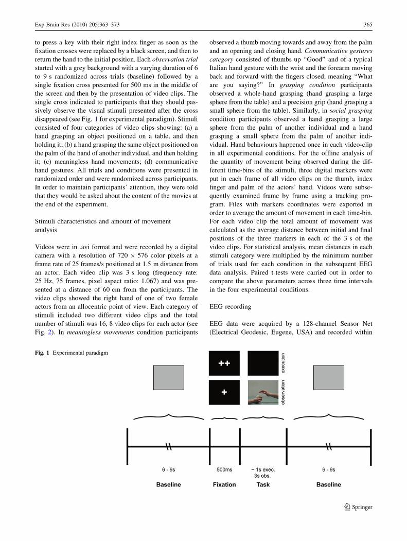
to press a key with their right index finger as soon as the
fixation crosses were replaced by a black screen, and then to
return the hand to the initial position. Each observation trial
started with a grey background with a varying duration of 6
to 9 s randomized across trials (baseline) followed by a
single fixation cross presented for 500 ms in the middle of
the screen and then by the presentation of video clips. The
single cross indicated to participants that they should pas-
sively observe the visual stimuli presented after the cross
disappeared (see Fig. 1 for experimental paradigm). Stimuli
consisted of four categories of video clips showing: (a) a
hand grasping an object positioned on a table, and then
holding it; (b) a hand grasping the same object positioned on
the palm of the hand of another individual, and then holding
it; (c) meaningless hand movements; (d) communicative
hand gestures. All trials and conditions were presented in
randomized order and were randomized across participants.
In order to maintain participants’ attention, they were told
that they would be asked about the content of the movies at
the end of the experiment.
Stimuli characteristics and amount of movement
analysis
Videos were in .avi format and were recorded by a digital
camera with a resolution of 720 9 576 color pixels at a
frame rate of 25 frames/s positioned at 1.5 m distance from
an actor. Each video clip was 3 s long (frequency rate:
25 Hz, 75 frames, pixel aspect ratio: 1.067) and was pre-
sented at a distance of 60 cm from the participants. The
video clips showed the right hand of one of two female
actors from an allocentric point of view. Each category of
stimuli included two different video clips and the total
number of stimuli was 16, 8 video clips for each actor (see
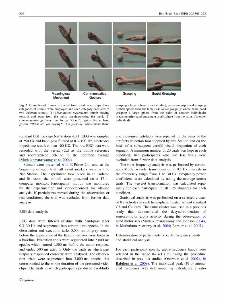
Fig. 2). In meaningless movements condition participants
observed a thumb moving towards and away from the palm
and an opening and closing hand. Communicative gestures
category consisted of thumbs up ‘‘Good’’ and of a typical
Italian hand gesture with the wrist and the forearm moving
back and forward with the fingers closed, meaning ‘‘What
are you saying?’’ In grasping condition participants
observed a whole-hand grasping (hand grasping a large
sphere from the table) and a precision grip (hand grasping a
small sphere from the table). Similarly, in social grasping
condition participants observed a hand grasping a large
sphere from the palm of another individual and a hand
grasping a small sphere from the palm of another indi-
vidual. Hand behaviours happened once in each video-clip
in all experimental conditions. For the offline analysis of
the quantity of movement being observed during the dif-
ferent time-bins of the stimuli, three digital markers were
put in each frame of all video clips on the thumb, index
finger and palm of the actors’ hand. Videos were subse-
quently examined frame by frame using a tracking pro-
gram. Files with markers coordinates were exported in
order to average the amount of movement in each time-bin.
For each video clip the total amount of movement was
calculated as the average distance between initial and final
positions of the three markers in each of the 3 s of the
video clips. For statistical analysis, mean distances in each
stimuli category were multiplied by the minimum number
of trials used for each condition in the subsequent EEG
data analysis. Paired t-tests were carried out in order to
compare the above parameters across three time intervals
in the four experimental conditions.
EEG recording
EEG data were acquired by a 128-channel Sensor Net
(Electrical Geodesic, Eugene, USA) and recorded within
Fig. 1 Experimental paradigm
Exp Brain Res (2010) 205:363–373 365
123
standard EGI package Net Station 4.3.1. EEG was sampled
at 250 Hz and band-pass filtered at 0.3–100 Hz, electrodes
impedance was less than 100 KX. The raw EEG data were
recorded with the vertex (Cz) as the online reference
and re-referenced off-line to the common average
(Muthukumaraswamy et al. 2004).
Stimuli were presented with E-Prime 2.0. and, at the
beginning of each trial, all event markers were sent to
Net Station. The experiment took place in an isolated
and lit room, the stimuli were presented on a 17-in.
computer monitor. Participants’ motion was monitored
by the experimenter and video-recorded for off-line
analysis; if participants moved during the observation or
rest conditions, the trial was excluded from further data
analysis.
EEG data analysis
EEG data were filtered off-line with band-pass filter
0.3–30 Hz and segmented into certain time epochs. In the
observation and execution tasks 3,000 ms of grey screen
before the appearance of the fixation crosses were taken as
a baseline. Execution trials were segmented into 2,000 ms
epochs which started 1,500 ms before the motor response
and ended 500 ms after it. Only the trials in which par-
ticipants responded correctly were analysed. The observa-
tion trials were segmented into 3,000 ms epochs that
corresponded to the whole duration of the presented video
clips. The trials in which participants produced eye-blinks
and movement artefacts were rejected on the basis of the
artefacts detection tool supplied by Net Station and on the
basis of a subsequent careful visual inspection of each
segment. A minimum number of 20 trials was kept in each
condition; two participants who had less trials were
excluded from further data analysis.
The time–frequency analysis was performed by contin-
uous Morlet wavelet transformation in 0.5 Hz intervals in
the frequency range from 1 to 30 Hz. Frequency-power
coefficients were calculated by taking the average across
trials. The wavelet transformation was calculated sepa-
rately for each participant in all 128 channels for each
condition.
Statistical analysis was performed on a selected cluster
of 8 electrodes in each hemisphere located around standard
C3 and C4 sites. The same cluster was used in a previous
study that demonstrated the desynchronization of
sensory-motor alpha activity during the observation of
hand motor acts (Muthukumaraswamy and Johnson 2004a,
b; Muthukumaraswamy et al. 2004; Bernier et al. 2007).
Determination of participants’ specific frequency bands
and statistical analysis
For each participant specific alpha-frequency bands were
selected in the range 8–14 Hz following the procedure
described in previous studies (Oberman et al. 2007a, b;
Babiloni et al. 2009). The individual peak (F) of attenu-
ated frequency was determined by calculating a ratio
Fig. 2 Examples of frames extracted from used video clips. Four
categories of stimuli were employed and each category consisted of
two different stimuli. (1) Meaningless movements: thumb moving
towards and away from the palm; opening/closing the hand; (2)
communicative gestures: thumbs up ‘‘Good’’; typical Italian hand
gesture ‘‘What are you saying?’’; (3) grasping: whole hand (hand
grasping a large sphere from the table); precision grip (hand grasping
a small sphere from the table); (4) social grasping: whole hand (hand
grasping a large sphere from the palm of another individual);
precision grip (hand grasping a small sphere from the palm of another
individual)
366 Exp Brain Res (2010) 205:363–373
123
between the frequency power in the execution and in the
baseline conditions in the six following sub-frequency
bands: 8–9, 9–10, 10–11, 11–12, 12–13, 13–14 Hz. Each
value was then transformed into a log-ratio and the fre-
quency which corresponded to log-ratio with the most
negative value was taken as F. A 3 Hz range frequency
band was chosen for each participant: the interval (F - 1;
F ? 1) in which a lower frequency power was revealed in
execution compared to the baseline condition. For the
following statistical analyses, the frequency power in this
3 Hz range was extracted in all conditions (see Table 1
and Fig. 3).
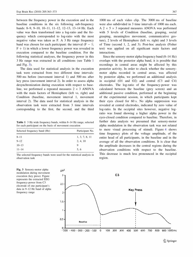
The data used for statistical analysis in the execution
task were extracted from two different time intervals:
500 ms before (movement interval 1) and 500 ms after
key press (movement interval 2). In order to assess alpha
desynchronization during execution with respect to base-
line, we performed a repeated measures 2 9 3 ANOVA
with the main factors of Hemisphere (left vs. right) and
Condition (baseline, movement interval 1, movement
interval 2). The data used for statistical analysis in the
observation task were extracted from 3 time intervals
corresponding to the first, the second, and the third
1000 ms of each video clip. The 3000 ms of baseline
were also subdivided in 3 time intervals of 1000 ms each.
A 2 9 5 9 3 repeated measures ANOVA was performed
with 5 levels of Condition (baseline, grasping, social
grasping, meaningless movement, communicative ges-
ture), 2 levels of Hemisphere (left vs. right) and 3 levels
of Time (second 1, 2, and 3). Post-hoc analysis (Fisher
test) was applied on all significant main factors and
interactions.
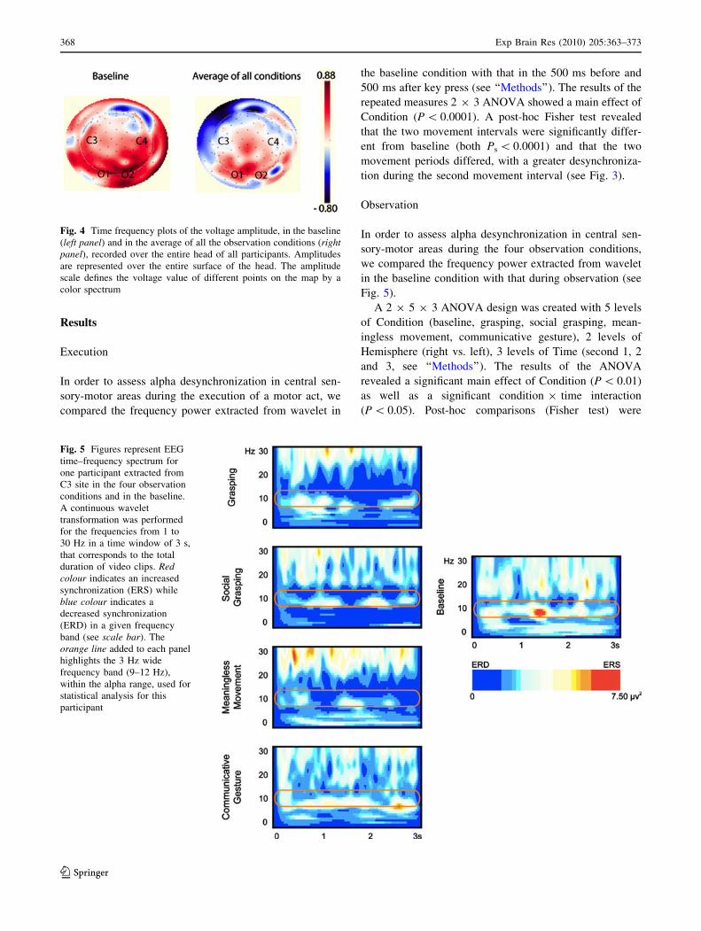
Since the sensory-motor alpha frequency band (8–14 Hz)
overlaps with the posterior alpha band, it is possible that
recordings in central areas might be affected by this
posterior activity. In order to check whether the sensory-
motor alpha recorded in central areas, was affected
by posterior alpha, we performed an additional analysis
in occipital (O1 and O2) and central (C3 and C4)
electrodes. The log-ratio of the frequency-power was
calculated between the baseline (grey screen) and an
additional passive condition, performed at the beginning
of the experimental session, in which participants kept
their eyes closed for 60 s. No alpha suppression was
revealed at central electrodes, indicated by zero value of
log-ratio. In the occipital sites however, negative log-
ratio was found showing a higher alpha power in the
eyes-closed condition compared to baseline. Therefore, in
further data analysis we presumed that sensory-motor
alpha modulation in the observation task was not related
to mere visual processing of stimuli. Figure 4 shows
time frequency plots of the voltage amplitude, of the
entire head of all participants, in the baseline and in the
average of all the observation conditions. It is clear that
the amplitude decreases in the central regions during the
observation conditions with respect to the baseline.
This decrease is much less pronounced in the occipital
region.
Table 1 3 Hz wide frequency bands, within 8–14 Hz range, selected
for each participant on the basis of movement execution
Selected frequency band (Hz) Participant No.
8–11 1, 3, 7, 8, 11
9–12 2, 4, 10
10–13 9
11–14 5, 6
The selected frequency bands were used for the statistical analysis in
observation task
Fig. 3 Sensory-motor alpha
modulation during movement
execution (key press). Figure
represents the extracted EEG
frequency-power from C3
electrode of one participant’s
data in 9–12 Hz band of alpha
frequency range
Exp Brain Res (2010) 205:363–373 367
123
Results
Execution
In order to assess alpha desynchronization in central sen-
sory-motor areas during the execution of a motor act, we
compared the frequency power extracted from wavelet in
the baseline condition with that in the 500 ms before and
500 ms after key press (see ‘‘Methods’’). The results of the
repeated measures 2 9 3 ANOVA showed a main effect of
Condition (P \ 0.0001). A post-hoc Fisher test revealed
that the two movement intervals were significantly differ-
ent from baseline (both Ps \ 0.0001) and that the two
movement periods differed, with a greater desynchroniza-
tion during the second movement interval (see Fig. 3).
Observation
In order to assess alpha desynchronization in central sen-
sory-motor areas during the four observation conditions,
we compared the frequency power extracted from wavelet
in the baseline condition with that during observation (see
Fig. 5).
A 2 9 5 9 3 ANOVA design was created with 5 levels
of Condition (baseline, grasping, social grasping, mean-
ingless movement, communicative gesture), 2 levels of
Hemisphere (right vs. left), 3 levels of Time (second 1, 2
and 3, see ‘‘Methods’’). The results of the ANOVA
revealed a significant main effect of Condition (P \ 0.01)
as well as a significant condition 9 time interaction
(P \ 0.05). Post-hoc comparisons (Fisher test) were
Fig. 4 Time frequency plots of the voltage amplitude, in the baseline
(left panel) and in the average of all the observation conditions (rightpanel), recorded over the entire head of all participants. Amplitudes
are represented over the entire surface of the head. The amplitude
scale defines the voltage value of different points on the map by a
color spectrum
Fig. 5 Figures represent EEG
time–frequency spectrum for
one participant extracted from
C3 site in the four observation
conditions and in the baseline.
A continuous wavelet
transformation was performed
for the frequencies from 1 to
30 Hz in a time window of 3 s,
that corresponds to the total
duration of video clips. Redcolour indicates an increased
synchronization (ERS) while
blue colour indicates a
decreased synchronization
(ERD) in a given frequency
band (see scale bar). The
orange line added to each panel
highlights the 3 Hz wide
frequency band (9–12 Hz),
within the alpha range, used for
statistical analysis for this
participant
368 Exp Brain Res (2010) 205:363–373
123
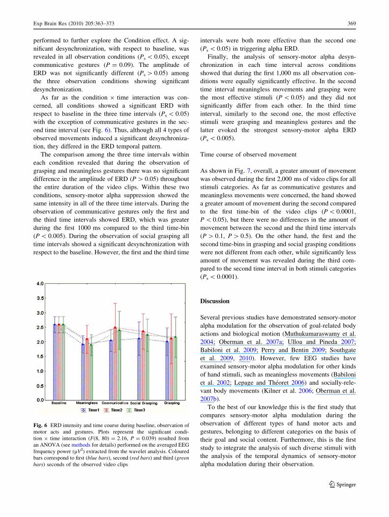
performed to further explore the Condition effect. A sig-
nificant desynchronization, with respect to baseline, was
revealed in all observation conditions (Ps \ 0.05), except
communicative gestures (P = 0.09). The amplitude of
ERD was not significantly different (Ps [ 0.05) among
the three observation conditions showing significant
desynchronization.
As far as the condition 9 time interaction was con-
cerned, all conditions showed a significant ERD with
respect to baseline in the three time intervals (Ps \ 0.05)
with the exception of communicative gestures in the sec-
ond time interval (see Fig. 6). Thus, although all 4 types of
observed movements induced a significant desynchroniza-
tion, they differed in the ERD temporal pattern.
The comparison among the three time intervals within
each condition revealed that during the observation of
grasping and meaningless gestures there was no significant
difference in the amplitude of ERD (P [ 0.05) throughout
the entire duration of the video clips. Within these two
conditions, sensory-motor alpha suppression showed the
same intensity in all of the three time intervals. During the
observation of communicative gestures only the first and
the third time intervals showed ERD, which was greater
during the first 1000 ms compared to the third time-bin
(P \ 0.005). During the observation of social grasping all
time intervals showed a significant desynchronization with
respect to the baseline. However, the first and the third time
intervals were both more effective than the second one
(Ps \ 0.05) in triggering alpha ERD.
Finally, the analysis of sensory-motor alpha desyn-
chronization in each time interval across conditions
showed that during the first 1,000 ms all observation con-
ditions were equally significantly effective. In the second
time interval meaningless movements and grasping were
the most effective stimuli (P \ 0.05) and they did not
significantly differ from each other. In the third time
interval, similarly to the second one, the most effective
stimuli were grasping and meaningless gestures and the
latter evoked the strongest sensory-motor alpha ERD
(Ps \ 0.005).
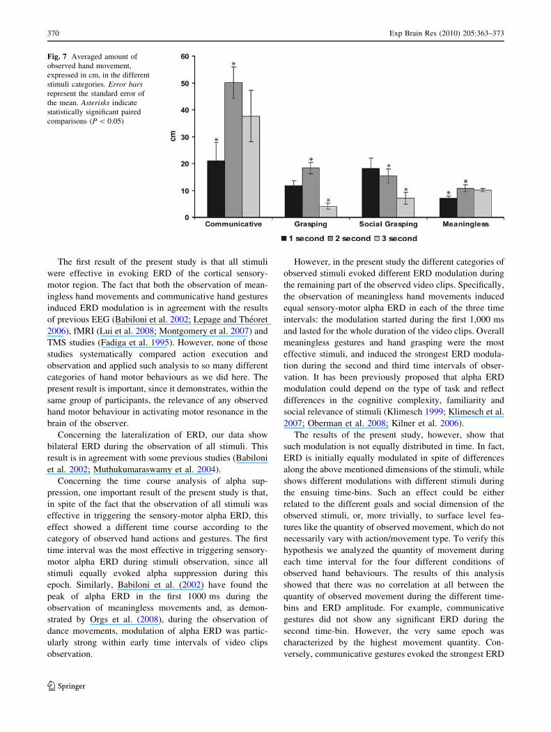
Time course of observed movement
As shown in Fig. 7, overall, a greater amount of movement
was observed during the first 2,000 ms of video clips for all
stimuli categories. As far as communicative gestures and
meaningless movements were concerned, the hand showed
a greater amount of movement during the second compared
to the first time-bin of the video clips (P \ 0.0001,
P \ 0.05), but there were no differences in the amount of
movement between the second and the third time intervals
(P [ 0.1, P [ 0.5). On the other hand, the first and the
second time-bins in grasping and social grasping conditions
were not different from each other, while significantly less
amount of movement was revealed during the third com-
pared to the second time interval in both stimuli categories
(Ps \ 0.0001).
Discussion
Several previous studies have demonstrated sensory-motor
alpha modulation for the observation of goal-related body
actions and biological motion (Muthukumaraswamy et al.
2004; Oberman et al. 2007a; Ulloa and Pineda 2007;
Babiloni et al. 2009; Perry and Bentin 2009; Southgate
et al. 2009, 2010). However, few EEG studies have
examined sensory-motor alpha modulation for other kinds
of hand stimuli, such as meaningless movements (Babiloni
et al. 2002; Lepage and Theoret 2006) and socially-rele-
vant body movements (Kilner et al. 2006; Oberman et al.
2007b).
To the best of our knowledge this is the first study that
compares sensory-motor alpha modulation during the
observation of different types of hand motor acts and
gestures, belonging to different categories on the basis of
their goal and social content. Furthermore, this is the first
study to integrate the analysis of such diverse stimuli with
the analysis of the temporal dynamics of sensory-motor
alpha modulation during their observation.
Fig. 6 ERD intensity and time course during baseline, observation of
motor acts and gestures. Plots represent the significant condi-
tion 9 time interaction (F(8, 80) = 2.16, P = 0.039) resulted from
an ANOVA (see methods for details) performed on the averaged EEG
frequency power (lV2) extracted from the wavelet analysis. Coloured
bars correspond to first (blue bars), second (red bars) and third (greenbars) seconds of the observed video clips
Exp Brain Res (2010) 205:363–373 369
123
The first result of the present study is that all stimuli
were effective in evoking ERD of the cortical sensory-
motor region. The fact that both the observation of mean-
ingless hand movements and communicative hand gestures
induced ERD modulation is in agreement with the results
of previous EEG (Babiloni et al. 2002; Lepage and Theoret
2006), fMRI (Lui et al. 2008; Montgomery et al. 2007) and
TMS studies (Fadiga et al. 1995). However, none of those
studies systematically compared action execution and
observation and applied such analysis to so many different
categories of hand motor behaviours as we did here. The
present result is important, since it demonstrates, within the
same group of participants, the relevance of any observed
hand motor behaviour in activating motor resonance in the
brain of the observer.
Concerning the lateralization of ERD, our data show
bilateral ERD during the observation of all stimuli. This
result is in agreement with some previous studies (Babiloni
et al. 2002; Muthukumaraswamy et al. 2004).
Concerning the time course analysis of alpha sup-
pression, one important result of the present study is that,
in spite of the fact that the observation of all stimuli was
effective in triggering the sensory-motor alpha ERD, this
effect showed a different time course according to the
category of observed hand actions and gestures. The first
time interval was the most effective in triggering sensory-
motor alpha ERD during stimuli observation, since all
stimuli equally evoked alpha suppression during this
epoch. Similarly, Babiloni et al. (2002) have found the
peak of alpha ERD in the first 1000 ms during the
observation of meaningless movements and, as demon-
strated by Orgs et al. (2008), during the observation of
dance movements, modulation of alpha ERD was partic-
ularly strong within early time intervals of video clips
observation.
However, in the present study the different categories of
observed stimuli evoked different ERD modulation during
the remaining part of the observed video clips. Specifically,
the observation of meaningless hand movements induced
equal sensory-motor alpha ERD in each of the three time
intervals: the modulation started during the first 1,000 ms
and lasted for the whole duration of the video clips. Overall
meaningless gestures and hand grasping were the most
effective stimuli, and induced the strongest ERD modula-
tion during the second and third time intervals of obser-
vation. It has been previously proposed that alpha ERD
modulation could depend on the type of task and reflect
differences in the cognitive complexity, familiarity and
social relevance of stimuli (Klimesch 1999; Klimesch et al.
2007; Oberman et al. 2008; Kilner et al. 2006).
The results of the present study, however, show that
such modulation is not equally distributed in time. In fact,
ERD is initially equally modulated in spite of differences
along the above mentioned dimensions of the stimuli, while
shows different modulations with different stimuli during
the ensuing time-bins. Such an effect could be either
related to the different goals and social dimension of the
observed stimuli, or, more trivially, to surface level fea-
tures like the quantity of observed movement, which do not
necessarily vary with action/movement type. To verify this
hypothesis we analyzed the quantity of movement during
each time interval for the four different conditions of
observed hand behaviours. The results of this analysis
showed that there was no correlation at all between the
quantity of observed movement during the different time-
bins and ERD amplitude. For example, communicative
gestures did not show any significant ERD during the
second time-bin. However, the very same epoch was
characterized by the highest movement quantity. Con-
versely, communicative gestures evoked the strongest ERD
Fig. 7 Averaged amount of
observed hand movement,
expressed in cm, in the different
stimuli categories. Error barsrepresent the standard error of
the mean. Asterisks indicate
statistically significant paired
comparisons (P \ 0.05)
370 Exp Brain Res (2010) 205:363–373
123
during the first time-bin, in which the quantity of move-
ment was less than in the subsequent epoch. Thus, the
equal modulation of ERD during the first 1,000 ms for all
categories of observed hand behaviours is most likely due
to the motor resonance that occurs immediately and auto-
matically. The presence of a goal or social value in the
observed hand behaviour or its occurrence within a social
context are not necessary conditions to evoke motor reso-
nance. Once the hand motor representation of the observed
behaviour has been activated in the observer, the intensity
and stability of its activation appear to depend upon the
goal-relatedness and social relevance of the observed
behaviour. Indeed, only communicative gestures and social
grasping—sharing both goal-relatedness and social value—
showed a dynamic pattern of ERD modulation during the
second and third 1,000 ms of the video clips presentation.
The observation of social hand behaviours, such as
social grasping triggers a more dynamically modulated
motor resonance mechanism compared to that driven by
the observation of simple grasping. Simple grasping fol-
lowed by holding of the object evokes a prolonged alpha
suppression during the entire duration of its observation,
likely reflecting motor resonance occurring during the
observation of hand pre-shaping, actual grasping and object
holding. Once the most trivial and parsimonious explana-
tion of these dynamic modulations is ruled out (see above),
two not mutually exclusive explanations can be proposed
for the different ERD temporal modulation evoked by
social grasping observation. On the one hand, it has been
shown that the kinematics of the same hand motor act
differs according to its social or non social nature (Becchio
et al. 2008a; Sartori et al. 2009a, b). More specifically, the
authors (Becchio et al. 2008b) have found a lower move-
ment amplitude and earlier peak of velocity during the
execution of a goal-directed action in social context (giving
an object to another individual) than in ‘single-agent’
condition (placing an object on a base). In other words,
deceleration phase was longer (thus action more accurate)
when the hand was approaching another individual. It
could be proposed that the specific kinematics pattern of an
executed social hand action may influence the differences
in the temporal pattern of ERD also during the observation
of social grasping when this condition is compared with
grasping. Thus, the observation of social grasping most
likely induces in the observer a different motor resonance
compared to the observation of simple grasping. Our data
suggest that such difference develops in time. On the other
hand, social grasping also appears more ambiguous than
simple grasping. While in the latter there is only one acting
agent, in social grasping there are two agents, and the
observer in principle might not know with whom to reso-
nate, because social grasping potentially implies two dif-
ferent motor acts, offering an object and/or grasping it. It is
no coincidence that at debriefing several participants
pointed out that with social grasping they were not sure
about the goal of the observed motor act, described alter-
natively as a grasping or as a giving. Our data show that
only during the third time interval of video clip presenta-
tion, when social grasping is completed with the holding
phase, thus when the goal of grasping becomes clear, ERD
increases and becomes as strong as during the first
1,000 ms of observation. Finally, another factor could
influence the reduction of ERD in the second time interval
of social grasping. As the motor act develops, visual
attention is shared between two interacting hands. It is
plausible that during the second time interval participants
shift their attention to the static hand of a potential ‘giver’
as they are unsure whether the giver will do something of
interest. Although, we cannot provide further elements to
this hypothesis, as the eye-tracker was not used in the
present study, the attentional account could have strong
implications when comparing conditions with one versus
two biological stimuli/agents.
Comparing the two types of stimuli within the ‘gesture’
category, again we observe a clear difference in the temporal
pattern of sensory-motor alpha modulation. ERD modula-
tion was more stable for meaningless gestures. With such
stimuli, alpha remained desynchronised for the whole period
of observation, while for communicative gestures alpha
desynchronised during the first 1,000 ms of observation, and
resynchronized during the second time epoch, when ERD
disappeared with respect to baseline. During the third second
of observation, ERD reappeared, although being signifi-
cantly weaker when compared with meaningless gestures.
We presume that this difference might again depend upon
the different goal-relatedness of the observed hand gestures.
The observation of meaningless gestures triggers ERD dur-
ing the whole duration of the stimuli without any time course
modulation. The prolonged ERD could be due to an activa-
tion of the motor resonance mechanism lasting as long as the
hand continues to move, perhaps waiting for a potential goal
to emerge. It is possible to hypothesize that when observing
hand behaviors devoid of any meaning, humans nevertheless
cannot refrain from trying to find a meaning, hence the
necessity to keep motor resonance active.
In contrast, during the observation of communicative
gestures, motor resonance finishes as soon as the meaning
is understood, that is, in our case at the end of the first
1,000 ms of observation. In agreement with this interpre-
tation, communicative gestures evoke reduced N400
compared to meaningless gestures, and this effect has a
centro-posterior distribution (Gunter and Bach 2004). The
resumed desynchronization occurring during the last sec-
ond of observation might be due to the persistence of a
static hand, which nevertheless conveys a social meaning,
thus inducing motor resonance to be resumed.
Exp Brain Res (2010) 205:363–373 371
123
In our view, there are two different functional aspects
related to the activation of the mirror mechanism during
movement observation in humans. The first one represents
an automatic low-level motor resonance, starting as soon as
a movement or a goal-related motor act is observed, which
triggers eventually the understanding process. Differently
from the available evidence in monkeys, in humans motor
resonance can be induced also when a motor goal is not
present in the observed behavior of others (Fadiga et al.
1995; Babiloni et al. 2002). Most likely, the cortical
motor system of humans contains neurons that can be
activated by the observation of meaningless movements.
Action understanding is the second functional aspect rela-
ted to the mirror mechanism. Such aspect implies the
activation of goal-related motor neurons in the brain of the
observer matching the goal of the observed motor behavior
of others. For meaningless movements, there is only the
first aspect of the mirror mechanism activation. Brain
imaging experiments are being designed to investigate the
possible segregation of these two different aspects of the
mirror mechanism for hand motor behaviors.
In summary, the present study clearly shows that motor
resonance can be induced whenever hand motor behavior is
observed, irrespective of its goal-relatedness and social
content. However, motor resonance is not an all-or nothing
mechanism, but can be strongly modulated in time
according to—at least—two different dimensions of the
observed hand behaviors: the presence/absence of a goal
and its social relevance. It is important to emphasize that
such differences can be detected, only if one investigates
the temporal pattern of the mirror mechanism activation.
Acknowledgments The authors wish to thank Prof. C.A. Tassinari
for most valuable comments on EEG data collection and analysis,
Dr. Pietro Avanzini for his assistance in wavelet analysis and
Dr. Patricia Gough for helpful comments on earlier draft of this man-
uscript, Dr. Marco Bimbi and Dr. Fabian Chersi for providing the
technical support for movement analysis This research was supported
by Ministero dell’Universita e della Ricerca [Relevant National Interest
Project]. A.S. was funded by the Marie Curie Research Training
Network ‘‘Disorders and Coherence of the Embodied Self’’. C.B. was
supported by fellowship Ricerca Finalizzata 2007—Programma
Strategico ‘‘Inquiry into disruption of inter-subjective equipment in
autistic spectrum disorders in childhood’’.
Open Access This article is distributed under the terms of the
Creative Commons Attribution Noncommercial License which per-
mits any noncommercial use, distribution, and reproduction in any
medium, provided the original author(s) and source are credited.
References
Altschuler EL, Vankov A, Wang V, Ramachandran VS, Pineda JA.
(1997) Person see, person do: human cortical electrophysiological
correlates of monkey see monkey do cell. Soc Neurosci 719 (17)
Altschuler EL, Vankov A, Hubbard EM, Roberts E, Ramachandran
VS, Pineda JA (2000) Mu wave blocking by observation of
movement and its possible use as a tool to study theory of other
minds. Soc Neurosci 68 (1)
Aziz-Zadeh L, Wilson SM, Rizzolatti G, Iacoboni M (2006)
Congruent embodied representations for visually presented
actions and linguistic phrases describing actions. Curr Biol
16(18):1818–1823
Babiloni C, Babiloni F, Carducci F, Cincotti F, Cocozza G, Del Percio
C, Moretti DV, Rossini PM (2002) Human cortical electroen-
cephalography (EEG) rhythms during the observation of simple
aimless movements: a high resolution EEG study. Neuroimage
17:559–572
Babiloni C, Del Percio C, Rossini PM, Marzano N, Iacoboni M,
Infarinato F, Lizio R, Piazza M, Pirritano M, Berlutti G, Cibelli G,
Eusebi F (2009) Judgment of actions in experts: a high-resolution
EEG study in elite athletes. Neuroimage 45(2):512–521
Becchio C, Sartori L, Bulgheroni M, Castiello U (2008a) Both your
intention and mine are reflected in the kinematics of my reach-
to-grasp movement. Cognition 106(2):894–912
Becchio C, Sartori L, Bulgheroni M, Castiello U (2008b) The case of
Dr. Jekyll and Mr. Hyde: a kinematic study on social intention.
Conscious Cogn 17(3):557–564
Bernier R, Dawson G, Webb S, Murias M (2007) EEG mu rhythm
and imitation impairments in individuals with autism spectrum
disorder. Brain Cogn 64:228–237
Buccino G, Binkofski F, Fink GR, Fadiga L, Fogassi L et al (2001)
Action observation activates premotor and parietal areas in a
somatotopic manner: an fMRI study. Eur J Neurosci 13:400–404
Calmels C, Holmes P, Jarry G, Leveque JM, Hars M, Stam CJ (2006)
Cortical activity prior to, and during, observation and execution
of sequential finger movements. Brain Topogr 19(1–2):77–88
Cattaneo L, Rizzolatti G (2009) The mirror neuron system. Review.
Arch Neurol 66(5):557–560
Cochin S, Barthelemy C, Lejeune B, Roux S, Martineau J (1998)
Perception of motion and qEEG activity in human adults.
Electroencephalogr Clin Neurophysiol 107:287–295
Cochin S, Barthelemy C, Roux S, Martineau J (1999) Observation
and execution of movement: similarities demonstrated by
quantified electroencephalograpy. Eur J Neurosci 11:1839–1842
Cohen-Seat G, Gastaut H, Faure J, Heuyer G (1954) Etudes
experiementales de l’activite nerveuse pendant la projection
cinematographique. Rev Int de Filmologie 5:7–64
Di Pellegrino G, Fadiga L, Fogassi L, Gallese V, Rizzolatti G (1992)
Understanding motor events: a neurophysiological study. Exp
Brain Res 91:176–180
Fadiga L, Fogassi L, Pavesi G, Rizzolatti G (1995) Motor facilitation
during action observation: a magnetic stimulation study. J Neu-
rophysiol 73:2608–2611
Gallese V, Fadiga L, Fogassi L, Rizzolatti G (1996) Action
recognition in the premotor cortex. Brain 119:593–609
Gallese V, Keysers C, Rizzolatti G (2004) A unifying view of the
basis of social cognition. Trends Cogn Sci 8(9):396–403
Gastaut HJ, Bert J (1954) EEG changes during cinematographic
presentation, moving picture activation of the EEG. Electroen-
cephalogr Clin Neurophysiol 6:433–444
Gazzola V, Aziz-Zadeh L, Keysers C (2006) Empathy and the
somatotopic auditory mirror system in human. Curr Biol
16:1824–1829
Gunter TC, Bach P (2004) Communicating hands: ERPs elicited
by meaningful symbolic hand postures. Neurosci Lett
372(1–2):52–56
Hari R (2006) Action-perception connection and the cortical mu
rhythm. Prog Brain Res 159:253–260
Hari R, Salmelin R (1997) Human cortical oscillations: a neuromag-
netic view through the skull. Trends Neurosci 20(1):44–49
372 Exp Brain Res (2010) 205:363–373
123

Hari R, Forss N, Avikainen S, Kirveskari S, Salenius S, Rizzolatti G
(1998) Activation of human primary motor cortex during action
observation: a neuromagnetic study. Proc Natl Acad Sci USA
95:15061–15065
Kilner JM, Marchant JL, Frith CD (2006) Modulation of the mirror
system by social relevance. Soc Cogn Affect Neurosci
1(2):143–148
Klimesch W (1999) EEG alpha and theta oscillations reflect cognitive
and memory performance: a review and analysis. Brain Res Rev
29:169–195
Klimesch W, Sauseng P, Hanslmayr S (2007) EEG alpha oscillations:
the inhibition-timing hypothesis. Brain Res Rev 53:63–88
Lepage JF, Theoret H (2006) EEG evidence for the presence of an
action observation-execution matching system in children. Eur J
Neurosci 23(9):2505–2510
Lui F, Buccino G, Duzzi D, Benuzzi F, Crisi G, Baraldi P, Nichelli P,
Porro CA (2008) Neural substrates of observing and imaging
non-object directed action. Soc Neurosci 3:261–275
Montgomery KJ, Isenberg N, Haxby JV (2007) Communicative hand
gestures and object-directed hand movements activated the
mirror neuron system. Soc Cogn Affect Neurosci 2(2):114–122
Mukamel R, Ekstrom AD, Kaplan J, Iacoboni M, Fried I (2010)
Single-neuron responses in humans during execution and
observation of actions. Curr Biol 20(8):750–756
Muthukumaraswamy SD, Johnson BW (2004a) Changes in rolandic
mu rhythm during observation of a precision grip. Psychophys-
iolgy 41:152–156
Muthukumaraswamy SD, Johnson BW (2004b) Primary motor cortex
activation during action observation revealed by wavelet anal-
ysis of the EEG. Clin Neurophysiol 115:1760–1766
Muthukumaraswamy SD, Johnson BW, McNair NA (2004) Mu
rhythm modulation during observation of an object-directed
grasp. Cogn Brain Res 19:195–201
Nakamura K, Kawashima R, Ito K, Sugiura M, Kato T, Nakamura A,
Hatano K, Nagumo S, Kubota K, Fukuda H, Kojima S (1999)
Activation of the right inferior frontal cortex during assessment
of facial emotion. J Neurophysiol 82:1610–1614
Nakamura A, Maess B, Knosche TR, Gunter TC, Bach P, Friederici
AD (2004) Cooperation of different neuronal systems during
hand sign recognition. Neuroimage 23(1):25–34
Oberman LM, McCleery JP, Ramachandran VS, Pineda JA (2007a)
EEG evidence for mirror neuron activity during the observation
of human and robot actions: toward an analysis of the human
qualities of interactive robots. Neurocomputing 70:2194–2203
Oberman LM, Pineda JA, Ramachandran VS (2007b) The human
mirror neuron system: a link between action observation and
social skills. Soc Cogn Affect Neurosci 6:62–66
Oberman LM, Ramachandran VS, Pineda JA (2008) Modulation of
mu suppression in children with autism spectrum disorders in
response to familiar or unfamiliar stimuli: the mirror neuron
hypothesis. Neuropsychology 46(5):1558–1565
Oldfield RC (1971) The assessment and analysis of handedness: the
Edinburgh Inventory. Neuropsychology 9:77–113
Orgs G, Dombrowski JH, Heil M, Jansen-Osmann P (2008) Expertise
in dance modulates alpha/beta event-related desynchronization
during action observation. Eur J Neurosci 27:3380–3384
Perry A, Bentin S (2009) Mirror activity in the human brain while
observing hand movements: a comparison between EEG desyn-
chronization in the mu-range and previous fMRI results. Brain
Res 1282:126–132
Pfurtscheller G, Lopes da Silva FH (1999) Event-related EEG/MEG
synchronization and desynchronization: basic principles. Clin
Neurophysiol 110:1842–1857
Pineda JA (2005) The functional significance of mu rhythms:
translating ‘‘seeing’’ and ‘‘hearing’’ into ‘‘doing’’. Brain Res
Rev 50:57–68
Rizzolatti G, Fadiga L, Gallese V, Fogassi L (1996) Premotor cortex
and the recognition of motor actions. Cogn Brain Res 3:131–141
Rizzolatti G, Craighero L (2004) The mirror-neuron system. Annu
Rev Neurosci 27:169–192
Salmelin R, Hari R (1994) Spatiotemporal characteristics of senso-
rimotor neuromagnetic rhythms related to thumb movement.
Neuroscience 60:537–550
Sartori L, Becchio C, Bulgheroni M, Castiello U (2009a) Modulation
of the action control system by social intention: unexpected
social requests override preplanned action. J Exp Psychol Hum
Percept Perform 35(5):1490–1500
Sartori L, Becchio C, Bara BG, Castiello U (2009b) Does the
intention to communicate affect action kinematics? Conscious
Cog 18(3):766–772
Southgate V, Johnson MH, Osborne T, Csibra G (2009) Predictive
motor activation during action observation in human infants.
Biol Lett 5(6):769–772
Southgate V, Johnson MH, El Karoui I, Csibra G (2010) Motor
system activation reveals infants’ on-line prediction of others’
goals. Psychol Sci 21(3):355–359
Ulloa ER, Pineda JA (2007) Recognition of point-light biological
motion: mu rhythms and mirror neuron activity. Behav Brain
Res 183(2):188–194
Villarreal M, Fridman EA, Amengual A, Falasco G, Gerscovich ER,
Ulloa ER, Leiguarda RC (2008) The neural substrate of gesture
recognition. Neuropsychology 46(9):2371–2382
Exp Brain Res (2010) 205:363–373 373
123
Related Documents