Neuropsychologia 49 (2011) 1016–1023 Contents lists available at ScienceDirect Neuropsychologia journal homepage: www.elsevier.com/locate/neuropsychologia Time and spatial attention: Effects of prism adaptation on temporal deficits in brain damaged patients Barbara Magnani a , Massimiliano Oliveri b,c,∗ , Giovanni Mancuso d , Emanuela Galante e , Francesca Frassinetti a a Department of Psychology, University of Bologna, 40127 Bologna, Italy b Department of Psychology, University of Palermo, 90128 Palermo, Italy c Fondazione “Santa Lucia” IRCCS, 00179 Rome, Italy d San Giacomo Hospital, Ponte dell’Olio (PC), Italy e Fondazione Salvatore Maugeri, Clinica del Lavoro e della Riabilitazione, IRCCS – Istituto Scientifico di Castel Goffredo, Mantova, Italy article info Article history: Received 24 July 2010 Received in revised form 6 December 2010 Accepted 7 December 2010 Available online 14 January 2011 Keywords: Space Time Prisms Right hemisphere Left hemisphere Stroke abstract Growing evidence indicates that the representations of space and time interact in the brain but the exact neural correlates of such interaction remain unknown. Neuroimaging and neuropsychological studies show that processing of temporal information engages a distributed network in the right hemisphere and suggest a link between deficits in spatial attention and deficits in time perception. In the present study we used the procedure of prismatic adaptation (PA) to directionally manipulate spatial attention in order to explore the effect of attentional deviation on time perception in patients with right (RBD) vs. left (LBD) brain damage. In a first experiment, two groups of RBD and LBD patients and two groups of age-matched healthy subjects were submitted to a time reproduction task before and after rightward or leftward PA (between-group design). In a second experiment RBD and LBD patients were submitted to the same task, before and after rightward and leftward PA (within-group design). RBD but not LBD patients presented a time deficit with a clear tendency to underestimate the real time. PA inducing leftward attentional deviation biased time perception toward an underestimation in RBD patients and controls, while it was ineffective in LBD patients. PA inducing a rightward attentional deviation failed to affect time perception in either group. These results underline the effects of PA on temporal deficits in brain damaged patients. The novel finding is that, while a right hemispheric network is critical for explicit timing, a left hemispheric network is necessary for mediating the effects of prismatic adaptation on spatial and temporal perception. © 2010 Elsevier Ltd. All rights reserved. 1. Introduction Psychophysical studies on healthy subjects document that spa- tial factors affect timing. In fact, lateralised saccades (Burr, Tozzi, & Morrone, 2007; Morrone, Ross, & Burr, 2005) as well as visual adap- tation to high-frequency lateralised gratings (Johnston, Arnold, & Nishida, 2006) compress time perception. Moreover, time percep- tion is underestimated in the left- and overestimated in the right space (Oliveri, Koch, & Caltagirone, 2009; Vallesi, Binns, & Shallice, 2008; Vicario, Caltagirone, & Oliveri, 2007; Vicario et al., 2008; Vicario, Rappo, Pepi, & Oliveri, 2009). In line with the finding of lateralised distortions of perceived time, time underestimation or overestimation are observed follow- ∗ Corresponding author at: Dipartimento di Psicologia, Università di Palermo, Viale delle Scienze, Edificio 15, 90128 Palermo, Italy. Tel.: +39 09123897736. E-mail address: [email protected] (M. Oliveri). ing leftward and rightward attentional shifts (Frassinetti, Magnani, & Oliveri, 2009). In this study the direction of spatial attention was manipulated by using prismatic lenses. Indeed previous stud- ies have demonstrated that prismatic lenses reduce the attentional bias in patients with neglect, not only in visual and in tactile modal- ity (see Frassinetti, Angeli, Meneghello, Avanzi, & Làdavas, 2002; Maravita et al., 2003) but also at a representational level (Rode, Rossetti, & Boisson, 2001). In Frassinetti et al.’s study (2009), right- ward displacing-prisms, inducing leftward after-effect, affect time reproduction toward an underestimation. Leftward displacing- prisms, inducing rightward after-effect, cause an opposite pattern of temporal overestimation. These results were explained in terms of a linear representation of time intervals in ascending order from left to right, where short time intervals are coded leftward whereas longer time intervals are represented more rightward. According to this model of a spatial representation of temporal information, moving attentive focus along the mental time-line can bias time perception. 0028-3932/$ – see front matter © 2010 Elsevier Ltd. All rights reserved. doi:10.1016/j.neuropsychologia.2010.12.014

Welcome message from author
This document is posted to help you gain knowledge. Please leave a comment to let me know what you think about it! Share it to your friends and learn new things together.
Transcript

Tb
BFa
b
c
d
e
a
ARRAA
KSTPRLS
1
tMtNts2V
t
V
0d
Neuropsychologia 49 (2011) 1016–1023
Contents lists available at ScienceDirect
Neuropsychologia
journa l homepage: www.e lsev ier .com/ locate /neuropsychologia
ime and spatial attention: Effects of prism adaptation on temporal deficits inrain damaged patients
arbara Magnania, Massimiliano Oliverib,c,∗, Giovanni Mancusod, Emanuela Galantee,rancesca Frassinetti a
Department of Psychology, University of Bologna, 40127 Bologna, ItalyDepartment of Psychology, University of Palermo, 90128 Palermo, ItalyFondazione “Santa Lucia” IRCCS, 00179 Rome, ItalySan Giacomo Hospital, Ponte dell’Olio (PC), ItalyFondazione Salvatore Maugeri, Clinica del Lavoro e della Riabilitazione, IRCCS – Istituto Scientifico di Castel Goffredo, Mantova, Italy
r t i c l e i n f o
rticle history:eceived 24 July 2010eceived in revised form 6 December 2010ccepted 7 December 2010vailable online 14 January 2011
eywords:paceimerismsight hemisphere
a b s t r a c t
Growing evidence indicates that the representations of space and time interact in the brain but the exactneural correlates of such interaction remain unknown. Neuroimaging and neuropsychological studiesshow that processing of temporal information engages a distributed network in the right hemisphereand suggest a link between deficits in spatial attention and deficits in time perception.
In the present study we used the procedure of prismatic adaptation (PA) to directionally manipulatespatial attention in order to explore the effect of attentional deviation on time perception in patientswith right (RBD) vs. left (LBD) brain damage. In a first experiment, two groups of RBD and LBD patientsand two groups of age-matched healthy subjects were submitted to a time reproduction task before andafter rightward or leftward PA (between-group design). In a second experiment RBD and LBD patientswere submitted to the same task, before and after rightward and leftward PA (within-group design).
eft hemispheretroke
RBD but not LBD patients presented a time deficit with a clear tendency to underestimate the realtime. PA inducing leftward attentional deviation biased time perception toward an underestimation inRBD patients and controls, while it was ineffective in LBD patients. PA inducing a rightward attentionaldeviation failed to affect time perception in either group.
These results underline the effects of PA on temporal deficits in brain damaged patients. The novelfinding is that, while a right hemispheric network is critical for explicit timing, a left hemispheric network
g the
is necessary for mediatin. Introduction
Psychophysical studies on healthy subjects document that spa-ial factors affect timing. In fact, lateralised saccades (Burr, Tozzi, &
orrone, 2007; Morrone, Ross, & Burr, 2005) as well as visual adap-ation to high-frequency lateralised gratings (Johnston, Arnold, &ishida, 2006) compress time perception. Moreover, time percep-
ion is underestimated in the left- and overestimated in the rightpace (Oliveri, Koch, & Caltagirone, 2009; Vallesi, Binns, & Shallice,
008; Vicario, Caltagirone, & Oliveri, 2007; Vicario et al., 2008;icario, Rappo, Pepi, & Oliveri, 2009).In line with the finding of lateralised distortions of perceivedime, time underestimation or overestimation are observed follow-
∗ Corresponding author at: Dipartimento di Psicologia, Università di Palermo,iale delle Scienze, Edificio 15, 90128 Palermo, Italy. Tel.: +39 09123897736.
E-mail address: [email protected] (M. Oliveri).
028-3932/$ – see front matter © 2010 Elsevier Ltd. All rights reserved.oi:10.1016/j.neuropsychologia.2010.12.014
effects of prismatic adaptation on spatial and temporal perception.© 2010 Elsevier Ltd. All rights reserved.
ing leftward and rightward attentional shifts (Frassinetti, Magnani,& Oliveri, 2009). In this study the direction of spatial attentionwas manipulated by using prismatic lenses. Indeed previous stud-ies have demonstrated that prismatic lenses reduce the attentionalbias in patients with neglect, not only in visual and in tactile modal-ity (see Frassinetti, Angeli, Meneghello, Avanzi, & Làdavas, 2002;Maravita et al., 2003) but also at a representational level (Rode,Rossetti, & Boisson, 2001). In Frassinetti et al.’s study (2009), right-ward displacing-prisms, inducing leftward after-effect, affect timereproduction toward an underestimation. Leftward displacing-prisms, inducing rightward after-effect, cause an opposite patternof temporal overestimation. These results were explained in termsof a linear representation of time intervals in ascending order from
left to right, where short time intervals are coded leftward whereaslonger time intervals are represented more rightward. Accordingto this model of a spatial representation of temporal information,moving attentive focus along the mental time-line can bias timeperception.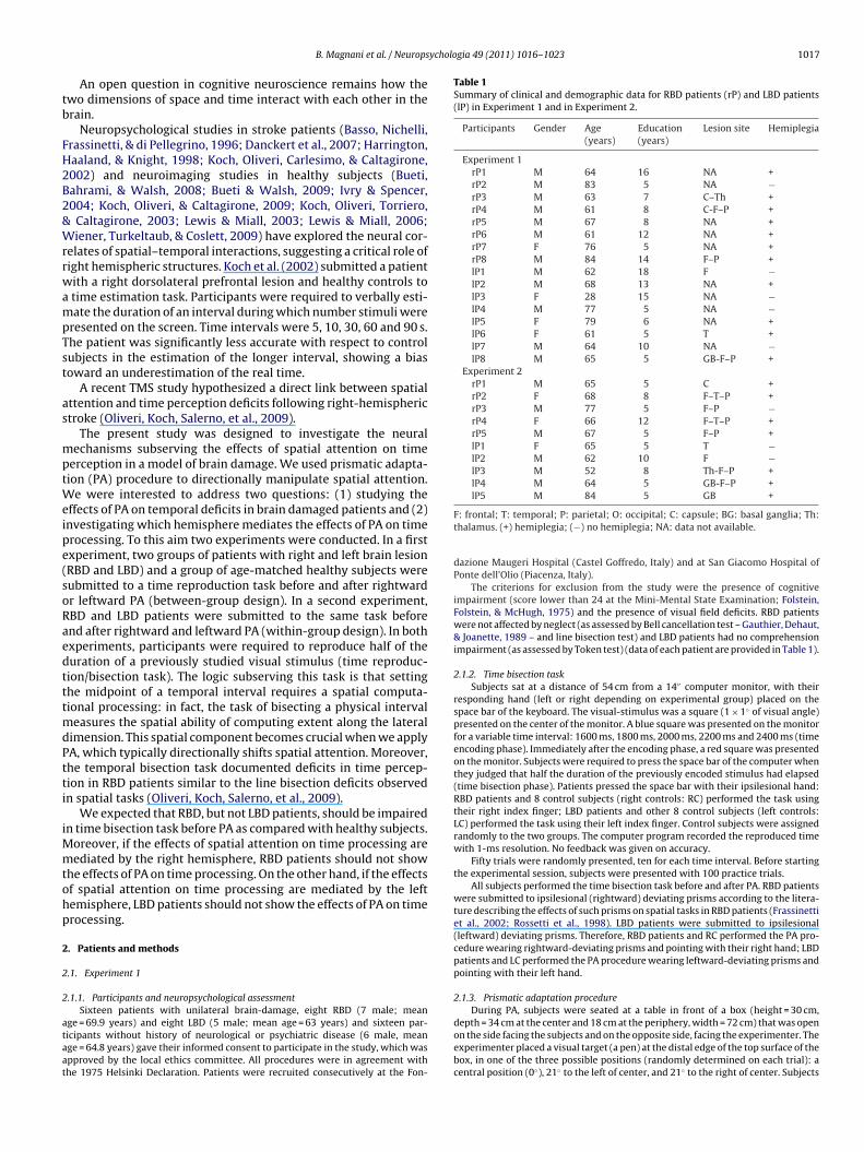
ychologia 49 (2011) 1016–1023 1017
tb
FH2B2&WrrwampTst
as
mptWeipe(soRaedtttmdPtti
iMmtohp
2
2
2
ataat
Table 1Summary of clinical and demographic data for RBD patients (rP) and LBD patients(lP) in Experiment 1 and in Experiment 2.
Participants Gender Age(years)
Education(years)
Lesion site Hemiplegia
Experiment 1rP1 M 64 16 NA +rP2 M 83 5 NA −rP3 M 63 7 C–Th +rP4 M 61 8 C-F–P +rP5 M 67 8 NA +rP6 M 61 12 NA +rP7 F 76 5 NA +rP8 M 84 14 F–P +lP1 M 62 18 F −lP2 M 68 13 NA +lP3 F 28 15 NA −lP4 M 77 5 NA −lP5 F 79 6 NA +lP6 F 61 5 T +lP7 M 64 10 NA −lP8 M 65 5 GB-F–P +
Experiment 2rP1 M 65 5 C +rP2 F 68 8 F–T–P +rP3 M 77 5 F–P −rP4 F 66 12 F–T–P +rP5 M 67 5 F–P +lP1 F 65 5 T −lP2 M 62 10 F −lP3 M 52 8 Th-F–P +lP4 M 64 5 GB-F–P +
B. Magnani et al. / Neurops
An open question in cognitive neuroscience remains how thewo dimensions of space and time interact with each other in therain.
Neuropsychological studies in stroke patients (Basso, Nichelli,rassinetti, & di Pellegrino, 1996; Danckert et al., 2007; Harrington,aaland, & Knight, 1998; Koch, Oliveri, Carlesimo, & Caltagirone,002) and neuroimaging studies in healthy subjects (Bueti,ahrami, & Walsh, 2008; Bueti & Walsh, 2009; Ivry & Spencer,004; Koch, Oliveri, & Caltagirone, 2009; Koch, Oliveri, Torriero,
Caltagirone, 2003; Lewis & Miall, 2003; Lewis & Miall, 2006;iener, Turkeltaub, & Coslett, 2009) have explored the neural cor-
elates of spatial–temporal interactions, suggesting a critical role ofight hemispheric structures. Koch et al. (2002) submitted a patientith a right dorsolateral prefrontal lesion and healthy controls totime estimation task. Participants were required to verbally esti-ate the duration of an interval during which number stimuli were
resented on the screen. Time intervals were 5, 10, 30, 60 and 90 s.he patient was significantly less accurate with respect to controlubjects in the estimation of the longer interval, showing a biasoward an underestimation of the real time.
A recent TMS study hypothesized a direct link between spatialttention and time perception deficits following right-hemispherictroke (Oliveri, Koch, Salerno, et al., 2009).
The present study was designed to investigate the neuralechanisms subserving the effects of spatial attention on time
erception in a model of brain damage. We used prismatic adapta-ion (PA) procedure to directionally manipulate spatial attention.
e were interested to address two questions: (1) studying theffects of PA on temporal deficits in brain damaged patients and (2)nvestigating which hemisphere mediates the effects of PA on timerocessing. To this aim two experiments were conducted. In a firstxperiment, two groups of patients with right and left brain lesionRBD and LBD) and a group of age-matched healthy subjects wereubmitted to a time reproduction task before and after rightwardr leftward PA (between-group design). In a second experiment,BD and LBD patients were submitted to the same task beforend after rightward and leftward PA (within-group design). In bothxperiments, participants were required to reproduce half of theuration of a previously studied visual stimulus (time reproduc-ion/bisection task). The logic subserving this task is that settinghe midpoint of a temporal interval requires a spatial computa-ional processing: in fact, the task of bisecting a physical interval
easures the spatial ability of computing extent along the lateralimension. This spatial component becomes crucial when we applyA, which typically directionally shifts spatial attention. Moreover,he temporal bisection task documented deficits in time percep-ion in RBD patients similar to the line bisection deficits observedn spatial tasks (Oliveri, Koch, Salerno, et al., 2009).
We expected that RBD, but not LBD patients, should be impairedn time bisection task before PA as compared with healthy subjects.
oreover, if the effects of spatial attention on time processing areediated by the right hemisphere, RBD patients should not show
he effects of PA on time processing. On the other hand, if the effectsf spatial attention on time processing are mediated by the leftemisphere, LBD patients should not show the effects of PA on timerocessing.
. Patients and methods
.1. Experiment 1
.1.1. Participants and neuropsychological assessment
Sixteen patients with unilateral brain-damage, eight RBD (7 male; meange = 69.9 years) and eight LBD (5 male; mean age = 63 years) and sixteen par-icipants without history of neurological or psychiatric disease (6 male, meange = 64.8 years) gave their informed consent to participate in the study, which waspproved by the local ethics committee. All procedures were in agreement withhe 1975 Helsinki Declaration. Patients were recruited consecutively at the Fon-
lP5 M 84 5 GB +
F: frontal; T: temporal; P: parietal; O: occipital; C: capsule; BG: basal ganglia; Th:thalamus. (+) hemiplegia; (−) no hemiplegia; NA: data not available.
dazione Maugeri Hospital (Castel Goffredo, Italy) and at San Giacomo Hospital ofPonte dell’Olio (Piacenza, Italy).
The criterions for exclusion from the study were the presence of cognitiveimpairment (score lower than 24 at the Mini-Mental State Examination; Folstein,Folstein, & McHugh, 1975) and the presence of visual field deficits. RBD patientswere not affected by neglect (as assessed by Bell cancellation test – Gauthier, Dehaut,& Joanette, 1989 – and line bisection test) and LBD patients had no comprehensionimpairment (as assessed by Token test) (data of each patient are provided in Table 1).
2.1.2. Time bisection taskSubjects sat at a distance of 54 cm from a 14′′ computer monitor, with their
responding hand (left or right depending on experimental group) placed on thespace bar of the keyboard. The visual-stimulus was a square (1 × 1◦ of visual angle)presented on the center of the monitor. A blue square was presented on the monitorfor a variable time interval: 1600 ms, 1800 ms, 2000 ms, 2200 ms and 2400 ms (timeencoding phase). Immediately after the encoding phase, a red square was presentedon the monitor. Subjects were required to press the space bar of the computer whenthey judged that half the duration of the previously encoded stimulus had elapsed(time bisection phase). Patients pressed the space bar with their ipsilesional hand:RBD patients and 8 control subjects (right controls: RC) performed the task usingtheir right index finger; LBD patients and other 8 control subjects (left controls:LC) performed the task using their left index finger. Control subjects were assignedrandomly to the two groups. The computer program recorded the reproduced timewith 1-ms resolution. No feedback was given on accuracy.
Fifty trials were randomly presented, ten for each time interval. Before startingthe experimental session, subjects were presented with 100 practice trials.
All subjects performed the time bisection task before and after PA. RBD patientswere submitted to ipsilesional (rightward) deviating prisms according to the litera-ture describing the effects of such prisms on spatial tasks in RBD patients (Frassinettiet al., 2002; Rossetti et al., 1998). LBD patients were submitted to ipsilesional(leftward) deviating prisms. Therefore, RBD patients and RC performed the PA pro-cedure wearing rightward-deviating prisms and pointing with their right hand; LBDpatients and LC performed the PA procedure wearing leftward-deviating prisms andpointing with their left hand.
2.1.3. Prismatic adaptation procedure
During PA, subjects were seated at a table in front of a box (height = 30 cm,depth = 34 cm at the center and 18 cm at the periphery, width = 72 cm) that was openon the side facing the subjects and on the opposite side, facing the experimenter. Theexperimenter placed a visual target (a pen) at the distal edge of the top surface of thebox, in one of the three possible positions (randomly determined on each trial): acentral position (0◦), 21◦ to the left of center, and 21◦ to the right of center. Subjects

1018 B. Magnani et al. / Neuropsychologia 49 (2011) 1016–1023
F d usini olor in
wtti
6cewt(tti
2
ypwtwsE
u1ps
r
ig. 1. CT/MRI digitalized images of five RBD (a) and of four LBD (b) patients mappenterest); red: all ROIs. See text for details. (For interpretation of the references to c
ere asked to keep their responding hand at the level of the sternum and to pointoward the pen using the index finger of the same hand; the experimenter recordedhe end position of the subject’s pointing direction. The pointing task was performedn three experimental conditions: pre-exposure, exposure, and post-exposure.
In the pre-exposure condition, subjects performed two types of trials (total of0 trials). On half of the trials, their pointing was visible to them (as in the exposureondition), and on the other half, they could not see their pointing (as in the post-xposure condition). In the exposure condition, subjects performed the task whileearing prismatic lenses that induced a 10◦ shift of the visual field to the right or
o the left (90 trials). Subjects could see the last third of the trajectory of their armi.e. visible pointing). In the post-exposure condition, immediately after removal ofhe prism, subjects were required to make their pointing movements underneathhe top surface of the box so that the index finger was not visible at any stage (i.e.nvisible pointing; 30 trials).
.2. Experiment 2
Ten patients with unilateral brain-damage, five RBD (3 male; mean age = 68.6ears) and five LBD (4 male; mean age = 65.4 years), who did not take part in therevious experiment, performed the time bisection task before and after both right-ard and leftward PA, following the same procedure of experiment 1. The order of
he direction of prismatic deviation was counterbalanced across subjects. Subjectsere examined in two sessions, separated by an interval of one week. The exclu-
ion criterions used for Experiment 1 were also applied for selecting patients forxperiment 2 (data of each patient are provided in Table 1).
In the first session, they performed the task before PA (baseline), then they
nderwent PA procedure and then performed the task again (as in Experiment). One week later, in the second session, subjects were submitted to the samerocedure with prisms inducing the opposite deviation with respect to the firstession.In all experiments, in line with previous findings (Frassinetti et al., 2009), wheneproduced time is longer than the real time we refer to under-estimation; when
g MRIcro software. One color refers to one patient. Dark violet: one ROI (region ofthis figure legend, the reader is referred to the web version of this article.)
reproduced time is shorter than the real time, we refer to over-estimation. Repro-ducing a time interval longer than the real time is considered time underestimationbecause subjects press the key later as if they believed that time is elapsing slower.In line with this interpretation, if participants reproduced a time interval longerafter than before PA, the effect induced by prisms is toward an underestimation oftime.
CT/MRI digitalized images of five RBD and four LBD patients, who par-ticipated in Experiment 2, were mapped using MRIcro software (available onhttp://www.cabiatl.com/mricro, Rorden C.) The region of maximum overlap, whichcontained the overlap of at least three patients’ lesions, was extracted. Thereafter,the mean number of voxels of patients’ lesions overlapping was calculated. TheBrodmann areas involved by the lesion for more voxels than the mean were identi-fied.
In RBD patients the Brodmann areas identified were in frontal (BA 47), parietal(BA 7, 39 and 40), temporal (BA 20, 21, 38, 41 and 42) and occipital (BA 19) areasand the region of maximum overlap was located in the deep white matter in atemporo-parietal region.
In LBD patients the Brodmann areas identified were in frontal (BA 4, 6, 44, 45 and47), parietal (BA 2 and 40), temporal (BA 22, 32 and 38) areas and the region of maxi-mum overlap of at least two patients’ lesions was located in the frontal cortex (Fig. 1).
3. Results
3.1. Experiment 1
3.1.1. Time bisection task3.1.1.1. RBD patients and RC (leftward attentional shift). In orderto verify the effects of leftward shifts of spatial attention ontime processing, an ANOVA was performed with Group (RBD

B. Magnani et al. / Neuropsychologia 49 (2011) 1016–1023 1019
Fig. 2. Experiment 1. (a) Mean reproduced time in RBD patients vs. right-controls(ifi
vp(
pwsuviiRTGrvp
Fig. 3. Experiment 1. (a) Mean reproduced time in LBD patients vs. left-controls (LC).
RC). (b) Mean reproduced time before and following rightward prismatic adaptationn RBD patients and right-controls (RC) together. (c) Mean reproduced time as aunction of the five time intervals in RBD patients and right-controls (RC). Error barsndicate standard error of mean.
s. RC) as between-subjects factor and Condition (B-PA: beforerism-adaptation vs. A-PA: after prism-adaptation) and Interval800–900–1000–1100–1200 ms) as within-subjects factors.
Results showed a significant effect of Group [F(1,14) = 8.85;< .01]: RBD patients underestimated time durations as comparedith RC (1237 ms vs. 985 ms) (Fig. 2a). The Condition factor was also
ignificant [F(1,14) = 4.63; p < .05]: in the A-PA condition, time wasnderestimated as compared with B-PA condition (A-PA: 1169 mss. B-PA: 1054 ms). Time underestimation following PA was sim-lar in both groups, as revealed by the lack of significance of thenteraction Group × Condition (p = .49; RBD: 1314 ms vs. 1161 ms;C: 1024 ms vs. 947 ms) (Fig. 2b) (see Table 2 for RT values).he factor Interval [F(4,56) = 5.66; p < .0007] and the interaction
roup × Interval [F(4,56) = 3.02; p < .02] were significant. In RC,eproduced time in the time bisection task increased as the inter-als to-be-timed increased (800 ms vs. 1000, 1100 and 1200 ms:< .03; 900 ms vs. from 1200 ms: p < .03) whereas in RBD patients
(b) Mean reproduced time before and following rightward prismatic adaptation inLBD patients and left-controls (LC) together. (c) Mean reproduced time as a functionof the four time intervals in LBD patients and left-controls (LC). Error bars indicatestandard error of mean.
there was not any difference between intervals. Furthermore, RBDpatients underestimated all time intervals as compared with RC(p < .04) (Fig. 2c).
3.1.1.2. LBD patients and LC (rightward attention shift). An ANOVAwas performed with Group (LBD vs. LC) as between-group fac-tor and Condition (B-PA: before prism adaptation vs. A-PA: afterprism adaptation) and Intervals (800–900–1000–1100–1200 ms)as within-subjects factors.
Results showed no differences in timing between patients andcontrols (LBD: 1062 ms vs. LC: 1086 ms: p = .86) (Fig. 3a). Conditionand the interaction Group × Condition (p = .89) were not significant:leftward PA (rightward after-effect) did not influence time process-
ing both in LBD patients (1085 ms vs. 1039 ms) and LC (1104 ms vs.1067 ms) (Fig. 3b) (see Table 3 for RT values). The factor Intervalwas significant [F(4,56) = 9.70; p < .00001]: in fact, reproduced timein the time bisection task increased as the intervals to-be-timed
1020 B. Magnani et al. / Neuropsychologia 49 (2011) 1016–1023
Table 2Summary of RTs in time bisection task in RBD patients (rP), right-controls (RC), LBD patients (lP) and left-controls (LC) in Experiment 1.
Participants Before-PA After-PA Participants Before-PA After-PA
rP1 1204 1281 lP1 1029 1200rP2 1284 1532 lP2 1064 979rP3 1069 1159 lP3 951 1009rP4 808 903 lP4 1366 1193rP5 1131 1065 lP5 732 806rP6 1475 1390 lP6 966 968rP7 1353 1408 lP7 717 695rP8 961 1770 lP8 1487 1831
RC1 851 929 LC1 626 759RC2 987 1168 LC2 1060 1002RC3 850 967 LC3 889 1054RC4 715 819 LC4 1377 1495RC5 1057 981 LC5 1191 1142RC6 932 986 LC6 1387 1386RC7 1128 1072 LC7 1043 1027RC8 1054 1268
RT values (milliseconds) of RBD patients (rP), right-controls (RC), LBD patients (lP) and leand after prismatic adaptation (After-PA) in Experiment 1.
Table 3Summary of RTs in time bisection task in RBD patients (rP) and LBD patients (lP) inExperiment 2.
Participant Leftward attentional shift Rightward attentional shift
Before-PA After-PA Before-PA After-PA
rP1 887 1198 1379 1493rP2 1291 1346 1317 1129rP3 1787 1855 1600 1617rP4 1582 1563 1091 1158rP5 1046 1301 1275 1293
lP1 1555 1404 1487 1831lP2 889 1004 1029 1200lP3 981 973 1043 1014lP4 1038 958 966 968lP5 684 681 998 963
Rbtr
i1nL
gyhprjAaGrt
3
wa
pt
bias in response to the rightward deviation induced by prism, a
T values (milliseconds) of RBD patients (rP) and of LBD patients (lP) in the timeisection task before prismatic adaptation (Before-PA) and after prismatic adapta-ion (After-PA), for leftward shift of spatial attention (Leftward attentional shift) andightward shift of spatial attention (Rightward attentional shift) in Experiment 2.
ncreased: 800 ms vs. 1000, 1100 and 1200 ms: p < .01; 900 ms vs.100 and 1200 ms: p < .01). The interaction Group × Interval wasot significant (p = .39), indicating that this effect was similar inBD patients and controls (Fig. 3c).
To control for the role of the responding hand, a new control-roup of six age-matched healthy subjects (4 male; mean age = 66ears) was submitted to the time bisection task using their rightand, before and after leftward PA (rightward after effect). Theerformance of subjects performing the pointing task with theiright hand (LC-RH) was compared with the performance of sub-ect performing the pointing task with their left hand (LC-LH). AnNOVA with Group (LC-RH vs. LC-LH) as between-subjects factornd Condition (B-PA vs. A-PA) as within-group factor, showed thatroup, Condition and their interaction were not significant. This
esult rules out a crucial role of the responding hand in mediatinghe effects of PA on time processing.
.1.2. Prismatic adaptation resultsTo ensure that any potential difference in time processing
ere due to prism exposure, error reduction and after-effect weressessed.1
1 The error-reduction is the tendency to compensate, during prism exposure, forrism-induced spatial errors in pointing. The after-effect is the subsequent tendencyo point to the direction opposite to the optical displacement induced by prism, after
LC8 966 968
ft-controls (LC) in the time bisection task before prismatic adaptation (Before-PA)
Error reduction: To demonstrate the presence of error displace-ment, in the first trials, and of error reduction, in the last trialsof prisms exposure condition, visible pointing performance dur-ing pre-exposure and exposure condition were compared with thefollowing predictions. First, if subjects were influenced by prismsexposure, a difference should be found between the first trials of theexposure condition and the pre-exposure condition. Second, if sub-jects were actually able to adapt to the prisms, no difference shouldbe found between the last trials of the exposure condition and thepre-exposure condition, i.e. 0◦ or close to 0◦ pointing displacementshould be registered in both conditions.
Two different ANOVAs were performed for subjects (patientsand controls) submitted to rightward and leftward prismatic devi-ation respectively, taking Group as between-group variable andCondition (pre-exposure, exposure first three trials and exposurelast three trials) as within-subjects variable.
Rightward-deviating prisms (RBD patients and RC). ANOVA indi-cated a significant effect of Condition [F(2,28) = 57,19; p < .0001].Post hoc analysis reveals that pointing displacement before PA(−.001) was different from exposure condition in the first threetrials (2.19, p < .0001) but not from exposure condition in the lastthree trials (.16, p = .47). This effect was present both in RBD andin RC, as proven by the lack of significance of the interactionGroup × Condition (p = .16).
Leftward-deviating prisms (LBD patients and LC). ANOVA indi-cated a significant effect of Condition and of the interactionGroup × Condition [F(2,28) = 4.50; p < .02]. Post hoc analysis revealsthat in both LBD patients and LC, pointing displacement before PAwas different from that in the first three trials of exposure condition(LBD: .02 vs. −1.15; LC: .01 vs. −2.06, p < .0001 for both compar-isons) but not from that in the last three trials of exposure condition(LBD: .03, p = .99; LC: −.01, p = .92). Pointing deviation in the firstthree trials was smaller in LBD than in LC (p < .0001) (see Fig. 4a).
After-effect: To show the presence of an after-effect, invisiblepointing was compared between the post-exposure condition andthe pre-exposure condition. If PA produced a leftward visuo-motor
leftward (i.e. negative) or rightward (i.e. positive) error duringpointing, after rightward or leftward prisms respectively, shouldbe found when prismatic goggles have been removed, whereas
prisms removal. Pointing displacement measure carries a negative sign (−) whendirected to the left and a positive sign (+) when directed to the right with respect tothe target actual location.

B. Magnani et al. / Neuropsycholo
Fig. 4. Pointing deviation. (A) Mean pointing displacement (expressed in degreesof visual angle) of subjects’ visible pointing (VP) responses before prism adapta-tion (Before-PA) and mean pointing displacement of the first three (PA-first t.) andthe last three trials (PA-last t.) during prism adaptation. (B) Mean displacement(expressed in degrees of visual angle) of subjects’ invisible pointing (IP) responsesbal
tTapv
rpbpG
rPcdw
3
3
u(1npR
efore prism adaptation (Before-PA) and mean pointing displacement after prismdaptation (After-PA). RBD, right brain damaged patients; RC, right controls; LBD,eft brain damaged patients; RC, left controls.
his effect should not be present during pre-exposure condition.o verify this prediction, an ANOVA was performed taking Groups between-group variable and Condition (pre-exposure invisibleointing and post-exposure invisible pointing) as within-subjectsariable.
Rightward-deviating prisms (RBD patients and RC). ANOVAevealed a significant effect of Condition [F(1,14) = 146.73;< .0001]. Post hoc analysis showed that pre-exposure invisi-le pointing condition was different from post-exposure invisibleointing condition (after-effect) (.02 vs. −2.3). The interactionroup × Condition was not significant (p = .60).
Leftward-deviating prisms (LBD patients and LC). ANOVAevealed a significant effect of Condition [F(1,14) = 209.2; p < .0001].ost hoc analysis showed that pre-exposure invisible pointingondition was different from post-exposure invisible pointing con-ition (after-effect) (.07 vs. 2.5). The interaction Group × Conditionas not significant (p = .13) (see Fig. 4b).
.2. Experiment 2
.2.1. Time bisection taskA shift of spatial attention to the left space induced time
nderestimation as compared with before PA in RBD patients
1453 ms vs. 1318 ms; p < .05) but not in LBD patients (1004 ms vs.029 ms; p = .30). A shift of spatial attention to the right space didot influence time perception either in RBD (1338 ms vs. 1332 ms,= .45) or in LBD patients (1195 ms vs. 1105 ms p = .14). Before PA,BD patients tended to underestimate time durations as comparedgia 49 (2011) 1016–1023 1021
with LBD patients (1325 ms vs. 1067 ms, p = .08) (see Table 3 for RTvalues).
3.2.2. Prismatic adaptation resultsError reduction: To verify that subjects showed an error reduc-
tion, we conducted an ANOVA with Group (RBD and LBD patients)as between-group variable and Prismatic Deviation (right and left)and Condition (pre-exposure condition, first three trials of theexposure condition, last three trials of the exposure condition) aswithin-subjects variables. The interaction Group × Prismatic Devi-ation × Condition was significant [F(1,16) = 24.26; p < .0001].
This analysis revealed a significant pointing deviation, in the firstthree trials of the exposure condition, relative to the pre-exposurecondition, in RBD patients for rightward (.04 vs. 2.08) and leftwardprisms (−.06 vs. −2.42, p < .0001 for both comparisons) and in LBDpatients for rightward (0 vs. 1.02, p < .01) but not for leftward prisms(0 vs. −.56, p = .27). No difference was found between pre-exposurecondition and the last three trials of the exposure condition in RBDas well as in LBD patients, for both rightward and leftward pris-matic deviation. Thus, LBD patients did not exhibit the expectedpointing deviation during leftward prisms exposure. Moreover, inthe first three trials of the exposure condition, LBD patients showeda smaller pointing deviation than RBD patients, both with rightward(1.02 vs. 2.08 p < .0001) and with leftward prisms (−.56 vs. −2.42).
To better investigate the beginning pointing deviation and therapidity to correct the pointing deviation, RBD and LBD patients’pointing displacement (absolute values) was submitted to anANOVA with Group as between-group variable and PrismaticDeviation and Blocks (trials 1–3 = block 1; trials 4–6 = block 2;7–9 = block 3) as within-subjects variables. The deviation in thefirst three trials is a measure of the immediate effects of prismaticlenses on pointing accuracy, whereas the deviation in the followingtrials is a measure of the ability to correct the pointing deviation.Indeed, if patients rapidly correct their pointing deviation, a dif-ference should be found between the first and the second block oftrials; on the other hand, if patients slowly correct their pointingdeviation, the difference should not be found between the first andthe second block, but rather between the second and the third blockof trials.
The variables Group and Blocks and their interaction were sig-nificant [F(2,16) = 21.28; p < .0001]. The pointing deviation in thefirst block of trials was bigger in RBD than in the LBD patients (.22 vs..79, p < .0002). No differences between RBD and LBD patients werefound in the second (.21 vs. .02, p = .50) and in the third block (.04vs. 0, p = .97). The rapidity of error reduction was similar in RBD andLBD patients, since pointing deviation was significantly reduced inthe second (as well as in the third), compared with the first block oftrials, in both groups of patients (p < .001, in all comparisons) (seeFig. 5).
After-effect: To verify the presence of an after-effect, we con-ducted an ANOVA on displacement in invisible pointing withGroup (RBD and LBD) as between-group variable and After-Effect(left and right) and Condition (pre-exposure and post-exposurecondition) as within-subjects variables. This analysis showed a sig-nificant interaction Group × After-Effect × Condition [F(1,8) = 22.5;p < .001]: with prisms inducing a leftward after-effect, RBD and LBDpatients showed a leftward pointing deviation in the post-exposurecondition, relative to the pre-exposure condition (RBD: −3.36 vs..01; LBD: −1.49 vs. .04, p < .0001 in both comparisons); with prismsinducing a rightward after-effect, RBD and LBD patients showed arightward pointing deviation in the post-exposure condition, rela-
tive to the pre-exposure condition (RBD 2.13 vs. .13; LBD 2.21 vs.−.04, p < .0001 in both comparison). Crucially, the leftward aftereffect in LBD patients was smaller than in RBD patients (−1.49 vs.−3.36, p < .0001) whereas the rightward after effect was not signif-icantly different in LBD and RBD patients (2.21 vs. 2.13, p = .68).
1022 B. Magnani et al. / Neuropsycholo
FaaL
4
tsisso
w2SimstBerese
top(itStme
aRpc2(ctb
ig. 5. Mean pointing displacement (absolute values, expressed in degrees of visualngle) of trials 1–3 (block 1), trials 4–6 (block 2) and trials 7–9 (block 3), during prismdaptation in RBD patients and LBD patients. RBD, right brain damaged patients;BD, left brain damaged patients.
. Discussion
The first aim of the research was to study the effects of PA onemporal deficits in brain damaged patients. Prismatic adaptationhifting spatial attention to the left induces time underestimationn both healthy subjects and RBD patients. Prismatic adaptationhifting spatial attention to the right fails to affect timing in healthyubjects or in patients. LBD patients do not present any distortionf timing following prismatic adaptation.
Time underestimation following a right hemisphere damageas found in previous patients’ and TMS studies (Danckert et al.,
007; Harrington et al., 1998; Koch et al., 2002, 2003; Oliveri, Koch,alerno, et al., 2009). Mapping of the distribution of brain lesionsn our RBD patients presenting temporal deficits showed involve-
ent of temporo-parietal cortex. These data are in agreement withtudies suggesting a specific role of the inferior parietal cortex inime processing (Battelli, Walsh, Pascual-Leone, & Cavanagh, 2008;ueti & Walsh, 2009; Harrington et al., 1998; Oliveri, Koch, Salerno,t al., 2009). Interestingly, a greater involvement of posterior brainegions (parietal and/or temporal cortex) is reported in studiesmploying temporal tasks that emphasize the use of spatial codes,uch as the present study and the study by Oliveri, Koch, Salerno,t al. (2009).
As to the phase of time processing impaired in RBD patients, inhe adopted time bisection task the supposed timing deficit couldperate in the encoding phase, when the temporal interval is firstresented, or in the reproduction phase, when the same intervali.e. half of it) has to be reproduced. The more probable hypothesiss that right hemispheric damage impairs selection of response inhe reproduction phase as suggested by recent data (Oliveri, Koch,alerno, et al., 2009), showing that time is underestimated whenhe activity of the right hemisphere is disrupted with transcranial
agnetic stimulation during the reproduction and not during thencoding phase.
The time deficit showed by RBD patients is in the direction oftime underestimation. The tendency to underestimate time in
BD patients could depend on impairment of a timing mechanismer se (Wiener et al., 2009), as well as on impairment of otherognitive functions such as attention (Oliveri, Koch, & Caltagirone,
009; Casini & Ivry, 1999), working memory or long-term memoryKoch et al., 2002, 2003). In particular, working memory deficitsould have played an important role in the present study, wherehe temporal task required subjects to hold in mind the intervalefore bisecting it. The memory load is indeed greater in this taskgia 49 (2011) 1016–1023
compared to a classical line bisection task, where the line’s lengthis immediately available. For this reason, the correlation betweenworking memory abilities and time processing in brain damagedpatients should be considered in future studies.
As far as the role of attention in time processing, a debated pointin the literature is whether temporal processing deficits in RBDpatients are correlated with the presence of contralesional spatialneglect. In fact, time underestimation in time bisection tasks wasfound in patients with spatial neglect (Basso et al., 1996; Danckertet al., 2007; Oliveri, Koch, Salerno, et al., 2009). In Danckert et al.’sstudy (2007), RBD patients with and without neglect estimatedtime intervals as shorter compared to controls. To estimate a timeinterval as shorter corresponds to reproduce it as longer, that isto underestimate time interval. On the other hand, in the RBDpatients of the above mentioned studies, the lesion was largerin patients with neglect compared to those without neglect. Thisdifferent lesion pattern could explain the presence or absence oftime underestimation. Data of the present paper suggest that timeunderestimation can follow lesion of the right hemisphere per seregardless of the presence of neglect, as suggested by other authors(Harrington et al., 1998; Koch et al., 2002). However, this does notexclude that spatial attention could influence time processing.
Indeed, a manipulation of spatial attention by PA influences timeprocessing: after prismatic deviation inducing a leftward shift ofspatial attention, RBD patients and healthy subjects showed a sig-nificant underestimation of time duration (relative to before PA).This result is in line with the hypothesis of the existence of a men-tal temporal line, where short durations are represented on theleft side of space and long durations on the right side of space(Frassinetti et al., 2009; Oliveri, Koch, & Caltagirone, 2009; Vicarioet al., 2007, 2008). According to the proposed mechanism of actionof prismatic adaptation procedure on time perception (Frassinettiet al., 2009), one could hypothesize that the leftward shift of spatialattention biases the temporal encoding phase of the time bisectiontask. Because of this bias, subjects would perceive the first part ofthe presented temporal interval as shorter, such that when askedto reproduce it they would produce an interval longer than the realhalf. In RBD patients the bias in encoding produced by rightwardprismatic adaptation interacts with the bias in reproduction depen-dent on right brain damage, leading to a greater underestimationof the reproduced time as compared with control subjects.
On the other hand, after leftward prismatic deviation (induc-ing a rightward shift of spatial attention) neither RBD patients norcontrols showed the attended time overestimation. This findingonly partially confirms previous data obtained in healthy subjects,where time underestimation and overestimation were observedrespectively following leftward and rightward attentional shifts(Frassinetti et al., 2009; Vicario et al., 2007). A possible explana-tion could be related to the subjects’ age, being significantly higherin the control subjects of the present as compared with those of pre-vious studies. Indeed, aging can influence mechanisms involved incognitive functions, and it has been associated with a reduction ofhemispheric asymmetries (Cabeza, 2002) and with a progressivereduction in the activity of posterior brain regions (Davis, Dennis,Daselaar, Fleck, & Cabeza, 2008). Interestingly, in spatial attentiontasks, Fujii, Fukatsu, Yamadori, and Kimura (1995) examining old,middle aged, and young subjects in a traditional line bisection task,found a trend of greater rightward error with increasing age. Theeffect of age on bisection performance has been ascribed to asym-metrical decline of hemispheres, with greater decline of the rightas compared with the left hemisphere. Further studies conducted
on subjects of several ages could better clarify any role of age inmediating the spatial attentional effects on time perception.As regards the second aim of the study, that was to investigatewhich hemisphere mediates the effects of PA on time process-ing, the novel finding was that LBD patients did not show any

ycholo
ed
LsetfpatMLotdhtlpio(iiabauswmnbotd
R
B
B
B
B
B
C
C
B. Magnani et al. / Neurops
ffects of PA on time processing, regardless of the side of prism-eviation.
In fact, there were differences in the effects of PA procedure inBD as compared with RBD patients and controls: LBD patients pre-ented less pointing deviation during leftward and rightward prismxposure as compared with controls and RBD patients respec-ively. The reduced pointing deviation with rightward prism wasollowed by a reduced leftward after-effect in LBD than in RBDatients. It is important to note that even though LBD patients showreduced pointing deviation during rightward prism exposure,
hey are able to adapt to prismatic lenses likewise RBD patients.oreover, the rapidity of error reduction was similar in RBD and
BD patients, as shown by the analysis conducted on the blocksf first trials during adaptation procedure. This interesting result,hat was never reported in previous studies on prismatic proce-ure, puts forward the hypothesis of a role played by the leftemisphere in PA. Left hemisphere mediates prismatic effect onhe first phase of visuo-motor adaptation. Thus, LBD patients areess sensitive to the visuo-motor shift induced by prism, inde-endently from the side of prismatic deviation. Our hypothesis
s consistent with a recent neuroimaging study showing a rolef the left hemisphere in the initial pointing errors during PALuauté et al., 2009). The authors found that the left anteriorntraparietal sulcus was activated in direct proportion to point-ng deviation, while the superior temporal cortex was selectivelyctivated during the later phase of prism exposure. Interestingly,rain lesions in our LBD patients mainly involved parieto-temporalnd premotor cortex. Furthermore, studies on RBD patients withnilateral neglect (Frassinetti et al., 2002; Rossetti et al., 1998),howing an amelioration of the visual spatial deficit after right-ard PA, suggest the contribution of the left intact hemisphere inediating the effects of prism on spatial representation. Ongoing
euro-physiological studies could better clarify the specific contri-ution of the right and the left hemisphere in mediating the effectsf prismatic adaptation on spatial and temporal perception andhe potential of PA to manipulate temporal in addition to spatialeficits.
eferences
asso, G., Nichelli, P., Frassinetti, F., & di Pellegrino, G. (1996). Time perception in aneglected space. Neuroreport, 7(13), 2111–2114.
attelli, L., Walsh, V., Pascual-Leone, A., & Cavanagh, P. (2008). The ‘when’ pari-etal pathway explored by lesion studies. Current Opinion in Neurobiology, 18(2),120–126 (review)
ueti, D., Bahrami, B., & Walsh, V. (2008). Sensory and association cortex in timeperception. Journal of Cognitive Neuroscience, 20(6), 1054–1062.
ueti, D., & Walsh, V. (2009). The parietal cortex and the representation of time,space, number and other magnitudes. Philosophical Transaction of the Royal Soci-ety London B: Biological Science, 364(1525), 1831–1840 (review).
urr, D., Tozzi, A., & Morrone, M. C. (2007). Neural mechanisms for timing visualevents are spatially selective in real-world coordinates. Nature Neuroscience,
10(4), 423–425.abeza, R. (2002, March). Hemispheric asymmetry reduction in older adults: TheHAROLD model. Psychological Aging, 17(1), 85–100.
asini, L., & Ivry, R. B. (1999). Effects of divided attention on temporal processing inpatients with lesions of the cerebellum or frontal lobe. Neuropsychology, 13(1),10–21.
gia 49 (2011) 1016–1023 1023
Danckert, J., Ferber, S., Pun, C., Broderick, C., Striemer, C., Rock, S., et al. (2007).Neglected time: Impaired temporal perception of multisecond intervals in uni-lateral neglect. Journal of Cognitive Neuroscience, 19(10), 1706–1720.
Davis, S. W., Dennis, N. A., Daselaar, S. M., Fleck, M. S., & Cabeza, R. (2008). Que PASA?The posterior-anterior shift in aging. Cerebral Cortex, 18(5), 1201–1209.
Folstein, M. F., Folstein, S. E., & McHugh, P. R. (1975). “Minimental state.” A practicalmethod for grading the cognitive state of patients for the clinician. Journal ofPsychiatric Research, 12, 189–198.
Frassinetti, F., Angeli, V., Meneghello, F., Avanzi, S., & Làdavas, E. (2002). Long-lasting amelioration of visuospatial neglect by prism adaptation. Brain, 125(Pt3), 608–623.
Frassinetti, F., Magnani, B., & Oliveri, M. (2009). Prismatic lenses shift time percep-tion. Psychological Science, 20(8), 949–954.
Fujii, T., Fukatsu, R., Yamadori, A., & Kimura, I. (1995). Effect of age on the linebisection test. Journal of Clinical and Experimental Neuropsychology, 17, 941–944.
Gauthier, L., Dehaut, F., & Joanette, Y. (1989). The Bells Test: A quantitative and quali-tative test for visual neglect. International Journal of Clinical Neuropsychology, 11,49–54.
Harrington, D. L., Haaland, K. Y., & Knight, R. T. (1998). Cortical networks underlyingmechanisms of time perception. Journal of Neuroscience, 18(3), 1085–1095.
Ivry, R. B., & Spencer, R. M. (2004). The neural representation of time. Current Opinionin Neurobiology, 14(2), 225–232.
Johnston, A., Arnold, D. H., & Nishida, S. (2006). Spatially localized distortions ofevent time. Current Biology, 16(5), 472–479.
Koch, G., Oliveri, M., & Caltagirone, C. (2009). Neural networks engaged in mil-liseconds and seconds time processing: Evidence from transcranial magneticstimulation and patients with cortical or subcortical dysfunction. Philosoph-ical Transaction of the Royal Society London B: Biological Science, 364(1525),1907–1918 (review).
Koch, G., Oliveri, M., Carlesimo, G. A., & Caltagirone, C. (2002). Selective deficit of timeperception in a patient with right prefrontal cortex lesion. Neurology, 59(10),1658–1659.
Koch, G., Oliveri, M., Torriero, S., & Caltagirone, C. (2003). Underestimation of timeperception after repetitive transcranial magnetic stimulation. Neurology, 60(11),1844–1846.
Lewis, P. A., & Miall, R. C. (2003). Distinct systems for automatic and cognitivelycontrolled time measurement: Evidence from neuroimaging. Current OpinionNeurobiology, 13(2), 250–255.
Lewis, P. A., & Miall, R. C. (2006). Remembering the time: A continuous clock. Trendsin Cognitive Science, 10(9), 401–406.
Luauté, J., Schwartz, S., Rossetti, Y., Spiridon, M., Rode, G., Boisson, D., et al. (2009).Dynamic changes in brain activity during prism adaptation. Journal of Neuro-science, 29(1), 169–178.
Maravita, A., McNeil, J., Malhotra, P., Greenwood, R., Husain, M., & Driver, J. (2003).Prism adaptation can improve controlesional tactile perception in neglect. Neu-rology, 60(11), 1829–1831.
Morrone, M. C., Ross, J., & Burr, D. (2005). Saccadic eye movements cause compres-sion of time as well as space. Nature Neuroscience, 8(7), 950–954.
Oliveri, M., Koch, G., & Caltagirone, C. (2009). Spatial–temporal interactions in thehuman brain. Experimental Brain Research, 195(4), 489–497 (review).
Oliveri, M., Koch, G., Salerno, S., Torriero, S., Lo Gerfo, E, & Caltagirone, C. (2009). Rep-resentation of time intervals in the right posterior parietal cortex: Implicationsfor a mental time line. Neuroimage, 46(4), 1173–1179.
Rode, G., Rossetti, Y., & Boisson, D. (2001). Prism adaptation improves representa-tional neglect. Neuropsychologia, 39(11), 1250–1254.
Rossetti, Y., Rode, G., Pisella, L., Farné, A., Li, L., Boisson, D., et al. (1998). Prism adap-tation to a rightward optical deviation rehabilitates left hemispatial neglect.Nature, 395(6698), 166–169.
Vallesi, A., Binns, M. A., & Shallice, T. (2008). An effect of spatial–temporal associ-ation of response codes: Understanding the cognitive representations of time.Cognition, 107, 501–527.
Vicario, C. M., Caltagirone, C., & Oliveri, M. (2007). Optokinetic stimulation affectstemporal estimation in healthy humans. Brain Cognition, 64(1), 68–73.
Vicario, C. M., Pecoraro, P., Turriziani, P., Koch, G., Caltagirone, C., & Oliveri, M. (2008).
Relativistic compression and expansion of experiential time in the left and rightspace. PLoS ONE, 3(3), e1716.Vicario, C. M., Rappo, G., Pepi, A. M., & Oliveri, M. (2009). Timing flickers acrosssensory modalities. Perception, 38(8), 1144–1151.
Wiener, M., Turkeltaub, P., & Coslett, H. B. (2009). The image of time: A voxel-wisemeta-analysis. Neuroimage,. October 2 (Epub ahead of print).
Related Documents