1 23 Parasitology Research Founded as Zeitschrift für Parasitenkunde ISSN 0932-0113 Volume 109 Number 1 Parasitol Res (2011) 109:147-162 DOI 10.1007/ s00436-010-2238-7 Ticks’ response to feeding on host immunized with glandular extracts of Rhipicephalus sanguineus females fed for 2, 4, and 6 days. I. Inactivity or early degeneration of salivary glands? Karim Christina Scopinho Furquim, Maria Izabel Camargo Mathias, Letícia Maria Gráballos Ferraz Hebling, Gislaine Cristina Roma, et al.

Welcome message from author
This document is posted to help you gain knowledge. Please leave a comment to let me know what you think about it! Share it to your friends and learn new things together.
Transcript

1 23
Parasitology ResearchFounded as Zeitschrift fürParasitenkunde ISSN 0932-0113Volume 109Number 1 Parasitol Res (2011)109:147-162DOI 10.1007/s00436-010-2238-7
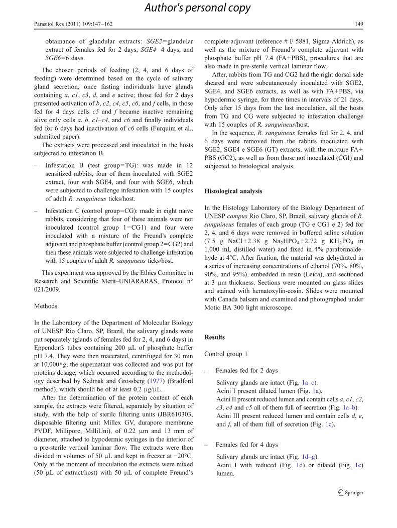
Ticks’ response to feeding on hostimmunized with glandular extracts ofRhipicephalus sanguineus females fedfor 2, 4, and 6 days. I. Inactivity or earlydegeneration of salivary glands?Karim Christina Scopinho Furquim,Maria Izabel Camargo Mathias, LetíciaMaria Gráballos Ferraz Hebling, GislaineCristina Roma, et al.
1 23
Your article is protected by copyright and
all rights are held exclusively by Springer-
Verlag. This e-offprint is for personal use only
and shall not be self-archived in electronic
repositories. If you wish to self-archive your
work, please use the accepted author’s
version for posting to your own website or
your institution’s repository. You may further
deposit the accepted author’s version on a
funder’s repository at a funder’s request,
provided it is not made publicly available until
12 months after publication.

ORIGINAL PAPER
Ticks’ response to feeding on host immunized with glandularextracts of Rhipicephalus sanguineus females fed for 2, 4,and 6 days. I. Inactivity or early degenerationof salivary glands?
Karim Christina Scopinho Furquim & Maria Izabel Camargo Mathias &
Letícia Maria Gráballos Ferraz Hebling & Gislaine Cristina Roma &
Gervásio Henrique Bechara
# Springer-Verlag 2011
Abstract The present study histologically analyzed thesalivary glands of Rhipicephalus sanguineus females fedfor 2, 4, and 6 days in hosts which had been previouslyimmunized with glandular extracts obtained from femalesfrom this same species in different periods of feeding,having as main objective verify the action of these extractsin the secretor cycle of these glands. For this, glandularextract of females fed for 2 days (SGE2), glandular extractof females fed for 4 days (SGE4), and glandular extract offemales fed for 6 days (SGE6) extracts were obtained fromsalivary glands of R. sanguineus females fed for 2, 4, and6 days respectively. Then, New Zealand White naiverabbits were inoculated either with extracts (test group=TG), or with a mixture of phosphate buffer and Freund'scomplete adjuvant (control group 2=CG2). Each inoculatedrabbit (TG and CG2) and non-inoculated (control group 1=CG1) was posteriorly infested with 15 couples of fasting R.sanguineus from which the salivary glands had beencollected from females fed for 2, 4, and 6 days. The resultsrevealed that the resistance the hosts had acquired by theimmunization with the extracts affected differently thesecretory activity of the glandular cells. It was verifiedthat the resistance to SGE2 and SGE4 extracts acted in
the cells of acini II and III, being c1 and c5 from II and dfrom III inactivated due to the action of SGE2 and c1 andc4 from II and f from III inactivated by the action ofSGE4. As for the resistance to SGE6 the effect was onlyon cells of acini II (c1, c3 e c4), which were alsoinactivated. In addition, the hosts’ resistance to SGE2–SGE6 extracts made the degenerative process earlier incomparison to CG1. On the other hand, the resistance tothe extracts did not influence the characteristics of thedegenerative process normally found in salivary glands.The assynchronism of the degenerative process wasmaintained—acini III were always the most affected andI the less affected. The structural cell alterations, such ascytoplasmic vacuolation, nuclear alterations and formationof apoptotic bodies which characterize the occurrence ofatypical apoptosis were also maintained in the glands ofindividuals from TG making it clear that the immunizationof the hosts with glandular extracts SGE2–SGE6 hadinfluenced the glandular physiology of R. sanguineus,which is an important piece of information in the searchfor a way to control these ectoparasites.
Introduction
The pharmacological and immunological properties ofticks’ saliva provide them with a very successful spoliativeaction on different species of animals (Ribeiro et al. 1985;Wikel 1999). The saliva is a complex and efficient mixturewhich acts in the modulation of immune-inflammatorymodulation of the host, allowing feeding and the permanenceon the host (Ribeiro et al. 1985; Wikel 1999; Sauer et al.2000). In addition, the fact that the saliva immunosuppresses
K. C. S. Furquim :G. H. Bechara (*)Departamento de Patologia Veterinária, FCAV, UNESP,Via de Acesso Prof. Paulo Castellane, s/n, CEP: 14884-900,Jaboticabal, SP, Brazile-mail: [email protected]
M. I. C. Mathias : L. M. G. F. Hebling :G. C. RomaDepartamento de Biologia, Instituto de Biociências, UNESP,Av. 24 A, nº 1515, Cx, Postal 199, CEP: 13506-900, Rio Claro,São Paulo, Brazil
Parasitol Res (2011) 109:147–162DOI 10.1007/s00436-010-2238-7
Received: 27 October 2010 /Accepted: 13 December 2010 /Published online: 11 January 2011
Author's personal copy

the host makes the ticks distinguished transmitters ofpathogenic agents which are veiculated by the saliva itself(Wikel 1999).
The ticks’ saliva is produced by salivary glands, organsthat present great morphofunctional complexity (Till 1961;Binnington 1978; Balashov 1983; Walker et al. 1985;Fawcett et al. 1986; Gill and Walker 1987; Sonenshine1991; Marzouk and Darwish 1994). In the females they arecomposed by types I, II, and III acini (Binnington 1978;Walker et al. 1985; Gill and Walker 1987; Sonenshine1991). Considering the importance of acini II and III for thefeeding and attachment of the ticks (Binnington 1978;Walker et al. 1985), as well as in the release of pathogens,these have to be deeply studied (Jittapalapong et al. 2008).
In the ticks, acini II are constituted by a, b, c1, c2, c3 ec4 cells (Binnington 1978) and in Rhipicephalus sangui-neus females c5 and c6 (Furquim et al., submitted paper)were additionally found. It is known that a cells areinvolved with cement secretion for the construction of thefixation cone (Binnington 1978; Walker et al. 1985;Fawcett et al. 1986; Gill and Walker 1987), and b and cwith several functions that have been attributed to the salivain the modulation of the host’s immune-inflammatoryresponse (Binnington 1978; Walker et al. 1985). Acini IIIare formed by d, e and f cells (Binnington 1978; Walker etal. 1985; Gill and Walker 1987). d and e secretecomponents of the cement (Binnington 1978; Walker etal. 1985; Gill and Walker 1987) and f have two functions:secretory and osmoregulative (Binnington 1978; Walker etal. 1985; Gill and Walker 1987).
In the ticks, the salivary glands (Wikel et al. 1978; Gill etal. 1986; Almeida et al. 1994; Ferreira et al. 1996; Szabó andBechara 1997; Jittapalapong et al. 2000a; Nunes et al. 2010)and the intestine (Szabó and Bechara 1997; Jittapalaponget al. 2000a; Caperucci et al. 2009, 2010; Veronez et al.2010) are important sites of antigens production. Because ofthis, after the first contact of these ectoparasites with somehosts, the latter develop resistance (Wikel et al. 1978; Gillet al. 1986; Jittapalapong et al. 2000a; Zhou et al. 2006). Inthis sense, several studies are carried out to verify theacquisition of resistance by the hosts when these areimmunized by successive infestations (Nunes et al. 2010;Monteiro and Bechara 2008; Monteiro et al. 2010; Veronezet al. 2010; Caperucci et al. 2009, 2010; Jittapalapong et al.2000a, b) or by the inoculation of extracts of whole ticks orpart of them (Wikel 1981; Ferreira et al. 1996; Szabó andBechara 1997; Jittapalapong et al. 2000a, b, 2008).
The acquisition of resistance by the hosts is measured bythe analysis of feeding and reproductive parameters ofinfesting ticks of previously immunized animals (Wikel1981; Szabó and Bechara 1997; Jittapalapong et al. 2000b),as well as by the analysis of the impact of this resistance inthe salivary glands (Sanders et al. 1996; Jittapalapong et al.
2008; Nunes et al. 2010) and intestines (Veronez et al.2010; Caperucci et al. 2009, 2010) of different species.
According to Wikel (1981) and Jittapalapong et al.(2000b, 2008) the immunization from antigens originated inthe salivary glands would stimulate the immunologicalresponse of the hosts which would affect the ticks ofsubsequent infestations. This would be caused by the directaction of the resistance acquired by the host in the salivaryglands’ secretory cycle reducing the efficiency of thefeeding process and the transmission of pathogens by theticks (Jittapalapong et al. 2008).
There are no studies which address the immunization ofhosts with extracts of salivary glands of R. sanguineusfemales in different periods of the glandular cycle, whichwould provide important information to understand how thehost’s organism would respond to pre-immunization withthe different glandular components of this species. Theinterest in studies of this nature would be justified by thefact that R. sanguineus species is considered cosmopoliteand the most predominant of Ixodidae family (Pegram et al.1987a, b). In addition to being an important transmitter ofdiseases like botounneuse fever for the men and babesiosisand erchilliosis for dogs (Walker et al. 2000). Furthermore,the ability of R. sanguineus to transmit Rickettsia rickettsii—the causing agent of Rocky Mountain spotted fever to thehuman being (Demma et al. 2005) and canine visceralleishmaniosis (Coutinho et al. 2005; Dantas-Torres 2008).
Considering the information above, the present studyhad the objective to histologically analyze the salivaryglands of R. sanguineus females with 2, 4, and 6 days offeeding subjected to the infestation on hosts previouslyimmunized with glandular extracts.
Material and methods
Material
To perform this study, salivary glands of adult R. sanguineusfemales fed for 2, 4, and 6 days subjected to infestation onNew Zealand White rabbits immunized with glandularextracts were used. For this, fasting individuals (males andfemales) from a colony kept in BOD incubator, in controlledconditions (29°C, 80% of humidity and photoperiod of12 h), in the Biotery of the Biology Department of UNESPcampus Rio Claro, SP were used in the infestations A, B andC made in rabbits according to the procedure described byBechara et al. (1995).
– Infestation A: made in naive rabbits using 25 couplesof R. sanguineus couples/host, for the acquisition offemales of ticks fed for 2 (55 individuals), 4 (36individuals), and 6 (25 individuals) days, for the
148 Parasitol Res (2011) 109:147–162
Author's personal copy
obtainance of glandular extracts: SGE2=glandularextract of females fed for 2 days, SGE4=4 days, andSGE6=6 days.
The chosen periods of feeding (2, 4, and 6 days offeeding) were determined based on the cycle of salivarygland secretion, once fasting individuals have glandscontaining a, c1, c3, d, and e active; those fed for 2 dayspresented activation of b, c2, c4, c5, c6, and f cells, in thosefed for 4 days cells c5 and f became inactive remainingalive only cells a, b, c1–c4, and c6 and finally individualsfed for 6 days had inactivation of c6 cells (Furquim et al.,submitted paper).
The extracts were processed and inoculated in the hostssubjected to infestation B.
– Infestation B (test group=TG): was made in 12sensitized rabbits, four of them inoculated with SGE2extract, four with SGE4, and four with SGE6, whichwere subjected to challenge infestation with 15 couplesof adult R. sanguineus ticks/host.
– Infestation C (control group=CG): made in eight naiverabbits, considering that four of these animals were notinoculated (control group 1=CG1) and four wereinoculated with a mixture of the Freund’s completeadjuvant and phosphate buffer (control group 2=CG2) andthen these animals were subjected to challenge infestationwith 15 couples of adult R. sanguineus ticks/host.
This experiment was approved by the Ethics Committee inResearch and Scientific Merit–UNIARARAS, Protocol n°021/2009.
Methods
In the Laboratory of the Department of Molecular Biologyof UNESP Rio Claro, SP, Brazil, the salivary glands wereput separately (glands of females fed for 2, 4, and 6 days) inEppendorfs tubes containing 200 μL of phosphate bufferpH 7.4. They were then macerated, centrifuged for 30 minat 10,000×g, the supernatant was collected and was put forproteins dosage, which occurred according to the methodol-ogy described by Sedmak and Grossberg (1977) (Bradfordmethod), which should be of at least 0.2 μg/μL.
After the determination of the protein content of eachsample, the extracts were filtered, separately by situation ofstudy, with the help of sterile filtering units (JBR610303,disposable filtering unit Millex GV, durapore membranePVDF, Millipore, MilliUni), of 0.22 μm and 13 mm ofdiameter, attached to hypodermic syringes in the interior ofa pre-sterile vertical laminar flow. The extracts were thendivided in volumes of 50 μL and kept in freezer at −20°C.Only at the moment of inoculation the extracts were mixed(50 μL of extract/host) with 50 μL of complete Freund’s
complete adjuvant (reference # F 5881, Sigma-Aldrich), aswell as the mixture of Freund’s complete adjuvant withphosphate buffer pH 7.4 (FA+PBS), procedures that arealso made in pre-sterile vertical laminar flow.
After, rabbits from TG and CG2 had the right dorsal sidesheared and were subcutaneously inoculated with SGE2,SGE4, and SGE6 extracts, as well as with FA+PBS, viahypodermic syringe, for three times in intervals of 21 days.Only after 15 days from the last inoculation, all the hostsfrom TG and CG were subjected to infestation challengewith 15 couples of R. sanguineus/host.
In the sequence, R. sanguineus females fed for 2, 4, and6 days were removed from the rabbits inoculated withSGE2, SGE4 e SGE6 (GT) extracts, with the mixture FA+PBS (GC2), as well as from those not inoculated (CGI) andsubjected to histological analysis.
Histological analysis
In the Histology Laboratory of the Biology Department ofUNESP campus Rio Claro, SP, Brazil, salivary glands of R.sanguineus females of each group (TG e CG1 e 2) fed for2, 4, and 6 days were removed in buffered saline solution(7.5 g NaCl+2.38 g Na2HPO4+2.72 g KH2PO4 in1,000 mL distilled water) and fixed in 4% paraformalde-hyde at 4°C. After fixation, the material was dehydrated ina series of increasing concentrations of ethanol (70%, 80%,90%, and 95%), embedded in resin (Leica), and sectionedat 3 μm thickness. Sections were mounted on glass slidesand stained with hematoxylin-eosin. Slides were mountedwith Canada balsam and examined and photographed underMotic BA 300 light microscope.
Results
Control group 1
– Females fed for 2 days
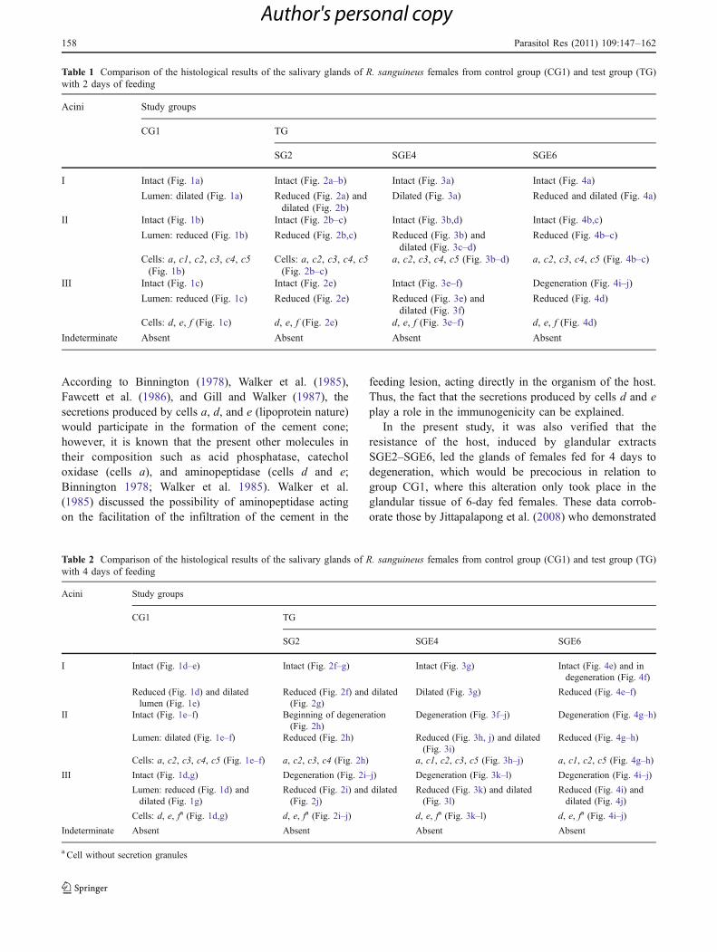
Salivary glands are intact (Fig. 1a–c).Acini I present dilated lumen (Fig. 1a).Acini II present reduced lumen and contain cells a, c1, c2,c3, c4 and c5 all of them full of secretion (Fig. 1a–b).Acini III present reduced lumen and contain cells d, e,and f, all of them full of secretion (Fig. 1c).
– Females fed for 4 days
Salivary glands are intact (Fig. 1d–g).Acini I with reduced (Fig. 1d) or dilated (Fig. 1e)lumen.
Parasitol Res (2011) 109:147–162 149
Author's personal copy

Acini II present slight increase in the diameter of thelumen (Fig. 1e–f) and cells a, c2, c3, c4, and c5 areobserved, all of them full of secretion granules(Fig. 1e–f).Acini III present reduced lumen (Fig. 1d) or a slightincrease in the diameter (Fig. 1g), in addition, cells d, eand f are also observed, d and e full of secretiongranules (Fig. d, g).
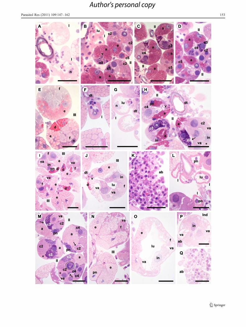
– Females fed for 6 days
In these, acini I, II, and III and Indeterminate arepresent, the latter scarce and this way named due to theadvanced stage of degeneration which made theiridentification difficult and countless apoptotic bodies(Fig. 1h–n). Acini II and III also present degenerativecharacteristics, more intense in the latter (Fig. 1j–l).Acini I present very dilated lumen (Fig.1h).Acini II present reduced lumen, intact (Fig. 1i) or indegeneration (beginning of vacuolation; Fig. 1j), thelatter being more numerous. Secretory cells a, c1, c2,c3, and c4 are also observed, full of secretion granules(Fig. 1i–j).As for acini III, these can present reduced (Fig. 1k) ordilated (Fig. 1l) lumen. In addition, they present d, e,and f cells, only d and e contain secretion granules andf, cytoplasmic vacuolation (Fig. 1k–l).
Control group
The salivary glands of females fed for 2, 4, and 6 days onthe hosts of this group presented similar characteristics tothe females from CG1 fed for 2, 4, and 6 days, respectively.
Test group
Inoculation of glandular extract of 2 days
– Females fed for 2 days
These salivary glands were intact (Fig. 2a–e).Acini I presented reduced (Fig. 2a) or dilated (Fig. 2b)lumen.Acini II presented reduced lumen (Fig. 2b–d). Cells a,c2, c3, c4 and c5 are present, all of them full ofsecretion (Fig. 2b–d).Acini III present reduced lumen and in them there ared, e, and f cells, all of them full of secretion (Fig. 2e).
– Females fed for 4 days
These glands present signs of degeneration such asbeginning of cytoplasmic vacuolation (Fig. 2h–j) andthe presence of apoptotic bodies (Fig. 2k).
Acini I are intact and can present reduced (Fig. 2f) ordilated (Fig. 2g) lumen.Acini II are starting to degenerate, present increase sizein relation to those from females fed for 2 days andsubjected to the extract of 2 days and reduced lumen(Fig. 2h). In addition, many acini II present cellswithout secretion granules (Fig. 2h).In acini II cells a, c2, c3, and c4 are observed, all ofthem full of secretion granules (Fig. 2h).Acini III are in process of degeneration and can presentreduced (Fig. 2i) or slightly dilated (Fig. 2j) lumen,being the former more numerous.In acini III cells d, e, and f are found (Fig. 2i–j). Cells dand e are full of secretion granules and in f, thecytoplasm does not contain secretion granules inaddition to being disorganized due to the beginningof vacuolation(Fig. 2i–j).
– Females fed for 6 days
Glands are in degeneration (Fig. 2l–p), beginningcytoplasmic vauolation (Fig. 2m–p), which is moreintense than that observed in the females fed for 4 daysand subjected to glandular extract of days as well asthe presence of apoptotic bodies (Fig. 2q).All kinds of acini with degenerative characteristics arefound (Fig. 2l–p).Acini I are in degeneration and present very dilatedlumen (Fig. 2l).Acini II are in degeneration, with reduced lumen andwith cells a, c2, c3, and c4, all full of secretiongranules (Fig. 2m). In addition, some acini with cellsfree of secretion granules were found (Fig. 2m).Acini III are in degeneration, therefore presentingirregular shape, cytoplasmic vacuolation, with reduced(Fig. 2n) or dilated (Fig. 2o) lumen, being the lattermore numerous.
Fig. 1 Histological sections of salivary glands of Rhipicephalussanguineus females used as control group 1 (CG1). a–c Two days offeeding presenting the glandular tissue intact, synthesis activity andsecretion and cells onset a (a), c1, c2, c3, c4, and c5 of acini II (II) (a–b) and d, e, and f of III (III) (c) full of secretion. d–g Four days offeeding with intact glands and intense secretory activity. In acini II (II)there are cells a, c2, c3, c4, and c5 (e, f) and in III cells d (d), e and f(III) (d, g), the latter without secretion. Acini II (II) present dilatedlumen (lu) (e–f). Acini III (III) present reduced (d) or dilated (g)lumen (lu). h–n Six days of feeding with acini II (II) (j), III (III) (k–l)and indeterminate (Ind) (m) presenting signs of degeneration. Acini II(II) are intact or in degeneration (j). Acini III (III) present reduced (k)or dilated (l) lumen (lu) and their cells f (f) (k–l) present intensecytoplasmic vacuolation (va). Acini in degeneration present loss ofcellular contact (arrow) (j–k), cytoplasmic vacuolation (va) (j–m),irregular (in) (j, k, m), picnotic (pn) (l), and fragmented (dottedrectangle) (k) nuclei. There are also apoptotic bodies (ab) (n). I acinustype I, lu acinus lumen, n nucleus; a cell a, c1 cell c1, c2 cell c2, c3cell c3, c4 cell c4, d cell d, e cell e. Bars 50 μm
b
150 Parasitol Res (2011) 109:147–162
Author's personal copy

Parasitol Res (2011) 109:147–162 151
Author's personal copy
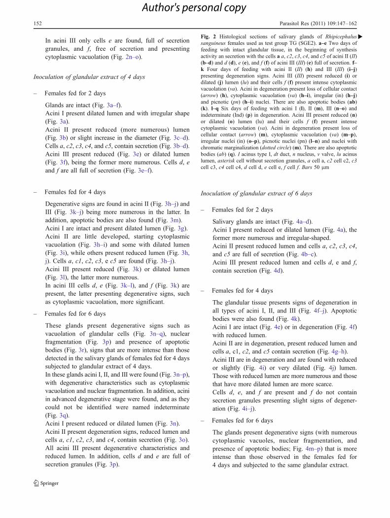
In acini III only cells e are found, full of secretiongranules, and f, free of secretion and presentingcytoplasmic vacuolation (Fig. 2n–o).
Inoculation of glandular extract of 4 days
– Females fed for 2 days
Glands are intact (Fig. 3a–f).Acini I present dilated lumen and with irregular shape(Fig. 3a).Acini II present reduced (more numerous) lumen(Fig. 3b) or slight increase in the diameter (Fig. 3c–d).Cells a, c2, c3, c4, and c5, contain secretion (Fig. 3b–d).Acini III present reduced (Fig. 3e) or dilated lumen(Fig. 3f), being the former more numerous. Cells d, eand f are all full of secretion (Fig. 3e–f).
– Females fed for 4 days
Degenerative signs are found in acini II (Fig. 3h–j) andIII (Fig. 3k–j) being more numerous in the latter. Inaddition, apoptotic bodies are also found (Fig. 3m).Acini I are intact and present dilated lumen (Fig. 3g).Acini II are little developed, starting cytoplasmicvacuolation (Fig. 3h–i) and some with dilated lumen(Fig. 3i), while others present reduced lumen (Fig. 3h,j). Cells a, c1, c2, c3, e c5 are found (Fig. 3h–j).Acini III present reduced (Fig. 3k) or dilated lumen(Fig. 3l), the latter more numerous.In acini III cells d, e (Fig. 3k–l), and f (Fig. 3k) arepresent, the latter presenting degenerative signs, suchas cytoplasmic vacuolation, more significant.
– Females fed for 6 days
These glands present degenerative signs such asvacuolation of glandular cells (Fig. 3n–q), nuclearfragmentation (Fig. 3p) and presence of apoptoticbodies (Fig. 3r), signs that are more intense than thosedetected in the salivary glands of females fed for 4 dayssubjected to glandular extract of 4 days.In these glands acini I, II, and III were found (Fig. 3n–p),with degenerative characteristics such as cytoplasmicvacuolation and nuclear fragmentation. In addition, aciniin advanced degenerative stage were found, and as theycould not be identified were named indeterminate(Fig. 3q).Acini I present reduced or dilated lumen (Fig. 3n).Acini II present degeneration signs, reduced lumen andcells a, c1, c2, c3, and c4, contain secretion (Fig. 3o).All acini III present degenerative characteristics andreduced lumen. In addition, cells d and e are full ofsecretion granules (Fig. 3p).
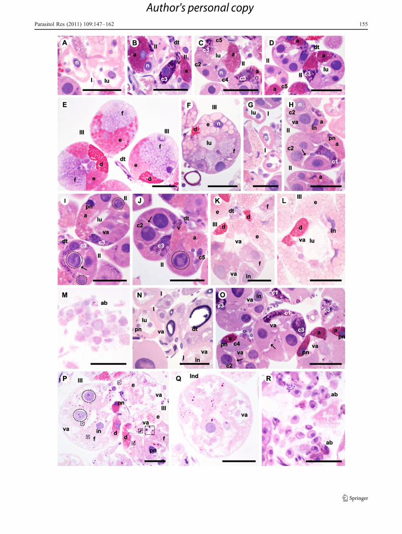
Inoculation of glandular extract of 6 days
– Females fed for 2 days
Salivary glands are intact (Fig. 4a–d).Acini I present reduced or dilated lumen (Fig. 4a), theformer more numerous and irregular-shaped.Acini II present reduced lumen and cells a, c2, c3, c4,and c5 are full of secretion (Fig. 4b–c).Acini III present reduced lumen and cells d, e and f,contain secretion (Fig. 4d).
– Females fed for 4 days
The glandular tissue presents signs of degeneration inall types of acini I, II, and III (Fig. 4f–j). Apoptoticbodies were also found (Fig. 4k).Acini I are intact (Fig. 4e) or in degeneration (Fig. 4f)with reduced lumen.Acini II are in degeneration, present reduced lumen andcells a, c1, c2, and c5 contain secretion (Fig. 4g–h).Acini III are in degeneration and are found with reducedor slightly (Fig. 4i) or very dilated (Fig. 4j) lumen.Those with reduced lumen are more numerous and thosethat have more dilated lumen are more scarce.Cells d, e, and f are present and f do not containsecretion granules presenting slight signs of degener-ation (Fig. 4i–j).
– Females fed for 6 days
The glands present degenerative signs (with numerouscytoplasmic vacuoles, nuclear fragmentation, andpresence of apoptotic bodies; Fig. 4m–p) that is moreintense than those observed in the females fed for4 days and subjected to the same glandular extract.
Fig. 2 Histological sections of salivary glands of Rhipicephalussanguineus females used as test group TG (SGE2). a–e Two days offeeding with intact glandular tissue, in the beginning of synthesisactivity an secretion with the cells a a, c2, c3, c4, and c5 of acini II (II)(b–d) and d (d), e (e), and f (f) of acini III (III) (e) full of secretion. f–k Four days of feeding with acini II (II) (h) and III (III) (i–j)presenting degeneration signs. Acini III (III) present reduced (i) ordilated (j) lumen (lu) and their cells f (f) present intense cytoplasmicvacuolation (va). Acini in degeneration present loss of cellular contact(arrow) (h), cytoplasmic vacuolation (va) (h–i), irregular (in) (h–j)and picnotic (pn) (h–i) nuclei. There are also apoptotic bodies (ab)(k). l–q Six days of feeding with acini I (l), II (m), III (n–o) andindeterminate (Ind) (p) in degeneration. Acini III present reduced (n)or dilated (o) lumen (lu) and their cells f (f) present intensecytoplasmic vacuolation (va). Acini in degeneration present loss ofcellular contact (arrow) (m), cytoplasmic vacuolation (va) (m–p),irregular nuclei (in) (o–p), picnotic nuclei (pn) (l–n) and nuclei withchromatic marginalization (dotted circle) (m). There are also apoptoticbodies (ab) (q). I acinus type I, dt duct, n nucleus, v valve, lu acinuslumen, asterisk cell without secretion granules, a cell a, c2 cell c2, c3cell c3, c4 cell c4, d cell d, e cell e, f cell f. Bars 50 μm
�
152 Parasitol Res (2011) 109:147–162
Author's personal copy
Parasitol Res (2011) 109:147–162 153
Author's personal copy

Acini I, II, III and indeterminate, all of them are withdegenerative characteristics (Fig. 4l–p).Acini I present dilated lumen (Fig. 4l).Acini II present reduced lumen and cells a, c1, c2, c3,and c4 contain secretion granules (Fig. 4m–n).Acini III present reduced lumen and cells d, e, and f(Fig. 4O); f present intense vacuolation in the cyto-plasm without secretion granules (Fig. 4o).The results of the glandular alterations found infemales fed for 2, 4, and 6 days form groups CG1and TG are summarized in Tables 1, 2, 3, 4, and 5.
Discussion
The present study provided histological results of salivaryglands of R. sanguineus females fed for 2, 4, and 6 days inrabbits which were previously immunized with glandularextracts SGE2–SGE6. It is clear that the resistance of thehost influenced in the secretory cycle of the infestingfemales glands, acting both in the secretory activity of theglandular cells (making them inactive) and leading theglands to early degeneration. Nunes et al. (2010) andJittapalapong et al. (2008) have reported that the resistanceacquired by the hosts studied by them induced the salivaryglands of Amblyomma cajennense and R. (Boophilus)microplus females, respectively, to necrosis. On the otherhand, the present study showed that the resistance of thehost led to the loss of function (inactivity) of somesecretory cells as well as to the early degeneration of thesalivary glands due to the occurrence of apoptosis.However, it is known that different species of ticks responddifferently to the resistance acquired by the host (Nunes et al.2010).
The results here obtained showed that under thehistological point of view, the glands of females fed for 2,4, and 6 days from CG1 (control) are similar to those fromCG2, demonstrating that Freund’s adjuvant, used withglandular extracts did not influence the glandular physiol-ogy. As for the glandular extracts, it is clear that they wouldinduce the development of resistance by the hosts, whichaffected the glandular physiology, i.e., the glands had thesecretory activity reduced, becoming less efficient whencompared to individuals from CG1, data that corroborateJittapalapong et al. (2008), who reported that the immuni-zation of the host with glandular extracts would affectdirectly the salivary glands of the infesting ticks, whichwould make the feeding process less efficient and wouldpossibly reduce the transmission of pathogens.
The results here presented showed that the resistanceacquired by the hosts immunized with SGE2, SGE4, andSGE6 extracts affected much more acini II and III cells than
those of acini I. This has probably occurred because thesecells present secretory function, while those of acini I act onthe osmoregulation and in the hydric balance of theectoparasite (Binnington 1978; Walker et al. 1985). Thus,cells of acini II and III are responsible for the secretion ofantigenic molecules and shape the immune-inflammatorysystem of the host, corroborating Jittapalapong et al. (2008)and Nunes et al. (2010) who also demonstrated that acini IIand III cells were more affected by the resistance of thehost.
Adding to this, Ferreira et al. (1996) demonstrated thatacini II and III of R. sanguineus ticks would be the onesthat synthesize more and secrete antigenic molecules whichwould cause the development of elevated immunologicresponse of the hosts. Gill et al. (1986) reported theoccurrence of intense immunological response to someglandular antigens of Hyalomma anatolicum anatolicumtick, such as glycoproteins, acid phosphatase, esterase andaminopetidase, molecules synthesized mainly by theglandular cells of acini II and III (Binnington 1978;Walker et al. 1985).
Other important point considered in the present studywas the resistance of the hosts of each of the GT groupsaffected the salivary glands of females fed for 2, 4, and6 days. In those fed on hosts immunized with SGE2 andSGE4, there were alterations in cells of acini II and III,while in those subjected to SGE6 only those of II wereaffected, demonstrating that the immune system of the hostsresponded in a different and specific way to the immunizationwith different extracts.
The resistance to extract SGE2 acted on the secretoryactivity of cells c1 of acini II, which were inactive for threeglandular periods, c5 of acini II, which were inactive in theglands of females fed for 4 days and in cells d of acini III,which would stay inactive in the glands of females fed for
Fig. 3 Histological sections of the salivary glands of Rhipicephalussanguineus femalesused as test group TG (SGE4). a–f Two days offeeding with intact cells, in the beginning of the secretory activity andcells a, c2, c3, c4, and c5 of acini II (II) (b–d) and d (d), e (e), e f (f) ofacini III (III) (e–f) full of secretion. Acini II (II) and III (III) presentreduced (b, e) or dilated (c–d, f) lumen (lu). g–m Four days of feedingwith degenerative signs in acini II (II) (h–j) and III (III) (k–l). Acini II(II) and III (III) present reduced (h, j–k) or dilated (i, l) lumen (lu).Acini in degeneration present loss of cellular contact (arrow) (h, j),cytoplasmic vacuolation (va) (h–i, k–l), irregular nuclei (in) (h, k–l),picnotic nuclei (pn) (h–i) and nuclei with chromatic marginalization(dotted circle) (i–j). There are also apoptotic bodies (ab) (m). n–r Sixdays of feeding with acini I (I) (n), II (II) (o), III (III) (p) andindeterminate (Ind) (q) in degeneration. In acini III (III) the cells f (f)present intense cytoplasmic vacuolation (va). Acini in degenerationpresent loss of cellular contact (arrow) (o), cytoplasmic vacuolation(va) (n–q), irregular (in) (n–p), picnotic (dotted circle) (n–p) andfragmented (dotted rectangle) (p) nuclei. Apoptotic bodies (ab) (r) arealso observed. I acinus type I, lu acinus lumen, n nucleus, v valve, dtduct, a cell a, c1 cell c1, c2 cell c2, c3 cell c3, c4 cell c4, c5 cell c5, dcell d, e cell e, f cell f. Bars 50 μm
�
154 Parasitol Res (2011) 109:147–162
Author's personal copy
Parasitol Res (2011) 109:147–162 155
Author's personal copy

6 days. As for the resistance to SGE4, it acted on cells c1and c4 of acini II and f of acini III. Cells c1 were inactive inglands of 2 days and started to present activity in those of4 days and 6 days. Cells c4 were active in glands of 2 days,lost activity in those of 4 days and became active again inglands of 6 days. As for cells f, they were active in glandsof 2 and 4 days and became inactive in those of 6 days.
The resistance acquired with the immunization withextract SGE6 acted only on cells c1, c3, c4, c1 wereinactive in the glands of 2 days and started to presentactivity in those of 4 and 6 days. Cells c3 e c4 were activein glands of 2 days, became active in those of 4 and start topresent activity again in those of 6 days.
It was demonstrated here that the glandular cells that hadtheir secretory performance more affected by the resistanceof the host were c1. The c4 presented intermediatealterations, followed by c3, c5, d, and f. Data here obtainedabout cells from group c and f corroborate those obtainedby Jittapalapong et al. (2008), who showed that these cellswould be very affected by the resistance of the host and thestudy by Nunes et al. (2010) corroborated data hereobtained for cells c1, c3, and c4, once according to theauthors the cells from group c would also be very affectedby the action of the resistance of the host.
Here, the fact that cells c1, c3, c4, c5, d, and f wereaffected by the resistance indicated that the secretionsproduced by them would be highly immunogenic inrelation to those from other types of cells present in thesalivary glands. In addition, the alterations suffered by cellsc1, c3, c4, c5, d, and f caused alterations in the glandularsecretory process, reducing it, which caused a reduction inthe feeding efficiency of R. sanguineus females anddemonstrated that such cells would possibly play animportant role in the modulation of the host. Differentlyfrom what was here demonstrated, Almeida et al. (1994)reported that the molecules from cells a, d, and e would bethe most immunogenic, while those from group c would notpresent this characteristic.
The fact that the secretions of cells c1, c3, and c4 arerelated to the modulation of the host corroborates Binning-ton (1978) and Walker et al. (1985), who reported that thecells from group c would act in the manipulation of theresponse of the host. According to Binnington (1978), cellsc1 of acini II of the salivary glands of Boophilus microplusfemales would be responsible for the production of esterase,which, according to Geczy et al. (1971) would act in theincrease of the vascular permeability and in the hydrolisisof cholesteryl ester.
From the analysis of the glands of females fed for 4 and6 days on host immunized with extract SGE4 importantinformation was obtained about cells c4 concerning theirparticipation in the modulation of the host. It was observedthat these cells were inactive in glands of 4 days, impairing
their glandular performance, which made the ectoparasite’sfeeding deficient. This can be proved by the fact that cells fof glands of 6 days were inactive, because in normalconditions they would be hypertrophied due to their intenseosmoregulating action, which would occur due to the fullconsumption of blood by the tick, making it clear that thesecretion produced by cells c4 would present, in addition toothers, the function of modulating the vasodilating mech-anisms and coagulation of the host’s blood, which wouldallow the great consumption of blood by the ectoparasite.
Although literature has indicated that d cells are involvedwith the fabrication of components of the cement coneduring the attachment of the tick (Walker et al. 1985; Gilland Walker 1987), the results here obtained demonstratedthat, in R. sanguineus females, the secretion produced bythese cells also play a role in the modulation of the host,once they were affected by the resistance acquired by theanimals immunized with SGE2 extract. Binnington (1978),studying the salivary glands of B. microplus females,observed that cells d would also synthesize and secreteacid phosphatase, enzymes that catalyze the moleculesbreak (digestion). Thus, considering what has previouslybeen reported, it can be inferred that the secretion releasedby cells d of R. sanguineus females would also be acting inthe digestion of the tissues of the host’s skin in order tomaintain the feeding lesion open.
In addition to the ability of resistance of the hosts fromgroup TG in making some glandular cells inactive, it isclear that this resistance affected the glandular cells a andc5 of acini II and d and e of acini III in a way that theyaltered the composition of the synthesized secretion,alteration reflected in the histology and that varied bothaccording to the study group and to the period of feeding ofthe females, allowing the assumption that the secretionsynthesized by such cells would be immunogenic andwould probably act in the modulation of the host.
Fig. 4 Histological sections of salivary glands of Rhipicephalussanguineus females used as test group TG (SGE6). a–d Two days offeeding with intact glandular tissue, beginning of the secretion phasewith cells a, c2, c3, c4, and c5 dos acini II (II) (b–c) e d (d), e (e) e f(f) (d) of III (III) full of secretion. e–k Four days of feeding with intact(I) acini I (e) or in degeneration (f) and acini II (II) and III (III) indegeneration (g–j). Os acini III (III) present reduced (i) or dilated (j)lumen (lu). In acini in process of degeneration are observed loss ofcellular contact (arrow) (h–j), cytoplasmic vacuolation (va) (f, i–j),irregular (in) (e, g, i–j), and picnotic (pn) (g) nuclei. Apoptotic bodiesare also observed (ab) (k). l–q Six days of feeding with acini I (i) (l),II (II) (m–n), III (III) (o) and indeterminate (Ind) (p) in degeneration.In acini III (III) the cells f (f) (o) present intense cytoplasmicvacuolation (va). Acini in degeneration present loss of cellular contact(arrow) (m–n), cytoplasmic vacuolation (va) (m–o), irregular (in) (m,o–p) and picnotic (pn) (l, o) nuclei, with chromatic marginalization(dotted circle) (n, p) and fragmented nuclei (dotted rectangle) (o).Apoptotic bodies are also observed (ab) (q). I type I acinus, lu acinuslumen, n nucleus, dt duct, v valve, a cell a, c1 cell c1, c2 cell c2, c3cell c3, c4 cell c4, c5 cell c5, d cell d, e cell e, f cell f. Bars 50 μm
�
156 Parasitol Res (2011) 109:147–162
Author's personal copy

Parasitol Res (2011) 109:147–162 157
Author's personal copy
According to Binnington (1978), Walker et al. (1985),Fawcett et al. (1986), and Gill and Walker (1987), thesecretions produced by cells a, d, and e (lipoprotein nature)would participate in the formation of the cement cone;however, it is known that the present other molecules intheir composition such as acid phosphatase, catecholoxidase (cells a), and aminopeptidase (cells d and e;Binnington 1978; Walker et al. 1985). Walker et al.(1985) discussed the possibility of aminopeptidase actingon the facilitation of the infiltration of the cement in the
feeding lesion, acting directly in the organism of the host.Thus, the fact that the secretions produced by cells d and eplay a role in the immunogenicity can be explained.
In the present study, it was also verified that theresistance of the host, induced by glandular extractsSGE2–SGE6, led the glands of females fed for 4 days todegeneration, which would be precocious in relation togroup CG1, where this alteration only took place in theglandular tissue of 6-day fed females. These data corrob-orate those by Jittapalapong et al. (2008) who demonstrated
Table 2 Comparison of the histological results of the salivary glands of R. sanguineus females from control group (CG1) and test group (TG)with 4 days of feeding
Acini Study groups
CG1 TG
SG2 SGE4 SGE6
I Intact (Fig. 1d–e) Intact (Fig. 2f–g) Intact (Fig. 3g) Intact (Fig. 4e) and indegeneration (Fig. 4f)
Reduced (Fig. 1d) and dilatedlumen (Fig. 1e)
Reduced (Fig. 2f) and dilated(Fig. 2g)
Dilated (Fig. 3g) Reduced (Fig. 4e–f)
II Intact (Fig. 1e–f) Beginning of degeneration(Fig. 2h)
Degeneration (Fig. 3f–j) Degeneration (Fig. 4g–h)
Lumen: dilated (Fig. 1e–f) Reduced (Fig. 2h) Reduced (Fig. 3h, j) and dilated(Fig. 3i)
Reduced (Fig. 4g–h)
Cells: a, c2, c3, c4, c5 (Fig. 1e–f) a, c2, c3, c4 (Fig. 2h) a, c1, c2, c3, c5 (Fig. 3h–j) a, c1, c2, c5 (Fig. 4g–h)
III Intact (Fig. 1d,g) Degeneration (Fig. 2i–j) Degeneration (Fig. 3k–l) Degeneration (Fig. 4i–j)
Lumen: reduced (Fig. 1d) anddilated (Fig. 1g)
Reduced (Fig. 2i) and dilated(Fig. 2j)
Reduced (Fig. 3k) and dilated(Fig. 3l)
Reduced (Fig. 4i) anddilated (Fig. 4j)
Cells: d, e, fa (Fig. 1d,g) d, e, fa (Fig. 2i–j) d, e, fa (Fig. 3k–l) d, e, fa (Fig. 4i–j)
Indeterminate Absent Absent Absent Absent
a Cell without secretion granules
Table 1 Comparison of the histological results of the salivary glands of R. sanguineus females from control group (CG1) and test group (TG)with 2 days of feeding
Acini Study groups
CG1 TG
SG2 SGE4 SGE6
I Intact (Fig. 1a) Intact (Fig. 2a–b) Intact (Fig. 3a) Intact (Fig. 4a)
Lumen: dilated (Fig. 1a) Reduced (Fig. 2a) anddilated (Fig. 2b)
Dilated (Fig. 3a) Reduced and dilated (Fig. 4a)
II Intact (Fig. 1b) Intact (Fig. 2b–c) Intact (Fig. 3b,d) Intact (Fig. 4b,c)
Lumen: reduced (Fig. 1b) Reduced (Fig. 2b,c) Reduced (Fig. 3b) anddilated (Fig. 3c–d)
Reduced (Fig. 4b–c)
Cells: a, c1, c2, c3, c4, c5(Fig. 1b)
Cells: a, c2, c3, c4, c5(Fig. 2b–c)
a, c2, c3, c4, c5 (Fig. 3b–d) a, c2, c3, c4, c5 (Fig. 4b–c)
III Intact (Fig. 1c) Intact (Fig. 2e) Intact (Fig. 3e–f) Degeneration (Fig. 4i–j)
Lumen: reduced (Fig. 1c) Reduced (Fig. 2e) Reduced (Fig. 3e) anddilated (Fig. 3f)
Reduced (Fig. 4d)
Cells: d, e, f (Fig. 1c) d, e, f (Fig. 2e) d, e, f (Fig. 3e–f) d, e, f (Fig. 4d)
Indeterminate Absent Absent Absent Absent
158 Parasitol Res (2011) 109:147–162
Author's personal copy

that the immunization of the hosts with glandular extractswould precociously induce the degeneration of thesalivary glands of R. (Boophilus) microplus females. Inaddition, Nunes et al. (2010) also reported that theresistance acquired by successive infestations would alsoinduce salivary glands of A. cajennense females to earlydegeneration.
A possible explanation for the early induction ofdegeneration would be the mechanism which controls thesynthesis of ecdisteroids, hormone which stimulates theglandular degeneration (Mao et al. 1995; Lomas et al. 1998;Weiss and Kaufman 2001), which was intensively synthe-sized in females from group TG (SGE2 and GS24), fed for4 days, a precocious synthesis in relation to what was
obtained in those exposed to normal conditions (occurringin the end of the feeding process; Lomas 1993).
The information above is probably explained by theacquisition of resistance by the immunized hosts, whichreduced the infesting ectoparasites’ blood consumption bymaking it more difficult (Furquim et al., submitted paper).Even with the reduction of blood ingestion these femaleswould reach the “critical weight” (increase in ten times theweight of a fasting tick) which would trigger the initialproduction of ecdysteroid, as would happen in normalconditions (Weiss and Kaufman 2001). However, uponreaching 4 days of feeding they would not be able to reachthe stage of rapid engorgement (consumption of a largevolume of blood in a short period of time), which was
Acini Period offeeding
Study groups
CG1 TG
SGE2 SGE4 SGE6
II 2 Days a (5) (Fig. 1a–b) a (5) (Fig. 2b–d) a (5) (Fig. 3b–d) a (6) (Fig. 4b–c)
c5 (3) (Fig. 1a–b) a (4) (Fig. 2b–d) c5 (3) (Fig. 3b–d) c5 (4) (Fig. 4c)
4 Days a (3) (Fig. 1e–f) a (6) (Fig. 2h) a (4) (Fig. 3h–j) a (7) (Fig. 4g–h)
c5 (2) (Fig. 1e) c5a c5 (2) (Fig. 3j) c5 (3) (Fig. 4g)
6 Days a (3) (Fig. 1i–j) a (7) (Fig. 2m) a (5) (Fig. 3o) a (7) (Fig. 4m)
c5a c5a c5a c5a
III 2 Days d (5) (Fig. 1c) d (5) (Fig. 2e) d (5) (Fig. 3e–f) d (5) (Fig. 4d)
e (3) (Fig. 1c) e (3) (Fig. 2e) e (4) (Fig. 3e–f) e (4) (Fig. 4d)
4 Days d (3) (Fig. 1d, g) d (5) (Fig. 2i–j) d (5) (Fig. 3k–l) d (4) (Fig. 4i–j)
e (3) (Fig. 1d, g) e (2) (Fig. 2i–j) e (5) (Fig. 3k–l) e (3) (Fig. 4i–j)
6 Days d (3) (Fig. 1l) da d (5) (Fig. 3p) d (4) (Fig. 4o)
e (0) (Fig. 1k–l) e (1) (Fig. 2n–o) e (6) (Fig. 3p) e (4) (Fig. 4o)
Table 4 Comparison of theresult of eosin staining ofsecretion granules of cells aand c5 of acini II and d and eof III of the salivary glands offemales from CG1 and TG fedfor 2, 4, and 6 days
(0) no staining, (7) stronglypositive staininga Cell not observed
Table 3 Comparison of the histological results of the salivary glands of R. sanguineus females from control group (CG1) and test group (TG)with 6 days of feeding
Acini Study groups
CG1 TG
SG2 SGE4 SGE6
I Intact (Fig. 1h) Degeneration (Fig. 2l) Degeneration (Fig. 3n) Degeneration (Fig. 4l)
Lumen: dilated (Fig. 1h) Dilated (Fig. 2l) Dilated (Fig. 3n) Dilated (Fig. 4l)
II Intact (Fig. 1i) and indegeneration (Fig. 1j)
Degeneration (Fig. 2m) Degeneration (Fig. 3o) Degeneration(Fig. 4m–n)
Lumen: reduced (Fig. 1i–j) Reduced (Fig. 2m) Reduced (Fig. 3o) Reduced (Fig. 4m–n)
Cells: a, c1, c2, c3, c4 (Fig. 1i–j) a, c2, c3, c4 (Fig. 2m) a, c1, c2, c3, c5 (Fig. 3o) a, c1, c2, c3, c4 (Fig. 4m–n)
III Degeneration (Fig. 1k–l) Degeneration (Fig. 2n–o) Degeneration (Fig. 3p) Degeneration (Fig. 4o)
Lumen: reduced (Fig. 1k) anddilated (Fig. 1l)
Reduced (Fig. 2n) and dilated(Fig. 2o)
Reduced (Fig. 3p) Reduced (Fig. 4o)
Cells: d, e, fa (Fig. 1k–l) e, fa (Fig. 2n–o) d, e (Fig. 3p) d, e, fa (Fig. 4o)
Indeterminate Scarce (Fig. 1m) Scarce (Fig. 2p) Numerous (Fig. 3q) Numerous (Fig. 4p)
a Cell without secretion granules
Parasitol Res (2011) 109:147–162 159
Author's personal copy
proved by the need of a longer time to reach completeengorgement (Furquim et al., submitted paper).
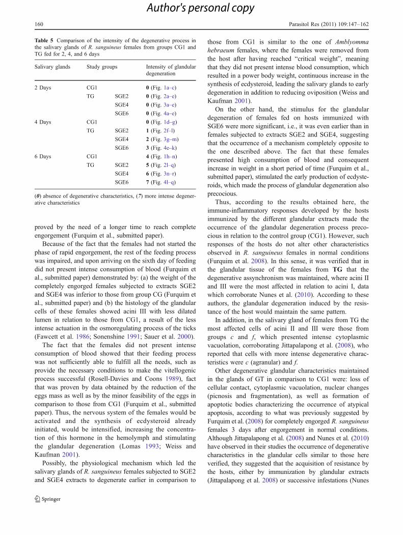
Because of the fact that the females had not started thephase of rapid engorgement, the rest of the feeding processwas impaired, and upon arriving on the sixth day of feedingdid not present intense consumption of blood (Furquim etal., submitted paper) demonstrated by: (a) the weight of thecompletely engorged females subjected to extracts SGE2and SGE4 was inferior to those from group CG (Furquim etal., submitted paper) and (b) the histology of the glandularcells of these females showed acini III with less dilatedlumen in relation to those from CG1, a result of the lessintense actuation in the osmoregulating process of the ticks(Fawcett et al. 1986; Sonenshine 1991; Sauer et al. 2000).
The fact that the females did not present intenseconsumption of blood showed that their feeding processwas not sufficiently able to fulfill all the needs, such asprovide the necessary conditions to make the vitellogenicprocess successful (Rosell-Davies and Coons 1989), factthat was proven by data obtained by the reduction of theeggs mass as well as by the minor feasibility of the eggs incomparison to those from CG1 (Furquim et al., submittedpaper). Thus, the nervous system of the females would beactivated and the synthesis of ecdysteroid alreadyinitiated, would be intensified, increasing the concentra-tion of this hormone in the hemolymph and stimulatingthe glandular degeneration (Lomas 1993; Weiss andKaufman 2001).
Possibly, the physiological mechanism which led thesalivary glands of R. sanguineus females subjected to SGE2and SGE4 extracts to degenerate earlier in comparison to
those from CG1 is similar to the one of Amblyommahebraeum females, where the females were removed fromthe host after having reached “critical weight”, meaningthat they did not present intense blood consumption, whichresulted in a power body weight, continuous increase in thesynthesis of ecdysteroid, leading the salivary glands to earlydegeneration in addition to reducing oviposition (Weiss andKaufman 2001).
On the other hand, the stimulus for the glandulardegeneration of females fed on hosts immunized withSGE6 were more significant, i.e., it was even earlier than infemales subjected to extracts SGE2 and SGE4, suggestingthat the occurrence of a mechanism completely opposite tothe one described above. The fact that these femalespresented high consumption of blood and consequentincrease in weight in a short period of time (Furquim et al.,submitted paper), stimulated the early production of ecdyste-roids, which made the process of glandular degeneration alsoprecocious.
Thus, according to the results obtained here, theimmune-inflammatory responses developed by the hostsimmunized by the different glandular extracts made theoccurrence of the glandular degeneration process preco-cious in relation to the control group (CG1). However, suchresponses of the hosts do not alter other characteristicsobserved in R. sanguineus females in normal conditions(Furquim et al. 2008). In this sense, it was verified that inthe glandular tissue of the females from TG that thedegenerative assynchronism was maintained, where acini IIand III were the most affected in relation to acini I, datawhich corroborate Nunes et al. (2010). According to theseauthors, the glandular degeneration induced by the resis-tance of the host would maintain the same pattern.
In addition, in the salivary gland of females from TG themost affected cells of acini II and III were those fromgroups c and f, which presented intense cytoplasmicvacuolation, corroborating Jittapalapong et al. (2008), whoreported that cells with more intense degenerative charac-teristics were c (agranular) and f.
Other degenerative glandular characteristics maintainedin the glands of GT in comparison to CG1 were: loss ofcellular contact, cytoplasmic vacuolation, nuclear changes(picnosis and fragmentation), as well as formation ofapoptotic bodies characterizing the occurrence of atypicalapoptosis, according to what was previously suggested byFurquim et al. (2008) for completely engorged R. sanguineusfemales 3 days after engorgement in normal conditions.Although Jittapalapong et al. (2008) and Nunes et al. (2010)have observed in their studies the occurrence of degenerativecharacteristics in the glandular cells similar to those hereverified, they suggested that the acquisition of resistance bythe hosts, either by immunization by glandular extracts(Jittapalapong et al. 2008) or successive infestations (Nunes
Table 5 Comparison of the intensity of the degenerative process inthe salivary glands of R. sanguineus females from groups CG1 andTG fed for 2, 4, and 6 days
Salivary glands Study groups Intensity of glandulardegeneration
2 Days CG1 0 (Fig. 1a–c)
TG SGE2 0 (Fig. 2a–e)
SGE4 0 (Fig. 3a–e)
SGE6 0 (Fig. 4a–e)
4 Days CG1 0 (Fig. 1d–g)
TG SGE2 1 (Fig. 2f–l)
SGE4 2 (Fig. 3g–m)
SGE6 3 (Fig. 4e–k)
6 Days CG1 4 (Fig. 1h–n)
TG SGE2 5 (Fig. 2l–q)
SGE4 6 (Fig. 3n–r)
SGE6 7 (Fig. 4l–q)
(0) absence of degenerative characteristics, (7) more intense degener-ative characteristics
160 Parasitol Res (2011) 109:147–162
Author's personal copy

et al. 2010), led the glandular cells of R. (Boophilus)microplus and A. cajennense females to necrosis.
Thus, it was here established that the immunization ofhosts with SGE2 and SGE4 glandular extracts of R.sanguineus females have influenced negatively in thesecretory cycle of salivary glands, reducing their secretoryability and consequently reducing the ectoparasites feeding(making it less efficient) important information for thebetter understanding and creation of alternatives to controlthese ectoparasites.
Acknowledgments This research has been supported by FAPESP(Fundação de Amparo à Pesquisa do Estado de São Paulo; grants no.08/58443-7 and 07/59020-0) and CNPq (Conselho Nacional deDesenvolvimento Científico e Tecnológico; grant no. 308733/2006-1and M.I. Camargo-Mathias and G.H. Bechara academic carrierresearch fellowships). Part of this work has been facilitated throughthe Integrated Consortium on Ticks and Tick-borne Diseases (ICTTD-3)supported by the European Union under contract number 510561-INCO.
References
Almeida APG, Bechara GH, Varma RMG (1994) Cross-reactivitybetween hard tick antigens. Braz J Med Biol Res 27:697–707
Balashov YuS (1983) An atlas of ixodid tick ultrastructure. In: RaikhelAS, Hoogstraal II (eds) Entomological Society of America.Special Publication, Lanham, pp 99–128
Bechara GH, Szabó MPJ, Ferreira BR, Garcia MV (1995)Rhipicephalus sanguineus tick in Brazil: feeding and reproduc-tive aspects under laboratorial conditions. Rev Bras ParasitolVet 4(2):61–66
Binnington KC (1978) Sequential changes in salivary gland structureduring attachment and feeding of the cattle tick Boophilusmicroplus. Int J Parasitol 8:97–115
Caperucci D, Bechara GH, Camargo-Mathias MI (2009) Histopathol-ogy and ultrastructure features of the midgut of adult females ofthe tick Amblyomma cajennense Fabricius, 1787 (Acari: Ixodi-dae) in various feeding stages and submitted to three infestations.Ultrastruct Pathol 33:249–259
Caperucci D, Bechara GH, Camargo-Mathias MI (2010) Ultrastruc-ture features of the midgut of the female adult Amblyommacajennense ticks Fabricius, 1787 (Acari: Ixodidae) in severalfeeding stages and subjected to three infestations. Micron.doi:10.1016/j.micron.2010.05.015
Coutinho MTZ, Bueno LL, Sterzik A, Fujiwara RT, Botelho JR, deMaria M, Genaro O, Linardi PM (2005) Participation ofRhipicephalus sanguineus (Acari: Ixodidae) in the epidemiologyof canine leishmaniasis. Vet Parasitol 128:149–155
Dantas-Torres F (2008) Canine vector-borne deseases in Brazil.Parasit Vectors 1(25):1–17
Demma LJ, Traeger MS, Nicholson WL, Paddock CD, Blau DM,Eremeeva ME, Dasch GA, Levin ML, Singleton J, Zaki SR,Cheek JE, Swerflow DL, McQuiston JH (2005) Rocky mountainspotted fever from an unexpected tick vector in Arizona. N EnglJ Med 353(6):587–594
Fawcett DW, Binnington KC, Voight WR (1986) The cell biology ofthe ixodid tick salivary gland. In: Sauer JR, Hair JA (eds)Morphology, physiology and behavioral biology of ticks. EllisHorwood, Chichester, pp 22–45
Ferreira BR, Machado RZ, Bechara GH (1996) Western blotanalysis of tick antigens from a Rhipicephalus sanguineus
unfed larval extract and identification of antigenic sites in ticksections using immunohistochemistry. A comparative studybetween resistant and susceptible host species. Vet Parasitol62:161–174
Furquim KCS, Bechara GH, Camargo-Mathias MI (2008) Death byapoptosis in salivary glands of females of the tick Rhipicephalussanguineus (Latreille, 1806) (Acari, Ixodidae). Exp Parasitol119:152–163
Geczy F, Nayghton MA, Clegg JB, Heweton RW (1971) Esterases anda carbohydrate-spliting enzyme in the saliva of the cattle tick,Boophilus microplus. J Parasitol 57:437–438
Gill HS, Walker AR (1987) The salivary glands of Hyalommaanatolicum anatolicum: nature of salivary gland componentsand their role in tick attachment and feeding. Int J Parasitol 18(1):83–93
Gill HS, Boid R, Ross CA (1986) Isolation and characterization ofsalivary antigens from Hyalomma anatolicum anatolicum.Parasite Immunol 8:11–25
Jittapalapong S, Stich RW, Gordon JC, Wittum TE, Barriga OO(2000a) Performance of female Rhipicephalus sanguineus (Acari:Ixodidae) fed dogs exposed to multiple infestations or immuni-zation with tick salivary gland or midgut tissues. J Med Entomol37(4):601–611
Jittapalapong S, Stich RW, Gordon JC, Bremer CA, Barriga OO(2000b) Humoral immune response of dogs immunized withsalivary gland, midgut, or repeated infestations with Rhipicephalussanguineus. Ann N Y Acad Sci 916:283–288
Jittapalapong S, Phichitrasilp T, Chanphao H, Rerkamnuychoke W,Stich RW (2008) Immunization with tick salivary gland extracts.Ann N Y Acad Sci 1149:200–204
Lomas LO (1993) The influence of a male-derived protein factor onsalivary gland degeneration in the ixodid tick, Amblyommahebraeum Koch. Ph.D Thesis, University of Alberta
Lomas LO, Gelman D, Kaufman WR (1998) Ecdysteroid regulationof salivary gland degeneration in the ixodid tick Amblyommahebraeum: a reconciliation of in vivo and in vitro observations.Gen Comp Endocrinol 109:200–211
Mao H, Macblain WA, Kaufman WR (1995) Some properties ofthe ecdysteroid receptor in the salivary gland of the ixodidtick, Amblyomma hebraeum. Gen Comp Endocrinol 99:340–348
Marzouk AS, Darwish ZEA (1994) Changes in the salivary glands offemale Hyalomma (Hyalomma) dromedarii during and afterfeeding. J Egypt Soc Parasitol 24(1):39–57
Monteiro GER, Bechara GH (2008) Cutaneus basophilia in theresistance of goats to Amblyomma cajennense nymphs afterrepeated infestations. Ann N Y Acad Sci 1149:221–225
Monteiro GER, Bechara GH, Franzin AM, de Miranda SantosIKF (2010) Antigen-presenting in draining lymph nodes ofgoads repeatedly infested by the Cayenne tick Amblyommacajennense nymphs. Exp Appl Acarol. doi:10.1007/s10493-010-9380-x
Nunes PH, Bechara GH, Camargo-Mathias MI (2010) Secretoryprocess of salivary glands of female Amblyomma cajennenseFabricius, 1787 (Acari: Ixodidae) ticks fed on resistant rabbits.Exp Appl Acarol. doi:10.1007/s10493-010-9388-2
Pegram RG, Clifford CM,Walker JB, Keirans JE (1987a) Clarification ofthe Rhipicephalus sanguineus group (Acari, Ixodoidea, Ixodidae).I. R. sulcatus Neuman 1908 and R. turanicus Pomerantsev 1936.Syst Parasitol 10:3–26
Pegram RG, Keirans JE, Clifford CM,Walker JB (1987b) Clarification ofthe Rhipicephalus sanguineus group (Acari, Ixodoidea, Ixodidae).II. R. sanguineus (Latreille, 1806) and related species. SystParasitol 10:27–44
Ribeiro JMC, Makoul GT, Levine J, Robinson DK, Spilman A(1985) Antihaemostatic, antiinflammatory and immunosupres-
Parasitol Res (2011) 109:147–162 161
Author's personal copy
sive properties of the saliva of a tick, Ixodes dammini. J ExpMed 161:332–344
Rosell-Davies R, Coons LB (1989) Relationship between feedingmating, vitellogenin production and vitellogenesis in the tickDermacentor variabilis. Exp Appl Acarol 7:95–105
Sanders ML, Scott AL, Glass GE, Schwartz BS (1996) Salivary glandchanges and host antibody responses associated with feeding ofmale lone star ticks (Acari: Ixodidae). J Med Entomol 33(4):628–634
Sauer JR, Essenberg RC, Bowman AS (2000) Salivary glands inixodid ticks: control and mechanism of secretion. J Insect Physiol46:1069–1078
Sedmak JJ, Grossberg SE (1977) A rapid, sensitive, and versatileassay for protein using Coomassie brilliant blue G250. AnalBiochem 79:544–552
Sonenshine DE (1991) Biology of ticks. Oxford University Press,New York
Szabó MPJ, Bechara GH (1997) Immunisation of dogs and guineapigs against Rhipicephalus sanguineus ticks using gut extract.Vet Parasitol 68:283–294
Till WM (1961) A contribution to the anatomy and histology of thebrown ear tick Rhipicephalus appendiculatus Neumann, vol 6.Memoirs of the Entomological Society of Southern Africa,Pretoria
Veronez VA, de Castro MB, Bechara GH, Szabó MPJ (2010)Histopathology of Rhipicephalus sanguineus (Acari: Ixodidae)ticks fed on resistant hosts. Exp Appl Acarol 50:151–161
Walker A, Fletcher JD, Gill HS (1985) Structural and histochemicalchanges in the salivary glands of Rhipicephalus appendiculatusduring feeding. Int J Parasitol 15(1):81–100
Walker JB, Keirans JE, Horak IG (2000) The genus Rhipicephalus(Acari, Ixodidae): a guide to the brown ticks of the world.Cambridge University Press, Cambridge
Weiss BL, Kaufman WR (2001) The relationship between ‘criticalweight’ and 20-hydroxyecdysone in the female ixodid tickAmblyomma hebraeum. J Insect Physiol 47:1261–1267
Wikel SK (1981) The induction of host resistance to tickinfestation with salivary gland antigen. Am J Trop MedHyg 30(1):284–288
Wikel SK (1999) Tick modulation of host immunity: an importantfactor in pathogen transmission. Int J Parasitol 29:851–859
Wikel SK, Graham JE, Allen JR (1978) Acquired resistance to ticks.IV. Skin reactivity and in vitro lymphocyte responsiveness tosalivary gland antigen. Immunology 34:257–263
Zhou J, Gong H, Zhou Y, Xuan X, Fujisaki K (2006) Identification ofa glycine-rich protein from the tick Rhipicephalus haemaphysa-loides and evaluation of its vaccine potential against tick feeding.Parasitol Res 100:77–84
162 Parasitol Res (2011) 109:147–162
Author's personal copy
Related Documents