56 The Open Biochemistry Journal, 2012, 6, 56-65 1874-091X/12 2012 Bentham Open Open Access Tianma Modulates Blood Vessel Tonicity Lin Feng 1,# , Arulmani Manavalan 1,2 , Manisha Mishra 1,2 , Siu Kwan Sze 1 , Jiang-Miao Hu 3 and Klaus Heese 1,2,* 1 School of Biological Sciences, Nanyang Technological University, 60 Nanyang Drive, 637551, Singapore 2 Institute of Advanced Studies, Nanyang Technological University, 60 Nanyang View, 639673, Singapore 3 Kunming Institute of Botany, Chinese Academy of Science, Kunming 650204, Yunnan, People’s Republic of China Abstract: Tianma is a traditional Chinese medicine (TCM) often used for the treatment of hypertension and heart dis- eases. To elucidate the function of tianma at the molecular level, we investigated the effect of tianma on vascular func- tions and aortic protein metabolism. We found that long-term treatment with tianma (~2.5g/kg/day for three months) in one-year-old rats could enhance acetylcholine (ACh)-induced vasorelaxation in endothelium-intact thoracic aortic rings against both KCl (80 mM)- and phenylephrine (PE)-induced contraction. By using the iTRAQ (isobaric tag for relative and absolute quantification) technique, we confirmed from the functional data at the proteome level that tianma treatment down-regulated the expressions of contractile proteins (e.g. Acta2) and other related structural proteins (e.g. desmin), and up-regulated the expressions of extracellular matrix (ECM) glycoproteins (e.g. Fbln5) and anti-thrombotic proteins (e.g. Anxa2) in aortic tissue. By inductive reasoning, tianma could perform its vasodilatory effect not only by inhibiting vascu- lar smooth muscle contraction, but also by enhancing blood vessel elasticity and stabilizing the arterial structure. Thus, tianma might become a novel therapeutic herbal medicine for cardiovascular diseases by regulating the aortic proteome metabolism. Keywords: Tianma, Vascular disease, Aorta, TCM. INTRODUCTION The research, development and use of natural products as therapeutic agents, especially those derived from higher plants, have been increasing in recent years. Traditional Chi- nese medicine (TCM) involves thousands of herbs for clini- cal treatments [1-3]. However, the underlying molecular and cellular mechanisms of most herbal medicines remain ob- scure. Tianma is the tuber of an orchid, Gastrodia elata Blume, and has been used as an ancient Chinese herbal medicine for treating various cardio- and cerebro-vascular and nervous diseases, including convulsion, headache, epilepsy, hyper- tension and coronary heart diseases [4-6]. Tianma contains many bioactive components such as vanilline, gastrodin, daucosterol, citric acid, succinct acid, parishin and minerals [7-11]. Besides its well known neuroprotective effects [12], tianma also plays a role in vascular circulation through con- trolling the functions of vascular smooth muscle cells (VSMCs) and inhibiting platelet aggregation [13]. Cardio- vascular remodeling caused by VSMCs overgrowth in the large and medium size vessels contributed to the pathogene- sis of hypertension and restenosis after angioplasty [14, 15]. *Address correspondence to this author at the Institute of Advanced Studies, Nanyang Technological University, 60 Nanyang View, 639673 Singapore; Tel: +65-6316-2848; Fax: +65-6791-3856; E-mail: [email protected] # Current address: Department of Obstetrics and Gynecology, University of California Irvine, Irvine, CA 92697, USA Tianma was found to inhibit VSMCs proliferation through decreasing the expressions of proliferating cell nuclear anti- gen (PCNA) and c-myc, thus inhibiting cardiovascular re- modeling [16]. Tianma could also elicit relaxant effects on smooth muscles in the blood vessels, gastrointestinal tract [17] and bladder [18] directly or through inhibiting neuro- genic contraction. In order to comprehensively understand the potential mechanisms by which tianma regulates vascular functions, we investigated the vasodilatory effects of tianma on vascu- lar smooth muscles by measuring the KCl- and phen- ylephrine (PE)-induced contractility of rat thoracic aorta ex vivo using myography. We then quantitatively analyzed the proteomic changes of arterial smooth muscle cells using iTRAQ (two-dimensional (2D) liquid chromatography cou- pled with tandem mass spectrometry (2D-LC-MS/MS)-based multidimensional protein identification technology combined with multiplex isobaric tag for relative and absolute quantifi- cation [19]) after long-term tianma treatment of one-year-old rats. The selective iTRAQ-detected changed proteins were further confirmed at the protein level by using western blot analyses (Fig. 1). Our experimental results showed for the first time that tianma could dilate blood vessels through regulating the cellular protein metabolism, including con- tractile/structural proteins as well as extra-cellular matrix glycoproteins and anti-thrombotic proteins.

Welcome message from author
This document is posted to help you gain knowledge. Please leave a comment to let me know what you think about it! Share it to your friends and learn new things together.
Transcript
56 The Open Biochemistry Journal, 2012, 6, 56-65
1874-091X/12 2012 Bentham Open
Open Access
Tianma Modulates Blood Vessel Tonicity
Lin Feng1,#
, Arulmani Manavalan1,2
, Manisha Mishra1,2
, Siu Kwan Sze1, Jiang-Miao Hu
3 and
Klaus Heese1,2,*
1School of Biological Sciences, Nanyang Technological University, 60 Nanyang Drive, 637551, Singapore
2Institute of Advanced Studies, Nanyang Technological University, 60 Nanyang View, 639673, Singapore
3Kunming Institute of Botany, Chinese Academy of Science, Kunming 650204, Yunnan, People’s Republic of China
Abstract: Tianma is a traditional Chinese medicine (TCM) often used for the treatment of hypertension and heart dis-
eases. To elucidate the function of tianma at the molecular level, we investigated the effect of tianma on vascular func-
tions and aortic protein metabolism. We found that long-term treatment with tianma (~2.5g/kg/day for three months) in
one-year-old rats could enhance acetylcholine (ACh)-induced vasorelaxation in endothelium-intact thoracic aortic rings
against both KCl (80 mM)- and phenylephrine (PE)-induced contraction. By using the iTRAQ (isobaric tag for relative
and absolute quantification) technique, we confirmed from the functional data at the proteome level that tianma treatment
down-regulated the expressions of contractile proteins (e.g. Acta2) and other related structural proteins (e.g. desmin), and
up-regulated the expressions of extracellular matrix (ECM) glycoproteins (e.g. Fbln5) and anti-thrombotic proteins (e.g.
Anxa2) in aortic tissue. By inductive reasoning, tianma could perform its vasodilatory effect not only by inhibiting vascu-
lar smooth muscle contraction, but also by enhancing blood vessel elasticity and stabilizing the arterial structure. Thus,
tianma might become a novel therapeutic herbal medicine for cardiovascular diseases by regulating the aortic proteome
metabolism.
Keywords: Tianma, Vascular disease, Aorta, TCM.
INTRODUCTION
The research, development and use of natural products as
therapeutic agents, especially those derived from higher
plants, have been increasing in recent years. Traditional Chi-
nese medicine (TCM) involves thousands of herbs for clini-
cal treatments [1-3]. However, the underlying molecular and
cellular mechanisms of most herbal medicines remain ob-
scure.
Tianma is the tuber of an orchid, Gastrodia elata Blume,
and has been used as an ancient Chinese herbal medicine for
treating various cardio- and cerebro-vascular and nervous
diseases, including convulsion, headache, epilepsy, hyper-
tension and coronary heart diseases [4-6]. Tianma contains
many bioactive components such as vanilline, gastrodin,
daucosterol, citric acid, succinct acid, parishin and minerals
[7-11]. Besides its well known neuroprotective effects [12],
tianma also plays a role in vascular circulation through con-
trolling the functions of vascular smooth muscle cells
(VSMCs) and inhibiting platelet aggregation [13]. Cardio-
vascular remodeling caused by VSMCs overgrowth in the
large and medium size vessels contributed to the pathogene-
sis of hypertension and restenosis after angioplasty [14, 15].
*Address correspondence to this author at the Institute of Advanced Studies,
Nanyang Technological University, 60 Nanyang View, 639673 Singapore;
Tel: +65-6316-2848; Fax: +65-6791-3856; E-mail: [email protected] #Current address: Department of Obstetrics and Gynecology, University of
California Irvine, Irvine, CA 92697, USA
Tianma was found to inhibit VSMCs proliferation through
decreasing the expressions of proliferating cell nuclear anti-
gen (PCNA) and c-myc, thus inhibiting cardiovascular re-
modeling [16]. Tianma could also elicit relaxant effects on
smooth muscles in the blood vessels, gastrointestinal tract
[17] and bladder [18] directly or through inhibiting neuro-
genic contraction.
In order to comprehensively understand the potential
mechanisms by which tianma regulates vascular functions,
we investigated the vasodilatory effects of tianma on vascu-
lar smooth muscles by measuring the KCl- and phen-
ylephrine (PE)-induced contractility of rat thoracic aorta ex
vivo using myography. We then quantitatively analyzed the
proteomic changes of arterial smooth muscle cells using
iTRAQ (two-dimensional (2D) liquid chromatography cou-
pled with tandem mass spectrometry (2D-LC-MS/MS)-based
multidimensional protein identification technology combined
with multiplex isobaric tag for relative and absolute quantifi-
cation [19]) after long-term tianma treatment of one-year-old
rats. The selective iTRAQ-detected changed proteins were
further confirmed at the protein level by using western blot
analyses (Fig. 1). Our experimental results showed for the
first time that tianma could dilate blood vessels through
regulating the cellular protein metabolism, including con-
tractile/structural proteins as well as extra-cellular matrix
glycoproteins and anti-thrombotic proteins.
Tianma Regulates Aorta Tonicity The Open Biochemistry Journal, 2012, Volume 6 57
MATERIALS AND METHODS
Reagents
Unless indicated, all reagents used for biochemical meth-ods were purchased from Sigma-Aldrich (St. Louis, MO, USA). Materials and reagents for SDS-PAGE (sodium dode-cyl sulfate-polyacrylamide gel electrophoresis) were from Bio-Rad (Bio-Rad Laboratories, Hercules, CA, USA). The iTRAQ reagent multi-plex kit, containing the iTRAQ rea-gents, was bought commercially (Applied Biosystems, Fos-ter City, CA, USA).
Animal Material
Experimental procedures, including the killing of ani-mals, were in accordance with the International Guiding Principles for Animal Research (WHO) and were approved
by the local Institutional Animal Care & Use Committee (NTU-IACUC). One-year-old male Wistar Kyoto rats (~250 g) were obtained from the laboratory animal centre (National University of Singapore) and randomly assigned to control and tianma-treated groups (10 each). According to previous reports and our own recent pilot studies, the average daily dose of tianma per rat was 2.5g/kg body weight [12, 18, 20-22]. They were fed a normal chow (Funabashi SP, Japan) and tap water was given freely. Room temperature (RT) was kept at 21 ± 2 ºC, with 60 % humidity, and a 12 h light/dark cycle. Tianma-feeding was done orally (intragastric admini-stration) with a blunt needle syringe by dispensing the tianma solution for the period of three months. Control rats were treated with the same volume of the solvent only. Ani-mals were sacrificed by CO2 asphyxiation. All efforts were made to minimize animal suffering and to reduce the number of animals used.
Herb Preparation
The rhizome of Gastrodia elata (tianma), grown under standardized conditions [23], was collected from Zhaotong City, China and provided by Dr. Jun Zhou (Kunming Insti-tute of Botany, Chinese Academy of Science, Yunnan, P.R. China). The species was identified and chemically analyzed as reported previously [12, 24]. A voucher specimen (0249742) was deposited in the herbarium of the Kunming Institute of Botany (Chinese Academy of Science, Yunnan, P.R. China). In this study, tianma was prepared according to previous reports [12, 16, 19, 20, 22, 25-27]. Whole dried tubers of the tianma were hammered into smaller pieces and subsequently ground to fine powder. 7.5 g of tianma powder was mixed with 100 ml sterilized Milli-Q water and boiled for 1 h at 100 ºC. The solution was centrifuged at 5000xg for 10 min at RT. The supernatant was filtered with a Whatman filter paper-1 (GE Healthcare, Chalfont St Giles, UK), yield-ing approximately 85 ml. The tianma solution was concen-trated at 60 ºC under vacuum and the final volume was re-duced to 10 ml for further applications.
Endothelium-Dependent Contractility/Relaxation Analy-sis with Rat Aorta Rings using Myograph
Briefly, the thoracic aorta was excised from the rat gently
after dissection, immediately immersed into cold physiologic
saline solution (PSS, 119 mM NaCl, 25 mM NaHCO3, 4.7
mM KCl, 1.18 mM KH2PO4, 1.17 mM MgSO4, 2.5 mM
CaCl2, 0.026 mM EDTA and 5.5 mM glucose; the solution
was gassed with 5 % CO2 in O2 to maintain pH at 7.4) and
kept on ice for further careful cleaning with scissors and for-
ceps under the microscope to remove fat and connective tis-
sues as well as blood clots. The aorta was then cut into eight
segments of 2 mm rings and kept in 4 °C-cold PSS. The in-
tegrity of the endothelium was tested by constricting with 10
μM PE and after steady contraction obtained, relaxed with
10 μM ACh [28, 29]. The myograph (Danish Myo Technol-
ogy, ADInstruments S.E.Asia, Subang Jaya, Selangor, Ma-
laysia) chambers were filled with ‘Krebs’ solution and the
aorta rings mounted onto the hooks inside the chambers for
isometric tension recording [30]. The tension was adjusted to
1 g as the baseline. The tissues were then washed twice with
124 mM K+ PSS to ensure the viability. For experimental set
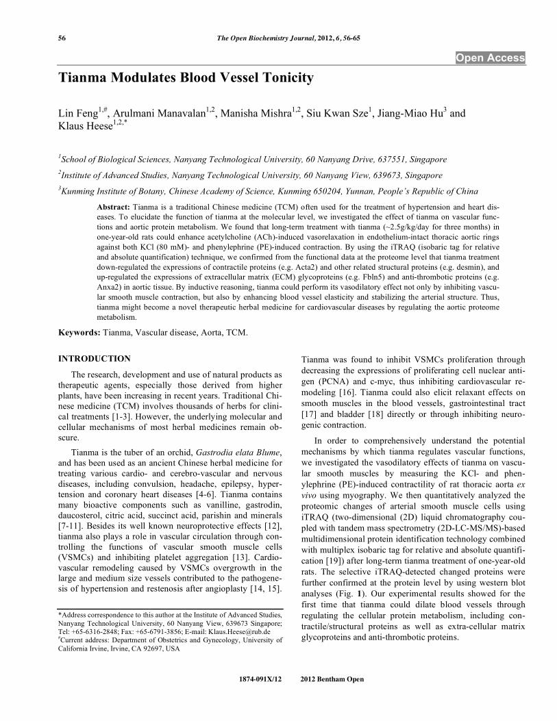
Fig. (1). Schematic representation of the experimental design and
the quantitative proteomics analyses showing biological and techni-
cal replicates. Following tianma treatment (batches B-I (5 rats) and
B-II (5 rats): +T; control = B-I (5 rats) and B-II (5 rats): -T) and
aortic tissue lysis, protein extracts were acetone precipitated and
quantified. These were then run in SDS-PAGE and subsequently
digested. The quantitative proteomics analyses of aortic tissue
lysates were performed by labeling with multi-plex isobaric tags
(114, 115, 116 and 117) for relative and absolute quantification
(iTRAQ) followed by Electrostatic Repulsion-Hydrophilic Interac-
tion Chromatography (ERLIC)-based fractionation, and liquid
chromatography coupled with tandem mass spectrometry (LC-
MS/MS)-based multidimensional protein identification technology.
The obtained data was analyzed using ProteinPilot software and
validated by quantitative western blots. Finally, proteins were func-
tionally classified into various subgroups.
58 The Open Biochemistry Journal, 2012, Volume 6 Feng et al.
I, the segments were initially loaded to an optimum stretch,
which was previously determined by using high-K+ physio-
logical solution (80 mM) as the contracting agent after ap-
plying different passive tensions. The initial stretch and the
length of the segments (2 mm) were consistently maintained
across all arterial rings of either group. The aorta rings were
pre-contracted with 80 mM K+ PSS until they reached a pla-
teau. Thereafter, an ACh-mediated relaxation was performed
as a concentration response curve (CRC) by adding increas-
ing concentrations of ACh in half-log concentration incre-
ments, i.e. 10-8
M, 3x10⁻⁸
M, 10-7
M, 3x10⁻⁷
M, 10-6
M, 3x10-
6 M, 10
-5 M, 3x10
-5 M, and 10
-4 M. After the ACh CRC, the
tissues were end-relaxed by applying 5x10⁻⁵
M sodium ni-
troprusside (SNP). For experimental set II, the aorta rings
were pre-contracted with 10-6
M PE and the ACh CRC ap-
plied. Upon reaching the PE plateau, the ACh CRC was per-
formed by half-log (10-8
M to 10-4
M) applications of ACh.
Again, after the ACh CRC, the tissues were further relaxed
by applying 5x10⁻⁵
M SNP. For data analysis the weight of
the dried aorta rings were measured for appropriate normali-
zation. Contraction responses to PE were calculated as per-
cent of its maximal contraction. Relaxant responses to ACh
were calculated as percent inhibition of the K+-/PE-induced
contraction. Data analysis was done using GraphPad Prism
for Windows (Graph-Pad Software, San Diego, CA, USA).
Data from several (8) vascular rings of the same rat were
averaged and presented as the datum for 1 rat, with the n
value (= 5) representing the number of rats for each group
(control or tianma-treated). Differences were considered
statistically significant at P < 0.05.
Maximal Contractile Force Measurement of the Aortal Ring in Response to PE
For this purpose, PE was initially added at a final concen-
tration of 10-7
M to the bath to contract the ring, and force
was allowed to stabilize for 5 min. ACh at a final concentra-
tion of 10-5
M was then added to the pre-contracted rings for
5 min to test for endothelial integrity and aortal ring viabil-
ity. If a ring failed to contract in response to PE or failed to
relax in response to ACh, it was replaced with another aortic
ring from the same rat. After the initial test for vessel viabil-
ity and endothelial integrity, the ring was washed 3 times
with PSS, allowed to equilibrate, and then re-washed with
fresh PSS at 10 min intervals until the measured active force
stabilized at 0 g. The maximum contraction achievable by
the ring was then determined by filling the bath with 80 mM
K+ and adding increasing concentrations of PE up to a final
concentration of 10-4
M (a PE CRC with half-log concentra-
tions was performed (i.e. 10-8
M, 3x10⁻⁸
M, 10-7
M, 3x10⁻⁷
M, 10-6
M, 3x10⁻⁶
M, 10-5
M, 3x10⁻⁵
M, and 10-4
M)). Maxi-
mal contractile force generated in response to the combina-
tion of 80 mM K+ and PE was normalized to the wet weight
of the aortic ring (determined at the end of the experiment).
After determining the maximum contraction of the aortic
rings, the vessels were allowed to stabilize and washed with
PSS every 10 min until the measured active force returned to
0 g.
Aorta-Tissue-Specific Protein Expression Analysis
For the aorta-tissue-specific proteome analyses, aorta tissues were isolated from tianma-treated and control rats. Briefly, the thoracic aorta was excised from the rats gently after dissection, immediately immersed into liquid nitrogen, and then powdered using a mortar and pestle. Upon the addi-tion of lysis buffer (2 % SDS, 0.5 M Triethyl ammonium bicarbonate buffer (TEAB), 1 Complete™ protease inhibitor cocktail tablet (Roche, Mannheim, Germany) and 1 Phos-STOP phosphatase inhibitor cocktail tablet (Roche)), the samples were vortexed for 1 min and incubated on ice for an additional 45 min prior to homogenisation (sonication pa-rameters: amplitude, 23 %; pulse: 5 s/ 5 s for 5 min) using a Vibra Cell high intensity ultrasonic processor (Jencon Scien-tific Ltd, Leighton Buzzard, Bedfordshire, UK). After cen-trifugation (20,000 x g / 4 °C / 30 min), supernatant was col-lected and stored at -80 °C until further use. The protein con-centration was quantified by a ‘2-D Quant’ kit (Amersham, Piscataway, NJ, USA) according to the manufacturer’s pro-tocol.
ITRAQ Protocol
A detailed description of the 2D-LC-MS/MS-iTRAQ procedures [19, 31-33], including post-proteomic data verifi-cation by SDS-PAGE - western blot analysis [34-36], can be found in the supplementary content document as described previously.
Antibodies
Anti-Anxa2 (Annexin-A2, 1:4000, polyclonal; Abcam, Cambridge, UK), anti-Des (Desmin, 1:1000, rabbit poly-clonal; Cell Signaling Technology Inc., Danvers, MA, USA), anti-Gapdh (1:1000, mouse monoclonal; Abcam), anti-Gstm2 (Glutathione S-transferase mu 2, 1:1000, goat poly-clonal; Santa Cruz Biotechnology Inc., Santa Cruz, CA, USA), anti-Serpinh1 (Serpin H1, 1:1000, rabbit polyclonal; Abnova, Taipei, Taiwan), anti-Vcl (Vinculin, 1:4000, mono-clonal; Abcam).
Statistical Evaluation
The mechanical responses of the vessels were measured as force and expressed as active wall tension, which is the increase in measured force divided by twice the segment length [30]. By using a computer program (GraphPad, Insti-tute for Scientific Information, San Diego, CA, USA), the CRCs were fitted to the classical Hill equation, as described earlier [29]. The results are expressed as mean ± SD. Differ-ences between means were analyzed using either one-way analysis of variance (ANOVA) followed by a Bonferroni t-test, Student’s t-test or paired t-test (SPSS (Statistical Prod-ucts and Service Solutions) for Windows Version 19 was used to perform ANOVA, optionally followed by Fisher’s Protected Least Significant Difference (PLSD) post hoc tests, when warranted). For the western blot analyses the Student’s t-test was applied accordingly. For the iTRAQ analysis ProteinPilot Software 3.0 was used as described above. To be considered statistically significant, we required a probability value to be at least < 0.05 (95 % confidence limit, *P < 0.05).
Tianma Regulates Aorta Tonicity The Open Biochemistry Journal, 2012, Volume 6 59
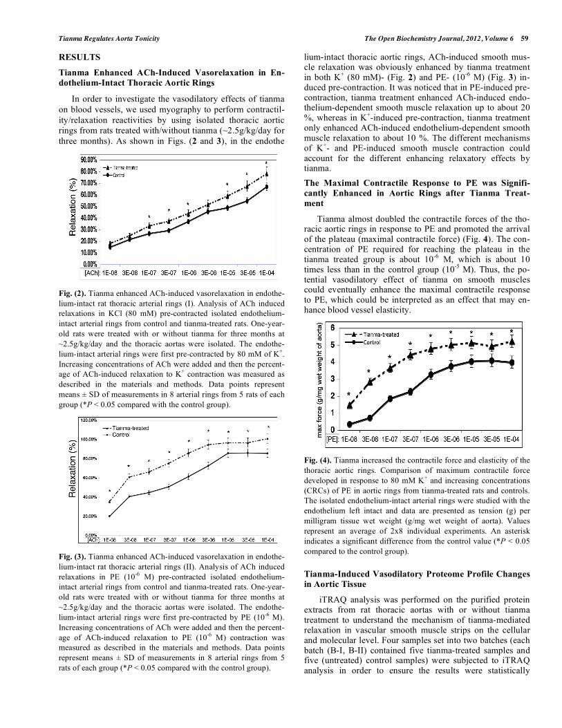
RESULTS
Tianma Enhanced ACh-Induced Vasorelaxation in En-
dothelium-Intact Thoracic Aortic Rings
In order to investigate the vasodilatory effects of tianma
on blood vessels, we used myography to perform contractil-
ity/relaxation reactivities by using isolated thoracic aortic
rings from rats treated with/without tianma (~2.5g/kg/day for
three months). As shown in Figs. (2 and 3), in the endothe
Fig. (2). Tianma enhanced ACh-induced vasorelaxation in endothe-
lium-intact rat thoracic arterial rings (I). Analysis of ACh induced
relaxations in KCl (80 mM) pre-contracted isolated endothelium-
intact arterial rings from control and tianma-treated rats. One-year-
old rats were treated with or without tianma for three months at
~2.5g/kg/day and the thoracic aortas were isolated. The endothe-
lium-intact arterial rings were first pre-contracted by 80 mM of K+.
Increasing concentrations of ACh were added and then the percent-
age of ACh-induced relaxation to K+ contraction was measured as
described in the materials and methods. Data points represent
means ± SD of measurements in 8 arterial rings from 5 rats of each
group (*P < 0.05 compared with the control group).
Fig. (3). Tianma enhanced ACh-induced vasorelaxation in endothe-
lium-intact rat thoracic arterial rings (II). Analysis of ACh induced
relaxations in PE (10-6
M) pre-contracted isolated endothelium-
intact arterial rings from control and tianma-treated rats. One-year-
old rats were treated with or without tianma for three months at
~2.5g/kg/day and the thoracic aortas were isolated. The endothe-
lium-intact arterial rings were first pre-contracted by PE (10-6
M).
Increasing concentrations of ACh were added and then the percent-
age of ACh-induced relaxation to PE (10-6
M) contraction was
measured as described in the materials and methods. Data points
represent means ± SD of measurements in 8 arterial rings from 5
rats of each group (*P < 0.05 compared with the control group).
lium-intact thoracic aortic rings, ACh-induced smooth mus-cle relaxation was obviously enhanced by tianma treatment in both K
+ (80 mM)- (Fig. 2) and PE- (10
-6 M) (Fig. 3) in-
duced pre-contraction. It was noticed that in PE-induced pre-contraction, tianma treatment enhanced ACh-induced endo-thelium-dependent smooth muscle relaxation up to about 20 %, whereas in K
+-induced pre-contraction, tianma treatment
only enhanced ACh-induced endothelium-dependent smooth muscle relaxation to about 10 %. The different mechanisms of K
+- and PE-induced smooth muscle contraction could
account for the different enhancing relaxatory effects by tianma.
The Maximal Contractile Response to PE was Signifi-
cantly Enhanced in Aortic Rings after Tianma Treat-
ment
Tianma almost doubled the contractile forces of the tho-racic aortic rings in response to PE and promoted the arrival of the plateau (maximal contractile force) (Fig. 4). The con-centration of PE required for reaching the plateau in the tianma treated group is about 10
-6 M, which is about 10
times less than in the control group (10-5
M). Thus, the po-tential vasodilatory effect of tianma on smooth muscles could eventually enhance the maximal contractile response to PE, which could be interpreted as an effect that may en-hance blood vessel elasticity.
Fig. (4). Tianma increased the contractile force and elasticity of the
thoracic aortic rings. Comparison of maximum contractile force
developed in response to 80 mM K+ and increasing concentrations
(CRCs) of PE in aortic rings from tianma-treated rats and controls.
The isolated endothelium-intact arterial rings were studied with the
endothelium left intact and data are presented as tension (g) per
milligram tissue wet weight (g/mg wet weight of aorta). Values
represent an average of 2x8 individual experiments. An asterisk
indicates a significant difference from the control value (*P < 0.05
compared to the control group).
Tianma-Induced Vasodilatory Proteome Profile Changes
in Aortic Tissue
iTRAQ analysis was performed on the purified protein extracts from rat thoracic aortas with or without tianma treatment to understand the mechanism of tianma-mediated relaxation in vascular smooth muscle strips on the cellular and molecular level. Four samples set into two batches (each batch (B-I, B-II) contained five tianma-treated samples and five (untreated) control samples) were subjected to iTRAQ analysis in order to ensure the results were statistically

60 The Open Biochemistry Journal, 2012, Volume 6 Feng et al.
meaningful. Identified proteins were also visualized in a vir-tual two-dimensional protein gel, JVirGel, and categorized by their calculated isoelectric points and molecular weights [37]. The protein spots were well separated without aggrega-tion, indicating a well-qualified whole cell proteomic pattern (Fig. 5).
Fig. (5). Simulated 2D gel presentation of rat thoracic aorta-derived
quantified proteins. MW and pI values generated from JVirGel
(http://www.jvirgel.de/). The proteins identified by LC-MS/MS
were uploaded onto JVirGel, online software used to create a 2D
gel image. This image confirmed that the tissue lysis performed was
adequate and the entire proteome within cells was extracted.
We identified a total of 1298 proteins through iTRAQ analysis out of which 1166 proteins were quantified (with a strict cutoff of unused ProtScore 2 as the qualification cri-teria, which corresponds to a peptide confidence level of 99 % and an applied FDR (false discovery rate) of 0.33% (<1.0%)). Comparing the expression levels between tianma-treated and untreated samples, 54 proteins showed an altered expression level (30 proteins were down-regulated and 24 proteins were up-regulated, (Supplementary data: Table 1)). We used Panther, UniProt, and NCBI online databases for classifying the altered proteins based on their role and local-ization. Approximately 33.30 % of the altered proteins were cytoskeletal, 26 % were catalytic and 10 % were extracellu-lar matrix (ECM) proteins (Fig. 6).
We focused our interest on the structural protein sub-group: In tianma treated aortic rings, actin (Acta2) decreased to about half the level of the control (with p<0.02) while other structural proteins, like desmin (Des), vinculin (Vcl), PDZ and LIM domain protein 1 (Pdlim1), tubulin beta 6 (Tubb6), alpha-parvin (Parva) and microtubule-associated protein-4 (Map4) were also down-regulated by tianma treat-ment upto 30 % - 60 %, whereas Myo1c and lamin-B1 (Lmnb1) were up-regulated. Most ECM/cell-surface proteins were also up-regulated. Elastin (Eln) and proline arginine-rich end leucine-rich repeat protein (Prelp) expressions were increased to more than twice of the control while fibulin-5 (Fbln5), biglycan (Bgn) and fibromodulin (Fmod) expres-sions were also increased to more than 1.3 times of the con-trol. In contrast, fibronectin (Fn1) and periostin (Postn) were down-regulated to about half of the control level (Fig. 7).
Fig. (7). Effect of tianma on the expression levels of cytoskeletal
and ECM proteins. Protein expression levels were quantitatively
analyzed ex-vivo by using iTRAQ after tianma treatment for three
months as described in the experimental procedures. The Y-axis
shows the iTRAQ ratios of proteins between tianma-treated and
untreated controls. Values above 1.2 indicate up-regulation and
below 0.83 indicate down-regulation of the proteins. Proteins
shown (from left to right): black bars: down-regulated: Acta2, Actb,
Tubb6, Pdlim1, Des, Vcl, Parva, Cryab, Map4, Coro1c, Eml2, up-
regulated: Myo1c, Pxn, Lmnb1, Flnc. White bars: up-regulated:
Eln, Fbln5, Prelp, Bgn, Fmod; down-regulated: Fn1, Postn.
Validation of Tianma-Modulated Proteins by Western Blot
Following the database search and classification of pro-teins, western blots were performed on randomly selected
Fig. (6). Pie chart depicting the iTRAQ identified proteins characterized by the molecular function GO category. Proteins identified and
quantified by iTRAQ, were classified in terms of their role in biological processes (A) and molecular functions (B).
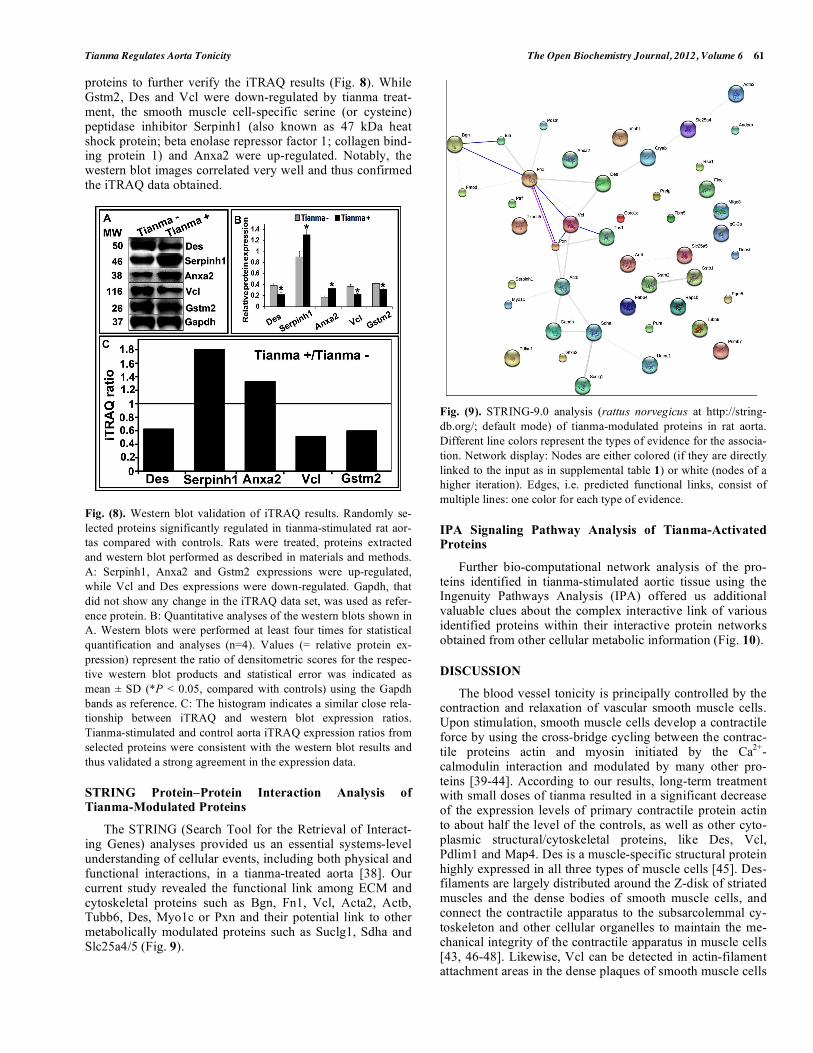
Tianma Regulates Aorta Tonicity The Open Biochemistry Journal, 2012, Volume 6 61
proteins to further verify the iTRAQ results (Fig. 8). While Gstm2, Des and Vcl were down-regulated by tianma treat-ment, the smooth muscle cell-specific serine (or cysteine) peptidase inhibitor Serpinh1 (also known as 47 kDa heat shock protein; beta enolase repressor factor 1; collagen bind-ing protein 1) and Anxa2 were up-regulated. Notably, the western blot images correlated very well and thus confirmed the iTRAQ data obtained.
Fig. (8). Western blot validation of iTRAQ results. Randomly se-
lected proteins significantly regulated in tianma-stimulated rat aor-
tas compared with controls. Rats were treated, proteins extracted
and western blot performed as described in materials and methods.
A: Serpinh1, Anxa2 and Gstm2 expressions were up-regulated,
while Vcl and Des expressions were down-regulated. Gapdh, that
did not show any change in the iTRAQ data set, was used as refer-
ence protein. B: Quantitative analyses of the western blots shown in
A. Western blots were performed at least four times for statistical
quantification and analyses (n=4). Values (= relative protein ex-
pression) represent the ratio of densitometric scores for the respec-
tive western blot products and statistical error was indicated as
mean ± SD (*P < 0.05, compared with controls) using the Gapdh
bands as reference. C: The histogram indicates a similar close rela-
tionship between iTRAQ and western blot expression ratios.
Tianma-stimulated and control aorta iTRAQ expression ratios from
selected proteins were consistent with the western blot results and
thus validated a strong agreement in the expression data.
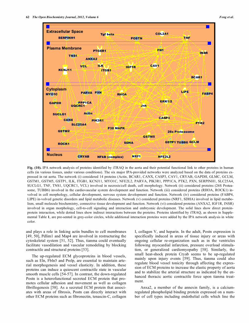
STRING Protein–Protein Interaction Analysis of Tianma-Modulated Proteins
The STRING (Search Tool for the Retrieval of Interact-ing Genes) analyses provided us an essential systems-level understanding of cellular events, including both physical and functional interactions, in a tianma-treated aorta [38]. Our current study revealed the functional link among ECM and cytoskeletal proteins such as Bgn, Fn1, Vcl, Acta2, Actb, Tubb6, Des, Myo1c or Pxn and their potential link to other metabolically modulated proteins such as Suclg1, Sdha and Slc25a4/5 (Fig. 9).
Fig. (9). STRING-9.0 analysis (rattus norvegicus at http://string-
db.org/; default mode) of tianma-modulated proteins in rat aorta.
Different line colors represent the types of evidence for the associa-
tion. Network display: Nodes are either colored (if they are directly
linked to the input as in supplemental table 1) or white (nodes of a
higher iteration). Edges, i.e. predicted functional links, consist of
multiple lines: one color for each type of evidence.
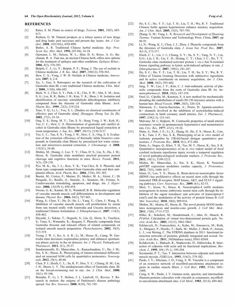
IPA Signaling Pathway Analysis of Tianma-Activated Proteins
Further bio-computational network analysis of the pro-teins identified in tianma-stimulated aortic tissue using the Ingenuity Pathways Analysis (IPA) offered us additional valuable clues about the complex interactive link of various identified proteins within their interactive protein networks obtained from other cellular metabolic information (Fig. 10).
DISCUSSION
The blood vessel tonicity is principally controlled by the contraction and relaxation of vascular smooth muscle cells. Upon stimulation, smooth muscle cells develop a contractile force by using the cross-bridge cycling between the contrac-tile proteins actin and myosin initiated by the Ca
2+-
calmodulin interaction and modulated by many other pro-teins [39-44]. According to our results, long-term treatment with small doses of tianma resulted in a significant decrease of the expression levels of primary contractile protein actin to about half the level of the controls, as well as other cyto-plasmic structural/cytoskeletal proteins, like Des, Vcl, Pdlim1 and Map4. Des is a muscle-specific structural protein highly expressed in all three types of muscle cells [45]. Des-filaments are largely distributed around the Z-disk of striated muscles and the dense bodies of smooth muscle cells, and connect the contractile apparatus to the subsarcolemmal cy-toskeleton and other cellular organelles to maintain the me-chanical integrity of the contractile apparatus in muscle cells [43, 46-48]. Likewise, Vcl can be detected in actin-filament attachment areas in the dense plaques of smooth muscle cells
62 The Open Biochemistry Journal, 2012, Volume 6 Feng et al.
and plays a role in linking actin bundles to cell membranes [49, 50], Pdlim1 and Map4 are involved in restructuring the cytoskeletal system [51, 52]. Thus, tianma could eventually facilitate vasodilation and vascular remodeling by blocking contractile and structural proteins [53].
The up-regulated ECM glycoproteins in blood vessels, such as Eln, Fbln5 and Prelp, are essential to maintain arte-rial morphogenesis and vessel elasticity. In addition, these proteins can induce a quiescent contractile state in vascular smooth muscle cells [54-57]. In contrast, the down-regulated Postn is a heterofunctional secreted ECM protein that pro-motes cellular adhesion and movement as well as collagen fibrillogenesis [58]. As a secreted ECM protein that associ-ates with areas of fibrosis, Postn can directly interact with other ECM proteins such as fibronectin, tenascin-C, collagen
I, collagen V, and heparin. In the adult, Postn expression is specifically induced in areas of tissue injury or areas with ongoing cellular re-organization such as in the ventricles following myocardial infarction, pressure overload stimula-tion, or generalized cardiomyopathy [58]. Similarly, the small heat-shock protein Cryab seems to be up-regulated mainly upon injury events [59]. Thus, tianma could also regulate blood vessel tonicity through affecting the expres-sion of ECM proteins to increase the elastic property of aorta and to stabilize the arterial structure as indicated by the en-hanced thoracic aortic contractile force upon tianma treat-ment.
Anxa2, a member of the annexin family, is a calcium-regulated phospholipid binding protein expressed on a num-ber of cell types including endothelial cells which line the
Fig. (10). IPA network analysis of proteins identified by iTRAQ in the aorta and their potential functional link to other proteins in human
cells (in various tissues, under various conditions). The six major IPA-provided networks were analyzed based on the data of proteins ex-
pressed in rat aorta. The network (i) considered 14 proteins (Actin, BCAR1, CANX, CASP3, CAV1, CRYAB, GAPDH, GLMC, GCLM,
GSTM1, GSTM5, GSTP1, ILK, ITGB1, KCNJ11, MYO1C, NFE2L2, PARVA, PIK3R1, PPP1CA, PTK2, PXN, SERPINH1, SLC25A4,
SUCLG1, TNF, TNS1, UQCRC1, VCL) involved in necrosis/cell death, cell morphology. Network (ii) considered proteins (26S Protea-
some, TUBB6) involved in the cardiovascular system development and function. Network (iii) considered proteins (RHOA, ROCK1) in-
volved in cell morphology, cellular development, nervous system development and function. Network (iv) considered proteins (FABP4,
LIPE) in-volved genetic disorders and lipid metabolic diseases. Network (v) considered proteins (NRF1, SDHA) involved in lipid metabo-
lism, small molecule biochemistry, connective tissue development and function. Network (vi) considered proteins (ANXA2, IGF1R, INSR)
involved in organ morphology, cell-to-cell signaling and interaction and embryonic development. The solid lines show direct protein-
protein interaction, while dotted lines show indirect interactions between the proteins. Proteins identified by iTRAQ, as shown in Supple-
mental Table 1, are pre-sented in grey-color circles, while additional interaction proteins were added by the IPA network analysis in white
color.

Tianma Regulates Aorta Tonicity The Open Biochemistry Journal, 2012, Volume 6 63
blood vessel wall [60-63]. Decreased binding of Anxa2 to endothelial cells contributes to atherothrombotic disease and elevated Anxa2 expression by tianma treatment indicates that it could improve blood circulation through increasing blood flow and preventing thrombosis.
Furthermore, tianma inhibited the protein expression of the fatty acid binding protein 4 (Fabp4) which belongs to the family of lipid chaperones that control intracellular fluxes and compartmentalization of their respective ligands (e.g. fatty acids). Recently it has been demonstrated that reduced levels of Fabp4, as shown in tianma-treated aortic tissue, may protect against atherosclerosis and other cardiovascular and metabolic diseases such as diabetes and obesity [64-68].
In addition, IPA analysis could demonstrate the involve-ment of the iTRAQ-based analysis-identified proteins and their metabolic interactive pathways within a cardiovascular network associated with specific cardiovascular diseases, eventually important for the proper functions of active blood vessels in the circulation system during regenerative proc-esses (Fig. 11).
In summary, our ex vivo study reveals a vasodilatory ef-fect of tianma and the potential molecular mechanisms in-volved in this process: (i) restriction of contractile activity in smooth muscle cells through the inhibition of contractile and
structural proteins; and (ii) increasing the arterial elasticity and stabilizing the arterial structure through modulating the expression of ECM proteins. Since many cardio- and cere-bro-vascular diseases, such as hypertension, atherosclerosis, stroke and headache, are closely related to the abnormal con-traction of vascular smooth muscles, decrease of arterial elasticity and increase of blood coagulation [44, 69], the dis-closure of all the bioactive ingredients would facilitate the application of tianma as an efficient therapeutic herbal medi-cine [6, 12, 24, 70-73].
CONFLICT OF INTEREST
None declared.
ACKNOWLEDGEMENTS
This study was supported by the Institute of Advanced Studies, Nanyang Technological University. We thank Mr. M. Li, (Institute of Advanced Studies) and Dr. W. Liang (School of Biological Sciences) for technical assistance.
SUPPLEMENTARY MATERIAL
Supplementary material is available on the publisher’s web site along with the published article.
Fig. (11). Protein function and human diseases (Fx). Five important proteins that are involved in various cardiovascular disorders are shown.
The significance of their role in these diseases needs to be explored to develop more specific drugs targeting those genes or proteins.
64 The Open Biochemistry Journal, 2012, Volume 6 Feng et al.
REFERENCES
[1] Rates, S. M. Plants as source of drugs. Toxicon, 2001, 39(5), 603-
613. [2] Rishton, G. M. Natural products as a robust source of new drugs
and drug leads: past successes and present day issues. Am. J. Car-diol., 2008, 101(10A), 43D-49D.
[3] Butler, A. R. Traditional Chinese herbal medicine. Rep. Proc. Scott. Soc. Hist. Med., 1992, (93-94), 16-18.
[4] Ojemann, L. M.; Nelson, W. L.; Shin, D. S.; Rowe. A. O.; Bu-chanan. R. A. Tian ma, an ancient Chinese herb, offers new options
for the treatment of epilepsy and other conditions. Epilepsy Behav., 2006, 8(2), 376-383.
[5] Bulpitt, C. J.; Li, Y.; Bulpitt, P. F.; Wang, J. The use of orchids in Chinese medicine. J. R. Soc. Med., 2007, 100(12), 558-563.
[6] Hew, C. S.; Yong, J. W. H. Orchids in Chinese medicine. Innova-tion, 2007, 6, 2-4.
[7] Xu, J.; Guo, S. Retrospect on the research of the cultivation of Gastrodia elata Bl, a rare traditional Chinese medicine. Chin. Med.
J. 2000, 113(8), 686-692. [8] Baek, N. I.; Choi, S. Y.; Park, J. K.; Cho, S. W.; Ahn, E. M.; Jeon,
S. G.; Lee, B. R.; Bahn, J. H.; Kim, Y. K.; Shon, I. H. Isolation and identification of succinic semialdehyde dehydrogenase inhibitory
compound from the rhizome of Gastrodia elata Blume. Arch. Pharm. Res., 1999, 22(2), 219-224.
[9] Xiao, Y. Q.; Li, L.; You, X. L. [Studies on chemical constituents of effective part of Gastrodia elata]. Zhongguo Zhong Yao Za Zhi,
2002, 27(1), 35-36. [10] Ong, E. S.; Heng, M. Y.; Tan, S. N.; Hong Yong, J. W.; Koh, H.;
Teo, C. C.; Hew, C. S. Determination of gastrodin and vanillyl al-cohol in Gastrodia elata Blume by pressurized liquid extraction at
room temperature. J. Sep. Sci., 2007, 30(13), 2130-2137. [11] Teo, C. C.; Tan, S. N.; Yong, J. W.; Hew, C. S.; Ong, E. S. Evalua-
tion of the extraction efficiency of thermally labile bioactive com-pounds in Gastrodia elata Blume by pressurized hot water extrac-
tion and microwave-assisted extraction. J. Chromatogr. A, 2008, 1182(1), 34-40.
[12] Mishra, M.; Huang, J.; Lee, Y. Y.; Chua, D. S.; Lin, X.; Hu, J. M.; Heese, K. Gastrodia elata modulates amyloid precursor protein
cleavage and cognitive functions in mice. Biosci. Trends, 2011, 5(3), 129-138.
[13] Pyo, M. K.; Jin, J. L.; Koo, Y. K.; Yun-Choi, H. S. Phenolic and furan type compounds isolated from Gastrodia elata and their anti-
platelet effects. Arch. Pharm. Res., 2004, 27(4), 381-385. [14] Buemi, M.; Corica, F.; Marino, D.; Medici, M. A.; Aloisi, C.; Di
Pasquale, G.; Ruello, A.; Sturiale, A.; Senatore, M.; Frisina, N. Cardiovascular remodeling, apoptosis, and drugs. Am. J. Hyper-
tens., 2000, 13(4 Pt 1), 450-454. [15] Owens, G. K.; Kumar, M. S.; Wamhoff, B. R. Molecular regulation
of vascular smooth muscle cell differentiation in development and disease. Physiol. Rev., 2004, 84(3), 767-801.
[16] Wang, S.; Chen, Y.; He, D.; He, L.; Yang, Y.; Chen, J.; Wang, X. Inhibition of vascular smooth muscle cell proliferation by serum
from rats treated orally with Gastrodia and Uncaria decoction, a traditional Chinese formulation. J. Ethnopharmacol., 2007, 114(3),
458-462. [17] Hayashi, J.; Sekine, T.; Deguchi, S.; Lin, Q.; Horie, S.; Tsuchiya,
S.; Yano, S.; Watanabe. K.; Ikegami, F. Phenolic compounds from Gastrodia rhizome and relaxant effects of related compounds on
isolated smooth muscle preparation. Phytochemistry, 2002, 59(5), 513-519.
[18] Teong, I. W. J.; Ko, A. A. R.; Li, M.; Heese, K.; Liang, W. Gas-trodia elata decreases isoprenaline potency and enhances spontane-
ous phasic activity in the rat detrusor. Int. J. Physiol. Pathophysiol. Pharmacol., 2011, 3(1), 29-37.
[19] Sundaramurthi, H.; Manavalan, A.; Ramachandran, U.; Hu. J. M.; Sze, S. K.; Heese, K. Phenotyping of tianma-stimulated differenti-
ated rat neuronal b104 cells by quantitative proteomics. Neurosig-nals, 2012, 20(1), 48-60.
[20] Chen, P. J.; Hsieh, C. L.; Su, K. P.; Hou, Y. C.; Chiang, H. M.; Lin, I. H.; Sheen, L. Y. The antidepressant effect of Gastrodia elata. Bl.
on the forced-swimming test in rats. Am. J. Chin. Med., 2008, 36(1), 95-106.
[21] Brundin, P.; Li, J. Y. Holton, J. L. Lindvall, O.; Revesz, T. Re-search in motion: the enigma of Parkinson's disease pathology
spread. Nat. Rev. Neurosci. 2008, 9(10), 741-745.
[22] Ho, S. C.; Ho, Y. F.; Lai, T. H.; Liu, T. H.; Wu, R. Y. Traditional
Chinese herbs against hypertension enhance memory acquisition. Am. J. Chin. Med., 2005, 33(5), 787-795.
[23] Zhang, G. M.; Yang, L. X. Research and Development of Zhaotong Tianma; Yunnan Science and Techonology Press: China, 2007; pp
86-91. [24] Li, N.; Wang, K. J.; Chen, J. J.; Zhou, J. Phenolic compounds from
the rhizomes of Gastrodia elata. J. Asian Nat. Prod. Res., 2007, 9(3-5), 373-377.
[25] Hsieh, C. L.; Lin, J. J.; Chiang, S. Y.; Su, S. Y.; Tang, N. Y.; Lin, G.G.; Lin, I. H.; Liu, C. H.; Hsiang, C. Y.; Chen, J. C.; Ho, T. Y.
Gastrodia elata modulated activator protein 1 via c-Jun N-terminal kinase signaling pathway in kainic acid-induced epilepsy in rats. J.
Ethnopharmacol., 2007, 109(2), 241-247. [26] Ho, S. C.; Ho, Y. F.; Lai, T. H.; Liu, T. H.; Su, S. Y.; Wu, R. Y.
Effect of Tianma Gouteng Decoction with subtractive ingredients and its active constituents on memory acquisition. Am. J. Chin.
Med., 2008, 36(3), 593-602. [27] Jang, Y. W.; Lee, J. Y.; Kim, C. J. Anti-asthmatic activity of phe-
nolic compounds from the roots of Gastrodia elata Bl. Int. Im-munopharmacol., 2010, 10(2), 147-154.
[28] Osol, G.; Cipolla, M.; Knutson, S. A new method for mechanically denuding the endothelium of small (50-150 microns) arteries with a
human hair. Blood Vessels, 1989, 26(5), 320-324. [29] Simonsen, U.; Garcia-Sacristan, A.; Prieto, D. Apamin-sensitive
K+ channels involved in the inhibition of acetylcholine-induced contractions in lamb coronary small arteries. Eur. J. Pharmacol.,
1997, 329(2-3), 153-163. [30] Mulvany, M. J.; Halpern, W. Contractile properties of small arterial
resistance vessels in spontaneously hypertensive and normotensive rats. Circ. Res., 1977, 41(1), 19-26.
[31] Datta, A.; Park, J. E.; Li, X.; Zhang, H.; Ho, Z. S.; Heese, K.; Lim, S. K.; Tam, J. P.; Sze, S. K. Phenotyping of an in vitro model of
ischemic penumbra by iTRAQ-based shotgun quantitative pro-teomics. J. Proteome Res., 2010, 9(1), 472-484.
[32] Datta, A.; Jingru, Q.; Khor, T. H.; Teo, M. T.; Heese, K.; Sze, S. K. Quantitative neuroproteomics of an in vivo rodent model of focal
cerebral ischemia reperfusion injury reveals a temporal regulation of novel pathophysiological molecular markers. J. Proteome Res.,
2011, 10(11), 5199-5213. [33] Mishra, M.; Manavalan, A.; Sze, S. K.; Heese, K. Neuronal
p60TRP expression modulates cardiac capacity. J. Proteomics, 2012, 75(5), 1600-1617.
[34] Islam, O.; Loo, T. X.; Heese, K. Brain-derived neurotrophic factor (BDNF) has proliferative effects on neural stem cells through the
truncated TRK-B receptor, MAP kinase, AKT, and STAT-3 signal-ing pathways. Curr. Neurovasc. Res., 2009, 6(1), 42-53.
[35] Shen, Y.; Inoue, N.; Heese, K. Neurotrophin-4 (ntf4) mediates neurogenesis in mouse embryonic neural stem cells through the in-
hibition of the signal transducer and activator of transcription-3 (stat3) and the modulation of the activity of protein kinase B. Cell
Mol. Neurobiol., 2010, 30(6), 909-916. [36] Mishra, M.; Akatsu, H.; Heese, K. The novel protein MANI modu-
lates neurogenesis and neurite-cone growth. J. Cell Mol. Med., 2011, 15(8), 1713-1725.
[37] Hiller, K.; Schobert, M.; Hundertmark, C.; Jahn, D.; Munch, R. JVirGel: Calculation of virtual two-dimensional protein gels. Nu-
cleic Acids Res., 2003, 31(13), 3862-3865. [38] Szklarczyk, D.; Franceschini, A.; Kuhn, M.; Simonovic, M.; Roth,
A.; Minguez, P.; Doerks, T.; Stark, M.; Muller, J.; Bork, P.; Jensen, L. J.; von Mering, C. The STRING database in 2011: functional in-
teraction networks of proteins, globally integrated and scored. Nu-cleic Acids Res., 2011, 39(Database issue), D561-D568.
[39] Kolakowski, J.; Makuch, R.; Stepkowski, D.; Dabrowska, R. Inter-action of calponin with actin and its functional implications. Bio-
chem. J., 1995, 306 ( Pt 1), 199-204. [40] Szymanski, P. T.; Tao, T. Interaction between calponin and smooth
muscle myosin. FEBS Lett., 1993, 334(3), 379-382. [41] Pardo, J. V.; Siliciano, J. D.; Craig, S. W. Vinculin is a component
of an extensive network of myofibril-sarcolemma attachment re-gions in cardiac muscle fibers. J. Cell Biol., 1983, 97(4), 1081-
1088. [42] Craig, S. W.; Pardo, J. V. Gamma actin, spectrin, and intermediate
filament proteins colocalize with vinculin at costameres, myofibril-to-sarcolemma attachment sites. Cell Motil., 1983, 3(5-6), 449-462.
Tianma Regulates Aorta Tonicity The Open Biochemistry Journal, 2012, Volume 6 65
[43] Costa, M. L.; Escaleira, R.; Cataldo, A.; Oliveira, F.; Mermelstein,
C. S. Desmin: molecular interactions and putative functions of the muscle intermediate filament protein. Braz. J. Med. Biol. Res.,
2004, 37(12), 1819-1830. [44] Webb, R. C. Smooth muscle contraction and relaxation. Adv.
Physiol. Educ., 2003, 27(1-4), 201-206. [45] Lazarides, E.; Hubbard, B. D. Immunological characterization of
the subunit of the 100 A filaments from muscle cells. Proc., Natl. Acad. Sci., USA, 1976, 73(12), 4344-4348.
[46] Hubbard, B. D.; Lazarides, E. Copurification of actin and desmin from chicken smooth muscle and their copolymerization in vitro to
intermediate filaments. J. Cell Biol., 1979, 80(1), 166-1082. [47] Paulin, D.; Li, Z. Desmin: a major intermediate filament protein
essential for the structural integrity and function of muscle. Exp. Cell Res., 2004, 301(1), 1-7.
[48] Milner, D. J; Weitzer, G.; Tran, D.; Bradley, A.; Capetanaki, Y. Disruption of muscle architecture and myocardial degeneration in
mice lacking desmin. J. Cell Biol., 1996, 134(5), 1255-1270. [49] Geiger, B.; Tokuyasu, K. T.; Dutton, A. H.; Singer, S. J. Vinculin,
an intracellular protein localized at specialized sites where micro-filament bundles terminate at cell membranes. Proc. Natl. Acad.
Sci. USA, 1980, 77(7), 4127-4131. [50] Evans, R. R.; Robson, R. M.; Stromer, M. H. Properties of smooth
muscle vinculin. J. Biol., Chem., 1984, 259(6), 3916-3924. [51] Chinnakkannu, P.; Samanna, V.; Cheng, G.; Ablonczy, Z.; Baicu,
C. F.; Bethard, J. R.; Menick, D. R.; Kuppuswamy, D.; Cooper, G. T. Site-specific microtubule-associated protein 4 dephosphorylation
causes microtubule network densification in pressure overload car-diac hypertrophy. J. Biol. Chem., 2010, 285(28), 21837-21848.
[52] Tamura, N.; Ohno, K.; Katayama, T.; Kanayama, N.; Sato, K. The PDZ-LIM protein CLP36 is required for actin stress fiber formation
and focal adhesion assembly in BeWo cells. Biochem. Biophys. Res. Commun., 2007, 364(3), 589-594.
[53] Makinen, T.; Adams, R. H.; Bailey, J.; Lu, Q.; Ziemiecki, A.; Ali-talo, K.; Klein, R.; Wilkinson, G. A. PDZ interaction site in eph-
rinB2 is required for the remodeling of lymphatic vasculature. Genes Dev., 2005, 19(3), 397-410.
[54] Li, D.Y.; Brooke, B.; Davis, E. C.; Mecham, R. P.; Sorensen, L. K.; Boak, B. B.; Eichwald, E.; Keating, M. T. Elastin is an essential de-
terminant of arterial morphogenesis. Nature, 1998, 393(6682), 276-280.
[55] Karnik, S. K.; Brooke, B. S.; Bayes-Genis, A.; Sorensen, L.; Wythe, J. D.; Schwartz, R. S.; Keating, M. T.; Li, D. Y. A critical
role for elastin signaling in vascular morphogenesis and disease. Development, 2003, 130(2), 411-423.
[56] Argraves, W. S.; Tran, H.; Burgess, W. H.; Dickerson, K. Fibulin is an extracellular matrix and plasma glycoprotein with repeated do-
main structure. J. Cell Biol., 1990, 111(6 Pt 2), 3155-3164. [57] Bengtsson, E.; Morgelin, M.; Sasaki, T.; Timpl, R.; Heinegard, D.;
Aspberg, A. The leucine-rich repeat protein PRELP binds perlecan and collagens and may function as a basement membrane anchor. J.
Biol. Chem., 2002, 277(17), 15061-16068.
[58] Conway, S. J.; Molkentin, J. D. Periostin as a heterofunctional
regulator of cardiac development and disease. Curr. Genomics, 2008, 9(8), 548-555.
[59] Liu, S.; Piatigorsky, J. Regulation of mouse small heat shock pro-tein alphab-crystallin gene by aryl hydrocarbon receptor. PLoS
ONE, 2011, 6(4), e17904. [60] Cesarman, G. M.; Guevara, C. A.; Hajjar, K. A. An endothelial cell
receptor for plasminogen/tissue plasminogen activator (t-PA). II. Annexin II-mediated enhancement of t-PA-dependent plasminogen
activation. J. Biol. Chem., 1994, 269(33), 21198-21203. [61] Hajjar, K. A.; Jacovina, A. T.; Chacko, J. An endothelial cell recep-
tor for plasminogen/tissue plasminogen activator. I. Identity with annexin II. J. Biol. Chem., 1994, 269(33), 21191-21197.
[62] Dudani, A. K.; Pluskota, A.; Ganz, P. R. Interaction of tissue plas-minogen activator with a human endothelial cell 45-kilodalton
plasminogen receptor. Biochem. Cell Biol., 1994, 72(3-4), 126-131. [63] Flood, E. C.; Hajjar, K. A. The annexin A2 system and vascular
homeostasis. Vascul. Pharmacol., 2011, 54(3-6), 59-67. [64] Suhre, K.; Romisch-Margl, W.; de Angelis, M. H.; Adamski, J.;
Luippold, G.; Augustin, R. Identification of a potential biomarker for FABP4 inhibition: the power of lipidomics in preclinical drug
testing. J. Biomol. Screen., 2011, 16(5), 467-475. [65] Ordovas, J. M. Identification of a functional polymorphism at the
adipose fatty acid binding protein gene (FABP4) and demonstration of its association with cardiovascular disease: a path to follow.
Nutr. Rev., 2007, 65(3), 130-134. [66] Frayn, K. N.; Fielding, B. A.; Karpe, F. Adipose tissue fatty acid
metabolism and cardiovascular disease. Curr. Opin. Lipidol. 2005, 16(4), 409-415.
[67] Storch, J.; Thumser, A. E. Tissue-specific functions in the fatty acid-binding protein family. J. Biol. Chem., 2010, 285(43), 32679-
32683. [68] Furuhashi, M.; Hotamisligil, G. S. Fatty acid-binding proteins: role
in metabolic diseases and potential as drug targets. Nat. Rev. Drug Discov., 2008, 7(6), 489-503.
[69] Cernes, R.; Zimlichman, R.; Shargorodsky, M. Arterial elasticity in cardiovascular disease: focus on hypertension, metabolic syndrome
and diabetes. Adv. Cardiol., 2008, 45, 65-81. [70] Sa, Q.; Wang, Y.; Li, W. Zhang, L.; Sun, Y. The promoter of an
antifungal protein gene from Gastrodia elata confers tissue-specific and fungus-inducible expression patterns and responds to both sali-
cylic acid and jasmonic acid. Plant Cell Rep., 2003, 22(1), 79-84. [71] Liu, W.; Hu, Y. L.; Wang, M.; Xiang, Y.; Hu, Z.; Wang, D. C.
Purification, crystallization and preliminary X-ray diffraction analysis of a novel mannose-binding lectin from Gastrodia elata
with antifungal properties. Acta Crystallogr. D Biol. Crystallogr., 2002, 58(Pt 10 Pt 2), 1833-1835.
[72] Bulpitt, C. J. The uses and misuses of orchids in medicine. Q. J. M., 2005, 98(9), 625-31.
[73] Schachter, S. C. Botanicals and herbs: a traditional approach to treating epilepsy. Neurotherapeutics, 2009, 6(2), 415-420.
Received: March 04, 2012 Revised: April 10, 2012 Accepted: April 24, 2012
© Feng et al.; Licensee Bentham Open.
This is an open access article licensed under the terms of the Creative Commons Attribution Non-Commercial License (http://creativecommons.org/licenses/by-nc/3.0/) which permits unrestricted, non-commercial use, distribution and reproduction in any medium, provided the
work is properly cited.
Related Documents