Kidney International, Vol. 60 (2001), pp. 831–846 PERSPECTIVES IN BASIC SCIENCE Thrombotic microangiopathy, hemolytic uremic syndrome, and thrombotic thrombocytopenic purpura PIERO RUGGENENTI,MARINA NORIS, and GIUSEPPE REMUZZI Mario Negri Institute for Pharmacological Research and Unit of Nephrology and Dialysis, Ospedali Riuniti di Bergamo, Italy Thrombotic microangiopathy, hemolytic uremic syndrome, and the consequent organ dysfunction. Depending on whether thrombotic thrombocytopenic purpura. The term thrombotic renal or brain lesions prevail, two pathologically indistin- microangiopathy (TMA) defines a lesion of vessel wall thick- guishable, but somehow clinically different entities have ening (mainly arterioles or capillaries), intraluminal platelet been described. They have been uniformly termed in the thrombosis, and partial or complete obstruction of the vessel last few decades as hemolytic uremic syndrome (HUS), lumina. Depending on whether renal or brain lesions prevail, two pathologically indistinguishable but somehow clinically dif- after the five children with hemolytic anemia, thrombo- ferent entities have been described: the hemolytic uremic syn- cytopenia, and acute renal failure reported by Gasser et drome (HUS) and the thrombotic thrombocytopenic purpura al in 1955 [3], or as thrombotic thrombocytopenic pur- (TTP). Injury to the endothelial cell is the central and likely pura (TTP), after the case of a 16-year-old female with inciting factor in the sequence of events leading to TMA. Loss of physiological thromboresistance, leukocyte adhesion to dam- acute febrile pleiochromic anemia, petechiae, paralysis, aged endothelium, complement consumption, abnormal von and coma reported by Moschowitz in 1923 [4]. The for- Willebrand factor release and fragmentation, and increased mer syndrome usually affects young children; the latter vascular shear stress may then sustain and amplify the microan- occurs in adults. In the only comparative analysis of adult giopathic process. Intrinsic abnormalities of the complement forms of HUS or TTP available so far, hematological system and of the von Willebrand factor pathway may account for a genetic predisposition to the disease that may play a para- and neurological abnormalities at disease onset were indis- mount role in particular in familial and recurrent forms. Out- tinguishable [5, 6]. Moreover, regardless of the severity come is usually good in childhood, Shiga toxin-associated HUS, of renal involvement, adults have similar outcomes that whereas renal and neurological sequelae are more frequently mostly depend on when treatment with plasma is effective reported in adult, atypical, and familial forms of HUS and in TTP. Plasma infusion or exchange is the only treatment of in normalizing laboratory signs and inducing remission proven efficacy. Bilateral nephrectomy and splenectomy may of clinical symptoms [5, 6]. To be practical and avoid serve as rescue therapies in very selected cases of plasma resis- confusion, here we first discuss children with HUS initi- tant HUS or recurrent TTP, respectively. ated by infections with Shiga toxin (Stx)-producing strains of Shigella or Escherichia coli infection (Stx-associated HUS). This form of HUS of children is separated from The term thrombotic microangiopathy (TMA), first all the other forms of children HUS and from HUS and introduced by Symmers in 1952 [1], defines a lesion of TTP of adults because the outcome and response to vessel wall thickening (mainly arterioles or capillaries) therapy is different in Stx-associated childhood HUS as with swelling or detachment of the endothelial cell from compared with all the other forms of HUS and TTP [7]. the basement membrane, accumulation of fluffy material As well, simply for practical reasons, for all forms of in the subendothelial space, intraluminal platelet throm- non-Stx–associated HUS or TTP, the broader term HUS/ bosis, and partial or complete obstruction of the vessel TTP [8] will be used. lumina [2]. This lesion is common to various diseases [reviewed in 2]. Laboratory features of thrombocyto- penia and hemolytic anemia are almost invariably pres- ROLE OF ENDOTHELIAL INJURY: ent in patients with TMA lesions, and reflect consump- AN OLD IDEA STILL IN PLACE tion and disruption of platelets and erythrocytes in the Most authorities consider endothelial injury as the cen- microvasculature. Additional clinical signs depend on tral and likely inciting factor that sustains the microangio- the diverse distribution of the microvascular lesions and pathic process (Fig. 1). This is not a recent idea, however. As early as in 1942, Mark Altschule of Harvard Medical Key words: Shigatoxin, endothelial activation, von Willebrand factor. School suggested that microvascular endothelial activa- tion was the primary event causing platelet deposition 2001 by the International Society of Nephrology 831

Welcome message from author
This document is posted to help you gain knowledge. Please leave a comment to let me know what you think about it! Share it to your friends and learn new things together.
Transcript

Kidney International, Vol. 60 (2001), pp. 831–846
PERSPECTIVES IN BASIC SCIENCE
Thrombotic microangiopathy, hemolytic uremic syndrome, andthrombotic thrombocytopenic purpura
PIERO RUGGENENTI, MARINA NORIS, and GIUSEPPE REMUZZI
Mario Negri Institute for Pharmacological Research and Unit of Nephrology and Dialysis, Ospedali Riuniti di Bergamo, Italy
Thrombotic microangiopathy, hemolytic uremic syndrome, and the consequent organ dysfunction. Depending on whetherthrombotic thrombocytopenic purpura. The term thrombotic renal or brain lesions prevail, two pathologically indistin-microangiopathy (TMA) defines a lesion of vessel wall thick- guishable, but somehow clinically different entities haveening (mainly arterioles or capillaries), intraluminal platelet
been described. They have been uniformly termed in thethrombosis, and partial or complete obstruction of the vessellast few decades as hemolytic uremic syndrome (HUS),lumina. Depending on whether renal or brain lesions prevail,
two pathologically indistinguishable but somehow clinically dif- after the five children with hemolytic anemia, thrombo-ferent entities have been described: the hemolytic uremic syn- cytopenia, and acute renal failure reported by Gasser etdrome (HUS) and the thrombotic thrombocytopenic purpura
al in 1955 [3], or as thrombotic thrombocytopenic pur-(TTP). Injury to the endothelial cell is the central and likelypura (TTP), after the case of a 16-year-old female withinciting factor in the sequence of events leading to TMA. Loss
of physiological thromboresistance, leukocyte adhesion to dam- acute febrile pleiochromic anemia, petechiae, paralysis,aged endothelium, complement consumption, abnormal von and coma reported by Moschowitz in 1923 [4]. The for-Willebrand factor release and fragmentation, and increased mer syndrome usually affects young children; the lattervascular shear stress may then sustain and amplify the microan-
occurs in adults. In the only comparative analysis of adultgiopathic process. Intrinsic abnormalities of the complementforms of HUS or TTP available so far, hematologicalsystem and of the von Willebrand factor pathway may account
for a genetic predisposition to the disease that may play a para- and neurological abnormalities at disease onset were indis-mount role in particular in familial and recurrent forms. Out- tinguishable [5, 6]. Moreover, regardless of the severitycome is usually good in childhood, Shiga toxin-associated HUS, of renal involvement, adults have similar outcomes thatwhereas renal and neurological sequelae are more frequently
mostly depend on when treatment with plasma is effectivereported in adult, atypical, and familial forms of HUS and inTTP. Plasma infusion or exchange is the only treatment of in normalizing laboratory signs and inducing remissionproven efficacy. Bilateral nephrectomy and splenectomy may of clinical symptoms [5, 6]. To be practical and avoidserve as rescue therapies in very selected cases of plasma resis- confusion, here we first discuss children with HUS initi-tant HUS or recurrent TTP, respectively.
ated by infections with Shiga toxin (Stx)-producing strainsof Shigella or Escherichia coli infection (Stx-associatedHUS). This form of HUS of children is separated fromThe term thrombotic microangiopathy (TMA), firstall the other forms of children HUS and from HUS andintroduced by Symmers in 1952 [1], defines a lesion ofTTP of adults because the outcome and response tovessel wall thickening (mainly arterioles or capillaries)therapy is different in Stx-associated childhood HUS aswith swelling or detachment of the endothelial cell fromcompared with all the other forms of HUS and TTP [7].the basement membrane, accumulation of fluffy materialAs well, simply for practical reasons, for all forms ofin the subendothelial space, intraluminal platelet throm-non-Stx–associated HUS or TTP, the broader term HUS/bosis, and partial or complete obstruction of the vesselTTP [8] will be used.lumina [2]. This lesion is common to various diseases
[reviewed in 2]. Laboratory features of thrombocyto-penia and hemolytic anemia are almost invariably pres- ROLE OF ENDOTHELIAL INJURY:ent in patients with TMA lesions, and reflect consump- AN OLD IDEA STILL IN PLACEtion and disruption of platelets and erythrocytes in the
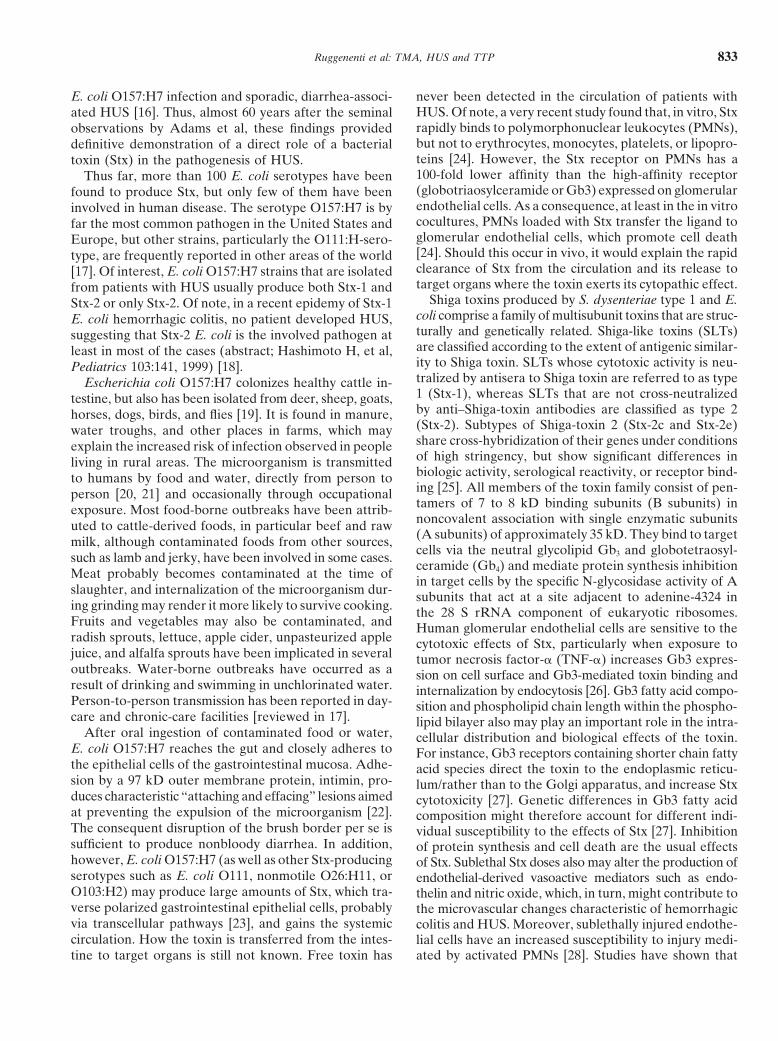
Most authorities consider endothelial injury as the cen-microvasculature. Additional clinical signs depend ontral and likely inciting factor that sustains the microangio-the diverse distribution of the microvascular lesions andpathic process (Fig. 1). This is not a recent idea, however.As early as in 1942, Mark Altschule of Harvard Medical
Key words: Shigatoxin, endothelial activation, von Willebrand factor. School suggested that microvascular endothelial activa-tion was the primary event causing platelet deposition 2001 by the International Society of Nephrology
831

Ruggenenti et al: TMA, HUS and TTP832
Fig. 1. A suggested sequence of events leading to thrombotic microangiopathy in predisposed individuals exposed to triggers of vascular injury. Geneticpredisposition may have a predominant role in familial and recurrent forms, exposition to triggers of vascular injury in Shiga toxin E. coli-associatedHUS.
in arterioles and capillaries with secondary “clearance mice [12]. These findings were taken to indicate that atoxin possibly released by the microorganism could in-of enormous numbers of platelets from the circulation”
[9]. Actually, of the agents that have been associated duce hemorrhagic necrosis of the gastrointestinal mucosaand—once absorbed into the blood stream—of otherwith the disease in subsequent years, all are toxic to
microvascular endothelium [reviewed in 2]. Moreover, organs. Several years later, Konowalciuck, Speirs andStavric noted that E. coli isolated from human cases withplasma from patients with acute HUS/TTP induces apo-
ptosis of human endothelial cells from the renal and diarrhea, produced a toxin similar to the one of Shigellacerebral microvasculature, but not from large vessels [10]. dysenteriae type 1 (Shiga toxin or Stx) found cytopathic
to Vero cells (African green monkey kidney cells) [13].This toxin was subsequently given different names such
AGENTS IMPLICATED IN ENDOTHELIALas Shiga-like toxin (SLT), or verotoxin. In 1978, Koster
ACTIVATION AND DAMAGEet al suggested that a circulating toxin was the cause of
Shiga toxins colitis, hemolysis, and renal failure in children HUS withevidence of S. dysenteriae type 1 infection [14]. A fewDespite that, the term HUS was introduced in 1954
[3]. A careful look at the older literature reveals some years later, a case-control study by Riley et al found astrong association between hemorrhagic colitis and inges-relevant observations. In 1927, Adam first reported that
an infant epidemiology of gastroenteritis was associated tion of E. coli O157:H7-infected beef patties at outlets ofa well-known fast-food restaurant chain [15]. Recoverywith a special type of bacterium coli, biochemically unique
for its fermentation properties [11]. Subsequent observa- of the same strain of E. coli (which could be readily distin-guished from other fecal E. coli because of its uniquetions described a similar disease with diarrhea, occasion-
ally complicated by purpura, anuria, and neurological signs, inability to ferment sorbitol) from the stools of about halfthe 47 children with diarrhea, but from none of the healthythat was characterized at autopsy by capillary and arteriolar
thrombosis and glomerular tuft occlusion by fibrin thrombi. controls, suggested a cause-and-effect relationship betweenfood-borne E. coli infection and hemorrhagic colitis [15].E. coli serotype O111:B4 was considered the causative
agent in more than 90% of these cases [12]. Further studies During the same period, Karmali et al demonstrated anincreased Stx activity in fecal filtrates and increased Stx-found a filterable agent isolated from the stools of these
children that caused diarrhea in calves and was lethal to neutralizing antibody titer in sera from children with
Ruggenenti et al: TMA, HUS and TTP 833
E. coli O157:H7 infection and sporadic, diarrhea-associ- never been detected in the circulation of patients withHUS. Of note, a very recent study found that, in vitro, Stxated HUS [16]. Thus, almost 60 years after the seminalrapidly binds to polymorphonuclear leukocytes (PMNs),observations by Adams et al, these findings providedbut not to erythrocytes, monocytes, platelets, or lipopro-definitive demonstration of a direct role of a bacterialteins [24]. However, the Stx receptor on PMNs has atoxin (Stx) in the pathogenesis of HUS.100-fold lower affinity than the high-affinity receptorThus far, more than 100 E. coli serotypes have been(globotriaosylceramide or Gb3) expressed on glomerularfound to produce Stx, but only few of them have beenendothelial cells. As a consequence, at least in the in vitroinvolved in human disease. The serotype O157:H7 is bycocultures, PMNs loaded with Stx transfer the ligand tofar the most common pathogen in the United States andglomerular endothelial cells, which promote cell deathEurope, but other strains, particularly the O111:H-sero-[24]. Should this occur in vivo, it would explain the rapidtype, are frequently reported in other areas of the worldclearance of Stx from the circulation and its release to[17]. Of interest, E. coli O157:H7 strains that are isolatedtarget organs where the toxin exerts its cytopathic effect.from patients with HUS usually produce both Stx-1 and
Shiga toxins produced by S. dysenteriae type 1 and E.Stx-2 or only Stx-2. Of note, in a recent epidemy of Stx-1coli comprise a family of multisubunit toxins that are struc-E. coli hemorrhagic colitis, no patient developed HUS,turally and genetically related. Shiga-like toxins (SLTs)suggesting that Stx-2 E. coli is the involved pathogen atare classified according to the extent of antigenic similar-least in most of the cases (abstract; Hashimoto H, et al,ity to Shiga toxin. SLTs whose cytotoxic activity is neu-Pediatrics 103:141, 1999) [18].tralized by antisera to Shiga toxin are referred to as typeEscherichia coli O157:H7 colonizes healthy cattle in-1 (Stx-1), whereas SLTs that are not cross-neutralizedtestine, but also has been isolated from deer, sheep, goats,by anti–Shiga-toxin antibodies are classified as type 2horses, dogs, birds, and flies [19]. It is found in manure,(Stx-2). Subtypes of Shiga-toxin 2 (Stx-2c and Stx-2e)water troughs, and other places in farms, which mayshare cross-hybridization of their genes under conditionsexplain the increased risk of infection observed in peopleof high stringency, but show significant differences inliving in rural areas. The microorganism is transmittedbiologic activity, serological reactivity, or receptor bind-to humans by food and water, directly from person toing [25]. All members of the toxin family consist of pen-person [20, 21] and occasionally through occupationaltamers of 7 to 8 kD binding subunits (B subunits) inexposure. Most food-borne outbreaks have been attrib-noncovalent association with single enzymatic subunitsuted to cattle-derived foods, in particular beef and raw(A subunits) of approximately 35 kD. They bind to targetmilk, although contaminated foods from other sources,cells via the neutral glycolipid Gb3 and globotetraosyl-such as lamb and jerky, have been involved in some cases.ceramide (Gb4) and mediate protein synthesis inhibition
Meat probably becomes contaminated at the time of in target cells by the specific N-glycosidase activity of Aslaughter, and internalization of the microorganism dur- subunits that act at a site adjacent to adenine-4324 ining grinding may render it more likely to survive cooking. the 28 S rRNA component of eukaryotic ribosomes.Fruits and vegetables may also be contaminated, and Human glomerular endothelial cells are sensitive to theradish sprouts, lettuce, apple cider, unpasteurized apple cytotoxic effects of Stx, particularly when exposure tojuice, and alfalfa sprouts have been implicated in several tumor necrosis factor-� (TNF-�) increases Gb3 expres-outbreaks. Water-borne outbreaks have occurred as a sion on cell surface and Gb3-mediated toxin binding andresult of drinking and swimming in unchlorinated water. internalization by endocytosis [26]. Gb3 fatty acid compo-Person-to-person transmission has been reported in day- sition and phospholipid chain length within the phospho-care and chronic-care facilities [reviewed in 17]. lipid bilayer also may play an important role in the intra-
After oral ingestion of contaminated food or water, cellular distribution and biological effects of the toxin.E. coli O157:H7 reaches the gut and closely adheres to For instance, Gb3 receptors containing shorter chain fattythe epithelial cells of the gastrointestinal mucosa. Adhe- acid species direct the toxin to the endoplasmic reticu-sion by a 97 kD outer membrane protein, intimin, pro- lum/rather than to the Golgi apparatus, and increase Stxduces characteristic “attaching and effacing” lesions aimed cytotoxicity [27]. Genetic differences in Gb3 fatty acidat preventing the expulsion of the microorganism [22]. composition might therefore account for different indi-The consequent disruption of the brush border per se is vidual susceptibility to the effects of Stx [27]. Inhibitionsufficient to produce nonbloody diarrhea. In addition, of protein synthesis and cell death are the usual effectshowever, E. coli O157:H7 (as well as other Stx-producing of Stx. Sublethal Stx doses also may alter the production ofserotypes such as E. coli O111, nonmotile O26:H11, or endothelial-derived vasoactive mediators such as endo-O103:H2) may produce large amounts of Stx, which tra- thelin and nitric oxide, which, in turn, might contribute toverse polarized gastrointestinal epithelial cells, probably the microvascular changes characteristic of hemorrhagicvia transcellular pathways [23], and gains the systemic colitis and HUS. Moreover, sublethally injured endothe-circulation. How the toxin is transferred from the intes- lial cells have an increased susceptibility to injury medi-
ated by activated PMNs [28]. Studies have shown thattine to target organs is still not known. Free toxin has

Ruggenenti et al: TMA, HUS and TTP834
up-regulation of cytokine production, PMN activation, Bacterial neuraminidasesand enhanced oxidant injury potentiate the direct cyto- Following a few reports in the European literature,pathic effects of Stx; this amplifies vascular damage and, Klein et al found Thomsen-Friedenreich antigen on eryth-conceivably, alters the normal thromboresistant pheno- rocytes and in the glomeruli of two one-year-old childrentype of the endothelial cell [reviewed in 29]. who died from HUS secondary to pneumococcus pneu-
However, endothelial cells are not the only targets for monia and sepsis [35]. Binding studies also found weakStx in the kidney, and several renal cell types (such as IgM deposits in the glomeruli indicative of a specific IgMproximal and distal tubular cells or mesangial and epithe- binding to the Thomsen-Friedenreich antigen. Subsequentlial glomerular cells) have receptors for Stx and are dam- studies demonstrated neuraminidase activity in organism-aged by the toxin in vitro [30]. These findings suggest free filtrates of sera from three infants with Streptococcusthat renal injury in Stx-HUS, unlike other forms of HUS/ pneumoniae-associated HUS, but not in filtrates of seraTTP, is not simply a consequence of vascular involve- from a large series of samples from children with Strepto-ment in the microangiopathic process, but may reflect, at coccus pneumoniae infection and no evidence of HUS.least in part, a primary damage to parenchymal cells [30]. Since the Thomsen-Friedenreich antigen is normally cov-
Experimental evidence indicates that sites of Gb3 ex- ered by N-acetyl neuraminic acid (sialic acid) moieties,pression coincide with sites of Stx-induced tissue damage it has been speculated that Streptococcus pneumoniae-[2]. Indeed, after a challenge with Stx-1 purified from E. derived neuraminidase, by removing sialic acid from thecoli O157:H7, rabbits presented neurologic and enteric cell membranes, exposes this cryptic antigen to preformedfeatures of endothelial swelling and luminal occlusion circulating IgM antibodies [35, 36]. Then, IgM bindingof small arteries and arterioles, but no signs of renal to Thomsen-Friedenreich antigen exposed on platelet andinvolvement [31]. Binding studies identified Stx recep- endothelial cell surface would cause platelet aggregationtors in the central nervous system and in the gastrointes- and endothelial damage [35, 36]. Binding of IgM antibod-tinal tract, but not in the kidney [31]. In humans, the ies to the antigen expressed on circulating erythrocytesGb3 receptor is expressed not only in the kidney, but may explain why Coombs-positive hemolytic anemia isalso on the membrane of epithelial and endothelial cells so frequently reported in patients with neuraminidase-of gastrointestinal mucosa and submucosa, respectively.
induced HUS.Of interest, a recent study in baboons, which share withhumans a similar distribution of gastrointestinal and re- Immune complexesnal Gb3 receptors, demonstrated that Stx injection in-
Complement-dependent antibodies, which are cyto-duces direct injury to the endothelial and epithelial cellstoxic endothelial cells, have been found in the serumof the gastrointestinal and renal microcirculation [32].and plasma of patients with HUS/TTP [37]. ComplementIn this animal model, structural changes reminiscent ofand IgG and IgM antibodies can be found in kidney biop-the autoptic changes observed in children with HUS andsies [2]. Together, these findings suggest a pathogeneticneurological involvement also were detected in the cen-role for immune complexes. This possibility is consistenttral nervous system, despite a relatively low expressionwith findings that endothelial cells exposed in vitro toof the Gb3 receptor in this organ [32]. Increased localsera of patients with HUS/TTP in the presence of com-release of inflammatory mediators was advocated to con-plement show ultrastructural changes of cytoplasmic in-tribute to renal tissue injury. This possibility is supportedclusions and cytoplasmic and nuclear degeneration sug-by the observation that neurological changes can be asso-gestive of endothelial cell apoptosis [37].ciated with increased interleukin (IL)-6 concentration in
Hemolytic uremic syndrome/TTP complicates immuneliquor. Thus, the finding that, despite negligible Gb3 re-diseases, in particular systemic lupus erythematosus, andceptor expression, adult humans may develop central ner-increasingly is reported in association with the antiphos-vous system lesions secondary to Stx-producing E. colipholipid syndrome [reviewed in 38].infection [20, 33] could be due to a facilitating effect of
enhanced local release of inflammatory mediators [31, 32].Drugs
Endotoxins Several chemotherapeutic agents, including mitomy-cin C (MMC), cisplatin, daunorubimicin, cytosine arabi-Upon E. coli infection, bacterial-derived endotoxinsnoside, chlorozotocin, neocarcinostatin, and deoxycofor-(lipopolysaccharides or LPS) and Stx act synergeticallymycin, have been implicated in a syndrome of thromboticin initiating damage to target organs, including the kid-microangiopathy designated as chemotherapy-associ-ney. LPS prime the endothelial cells to undergo apopto-ated HUS (C-HUS) [reviewed in 39]. MMC causes TMAsis when exposed to picomolar amounts of Stx. More-after direct infusion into rat kidneys. Endothelial cellsover, LPS derived from Stx producing E. coli activatemetabolize MMC to a reactive intermediate that cross-PMNs to release TNF-�, IL-1, elastase, and free radicals
that are highly toxic to microvascular endothelium [34]. links cellular DNA and proteins. Endothelial cells, ex-
Ruggenenti et al: TMA, HUS and TTP 835
posed to MMC in vitro at concentrations equivalent to CONSEQUENCES OF VASCULAR INJURYtherapeutic drug levels in vivo, survive but subsequently Virtually all properties of the normal microvascularshow both an impaired ability to divide and a reduced endothelium are altered in HUS/TTP. Endothelial cellsthrombin-stimulated production of prostacyclin. synthesize many substances involved in coagulation and
Cyclosporine. Cyclosporine A (CsA) has been associ- fibrinolysis, including prostacyclin, nitric oxide (NO),ated with de novo thrombotic microangiopathy in renal vWF, thrombomodulin, tissue-type plasminogen activa-and nonrenal transplantation and in patients with Beh- tor inhibitor and protein S. Loss of prostacyclin [47] andcets disease [40]. Thrombotic microangiopathy in renal increase in vWF [48] have been claimed to account forallograft recipients is most common in the first weeks, the loss of physiologic thromboresistance and for thewhen CsA drug levels are highest, and should be differ- consequent widespread platelet aggregation in vascularentiated from both humorally and/or cell-mediated rejec- beds throughout the body, creating a cycle of vasocon-tion. In renal allograft recipients, CsA enhances platelet striction with platelet and fibrin deposition and furtheraggregation and thromboxane A2 production, and high thrombus formation.CsA levels correlate with increased thrombomodulinand von Willebrand factor serum levels. Since the first Leukocyte activation and complement consumptionreport by Schmidt et al in 1991, more than 20 cases have The interaction between leukocytes and damaged en-been described providing evidence that tacrolimus may dothelial cells is instrumental to the development of mi-cause HUS/TTP [41, 42]. The disease is reported in 1 to crovascular injury in Stx-associated HUS, as documented4.7% of tacrolimus treated patients. It is more frequently by experimental and clinical evidence [34]. Neutrophilsdiagnosed in the first year after transplantation, although from children in the acute phase of HUS adhere to endo-it may occur at any time. thelial cells in vitro more tightly than normal neutrophils
Ticlopidine and clopidogrel. Ticlopidine has been asso- and induce endothelial injury by degrading endothelial cellciated with the development of HUS/TTP, but this event fibronectin, possibly by the release of neutrophil-specificis rare (1 in 1600 patients treated with ticlopidine after proteases [34]. Stx in vitro promotes massive leukocytecardiac stenting) [43]. Clopidogrel, a new antiplatelet
adhesion and transmigration to endothelium under flowagent that has achieved widespread clinical acceptance
conditions by up-regulating endothelial expression of ad-because of a more favorable safety profile than ticlopi-
hesive proteins and chemokines (abstract; Zoja et al,dine, also has been associated with the disease, and 11J Am Soc Nephrol 10:595A, 1999). Platelet and leukocytecases have been reported recently [44]. Ten of theseactivation concur to promote the formation of C3bBbpatients developed TTP within two weeks from begin-convertase, which in turn leads to cleavage of the thirdning the drug treatment. Ticlopidine and clopidogrel arefraction of complement (C3) to the active form C3b,structurally related derivatives of thienopyridine and actready to react with any cell surface or membrane nucleo-by blocking an adenosine diphosphate-binding site onphile. Low C3 levels were first described in diarrhea-platelets, which inhibits the expression of the glycopro-associated HUS in the early 1970s [49]. Persistently lowtein IIb/IIIa receptor in the high-affinity configurationcomplement levels were found in different forms of HUSthat binds fibrinogen and large von Willebrand factor[49]. Granular C3 deposits in glomeruli and arterioles of(vWF) multimers. Why these two drugs cause TTP ispatients with either diarrhea-associated or adult formsnot fully clear, but of great interest, as patients withof HUS/TTP and evidence of C3 breakdown productsticlopidine- or clopidogrel-associated TTP have a defi-in patients’ sera were taken to reflect complement con-ciency of vWF-cleaving protease activity in plasma thatsumption in the microvasculature [50].appears quite comparable to the deficiency observed in
idiopathic TTP [44].Abnormal von Willebrand factorQuinine. Quinine is widely used for the prophylaxisrelease and processingof nocturnal cramps, but is also present in tonic water and
In normal individuals, vWF is formed as large multi-bitter lemon drinks. Occasionally, it may cause hemolyticmers [ultralarge (UL) multimers] due to the polymeriza-anemia, thrombocytopenia, disseminated intravasculartion in endothelial cells and megakaryocytes of a nativecoagulation, and HUS [45]. HUS typically occurs in pa-subunit with an apparent molecular mass of 225 kD, andtients presensitized by prior exposure to quinine andis stored as such in Weibel-Palade bodies and plateletrapidly follows re-ingestion of the compound. Quinine-granules. UL multimers do not normally circulate, sincedependent antibodies to red blood cells, granulocytes,they are rapidly reduced into smaller multimers soonand the glycoprotein Ib/IX and IIb/IIIa on platelet sur-after their secretion by cleavage at position 842 Tyr-843face have been involved in the pathogenesis of the dis-Met of the mature subunit. A major contribution to theease. Renal failure is usually severe, but the outcome isunderstanding of vWF processing was provided by Fur-good, provided that patients are treated early enough
with plasma exchange [46]. lan et al [51] and Tsai and Lia [52], who partially purified

Ruggenenti et al: TMA, HUS and TTP836
and characterized a plasma metalloprotease that physio- tease activity [59]. Of interest, in both patients with consti-logically cleaves ultralarge vWF multimers. The prote- tutional protease deficiency, disease remission achievedase, of approximately 300 kD, needs bivalent cations for by plasma therapy was concurrent with an almost fullits activation, is inhibited by calcium-chelating agents, recovery of the vWF-cleaving protease activity. Bothand is activated only in conditions of low ionic strength patients achieved a long-lasting remission, although pro-or high shear stress. In vivo evidence that proteolytic tease activity decreased to less than 20% over 20 dayscleavage is involved in the modification of plasma multi- after plasma therapy withdrawal.mers after secretion has been provided by studies show- In nonfamilial cases, the protease deficiency appearsing that circulating vWF multimers are heterogeneous to be a consequence of a specific autoantibody that devel-oligomers of a native 225 kD subunit and of proteolytic ops transiently and tends to disappear during remissionfragments with apparent molecular masses of 189, 176, [51]. In a patient with recurrent episodes of TTP shown toand 140 kD [53]. In patients with HUS and TTP, in con- have an acquired inhibitor of the vWF-cleaving proteasetrast to healthy subjects, UL multimers similar to the [60], splenectomy, performed one year after the first TTPones stored in endothelial cells and platelets were occa- event, resulted in the disappearance of the autoantibodysionally detected in plasma [54]. The presence in patients and normalization of the protease activity, platelet count,with HUS/TTP of circulating UL multimers that in vitro and hemoglobin levels [60].are capable of supporting platelet aggregation more effi- At variance with TTP, in the previously mentionedciently than normal multimers was evidence for their studies, no deficiency was found in patients with familialpathogenic role in microvascular thrombi [55]. However, or sporadic forms of HUS [51], suggesting that the pres-direct proof that this was indeed the case was never pro- ence or absence of this activity were enough to classifyvided. Interestingly, the opposite appears true in vivo, patients as having HUS or TTP, respectively. More recentsince the available evidence is against the possibility that data, however, indicate that low or zero protease activityUL vWF multimers when present in the circulation can is not confined to TTP and can even be found in a numbereven cause intravascular thrombosis. UL multimers have of diseases associated with an increased tendency to throm-been found in fetuses and newborns, but disappear within bosis, such as inflammatory states, liver cirrhosis, majorthe first months of life, indicating that physiologic cleav- surgery (Mannucci et al, personal communication, Work-age of vWF is less efficient at birth and fully develops shop on vWF and TTP, Bethesda, July 31, 2000), andlater in life [56]. Moreover, in patients with a rare variant disseminated malignancies [7]. Another child who wasof von Willebrand disease, named “Vicenza,” UL vWF diagnosed as HUS/TTP, although he had renal failuremultimers were constantly found in the circulation; none- and no neurological symptoms, showed with zero vWFtheless, these patients never experienced thrombotic epi-
protease activity [61]. Our group has obtained similarsodes; rather, they suffered with bleeding tendency [57].results of complete lack of protease in two cases of recur-In patients with HUS/TTP who recovered after a sin-rent HUS (unpublished results). These data indicate thatgle episode, UL multimers were found almost exclusivelythe issue is controversial and the distinction betweenin the acute phase but not in remission, suggesting a mas-HUS and TTP is much more complicated than presentedsive release from storage sites of acutely injured endothe-in the New England Journal of Medicine articles [51, 52]lial cells that possibly transiently overwhelmed theand accompanying editorial [55].plasma proteolytic capacity [58]. In contrast, those cases
A constant finding in the acute phase of different formsthat had a tendency to recur had circulating UL multi-of HUS/TTP is an increase of low molecular weight mul-mers either in the acute phase or consistently in thetimers and a decrease of high molecular weight multi-remission phase of the disease, which was initially takenmers, which reflects an enhanced proteolytic fragmen-as evidence of a state of persistent endothelial perturba-tation of the molecule [62, 63]. That vWF undergoestion [54]. The hypothesis that circulating UL vWF multi-excessive fragmentation in the acute phase of these dis-mers may reflect a condition of endothelial perturbationeases is remarkably consistent with previous findings ofhas been recently challenged by findings that circulatinga relative decrease in the native 225 kD vWF subunit,UL multimers in chronic relapsing TTP were associatedwhich only occurs in the acute phase, accompanied bywith a reduced or totally absent activity of the abovea relative increase of fragments that can only derive fromvWF-cleaving protease activity that normally cleaves vWFthe cleavage of the native subunit [64].multimers to smaller molecular forms. In two large stud-
ies, vWF-cleaving protease deficiency was described inEnhanced vascular shear stress as a determinant ofpatients with different forms of TTP [51, 52]. In familialabnormal von Willebrand factor fragmentationforms of TTP, the deficiency is probably inherited as an
von Willebrand factor’s susceptibility to fragmentationautosomal recessive trait. Consistent with this possibility,increases in response to rising levels of shear stress [65],complete deficiency of vWF-cleaving protease activitywhich induces protein unfolding and makes vWF proteo-was found in two brothers with chronic relapsing TTP,
whereas their parents had approximately half-normal pro- lytic cleavage sites more accessible to specific plasma
Ruggenenti et al: TMA, HUS and TTP 837
protease(s). It is speculated that enhanced shear stress of acute disease, and only two had moderately increasedLDH levels [67].in the severely narrowed damaged microvessels accounts
for the abnormal vWF fragmentation observed during Actually, low C3 levels are well known to accompanythe acute phases either of diarrhea-associated or idio-the acute phase of HUS/TTP. Evidence of increased
capacity of fragmented vWF to bind receptors on acti- pathic HUS [49] and likely reflect C3 consumption inthe microvasculature. Granular C3 deposits in glomerulivated platelets suggests that shear stress-induced vWF
fragmentation may contribute to maintain and further and arterioles of HUS patients [50] and evidence of C3breakdown products in HUS sera [49] further documentspread microvascular thrombosis. In severe forms of
HUS that are resistant to plasma therapy, removal of the activation of the complement system in the acutephase of the disease. Enhanced release of complementthe kidneys—a major site of vascular bed occlusion and
augmented shear stress—was followed by hematologic cleavage products (including C3a and C5a) consequentto uncontrolled complement activation may contributeand clinical remission associated with restoring the vWF
fragmentation pathway to normal [62]. Consistent with to the microangiopathic process by stimulating neutro-phil activation phagocytic adhesion to vascular endothe-this possibility in patients with recurrent and sporadic
HUS/TTP, increased vWF fragmentation normalized lium, or platelet aggregation, and by directly injuring theendothelium through enhanced production of the mem-after resolution of the microangiophatic process.brane attack complex, the final multimolecular unit ofthe complement C5b-9.
GENETIC PREDISPOSITIONTwo complement pathways can generate C3-activating
Congenital complement abnormalities enzymes: the classic convertase generated by the sequen-tial reaction of C1, C4, and C2, and the alternative path-Over the last 20 years, approximately 140 cases of
familial HUS and TTP have been described in 70 fami- way convertase. Activation of classic and alternativecomplement pathway—possibly triggered by circulatinglies, with the predominant features of HUS in two thirds
of the patients. Both autosomal-recessive and autosomal- immune complexes and damaged erythrocytes, respec-dominant modes of inheritance have been recognized [66]. tively—is well documented in acute HUS [68], but consis-Precipitating events such as pregnancy, virus-like dis- tently subsides with remission of the disease. In contrast,ease, or sepsis have been reported only in a minority of in cases of familial HUS/TTP, serum C3 levels were consis-cases. Evidence that some of these cases responded, at tently and remarkably depressed in cases as comparedleast transiently, to plasma infusion or exchange, sug- with controls, even during remission of the disease [67].gests that the genetic defect(s) associated with familial Even more interestingly, low C3 levels also were foundHUS/TTP were the cause of one or more abnormalities in the patients’ relatives, who had never suffered within plasma component(s) essential to the integrity of mi- HUS or TTP in the past and had no sign of the diseasecrovascular circulation and/or to the defense mechanism at the time of the study [67]. Furthermore, either in casesof the host endothelium against injurious agents. Thus, or in case-relatives depressed C3 values did not parallelreduced serum levels of the third component (C3) of the similar changes in C4 levels. These data definitely ruledcomplement system have been reported since 1974 [49] out the possibility that classic pathway activation ac-in both the sporadic and familial forms of HUS [67]. That counted for hypocomplementemia in this series [67]. Anthe disease may be related to an inherited congenital inherited defect in C3 synthesis has been suggested toabnormality is consistent with the finding that more than account for decreased C3 serum concentration [67], but50% of patients with familial HUS/TTP—as compared much more convincing data are now available that lowwith only 10 to 20% in nonfamilial forms—had repeated C3 in HUS may derive from either a lack or altered functionrecurrences. This genetical abnormality is likely to in- of factor H, a regulatory protein that inhibits the comple-volve the complement system, as suggested by levels of ment activation through the alternative pathway [69].circulating C3, which were extremely low in a large seriesof familial cases as compared with controls [67]. Reduced
INHERITED FACTOR H DEFICIENCYC3 levels in cases and case relatives, but not in controlsIn one patient with HUS and in his healthy brother,and the controls’ relatives, further indicate that the de-
a persistent reduction in C3 levels was found with veryfect clusters in families. Evidence that a low C3 concen-low levels of factor H [70]. Finding that the parents, whotration was strongly associated with the disease evenwere first cousins, had half-normal levels of factor H,more convincingly suggests the possibility of a tight (pos-convincingly indicated that the defect was inherited. Asibly causal) relationship between decreased C3 and dis-similar finding was later reported in another family. Inease manifestation [67]. On the other hand, in the pre-a recent report, the association between inherited factorviously mentioned series, low C3 levels could not dependH deficiency and low C3 levels was investigated in aon consumption in a still-ongoing microangiopathic pro-
cess, since no patient at the time of the study had any sign large series of families with a history of HUS/TTP [67].
Ruggenenti et al: TMA, HUS and TTP838
By radial immunodiffusion and Western blot analysis, a C to G transversion causing an arginine to glycine changetwo affected subjects of one family were identified who in the short consensus repeat 20 (SCR20) [72]. This muta-had very low circulating factor H levels, and moderately tion is likely to alter structure and hence function oflow levels were found in two healthy relatives. In these factor H protein without modifying its circulating levels.patients, the cofactor activity of factor H, measured as The same authors also described a nonsense mutation,the capacity to degrade C3b, was also reduced. In the located in SCR1 of factor H gene, in a single case withother families, serum factor H concentration was normal late onset [72]. In a large series of patients, four new[67]; however, normal serum levels do not necessarily heterozygous mutations in factor H gene were foundexclude an underlying biochemical abnormality in circu- [73]. Three of the mutations were observed in familieslating factor H. In this regard, in another family the with dominant transmission, the fourth was found in atwo affected members, and the healthy father, who had single case who experienced disease recurrence on thenormal serum concentrations of factor H by radial immu- renal allograft. Loirat et al have recently reported fivenodiffusion, Western blots showed additional bands of cases of atypical HUS with neonatal onset and 2 to 15higher molecular weights that were not found in any relapses over 2 to 14 years follow-up, who had completecontrols [67]. The bands might represent dimeric forms and persistent vWF-cleaving protease deficiency (abstract;of factor H. At variance, no differences were found in Loirat et al, Twelfth Congress of the International Pedi-serum levels and patterns of FHL-1 and FHR proteins atric Nephrology Association, Seattle, WA, USA, Sep-[69]. Similar results were obtained in another series of tember 1–2, 2001) [74, 75]. Another study reported anpatients with HUS [71]. apparent mutation in factor H (a C to T transition in
Factor H has several biological activities. It prevents SCR 20) in a family with HUS with a recessive mode ofthe formation of C3bBb complex and accelerates the inheritance and severely depressed factor H levels [76].dissociation of Bb from the C3 convertase. It acts as a However, two independent groups subsequently pro-cofactor for factor I that degrades C3b [69], and it also vided evidence that the apparent mutation was actuallydistinguishes between activator and nonactivator sur- an artifact caused by coamplification of a factor H-relatedfaces [69]. Other less well-defined functions of factor H gene, and demonstrated that the real mutation in thishave been suggested by the presence of at least two family is an A to T transversion and a 24 bp deletionheparin binding sites in factor H protein that could facili- that was present in homozygosity in affected memberstate interaction with extracellular matrix [69]. An inter- patients and in heterozygosity in the healthy carrierscurrent exposure to agents potentially toxic to the vascu- (abstract; Caprioli et al, J Am Soc Nephrol 11:404A,lar endothelium (such as certain virus, bacteria, toxins, 2000) [77]. Altogether, the available data provide com-immunocomplexes, and cytotoxic drugs [2]) may initiate pelling molecular evidence that genetic alterations in fac-a local intravascular thrombosis, which promotes C3bBb tor H are involved in both autosomal dominant and reces-convertase formation and complement deposition within sive HUS. Data that followed exposure to SLT producingcapillary vessels. In normal conditions, however, by mod- E. coli, where only 2 to 7% of the patients progressedulating C3bBb activity factor H may effectively limit to overt HUS, suggest that a genetic predisposition alsocomplement deposition and further extension of the pro- may have a role in the typical diarrhea-associated forms.cess [69]. In contrast, when the bioavailability and/or Polymorphisms in the promoter and in the coding regionsactivity of factor H is congenitally defective, C3bBb con- of the factor H gene have been recently described. It isvertase formation and complement deposition may be- intriguing to speculate that some of these polymorphiccome uncontrolled, with further extension of the micro- variants confer a genetic predisposition to SLT-inducedangiopathic process up to full manifestation of the disease. HUS. Although it seems clear that familial HUS is re-
lated to an inherited congenital defect, the cause of theGENE MUTATIONS ACCOUNTING FOR syndrome is probably multifactorial and the inheritedFACTOR H DEFICIENCY OR ABNORMALITIES complement defects simply may represent a predisposing
condition that increases the risk of the disease in combi-The consistent association found in families betweennation to other intercurrent environmental or acquiredfactor H abnormalities and low C3 levels supports thefactors. Thus, in a family with hereditary hypocomplem-hypothesis that low C3 in the setting of familial HUS/entemia, only one of the six members with low C3 levelsTTP may depend on a genetic deficiency of factor H.developed HUS, apparently following a flu-like episodeA recent report on three large families with HUS[67]. It is also possible that genetic defects in other com-documented that an area on chromosome 1q, where fac-plement regulatory proteins (DAF, CR1, CR2, C4bp)tor H is mapped, segregates with HUS [72]. All subjectsmight have a role in determining low C3 in familial HUSin the three families had normal serum factor H levels.and TTP. Data support this possibility that the aboveHowever, affected members and obligate carriers withingenes map on the same region of chromosome 1q asone family were found, by mutation analysis, to have a
heterozygous point mutation in factor H, consisting of factor H [78].

Ruggenenti et al: TMA, HUS and TTP 839
CLINICAL FEATURES OF THE DIFFERENT �80 mL/min as compared with 15 of 35 (42.8%) of thosewith anuria duration �16 days [82]. The extent of acuteFORMS OF THROMBOTIC MICROANGIOPATHYstructural injury and loss of functioning nephron unitsChildhood HUSmay account for long-term sequelae of the disease. At 15
Stx-associated HUS. This form, also referred to as di- years of follow-up, Gagnadoux et al found renal sequelaearrhea associated (D�) HUS, is most often due to E. coli in 83% of children with patchy cortical necrosis, one thirdO157:H7 infection and, less frequently to the O111:non- of whom were in ESRD, as compared with none of thosemotile, O26:HII, and O103:H2 [reviewed in 79]. It is with pure glomerular TMA) [82]. Regardless of the renalcharacterized by prodromal diarrhea followed by acute damage, however, in the long term, 10 to 42% of childrenrenal failure. The overall incidence is estimated to be have some renal sequelae (such as proteinuria and/or mod-2.1 cases per 100,000 persons/year with a peak incidence erate hypertension or mildly reduced GFR), 10 to 22%in children younger than five years of age (6.1/100,000/ have chronic renal failure, and 2 to 9% ESRD) [81, 82].year) and the lowest rate in adults 50 to 59 years old Neuraminidase-associated HUS. This is a rare but po-(0.5/100,000/year) [79]. tentially fatal disease that may complicate pneumonia,
E. coli O157 infection is most common in the warm or less frequently, meningitis caused by Streptococcussummer months. The average interval between Stx-E. coli pneumoniae [35, 36]. The clinical picture is usually severe,exposure and illness is three (range of 1 to 8) days [17]. with respiratory distress, anuria, neurological involve-Illness typically begins with abdominal cramps and non- ment, and coma.bloody diarrhea; diarrhea may become hemorrhagic in Idiopathic or atypical HUS. This is a subgroup with70% of cases usually within one or two days. Vomiting unknown etiology and no seasonal pattern that differsoccurs in 30 to 60% of cases and fever in only 30%. from Stx-associated HUS on epidemiologic, clinical, lab-Serum leukocyte count is usually elevated, and a barium oratory, and prognostic grounds. The onset is usually insid-enema may demonstrate “thumb-printing,” suggestive ious, without antecedent diarrhea, and may be precededof edema and submucosal hemorrhage, especially in the by nephrotic syndrome. The disease occasionally is famil-region of the ascending and transverse colon. Superficial ial, has a tendency to relapse, and carries a worse progno-ulcerations or pseudomembranes are common. E. coli sis than Stx-associated HUS [83]. Upper respiratory tractO157 infection is complicated by HUS in 3 to 7% of the infection may trigger the disease in approximately 30%sporadic cases and up to approximately 20% or more of of cases [83]. The clinical syndrome resembles TTP. One-the epidemic forms [17]. HUS is usually diagnosed six third of these patients have central nervous systemdays after the onset of diarrhea. After HUS infection, involvement at onset, with convulsions and alterationsStx-E. coli may be shed in the stool for several weeks of consciousness that persist despite correction of hyper-after the symptoms are resolved, particularly in younger tension and metabolic complications of renal failure. Thechildren (�5 years age) who in one-third of the cases may outcome is severe [83–85].carry the organism for over 20 days. Use of antimotility
Adult HUS/TTPagents and antibiotics, bloody diarrhea, fever, vomiting,elevated serum leukocyte count, extremes of age (in STX-associated HUS/TTP. Albeit less frequently thanparticular children �5 years old), and female sex have in children, Stx HUS has been reported even in adults,been associated with an increased risk of HUS following either in epidemics involving elderly people in nursingStx-E. coli infection [17]. homes or in sporadic cases [20, 33]. The etiology and
Fifty percent of patients who develop HUS need dial- pathogenesis of childhood and adult Stx HUS are theysis, 75% require red blood cell transfusions, and 25% same, but renal and neurological involvement are usuallyhave neurological signs, including stroke, seizure, and more severe and sequelae more frequent in adults thancoma [17]. Stx-associated HUS is erroneously regarded in children [86]. Indeed, in older patients, a mortality rateas a benign disease. Indeed, 3 to 5% of children die in close to 90% has been reported, a figure that is at leastthe acute phase and a similar percentage develop end- 10- to 20-fold higher than in pediatric series [20, 33, 86].stage renal disease (ESRD) [80]. Moreover, long-term Acute idiopathic HUS/TTP. The defining clinical fea-sequelae are common. In a series of 88 children, Fitzpa- tures include thrombocytopenia, microangiopathic he-trick et al reported long-term renal dysfunction in 39% molytic anemia, neurological and renal abnormalities,of cases, a residual glomerular filtration rate (GFR) �80 and fever. Neurological symptoms usually dominate themL/min 173 m2 in 18%, and persistent microalbuminuria clinical picture, but usually subside within 48 hours afterin 31% [81]. In different series, the incidence of ESRD initiation of plasma therapy [7]. Clinical response andranged from 3 to 18% [17]. The duration of anuria is a survival are comparable in patients with and withoutstrong predictor of residual renal dysfunction. Indeed, renal failure [5, 6].only 4 of 53 children (7.5%) with a duration of anuria Relapsing and frequently relapsing HUS/TTP. Relaps-
ing forms are increasingly reported [87, 88], as moreless than ten 10 days were found to have a residual GFR
Ruggenenti et al: TMA, HUS and TTP840
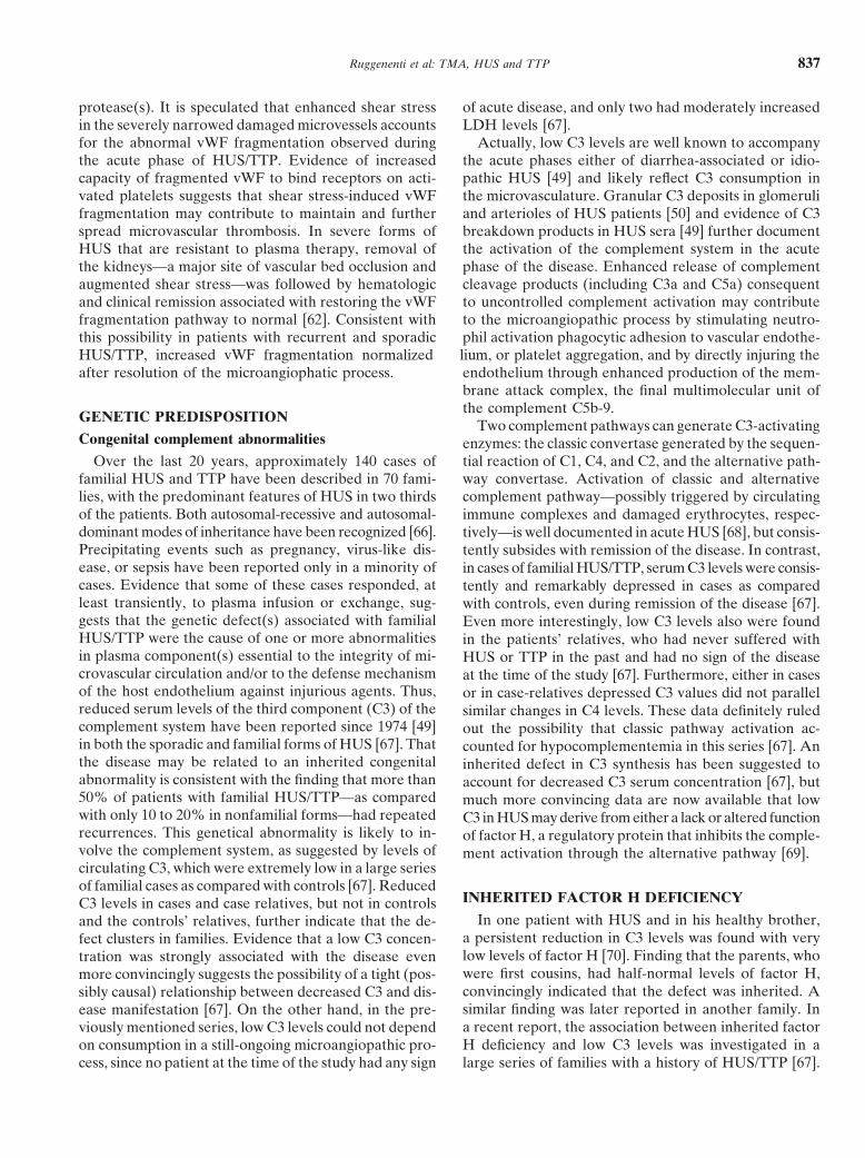
fluffy material in the subendothelium. These changes arepatients than in the past recover from the initial acutevirtually identical and often indistinguishable from theepisode thanks to improved supportive and specific treat-microvascular lesions of scleroderma and malignant hy-ments. An extremely rare variant of relapsing HUS/TTP,pertension [2]. In HUS, the microthrombi are confinedalso known as “chronic relapsing” or “frequent relaps-primarily to the kidneys (Fig. 2), and thus, renal failureing” HUS/TTP, is characterized by frequent episodes,is the dominant feature. TTP mainly involves the brain,recurring after symptom-free intervals of a predictableand intravascular thrombi apparently form and disperseduration (�1 month in most cases) [89].repeatedly, producing intermittent neurological signs. Inpediatric patients, particularly in children younger thanFAMILIAL HUS/TTPtwo years of age and in most cases of Stx-associated HUS,This form has a heterogeneous pattern of inheritancea pattern of glomerular injury is prominent (Fig. 2). Leu-and presents as HUS or as TTP in several members ofkocyte infiltrates and thrombi are common during the
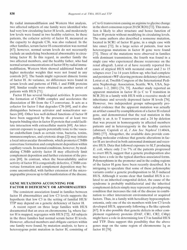
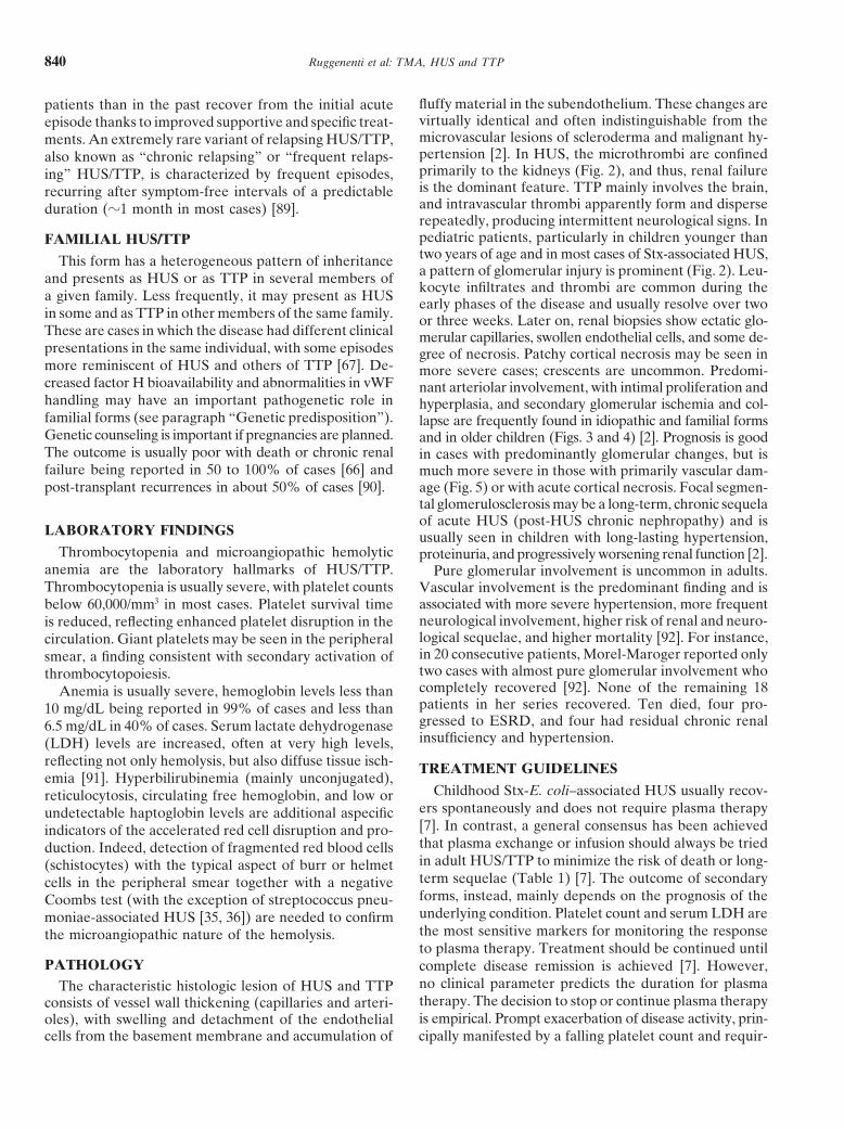
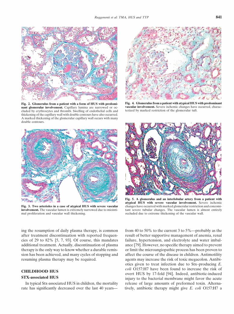
a given family. Less frequently, it may present as HUS early phases of the disease and usually resolve over twoin some and as TTP in other members of the same family. or three weeks. Later on, renal biopsies show ectatic glo-These are cases in which the disease had different clinical merular capillaries, swollen endothelial cells, and some de-presentations in the same individual, with some episodes gree of necrosis. Patchy cortical necrosis may be seen inmore reminiscent of HUS and others of TTP [67]. De- more severe cases; crescents are uncommon. Predomi-creased factor H bioavailability and abnormalities in vWF nant arteriolar involvement, with intimal proliferation andhandling may have an important pathogenetic role in hyperplasia, and secondary glomerular ischemia and col-familial forms (see paragraph “Genetic predisposition”). lapse are frequently found in idiopathic and familial formsGenetic counseling is important if pregnancies are planned. and in older children (Figs. 3 and 4) [2]. Prognosis is goodThe outcome is usually poor with death or chronic renal in cases with predominantly glomerular changes, but isfailure being reported in 50 to 100% of cases [66] and much more severe in those with primarily vascular dam-post-transplant recurrences in about 50% of cases [90]. age (Fig. 5) or with acute cortical necrosis. Focal segmen-
tal glomerulosclerosis may be a long-term, chronic sequelaof acute HUS (post-HUS chronic nephropathy) and is
LABORATORY FINDINGS usually seen in children with long-lasting hypertension,Thrombocytopenia and microangiopathic hemolytic proteinuria, and progressively worsening renal function [2].
anemia are the laboratory hallmarks of HUS/TTP. Pure glomerular involvement is uncommon in adults.Thrombocytopenia is usually severe, with platelet counts Vascular involvement is the predominant finding and is
associated with more severe hypertension, more frequentbelow 60,000/mm3 in most cases. Platelet survival timeneurological involvement, higher risk of renal and neuro-is reduced, reflecting enhanced platelet disruption in thelogical sequelae, and higher mortality [92]. For instance,circulation. Giant platelets may be seen in the peripheralin 20 consecutive patients, Morel-Maroger reported onlysmear, a finding consistent with secondary activation oftwo cases with almost pure glomerular involvement whothrombocytopoiesis.completely recovered [92]. None of the remaining 18Anemia is usually severe, hemoglobin levels less thanpatients in her series recovered. Ten died, four pro-10 mg/dL being reported in 99% of cases and less thangressed to ESRD, and four had residual chronic renal6.5 mg/dL in 40% of cases. Serum lactate dehydrogenaseinsufficiency and hypertension.(LDH) levels are increased, often at very high levels,
reflecting not only hemolysis, but also diffuse tissue isch- TREATMENT GUIDELINESemia [91]. Hyperbilirubinemia (mainly unconjugated),
Childhood Stx-E. coli–associated HUS usually recov-reticulocytosis, circulating free hemoglobin, and low orers spontaneously and does not require plasma therapyundetectable haptoglobin levels are additional aspecific[7]. In contrast, a general consensus has been achievedindicators of the accelerated red cell disruption and pro-that plasma exchange or infusion should always be triedduction. Indeed, detection of fragmented red blood cellsin adult HUS/TTP to minimize the risk of death or long-(schistocytes) with the typical aspect of burr or helmetterm sequelae (Table 1) [7]. The outcome of secondarycells in the peripheral smear together with a negativeforms, instead, mainly depends on the prognosis of theCoombs test (with the exception of streptococcus pneu-underlying condition. Platelet count and serum LDH aremoniae-associated HUS [35, 36]) are needed to confirmthe most sensitive markers for monitoring the responsethe microangiopathic nature of the hemolysis.to plasma therapy. Treatment should be continued until
PATHOLOGY complete disease remission is achieved [7]. However,no clinical parameter predicts the duration for plasmaThe characteristic histologic lesion of HUS and TTPtherapy. The decision to stop or continue plasma therapyconsists of vessel wall thickening (capillaries and arteri-is empirical. Prompt exacerbation of disease activity, prin-oles), with swelling and detachment of the endothelial
cells from the basement membrane and accumulation of cipally manifested by a falling platelet count and requir-
Ruggenenti et al: TMA, HUS and TTP 841
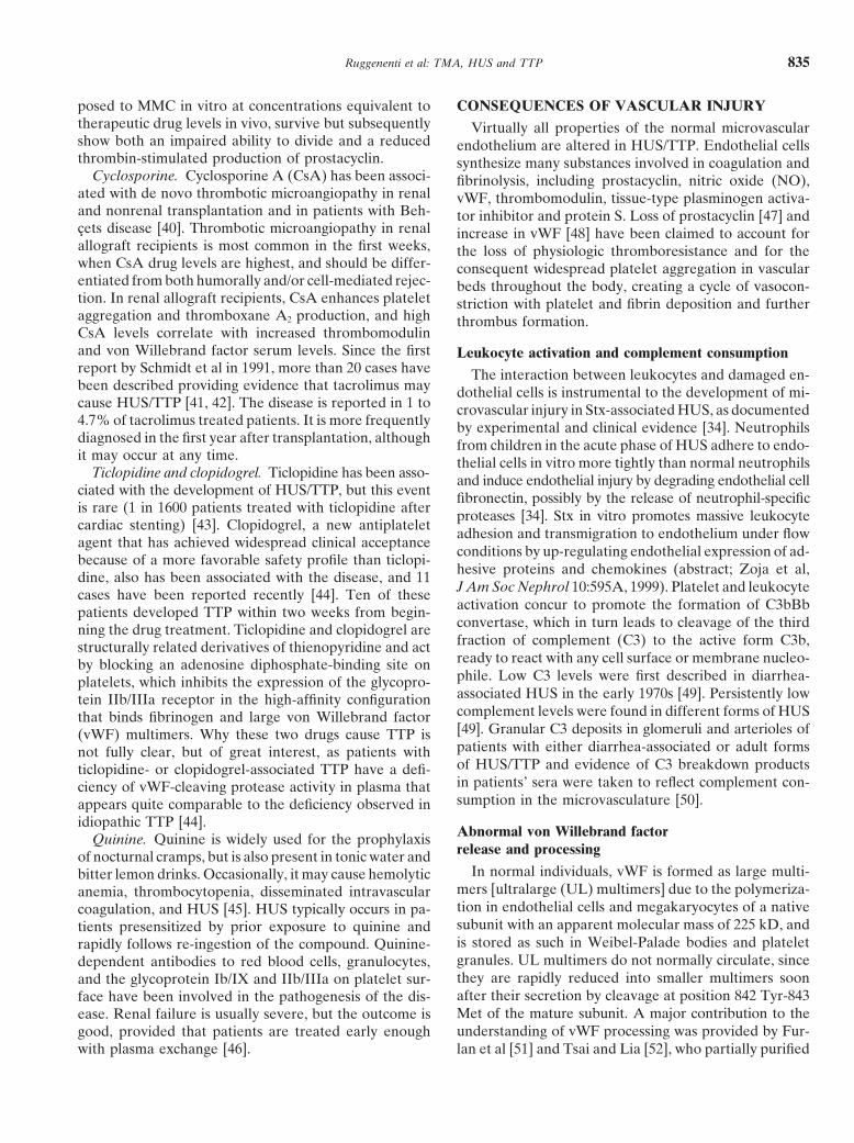
Fig. 4. Glomerulus from a patient with atypical HUS with predominantFig. 2. Glomerulus from a patient with a form of HUS with predomi-vascular involvement. Severe ischemic changes have occurred, charac-nant glomerular involvement. Capillary lumina are narrowed or oc-terized by marked restriction of the glomerular tuft.cluded by erythrocytes and thrombi. Swelling of endothelial cells and
thickening of the capillary wall with double contours have also occurred.A marked thickening of the glomerular capillary wall occurs with manydouble contours.
Fig. 5. A glomerulus and an interlobular artery from a patient withatypical HUS with severe vascular involvement. Severe ischemic
Fig. 3. Two arterioles in a case of atypical HUS with severe vascular changes have occurred with marked glomerular restriction and concomi-involvement. The vascular lumen is extremely narrowed due to miointi- tant severe tubular changes. The vascular lumen is almost entirelymal proliferation and vascular wall thickening. occluded due to extreme thickening of the vascular wall.
ing the resumption of daily plasma therapy, is common from 40 to 50% to the current 3 to 5%—probably as theresult of better supportive management of anemia, renalafter treatment discontinuation with reported frequen-
cies of 29 to 82% [5, 7, 93]. Of course, this mandates failure, hypertension, and electrolyte and water imbal-ance [79]. However, no specific therapy aimed to preventadditional treatment. Actually, discontinuation of plasma
therapy is the only way to know whether a durable remis- or limit the microangiopathic process has been proven toaffect the course of the disease in children. Antimotilitysion has been achieved, and many cycles of stopping and
resuming plasma therapy may be required. agents may increase the risk of toxic megacolon. Antibi-otics given to treat infection due to Stx–producing E.coli O157:H7 have been found to increase the risk of
CHILDHOOD HUSovert HUS by 17-fold [94]. Indeed, antibiotic-induced
STX-associated HUS injury to the bacterial membrane might favor the acuterelease of large amounts of preformed toxin. Alterna-In typical Stx-associated HUS in children, the mortality
rate has significantly decreased over the last 40 years— tively, antibiotic therapy might give E. coli O157:H7 a

Ruggenenti et al: TMA, HUS and TTP842
Tab
le1.
Spec
ific
trea
tmen
tsof
thro
mbo
tic
mic
roan
giop
athy
:Dos
es,m
odal
itie
sof
adm
inis
trat
ioin
,ind
icat
ions
and
cont
rain
dica
tion
s
Tre
atm
ent
Adm
inis
trat
ion
Indi
cati
on-c
omm
ent
Tre
atm
ents
ofpr
oven
effi
cacy
Pla
sma
exch
ange
1–2
plas
ma
vol/d
ayF
irst
-lin
eth
erap
yin
adul
tan
dat
ypic
alfo
rms;
life
savi
ngin
case
sw
ith
neur
olog
ical
invo
lvem
ent;
may
favo
rre
nal
func
tion
reco
very
;no
risk
offl
uid
over
load
even
inpa
tien
tsw
ith
seve
reca
rdia
cor
rena
ldy
sfun
ctio
nP
lasm
ain
fusi
on30
–40
mL
/kg
onda
y1,
then
Fir
st-l
ine
ther
apy
whe
nex
chan
geis
not
avai
labl
e;ef
fect
ive
inth
etr
eatm
ent
orpr
even
-10
–20
mL
/kg/
day
tion
ofre
curr
ent
epis
odes
Pla
sma
cryo
supe
rnat
ant
See
plas
ma
infu
sion
/exc
hang
eSe
cond
-lin
eth
erap
y;m
aybe
effe
ctiv
ein
case
sw
hodo
not
resp
ond
tow
hole
plas
ma
infu
sion
/exc
hang
eSo
lven
tde
terg
ent-
trea
ted
plas
ma
See
plas
ma
infu
sion
/exc
hang
eSa
me
indi
cati
ons
ofpl
asm
ain
fusi
on/e
xcha
nge;
may
limit
the
risk
ofvi
ralc
onta
min
atio
nR
escu
etr
eatm
ents
Bila
tera
lne
phre
ctom
yM
aybe
life
savi
ngin
case
sw
ith
refr
acto
ryhy
pert
ensi
on/t
hrom
bocy
tope
nia
and
hype
r-te
nsiv
een
ceph
alop
athy
;to
bere
stri
cted
topa
tien
tsw
ith
biop
syev
iden
ceof
seve
rere
nal
vasc
ular
invo
lvem
ent
Sple
nect
omy
May
beef
fect
ive
inre
laps
ing
form
s;to
bere
stri
cted
topa
tien
tsw
ith
freq
uent
rela
pses
and
requ
irin
gla
rge
amou
nts
ofpl
asm
aO
ther
trea
tmen
tsP
redn
ison
eO
ral,
60–2
00m
g/da
y,ta
pere
dby
5m
g/w
eek
Pos
sibl
yef
fect
ive
inm
ild,a
dult
form
s;ad
diti
onal
ther
apy
inpo
orre
spon
ders
topl
asm
ain
fusi
on/e
xcha
nge
(?)
Gam
ma
glob
ulin
sIn
trav
enou
s40
0m
g/kg
/day
Unp
rove
nef
fica
cyV
incr
isti
ne1.
4m
g/m
2IV
onda
y1,
than
1m
gev
ery
4da
ysup
to4
dose
sU
npro
ven
effi
ciac
y;m
ight
decr
ease
the
risk
ofre
curr
ence
sA
ntit
hrom
boti
cag
ents
(hep
arin
,str
epto
kina
se)
Intr
aven
ous
Unp
rove
nef
fica
cy;i
ncre
ased
risk
ofbl
eedi
ngA
ntip
late
let
agen
ts(a
spir
in,p
rost
acyc
lin)
Ora
lor
intr
aven
ous
Unp
rove
nef
fica
cy;i
ncre
ased
risk
ofbl
eedi
ngV
itam
inE
Ora
l,10
00m
g/m
2 /day
Pro
babl
yef
fect
ive
inSt
x-as
soci
ated
HU
S;to
bete
sted
inco
ntro
lled
tria
lsT
reat
men
tsun
der
inve
stig
atio
nSy
nsor
b-P
KO
ral,
0.5
gpe
rkg
body
wei
ght,
�7
days
;M
aypr
even
t/lim
itSh
iga
toxi
nab
sorp
tion
and
the
risk
offu
ll-bl
own
Stx-
asso
ciat
edH
US;
tobe
adm
inis
tere
dat
the
onse
tof
unde
rcl
inic
alin
vest
igat
ion.
diar
rhea
orw
hen
inge
stio
nof
cont
amin
ated
food
issu
spec
ted

Ruggenenti et al: TMA, HUS and TTP 843
selective advantage if these organisms are not as readily plasma infusion or exchange, occasionally in combina-tion with steroids, has been associated with recovery.eliminated from the bowel as are the normal intestinal
Idiopathic/atypical HUS. Plasma therapy is usually rec-flora. Moreover, several antimicrobial drugs, particularlyommended in these forms, although they have an almostthe quinolones, trimethoprim, and furazolidone, are po-invariably poor outcome despite the therapy.tent inducers of the expression of the Stx 2 gene and may
increase the level of toxin in the intestine [95]. However,ADULT HUS/TTPthese considerations do not necessarily apply to many cases
STX-associated HUS/TTP. The evaluation of treat-of bloody diarrhea, in particular in South America andment efficacy in adult patients is difficult because mostIndia, which are precipitated by E. coli strains differentof the information is derived by uncontrolled series,from O157:H7 or by other bacteria, such as S. dysenteriaewhich may include also non-Stx-HUS cases [86]. In par-type 1 [96]. For instance, when hemorrhagic colitis is causedticular, no prospective, randomized trials are available toby S. dysenteriae type 1, early and empirical antibioticdefinitely establish whether plasma infusion or exchangetreatment shortens the duration of diarrhea, decreasesmay offer some specific benefit as compared to support-the incidence of complications, and reduces the risk ofive treatment alone. However, comparative analyses oftransmission by shortening the duration of bacterial shed-two large series of patients treated [33] or not [20] withding [97]. Thus, in developing countries where Shigella isplasma suggest that plasma therapy may dramaticallythe most frequent cause of hemorrhagic colitis, antibioticdecrease overall mortality of Stx-E. coli O157:H7-associ-therapy should be started early and even before the in-ated HUS. These findings lead others and us to considervolved pathogen is identified [96].plasma infusion or exchange indicated in adult patients,New agents targeted to prevent organ exposition toin particular in those with severe renal insufficiency andStx are currently under evaluation. The most promisingcentral nervous system involvement [6, 7].are Synsorb-PK, a resin composed of repeated synthetic
Acute idiopathic HUS/TTP. Plasma manipulation is acarbohydrate determinants linked to colloidal silica thatcornerstone in the therapy of acute idiopathic HUS/TTP.binds Stx; recombinant modified E. coli that displays aExchange has been claimed to be superior than to plasmaStx receptor mimic on its surface and adsorbs and neutral-infusion in one study [5]. However, patients treated withizes Stx with very high efficiency; and “STARFISH,” anexchange were given larger amounts of plasma than thoseoligovalent, water-soluble carbohydrate ligand that cantreated with plasma infusion alone. Indeed, when equiva-simultaneously engage all five B subunits of two toxinlent volumes of plasma were given, infusion and exchangemolecules. These approaches offer potent new weaponsappeared to be equally effective [100]. Plasma exchangeagainst Stx-E. coli–induced hemorrhagic diarrhea andshould be considered as first-choice therapy when renal
HUS. Oral administration of Synsorb or recombinantinsufficiency or heart failure limit the amount of plasma
modified E. coli could efficiently clear toxin from the that can be provided with infusion alone. Cryosuperna-gut, whereas intravenous administration of “STARFISH” tant fraction (that is, plasma from which a cryoprecypi-might help to prevent toxin that already has entered the tate containing the largest plasma vWF multimers, fibrin-circulation from destroying kidney function and micro- ogen, and fibronectin has been removed) instead of freshvessels. Preliminary analyses of an ongoing trial in Can- frozen plasma has been successful in treating a smallada found that early treatment (within two days after number of patients who did not respond to repeated ex-the onset of diarrhea) with Synsorb-PK decreased the changes or infusions with fresh frozen plasma [101]. Therisk of HUS from 17 to 7% [98]. Toxin-neutralizing anti- rationale for this approach is that plasma cryosuperna-bodies might be a future possible prevention for E. coli tant may provide the same beneficial factor(s) found ininfection. Actually, natural infection with E. coli O157 whole plasma, but does not contain those factors (includ-does not confer immunity, and no human vaccine is cur- ing large vWF multimers) that may actually sustain therently available. Nevertheless, Shiga toxoid vaccines have microangiopathic process until remission is achieved. Onbeen shown to be effective in preventing related diseases the basis of the previously mentioned information, thein animals. Generalized pasteurization of ground beef use of plasma cryosupernatant has been suggested as firstthrough irradiation will further help to limit/prevent E. line therapy of adult HUS/TTP. However, results of thecoli O157 and other food-borne pathogen infections. few small, randomized studies failed to find differences
Neuroaminidase-associated HUS. The role of plasma between the two products.therapy is controversial. In theory, whole blood and Usually one plasma volume (40 mL/kg) is exchangedplasma should be avoided, since they contain IgM and per session. Treatment can be intensified by increasingmay accelerate polyagglutination and hemolysis through the volume of plasma replaced. The twice-daily exchangesIgM-mediated erythrocyte and endothelial cell injury of one plasma volume is probably the treatment of choice[99]. Thus, patients should be treated only with antibiot- for refractory patients in order to minimize the recycling
of infused plasma [7]. Serum LDH and platelet countics and washed red cells [36]. In some cases, however,
Ruggenenti et al: TMA, HUS and TTP844
are the most reliable markers of response, and plasma recover with delivery, and plasma therapy should betherapy should be continued until they are persistently restricted only to cases at imminent risk of death or withnormalized. Occasional patients with idiopathic HUS/ no evidence of disease recovery over 24 to 48 hours afterTTP do not achieve remission despite plasma therapy. delivery [103]. Cases occurring earlier in the course ofSome of them become plasma-dependent and require gestation are usually episodes of acute, idiopathic HUS/continued treatment, since disease relapses as soon as TTP and are best treated with plasma therapy. Thoseplasma infusion or exchange is stopped. Splenectomy occurring after a normal delivery are usually defined ashas been found to induce remission in some plasma- postpartum HUS. The pathogenesis is unclear, and theresistant cases, but was ineffective and actually increased outcome is usually poor with a high incidence of renalmorbidity and mortality in others [93]. Recently, the sequelae even despite aggressive plasma therapy. Casespresence in the circulation of autoantibodies against recurring after a kidney transplant are almost invariablyvWF-cleaving protease has been claimed to help identify found in the context of a familial or recurrent backgroundpatients who may benefit of glucocorticoid or vincristine and are usually associated with a poor outcome [90].treatment, removal of IgG by immunoadsorption on pro- Quite different is the outcome of de novo post-transplanttein A, or splenectomy [51]. HUS that may be associated with CsA or tacrolimus
Consistent data are available on the beneficial effect therapy or with vascular rejection. Removal of the drugof bilateral nephrectomy, in particular in patients with and effective treatment of the rejection, plus plasmasevere renal involvement, refractory hypertension, and therapy, can cure the disease.signs of hypertensive encephalopathy [62]. Combined toplasma therapy, antiplatelet agents have no additional ACKNOWLEDGMENTSeffect on the outcome of the disease [5, 93]. The same
This work was supported by the “Comitato 30 ore per la vita” andconsiderations apply to prostacyclin, heparin, and fibrino- Credito Bergamasco. The authors thank Mr. Gianfranco Marchetti for
his invaluable help in preparing and evaluating the histologic material,lytic agents such as streptokinase or urokinase. Plateletand Ms. Laura Arioli for assistance in preparing the manuscript.transfusions are contraindicated: They may fuel the throm-
botic process, and their use is recommended only in pa- Reprint requests to Piero Ruggenenti, M.D., “Mario Negri” Institutefor Pharmacological Research, Via Gavazzeni, 11, 24125 Bergamo,tients with active bleeding or before invasive procedures.Italy.The use of corticosteroids has been suggested with theE-mail: [email protected]
rationale to decrease splenic platelet sequestration andlimit vascular injury. However, before plasma therapy REFERENCESwas available, corticosteroids induced remission in fewer
1. Symmers WStC: Thrombotic microangiopathic haemolytic ane-than 10% of patients, an outcome comparable to thatmia (thrombotic microangiopathy). Br Med J 2:897–903, 1952
of untreated patients. The same considerations apply to 2. Remuzzi G, Ruggenenti P, Bertani T: Thrombotic microangio-immunoglobulins and other immunosuppressive treat- pathies, in Renal Pathology: With Clinical and Functional Correla-
tions, edited by Tisher CC, Brenner BM, Philadelphia, J.B. Lip-ments.pincott Company, 1994, p 1154
3. Gasser C, Gautier E, Steck A, et al: Hamolytisch-uramischeRECURRENT, FAMILIAL, AND syndromes bilaterale nierenrindennekrosen bei akuten erwor-“SMOLDERING” HUS/TTP benchen hamolytischen anamien. Schweiz Med Wschr 85:905–909,
1955Plasma infusion or exchange is usually effective to 4. Moschcowitz E: Acute febrile pleiochromic anemia with hyalinetreat or prevent acute episodes of relapsing forms [7]. thrombosis of a terminal arterioles and capillaries: An undescribed
disease. Arch Intern Med 36:89–93, 1925Preliminary evidence is available that elective splenec-5. Rock GA, Shumak KH, Buskard NA, et al: Comparison oftomy during hematologic remission reduces the relapse plasma exchange with plasma infusion in the treatment of throm-
rate and the need for plasma therapy [102]. Thus, sple- botic thrombocytopenic purpura. N Engl J Med 325:393–397, 19916. Rock G, Shumak K, Kelton J, et al: Thrombotic thrombocytope-nectomy should be considered in those patients with
nic purpura: Outcome in 24 patients with renal impairment treateddisabling disease requiring frequent and prolonged with plasma exchange. Transfusion 32:710–714, 1992courses of plasma therapy. After recovery, few patients 7. George JN: How I treat patients with thrombotic thrombocytope-
nic purpura-hemolytic uremic syndrome. Blood 96:1223–1229, 2000may have persistent, asymptomatic thrombocytopenia.8. Remuzzi G: HUS and TTP: Variable expression of a single entity.This “smoldering” form does not need specific treatment,
Kidney Int 32:292–308, 1987but warrants close monitoring since it may identify pa- 9. Altschule MD: A rare type of acute thrombocytopenic purpura:
Widespread formation of platelet thrombi in capillaries. N Engltients at risk of future recurrences [102].J Med 227:477–479, 1942
10. Mitra D, Jaffe EA, Weksler B, et al: Thrombotic thrombocyto-SECONDARY HUS/TTPpenic purpura and sporadic hemolytic-uremic syndrome plasmasinduce apoptosis in restricted lineages of human microvascularPatients with drug-induced HUS/TTP usually promptlyendothelial cells. Blood 89:1224–1234, 1997recover after treatment withdrawal and a short course
11. Adam A: Jahrb f Kinderh 116:1927of plasma therapy. Pregnancy-associated forms, when 12. Belnap WD, O’Donnell JJ: Epidemic gastroenteritis due to
Escherichia coli O–111. J Pediatr 47:178–193, 1955they manifest in the last trimester of gestation, normally
Ruggenenti et al: TMA, HUS and TTP 845
13. Konowalchuk J, Speirs JI, Stavric S: Vero response to a cyto- cell antibodies in hemolytic-uremic syndrome. Lancet 2:183–186,1988toxin of Escherichia coli. Infect Immunol 18:775–779, 1977
38. Asherson RA, Cervera R, Piette J-C, et al: Catastrophic anti-14. Koster F, Levin J, Walker L, et al: Hemolytic uremic syndromephospholipid syndrome: Clinical and laboratory features of 50after shigellosis: Relation to endotoxemia and circulating immunepatients. Medicine 77:195–207, 1998complexes. N Engl J Med 298:927–933, 1978
39. Murgo AJ: Cancer- and chemotherapy-associated thrombotic mi-15. Riley LW, Remis RS, Helgerson SD, et al: Hemorrhagic colitiscroangiopathy, in Hemolytic Uremic Syndrome and Thromboticassociated with a rare Escherichia coli O157:H7 serotype. N EnglThrombocytopenic Purpura, edited by Kaplan BS, TrompeterJ Med 308:681–685, 1983RS, Moake JL, New York, Marcel Dekker, Inc., 1992, p 27116. Karmali MA, Steele BT, Petric M, Lim C: Sporadic cases of
40. Remuzzi G, Bertani T: Renal vascular and thrombotic effectshemolytic-uremic syndrome associated with fecal verotoxin andof cyclosporine. Am J Kidney Dis 13:261–272, 1989cytotoxin producing Escherichia coli in stools. Lancet 1:619–620,
41. Schmidt RY, Venkat KK, Dumler F: Hemolytic uremic syn-1983drome in a renal transplant recipient on FK506 immunosuppres-17. Mead PS, Griffin PM: Escherichia coli O157:H7. Lancetsion. Transplant Proc 23:3156–3157, 1991352:1207–1212, 1998
42. Trimarchi HM, Truong LD, Brennan S, et al: FK-506-associated18. Deleted in proofthrombotic microangiopathy. Transplantation 67:539–544, 199919. Griffin PM, Tauxe RV: The epidemiology of infections caused
43. Bennett CL, Weinberg PD, Rozenberg-Ben-Dror K, et al:by Escherichia coli O157:H7, other enterohemorrhagic E coli,Thrombotic thrombocytopenic purpura associated with ticlopi-and the associated hemolytic uremic syndrome. Epidemiol Revdine: A review of 60 cases. Ann Intern Med 128:541–544, 199813:60–98, 1991
44. Bennett CL, Connors JM, Carwile JM, et al: Thrombotic throm-20. Carter AO, Borczyk AA, Carlson JA, et al: A severe outbreakbocytopenic purpura associated with clopidogrel. N Engl J Medof Escherichia coli O157:H7-associated hemorrhagic colitis in a342:1773–1777, 2000nursing home. N Engl J Med 317:1496–1500, 1987
45. Gottschall JL, Elliot W, Lianos E, et al: Quinine induced21. Pavia AT, Nichols CR, Green DP, et al: Hemolytic-uremic syn-immune thrombocytopenia associated with hemolytic uremic syn-drome during an outbreak of Escherichia coli O157:H7 infectionsdrome: A new clinical entity. Blood 77:306–310, 1991in institutions for mentally retarded persons: Clinical and epidemi-
46. McDonald SP, Shanahan EM, Thomas AC, et al: Quinine-ologic observations. J Pediatr 116:544–551, 1990induced hemolytic uremic syndrome. Clin Nephrol 47:397–400,22. Donnenberg MS, Tacket CO, James SP, et al: Role of the eaeA1997gene in experimental enteropathogenic Escherichia coli infection.
47. Noris M, Benigni A, Siegler R, et al: Renal prostacyclin biosyn-J Clin Invest 92:1412–1417, 1993thesis is reduced in children with hemolytic-uremic syndrome in23. Acheson DWK, Moore R, DeBreuler S: Translocation of Shiga-the context of systemic platelet activation. Am J Kidney Dislike toxins across polarized intestinal cells in tissue culture. Infect20:144–149, 1992Immunol 64:3294–3300, 1996
48. Rose PE, Enayat SM, Sunderland R, et al: Abnormalities of24. te Loo DMWM, Monnens LAH, van der Velden TJAM, et al:factor VIII related protein multimers in the haemolytic uraemicBinding and transfer of verocytotoxin by polymorphonuclear leu- syndrome. Arch Dis Child 59:1135–1140, 1984kocytes in hemolytic uremic syndrome. Blood 95:3396–3402, 2000 49. Stuhlinger W, Kourilsky O, Kanfer A, Sraer JD: Haemolytic-
25. Calderwood SB, Achewaz DWK, Krusch GT, et al: Proposed uraemic syndrome: Evidence for intravascular C3 activation. Lan-new nomenclature for SLI (VT) family. Infect Immunol 62:118– cet 2:788–780, 1974119, 1998 50. Hammar SP, Bloomer HA, McCloskey D: Adult hemolytic ure-
26. van Setten PA, van Hinsbergh VW, van der Velden TJ, et al: mic syndrome with renal arteriolar deposition of IgM and C3.Effects of TNF alpha on verocytotoxin cytotoxicity in purified Am J Clin Pathol 70:434–439, 1978human glomerular microvascular endothelial cells. Kidney Int 51. Furlan M, Robles R, Galbusera M, et al: von Willebrand factor-51:1245–1256, 1997 cleaving protease in thrombotic thrombocytopenic purpura and
27. Arab S, Lingwood CA: Intracellular targeting of the endoplasmic the hemolytic-uremic syndrome. N Engl J Med 339:1578–1584,reticulum/nuclear envelope by retrograde transport may deter- 1998mine cell hypersensitivity to verotoxin via globotriosyl ceramide 52. Tsai HM, Lia ECY: Antibodies to von Willebrand factor-cleavingfatty acid isoform traffic. J Cell Physiol 117:646–660, 1998 protease in acute thrombotic thrombocytopenic purpura. N Engl
28. Bitzan MM, Wang Y, Lin J, Marsden PA: Verotoxin and ricin J Med 339:1585–1594, 1998have novel effects on preproendothelin-1 expression but fail to 53. Dent JA, Galbusera M, Ruggeri ZM: Heterogeneity of plasmamodify nitric oxide synthase (ecNOS) expression and NO produc- von Willebrand factor multimers resulting from proteolysis oftion in vascular endothelium. J Clin Invest 101:372–382, 1998 constituent subunit. J Clin Invest 88:774–782, 1991
29. Andreoli SP: The pathophysiology of the hemolytic uremic syn- 54. Moake JL, McPherson PD: Abnormalities of von Willebranddrome. Curr Opin Nephrol Hypertens 8:459–464, 1999 factor multimers in thrombotic thrombocytopenic purpura and
30. Kevin EC, Kaplan BS: Many cell types are Shiga toxin targets. the hemolytic-uremic syndrome. Am J Med 87:9N–15N, 1989Kidney Int 57:2650–2651, 2000 55. Moake JL: Moschcowitz, multimers, and metalloprotease. N Engl
31. Zoja C, Corna D, Farina C, et al: Verotoxin glycolipid receptors J Med 339:1629–1631, 1998determine the localization of microangiopathic process in rabbits 56. Weinstein MJ, Blanchard R, Moake JL, et al: Fetal and neonatalgiven verotoxin-1. J Lab Clin Med 120:229–238, 1992 von Willebrand factor (vWF) is unusually large and similar to
32. Taylor FB Jr, Tesh VL, DeBault L, et al: Characterization of the the vWF in patients with thrombotic thrombocytopenic purpura.baboon responses to Shiga-like toxin. Am J Pathol 154:1285–1299, Br J Haematol 72:68–72, 19891999 57. Mannucci PM, Lombardi R, Castaman G, et al: von Willebrand
33. Dundas S, Murphy J, Soutar RL, et al: Effectiveness of therapeu- disease “Vicenza” with larger-than-normal (supranormal) vontic plasma exchange in the 1996 Lanarkshire Escherichia coli Willebrand factor multimers. Blood 71:65–70, 1988O157:H7 outbreak. Lancet 354:1327–1330, 1999 58. Moake JL: Hemolytic-uremic syndrome: Basic science. Lancet
34. Forsyth KD, Simpson AC, Fitzpatrick MM, et al: Neutrophil- 343:393–397, 1994mediated endothelial injury in hemolytic uremic syndrome. Lan- 59. Furlan M, Robles R, Solenthaler M, et al: Deficient activitycet 2:411–414, 1989 of von Willebrand factor-cleaving protease in chronic relapsing
35. Klein PJ, Bulla M, Newman RA, et al: Thomsen-Friedenreich thrombotic thrombocytopenic purpura. Blood 89:3097–3103, 1997antigen in hemolytic-uremic syndrome. Lancet 2:1024–1025, 1977 60. Furlan M, Robles R, Solenthaler M, Lammle B: Acquired
36. McGraw ME, Lendon M, Stevens RF, et al: Hemolytic uremic deficiency of von Willebrand factor-cleaving protease in a patientsyndrome and the Thomsen Friedenreich antigen. Pediatr Nephrol with thrombotic thrombocytopenic purpura. Blood 91:2839–2846,3:135–139, 1989 1998
61. te Loo DMWM, Levtchenko E, Furlan M, et al: Autosomal37. Leung YD, Moake JL, Havens PL, et al: Lytic anti-endothelial

Ruggenenti et al: TMA, HUS and TTP846
recessive inheritance of von Willebrand factor-cleaving protease 79. Remuzzi G, Ruggenenti P: The hemolytic uremic syndrome.Kidney Int 47:2–19, 1995deficiency. Pediatr Nephrol 14:762–765, 2000
80. Siegler RL: The hemolytic uremic syndrome. Pediatr Clin North62. Remuzzi G, Galbusera M, Salvadori M, et al: Bilateral nephrec-Am 42:1505–1529, 1995tomy stopped disease progression in plasma-resistant hemolytic
81. Fitzpatrick MM, Shah V, Trompeter RS, et al: Long term renaluremic syndrome with neurological signs and coma. Kidney Intoutcome of childhood hemolytic uremic syndrome. Br Med J 303:49:282–286, 1996489–492, 199163. Galbusera M, Noris M, Rossi C, et al: Increased fragmentation
82. Gagnadoux MF, Habib R, Gubler MC, et al: Long-term (15–25of von Willebrand factor, due to abnormal cleavage of the subunit,years) outcome of childhood hemolytic-uremic syndrome. Clinparallels disease activity in recurrent hemolytic uremic syndromeNephrol 46:39–41, 1996and thrombotic thrombocytopenic purpura and discloses predis-
83. Fitzpatrick MM, Walters MDS, Trompeter RS, et al: Atypicalposition in families: The Italian Registry of Familial and Recur-(non-diarrhea-associated) hemolytic-uremic syndrome in child-rent HUS/TTP. Blood 94:610–620, 1999hood. J Pediatr 122:532–537, 199364. Mannucci PM, Lombardi R, Lattuada A, et al: Enhanced prote-
84. Proesmans W: Typical and atypical hemolytic uremic syndrome.olysis of plasma von Willebrand factor in thrombotic thrombocy-Kidney Blood Press Res 19:205–208, 1996topenic purpura and the hemolytic uremic syndrome. Blood
85. Neuhaus TJ, Calonder S, Leumann EP: Heterogeneity of atypi-74:978–983, 1989cal hemolytic uremic syndrome. Arch Dis Child 76:518–521, 199765. Tsai H-M, Sussman II, Nagel RL: Shear stress enhances the
86. Kaplan BS, Meyers KE, Schulman SL: The pathogenesis andproteolysis of von Willebrand factor in normal plasma. Bloodtreatment of hemolytic uremic syndrome. J Am Soc Nephrol83:2171–2179, 19949:1126–1133, 199866. Kaplan BS, Kaplan P: Hemolytic uremic syndrome in families, in
87. Rose M, Eldor A: High incidence of relapses in thromboticHemolytic Uremic Syndrome and Thrombotic Thrombocytopenicthrombocytopenic purpura: Clinical study of 38 patients. Am JPurpura, edited by Kaplan BS, Trompeter RS, Moake JL, NewMed 83:437–444, 1987York, Marcel Dekker, Inc., 1992, p 213 88. Shumak KH, Rock GA, Nair RC, Canadian Apheresis Group:67. Noris M, Ruggenenti P, Perna A, et al: Hypocomplementemia Late relapses in patients successfully treated for thrombotic
discloses genetic predisposition to hemolytic uremic syndrome thrombocytopenic purpura. Ann Intern Med 122:569–572, 1995and thrombotic thrombocytopenic purpura: Role of factor H ab- 89. Ruggenenti P, Remuzzi G, Rossi EC: Epidemiology of the he-normalities: Italian Registry of Familial and Recurrent Hemolytic molytic-uremic syndrome. N Engl J Med 324:1065–1066, 1991Uremic Syndrome/Thrombotic Thrombocytopenic Purpura. J Am 90. Kaplan BS, Leonard MB: Autosomal dominant hemolytic ure-Soc Nephrol 10:281–293, 1999 mic syndrome: Variable phenotypes and transplant results. Pedi-
68. Monnens L, Malenaar J, Lambert PH, et al: The complement atric Nephrol 14:464–468, 2000system in hemolytic-uremic syndrome in childhood. Clin Nephrol 91. Cohen JD, Brecher ME, Bandarenko N: Cellular source of13:168–171, 1980 serum lactate dehydrogenase elevation in patients with throm-
69. Zipfel PF, Hellwage J, Friese M, et al: Factor H and disease: botic thrombocytopenic purpura. J Clin Apheresis 13:16–19, 1998A complement regulator affects vital body function. Mol Immunol 92. Morel-Maroger L: Adult hemolytic-uremic syndrome. Kidney36:241–248, 1999 Int 18:125–134, 1980
70. Thompson RA, Winterborn MH: Hypocomplementaemia due 93. Bell WR, Braine HG, Ness PM, Kickler TS: Improved survivalto a genetic deficiency of b1H globulin. Clin Exp Immunol 46:110– in thrombotic thrombocytopenic purpura-hemolytic uremic syn-119, 1981 drome. Clinical experience in 108 patients. N Engl J Med 325:398–
71. Rougier N, Kazatchkine MD, Rougier J-P, et al: Human com- 403, 1991plement factor H deficiency associated with hemolytic uremic 94. Wong CS, Jelacic S, Habeeb RL, et al: The risk of the hemolytic-syndrome. J Am Soc Nephrol 9:2318–2326, 1998 uremic syndrome after antibiotic treatment of Escherichia coli
72. Warwicker P, Goodship THJ, Donne RL, et al: Genetic studies O157:H7 infections. N Engl J Med 342:1930–1936, 2000into inherited and sporadic hemolytic uremic syndrome. Kidney 95. Zimmerhackl LB: E. coli, antibiotics, and the hemolytic-uremicInt 53:836–844, 1998 syndrome. N Engl J Med 342:1990–1991, 2000
96. O’Ryan M, Prado V: Risk of the hemolytic uremic syndrome73. Caprioli J, Bettinaglio P, Zipfel PF, et al: The molecular basis ofafter antibiotic treatment of Escherichia coli O157:H7 infections.familial hemolytic uremic syndrome: Mutation analysis of factor HN Engl J Med 343:1271, 2000gene reveals a hot spot in short consensus repeat 20. J Am Soc
97. Haltalin KC, Nelson JD, Ring R III, et al: Double-blind treat-Nephrol 12:287–307, 2001ment study of shigellois comparing ampiellin, sulfadlazine, and74. Richards A, Buddles MR, Donne RL, et al: Factor H mutationsplacebo. J Pediatr 70:970–981, 1967in hemolytic uremic syndrome cluster in exons 18-20, a domain
98. Donnelly JJ, Rappuoli R: Blocking bacterial enterotoxins. Na-important for host cell recognition. Am J Hum Genet 68:485–490,ture Medicine 6:257–258, 20002001
99. Erickson LC, Smith WS, Biswas AK, et al: Streptococcus pneu-75. Perez-Caballero D, Gonzalez-Rubio C, Gallardo ME, et al:moniae-induced hemolytic uremic syndrome: A case for earlyClustering of missense mutations in the C terminal region ofdiagnostic. Pediatr Nephrol 8:211–213, 1994factor H in atypical hemolytic uremic syndrome. Am J Hum Genet
100. Novitzky N, Jacobs P, Rosenstrauch W: The treatment of68:478–484, 2001 thrombotic thrombocytopenic purpura: Plasma infusion or ex-76. Ying L, Katz Y, Schlesinger M, et al: Complement factor H gene change? Br J Haematol 87:317–320, 1994
mutation associated with autosomal recessive atypical hemolytic 101. Byrnes JJ, Moake JL, Klug P, Periman P: Effectiveness of theuremic syndrome. Am J Hum Genet 65:1538–1546, 1999 cryosupernatant fraction of plasma in the treatment of refractory
77. Buddles MRH, Donne RL, Richards A, et al: Complement thrombotic thrombocytopenic purpura. Am J Hematol 34:169–factor H gene mutation associated with autosomal recessive atypi- 174, 1990cal hemolytic uremic syndrome. Am J Hum Genet 66:1721–1722, 102. Hayward CP, Sutton DM, Carter WH, Jr, et al: Treatment2000 outcomes in patients with adult thrombotic thrombocytopenic
78. Rey-Campos J, Rubinstain P, De Cordoba SR: A physical map purpura-hemolytic uremic syndrome. Arch Intern Med 154:982–of the human regulator of complement activation gene cluster 987, 1994linking the complement genes CR1, CR2, DAF and C4PB. J Exp 103. Weiner CP: Thrombotic microangiopathy in pregnancy and the
postpartum period. Semin Hematol 24:119–129, 1987Med 167:664–669, 1988
Related Documents