Thrombophilia Testing Robert Gosselin MT (ASCP), CLS

Thrombophilia Testing Robert Gosselin MT (ASCP), CLS.
Dec 30, 2015
Welcome message from author
This document is posted to help you gain knowledge. Please leave a comment to let me know what you think about it! Share it to your friends and learn new things together.
Transcript
Thrombophilia Testing
Robert Gosselin
MT (ASCP), CLS

• D-dimer– Indicates clot formation– Indicates clot degradation
• D-dimer test commonly used for exclusion:– Pulmonary embolism– Deep vein thrombosis– Consumptive coagulopathy– Aortic dissection

DE
D D
ED
Plasminogen PlasmintPA
uPA
FIBRIN
D
E
D
D
D
E
E
D
DD
D
E
D-dimer
Fragment X
Fragment D
Fragment Y
Fragments D & E
FIBRINOGEN

Microwell containing target Anti-human-XDP
XDP (+)
++ +
+++
Incubate
Conjugated Anti-human XDP antibody ¤
¤¤
¤
Wash
+++ ¤¤¤
Incubate
Wash Chromogenic tag
Color
Amount of color proportional to amount of XDP present. Quantitative result extrapolated from calibration curve
Patient XDP
Testing well
Reagent beads coated with anti-XDP
Instrument reading—changes in optical density
Incubate
Amount of light scattering proportional to XDP present. Quantitative result extrapolated from calibration curve
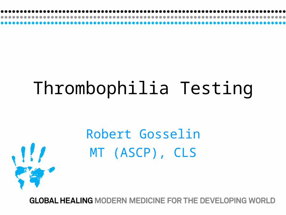
True Positive
True Positives + False Negatives
Sensitivity
SpecificityTrue Negative
True Negatives + False Positives
True Negatives
All Negatives
Negative Predictive Value

Clinical signs and symptoms of DVT +3Heart rate >100/min +1.5Hemoptysis +1Active cancer +1Bedridden (>3 days) or major (>12 weeks)
+1.5Previously history of DVT or PE +1.5PE most likely diagnosis +3
Clinical Probability for PE
Score: Low <2 Moderate 2-6 High >6
Wells PS, et al Thromb Haemost 2000; 83:416-20.

Active cancer +1
Paralysis, paresis, recent casting of leg +1
Bedridden (>3 days) or major (>12 weeks) +1
Entire leg swollen +1
Calf swelling (>3cm) compared to other leg +1
Pitting edema greater in symptomatic leg +1
Collateral nonvaricose superficial veins +1
Localized tenderness along deep venous system +1
Previously documented DVT +1
Alternative Dx as or more likely than DVT -2
Score: DVT unlikely <2 DVT likely >2
Clinical Probability for DVT
Wells PS, et al Lancet 1997; 350:1795-98; N Engl J Med 2003;349: 1227-35

Compression US
Positive Negative Low prob
Serial CUS (5-8 days) Mod or High Prob
DVT Positive
Positive VTE
DVT Negative
Negative VTE
3 month f/uPositive Negative
DVT
Algorithm
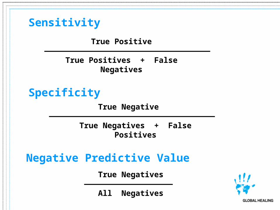
Spiral CT or Angiogram
Positive Negative
PE Positive
Positive VTE
PE Negative
Negative VTE
3 month f/u
PE
Algorithm
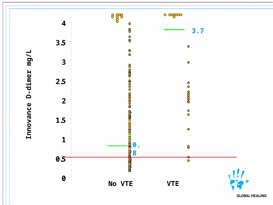
0
0.5
1
1.5
2
2.5
3
3.5
4
No VTE VTE
0.8
3.7In
no
van
ce D
-dim
er m
g/L
0.00
0.50
1.00
1.50
2.00
2.50
3.00
3.50
4.00
4.50
5.00
Inn
ova
nce
D-d
imer
, m
g/L
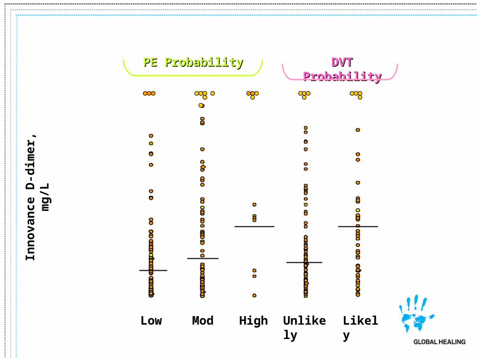
Low Mod High Unlikely Likely
PE ProbabilityPE Probability DVT ProbabilityDVT Probability
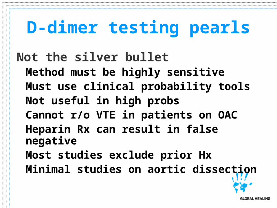
D-dimer testing pearls
Not the silver bulletMethod must be highly sensitiveMust use clinical probability tools
Not useful in high probsCannot r/o VTE in patients on OACHeparin Rx can result in false negativeMost studies exclude prior HxMinimal studies on aortic dissection
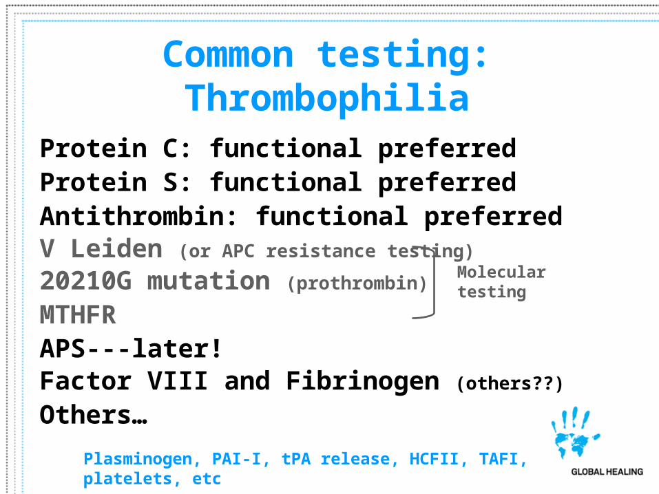
Common testing: Thrombophilia
Protein C: functional preferredProtein S: functional preferredAntithrombin: functional preferredV Leiden (or APC resistance testing)
20210G mutation (prothrombin)
MTHFRAPS---later!Factor VIII and Fibrinogen (others??)
Others…
Plasminogen, PAI-I, tPA release, HCFII, TAFI, platelets, etc
Molecular testing

Protein C-Amidolytic
Protein C Activated Protein C
Peptide-pNA Peptide + pNA
Copperhead rattlesnake venom
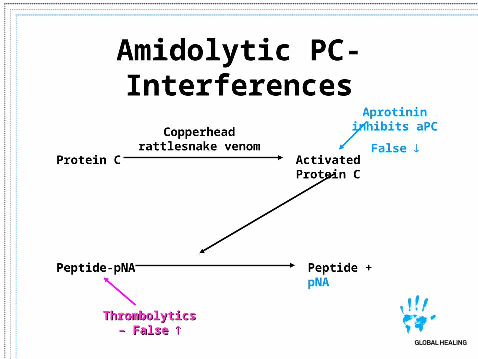
Amidolytic PC-Interferences
Protein C Activated Protein C
Peptide-pNA Peptide + pNA
Copperhead rattlesnake venom
Thrombolytics – Thrombolytics – False False
Aprotinin inhibits aPC
False
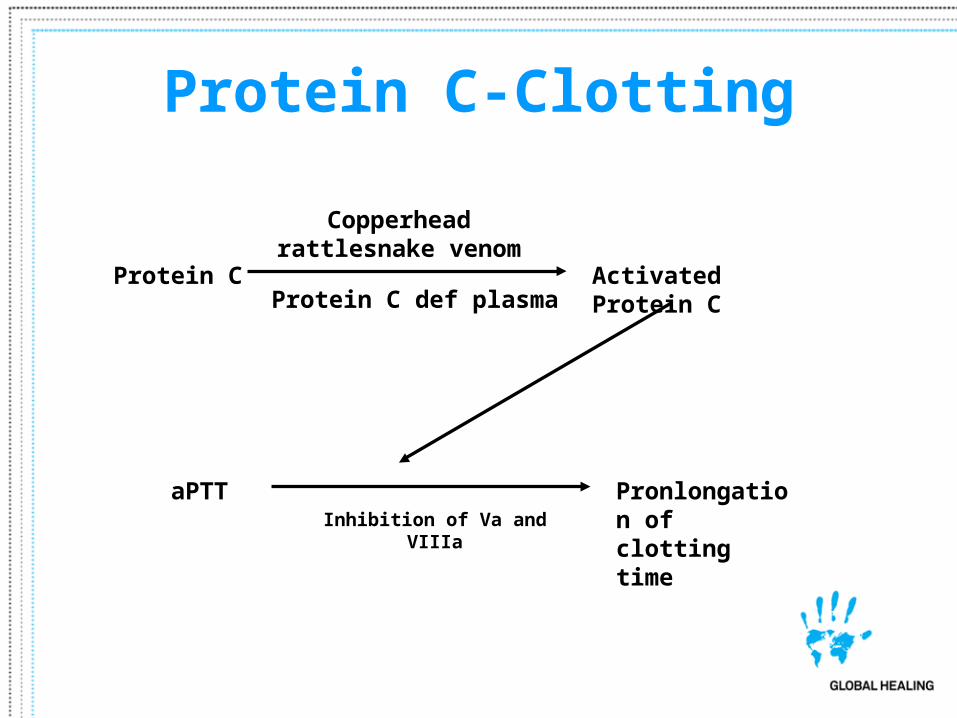
Protein C-Clotting
Protein C Activated Protein C
aPTT Pronlongation of clotting time
Copperhead rattlesnake venom
Protein C def plasma
Inhibition of Va and VIIIa

Clotting PC-Interferences
Protein C Activated Protein C
aPTT Prolongation of clotting time
Copperhead rattlesnake venom
Protein C def plasma
Inhibition of Va and VIIIaHeparin
DTI
Falsely
V Leiden mutation
Falsely
Aprotinin inhibits aPC
False
Increased Fbg or Factor VIII
Falsely
LA
Falsely
Pre-analytical
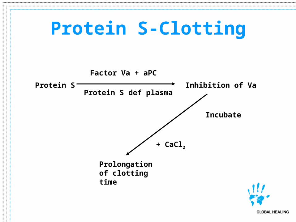
Protein S-Clotting
Protein S
Incubate
Prolongation of clotting time
Factor Va + aPC
Protein S def plasmaInhibition of Va
+ CaCl2
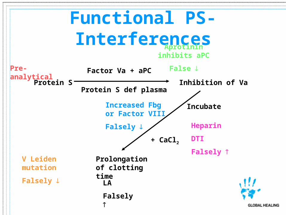
Functional PS-Interferences
Protein S
Incubate
Prolongation of clotting time
Factor Va + aPC
Protein S def plasmaInhibition of Va
+ CaCl2
Aprotinin inhibits aPC
False
Increased Fbg or Factor VIII
Falsely Heparin
DTI
Falsely
LA
Falsely
V Leiden mutation
Falsely
Pre-analytical
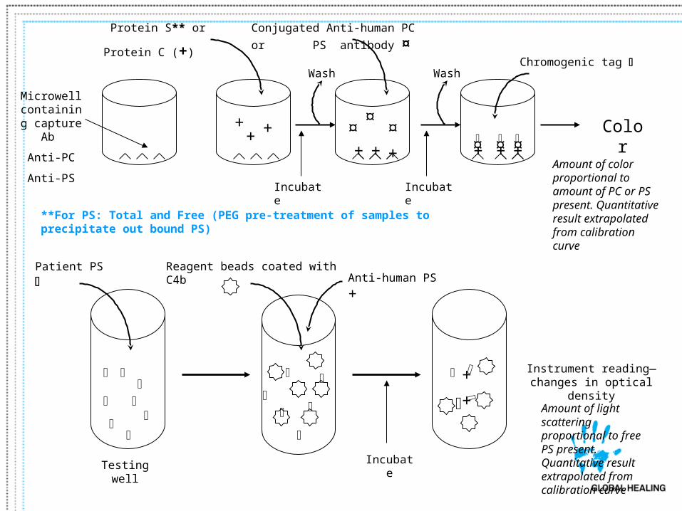
Microwell containing
capture Ab
Anti-PC
Anti-PS
Protein S**** or
Protein C (+)
++ +
+++
Incubate
Conjugated Anti-human PC or
PS antibody ¤
¤¤
¤
Wash
+++ ¤¤ ¤
Incubate
Wash Chromogenic tag
Color
Amount of color proportional to amount of PC or PS present. Quantitative result extrapolated from calibration curve
Patient PS
Testing well
Reagent beads coated with C4b
Instrument reading—changes in optical density
Incubate
Amount of light scattering proportional to free PS present. Quantitative result extrapolated from calibration curve
Anti-human PS +
+
+
**For PS: Total and Free (PEG pre-treatment of samples to precipitate out bound PS)
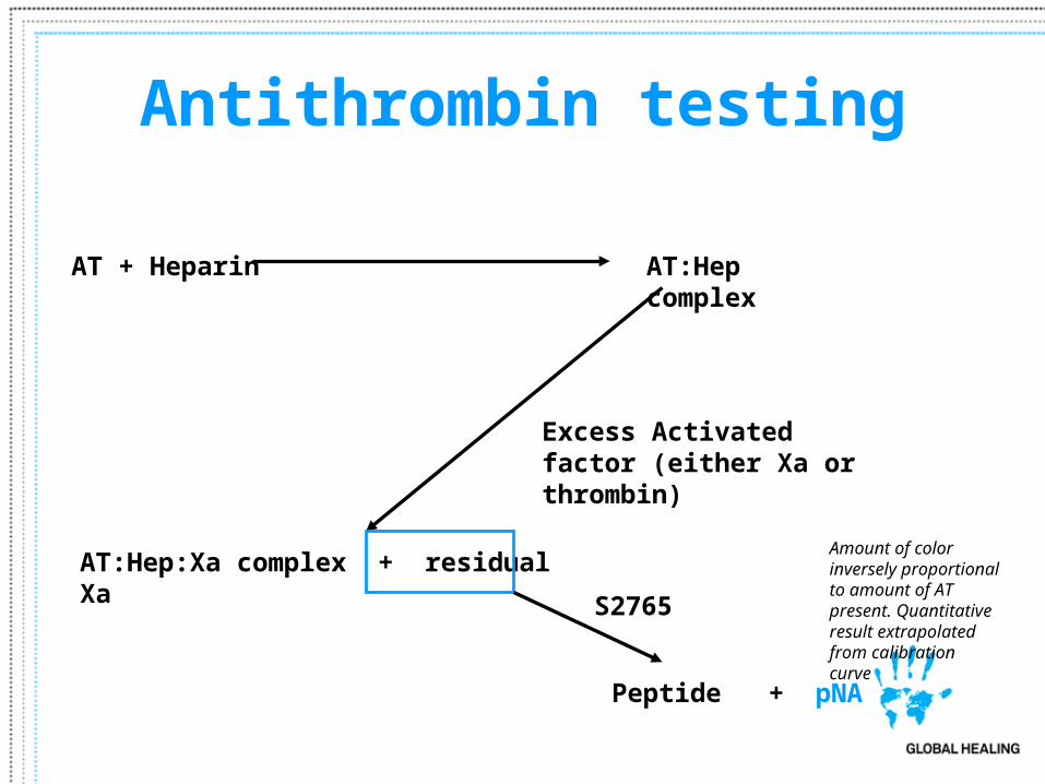
Antithrombin testing
AT + Heparin AT:Hep complex
Excess Activated factor (either Xa or thrombin)
AT:Hep:Xa complex + residual Xa
S2765
Peptide + pNA
Amount of color inversely proportional to amount of AT present. Quantitative result extrapolated from calibration curve
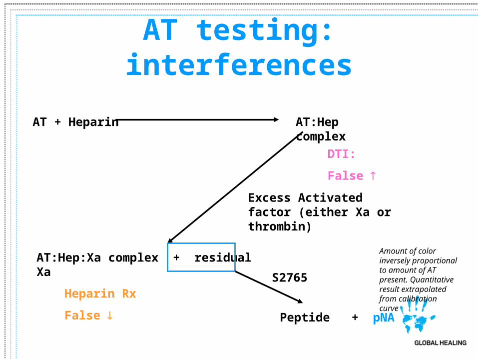
AT testing: interferences
AT + Heparin AT:Hep complex
Excess Activated factor (either Xa or thrombin)
AT:Hep:Xa complex + residual Xa
S2765
Peptide + pNA
Amount of color inversely proportional to amount of AT present. Quantitative result extrapolated from calibration curve
DTI:
False
Heparin Rx
False
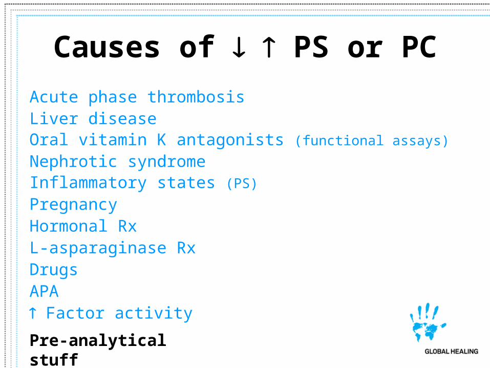
Causes of PS or PC
Acute phase thrombosisLiver diseaseOral vitamin K antagonists (functional assays)
Nephrotic syndrome Inflammatory states (PS)
PregnancyHormonal RxL-asparaginase RxDrugsAPA Factor activity
Pre-analytical stuff
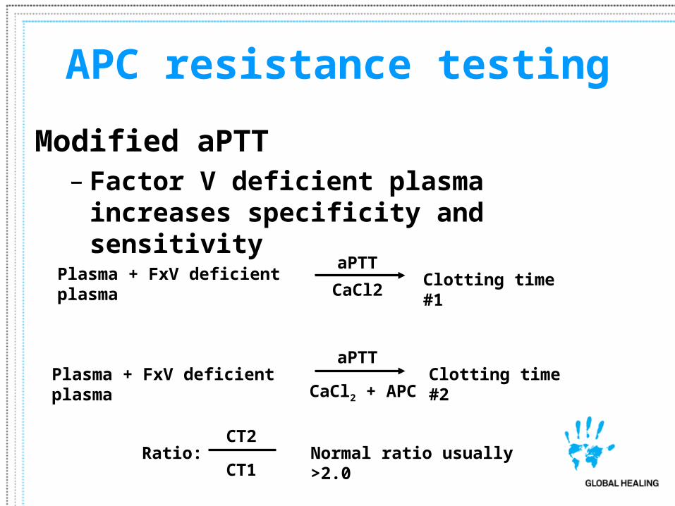
APC resistance testing
Modified aPTT– Factor V deficient plasma increases
specificity and sensitivity
Plasma + FxV deficient plasma Clotting time #1aPTT
Plasma + FxV deficient plasma Clotting time #2aPTT
CaCl2 + APC
CaCl2
Ratio: CT2
CT1Normal ratio usually >2.0

APC resistance: Interferences
Plasma + FxV deficient plasma Clotting timeaPTT
Plasma + FxV deficient plasma Clotting time #2aPTT
CaCl2 + APC
CaCl2
Ratio: CT2
CT1Normal ratio usually >2.0
Pre-analytical
Biases usually systematic -- tendency for lower ratios with APA. Patient on Xigris may effect results
Related Documents