REVIEW Open Access Three steps to the immortality of cancer cells: senescence, polyploidy and self-renewal Jekaterina Erenpreisa 1* and Mark S Cragg 2 Abstract Metastatic cancer is rarely cured by current DNA damaging treatments, apparently due to the development of resistance. However, recent data indicates that tumour cells can elicit the opposing processes of senescence and stemness in response to these treatments, the biological significance and molecular regulation of which is currently poorly understood. Although cellular senescence is typically considered a terminal cell fate, it was recently shown to be reversible in a small population of polyploid cancer cells induced after DNA damage. Overcoming genotoxic insults is associated with reversible polyploidy, which itself is associated with the induction of a stemness phenotype, thereby providing a framework linking these separate phenomena. In keeping with this suggestion, senescence and autophagy are clearly intimately involved in the emergence of self-renewal potential in the surviving cells that result from de-polyploidisation. Moreover, subsequent analysis indicates that senescence may paradoxically be actually required to rejuvenate cancer cells after genotoxic treatments. We propose that genotoxic resistance is thereby afforded through a programmed life-cycle-like process which intimately unites senescence, polyploidy and stemness. Keywords: Tumour cells, DNA damage, Senescence, Polyploidy, Self-renewal, Reprogramming, Totipotency, Resistance Introduction Accelerated cellular senescence (often simply termed ‘ senescence’) has been enigmatic since its first description. It was initially defined as an irreversible growth arrest induced in proliferating cells by a variety of stress stimuli, the most important being telomere attrition, DNA damage [1] and oncogene activation [2]; the latter two paradoxically representing cancer inhibiting and promoting responses, respectively. The biology of senescence and cancer are clearly closely related, although their inter-relationship remains poorly understood [3,4]. Currently, the com- plex regulation of these processes is thought to occur at the interface of signalling pathways involved in growth- arrest (p16INK4a/Rb and p19ARF/p53) and promotion (mTOR) [2,5,6]. Phenotypically, the features of accelerated senescence overlap with those of replicative senescence caused by telomere shortening; namely enlarged and flattened cell shape, increased cytoplasmic granularity, polyploidy, and expression of senescence-associated β-galactosidase (SA-β-gal) [7,8]. Hypertrophic senescent cells are also immunomodulatory and secrete cytokines [4]. Perhaps paradoxically, senescent cells can be cleared by CD4+ T cells and macrophages; however, if the immune response is suppressed, cancer develops [9,10]. The question then arises: Why do senescent cells which do not proliferate, pose a cancer risk and require elimination? This raises the possibility that at least a proportion of these cells can revert from terminal senescence [11]. In this article we review the recent evidence supporting this possibility and provide a hypothesis for the molecular and biological basis for how reversion may occur through induced polyploidy and reprogramming for totipotency. Escape from genotoxic insults is associated with reversible polyploidy DNA and spindle damage induce polyploidy in tumour cells, particularly when TP53 function is absent or dysregulated. Although previously the induction of polyploidy was viewed as a reproductive dead end, evidence has now accumulated (reviewed in [12-14]) to indicate that this is not the case. * Correspondence: [email protected] 1 Latvian Biomedical Research & Study Centre, Riga LV-1047, Latvia Full list of author information is available at the end of the article © 2013 Erenpreisa and Cragg; licensee BioMed Central Ltd. This is an Open Access article distributed under the terms of the Creative Commons Attribution License (http://creativecommons.org/licenses/by/2.0), which permits unrestricted use, distribution, and reproduction in any medium, provided the original work is properly cited. Erenpreisa and Cragg Cancer Cell International 2013, 13:92 http://www.cancerci.com/content/13/1/92

Welcome message from author
This document is posted to help you gain knowledge. Please leave a comment to let me know what you think about it! Share it to your friends and learn new things together.
Transcript
Erenpreisa and Cragg Cancer Cell International 2013, 13:92http://www.cancerci.com/content/13/1/92
REVIEW Open Access
Three steps to the immortality of cancer cells:senescence, polyploidy and self-renewalJekaterina Erenpreisa1* and Mark S Cragg2
Abstract
Metastatic cancer is rarely cured by current DNA damaging treatments, apparently due to the development ofresistance. However, recent data indicates that tumour cells can elicit the opposing processes of senescence andstemness in response to these treatments, the biological significance and molecular regulation of which is currentlypoorly understood. Although cellular senescence is typically considered a terminal cell fate, it was recently shownto be reversible in a small population of polyploid cancer cells induced after DNA damage. Overcoming genotoxicinsults is associated with reversible polyploidy, which itself is associated with the induction of a stemnessphenotype, thereby providing a framework linking these separate phenomena. In keeping with this suggestion,senescence and autophagy are clearly intimately involved in the emergence of self-renewal potential in thesurviving cells that result from de-polyploidisation. Moreover, subsequent analysis indicates that senescence mayparadoxically be actually required to rejuvenate cancer cells after genotoxic treatments. We propose that genotoxicresistance is thereby afforded through a programmed life-cycle-like process which intimately unites senescence,polyploidy and stemness.
Keywords: Tumour cells, DNA damage, Senescence, Polyploidy, Self-renewal, Reprogramming, Totipotency,Resistance
IntroductionAccelerated cellular senescence (often simply termed‘senescence’) has been enigmatic since its first description.It was initially defined as an irreversible growth arrestinduced in proliferating cells by a variety of stress stimuli,the most important being telomere attrition, DNA damage[1] and oncogene activation [2]; the latter two paradoxicallyrepresenting cancer inhibiting and promoting responses,respectively. The biology of senescence and cancer areclearly closely related, although their inter-relationshipremains poorly understood [3,4]. Currently, the com-plex regulation of these processes is thought to occur atthe interface of signalling pathways involved in growth-arrest (p16INK4a/Rb and p19ARF/p53) and promotion(mTOR) [2,5,6].Phenotypically, the features of accelerated senescence
overlap with those of replicative senescence caused bytelomere shortening; namely enlarged and flattened cellshape, increased cytoplasmic granularity, polyploidy, and
* Correspondence: [email protected] Biomedical Research & Study Centre, Riga LV-1047, LatviaFull list of author information is available at the end of the article
© 2013 Erenpreisa and Cragg; licensee BioMedCreative Commons Attribution License (http:/distribution, and reproduction in any medium
expression of senescence-associated β-galactosidase(SA-β-gal) [7,8]. Hypertrophic senescent cells are alsoimmunomodulatory and secrete cytokines [4]. Perhapsparadoxically, senescent cells can be cleared by CD4+ Tcells and macrophages; however, if the immune responseis suppressed, cancer develops [9,10]. The question thenarises: Why do senescent cells which do not proliferate,pose a cancer risk and require elimination? This raises thepossibility that at least a proportion of these cells can revertfrom terminal senescence [11]. In this article we review therecent evidence supporting this possibility and providea hypothesis for the molecular and biological basis forhow reversion may occur through induced polyploidyand reprogramming for totipotency.
Escape from genotoxic insults is associated withreversible polyploidyDNA and spindle damage induce polyploidy in tumour cells,particularly when TP53 function is absent or dysregulated.Although previously the induction of polyploidy was viewedas a reproductive dead end, evidence has now accumulated(reviewed in [12-14]) to indicate that this is not the case.
Central Ltd. This is an Open Access article distributed under the terms of the/creativecommons.org/licenses/by/2.0), which permits unrestricted use,, provided the original work is properly cited.

Erenpreisa and Cragg Cancer Cell International 2013, 13:92 Page 2 of 12http://www.cancerci.com/content/13/1/92
Using various DNA or spindle insults the reversibilityof induced polyploidy was shown definitively by directtime-lapse imaging [15-18] and/or isolation of thepolyploid fraction with subsequent sub-cloning. Thesecells can return to mitotic para-diploidy, giving rise todamage escape clones providing clonogenicity in vitro[19-21], and rapid malignant growth in vivo [20,21]. Thereversible polyploidy observed in these DNA-damagedtumour cells is however a complex, protracted processsuccessfully giving clonogenic escape to only 10-4-10-6 ofthe cells [8,20]. First, polyploidisation occurs in 10-50% of the cells, reaching a peak on day 5 post-damage,with ploidy numbers up to 32n. Extensive cell death(by apoptosis or mitotic catastrophe) ensues leavingonly 10-20% of polyploid giant cells alive [19-21], someof which undergo successful de-polyploidisation leadingto the establishment of the mitotically cycling survivorsfrom days 7–14 post-damage, while the other survivorsslowly senesce [17-20]. Subsequent re-treatment of thecells that recover elicits the same process again [19].This approximate schedule detailed by us for irradiatedNamalwa and HeLa cells [22] is also observed in tumourcell lines of multiple types and species treated with differentgenotoxic stimuli suggestive of a common underlyingbiological process with absence of TP53 function [19,21]or equivalent loss of the cell cycle control [20,23] a pre-requisite for its success.Somatic polyploidy (endopolyploidy) can be reversible
and irreversible, differing in several key aspects. Irreversiblepolyploidy [24-26] occurs through re-replication in the ab-sence of mitosis and can reach very high levels of genomeduplication (up to several thousand or more), for examplein the salivary glands of Diptera [27] and in the giant cellsof the rodent trophoblast [28]. In contrast, endopoly-ploidy of mammalian hepatocytes and cardiomyocytesoccurs through aborted mitoses, is less extensive; andtypically does not revert [25,26], although it retains thispotential [29,30], while transient polyploid mammaliantumour cells, which typically also do not exceed 32n,can revert to mitosis and initial para-diploidy [31-33].In tumours, this process is induced by DNA or spindledamage and occurs by aborted mitosis - ‘mitotic slippage’(reset of tetraploid interphase from aborted metaphase)or by a-cytotomic DNA-bridged bi-polar mitosis startingendopolyploidy from bi-nuclearity and often followed bymulti-nucleation [22,23,25,32,34].Another peculiar feature of the transient polyploidy is
that the tumour cells thus by-pass mitotic catastrophe(thereby uncoupling the spindle checkpoint from apoptosis)[35] and enter tetraploidy with unrepaired DNA doublestrand breaks (DSB). During the ensuing polyploidisationcycles these breaks are repaired by homologous recom-bination, which is also uncoupled from apoptosis [32].This behaviour supports the idea that entering polyploidy
is part of a tightly programmed process that provides apowerful survival advantage to cells carrying DNA damageand that the whole process has a clear purpose.It should be noted that cell fusions may also give rise
to polyploidy [36] or perhaps the parasexual eventsrepresent an intermediate step in the process of revers-ing tumour cell polyploidy, however their importanceand sequence in this process is unclear. Similarly, althoughthe means and consequences of the divisions that thepolyploidy tumour cells undergo have been extensivelystudied and discussed [25,34,37,38], currently the con-tribution and significance of each (for example bi-polarsymmetric and asymmetric, reductional, multi-polar(single and repeated) divisions and segregation of wholegenomes [21-23,31,39-41]) for clonogenic survival afterDNA damage is unclear. However, two of these may wellbe of central importance: (1) cell divisions with meioticfeatures – i.e. those featuring cohesed sister chromatids(segregating diplochromosomes or synapsed homologs),such as observed in 4n-8n cells [40-44] and (2) de-polyploidisation of the high ploidy cells (16n-32n) whichis completed by budding of para-diploid daughters, andperhaps represents the final stage in the step-wise survivalprocess [17,31,39,42,45-47] (see below).Our studies revealed that cyclin B1 and Aurora B kinase
overexpressed in endopolyploid tumour cells are importantregulators of the transition from the normal mitotic cycleto tetraploidy [22,32]. In line with this conclusion, Marxeret al. reported that tetraploid cancer cells are particularlysensitive to inhibition of Aurora B-kinase [48] and thatthe underlying mechanism is due to mitotic slippageand subsequent endoreduplication.
Reversible polyploidy coincides with reversible senescenceAccelerated senescence is also a product of DNA damagein treated tumour cells [1] and recent evidence has in-dicated that it may be reversible [49-52]. Puig et al. [20]have previously suggested that reversible polyploidy ofgenotoxic-damage induced tumour cells is associatedwith reversible senescence of the sa-β-gal-positive cells,a proposal supported by Daniel Wu’s group. The lattershowed that escape from accelerated senescence in botha p53-null non small cell lung cancer cell-line (NSCLC)in vitro and in primary tumours is due to overexpression ofcdk1 [53] and survivin [54] and that aberrant expression ofcdk1 promotes the formation of polyploid senescent cells,which are an important intermediary through which escapepreferentially occurs [55]. Cdk1 is a catalytic unit of cyclinB1 regulating entrance into mitosis, while Aurora B-kinasealongside INCENP and survivin regulate correct attach-ment of spindle microtubules to kinetochores [22,56]. Assuch, it appears that the illicit transition of cells from themitotic cycle into polyploidy, induced by DNA damage,paradoxically needs mitosis regulators and can be reversed.
Erenpreisa and Cragg Cancer Cell International 2013, 13:92 Page 3 of 12http://www.cancerci.com/content/13/1/92
The reversal of senescence is apparently also induced bythe same damage and depends on a common pathwayin a diverse array of tumour cells (human lymphoma,cervical and lung cancer, rat colon cancer, and mouseosteosarcoma). Moreover, this transition programmehas an additional dimension, most notably its ability tore-activate signalling pathways associated with meiosisand pluripotency.
Activation of meiotic genes during reversible polyploidyOur first observations of reversible polyploidy in irradiatedTP53 mutant Burkitt’s lymphoma cells induced byDNA damage lead us to propose an analogy with theevolutionarily-conserved ploidy cycles of unicellularorganisms [31]. It was based upon the view that theploidy cycles (reversible polyploidy) of unicellulariansevolved from mitosis to cope with DNA damage andserved as the evolutionary precursors of meiosis andsexual life-cycles [57,58]. This hypothesis is supported bythe close analogy observed between the signalling pathwaysresponding to exogenous and endogenous DSB introducedby DNA damage and the meiotic nuclease SPO11, respect-ively, evident in the molecular identity between the mitoticG2M DNA damage checkpoint and the recombinationcheckpoint of meiotic prophase [59]. As clear confirmationof their homology, sterile SPO11 mutants of C. eleganscan be rendered fertile by the application of radiationeliciting DNA DSB [60]. The idea that cancer cells maybe exploiting processes similar to these ancient unicellurianploidy cycles to recover from DNA damage and to supporttheir immortality was gradually developed in a series of arti-cles [31,43,61-63] resulting in the concept of a ‘cancer celllife cycle’ assigning germline properties to the recoveringcells. In line with this suggestion, up-regulation of keymeiotic genes (MOS, REC8, SGO1, SGO2, DMC1, SPO11,SCYP1,2,3, STAG3) was found and associated with revers-ible polyploidy in TP53-deficient lymphoma, breast, colon,ovarian, and cervical cancer cell lines after irradiationor spindle damage [21,23,43,61,64]. In addition, ectopicexpression of some key meiotic genes was also reportedin primary tumours, for example MOS in NSCLC [65],DMC1 in cervical cancer [23], SPO11, REC8, SGO1 andHORMAD1 in melanoma [66]. In particular, activationof Mos kinase in tumour cells after DNA damage wasreported as being coincident with overcoming prolongedG2-arrest [61,64] and necessary for the recovery of para-diploid descendants from tetraploid cells formed after spin-dle damage [21,61,63,64]. Key features of meiotic divisionswith the meiotic cohesin REC8 linking sister centromeres,the meiotic recombinase DMC1 colocalising with DSB/γH2AX foci, and the omission of S-phase before mitosiswere found in some polyploid lymphoma and HeLa cellsinduced after irradiation [23,43]. Together this informationallows us to suggest that prolonged arrest of damaged
tumour cells in G2 and their transition from it throughaborted mitoses into polyploidy (with its enhanced capacityfor DNA repair) as well as its reversal may be associatedwith the induced meiosis-like programme. In particular, theaberrant accumulation of Mos-juxta-localised cyclin B1in endomitotic cells, as well as the upregulation of AuroraB-kinase, both involved in many meiotic processes [59]may be critical for this transition towards polyploidy and inpreparing for its reversal. Intriguingly, the same complexes(cdk1/ cyclin B1 and Aurora B-kinase/survivin) are alsoinvolved in reversing senescence [53-55].
Accelerated senescence has overlapping molecularpathways with gametogenesisFurther links between senescence and meiosis can befound in the signalling pathways of two prominent proto-oncogenes, mos and ras. As reported above, mos activationis induced by DNA- or microtubule-damaging agentsin TP53-mutated somatic tumour cells of various ori-gins in association with their illicit shift to tetraploidy[21,23,61,63,64]. Mos, also known as MAP kinase kinasekinase, is a key driver of meiosis in the animal kingdom[59,67,68]. In female meiosis, activated Mos causes oocytematuration – inducing the first meiotic division of theoocytes paused at G2 phase-like prophase (by activatingcdk1/cyclin B1), triggering interkinesis with suppressionof DNA synthesis, and causing the subsequent arrest at thespindle checkpoint of meiosis II. Here, Mos prevents par-thenogenesis in the mature oocytes awaiting fertilisation,through the MEK-pMAPK42-Rsk90 complex and also byacting directly on the meiotic spindle [59,63,67-69]. Mos isdownstream of Ras in meiosis and equivalent to Raf in theRas-MEK-MAPK proliferative pathway. All constitutivelyactive downstream effectors of Mos: MEK, MAPK, andp90Rsk, are also able to induce meiotic maturation whenmicroinjected into oocytes [68]. Given its unique andpowerful role in meiosis, it is perhaps not surprisingthat overexpression of Mos in somatic cells can causean oocyte phenotype [70,71]. However, Mos, like Ras, isalso oncogenic [68,71]. Conversely, the same members ofthe Ras pathway, including Mos, can cause premature sen-escence through a MEK-MAPK-dependent p16inka4-pRbarrest of proliferation [7] and activate DNA damage sig-nalling [72]. In fact, strong oncogenic signalling throughthe constitutively active H-rasVal12 mutant is routinely usedexperimentally as a means to rapidly induce senescence [2].Intriguingly however Ras is required for meiosis as part ofthe productive germline programme (reviewed in [70,71])during the switch from meiosis to mitosis when it acti-vates the cleavage divisions after fertilisation of the ma-ture egg [68,71,73]. The activation of the mature oocyteto initiate post-fertilisation or parthenogenetic cleavagecycles also involves the activity of Akt and PKCα, whichcan be stimulated by activated Ras and likely mTOR

Erenpreisa and Cragg Cancer Cell International 2013, 13:92 Page 4 of 12http://www.cancerci.com/content/13/1/92
[71,73,74], both central regulators of senescence implicatedin cancer [75,76].Ras can also, directly and equivalently substitute for
endogenous Mos in frog oocyte maturation [77]. More-over, mutant H-rasVal12 is nearly 100-times more potent atinducing maturation [77]. It, unlike Mos, does not needstimulation by progesterone and can promote entry intomeiotic M phase and cdk1 activation independently ofMos [69]. Clearly then the molecular pathways induced byDNA damage and involved in the illicit transition totetraploidy and accelerated senescence (which shouldterminate proliferation), are intrinsically associated withthe molecular pathways of gametogenesis and earlyembryogenesis (which, in contrast, can restore immortalityand re-initiate the life-cycle) potentially allowing this switchbetween them.
Reversible polyploidy is associated with induction of theESC-type stemnessSince their description, cancer stem cells have been as-sociated with resistance to genotoxic therapy [78,79].In addition, a stem-like gene signature has been associatedwith aggressive tumours of various origins in vivo [80-82],while down-regulation is reported to cause suppression oftumour growth and invasion [83]. Typically resistance totherapy is attributed to the intrinsic properties of stem cells,most notably their enhanced expression of ABC drug effluxpumps, augmented DNA repair capacity and resistanceto apoptosis [84], however an alternative possibility ofstemness induction in differentiating tumour cells hasalso been proposed [85]. Our own observations on theinduction and reversal of polyploidy favour the latterhypothesis. We established that the key pluripotencyand self-renewal cassette (OCT4, NANOG and SOX2)was also induced after DNA and spindle damage in severaltumour cell types [45,86].Importantly, the core stemness gene expression cassette
(OCT4, NANOG, SOX2) was observed to be induced inthe vast majority of G2 - 4C cells before any completed celldivision, precluding the possibility that rare DNA-damageresistant stem cells had been selected. The induction ofstemness by DNA damage was further confirmed afterseparating phenotypically distinct tumour cell populationspossessing or lacking stem cell markers from myeloid[87], hepatocellular [88] and breast tumour cell lines [89].These studies showed that irradiation of differentiated(non-stem cell phenotype) tumour cells caused phenotypicshift to a stem cell-like state (as confirmed by the appropri-ate markers), with the associated transcriptional profiles,enhanced clonogenicity, growth as 3D-spheres and xeno-transplantation activity. Moreover, Lagadec and colleagues[89] convincingly showed that shift of breast cancer cells(including primary clinical material) to the pluripotencystate by ionising irradiation occurs principally in the
induced polyploid subpopulation. The link between in-duced tumour cell endopolyploidy and stemness reportedby these authors on breast cancer is in accordance with ourdata on lymphoma and HeLa cells [86] and so supports theexistence of a general mechanism.We showed that this induction is associated with the
transition from the mitotic cycle to tetraploidy and ispre-empted by the appearance of nuclear OCT4 foci atpromyelocytic (PML) nuclear bodies which further re-cruit the other members of the core ESC cassette, whiletreatment with retinoic acid which suppresses the OCT4promoter leads to dissociation of OCT4 from PML bod-ies, loss of nuclear localisation and the absence of Nanog[86]. In accordance with this observation, PML proteinwas reported to be required for activating chromatinremodelling of the Oct4 promoter in stem cells [90],while Bartova and colleagues showed in ESC that OCT4becomes recruited to chromatin at sites of DNA damage[91]. Oct-4 was further shown to be critical for the sur-vival/apoptosis-resistance of murine ES cells subjectedto stress through interactions with Stat3 and survivin[92]. The question arises then how these pluripotencyfactors, induced by DNA damage interact with the sen-escence machinery.
Senescence meets with stemness at the DNA damagecheckpointIn experiments with normal IMR90 fibroblasts wherepluripotency was induced by retroviral transfection ofthe Yamanaka factors (oct4, sox2, klf4 and c-myc) [93], aconcomitant and prevailing emergence of senescencewas seen [4,94], due to the upregulation of the cell cycleinhibitors p16Ink4a, p15Ink4b and Arf [95]. These observationsare interesting because induction of stemness in tumourcells by DNA damage might have mechanisms in commonwith how normal cells may be artificially re-programmed tobecome induced pluripotent stem cells (iPSC).Subsequent research has shown that chromatin relax-
ation (by histone deacetylase inhibitors), suppression ofROS, inhibition of mTOR, activation of glycolysis andupregulation of autophagy, all improve iPSC reprogram-ming efficiency. All of these mechanisms which serve todecrease senescence and increase longevity illustratethat accelerated senescence is an antagonist to, and naturalbarrier for, reprogramming [6,96]. However, this modeldoes not explain why senescent cells, when allowed toremain in the absence of a fully-functional immune system,result in cancer progression [9-11].We observed that when IMR90 fibroblasts are grown
in normal atmospheric oxygen they undergo limitedbut appreciable polyploidisation at the pre-senescencestage (very low mitotic index) through accumulation atthe G2/M DNA damage checkpoint [97]. A small pro-portion of these cells (4-6%) overcome the barrier to

Erenpreisa and Cragg Cancer Cell International 2013, 13:92 Page 5 of 12http://www.cancerci.com/content/13/1/92
tetraploidy and simultaneously display signs of DNAdamage (γH2AX positivity) [72,98], express the senescencemarkers p16inka4a (CDNK2a) and p21 (CDKN1a), as wellas the self-renewal and pluripotency factor NANOG. Thus,a mixed phenotype of accelerated senescence alongsidestemness markers appears at the abrogation of the DNAdamage checkpoint during the shift to tetraploidy. In fact,this response is also characteristic for stem cells themselveswhich lack the conventional G1/S checkpoint but retainthe checkpoint at G2/M [99] and can access reversiblepolyploidy through mitotic slippage uncoupled from apop-tosis during stress [100].Moreover, Mantel et al. [100,101] showed that mitotic
slippage in stressed ESC is associated with a peculiarsub-phase, where nuclear cyclin B1 remains undegraded.This same unusual enrichment of cyclin B1 was found inpolyploid tumour cells induced by irradiation [32,61],in parallel with the Aurora B-kinase enrichment [22].Furthermore, overexpression of the catalytic subunit ofcyclin B1-was also found responsible for the polyploidy-associated reversal of senescence in lung cancer [55]. It istempting therefore to link the ectopic expression of Mos(which prevents the degradation of cyclin B1 in meiosis)with the by-pass of mitotic catastrophe and slippageinto tetraploidy and endomitosis [61,63] as part of thereprogramming process overcoming senescence. Next,we must consider by which means induced polyploidycan favour stemness.
Polyploidy, stemness and cancer share a glycolyticmetabolismOne of the keys to this puzzle was provided by the recentwork of Zhang et al. [47]. Using CoCl2 to induce hypoxia,Zhang et al. showed that polyploid but not diploid ovariancancer cells are resistant to hypoxia and accumulatethe hypoxia-inducible factor HIF-1α, a key regulator ofglycolysis. Individual polyploid tumour cells selected in thisway are positive in sphere formation and tumorigenicityassays, and release small clonogenic descendents byasymmetric division and budding. Growth in the presenceof hypoxia indicates a reliance of the polyploid giant cellson a more glycolytic energy generation pathway unitingpolyploidy with the stemness phenotype as discussed by theauthors [47]. An increased glycolytic flux as an outcome ofwhole-genome duplication was found in yeast [102] andalso reported in endopolyploid mouse hepatocytes andhuman cardiomyocytes [103]. The preferential use of theaerobic glycolytic energy source by tumour cells discoveredby Otto Warburg nearly a century ago is now recognized asa key means of metabolic reprogramming [104], while in-creased glycolysis coupled to increased nucleotide and lipidsynthesis is a hallmark uniting tumours of various origins[105]. Zhang et al. also reported that these CoCl2-selectedpolyploid cells of ovarian cancer lacked sa-gal-β-positivity
and that their budded descendents acquired a mesenchymalphenotype [47]. The epithelial mesenchymal transition(EMT), which is a key phenotypic link in cancer pro-gression, is known to be associated with glycolysis/HIF-1α -dependent metabolic shift, conferring a powerfulgrowth advantage for tumours in hypoxic conditions. EMTis itself associated with increased stemness – i.e. phenotypicplasticity, sharing properties with cancer stem cells andmetastatic cancer [106]. Thus EMT coupled to the shift inmetabolism may be interpreted as a key step away fromsenescence. Interestingly, c-Myc is known to activate glu-tamine consumption and many of the genes involved inglucose metabolism contributing directly to the Warburgeffect [107]. It is furthermore linked with mTOR – a centralactivator of the Warburg effect in the HIF-1α-mediatedglycolysis signaling network [108].Moreover, c-Myc is long known as a powerful frequently-
activated oncogene conferring immortality to cancer cells[71] and perhaps most critically, it is a key reprogramminggene, targeting a large subset of the ESC- module genes,including telomerase [109]. As detailed above, it is also oneof the four Yamanaka transcription factors originally de-scribed for the generation of iPSC. In addition, c-Mycdirectly activates DNA replication [110], with its over-expression uncoupling DNA replication from mitosis,thereby favouring polyploidy [111]. Furthermore, thereis evidence that c-Myc is involved in polyploidisation ofnormal mouse hepatocytes; in particular it was shownthat c-Myc accelerates hepatic ploidy in transgenic mousemodels [112]. c-Myc also up-regulates Aurora B kinase[113] which is implicated in the maintenance of the malig-nant state and in mitotic slippage [48]; all effects whichcould contribute to the induction and maintenance of re-versible polyploidy. Therefore, the switch to a glycolyticmetabolism involving constitutional activation of c-Myccan be suggested as a key molecular event linking revers-ible polyploidy to stemness, immortality, and likely EMTphenotype of depolyploidised descendants and as a meansof shifting from senescence towards cancer progression. Italso worth noting that c-Myc accumulates extensively inthe cytoplasm of maturing oocytes, before migrating rapidlyinto the nucleus upon fertilization [114]. This phenomenonmay represent the key mechanism of germline immortalisa-tion [71] that be shared with the programmed reversiblepolyploidy of tumour cells.
Induced stemness and accelerated senescence emerge inthe same polyploid cellsSince the first observations by Roninson over a decade ago[1], it has been known that DNA damaging drug treatmentscause cellular senescence with permanent growth arrest oftumour cells. Moreover, as mentioned above, induced poly-ploidy is regarded as part of this process. At the same time,there are convincing data (initiated by the observations of

Erenpreisa and Cragg Cancer Cell International 2013, 13:92 Page 6 of 12http://www.cancerci.com/content/13/1/92
David Hansemann and Theodor Bovery more than acentury ago) suggesting that aneuploidy resulting fromthe emergence of tetraploidy is intimately involved incancer initiation and progression [25,34,38,115]. However,it now seems possible that the induction of stemness inthe induced tetraploid cells might be the primary driverof this effect, with genome instability and aneuploidy asecondary event.The question then becomes how does the senescence
machinery interact with that regulating stemness in thesepolyploid cells? Are they friends or enemies? Here weshould consider two different phases of the DNA damageresponse: (1) the early response occurring at the G2 DNAdamage checkpoint and the adaptation into tetraploidy, and(2) the later events in the smaller cohort of tumour cellswhich display higher levels of polyploidy and finally de-polyploidise by budding. In the former induced stemnessbecomes coupled with senescence by adapting the tetra-ploidy barrier [97,116] possibly through the activation ofmeiotic genes as discussed above. In contrast, in the secondphase of the response, stemness is apparently progressedand dissociates from senescence [17,45,47].
Phenotypic bi-potentiality of the tetraploid tumour cellsinduced by DNA damageAs already mentioned above, the first stage of the DNAdamage response is characterised by bi-potentiality,an observation that is supported from parallel studieson oncogene-induced senescence. Sherman et al. [117]showed that immortalised breast epithelial cells transducedby an oncogene do not undergo terminal growth arrest, butinstead display a dual state defined as ‘senescence withincomplete growth arrest’ or SWING, whereby ‘senescent’cells stained for sa-β-gal simultaneously express the prolif-eration marker Ki67 and occasionally divide [117]. It isinteresting to note that Ki-67-positivity was also describedas a feature of polyploidising human trophoblasts emerginginitially through restitution cycles (mitotic slippage) [118].‘SWING’ is dependent on TP53, its downstream cell
cycle kinase inhibitor, p21cip1 (CDKN1a) and telomerasecompetency [117]. Our most recent study of a TP53- andtelomerase-functional embryonal ovarian carcinoma PA1[116] is somewhat in accord with the above. Followingetoposide treatment we showed a TP53-dependent induc-tion of the self-renewal factor OCT4A alongside G2 arrestand the induction of the senescence regulator p21cip1. Asbefore, expression of both these factors was observed in thecells at the G2/M checkpoint and continued in tetraploidcells. Highly heterogenous levels of OCT4A and p21cip1were found in these cells, indicating a maintenance of insta-ble bi-potentiality for the two opposing cell fates. Silencingof TP53 lead to premature diversification of these fates,resulting in highly aberrant multicentrosomal divisions andsenescence with up-regulated p16(inka4a) and sa-β-gal, and
increased DNA damage signaling (chk2). Interestingly,competitive relationships between OCT4 and p21cip1 werealso revealed in ESC [119]. There, the p21cip1 promoterwas a direct repressional target of OCT4, leading theauthors to propose that this function of OCT4 maycontribute to the maintenance of ESC proliferation.Another study [120] treating transformed fibroblastswith etoposide revealed that silencing of p21cip1 para-doxically lead to a decrease of Rad51 repair foci and in-creased apoptosis, while Zheng and colleagues showedthat polyploid cells rewire the DNA damage responseand repair networks to escape senescence [121]. Col-lectively these data suggest that the two opposing regu-lators (Oct4 and p21cip1) initially cooperate to supportDNA damage repair, division potential, and protectionfrom aneuploidy. Our findings in embryonal carcinomaunderscore that the process following DNA damage occursin TP53-functional and telomerase competent tumour cellsthrough an intermediate state, which is bi-potential,unstable and perhaps non-determined in respect of individ-ual cell fates. Apparently, this dynamic type of regulationincluding stochastic elements may be important for theplasticity of the stem cell-like phenotype [122,123].
Release of rejuvenated descendants is associated withrejection of senescence by autophagyIn the final stages of depolyploidisation, paradiploid progenyderive from the polyploidy “mother” cell. According to the‘neosis’ hypothesis proposed by Rajaraman and colleagues[12,124] based on live-cell imaging and sub-cloning studies[17], tumour cell immortality was gained through the acqui-sition of transient stemness during this process. Accord-ingly, transient stemness is induced during the generation(“birth”) of rejuvenated de-polyploidised descendants fromthe senescing polyploid “mother” cell through budding.Weihua and colleagues manually isolated individual mouseosteosarcoma polyploid cells from untreated cultures andshowed them to be sa-β-gal-positive, but also capable togrow as spheroids, produce normal-sized cells resistant tochemotherapy, and give rise to tumours and lung metastasisin vivo [125]. Observations by Rajaraman’s group weresupported and extended by our findings [45] demonstratingthat the nuclear markers of stemness that initially ap-pear in the majority of tetraploid cells induced by DNAdamage, only persist and accumulate in rare, highlypolyploid (16-32n) cells surviving into the second weekpost-damage (while the other giant cells undergo mitoticcatastrophe, apoptosis or irreversible senescence). In thesesurviving polyploid cells a-cytotomic multipolar radial div-ision occurs followed by differentiation of subnuclei. Somesubnuclei continue to accumulate OCT4/NANOG/DMC1germline markers, while others halt DNA synthesis, losethese markers and undergo selective autophagic sequestra-tion and degradation within the viable polyploid mother

Erenpreisa and Cragg Cancer Cell International 2013, 13:92 Page 7 of 12http://www.cancerci.com/content/13/1/92
cell. Thus, macroautophagy, a programmed process largelyresponsible for sa-gal-β-positivity [126], becomes involvedin sorting and dismantling the degenerated sub-nuclei andfacilitates the release of the rejuvenating subnuclei, whichthen organise their individual cytoskeleton and cytoplasmand bud away [31,39,45], much as described by others[17,42,47]. These latter authors indicated that a stable EMTphenotype is established in the budding tumour cells. Thus,stemness, which appears first as an instable option in thebi-potential tetraploid cells induced by DNA damage, canpass through several endocycles, performing step-wiserecombination DNA repair, followed by segregation andsorting of sub-nuclei, before it becomes stabilised andindependent from the polyploid “mother”. It should benoted that during each polyploidisation cycle, cell fatedecisions are made, with the majority of cells deleted, ascan be seen from the progressively decreasing proportionof cells that reach the limit of polyploidy (32n) [19,31,33].The high DNA damage, step-wise character with negativesorting, and stochastic acquisition of a stem cell-like state[87] may explain why only a negligible proportion of poly-ploid cells (10-4-10-6) are able to accomplish it.
Immunogenicity of polyploidy cells and its disappearanceTumour cells are almost de facto immunogenic, basedupon their inherent mutations, and genomic and proteomicdysregulation. However, overcoming immunogenicityhas been recognised as a key hallmark of progressivemalignancy [127] being countered by numerous immune-evading tumour mechanisms (reviewed in [128]. To date,the best characterised group of tumour associated antigensare the so-called cancer testes associated (CTA) antigensencoded by genes that are normally expressed only in germ,placenta and embryonal cells, but which become ectopicallyexpressed in various tumours [129]. Furthermore, the ex-pression of immunogenic CTA is associated with poor prog-nosis [129,130]. Some authors have further associated poorprognosis specifically with the mitotic-meiotic transition in-volving proteins such as REC8, SPO11 and others [66,131].For these reasons, the immunogenic CTA proteins have
been pursued as targets for therapeutic cancer vaccines. Al-though clinically disappointing, these studies have heraldedin an era where the complexity of the immune system andthe multifarious tumour-driven modes of immune suppres-sion and evasion have come to be realised.However, the question remains: why does the immune
system not delete these cancer cells? indicating that theymay evade immune control or detection in some way.As a recent illustration, patients which show clear immuneresponses against major CTA antigens in gastric cancer(perhaps indicative of high CTA levels in the tumour)have a poorer survival [132].As already mentioned, senescent cells can be cleared
by CD4+ T cells and macrophages. This process appears
important for tumour control as if the immune response issuppressed, cancer develops [9,10]. Recent work shows thattetraploid cells specifically (in a colon cancer cell-line at least)upregulate the immunostimulatory molecule calreticulinon their cell surface [133] and appear to undergo immune-mediated control and destruction [134]. Therefore, tetraploidcancer cells, which are potentially dangerous precursors ofinvasive aneuploidy [19-21,34,55] can be detected and con-trolled by the immune system in a similar way to how sen-escent cells are controlled. How then do we explain theprocess by which tumour cells cause relapse after treatment?One possibility comes from findings associated with
EMT and autophagy. It is known that autophagy positivelyregulates the stem-like phenotype of cancer cells [135].Moreover, cells undergoing EMT were shown to be ableto upregulate autophagic mechanisms which serve toimpair target recognition and lysis of tumour cells bycytotoxic T lymphocytes (CTL) [136]. Within the revers-ible polyploidy process outlined above, autophagy is alsoupregulated during the senescence/stem cell reprogram-ming phase and during the generation of the final diploidprogeny [45,46]. Therefore, as EMT is also likely to occurduring this final de-polyploidisation of giant cells [47] it ispossible that autophagy serves a similar role here, redu-cing the immunogenicity of the polyploid cells and theirprogeny. Perhaps, the removal of the external cytoplasmof the polyploid mother cell in parallel with the sequestra-tion in autophagosomes of the diminuted sub-nuclei de-scribed by us previously [31,45,46] serves to reduce theimmunoreactivity of the rejuvenated descendents that arereleased. In this way the polyploid giant cells expressingimmunogenic CTA-associated epitopes may also diminishtheir CTL-reactivity and potentially avoid immune destruc-tion. Therefore descendants of polyploid cells transferringimmortality to the next tumour cell generation needboth the re-establishment of the germline programme(to up-regulate telomerase and restore self-renewal) andsenescence (to allow restructuring and release of the reju-venated descendents with the aid of autophagy). Rejectionof the germline by the senescent deteriorating polyploidmother cell involves the removal of the neo-immunogeniccell surface allowing escape from the immune system.In fact, this process, replicates exactly that which occursduring a life-cycle. However, the significance of thechromatin diminution which is observed, often extrudingthe whole sub-nuclei, remains obscure. One possibilityby way of analogy is with the emission of the polar bodiesduring oocyte maturation and activation [137]. Anotheranalogy may be found in the evolutionary karyology ofheterokaryotic protozoans.
Chromatin diminution in the life-cycle of TetrahymenaIn the life-cycles of some ciliates, such as Tetrahymena,both the vegetative polyploid macronucleus (MA) and

Growth arrest
Immortality
c-myc Reversiblepolyploidy
Stemness
Senescence
Autophagy
Oct4
SA-β-gal
Sox2/NanogTelomerase Aur B
kinase
Glycolysis
Genome replicationDNA repair
reduction
Mos
Negative regulators
TP53
Mut-Ras
p16 and p21Positiveregulators
DNA damage
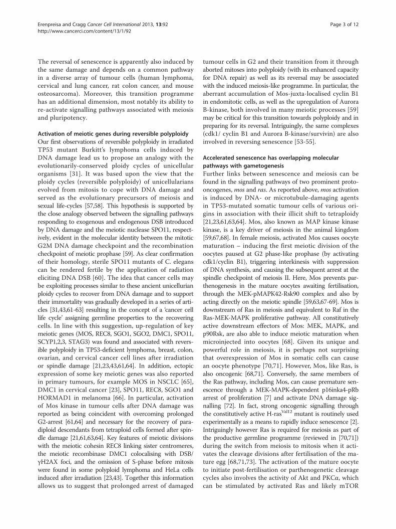
Figure 1 Inter-relationships between reversible polyploidy,senescence and stemness. This diagram highlights theinter-relationships and shared molecular pathways between thethree processes of polyploidy, senescence and stemness. DNAdamage potentiates this process leading to arrest at the G2Mdamage checkpoint from which cells that by-pass mitoticcatastrophe go on to enter the polyploid cycle, eliciting transientstemness to overcome senescence. TP53 serves as a strongnegative regulator of the process, favouring arrest at G1, apoptosisinduction and inhibiting entry into polyploidy.
Erenpreisa and Cragg Cancer Cell International 2013, 13:92 Page 8 of 12http://www.cancerci.com/content/13/1/92
germinative micronuclei (MI) originate in the same cell.The MA is degraded by nucleolysis and autophagy and be-comes extruded [138] during the conjugation and meiosisof the MI, whilst the removal of the MA prevents thisprocess (cited from [139]). Interestingly, extensive synthesisof the Rad51 recombinase in the MA is a necessary pre-requisite for successive meiosis of the MI [140]. A simi-lar collaboration may be required between persistingand further diminuted sub-nuclei in the late stage poly-ploid tumour cells, in which the diminuted chromatin isalso enriched with Rad51 and Rad52 proteins [45]. Ascommented by Zhang et al. [47] the budding of the EMTdescendants from the polyploid mother is reminiscentof the sporogenesis seen in Radiolaria, in which cyclingpolyploidy is part of the life-cycle [139]. The analogieswith protozoan ploidy cycles give support to the viewthat these cancer cell life cycles recapitulate some featuresof the earlier evolutionary ploidy cycles, preserved in someextant unicellular protozoans [141].
DNA damage can reprogramme tumour cells to totipotencySince the pioneering studies of Mintz and Illmensee[142] it has been known that the genome of cancer cellscan prime embryonal development. The molecular eventsinduced in TP53 deficient tumour cells in response toDNA damage indicate a re-activation of a meiosis-likeprogramme, a fundamental mechanism which serves tomaintain germline identity and provide the link betweenthe life-cycles. In addition, the core transcription cassetteof ESC appears to be evoked. The question then ariseshow these two pathways are linked through the DNAdamage response. Earlier studies revealed that Oct-4 ex-pression in the germline is regulated independently fromepiblast expression by its distal enhancer [143]. Additionalstudies [144] showed that the OCT4 transcriptional net-work might be part of the molecular signature of cellsfrom maternal origin from which the inner cell mass andthe ESC-associated pluripotency arise. In this way, Oct4provides the continuity of the totipotency (life) cycle innormal development. On the other hand, observations[86,91,116] indicate that Oct4 is the first of the coreESC cassette genes to respond to DNA damage both inTP53 wild-type stem cells and TP53 mutant somatictumour cells. Here the function of the different OCT4isoforms should be also considered [86,145]. Evidently,both the DNA damage responding and the totipotencycarrying functions of Oct4 are evolutionarily coupledand involved in the mitotic to meiotic transition oftumour cells. It can therefore be further suggested thatOct4, in its role as both a DNA damage-responder andtotipotency regulator, serves as a link between theearly meiosis-initiating and later cleavage-like eventsof de-polyploidisation and budding that give rise tothe rejuvenated descendants.
The feasibility of such a process is also seen from thebehaviour of ESC themselves: under special cultivationconditions, both female and male cells show gametogeneticpotential: i.e. they are capable of undergoing meiosis,oocyte maturation and parthenogenetic developmentup to the blastocyst stage [146].The formation of the endoclone by rejuvenated sub-
nuclei which acquire individual cytoplasm and initiatemitoses within a single giant cell [31,45] and the potentialof these individual cells to form a sphere and induce ma-lignant growth in vivo [47,89,125] is entirely in keepingwith the embryonal nature of this process. We previouslysuggested that to achieve this stage, the tumour cells needto undergo about four endocycles thus reaching the‘developmental totipotency checkpoint’ [33].
ConclusionThe failure of current cancer treatments to successfullyeradicate metastatic disease, likely results from a misunder-standing of the natural history of cancer. Rather than seeingmalignancy as a consequence of Darwinian microevolutiondriven by stochastic mutations, it can be considered as theresult of a programmed response illicitly accessed by a fewkey mutations. Thus the focus of research is transferredfrom the bewildering multiplicity of mutations to thekey transcriptional programme that is accessed and theunderlying epigenetics. This programme appears tohave been imprinted through evolution to cope with
Erenpreisa and Cragg Cancer Cell International 2013, 13:92 Page 9 of 12http://www.cancerci.com/content/13/1/92
DNA damage and stored in the evolutionary memoryof the genome. The mechanisms which gave rise firstto reversible polyploidy and then meiosis and sexuallife cycles in unicellurians allowing the transition tomulticellularity are in some way recapitulated duringcarcinogenesis. Unfortunately, it appears that the sameprogramme is stimulated in response to genotoxic treat-ments, leading to disease relapse.The concepts discussed in this review and the latest
available data give credence to the existence of an evolu-tionary ontogenetic relationship between senescence,carcinogenesis and gametogenesis and explain the paradoxof involving senescence in carcinogenesis and editing theimmunogenicity of tumour cells. This view brings us to anew twist in the centuries-old embryological theory of can-cer (reviewed by Erenpreiss [71]) with reversible polyploidyas a new aspect. While trying to unveil the relationships be-tween the overlapping pathways of polyploidy, senescenceand stemness (depicted in Figure 1), we have highlightedboth the synergism and heterogeneity of opposing regula-tors, the pleiotropism of key oncogenes and the plasticity ofcell fate determination. To fully understand these complexregulations a systems biology approach is required andthis has already led to an interesting variant of the em-bryological theory of cancer where ESC-like state attrac-tion is intrinsically linked to ontogenesis and phylogenesis[147,148]. Recognition that cancer, despite a diverse rangeof causes and driving mutations, is due to a similar epi-genetic acquisition of ilicit transcriptional programmesmay favour a shift away from current treatment paradigmsto a more holistic whole network approach. This shift isapparently already underway [149-152].
Competing interestsThe authors declare that they have no competing interests.
Authors’ contributionBoth senior authors have made substantial intellectual contribution to thisstudy. Both authors read and approved the final manuscript.
Authors’ informationJE and MSC, lead cancer research laboratories in Riga and Southampton,respectively and collaborated both experimentally and theoretically onidentifying and understanding the role of reversible polyploidy in cancerresistance over the last 15 years.
AcknowledgementsThe authors greatfully acknowledge the contributions made by the authorsof the experimental work performed in our laboratories since our firstpublication on reversible polyploidy in 2000. The authors are grateful toKirsten Walen for discussions on senescence and depolyploidisation aspects,Eugenia V and Tatyana G Zybina for discussions on developmentalpolyploidy, Francois Martin for discussions on reversible polyploidy, HarryScherthan for discussions on meiosis, Zane Kalnina for discussions on theimmunogenicity of tumours, Sui Huang and Alessandro Giulliani fordiscussions on aspects of systems biology, and Helmut Zacharius for providingliterature on the gender aspects of parthenogenesis. The contribution of Prof.Jānis O Ērenpreiss (1929–1996) relating to the gametogenetic theory of canceris gratefully commemorated. The e-version of his book cited in this reviewcan be found:http://bmc.biomed.lu.lv/en/research/directions-and-labs/
cancer-research/cancer-research/j-erenpreisa-lab/. The study was supportedby the Latvian Science Council grant Nr 341/2012.
Author details1Latvian Biomedical Research & Study Centre, Riga LV-1047, Latvia. 2Antibodyand Vaccine Group, Cancer Sciences Unit, Faculty of Medicine, GeneralHospital, University of Southampton, Southampton SO16 6YD, UK.
Received: 19 May 2013 Accepted: 24 July 2013Published: 11 September 2013
References1. Roninson IB: Tumor cell senescence in cancer treatment. Cancer Res 2003,
63(11):2705–2715.2. Serrano M, Lin AW, McCurrach ME, Beach D, Lowe SW: Oncogenic ras
provokes premature cell senescence associated with accumulation ofp53 and p16INK4a. Cell 1997, 88(5):593–602.
3. Finkel E: Telomeres: keys to senescence and cancer. Lancet 1998,351(9110):1186.
4. Kuilman T, Michaloglou C, Mooi WJ, Peeper DS: The essence ofsenescence. Genes Dev 2010, 24(22):2463–2479.
5. Hasty P, Sharp ZD, Curiel TJ, Campisi J: mTORC1 and p53: clash of thegods? Cell Cycle 2013, 12(1):20–25.
6. Lopez-Otin C, Blasco MA, Partridge L, Serrano M, Kroemer G: The hallmarksof aging. Cell 2013, 153(6):1194–1217.
7. Campisi J, d’Adda di Fagagna F: Cellular senescence: when bad thingshappen to good cells. Nat Rev Mol Cell Biol 2007, 8(9):729–740.
8. Wu PC, Wang Q, Grobman L, Chu E, Wu DY: Accelerated cellularsenescence in solid tumor therapy. Exp Oncol 2012, 34(3):298–305.
9. Kang TW, Yevsa T, Woller N, Hoenicke L, Wuestefeld T, Dauch D,Hohmeyer A, Gereke M, Rudalska R, Potapova A, et al: Senescencesurveillance of pre-malignant hepatocytes limits liver cancerdevelopment. Nature 2011, 479(7374):547–551.
10. Hoenicke L, Zender L: Immune surveillance of senescent cells–biologicalsignificance in cancer- and non-cancer pathologies. Carcinogenesis 2012,33(6):1123–1126.
11. Serrano M: Cancer: final act of senescence. Nature 2011, 479(7374):481–482.12. Rajaraman R, Guernsey DL, Rajaraman MM, Rajaraman SR: Stem cells,
senescence, neosis and self-renewal in cancer. Cancer Cell Int 2006, 6:25.13. Wheatley D: Growing evidence of the repopulation of regressed tumours
by the division of giant cells. Cell Biol Int 2008, 32(9):1029–1030.14. Lee HO, Davidson JM, Duronio RJ: Endoreplication: polyploidy with
purpose. Genes Dev 2009, 23(21):2461–2477.15. Ianzini F, Mackey MA: Development of the large scale digital cell analysis
system. Radiat Prot Dosimetry 2002, 99(1–4):289–293.16. Prieur-Carrillo G, Chu K, Lindqvist J, Dewey WC: Computerized video time-lapse
(CVTL) analysis of the fate of giant cells produced by X-irradiating EJ30human bladder carcinoma cells. Radiat Res 2003, 159(6):705–712.
17. Sundaram M, Guernsey DL, Rajaraman MM, Rajaraman R: Neosis: a noveltype of cell division in cancer. Cancer Biol Ther 2004, 3(2):207–218.
18. Chu K, Teele N, Dewey MW, Albright N, Dewey WC: Computerized videotime lapse study of cell cycle delay and arrest, mitotic catastrophe,apoptosis and clonogenic survival in irradiated 14-3-3sigma andCDKN1A (p21) knockout cell lines. Radiat Res 2004, 162(3):270–286.
19. Illidge TM, Cragg MS, Fringes B, Olive P, Erenpreisa JA: Polyploid giant cellsprovide a survival mechanism for p53 mutant cells after DNA damage.Cell Biol Int 2000, 24(9):621–633.
20. Puig PE, Guilly MN, Bouchot A, Droin N, Cathelin D, Bouyer F, Favier L,Ghiringhelli F, Kroemer G, Solary E, et al: Tumor cells can escapeDNA-damaging cisplatin through DNA endoreduplication and reversiblepolyploidy. Cell Biol Int 2008, 32(9):1031–1043.
21. Vitale I, Senovilla L, Jemaa M, Michaud M, Galluzzi L, Kepp O, Nanty L, Criollo A,Rello-Varona S, Manic G, et al: Multipolar mitosis of tetraploid cells: inhibitionby p53 and dependency on Mos. EMBO J 2010, 29(7):1272–1284.
22. Erenpreisa J, Ivanov A, Wheatley SP, Kosmacek EA, Ianzini F, Anisimov AP,Mackey M, Davis PJ, Plakhins G, Illidge TM: Endopolyploidy in irradiatedp53-deficient tumour cell lines: persistence of cell division activity ingiant cells expressing Aurora-B kinase. Cell Biol Int 2008, 32(9):1044–1056.
23. Ianzini F, Kosmacek EA, Nelson ES, Napoli E, Erenpreisa J, Kalejs M, MackeyMA: Activation of meiosis-specific genes is associated with
Erenpreisa and Cragg Cancer Cell International 2013, 13:92 Page 10 of 12http://www.cancerci.com/content/13/1/92
depolyploidization of human tumor cells following radiation-inducedmitotic catastrophe. Cancer Res 2009, 69(6):2296–2304.
24. Nagl W: Endopolyploidy and polyteny in differentiation and evolution.Amsterdam-New York: North-Holland: Publ. Comp.; 1978.
25. Davoli T, de Lange T: The causes and consequences of polyploidy innormal development and cancer. Annu Rev Cell Dev Biol 2011, 27:585–610.
26. Zybina TG, Zybina EV: Cell cycle modification in trophoblast cellpopulations in the course of placenta formation. A review. In DNAreplication and related cellular processes. Edited by Kusic-Tisma J. Rijeka:Croatia: InTech; 2011:227–258.
27. Beermann W: Control of differentiation at the chromosomal level.J Exp Zool 1964, 157:49–62.
28. Zybina E: Cytophotometric estimation of the amount of DNA in the nuclei ofthe giant cells of the trophoblast. DoklAkadNauk SSSR 1963, 153:1428–1431.
29. Rivello HG, Meckert PC, Vigliano C, Favaloro R, Laguens RP: Cardiacmyocyte nuclear size and ploidy status decrease after mechanicalsupport. Cardiovasc Pathol 2001, 10(2):53–57.
30. Duncan AW, Taylor MH, Hickey RD, Hanlon Newell AE, Lenzi ML, Olson SB,Finegold MJ, Grompe M: The ploidy conveyor of mature hepatocytes as asource of genetic variation. Nature 2010, 467(7316):707–710.
31. Erenpreisa JA, Cragg MS, Fringes B, Sharakhov I, Illidge TM: Release ofmitotic descendants by giant cells from irradiated Burkitt’s lymphomacell line. Cell Biol Int 2000, 24(9):635–648.
32. Ivanov A, Cragg MS, Erenpreisa J, Emzinsh D, Lukman H, Illidge TM:Endopolyploid cells produced after severe genotoxic damage have thepotential to repair DNA double strand breaks. J Cell Sci 2003, 116(Pt20):4095–4106.
33. Erenpreisa J, Cragg MS, Anisimov AP, Illidge TM: Tumor cell embryonalityand the ploidy number 32n: is it a developmental checkpoint? Cell Cycle2011, 10(11):1873–1874.
34. Vitale I, Galluzzi L, Senovilla L, Criollo A, Jemaa M, Castedo M, Kroemer G:Illicit survival of cancer cells during polyploidization anddepolyploidization. Cell Death Differ 2011, 18(9):1403–1413.
35. Vakifahmetoglu H, Olsson M, Zhivotovsky B: Death through a tragedy:mitotic catastrophe. Cell Death Differ 2008, 15(7):1153–1162.
36. Lu X, Kang Y: Cell fusion as a hidden force in tumor progression.Cancer Res 2009, 69(22):8536–8539.
37. Gisselsson D, Hakanson U, Stoller P, Marti D, Jin Y, Rosengren AH, SteweniusY, Kahl F, Panagopoulos I: When the genome plays dice: circumvention ofthe spindle assembly checkpoint and near-random chromosomesegregation in multipolar cancer cell mitoses. PLoS One 2008, 3(4):e1871.
38. Zasadil LM, Britigan EM, Weaver BA: 2n or not 2n: Aneuploidy, polyploidyand chromosomal instability in primary and tumor cells. Semin Cell DevBiol 2013, 24(4):370–379.
39. Erenpreisa J, Kalejs M, Ianzini F, Kosmacek EA, Mackey MA, Emzinsh D, CraggMS, Ivanov A, Illidge TM: Segregation of genomes in polyploid tumourcells following mitotic catastrophe. Cell Biol Int 2005, 29(12):1005–1011.
40. Walen KH: Meiotic-like division to a aneuploidy: chromosomal instability(CIN), cell-senescence and cancer. Cell Oncol 2008, 30(5):451–452.
41. Walen KH: Genetic stability of senescence reverted cells: genomereduction division of polyploidy cells, aneuploidy and neoplasia.Cell Cycle 2008, 7(11):1623–1629.
42. Walen KH: Mitosis is not the only distributor of mutated cells: non-mitoticendopolyploid cells produce reproductive genome-reduced cells. Cell BiolInt 2010, 34(8):867–872.
43. Erenpreisa J, Cragg MS, Salmina K, Hausmann M, Scherthan H: The roleof meiotic cohesin REC8 in chromosome segregation in gammairradiation-induced endopolyploid tumour cells. Exp Cell Res 2009,315(15):2593–2603.
44. Davoli T, Denchi EL, de Lange T: Persistent telomere damage inducesbypass of mitosis and tetraploidy. Cell 2010, 141(1):81–93.
45. Erenpreisa J, Salmina K, Huna A, Kosmacek EA, Cragg M, Ianzini F,Anisimov A: Polyploid tumour cells elicit para-diploid progenythrough de-polyploidising divisions and regulated autophagicdegradation. Cell Biol Int 2011, 35(7):687–695.
46. Erenpreisa J, Huna A, Salmina K, Jackson TR, Cragg MS: Macroautophagy-aidedelimination of chromatin: sorting of waste, sorting of fate? Autophagy 2012,8(12):1877–1881.
47. Zhang S, Mercado-Uribe I, Xing Z, Sun B, Kuang J, Liu J: Generation ofcancer stem-like cells through the formation of polyploid giant cancercells. Oncogene 2013:1–13.
48. Marxer M, Foucar CE, Man WY, Chen Y, Ma HT, Poon RY: Tetraploidizationincreases sensitivity to Aurora B kinase inhibition. Cell Cycle 2012,11(13):2567–2577.
49. Tam WL, Ang YS, Lim B: The molecular basis of ageing in stem cells.Mech Ageing Dev 2007, 128(1):137–148.
50. Sabisz M, Skladanowski A: Cancer stem cells and escape from drug-inducedpremature senescence in human lung tumor cells: implications for drugresistance and in vitro drug screening models. Cell Cycle 2009, 8(19):3208–3217.
51. Sliwinska MA, Mosieniak G, Wolanin K, Babik A, Piwocka K, Magalska A,Szczepanowska J, Fronk J, Sikora E: Induction of senescence withdoxorubicin leads to increased genomic instability of HCT116 cells. MechAgeing Dev 2009, 130(1–2):24–32.
52. Mosieniak G, Sikora E: Polyploidy: the link between senescence andcancer. Curr Pharm Des 2010, 16(6):734–740.
53. Roberson RS, Kussick SJ, Vallieres E, Chen SY, Wu DY: Escape from therapy-induced accelerated cellular senescence in p53-null lung cancer cellsand in human lung cancers. Cancer Res 2005, 65(7):2795–2803.
54. Wang Q, Wu PC, Roberson RS, Luk BV, Ivanova I, Chu E, Wu DY: Survivinand escaping in therapy-induced cellular senescence. Int J Cancer 2011,128(7):1546–1558.
55. Wang Q, Wu PC, Dong DZ, Ivanova I, Chu E, Zeliadt S, Vesselle H, Wu DY:Polyploidy road to therapy-induced cellular senescence and escape. Int JCancer 2013, 132(7):1505–1515.
56. Bolton MA, Lan W, Powers SE, McCleland ML, Kuang J, Stukenberg PT:Aurora B kinase exists in a complex with survivin and INCENP and itskinase activity is stimulated by survivin binding and phosphorylation.Mol Biol Cell 2002, 13(9):3064–3077.
57. Bernstein H, Hopf FA, Michod RE: The molecular basis of the evolution ofsex. Adv Genet 1987, 24:323–370.
58. Kondrashov AS: The asexual ploidy cycle and the origin of sex.Nature 1994, 370(6486):213–216.
59. Nebreda AR, Ferby I: Regulation of the meiotic cell cycle in oocytes.Curr Opin Cell Biol 2000, 12(6):666–675.
60. Dernburg AF, McDonald K, Moulder G, Barstead R, Dresser M, Villeneuve AM:Meiotic recombination in C. elegans initiates by a conserved mechanismand is dispensable for homologous chromosome synapsis. Cell 1998, 94(3):387–398.
61. Erenpreisa J, Kalejs M, Cragg MS: Mitotic catastrophe and endomitosis intumour cells: an evolutionary key to a molecular solution. Cell Biol Int2005, 29(12):1012–1018.
62. Erenpreisa J, Cragg MS: Cancer: a matter of life cycle? Cell Biol Int 2007, 31(12):1507–1510.
63. Erenpreisa J, Cragg MS: MOS, aneuploidy and the ploidy cycle of cancercells. Oncogene 2010, 29(40):5447–5451.
64. Kalejs M, Ivanov A, Plakhins G, Cragg MS, Emzinsh D, Illidge TM, Erenpreisa J:Upregulation of meiosis-specific genes in lymphoma cell lines followinggenotoxic insult and induction of mitotic catastrophe. BMC Cancer 2006, 6:6.
65. Gorgoulis VG, Zacharatos P, Mariatos G, Liloglou T, Kokotas S, Kastrinakis N,Kotsinas A, Athanasiou A, Foukas P, Zoumpourlis V, et al: Deregulatedexpression of c-mos in non-small cell lung carcinomas: relationship withp53 status, genomic instability, and tumor kinetics. Cancer Research 2001,61(2):538–549.
66. Rosa AM, Dabas N, Byrnes DM, Eller MS, Grichnik JM: Germ cell proteins inmelanoma: prognosis, diagnosis, treatment, and theories on expression.J Skin Cancer 2012, 2012:621968.
67. Nasmyth K: Disseminating the genome: joining, resolving, and separatingsister chromatids during mitosis and meiosis. Annu Rev Genet 2001,35:673–745.
68. Dupre A, Haccard O, Jessus C: Mos in the oocyte: how to use MAPKindependently of growth factors and transcription to control meioticdivisions. J Signal Transduct 2011, 2011:350412.
69. Dupre A, Suziedelis K, Valuckaite R, de Gunzburg J, Ozon R, Jessus C,Haccard O: Xenopus H-RasV12 promotes entry into meiotic M phase andcdc2 activation independently of Mos and p42(MAPK). Oncogene 2002,21(42):6425–6433.
70. Fukasawa K, Murakami MS, Blair DG, Kuriyama R, Hunt T, Fischinger P, VandeWoude GF: Similarities between somatic cells overexpressing the mosoncogene and oocytes during meiotic interphase. Cell Growth Differ 1994,5(10):1093–1103.
71. Erenpreiss J: Current concepts of malignant growth. Riga: Part A. From anormal cell to cancer Zvaigzne Publishers; 1993:191.

Erenpreisa and Cragg Cancer Cell International 2013, 13:92 Page 11 of 12http://www.cancerci.com/content/13/1/92
72. Mallette FA, Gaumont-Leclerc MF, Ferbeyre G: The DNA damage signalingpathway is a critical mediator of oncogene-induced senescence. Genes &Development 2007, 21(1):43–48.
73. Hasan AKMMT, Kihira M, Yoshida J, Sato K-I: In Phospho-Signaling at OocyteMaturation and Fertilization: Set Up for Embryogenesis and Beyond Part I.Protein Kinases, Embryogenesis. Edited by Sato K-I. ; 2012. InTech. DOI:10.5772/39369. http://www.intechopen.com/books/embryogenesis/phospho-signaling-at-oocyte-maturation-and-fertilization-set-up-for-embryogenesis-and-beyond-part-ii
74. Johnson AD, Cork RJ, Williams MA, Robinson KR, Smith LD: H-ras(val12)induces cytoplasmic but not nuclear events of the cell cycle in smallXenopus oocytes. Cell Regul 1990, 1(7):543–554.
75. Back JH, Kim AL: The expanding relevance of nuclear mTOR incarcinogenesis. Cell Cycle 2011, 10(22):3849–3852.
76. Laplante M, Sabatini DM: mTOR signaling in growth control and disease.Cell 2012, 149(2):274–293.
77. Birchmeier C, Broek D, Wigler M: Ras proteins can induce meiosis inXenopus oocytes. Cell 1985, 43(3 Pt 2):615–621.
78. Dean M, Fojo T, Bates S: Tumour stem cells and drug resistance. Nat RevCancer 2005, 5(4):275–284.
79. Jordan CT, Guzman ML, Noble M: Cancer stem cells. N Engl J Med 2006,355(12):1253–1261.
80. Ben-Porath I, Thomson MW, Carey VJ, Ge R, Bell GW, Regev A, Weinberg RA:An embryonic stem cell-like gene expression signature in poorlydifferentiated aggressive human tumors. Nat Genet 2008, 40(5):499–507.
81. Saigusa S, Tanaka K, Toiyama Y, Yokoe T, Okugawa Y, Ioue Y, Miki C,Kusunoki M: Correlation of CD133, OCT4, and SOX2 in rectal cancer andtheir association with distant recurrence after chemoradiotherapy. AnnSurg Oncol 2009, 16(12):3488–3498.
82. Ge N, Lin HX, Xiao XS, Guo L, Xu HM, Wang X, Jin T, Cai XY, Liang Y, Hu WH,et al: Prognostic significance of Oct4 and Sox2 expression inhypopharyngeal squamous cell carcinoma. J Transl Med 2010, 8:94.
83. Xiang R, Liao D, Cheng T, Zhou H, Shi Q, Chuang TS, Markowitz D, ReisfeldRA, Luo Y: Downregulation of transcription factor SOX2 in cancer stemcells suppresses growth and metastasis of lung cancer. Br J Cancer 2011,104(9):1410–1417.
84. Baumann M, Krause M, Hill R: Exploring the role of cancer stem cells inradioresistance. Nat Rev Cancer 2008, 8(7):545–554.
85. Blagosklonny MV: Target for cancer therapy: proliferating cells or stemcells. Leukemia 2006, 20(3):385–391.
86. Salmina K, Jankevics E, Huna A, Perminov D, Radovica I, Klymenko T, IvanovA, Jascenko E, Scherthan H, Cragg M, et al: Up-regulation of the embryonicself-renewal network through reversible polyploidy in irradiated p53-mutant tumour cells. Exp Cell Res 2010, 316(13):2099–2112.
87. Lee GY, Shim JS, Cho B, Jung JY, Lee DS, Oh IH: Stochastic acquisition of astem cell-like state and drug tolerance in leukemia cells stressed byradiation. Int J Hematol 2011, 93(1):27–35.
88. Ghisolfi L, Keates AC, Hu X, Lee DK, Li CJ: Ionizing radiation inducesstemness in cancer cells. PLoS One 2012, 7(8):e43628.
89. Lagadec C, Vlashi E, Della Donna L, Dekmezian C, Pajonk F: Radiation-inducedreprogramming of breast cancer cells. Stem Cells 2012, 30(5):833–844.
90. Chuang YS, Huang WH, Park SW, Persaud SD, Hung CH, Ho PC, Wei LN:Promyelocytic leukemia protein in retinoic acid-induced chromatinremodeling of Oct4 gene promoter. Stem Cells 2011, 29(4):660–669.
91. Bartova E, Sustackova G, Stixova L, Kozubek S, Legartova S, Foltankova V:Recruitment of Oct4 protein to UV-damaged chromatin in embryonicstem cells. PLoS One 2011, 6(12):e27281.
92. Guo Y, Mantel C, Hromas RA, Broxmeyer HE: Oct-4 is critical for survival/antiapoptosis of murine embryonic stem cells subjected to stress: effectsassociated with Stat3/survivin. Stem Cells 2008, 26(1):30–34.
93. Takahashi K, Yamanaka S: Induction of pluripotent stem cells from mouseembryonic and adult fibroblast cultures by defined factors. Cell 2006, 126(4):663–676.
94. Banito A, Rashid ST, Acosta JC, Li S, Pereira CF, Geti I, Pinho S, Silva JC,Azuara V, Walsh M, et al: Senescence impairs successful reprogrammingto pluripotent stem cells. Genes Dev 2009, 23(18):2134–2139.
95. Li H, Collado M, Villasante A, Strati K, Ortega S, Canamero M, Blasco MA,Serrano M: The Ink4/Arf locus is a barrier for iPS cell reprogramming.Nature 2009, 460(7259):1136–1139.
96. Menendez JA, Vellon L, Oliveras-Ferraros C, Cufi S, Vazquez-Martin A: mTOR-regulated senescence and autophagy during reprogramming of somatic
cells to pluripotency: a roadmap from energy metabolism to stem cellrenewal and aging. Cell Cycle 2011, 10(21):3658–3677.
97. Huna A, Salmina K, Jascenko E, Duburs G, Inashkina I, Erenpreisa J: Self-Renewal Signalling in Presenescent Tetraploid IMR90 Cells. J Aging Res2011, 2011:103253.
98. Rogakou EP, Pilch DR, Orr AH, Ivanova VS, Bonner WM: DNA double-stranded breaks induce histone H2AX phosphorylation on serine 139.J Biol Chem 1998, 273(10):5858–5868.
99. Boheler KR: Stem cell pluripotency: a cellular trait that depends ontranscription factors, chromatin state and a checkpoint deficient cellcycle. J Cell Physiol 2009, 221(1):10–17.
100. Mantel C, Guo Y, Lee MR, Kim MK, Han MK, Shibayama H, Fukuda S, YoderMC, Pelus LM, Kim KS, et al: Checkpoint-apoptosis uncoupling in humanand mouse embryonic stem cells: a source of karyotpic instability. Blood2007, 109(10):4518–4527.
101. Mantel C, Guo Y, Lee MR, Han MK, Rhorabough S, Kim KS, Broxmeyer HE:Cells enter a unique intermediate 4N stage, not 4N-G1, after abortedmitosis. Cell Cycle 2008, 7(4):484–492.
102. Conant GC, Wolfe KH: Increased glycolytic flux as an outcome of whole-genome duplication in yeast. Mol Syst Biol 2007, 3:129.
103. Anatskaya OV, Vinogradov AE: Genome multiplication as adaptation totissue survival: evidence from gene expression in mammalian heart andliver. Genomics 2007, 89(1):70–80.
104. Ward PS, Thompson CB: Metabolic reprogramming: a cancer hallmarkeven Warburg did not anticipate. Cancer Cell 2012, 21(3):297–308.
105. Hu J, Locasale JW, Bielas JH, O’Sullivan J, Sheahan K, Cantley LC, Heiden MGV,Vitkup D: Heterogeneity of tumor-induced gene expression changes in thehuman metabolic network. Nat Biotech 2013, 31(6):522–529.
106. Jiang J, Tang YL, Liang XH: EMT: a new vision of hypoxia promotingcancer progression. Cancer Biol Ther 2011, 11(8):714–723.
107. Miller DM, Thomas SD, Islam A, Muench D, Sedoris K: c-Myc and cancermetabolism. Clin Cancer Res 2012, 18(20):5546–5553.
108. Sun Q, Chen X, Ma J, Peng H, Wang F, Zha X, Wang Y, Jing Y, Yang H, ChenR, et al: Mammalian target of rapamycin up-regulation of pyruvate kinaseisoenzyme type M2 is critical for aerobic glycolysis and tumor growth.Proc Natl Acad Sci U S A 2011, 108(10):4129–4134.
109. Wong DJ, Liu H, Ridky TW, Cassarino D, Segal E, Chang HY: Module map ofstem cell genes guides creation of epithelial cancer stem cells. Cell StemCell 2008, 2(4):333–344.
110. Dominguez-Sola D, Ying CY, Grandori C, Ruggiero L, Chen B, Li M, GallowayDA, Gu W, Gautier J, Dalla-Favera R: Non-transcriptional control of DNAreplication by c-Myc. Nature 2007, 448(7152):445–451.
111. Li Q, Dang CV: c-Myc overexpression uncouples DNA replication frommitosis. Mol Cell Biol 1999, 19(8):5339–5351.
112. Conner EA, Lemmer ER, Sanchez A, Factor VM, Thorgeirsson SS: E2F1 blocksand c-Myc accelerates hepatic ploidy in transgenic mouse models.Biochem Biophys Res Commun 2003, 302(1):114–120.
113. den Hollander J, Rimpi S, Doherty JR, Rudelius M, Buck A, Hoellein A, KremerM, Graf N, Scheerer M, Hall MA, et al: Aurora kinases A and B are up-regulated by Myc and are essential for maintenance of the malignantstate. Blood 2010, 116(9):1498–1505.
114. Gusse M, Ghysdael J, Evan G, Soussi T, Mechali M: Translocation of a storeof maternal cytoplasmic c-myc protein into nuclei during earlydevelopment. Mol Cell Biol 1989, 9(12):5395–5403.
115. Gordon DJ, Resio B, Pellman D: Causes and consequences of aneuploidyin cancer. Nat Rev Genet 2012, 13(3):189–203.
116. Jackson TR, Salmina K, Huna A, Inashkina I, Jankevics E, Riekstina U, KalninaZ, Ivanov A, Townsend PA, Cragg MS, et al: DNA damage causes TP53-dependent coupling of self-renewal and senescence pathways inembryonal carcinoma cells. Cell Cycle 2013, 12(3):430–441.
117. Sherman MY, Meng L, Stampfer M, Gabai VL, Yaglom JA: Oncogenesinduce senescence with incomplete growth arrest and suppress theDNA damage response in immortalized cells. Aging Cell 2011,10(6):949–961.
118. Zybina TG, Stein GI, Zybina EV: Endopolyploid and proliferating trophoblastcells express different patterns of intracellular cytokeratin and glycogenlocalization in the rat placenta. Cell Biol Int 2011, 35(7):649–655.
119. Lee J, Go Y, Kang I, Han YM, Kim J: Oct-4 controls cell-cycle progression ofembryonic stem cells. Biochem J 2010, 426(2):171–181.
120. Raderschall E, Bazarov A, Cao J, Lurz R, Smith A, Mann W, Ropers HH, SedivyJM, Golub EI, Fritz E, et al: Formation of higher-order nuclear Rad51

Erenpreisa and Cragg Cancer Cell International 2013, 13:92 Page 12 of 12http://www.cancerci.com/content/13/1/92
structures is functionally linked to p21 expression and protection fromDNA damage-induced apoptosis. J Cell Sci 2002, 115(Pt 1):153–164.
121. Zheng L, Dai H, Zhou M, Li X, Liu C, Guo Z, Wu X, Wu J, Wang C, Zhong J,et al: Polyploid cells rewire DNA damage response networks toovercome replication stress-induced barriers for tumour progression. NatCommun 2012, 3:815.
122. Huang S: Reprogramming cell fates: reconciling rarity with robustness.Bioessays 2009, 31(5):546–560.
123. Huang S: Non-genetic heterogeneity of cells in development: more thanjust noise. Dev 2009, 136(23):3853–3862.
124. Rajaraman R, Guernsey DL, Rajaraman MM, Rajaraman SR: Neosis - Aparasexual somatic reduction division in cancer. Int J Hum Genet 2007,7(1):29–48.
125. Weihua Z, Lin Q, Ramoth AJ, Fan D, Fidler IJ: Formation of solid tumors bya single multinucleated cancer cell. Cancer 2011, 117(17):4092–4099.
126. Young AR, Narita M: Connecting autophagy to senescence inpathophysiology. Curr Opin Cell Biol 2010, 22(2):234–240.
127. Hanahan D, Weinberg RA: Hallmarks of cancer: the next generation. Cell2011, 144(5):646–674.
128. Dunn GP, Old LJ, Schreiber RD: The three Es of cancer immunoediting.Annu Rev Immunol 2004, 22:329–360.
129. Simpson AJ, Caballero OL, Jungbluth A, Chen YT, Old LJ: Cancer/testisantigens, gametogenesis and cancer. Nat Rev Cancer 2005, 5(8):615–625.
130. Fratta E, Coral S, Covre A, Parisi G, Colizzi F, Danielli R, Nicolay HJ, Sigalotti L,Maio M: The biology of cancer testis antigens: putative function,regulation and therapeutic potential. Mol Oncol 2011, 5(2):164–182.
131. Lindsey SF, Byrnes DM, Eller MS, Rosa AM, Dabas N, Escandon J, Grichnik JM:Potential role of meiosis proteins in melanoma chromosomal instability.J Skin Cancer 2013, 2013:1–9. http://dx.doi.org/10.1155/2013/1901099.
132. Zayakin P, Ancans G, Silina K, Meistere I, Kalnina Z, Andrejeva D, Endzelins E,Ivanova L, Pismennaja A, Ruskule A, et al: Tumor-associated autoantibodysignature for the early detection of gastric cancer. Int J Cancer 2013, 132(1):137–147.
133. Boileve A, Senovilla L, Vitale I, Lissa D, Martins I, Metivier D, van den Brink S,Clevers H, Galluzzi L, Castedo M, et al: Immunosurveillance againsttetraploidization-induced colon tumorigenesis. Cell Cycle 2013, 12(3):473–479.
134. Senovilla L, Vitale I, Martins I, Tailler M, Pailleret C, Michaud M, Galluzzi L,Adjemian S, Kepp O, Niso-Santano M, et al: An immunosurveillancemechanism controls cancer cell ploidy. Science 2012, 337(6102):1678–1684.
135. Cufi S, Vazquez-Martin A, Oliveras-Ferraros C, Martin-Castillo B, Vellon L,Menendez JA: Autophagy positively regulates the CD44(+) CD24(−/low)breast cancer stem-like phenotype. Cell Cycle 2011, 10(22):3871–3885.
136. Akalay I, Janji B, Hasmim M, Noman MZ, Andre F, De Cremoux P, BertheauP, Badoual C, Vielh P, Larsen AK, et al: Epithelial-to-mesenchymal transitionand autophagy induction in breast carcinoma promote escape from T-cell-mediated lysis. Cancer Res 2013, 73(8):2418–2427.
137. Wells D, Hillier SG: Polar bodies: their biological mystery and clinicalmeaning. Mol Hum Reprod 2011, 17(5):273–274.
138. Lu E, Wolfe J: Lysosomal enzymes in the macronucleus of Tetrahymenaduring its apoptosis-like degradation. Cell Death Differ 2001, 8(3):289–297.
139. Raikov IB: The protozoan nucleus – morphology and evolution. Wien u.a:Springer; 1982. 1983.
140. Marsh TC, Cole ES, Stuart KR, Campbell C, Romero DP: RAD51 is requiredfor propagation of the germinal nucleus in Tetrahymena thermophila.Genetics 2000, 154(4):1587–1596.
141. Erenpreisa J, Cragg MS: Life-cycle features of tumour cells, Evolutionary biologyfrom concept to application. Germany: Springer-Verlag Berlin; 2008:61–71.
142. Mintz B, Illmensee K: Normal genetically mosaic mice produced frommalignant teratocarcinoma cells. Proc Natl Acad Sci U S A 1975, 72(9):3585–3589.
143. Yeom YI, Fuhrmann G, Ovitt CE, Brehm A, Ohbo K, Gross M, Hubner K,Scholer HR: Germline regulatory element of Oct-4 specific for thetotipotent cycle of embryonal cells. Dev 1996, 122(3):881–894.
144. Zuccotti M, Merico V, Belli M, Mulas F, Sacchi L, Zupan B, Redi CA, PrigioneA, Adjaye J, Bellazzi R, et al: OCT4 and the acquisition of oocytedevelopmental competence during folliculogenesis. Int J Dev Biol 2012,56(10–12):853–858.
145. Wang X, Dai J: Concise review: isoforms of OCT4 contribute to theconfusing diversity in stem cell biology. Stem Cells 2010, 28(5):885–893.
146. Hubner K, Fuhrmann G, Christenson LK, Kehler J, Reinbold R, De La FuenteR, Wood J, Strauss JF 3rd, Boiani M, Scholer HR: Derivation of oocytes frommouse embryonic stem cells. Science 2003, 300(5623):1251–1256.
147. Huang S, Ernberg I, Kauffman S: Cancer attractors: a systems view oftumors from a gene network dynamics and developmental perspective.Semin Cell Dev Biol 2009, 20(7):869–876.
148. Huang S: On the intrinsic inevitability of cancer: from foetal to fatalattraction. Semin Cancer Biol 2011, 21(3):183–199.
149. Lipkin G: Plasticity of the cancer cell: implications for epigenetic controlof melanoma and other malignancies. J Invest Dermatol 2008,128(9):2152–2155.
150. Baylin SB, Jones PA: A decade of exploring the cancer epigenome -biological and translational implications. Nat Rev Cancer 2011, 11(10):726–734.
151. Tsai HC, Baylin SB: Cancer epigenetics: linking basic biology to clinicalmedicine. Cell Res 2011, 21(3):502–517.
152. Bissell M: Q&A: Mina Bissell on tumors as organs. Cancer Discov 2013, 3(1):7.
doi:10.1186/1475-2867-13-92Cite this article as: Erenpreisa and Cragg: Three steps to the immortalityof cancer cells: senescence, polyploidy and self-renewal. Cancer CellInternational 2013 13:92.
Submit your next manuscript to BioMed Centraland take full advantage of:
• Convenient online submission
• Thorough peer review
• No space constraints or color figure charges
• Immediate publication on acceptance
• Inclusion in PubMed, CAS, Scopus and Google Scholar
• Research which is freely available for redistribution
Submit your manuscript at www.biomedcentral.com/submit
Related Documents