International Journal of Aquatic Science ISSN: 2008-8019 Vol. 4, No. 2, 44-58, 2013 () [email protected] Three new species of Pseudorhabdosynochus (Monogenea: Diplectanidae) from Vietnamese grouper (Epinephelus spp.) (Perciformes: Serranidae) Binh T. Dang 1 *, Glenn A. Bristow 2 , Christoffer Schander 2 and Bjørn Berland 2 1) Institute for Biotechnology and Environment, University of Nha Trang, Nha Trang, Vietnam 2) Department of Biology, University of Bergen, P.O. Box 7800, N-5020 Bergen, Norway Received: 15 December 2012 Accepted: 5 February 2013 Published: 27 June 2013 Abstract: Three new species of monogenea are described from cultured and wild grouper from Nha Trang and Cam Ranh Bays, Vietnam. Pseudorhabdosynochus nhatrangensis n. sp and P. vietnamensis n. sp. were found on wild and cultured Epinephelus coioides and E. bleekeri, while Pseudorhabdosynochus brunei n. sp. parasitizes wild E. bruneus. P. nhatrangensis n. sp. is characterized by a sclerotized vagina with cup-shaped trumpet, open at the distal part, followed by a proximal tubular region, twisted to a circle at the posterior end and extending into thin branches; squamodiscs with 10 rows of rodlets with the central row being a closed circle. The tegument is scaly. P. vietnamensis n. sp. has a very large male copulatory organ with the tube being widened and slightly curved posteriorly. A sclerotized vagina comprised of an anterior trumpet, followed by convoluted structure which twists and divides into two distinct chambers. The central row of the squamodiscs forms a closed circle with a central core present. A heavily scaled tegument with scales on ventral and dorsal surfaces extends from the squamodiscs to the level of the ovary. P. nhatrangensiis n. sp. can be differentiated from other Pseudorhabdosynochus species by the vaginal structure. The complex vaginal structure and extremely large male copulatory organ of P. vietnamensis are similar to P. pai reported from E. tauvina, but these two can be distinguished by details of the vagina, and number of rows of squamodiscs (17–18 in P. vietnamensis and 10–11 in P. pai). P. brunei n. sp. possesses a sclerotized vagina with a tubular region and two serial chambers at the distal opening; squamodiscs have only 7 rows of rodlets. An egg of this species was found, being large and oval with a long sclerotized tail. Key Words: Grouper, Monogenea, Pseudorhabdosynochus, Nha Trang Bay

Welcome message from author
This document is posted to help you gain knowledge. Please leave a comment to let me know what you think about it! Share it to your friends and learn new things together.
Transcript

International Journal of Aquatic ScienceISSN: 2008-8019Vol. 4, No. 2, 44-58, 2013
Three new species of Pseudorhabdosynochus (Monogenea:
Diplectanidae) from Vietnamese grouper (Epinephelus spp.)(Perciformes: Serranidae)
Binh T. Dang1*, Glenn A. Bristow2, Christoffer Schander2 and Bjørn Berland2
1) Institute for Biotechnology and Environment, University of Nha Trang, Nha Trang, Vietnam
2) Department of Biology, University of Bergen, P.O. Box 7800, N-5020 Bergen, Norway
Received: 15 December 2012 Accepted: 5 February 2013 Published: 27 June 2013
Abstract: Three new species of monogenea are described from cultured and wild grouper from Nha
Trang and Cam Ranh Bays, Vietnam. Pseudorhabdosynochus nhatrangensis n. sp and P. vietnamensis n.
sp. were found on wild and cultured Epinephelus coioides and E. bleekeri, while Pseudorhabdosynochusbrunei n. sp. parasitizes wild E. bruneus. P. nhatrangensis n. sp. is characterized by a sclerotized vagina
with cup-shaped trumpet, open at the distal part, followed by a proximal tubular region, twisted to a
circle at the posterior end and extending into thin branches; squamodiscs with 10 rows of rodlets with
the central row being a closed circle. The tegument is scaly. P. vietnamensis n. sp. has a very large
male copulatory organ with the tube being widened and slightly curved posteriorly. A sclerotized vagina
comprised of an anterior trumpet, followed by convoluted structure which twists and divides into two
distinct chambers. The central row of the squamodiscs forms a closed circle with a central core present.
A heavily scaled tegument with scales on ventral and dorsal surfaces extends from the squamodiscs to
the level of the ovary. P. nhatrangensiis n. sp. can be differentiated from other Pseudorhabdosynochusspecies by the vaginal structure. The complex vaginal structure and extremely large male copulatory
organ of P. vietnamensis are similar to P. pai reported from E. tauvina, but these two can be
distinguished by details of the vagina, and number of rows of squamodiscs (17–18 in P. vietnamensisand 10–11 in P. pai). P. brunei n. sp. possesses a sclerotized vagina with a tubular region and two serial
chambers at the distal opening; squamodiscs have only 7 rows of rodlets. An egg of this species was
found, being large and oval with a long sclerotized tail.Key Words: Grouper, Monogenea, Pseudorhabdosynochus, Nha Trang Bay

Dang et al. (2013) Three new species of Pseudorhabdosynochus (Monogenea: Diplectanidae)…
Int. J. Aqu. Sci; 4(2): 44-58, 2013 45
IntordactionSpecies of Pseudorhabdosynochus are
increasingly reported parasitizing grouper
(Serranidae; Epinephelinae) throughout warm
and temperature waters of oceanic regions
(Oliver, 1986; Young, 1969; Dyer et al., 1994,
1995; Vidal-Martinez et al., 1997; Vidal-
Martínez and Mendoza-Franco, 1998; Bu et al.,1999; Santos et al., 2000; Yang et al.,2005a;Yang, et al. 2005b;Wu et al., 2005a; Wu
et al., 2005b; Justine and Euzet, 2005 a,b;
Hinsinger and Justine, 2006 a.b; Justine and
Sigura, 2007; Zeng and Yang, 2007; Neifar
and Euzet, 2007; Justine, 2007a,b and 2008;
Sigura et al., 2007; Justine, 2009; Justine and
Vignon, 2009). They are relatively host specific,
even when widely distributed (Santos et al.,2000). Santos et al. (2000) were the first to
discuss species totals within Pseudorhabd-osynochus, listing 23 species and describing 1
new species. Species richness currently
comprises over 40 species with and without
nominal status according to a summary by
Justine (2007a). Since 2007, a number of
Pseudorhabdosynochus species have been
described (Yang et al., 2005a, Yang et al.,2005b; Neifar and Euzet, 2007; Justine, 2007b;
Zeng and Yang, 2007; Sigura et al., 2007;
Justine and Sigura, 2007; Justine, 2008; 2009
and Justine and Vignon, 2009). The species in
this genus now number over 60. As an
increasing number of grouper are examined,
additional species may be described.
During a survey of monogenean fauna of
Epinephelus spp. (Serranidae) in Vietnam (Dang
et al., 2010), both new and previously known
species of Pseudorhabdosynochus were collect-
ed. Three new species are described herein.
Materials and MethodsParasite sampling and identification
Sea cage cultured Epinephelus coioides(Hamilton, 1822) (n=45), E. bleekeri (Vaillant,
1878) (n=40) and wild E. bruneus (Bloch,
1793) (n=28) (all Serranidae) were collected
from Nha Trang Bay (12° 15′ N, 109° 21′ E)
and Cam Ranh Bay (11° 52' 60 N, 109° 10' E),
Vietnam, from January to August 2007, and
from January 2008 to July 2009. Prior to
examination, fish were held in indoor aerated
water tanks. Skin, fins and gills of freshly
euthanized fish were subsequently examined for
monogenean parasites.
Freshly excised gills were kept in separate
Petri dishes filled with filtered sea water and
examined under a dissecting microscope.
Monogeneans were removed and examined
either alive or in 75% ethanol. Pseudorhabd-osynochus species identification was based
primarily on the morphology of the haptoral
sclerites and the reproductive organs using
various keys and species descriptions (Beverley-
Burton and Suriano, 1981; Kritsky and
Beverley-Burton, 1986; Bu et al., 1999; Justine

Dang et al. (2013) Three new species of Pseudorhabdosynochus (Monogenea: Diplectanidae)…
Int. J. Aqu. Sci; 4(2): 44-58, 2013 46
and Euzet, 2005a; Yang et al., 2005b; Justine
and Euzet, 2005b; Hinsinger and Justine,
2006a,b; Justine and Sigura, 2007; Justine,
2007a, b, Zeng and Yang, 2007; Justine and
Vignon, 2009). For morphological analysis,
whole mounts were prepared following Berland
(2005). Various diagnostic characters
were studied using differential interference
contrast microscopy (Olympus BX51) and Cell*
digital image analysis software (Olympus Soft
Imaging Solutions GmbH, Münster, Germany).
Body width was measured at mid-body or
vagina level. All measurements are given in µm
and presented as mean (range). The characters
measured and the terminologies used are in
accordance with Justine (2007a). Drawings of
the haptoral hardparts and copulatory
complexes were prepared with aid of a camera
lucida. The images were digitally processed in
Adobe Photoshop Elements®, version 5.0.
Holotype and paratypes specimens were
deposited in the Natural History collection,
Bergen museum (ZMBN), University of Bergen,
Bergen, Norway.
DescriptionPseudorhabdosynochus nhatrangensisn. sp.(Figures: 1, 2 A–J)
Diagnostic: Body slightly dorsoventrally
flattened, widest at level of ovary, length 458
(370–570, n=10), width 155 (120–185, n=10).
Tegument armed with scales in region of
haptoral peduncle. Anterior region with 3 pairs
of head organs, 2 pair of eye-spots with
posterior pair larger and more closely situated
than anterior. Distance between outer margins
of anterior eye-spot pair 18 (15– 20, n=5),
posterior eye-spot pair 12 (10–14, n=5).
Fig. 1: Pseudorhabdosynochus nhatrangensis n. sp.
from Epinephelus coioides and E. bleekeri in Nha
Trang Bay, Vietnam. Dorsal view, composite
drawing from holotype, paratypes and observation
of live specimens. Scale bar: 100 µm.
Haptor differentiated from rest of body,
almost as wide as body, width 157 (130–188,
n=7), provided with 2 similar squamodiscs, 2
pairs of lateral hamuli, 3 bars and 14 marginal
hooklets. Squamodiscs round in shape, made
Dang et al. (2013) Three new species of Pseudorhabdosynochus (Monogenea: Diplectanidae)…
Int. J. Aqu. Sci; 4(2): 44-58, 2013 47
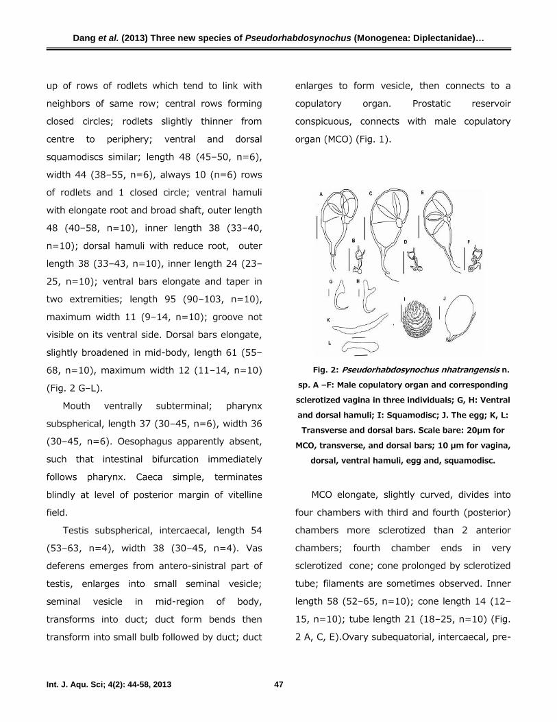
up of rows of rodlets which tend to link with
neighbors of same row; central rows forming
closed circles; rodlets slightly thinner from
centre to periphery; ventral and dorsal
squamodiscs similar; length 48 (45–50, n=6),
width 44 (38–55, n=6), always 10 (n=6) rows
of rodlets and 1 closed circle; ventral hamuli
with elongate root and broad shaft, outer length
48 (40–58, n=10), inner length 38 (33–40,
n=10); dorsal hamuli with reduce root, outer
length 38 (33–43, n=10), inner length 24 (23–
25, n=10); ventral bars elongate and taper in
two extremities; length 95 (90–103, n=10),
maximum width 11 (9–14, n=10); groove not
visible on its ventral side. Dorsal bars elongate,
slightly broadened in mid-body, length 61 (55–
68, n=10), maximum width 12 (11–14, n=10)
(Fig. 2 G–L).
Mouth ventrally subterminal; pharynx
subspherical, length 37 (30–45, n=6), width 36
(30–45, n=6). Oesophagus apparently absent,
such that intestinal bifurcation immediately
follows pharynx. Caeca simple, terminates
blindly at level of posterior margin of vitelline
field.
Testis subspherical, intercaecal, length 54
(53–63, n=4), width 38 (30–45, n=4). Vas
deferens emerges from antero-sinistral part of
testis, enlarges into small seminal vesicle;
seminal vesicle in mid-region of body,
transforms into duct; duct form bends then
transform into small bulb followed by duct; duct
enlarges to form vesicle, then connects to a
copulatory organ. Prostatic reservoir
conspicuous, connects with male copulatory
organ (MCO) (Fig. 1).
Fig. 2: Pseudorhabdosynochus nhatrangensis n.
sp. A –F: Male copulatory organ and corresponding
sclerotized vagina in three individuals; G, H: Ventral
and dorsal hamuli; I: Squamodisc; J. The egg; K, L:
Transverse and dorsal bars. Scale bare: 20µm for
MCO, transverse, and dorsal bars; 10 µm for vagina,
dorsal, ventral hamuli, egg and, squamodisc.
MCO elongate, slightly curved, divides into
four chambers with third and fourth (posterior)
chambers more sclerotized than 2 anterior
chambers; fourth chamber ends in very
sclerotized cone; cone prolonged by sclerotized
tube; filaments are sometimes observed. Inner
length 58 (52–65, n=10); cone length 14 (12–
15, n=10); tube length 21 (18–25, n=10) (Fig.
2 A, C, E).Ovary subequatorial, intercaecal, pre-
Dang et al. (2013) Three new species of Pseudorhabdosynochus (Monogenea: Diplectanidae)…
Int. J. Aqu. Sci; 4(2): 44-58, 2013 48
testicular, encircles right caecum. Ovary length
52 (48–55, n=4); width 31 (25–40, n=4).
Oviduct pass medially to form ootype,
surrounded by Mehlis’ gland; ootype short,
opens into uterus. Uterus dextral. Duct from
sclerotized vagina to ootype visible only in live
specimens. Vitelline fields extend posteriorly
from posterior to pharyngeal level in 2 lateral
bands, confluent in post-testicular region,
terminate anterior to peduncle. Bilateral
connections from vitelline fields to ootype
inconspicuous. Mature egg oval–shaped with
long filament, length 45, 66, n=2 width 35, 50,
n=2 (Fig. 2 L). Egg observed in uterus oval or
ellipse–shaped with thick wall. Vagina
comprised of unsclerotized region,
inconspicuous, visible only in living animal,
followed by sclerotized region. Sclerotized
vagina sinistral, a complex sclerotized structure;
length 29 (25–33, n=10) (Fig. 2 C, D, F).
Sclerotized vagina comprises cup–shaped
trumpet with opening at distal end, followed by
proximal tubular region. Tube twisted in circle
at posterior end, extending into thin branches.
Trumpet and tube more sclerotized with thick
wall.
Taxonomic summary
Type-host: Sea cage cultured Epinepheluscoioides (Hamilton, 1822) and E. bleekeri(Vaillant, 1878) (Serranidae).
Type-locality: Nha Trang Bay (12° 15′ N,
109° 21′ E), Vietnam.
Site: Between secondary gill lamellae.
Type-material: Holotype ZMBN collection N0:
84826, collected on 18 January 2008, Nha
Trang Bay, Vietnam on E. coioides. 3 paratypes
(ZMBN collection N0: 84827-84829), all in
ammonium picrate.
Material examined: 15 specimens, including 5 in
ammonium picrate, 5 cleared with lactophenol
and 5 live specimens. Measurements on 10
specimens.
Prevalence: 27% (12/45) on E. coioides and
10% (4/40) on E. bleeckeriEtymology: The name of this species refers to
Nha Trang Bay, where the species was found.
Remarks
Pseudorhabdosynochus nhatrangensis n. sp.
has the characteristic features of Pseudorhabd-osynochus, possessing an MCO of reniform,
sclerotized structure with four characteristic
compartments. With squamodiscs of 10 rows of
rodlets, P. nhatrangensis can be easily
separated from Pseudorhabdosynochus species
possessing more than 14 rows (P. riouxi (Oliver,
1986), P. monaensis Dyer et al., 1994, P.epinepheli (Yamaguti, 1938), P. americanus(Price, 1937) (synonymous with P. hargisi(Oliver and Paperna, 1984), P. coioidesis Bu etal., 1999, P. amplidiscatum (Bravo-Hollis,
1954), P. chinensis Zhang et al., 2001, P.sulamericanus Santos et al. 2000, P. bouaini
Dang et al. (2013) Three new species of Pseudorhabdosynochus (Monogenea: Diplectanidae)…
Int. J. Aqu. Sci; 4(2): 44-58, 2013 49
Neufar and Euzet, 2007, P. enitsuji Neufar and
Euzet, 2007).
The shape and number of the row of
rodlets, which form complete circles or
concentric rings (the so-called lamellosqua-
modisc in “P. cupatus group” (Hinsinger and
Justine, 2006b) are also important taxonomic
features. With only central row forming a
complete circle, P. nhatrangensis n. sp. can be
differentiated from P. cupatus Young, 1968, P.kritskyi Dyer et al. 1995, P. capurroi Vidal-
Martinez and Mendoza-Franco, 1998, P.melanesiensis Laird, 1958, P. beverleyburtonae(Oliver, 1984), P. buitoe Justine, 2007, P.cuitoe Justine, 2007, and, P. duitoe Justine,
2007, P. fuitoe Justine, 2007, and P. guitoeJustine, 2007, all having at least the two
innermost rows forming complete circles or
rings.
According to Justine (2007a), the
sclerotized vagina is likely the key structure for
Pseudorhabdosynochus identification. P.nhatrangensis n. sp. differentiates from the
remaining species, which have a similar number
of rows in the squamodisc, by the shape of the
sclerotized part of the vagina. P. nhatrangensisn. sp. is characterized by a short and thin
tubular vagina with a cup-shape trumpet
opening anteriorly. It is somewhat similar to
that of P. serrani Yamaguti, 1953, in that the
proximal tubular region twists in a round circle
before extending into thin chambers, but the
later species possesses an oval, saccular distal
part (trumpet), while it is cup-shaped in the
former. The sclerotized vagina is smaller in P.nhatrangensis n. sp. with a total length of 25–
33 vs 34.5–43.5 in P. serrani. P. shenzhenensisYang et al., 2005 is differentiated from the new
species by possessing a vagina with a flask-
shaped distal section and hook-shaped tube.
Pseudorhabdosynochus vietnamensis n.
sp.(Figure 3, 4 A–I)
Diagnostic: Body elongate, length 1040
(840–1240, n=6), width 213 (190–250, n=6).
Tegument scaly; scales on ventral and dorsal
faces from level of ovary to squamodiscs.
Anterior region with 3 pairs of head organs and
2 pairs of eye-spots; distance between outer
margins of anterior eye-spot pair 24 (19–26,
n=5), of posterior eye-spot pair 19 (15–22,
n=5).
Haptor distinctly differentiated from rest of
body, narrower than body, width 80 (75–85,
n=4); length 22 (20–25, n=4), provided with 2
similar squamodiscs, 2 pairs of lateral hamuli, 3
bars and 14 marginal hooklets. Squamodiscs
round in shape, made up of rows of rodlets;
central rows forming closed circles and
possessing an inner core; rodlets progressively
thinner from centre to periphery; last row with
very thin rodlets; ventral and dorsal
squamodiscs similar; length 82 (75–90, n=6),

Dang et al. (2013) Three new species of Pseudorhabdosynochus (Monogenea: Diplectanidae)…
Int. J. Aqu. Sci; 4(2): 44-58, 2013 50
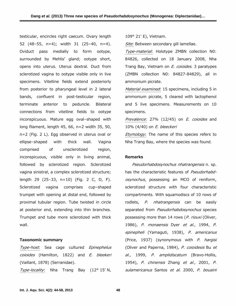
width 83 (73–95, n=6), with 17–18 (n=6) rows
of rodlets; ventral hamuli with shaft straight
and broad root, outer length 53 (18–58, n=6),
inner length 41 (38–45, n=6); dorsal hamuli
with reduced root, outer length 45 (43–50,
n=6), inner length 32 (30–35, n=6); ventral
bar lip-like shaped with elongate extremities;
length 87 (70–100, n=6), maximum width 15
(13–20, n=6); groove visible on its ventral side.
Dorsal bar broadened in mid-body, length 59
(53–70, n=6), maximum width 18 (13–20,
n=6) (Fig. 4 E–H).
Figure 3: Pseudorhabdosynochus vietnamensis n.
sp. from Epinephelus coioides in Nha Trang Bay,
Vietnam. Dorsal view, composite drawing from
holotype, paratype and observation of live
specimens. Scale bar: 100 µm.
Pharynx subspherical, length 44 (30–52,
n=5), width 42 (30–55, n=5). Oesophagus
apparently absent, such that intestinal
bifurcation immediately follows pharynx. Caeca
simple, terminate blindly at level of posterior
margin of vitelline field.
Fig. 4: Pseudorhabdosynochus vietnamensis n. sp.
A-D: Male copulatory organ and corresponding
sclerotized vagina in two individuals. E, F: Ventral
and dorsal hamuli; G, H Transverse and dorsal bars:
I: Squamodisc; Scale bare: 20µm for MCO,
transverse, and, dorsal bars; 10 µm for vagina,
dorsal, ventral hamuli and, squamodisc.
Testis subspherical, intercaecal. Vas
deferens emerges from antero-sinistral part of
testis, enlarges into small seminal vesicle;
seminal vesicle in middle region of body,
transforms into duct; duct forms bends, then
transform into small bulb followed by duct; duct
enlarges to form vesicle, then connects to male
copulatory organ (MCO). Prostatic reservoir
conspicuous, elongate, connects with male
copulatory organ (MCO) (Fig. 3B).

Dang et al. (2013) Three new species of Pseudorhabdosynochus (Monogenea: Diplectanidae)…
Int. J. Aqu. Sci; 4(2): 44-58, 2013 51
MCO very large, divided into four chambers
with fourth chamber more sclerotized than the
3 anterior chambers; first chamber with very
thin anterior wall; fourth chamber ends in very
sclerotized cone; characteristic thickening of
wall present inside fourth chamber at base of
cone; cone prolonged by elongate sclerotized
tube, usually widened and slightly curved
posteriorly, filaments not observed. Inner
length 108 (102–115, n=6); cone length 18
(15–20, n=6); tube length 46 (40–55, n=6)
(Fig. 4 A, C).
Ovary subequatorial, intercaecal, pre-
testicular, encircles right caecum. Oviduct
passes medially to form ootype, surrounded by
Mehlis’ gland; ootype short, uterus not
observed. Duct from sclerotized vagina to
ootype conspicuous. Vitelline fields extend
posteriorly from posterior to pharygeal level in 2
lateral bands, confluent in post-testicular
region, terminate anterior to peduncle (Fig 3
A,B). Egg not seen.
Vagina comprised of unsclerotized region,
visible only in living animal, followed by
sclerotized region. Sclerotized vagina sinistral in
a complex structure; length 31 (30–32, n=6),
width 24 (23–26, n=6). Sclerotized vagina
comprises small anterior trumpet, which is
continuous with unsclerotized vagina (Fig. 3).
Trumpet is followed by convoluted structure,
twisted and divided in to two distinct chambers,
which lay over each other (Fig. 4 B, D).
Taxonomic summary
Type-host: Epinephelus coioides (Hamilton,
1872).Type-locality: Nha Trang Bay (12° 15′ N,
109° 21′ E), Vietnam.
Site: Between secondary gill lamellae.
Type-material: Holotype ZMBL collection N0:
84830, collected on 20 July 2007, Nha Trang
Bay, Vietnam in ammonium picrate. 3
paratypes (ZMBN collection N0: 84831-84833)
all cleared with lactophenol.
Material examined: 10 specimens, including 3 in
ammonium picrate, 3 cleared with lactophenol
and 4 live specimens. Measurements from 6
specimens.
Prevalence: 7 % (3/45).
Etymology: The name of this species refers the
country Vietnam, where the species was found.
Remarks
Pseudorhabdosynochus vietnamensis n. sp.
possesses a characteristic vagina, trumpet
shaped anteriorly, followed by a convoluted
structure, twisted and divided into two distinct
chambers. The morphology of vagina serves as
a diagnostic character for P. vietnamensis n.
sp., easily differentiating it from Pseudorhabd-osynochus species with squamodisc having
more than 14 rows of rodlets (listed above).
Pseudorhabdosynochus fuitoe Justine, 2007
from Epinephelus maculates (Bloch, 1790), P.sinediscus Neifar and Euzet, 2007, on E. costae
Dang et al. (2013) Three new species of Pseudorhabdosynochus (Monogenea: Diplectanidae)…
Int. J. Aqu. Sci; 4(2): 44-58, 2013 52
(Steindachner, 1878) and P. pai Justine and
Vignon, 2009 on E. tauvina (Forsskål, 1775) (all
Serranidae) are characterized by sclerotized
vagina with anterior trumpet followed by a
primary canal, primary chamber, secondary
canal and secondary chamber. These species
share with P. vietnamensis n. sp. a similar
vagina with a trumpet anteriorly and a
complicated structure with several canals and
chambers. However, the above species all
possess squamodiscs with less than 14 rows of
rodlets, and P. sinediscus is differentiated from
other species by the absence of squamodiscs. P.vietnamensis n. sp. and P. pai both possess
extremely developed male cupulatory organs
(inner length: 108 and 118, Core length: 18
and 17; Tube length: 46 and 54; respectively)
(Measurement of P. pai specimens stored in
ammonium picrate (Justine and Vignon, 2009),
but differ in the number of rows in the
squamodisc (17–18 vs 11–13), and in the detail
in the structure of the vagina.
Pseudorhabdosynochus brunei n. sp.(Figure 5, 6 A–H, 7)
Diagnostic: Body slightly dorsoventrally
flattened, widest at level of ovary, length 245
(100–320, n=4), width 102 (90–150,
n=4).Tegument smooth. Anterior region with 3
pairs of head organs and 2 pairs of eye-spots;
distance between outer margins of anterior eye-
spot pair 13 (11–15, n=5, of posterior eye-spot
pair 7 (5–10, n=5).
Haptor differentiated from rest of body,
narrow than body, width 85 (80–90, n=3);
length 36 (34–38, n=3), provided with 2 similar
squamodiscs, 2 pairs of lateral hamuli, 3 bars
and 14 marginal hooklets. Squamodiscs round
in shape, made up of rows of rodlets; central
row not forming closed circles; rodlets
progressively thinner from centre to periphery;
last row with very thin rodlets; ventral and
dorsal squamodiscs similar; length 20 (19–21,
n=5), width 26 (25–28, n=5), with 7 (n=5)
rows of rodlets (Fig. 6H); Ventral hamuli with
shaft elongate and broad root, outer length 34
(30–36, n=5), inner length 29 (26–31, n=5);
Dorsal hamuli with reduced root and curved
shaft, outer length 32 (30–39, n=3), inner
length 21 (20–22, n=3); Ventral bar massive,
slightly constricted at medial part, tapering at
extremities; length 62 (60–66, n=5); groove
not visible on ventral side. Dorsal bar broadens
in mid-body, length 54 (50–58, n=5) (Fig. 6 D–
G).
Pharynx subspherical, length 33 (25–40,
n=4), width 33 (28–42, n=4). Oesophagus
apparently absent, such that intestinal
bifurcation immediately follows pharynx. Caeca
simple, terminates blindly at level of posterior
margin of vitelline field.
Testes subspherical, intercaecal. Vas
deferens emerge from antero-sinistral part of
testis, enlarge into small seminal vesicle;
Dang et al. (2013) Three new species of Pseudorhabdosynochus (Monogenea: Diplectanidae)…
Int. J. Aqu. Sci; 4(2): 44-58, 2013 53
seminal vesicle in middle region of body,
transform into duct; duct form bends, then
transform into small bulb followed by duct; duct
enlarge to form vesicle, then connect to male
copulatory organ (MCO).
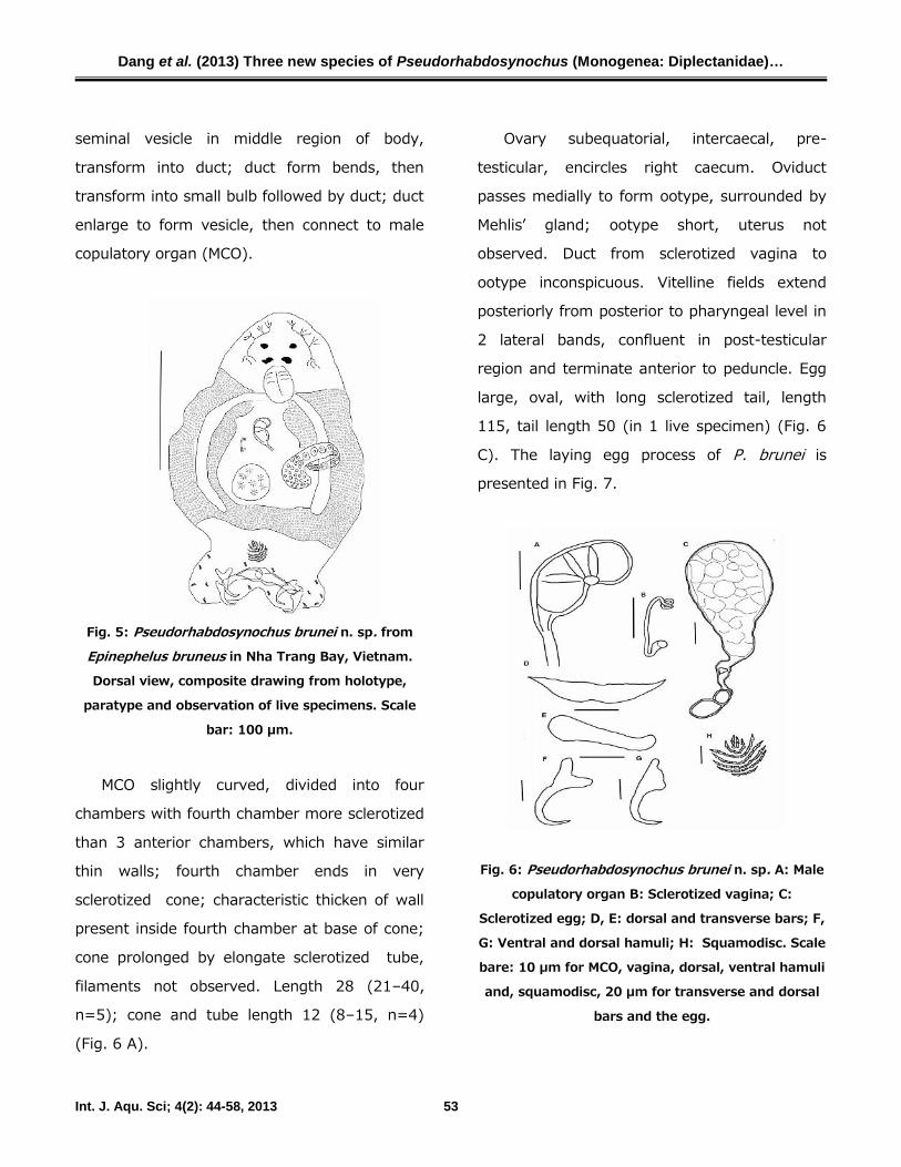
Fig. 5: Pseudorhabdosynochus brunei n. sp. from
Epinephelus bruneus in Nha Trang Bay, Vietnam.
Dorsal view, composite drawing from holotype,
paratype and observation of live specimens. Scale
bar: 100 µm.
MCO slightly curved, divided into four
chambers with fourth chamber more sclerotized
than 3 anterior chambers, which have similar
thin walls; fourth chamber ends in very
sclerotized cone; characteristic thicken of wall
present inside fourth chamber at base of cone;
cone prolonged by elongate sclerotized tube,
filaments not observed. Length 28 (21–40,
n=5); cone and tube length 12 (8–15, n=4)
(Fig. 6 A).
Ovary subequatorial, intercaecal, pre-
testicular, encircles right caecum. Oviduct
passes medially to form ootype, surrounded by
Mehlis’ gland; ootype short, uterus not
observed. Duct from sclerotized vagina to
ootype inconspicuous. Vitelline fields extend
posteriorly from posterior to pharyngeal level in
2 lateral bands, confluent in post-testicular
region and terminate anterior to peduncle. Egg
large, oval, with long sclerotized tail, length
115, tail length 50 (in 1 live specimen) (Fig. 6
C). The laying egg process of P. brunei is
presented in Fig. 7.
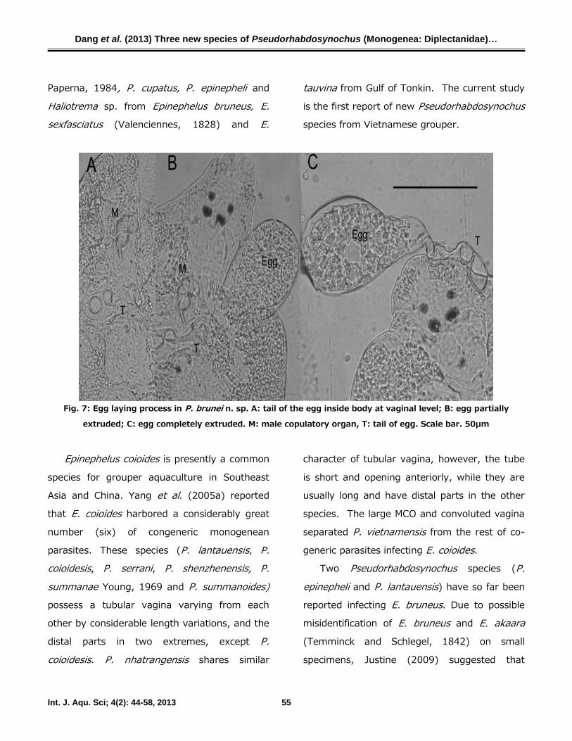
Fig. 6: Pseudorhabdosynochus brunei n. sp. A: Male
copulatory organ B: Sclerotized vagina; C:
Sclerotized egg; D, E: dorsal and transverse bars; F,
G: Ventral and dorsal hamuli; H: Squamodisc. Scale
bare: 10 µm for MCO, vagina, dorsal, ventral hamuli
and, squamodisc, 20 µm for transverse and dorsal
bars and the egg.

Dang et al. (2013) Three new species of Pseudorhabdosynochus (Monogenea: Diplectanidae)…
Int. J. Aqu. Sci; 4(2): 44-58, 2013 54
Sclerotized vagina, length 18 (15–20, n=4);
very difficult to observe, even in live specimens.
Sclerotized vagina with long sclerotized tube
extending close to distal opening. Distal region
make up of two small sequenced chambers.
Posteriorly, tube divides into broad branch (Fig.
6 B).
Taxonomic summary
Type-host: Epinephelus bruneus (Bloch, 1793).Type-locality: Cam Ranh Bay (11° 52' 60 N,
109° 10' E), Vietnam
Site: Between secondary gill lamellae.
Type-material: Holotype ZMBN collection N0:
84834, collected on 25 July 2007, Nha Trang
Bay, Vietnam; 3 paratypes (ZMBN 84835-
84837), all in ammonium picrate.
Material examined: 6 specimens, including 4 in
ammonium picrate, 2 cleared with lactophenol
and 2 live specimens.
Prevalence: 32 % (9/28)
Etymology: The specific name refers to the
scientific name of the host E. bruneus
Remarks
P. brunei n. sp. is the only species found on
E. bruneus in Nha Trang Bay, Vietnam. It is the
smallest Pseudorhabdosynochus so far found on
grouper in Vietnam. With all characteristics of
the genus, Pseudorhabdosynochus bruneipossesses a vagina which shares features with
Pseudorhabdosynochus summanoides Yang et
al., 2005 in having a sclerotized tube extending
close to the distal opening, and the distal region
being made up of sequenced chambers, but the
size is much smaller (18 (15–29) vs 29–56
(45.8)), and the distal region possesses two
sequential chambers, while it winds into a small
irregular convolution in P. summonoides. In
addition, it differs from P. summanoides in that
the posterior end of its tube does not form a
loop (twisted in round circle). The smaller size
of vagina and its distal region being made up of
sequential chambers separate P. brunei from P.lantauensis Beverley-Burton and Suriano, 1981
which was originally reported from E. bruneus.P. brunei n. sp. can easily be differentiated from
other Pseudorhabdosynochus species by the
squamodisc having only 7 rows of rodlets,
specific haptoral hadparts, and characteristics of
the egg. The egg laying process, presented in
Fig. 7, shows the sclerotized structure with the
tail. The eggs are also larger than eggs found in
other species of the genus.
DiscussionIn Vietnam, both E. coioides and E. bleekeri
are intensively cultured in sea cages, along with
E. fuscoguttatus (Forsskål, 1775) and
Plectroponus leopardus (Lacepède, 1802), and
in some cases with snapper (Lutjanusargentimaculatus (Forsskål, 1775) and other
finfish. Arthur and Te (2006) reported
Pseudorhabdosynochus harigisi Oliver and
Dang et al. (2013) Three new species of Pseudorhabdosynochus (Monogenea: Diplectanidae)…
Int. J. Aqu. Sci; 4(2): 44-58, 2013 55
Paperna, 1984, P. cupatus, P. epinepheli and
Haliotrema sp. from Epinephelus bruneus, E.sexfasciatus (Valenciennes, 1828) and E.
tauvina from Gulf of Tonkin. The current study
is the first report of new Pseudorhabdosynochusspecies from Vietnamese grouper.
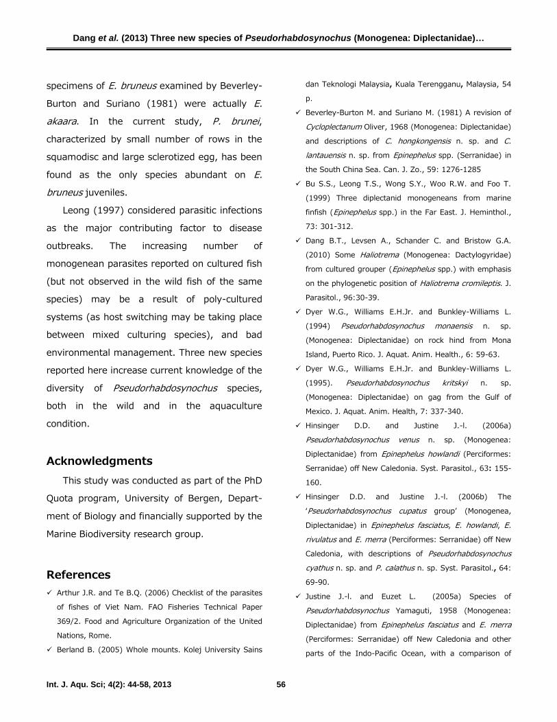
Fig. 7: Egg laying process in P. brunei n. sp. A: tail of the egg inside body at vaginal level; B: egg partially
extruded; C: egg completely extruded. M: male copulatory organ, T: tail of egg. Scale bar. 50µm
Epinephelus coioides is presently a common
species for grouper aquaculture in Southeast
Asia and China. Yang et al. (2005a) reported
that E. coioides harbored a considerably great
number (six) of congeneric monogenean
parasites. These species (P. lantauensis, P.coioidesis, P. serrani, P. shenzhenensis, P.summanae Young, 1969 and P. summanoides)possess a tubular vagina varying from each
other by considerable length variations, and the
distal parts in two extremes, except P.coioidesis. P. nhatrangensis shares similar
character of tubular vagina, however, the tube
is short and opening anteriorly, while they are
usually long and have distal parts in the other
species. The large MCO and convoluted vagina
separated P. vietnamensis from the rest of co-
generic parasites infecting E. coioides.Two Pseudorhabdosynochus species (P.
epinepheli and P. lantauensis) have so far been
reported infecting E. bruneus. Due to possible
misidentification of E. bruneus and E. akaara(Temminck and Schlegel, 1842) on small
specimens, Justine (2009) suggested that
Dang et al. (2013) Three new species of Pseudorhabdosynochus (Monogenea: Diplectanidae)…
Int. J. Aqu. Sci; 4(2): 44-58, 2013 56
specimens of E. bruneus examined by Beverley-
Burton and Suriano (1981) were actually E.akaara. In the current study, P. brunei,characterized by small number of rows in the
squamodisc and large sclerotized egg, has been
found as the only species abundant on E.bruneus juveniles.
Leong (1997) considered parasitic infections
as the major contributing factor to disease
outbreaks. The increasing number of
monogenean parasites reported on cultured fish
(but not observed in the wild fish of the same
species) may be a result of poly-cultured
systems (as host switching may be taking place
between mixed culturing species), and bad
environmental management. Three new species
reported here increase current knowledge of the
diversity of Pseudorhabdosynochus species,
both in the wild and in the aquaculture
condition.
AcknowledgmentsThis study was conducted as part of the PhD
Quota program, University of Bergen, Depart-
ment of Biology and financially supported by the
Marine Biodiversity research group.
References Arthur J.R. and Te B.Q. (2006) Checklist of the parasites
of fishes of Viet Nam. FAO Fisheries Technical Paper
369/2. Food and Agriculture Organization of the United
Nations, Rome.
Berland B. (2005) Whole mounts. Kolej University Sains
dan Teknologi Malaysia, Kuala Terengganu, Malaysia, 54
p.
Beverley-Burton M. and Suriano M. (1981) A revision of
Cycloplectanum Oliver, 1968 (Monogenea: Diplectanidae)
and descriptions of C. hongkongensis n. sp. and C.lantauensis n. sp. from Epinephelus spp. (Serranidae) in
the South China Sea. Can. J. Zo., 59: 1276-1285
Bu S.S., Leong T.S., Wong S.Y., Woo R.W. and Foo T.
(1999) Three diplectanid monogeneans from marine
finfish (Epinephelus spp.) in the Far East. J. Heminthol.,
73: 301-312.
Dang B.T., Levsen A., Schander C. and Bristow G.A.
(2010) Some Haliotrema (Monogenea: Dactylogyridae)
from cultured grouper (Epinephelus spp.) with emphasis
on the phylogenetic position of Haliotrema cromileptis. J.
Parasitol., 96:30-39.
Dyer W.G., Williams E.H.Jr. and Bunkley-Williams L.
(1994) Pseudorhabdosynochus monaensis n. sp.
(Monogenea: Diplectanidae) on rock hind from Mona
Island, Puerto Rico. J. Aquat. Anim. Health., 6: 59-63.
Dyer W.G., Williams E.H.Jr. and Bunkley-Williams L.
(1995). Pseudorhabdosynochus kritskyi n. sp.
(Monogenea: Diplectanidae) on gag from the Gulf of
Mexico. J. Aquat. Anim. Health, 7: 337-340.
Hinsinger D.D. and Justine J.-l. (2006a)
Pseudorhabdosynochus venus n. sp. (Monogenea:
Diplectanidae) from Epinephelus howlandi (Perciformes:
Serranidae) off New Caledonia. Syst. Parasitol., 63: 155-
160.
Hinsinger D.D. and Justine J.-l. (2006b) The
‘Pseudorhabdosynochus cupatus group’ (Monogenea,
Diplectanidae) in Epinephelus fasciatus, E. howlandi, E.rivulatus and E. merra (Perciformes: Serranidae) off New
Caledonia, with descriptions of Pseudorhabdosynochuscyathus n. sp. and P. calathus n. sp. Syst. Parasitol., 64:
69-90.
Justine J.-l. and Euzet L. (2005a) Species of
Pseudorhabdosynochus Yamaguti, 1958 (Monogenea:
Diplectanidae) from Epinephelus fasciatus and E. merra(Perciformes: Serranidae) off New Caledonia and other
parts of the Indo-Pacific Ocean, with a comparison of

Dang et al. (2013) Three new species of Pseudorhabdosynochus (Monogenea: Diplectanidae)…
Int. J. Aqu. Sci; 4(2): 44-58, 2013 57
measurements of specimens prepared using different
methods, and a description of P. caledonicus n. sp. Syst.
Parasitol., 62: 1-37.
Justine J.-l. and Euzet L. (2005b) Pseudorhabdosynochushirundineus n. sp. Monogenea: Diplectanidae) from
Variola louti (Perciformes: Serranidae) off New
Caledonia. Syst. Parasitol., 82: 39-45.
Justine J.-l. (2007a) Parasite biodiversity in a coral reef
fish: twelve species of monogeneans on the gills of the
grouper Epinephelus maculatus (Perciformes:
Serranidae) off New Caledonia, with a description of
eight new species of Pseudorhabdosynochus(Monogenea: Diplectanidae). Syst. Parasitol., 66: 81-
129.
Justine J.-l. (2007b) Pseudorhabdosynochus argus n. sp.
(Monogenea: Diplectanidae) from Cephalopholis argus, P.minutus n. sp. and Diplectanum nanus n. sp. from C.sonnerati and other monogeneans from Cephalopholisspp. (Perciformes: Serranidae) off Australia and New
Caledonia. Syst. Parasitol., 68: 195-215.
Justine J.-l. and Sigura A. (2007) Monogeneans of the
malabar grouper Epinephelus malabaricus (Perciformes,
Serranidae) off New Caledonia, with a description of six
new species of Pseudorhabdosynochus (Monogenea:
Diplectanidae). Zootaxa, 1543: 1–44.
Justine J.-l. (2008) Two new species of
Pseudorhabdosynochus Yamaguti, 1958 (Monogenea:
Diplectanidae) from the deep-sea grouper Epinephelusmorrhua (Val.) (Perciformes: Serranidae) off New
Caledonia. Syst. Parasitol., 71: 145-158.
Justine J.-l. (2009) A redescription of
Pseudorhabdosynochus epinepheli (Yamaguti, 1938), the
type-species of Pseudorhabdosynochus Yamaguti, 1958
(Monogenea: Diplectanidae) and the description of P.satyui n. sp. from Epinephelus akaara off Japan. Syst.
Parasitol., 72: 27-55.
Justine, J.-l. and Vignon M. (2009) Monogeneans of the
grouper Epinephelus tauvina (Perciformes: Serranidae)
off Moorea, French Polynesia, with a description of
Pseudorhabdosynochus pai n. sp. (Monogenea:
Diplectanidae). Syst. Parasitol., 72: 113-125.
Kritsky D.C. and Beverley-Burton M. (1986) The Status
of Pseudorhabdosynochus Yamaguti, 1958, and
Cycloplectanum Oliver, 1968 (Monogenea:
Diplectanidae). Proc. Biol. Soc. Wash., 99: 17-20.
Leong T.S. (1997) Control of Parasites in Cultured Marine
Finfishes in Southeast Asia-an Overview. Inrernorionnl J.
Parasirol., 27: 1177-1184.
Neifar L. and Euzet L. (2007) Five new species of
Pseudorhabdosynochus (Monogenea: Diplectanidae) from
the gills of Epinephelus costae (Teleostei: Serranidae).
Folia Parasitol., 54: 117-128.
Oliver G. (1986) Cycloplectanum riouxi n. sp., une
nouvelle espece de Diplectanidae (Monogenea,
Monopisthocotylea) parasite d'Epinephelusguaza(Linnaeus, 1758) (Pisces, Serranidae) Syst.
Parasitol., 8: 317-322.
Santos C., Buchmann P.K. and Gibson D. (2000)
Pseudorhabdosynochus spp. (Monogenea: Diplectanidae)
from the gills of Epinephelus spp. in Brazilian waters.
Syst. Parasitol., 45: 145-153.
Sigura A., Chauvet C. and Justine J.-L. (2007)
Pseudorhabdosynochus bacchus sp. nov. (Monogenea:
Diplectanidae) from Epinephelus coeruleopunctatus(Perciformes: Serranidae) off New Caledonia. Acta
Parasitol., 52: 196-200.
Vidal-Martinez V.M., Aguirre-Macedo L. and Mendoza-
Franco E.F. (1997) Pseudorhabdosynochus yucatanensissp. n. (Monogenea: Diplectanidae) from the gills of the
red grouper Epinephelus morio (Pisces: Serranidae) of
the Yucatan Peninsula, Mexico. Folia Parasitol., 4: 274-
278.
Vidal-Martinez V.M. and Mendoza-Franco E.F. (1998)
Pseudorhabdosynochus capurroi sp. n. (Monogenea:
Diplectanidae) from the gills of Mycteroperca bonaci(Pisces: Serranidae) of the Yucatan Peninsula, Mexico.
Folia Parasitol., 45: 221-224.
Wu X.Y., Chilton N.B., Zhu X.Q., Xie M.Q. and Li A.X.
(2005a) Molecular and morphological evidence indicates
that Pseudorhabdosynochus lantauensis (Monogenea:
Diplectanidae) represents two species. Parasitology, 130:
669-677.
Dang et al. (2013) Three new species of Pseudorhabdosynochus (Monogenea: Diplectanidae)…
Int. J. Aqu. Sci; 4(2): 44-58, 2013 58
Wu X.Y., Li A.X., Zhu X.Q. and Xie M.Q. (2005b)
Description of Pseudorhabdosynochus seabassi sp. n.
(Monogenea: Diplectanidae) from Lates calcarifer and the
revision of the phylogenetic position of Diplectanumgrouperi (Monogenea: Diplectanidae) based on rDNA
sequence data. Folia Parasitol., 52: 231-240.
Yang T.B., Gibson D. and Zeng B.J. (2005a)
Pseudorhabdosynochus summanoides n. sp (Monogenea:
Diplectanidae) from Epinephelus coioides in Dapeng Bay,
South China Sea, with observations on several similar
species of Pseudorhabdosynochus Yamaguti, 1958. Syst.
Parasitol., 62: 221-239.
Yang T.B., Zeng B.J. and Gibson D.I. (2005b)
Description of Pseudorhabdosynochus shenzhenensis n.
sp (Monogenea: Diplectanidae) and redescription of P.serrani Yamaguti, 1953 from Epinephelus coioides off
Dapeng Bay, Shenzhen, China. J. Parasitol., 91: 808-
813.
Young P.C. (1969) Some monogenoideans of the family
Diplectanidae Bychowsky, 1957 from Australia teleost
fishes. J. Helminthol., 18: 223-254.
Zeng B.J. and Yang T.B. (2007) Description of
Pseudorhabdosynochus justinei n. sp (Monogenea:
Diplectanidae) and redescription of P. vagampullum(Young, 1969) Kritsky and Beverley-Burton, 1986 from
the gills of the longfin grouper Epinephelus quoyanus(Valenciennes) (Perciformes: Serranidae) in Dapeng Bay,
South China Sea. Syst. Parasitol., 66: 223-235.
Related Documents


