Three-dimensional visualization and a deep-learning model reveal complex fungal parasite networks in behaviorally manipulated ants Maridel A. Fredericksen a , Yizhe Zhang b , Missy L. Hazen c , Raquel G. Loreto a,d , Colleen A. Mangold d,e , Danny Z. Chen b , and David P. Hughes a,d,f,1 a Department of Entomology, Pennsylvania State University, University Park, PA 16802; b Department of Computer Science and Engineering, University of Notre Dame, Notre Dame, IN 46556; c Huck Institutes of the Life Sciences Microscopy and Cytometry Facility, Pennsylvania State University, University Park, PA 16802; d Center for Infectious Disease Dynamics, Pennsylvania State University, University Park, PA 16802; e Department of Biochemistry and Molecular Biology, Pennsylvania State University, University Park, PA 16802; and f Department of Biology, Pennsylvania State University, University Park, PA 16802 Edited by Joan E. Strassmann, Washington University in St. Louis, St. Louis, MO, and approved October 16, 2017 (received for review June 29, 2017) Some microbes possess the ability to adaptively manipulate host behavior. To better understand how such microbial parasites control animal behavior, we examine the cell-level interactions between the species-specific fungal parasite Ophiocordyceps unilateralis sensu lato and its carpenter ant host (Camponotus castaneus) at a crucial moment in the parasite’ s lifecycle: when the manipulated host fixes itself per- manently to a substrate by its mandibles. The fungus is known to secrete tissue-specific metabolites and cause changes in host gene ex- pression as well as atrophy in the mandible muscles of its ant host, but it is unknown how the fungus coordinates these effects to manipulate its host’ s behavior. In this study, we combine techniques in serial block- face scanning-electron microscopy and deep-learning– based image seg- mentation algorithms to visualize the distribution, abundance, and in- teractions of this fungus inside the body of its manipulated host. Fungal cells were found throughout the host body but not in the brain, implying that behavioral control of the animal body by this microbe occurs peripherally. Additionally, fungal cells invaded host muscle fibers and joined together to form networks that encircled the muscles. These networks may represent a collective foraging behavior of this parasite, which may in turn facilitate host manipulation. deep learning | fungal networks | extended phenotype | behavioral manipulation | ants S ome parasitic microbes have evolved the ability to adaptively manipulate the behavior of the animals they infect (1). Ex- amples include: unicellular Trypanosomes (class Kinetoplastida) that alter salivary composition and feeding behavior in tsetse flies, thereby increasing the parasite’s transmission into mam- malian hosts (2); fungi such as Pandora and Ophiocordyceps that induce ants to bite vegetation and die in an elevated location suitable for parasite dispersal (3–5); and the well-studied api- complexan Toxoplasma gondii, which causes its rodent host to lose its innate fear of cats, enabling the parasite to transmit to the cat in which it reproduces (6). In each of these cases, the altered host behavior is an extended phenotype (7) of a microbial parasite’s genes being expressed through the body of an animal. An important question to ask is how these microbes, which are much smaller than their hosts, can control animal behavior to produce such spectacular extended phenotypes. For example, an individual T. gondii parasite is less than 5 μm in diameter (8), whereas there are an estimated 4 million neurons in the mouse brain (9). However, because T. gondii can form “tissue cysts” containing the slowly multiplying stage, bradyzoite (8), successful host manipulation may rely on a collective behavior among the microbial parasites. One observation that supports this possibility is a correlation between the amount of dopamine released and the number of dopaminergic cells infected with T. gondii, which is known to produce tyrosine hydroxylase, the rate-limiting en- zyme in dopamine synthesis (10). Although correlated changes in chemical activity (or gene expression) between parasites and hosts are important, such evi- dence alone is likely insufficient to determine what role, if any, collective behavior among microbes plays during manipulation. Because of the size asymmetry between microbes and the hosts whose behavior they control, it is important to have a micrometer- level view of the interface between the microbe and the host tissue with which it interacts. Such an approach would complement studies on the chemical cross-talk between parasite and host, and it would allow us to better understand what role collective be- havior among microbes may play during behavioral manipulation. One promising model system for such detailed, micrometer-level studies is the entomopathogenic fungal parasite Ophiocordyceps unilateralis sensu lato (s.l.) and its ant host. The fungus O. unilateralis s.l. is a complex of species that are all obligate parasites of ants from the tribe Camponotini, notably Camponotus and Polyrhachis (5). A distinct advantage of this system is that the ants can be infected in the laboratory (11). As with other entomopathogenic fungi, species in the unilateralis complex enter their hosts by penetrating the cu- ticle to then proliferate and fill the ant’s body (12). As such, the infection cycle begins when the parasite is a small free-floating cell in the open circulatory system of its insect host. After the required Significance Microbial parasites may behave collectively to manipulate their host’s behavior. We examine adaptations of a microbial para- site in its natural environment: the body of its coevolved and manipulated host. Electron microscopy and 3D reconstructions of host and parasite tissues reveal that this fungus invades muscle fibers throughout the ant’s body but leaves the brain intact, and that the fungal cells connect to form extensive net- works. The connections are likened to structures that aid in transporting nutrients and organelles in several plant-associated fungi. These findings alter the current view of parasite-extended phenotypes by demonstrating that behavior control does not require the parasite to physically invade the host brain and that parasite cells may coordinate to change host behavior. Author contributions: M.A.F. and D.P.H. designed research; M.A.F., Y.Z., R.G.L., C.A.M., D.Z.C., and D.P.H. performed research; Y.Z., M.L.H., and D.Z.C. contributed new reagents/ analytic tools; M.A.F., Y.Z., and D.P.H. analyzed data; and M.A.F., Y.Z., C.A.M., D.Z.C., and D.P.H. wrote the paper. The authors declare no conflict of interest. This article is a PNAS Direct Submission. This open access article is distributed under Creative Commons Attribution-NonCommercial- NoDerivatives License 4.0 (CC BY-NC-ND). Data deposition: Data generated in this study are available on Pennsylvania State University ScholarSphere, available at https://scholarsphere.psu.edu/concern/generic_works/6q524jn254. 1 To whom correspondence should be addressed. Email: [email protected]. This article contains supporting information online at www.pnas.org/lookup/suppl/doi:10. 1073/pnas.1711673114/-/DCSupplemental. www.pnas.org/cgi/doi/10.1073/pnas.1711673114 PNAS Early Edition | 1 of 6 MICROBIOLOGY

Welcome message from author
This document is posted to help you gain knowledge. Please leave a comment to let me know what you think about it! Share it to your friends and learn new things together.
Transcript

Three-dimensional visualization and a deep-learningmodel reveal complex fungal parasite networks inbehaviorally manipulated antsMaridel A. Fredericksena, Yizhe Zhangb, Missy L. Hazenc, Raquel G. Loretoa,d, Colleen A. Mangoldd,e, Danny Z. Chenb,and David P. Hughesa,d,f,1
aDepartment of Entomology, Pennsylvania State University, University Park, PA 16802; bDepartment of Computer Science and Engineering, University ofNotre Dame, Notre Dame, IN 46556; cHuck Institutes of the Life Sciences Microscopy and Cytometry Facility, Pennsylvania State University, University Park,PA 16802; dCenter for Infectious Disease Dynamics, Pennsylvania State University, University Park, PA 16802; eDepartment of Biochemistry and MolecularBiology, Pennsylvania State University, University Park, PA 16802; and fDepartment of Biology, Pennsylvania State University, University Park, PA 16802
Edited by Joan E. Strassmann, Washington University in St. Louis, St. Louis, MO, and approved October 16, 2017 (received for review June 29, 2017)
Some microbes possess the ability to adaptively manipulate hostbehavior. To better understand how such microbial parasites controlanimal behavior, we examine the cell-level interactions between thespecies-specific fungal parasite Ophiocordyceps unilateralis sensu latoand its carpenter ant host (Camponotus castaneus) at a crucial momentin the parasite’s lifecycle: when the manipulated host fixes itself per-manently to a substrate by its mandibles. The fungus is known tosecrete tissue-specific metabolites and cause changes in host gene ex-pression as well as atrophy in the mandible muscles of its ant host, butit is unknown how the fungus coordinates these effects to manipulateits host’s behavior. In this study, we combine techniques in serial block-face scanning-electronmicroscopy and deep-learning–based image seg-mentation algorithms to visualize the distribution, abundance, and in-teractions of this fungus inside the body of its manipulated host.Fungal cells were found throughout the host body but not in the brain,implying that behavioral control of the animal body by this microbeoccurs peripherally. Additionally, fungal cells invaded hostmuscle fibersand joined together to form networks that encircled themuscles. Thesenetworks may represent a collective foraging behavior of this parasite,which may in turn facilitate host manipulation.
deep learning | fungal networks | extended phenotype |behavioral manipulation | ants
Some parasitic microbes have evolved the ability to adaptivelymanipulate the behavior of the animals they infect (1). Ex-
amples include: unicellular Trypanosomes (class Kinetoplastida)that alter salivary composition and feeding behavior in tsetseflies, thereby increasing the parasite’s transmission into mam-malian hosts (2); fungi such as Pandora and Ophiocordyceps thatinduce ants to bite vegetation and die in an elevated locationsuitable for parasite dispersal (3–5); and the well-studied api-complexan Toxoplasma gondii, which causes its rodent host tolose its innate fear of cats, enabling the parasite to transmit tothe cat in which it reproduces (6). In each of these cases, thealtered host behavior is an extended phenotype (7) of a microbialparasite’s genes being expressed through the body of an animal.An important question to ask is how these microbes, which aremuch smaller than their hosts, can control animal behavior toproduce such spectacular extended phenotypes. For example, anindividual T. gondii parasite is less than 5 μm in diameter (8),whereas there are an estimated 4 million neurons in the mousebrain (9). However, because T. gondii can form “tissue cysts”containing the slowly multiplying stage, bradyzoite (8), successfulhost manipulation may rely on a collective behavior among themicrobial parasites. One observation that supports this possibilityis a correlation between the amount of dopamine released andthe number of dopaminergic cells infected with T. gondii, whichis known to produce tyrosine hydroxylase, the rate-limiting en-zyme in dopamine synthesis (10).
Although correlated changes in chemical activity (or geneexpression) between parasites and hosts are important, such evi-dence alone is likely insufficient to determine what role, if any,collective behavior among microbes plays during manipulation.Because of the size asymmetry between microbes and the hostswhose behavior they control, it is important to have a micrometer-level view of the interface between the microbe and the host tissuewith which it interacts. Such an approach would complementstudies on the chemical cross-talk between parasite and host, andit would allow us to better understand what role collective be-havior among microbes may play during behavioral manipulation.One promising model system for such detailed, micrometer-level
studies is the entomopathogenic fungal parasite Ophiocordycepsunilateralis sensu lato (s.l.) and its ant host. The fungusO. unilateraliss.l. is a complex of species that are all obligate parasites of ants fromthe tribe Camponotini, notably Camponotus and Polyrhachis (5). Adistinct advantage of this system is that the ants can be infected inthe laboratory (11). As with other entomopathogenic fungi, speciesin the unilateralis complex enter their hosts by penetrating the cu-ticle to then proliferate and fill the ant’s body (12). As such, theinfection cycle begins when the parasite is a small free-floating cellin the open circulatory system of its insect host. After the required
Significance
Microbial parasites may behave collectively to manipulate theirhost’s behavior. We examine adaptations of a microbial para-site in its natural environment: the body of its coevolved andmanipulated host. Electron microscopy and 3D reconstructionsof host and parasite tissues reveal that this fungus invadesmuscle fibers throughout the ant’s body but leaves the brainintact, and that the fungal cells connect to form extensive net-works. The connections are likened to structures that aid intransporting nutrients and organelles in several plant-associatedfungi. These findings alter the current view of parasite-extendedphenotypes by demonstrating that behavior control does notrequire the parasite to physically invade the host brain and thatparasite cells may coordinate to change host behavior.
Author contributions: M.A.F. and D.P.H. designed research; M.A.F., Y.Z., R.G.L., C.A.M.,D.Z.C., and D.P.H. performed research; Y.Z., M.L.H., and D.Z.C. contributed new reagents/analytic tools; M.A.F., Y.Z., and D.P.H. analyzed data; and M.A.F., Y.Z., C.A.M., D.Z.C., andD.P.H. wrote the paper.
The authors declare no conflict of interest.
This article is a PNAS Direct Submission.
This open access article is distributed under Creative Commons Attribution-NonCommercial-NoDerivatives License 4.0 (CC BY-NC-ND).
Data deposition: Data generated in this study are available on Pennsylvania State UniversityScholarSphere, available at https://scholarsphere.psu.edu/concern/generic_works/6q524jn254.1To whom correspondence should be addressed. Email: [email protected].
This article contains supporting information online at www.pnas.org/lookup/suppl/doi:10.1073/pnas.1711673114/-/DCSupplemental.
www.pnas.org/cgi/doi/10.1073/pnas.1711673114 PNAS Early Edition | 1 of 6
MICRO
BIOLO
GY

16–25 d of growth, the aggregation of microbial cells has reachedthe stage in which the fungus induces the ant to bite onto a plant ina location that is optimal for the fungus to grow and disperse (5,13). This conspicuous biting behavior is an unambiguous signal ofsuccessful manipulation. Such modified behaviors that increaseparasite transmission are not restricted to species in the Ophio-cordyceps genus. The ability to manipulate ants to bite onto a plantin an elevated location has evolved in at least two other phyloge-netically distant groups: the fungus Pandora formicae (phylumEntomophthoromycota) and the trematode Dicrocoelium den-driticum. Insights from the O. unilateralis s.l. system may thereforeset the stage for future research on the convergent evolution ofbehavioral manipulation.Previous work showed that O. unilateralis s.l. induces atrophy
in mandible muscles of the worker ants manipulated to bite leaves(14), and this atrophy was suggested to play a role in the bitingbehavior. Both ex vivo metabolomic (11) and in vivo transcriptomic(13) studies suggested the fungus actively secretes small moleculesthat affect the muscles and nervous tissue. Although these ap-proaches are informative, they provide only indirect observationsof the parasite as it affects host muscles and ultimately host be-havior, and they do not show how the individual cells of theparasite coordinate the manipulation. Additionally, at the sametime when the muscles are atrophied, the brain seems to be pre-served (13, 14), suggesting the parasite behaves differently towarddistinct tissues. This is in line with the observation that entomo-pathogenic fungi secrete specific metabolites when cultured in thepresence of muscle tissue or brain tissue (15). Such indirect ap-proaches where gene activity or secreted chemicals are measuredare useful, but direct observations of parasite and host cells wouldallow us to more precisely characterize how this fungus interactswith its ant host during manipulation.In the present study, we directly observe fungal cells inside the
manipulated host by integrating serial block-face scanning-electronmicroscopy (SEM) with automatic image segmentation and analy-ses that use deep-learning algorithms to distinguish host and par-asite tissue in serial stacks of images. This approach allows us toexamine the 3D structure and distribution of fungal cells in themuscle of manipulated ants. Because the atrophy reported inprevious work (14) could be a general effect of fungal pathogen-esis, we also infected ants with the generalist pathogen, Beauveriabassiana, which belongs to the same fungal order (Hypocreales) asO. unilateralis s.l. but does not manipulate its host as part of itslifecycle (16–18). This fungus thus serves as an important positivecontrol, allowing us to begin to separate characteristics that arespecific to the specialized insect/fungus interaction of O. unilateralis
s.l. (typified by complex behavioral manipulation) from those ef-fects on host tissue that result from general pathogenesis. Wespecifically aimed to describe the distribution, abundance, andinteractions of O. unilateralis s.l. cells inside their host during ma-nipulated biting behavior and to use these observations to hypoth-esize which fungal characteristics may play a role in producing thisextended phenotype.
ResultsThe Fungus O. unilateralis s.l. Is Present Throughout the Body butDoes Not Enter the Brain. Our first goal was to establish whethercells of the specialized fungus O. unilateralis s.l. were restricted toone location within the host ants. Previous work found a large ag-gregation of fungal cells in the head of manipulated ants, but otherparts of the body were not examined (14). In the present study, wefound O. unilateralis s.l. fungal cells in all three major body regions(head, thorax, and gaster; the latter is the ant-specific term for theterminal part of the abdomen) of the ant host (n = 8 O. unilateraliss.l.-infected ants) (Fig. S1). These fungal cells included two distinctforms: hyphal bodies, which are yeast-like cells that grow inside theinsect body and multiply by budding (19), and hyphae, which arethin filamentous projections (collectively termed mycelia) that arespecialized for foraging in new environments and invading hosttissues (20, 21). In our study, hyphae grew exclusively from the endsof hyphal bodies (see, for example, Fig. 3A). We also found fungalcells in abundance inside the legs (n = 12 additional O. unilateraliss.l.-infected ants) (Fig. S2). Taken together, our data demonstratethat O. unilateralis s.l. is not restricted to the head of its host.We also thoroughly examined the nervous tissue in the head
for O. unilateralis s.l. cells to determine whether the fungus en-ters the brain of the ant it manipulates. Using fluorescenceconfocal microscopy, we tested for the presence of fungal cellswithin the host brain. In all samples [n = 3 per group (infected,uninfected)], fungal cells were concentrated directly outside thebrain, but no fungal cells were observed inside the brain (Fig. 1).
Specialist and Generalist Fungal Pathogens Infiltrate Ant Muscle Tissue.We next wanted to determine the extent to whichO. unilateralis s.l.invades the muscles of the ant it manipulates. Here we used thegeneralist pathogen, B. bassiana, as a positive control for hostmuscle responses during infection by a fungal pathogen. We useda Zeiss Sigma serial block-face SEM with Gatan 3View technologyto examine 3D stacks of ant muscle. In these images, we observedmuscular atrophy in ants infected by both species of fungi. Thisatrophy was characterized by large spaces between the muscle fi-bers (cells of muscle tissue) compared with uninfected samples.
A B C
Fig. 1. Assessment of fungal invasion of the brain. Confocal images demonstrating the restriction of fungal invasion into the brain. Host brain is identified byimmunofluorescence of neuronal synapses (green, anti-SYNORF1), while fungal cells and host tracheae are stained with calcofluor white (red, antichitin). Allimages are represented as maximum projections of z-stacks. (A) Portion of the mushroom body (green) in the brain of an uninfected (control) ant, 156×magnification. (Scale bar, 10 μm.) (B) Portion of the optic lobe (green) in the brain of an infected, behaviorally manipulated ant, 60×magnification. (Scale bar,20 μm.) Arrows denote fungal hyphal bodies (red), identified by their short, rod-like structure and the presence of septa. Arrowheads denote tracheae (red),located within the brain (green), and identified by their long, filamentous structure and lack of septa. Note the restriction of fungal cell bodies to outside ofthe brain. (C) Zoom-in (96× magnification) of region of interest noted in B. (Scale bar, 20 μm.) While the presence of tracheae inside the brain is evident(arrowheads), there is no detection of fungal hyphal bodies (arrows) inside the brain.
2 of 6 | www.pnas.org/cgi/doi/10.1073/pnas.1711673114 Fredericksen et al.
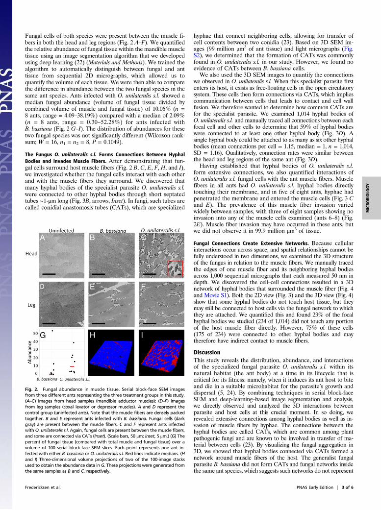
Fungal cells of both species were present between the muscle fi-bers in both the head and leg regions (Fig. 2 A–F). We quantifiedthe relative abundance of fungal tissue within the mandible muscletissue using an image segmentation algorithm that we developedusing deep learning (22) (Materials and Methods). We trained thealgorithm to automatically distinguish between fungal and anttissue from sequential 2D micrographs, which allowed us toquantify the volume of each tissue. We were then able to comparethe difference in abundance between the two fungal species in thesame ant species. Ants infected with O. unilateralis s.l. showed amedian fungal abundance (volume of fungal tissue divided bycombined volume of muscle and fungal tissue) of 10.06% (n =8 ants, range = 4.09–38.19%) compared with a median of 2.09%(n = 8 ants, range = 0.30–52.28%) for ants infected withB. bassiana (Fig. 2 G–I). The distribution of abundances for thesetwo fungal species was not significantly different (Wilcoxon rank-sum; W = 16, n1 = n2 = 8, P = 0.1049).
The Fungus O. unilateralis s.l. Forms Connections Between HyphalBodies and Invades Muscle Fibers. After demonstrating that fun-gal cells surround host muscle fibers (Fig. 2 B, C, E, F, H, and I),we investigated whether the fungal cells interact with each otherand with the muscle fibers they surround. We discovered thatmany hyphal bodies of the specialist parasite O. unilateralis s.l.were connected to other hyphal bodies through short septatedtubes ∼1-μm long (Fig. 3B, arrows, Inset). In fungi, such tubes arecalled conidial anastomosis tubes (CATs), which are specialized
hyphae that connect neighboring cells, allowing for transfer ofcell contents between two conidia (23). Based on 3D SEM im-ages (99 million μm3 of ant tissue) and light micrographs (Fig.S2), we determined that the formation of CATs was commonlyfound in O. unilateralis s.l. in our study. However, we found noevidence of CATs between B. bassiana cells.We also used the 3D SEM images to quantify the connections
we observed in O. unilateralis s.l. When this specialist parasite firstenters its host, it exists as free-floating cells in the open circulatorysystem. These cells then form connections via CATs, which impliescommunication between cells that leads to contact and cell wallfusion. We therefore wanted to determine how common CATs arefor the specialist parasite. We examined 1,014 hyphal bodies ofO. unilateralis s.l. and manually traced all connections between eachfocal cell and other cells to determine that 59% of hyphal bodieswere connected to at least one other hyphal body (Fig. 3D). Asingle hyphal body could be attached to as many as six other hyphalbodies (mean connections per cell = 1.15, median = 1, n = 1,014,SD = 1.16). Qualitatively, connection rates were similar betweenthe head and leg regions of the same ant (Fig. 3D).Having established that hyphal bodies of O. unilateralis s.l.
form extensive connections, we also quantified interactions ofO. unilateralis s.l. fungal cells with the ant muscle fibers. Musclefibers in all ants had O. unilateralis s.l. hyphal bodies directlytouching their membrane, and in five of eight ants, hyphae hadpenetrated the membrane and entered the muscle cells (Fig. 3 Cand E). The prevalence of this muscle fiber invasion variedwidely between samples, with three of eight samples showing noinvasion into any of the muscle cells examined (ants 6–8) (Fig.2E). Muscle fiber invasion may have occurred in these ants, butwe did not observe it in 99.9 million μm3 of tissue.
Fungal Connections Create Extensive Networks. Because cellularinteractions occur across space, and spatial relationships cannot befully understood in two dimensions, we examined the 3D structureof the fungus in relation to the muscle fibers. We manually tracedthe edges of one muscle fiber and its neighboring hyphal bodiesacross 1,000 sequential micrographs that each measured 50 nm indepth. We discovered the cell–cell connections resulted in a 3Dnetwork of hyphal bodies that surrounded the muscle fiber (Fig. 4and Movie S1). Both the 2D view (Fig. 3) and the 3D view (Fig. 4)show that some hyphal bodies do not touch host tissue, but theymay still be connected to host cells via the fungal network to whichthey are attached. We quantified this and found 23% of the focalhyphal bodies we studied (234 of 1,014) did not touch any portionof the host muscle fiber directly. However, 75% of these cells(175 of 234) were connected to other hyphal bodies and maytherefore have indirect contact to muscle fibers.
DiscussionThis study reveals the distribution, abundance, and interactionsof the specialized fungal parasite O. unilateralis s.l. within itsnatural habitat (the ant body) at a time in its lifecycle that iscritical for its fitness: namely, when it induces its ant host to biteand die in a suitable microhabitat for the parasite’s growth anddispersal (5, 24). By combining techniques in serial block-faceSEM and deep-learning–based image segmentation and analysis,we directly observed and analyzed the 3D interactions betweenparasite and host cells at this crucial moment. In so doing, werevealed extensive connections among hyphal bodies as well as in-vasion of muscle fibers by hyphae. The connections between thehyphal bodies are called CATs, which are common among plantpathogenic fungi and are known to be involved in transfer of ma-terial between cells (23). By visualizing the fungal aggregation in3D, we showed that hyphal bodies connected via CATs formed anetwork around muscle fibers of the host. The generalist fungalparasite B. bassiana did not form CATs and fungal networks insidethe same ant species, which suggests such networks do not represent
Head
Leg
O. unilateralis s.l.B. bassianaUninfected
Abun
dance
B. bassiana O. unilateralis s.l.
A B C
D E F
H IG50
40
30
20
10
0
Fig. 2. Fungal abundance in muscle tissue. Serial block-face SEM imagesfrom three different ants representing the three treatment groups in this study.(A–C) Images from head samples (mandible adductor muscles); (D–F) imagesfrom leg samples (coxal levator or depressor muscles). A and D represent thecontrol group (uninfected ants). Note that the muscle fibers are densely packedtogether. B and E represent ants infected with B. bassiana. Fungal cells (darkgray) are present between the muscle fibers. C and F represent ants infectedwithO. unilateralis s.l. Again, fungal cells are present between the muscle fibers,and some are connected via CATs (Inset). (Scale bars, 50 μm; Inset, 5 μm.) (G) Thepercent of fungal tissue (compared with total muscle and fungal tissue) over avolume of 100 serial block-face SEM slices. Each point represents one ant in-fected with either B. bassiana or O. unilateralis s.l. Red lines indicate medians. (Hand I) Three-dimensional volume projections of two of the 100-image stacksused to obtain the abundance data in G. These projections were generated fromthe same samples as B and C, respectively.
Fredericksen et al. PNAS Early Edition | 3 of 6
MICRO
BIOLO
GY

a universal strategy of entomopathogenic fungi (though otherentomopathogenic fungi must be examined with this technique).We suggest forming networks via CATs may be a specializedstrategy that allows O. unilateralis s.l. to exploit its specific habitat(the body of its host) and continue its lifecycle.Our first discovery was the apparent ubiquity of fungal cells
throughout the host but a distinct absence in the brain. To ma-nipulate its host, O. unilateralis s.l. requires precise control ofmandible muscles, which are responsible for the final biting be-havior (13, 14), so we expected fungal cells to be abundant in thehead region (Fig. 2). However, we also expected to find fungalcells in the brain, which is presumably important for manipulationof host behavior. Instead, we found no evidence ofO. unilateralis s.l.fungal cells in ant brains (Fig. 1), which supports suggestions fromprevious work (14). The manipulated biting behavior that typifiesthis system has convergently evolved in another fungus (4) and atrematode (1). The African liver fluke, Dicrocoelium hospes (classTrematoda), enters the ant’s antennal lobes, and the congenericspecies, D. dendriticum, enters the subesophageal ganglion, whereit abuts mandibular nerves (25). In both cases, the ants ascendvegetation where they are eaten by ruminants, allowing the par-asite to reproduce (1). But only with D. dendriticum are the antsinduced to bite plant tissue (grasses), which is the suggested rea-son for the parasite to abut the mandibular nerves (25). TheEntomophthoralean fungus Pandora formicae also induces in-fected ants to ascend and bite vegetation before death, but itsposition relative to the host’s central nervous system during thebiting behavior is unknown. In the O. unilateralis s.l. system, theabsence of fungal cells in the brain implies the biting behaviordoes not require direct contact with the brain. The species ofO. unilateralis s.l. studied here can dynamically alter the secondarymetabolite profile it secretes in the presence of brain versusmuscle tissue, and it also produces unique metabolites when itencounters a brain it normally manipulates compared with a brainfrom an ant species it does not control (11). Thus, the biting be-havior may be driven by chemical means, and more work isneeded to understand the effect of this specialist parasite givenour observation that the brain is not invaded.
Although we found no evidence that O. unilateralis s.l. cellsenter the host brain (Fig. 1), the cells were abundant in the restof the head capsule and in other sections of the ant’s body (Fig.S1), including the legs. This widespread distribution suggests thatthe fungus does not target a particular area that may be espe-cially suitable for inducing the biting behavior. This findingcontrasts with some other behavior-manipulating parasites, suchas Polypocephalus sp., tapeworms, and Dicrocoelium sp., trema-todes, which invade specific regions in the central nervous systemof their hosts (shrimp and ants, respectively) (25, 26). Thenonrandom localization of some parasites inside their hostscould be related to their ability to manipulate behavior (26).
An=60 n=60n=60 n=60n=60 n=60n=60 n=60n=115 n=74n=60 n=60n=60 n=45n=60 n=60
n=30n=29n=30n=30n=30n=30n=30n=30
Head Leg
HB
HB
HB
M
M
M
B
C
D
E Invasions per muscle fiber
0 1 2 3 4 5 6Connec�ons per hyphal body
0 2 4 6 8 10 12 14
***
AntID
12345678
***
AntID
12345678
Fig. 3. Fungal interactions observed in O. unilateralis s.l.-infected ant muscles. (A) Serial block-face SEM image showing fungal hyphal bodies (HB) andhyphae (arrowheads) occupying the spaces between ant mandible muscle fibers (M). Outlined boxes are shown larger in B and C. (Scale bar, 50 μm.)(B) Connections between hyphal bodies (arrows). (Scale bar, 10 μm.) (Inset) Close-up of connected hyphal bodies. (Scale bar, 1 μm.) (C) Muscle fiber invasion:hyphae have penetrated the membrane of this muscle fiber and are embedded within the muscle cell (arrows). (Scale bar, 10 μm.) (D) Connections per hyphalbody across leg and head regions for eight ants. Point sizes are proportional to number of occurrences. “n” refers to number of hyphal bodies sampled in eachant. (E) Invasions per muscle fiber (head only) across eight ants (same ants as in D). “n” refers to the number of muscle fibers examined in each ant. Asterisks(*) denote ants that were dead at the time of collection. Images from A–C were all taken from Ant #1.
Fig. 4. Three-dimensional reconstructions of fungal networks surroundingmuscle fibers. (A) A single fiber of an ant mandible adductor muscle (red) sur-rounded by 25 connected hyphal bodies (yellow). Connections between cells arevisible as short tubes, and many cells have hyphae growing from their ends.Some of these hyphae have grown along and parallel to the muscle fiber (ar-rowhead in Inset). This reconstruction was created using Avizo software. See alsoMovie S1 and interactive 3D pdf (Fig. S3). (B) Two different projections of a 3Dreconstruction showing several muscle fibers (blue) and fungal hyphal bodies(red) from the same area as seen in A. This reconstruction was created using amethod (developed here) that uses a U-Net deep-learning model.
4 of 6 | www.pnas.org/cgi/doi/10.1073/pnas.1711673114 Fredericksen et al.
However, it might also reflect an ability of some parasites, liketapeworms and trematodes, to actively move inside the animal,whereas others, like fungi, are moved around passively by theopen circulatory system.In addition to being distributed throughout the host body,
fungal cells were present in the interstices between muscle fi-bers (Fig. 2). Healthy muscle is normally a tightly packed systemof fibers with no spacing between the fibers (27). Previous workdemonstrated that the mandibular muscles of ants infected withO. unilateralis s.l. exhibited a distinct atrophy that resulted in theformation of such spaces (14). However, it was not known whetherthis muscle atrophy and the high abundance of fungal cells betweenthe muscle fibers were the results of specific adaptations ofO. unilateralis s.l., and thus related to a lifecycle that requires ma-nipulation, or whether these changes were general effects of ento-mopathogens. In the present study, we therefore included thefungus B. bassiana, which does not manipulate behavior, as a pos-itive control. This was a useful addition because it showed that cellsof this generalist fungal pathogen also occupied the spaces betweenmuscle cells. We expected that if fungal abundance and the relatedatrophy of host muscle were major factors in host manipulation, thespecialistO. unilateralis s.l.might have had a higher abundance thanB. bassiana. The finding that both fungal species were present andsimilarly abundant between the fibers implies the formation of theinterstices is likely not a specific adaptation of the specializedbehavior-manipulating fungus. It also suggests a shared mechanismof muscle degradation leading to separation of the fibers. Given thelarge energy reserves that muscles contain (in the form of mito-chondria), both generalist and specialist entomopathogens maymove into areas of high muscle density as part of their strategy togrow and eventually kill the host (both species can only reproducefrom dead insects). Previous work showed a significant reduction inmitochondrial density in muscles of manipulated ants (14). Mito-chondria are considered the power generators of the cells, so themyopathy we observed may be the result of fungal cells consumingthe energy reserves of the muscle fibers. The genus Ophiocordycepsinfects insects in 10 orders (including Hymenoptera, to which theants belong) (18, 24), and it would be instructive to study otherOphiocordyceps species as well as generalist fungal parasites to de-termine their effect on muscles of different insect hosts.Not only did fungal cells enter muscle bundles, they also pene-
trated the muscle fibers themselves. Since the first step in colonizingthe host body is the hyphal body stage, the appearance of the cell-invading mycelial phenotype implies a transition during which hy-phae grow from one or both poles of the hyphal body (Fig. 2A,arrowheads). In fungi, the mycelium is the vegetative growth stageinvolved in foraging, resource acquisition, and accessing new envi-ronments. Typified by the secretion of enzymes at the polar regionand subsequent cell wall synthesis localized to a surface of just a fewmicrometers, growing hyphae can penetrate substances that hyphalbodies cannot (28). An extreme example of such activity occurs inrock-eating fungi (29), but fungi routinely degrade cellulose andother recalcitrant biological material (30). We suggest the transitionto apical growth from hyphal bodies allows the fungal community toaccess resources inside muscle cells, but other functions are alsopossible. The penetration of muscle cells by hyphae also occurred,albeit rarely, in ants infected by the generalist parasite, B. bassiana,so muscle cell invasion may represent a general strategy that futurework should explore.A fungal characteristic that we only observed in the specialist
pathogen O. unilateralis s.l. was the formation of complex 3Dnetworks surrounding the muscle fibers (Fig. 4, Fig. S3, and MovieS1). The networks consisted of many hyphal bodies joined byconnections between the cell walls. The connections are known asCATs, which have been observed in 73 species (21 genera) offungi (23). These fungal species in which CATs have been de-scribed are pathogens of plants, and here we show similar con-nections in a fungus infecting an animal. One possible function of
the connections and network formation is suggested by our 3Dreconstruction of 25 connected hyphal bodies surrounding a singlemuscle fiber (Fig. 4, Fig. S3, and Movie S1). Visualizing the net-work in this way allowed us to observe its size and shape. Wedetermined that many hyphal bodies (23%, 234 of 1,014) did notdirectly touch the muscle fiber, but through their connections inthe network they could still interact indirectly with the muscle fi-ber. One suggestion from these observations is that hyphae se-quester resources from the muscles, and the connections betweenhyphal bodies allow nutrients to be transported to cells not directlytouching the host. Studies on ectomycorrhizal fungal networksshow that carbon can be transferred from one tree to anotheralong extensive underground networks (31). Therefore, hyphalbody networks could allow the fungus to acquire resources frommuscles and distribute these resources to other fungal cells thatare in the network but distant from the muscles.In summary, we have shown, through the use of serial block-
face SEM and automated image segmentation and analysis basedon a deep-learning model, the 3D structure of a microbial parasiteat the cellular level inside the body of its host. We demonstratedthat during the critical biting behavior of manipulated ants, thespecialist fungus O. unilateralis s.l. is present throughout the ant’sbody but not in the brain. The fungus takes the form of hyphalbodies that connect to form fungal networks, and it is also presentas filamentous mycelia that invade ant muscle fibers. The con-nections between hyphal bodies indicate that individual fungalcells communicate with each other and suggest that collectivebehavior may be an important strategy for this fungal parasite. Wespeculate that the resulting networks may aid in nutrient transportand formation of the structures that will later emerge from thehost’s body to continue the parasite’s lifecycle. Future work shouldcontinue to examine fungal behavior at the cellular level over theperiod of infection and across multiple areas within the ant’s body.
Materials and MethodsInfection and Sample Collection. Ant and fungal colonies were maintained inthe laboratory following procedures detailed in SI Materials and Methods.Colonies of the ant Camponotus castaneus were collected in AbbevilleCounty, South Carolina (georeference: 34.375215, −82.346937) in July 2014.Cultures of O. unilateralis s.l. (Strain SC09B, also collected in AbbevilleCounty) were grown from ascospores (sexually produced spores of Asco-mycete fungi) since July 2014. Manipulated ants were collected following ar-tificial infection with fungal material as described previously (11). Briefly, antswere injected under one foreleg with 1 μL of a fungal suspension (either freshascospores at 1 × 10⁵ spores per milliliter or 1 cm2 of cultured fungal materialsuspended in 500 μL of Grace’s medium) and kept at high humidity with accessto 10% sugar water ad libitum until collection (15–39 d after infection). It isunknown how many spores enter the ant during a natural infection andwhether those spores are clonal or not. While our injection method may resultin a different disease progression than what occurs in nature, the relevantoutcome for our study is the induced biting behavior in the host, and thisoccurs both in the field and in the laboratory. In total, 23 manipulated ants(8 for histology and SEM, 3 for immunofluorescence, 12 for leg dissections)were collected from three rounds of infection. Collected ants displayed acharacteristic biting behavior: they had been biting onto one of severalavailable substrates in the cage for at least 10 min (no more than 12 h) at thetime of collection. Control ants were not injected with fungus but werehoused with infected ants and exposed to the same laboratory conditions.Ants infected with B. bassianawere surface-infected with 2 μL or 3 μL of sporesuspension (108 or 109 spores per milliliter). Eight infected ants from twocolonies were collected between 3 and 5 d after infection when ants weremoribund (32) or within 1 h after death. Collected ants were either dissectedimmediately and fixed (for EM: 2.5% glutaraldehyde, 2% formaldehyde,2 mM calcium chloride in 0.15 M cacodylate buffer, pH 7.4; for immunohis-tochemistry: 4% paraformaldehyde in PBS) or flash-frozen in liquid nitrogenand stored at −80 °C to be thawed, dissected, and fixed at a later date.
Immunofluorescence of Neural Tissue. Immunofluorescence assays were per-formed similar to previously described work (33, 34). Briefly, fixed ant headswere washed in PBS, cryoprotected (10/15/20% sucrose in PBS for 2 h each),and embedded in O.C.T. Heads were cut into 50-μM sections along the
Fredericksen et al. PNAS Early Edition | 5 of 6
MICRO
BIOLO
GY

transverse plane using a cryostat (Leica Biosystems), and mounted slides werepostfixed briefly in 2% paraformaldehyde. Sections were rinsed with PBS,blocked and permeabilized in 10% donkey serum (Jackson ImmunoResearch)diluted in 0.1% Triton X-100/PBS. Slides were then incubated with primaryantibody (3C11 anti-SYNORF1, 1:50, DSHB) overnight at 4 °C, washed, andthen incubated in secondary antibody [Alexa Fluor 488 AffiniPure F(ab′)2fragment donkey anti-mouse IgG (H+L), 1:200; Jackson ImmunoResearch] andcalcofluor white antichitin stain (1:100; Sigma-Aldrich). Sections were thenwashed and mounted in ProLong Gold (ThermoFisher Scientific) and imagedusing a confocal laser scanning microscope (Olympus FluoView FV10i) and a60× water immersion objective. All images are presented as maximum pro-jections of z-stacks. Background subtraction was performed using the rollingball method (radius = 50) in ImageJ. Brightness and contrast were adjustedequally for all images using PhotoShop (Adobe).
Serial Block-Face SEM. The right half of the head and one forecoxa from eachant were prepared for serial block-face SEM according to the protocol of theNational Center for Microscopy and Imaging Research, University of Cal-ifornia, San Diego (35). Pin-mounted samples were sectioned using a serialblock-face SEM (Gatan 3View on Zeiss SIGMA VP-FESEM) at 50- or 100-nmthickness. Approximately 120 μm of tissue was sliced (1,200 or 2,400 slices)from each O. unilateralis s.l. sample and 10 μm (100 slices) from B. bassianaand control samples. Digital Micrograph (Gatan) software was used to alignimage stacks and quantify interactions between cells (for details of quanti-fication, see SI Materials and Methods). Image segmentation and 3D re-construction were performed using a deep-learning model (see below) aswell as the programs Avizo and Amira (FEI). Model smoothing and 3D pdfgeneration were performed with help from Thomas van de Kamp, KarlsruheInstitute of Technology, Karlsruhe, Germany, as described previously (36).
Deep-Learning Model for Image Segmentation. The automated image seg-mentation used to compute the relative abundance of fungus and muscletissue in each sample was performed using amodel developed by Zhang et al.(22). To perform quantitative analysis, our first step was to segment theimage data. In recent years, fully convolutional networks have shown highly
promising results in semantic segmentation (37). Our segmentation problem canbe viewed as a special case of general semantic segmentation. Compared withnatural scene images, the number of classes in our images is much smaller, butthe size of our training data is quite limited. Thus, our approach must be dif-ferent from the original fully convolutional networks, which were designed forgeneral computer vision tasks. U-Net (38) is a specially designed fully convolu-tional network for segmenting biomedical image data, and we applied thismodel for automatic fungus and muscle segmentation. For each stack, wemanually labeled two sections to train the model and then applied the trainedmodel to the remaining sections. We stacked the segmentation results for allsections to form the 3D segmentation. To calculate the segmentation accuracy,we compared the model results with ground truth data (one section from eachstack marked by a human expert). For simpler stacks, the F1 score is over 96%,and for harder stacks, the F1 score is over 93% (voxel level). Based on the seg-mentation, we used a computer program to count voxels of fungi, muscles, andother areas, and we calculated the volume ratios for each pair accordingly.
Data generated in this study are available on Pennsylvania State UniversityScholarSphere (https://scholarsphere.psu.edu/concern/generic_works/6q524jn254).
ACKNOWLEDGMENTS. We thank Kim Fleming for inviting us to collect antsand fungus on her land; Nina Jenkins (Pennsylvania State University) forproviding spores of Beauveria Bassiana; Greg Ning and John Cantolina (Pennsyl-vania State University Microscopy and Cytometry Facility) for assistance withmicroscopy and sample preparation; undergraduate assistant Saad Ahmad(Pennsylvania State University) for digitizing histology slides; and the Huck Insti-tutes of the Life Sciences for microscopy facilities and logistical support. TheSYNORF1 antibody, developed by Erich Buchner, was obtained from the Devel-opmental Studies Hybridoma Bank, created by the National Institute of ChildHealth and Human Development of the NIH and maintained at the University ofIowa, Department of Biology. This work was supported in part by NationalScience Foundation Grants IOS-1558062 (to D.P.H.), CCF-1217906 (to D.Z.C.),CNS-1629914 (to D.Z.C.), and CCF-1617735 (to D.Z.C.); and NIH Grant R01GM116927-02 (to D.P.H., D.C.Z., M.A.F., and Y.Z.). R.G.L. was funded by Comissaode Aperfeiçoamento de Pessoal de Nival Superior-Brazil (Project 6203-10-8).C.A.M. was funded by the American Heart Association (16POST29920001).
1. Moore J (2002) Parasites and the Behavior of Animals (Oxford Univ Press, Oxford, UK).2. Van Den Abbeele J, Caljon G, De Ridder K, De Baetselier P, Coosemans M (2010)
Trypanosoma brucei modifies the tsetse salivary composition, altering the fly feedingbehavior that favors parasite transmission. PLoS Pathog 6:e1000926.
3. Marikovsky PI (1962) On some features of behavior of the ants Formica rufa L. in-fected with fungus disease. Insectes Soc 9:173–179.
4. Małagocka J, Grell MN, Lange L, Eilenberg J, Jensen AB (2015) Transcriptome of anentomophthoralean fungus (Pandora formicae) shows molecular machinery adjustedfor successful host exploitation and transmission. J Invertebr Pathol 128:47–56.
5. Andersen SB, et al. (2009) The life of a dead ant: The expression of an adaptive ex-tended phenotype. Am Nat 174:424–433.
6. Berdoy M, Webster JP, Macdonald DW (2000) Fatal attraction in rats infected withToxoplasma gondii. Proc Biol Sci 267:1591–1594.
7. Dawkins R (1982) The Extended Phenotype (Oxford Univ Press, Oxford, UK).8. Dubey JP, Lindsay DS, Speer CA (1998) Structures of Toxoplasma gondii tachyzoites,
bradyzoites, and sporozoites and biology and development of tissue cysts. ClinMicrobiol Rev 11:267–299.
9. Roth G, Dicke U (2005) Evolution of the brain and intelligence. Trends Cogn Sci 9:250–257.
10. Prandovszky E, et al. (2011) The neurotropic parasite Toxoplasma gondii increasesdopamine metabolism. PLoS One 6:e23866.
11. de Bekker C, et al. (2014) Species-specific ant brain manipulation by a specializedfungal parasite. BMC Evol Biol 14:166.
12. Charnley AK (1989) Mechanisms of fungal pathogenesis in insects. Biotechnology ofFungi for Improving Plant Growth, eds Whipps JM, Lumsen RD (Cambridge Univ Press,Cambridge, UK), pp 85–125.
13. de Bekker C, et al. (2015) Gene expression during zombie ant biting behavior reflects thecomplexity underlying fungal parasitic behavioral manipulation. BMC Genomics 16:620.
14. Hughes DP, et al. (2011) Behavioral mechanisms and morphological symptoms ofzombie ants dying from fungal infection. BMC Ecol 11:13.
15. de Bekker C, Smith PB, Patterson AD, Hughes DP (2013) Metabolomics reveals theheterogeneous secretome of two entomopathogenic fungi to ex vivo cultured insecttissues. PLoS One 8:e70609.
16. Roy HE, Steinkraus DC, Eilenberg J, Hajek AE, Pell JK (2006) Bizarre interactions and end-games: Entomopathogenic fungi and their arthropod hosts. Annu Rev Entomol 51:331–357.
17. Humber RA (2008) Evolution of entomopathogenicity in fungi. J Invertebr Pathol 98:262–266.
18. Araújo JPM, Hughes DP (2016) Diversity of entomopathogenic fungi: Which groupsconquered the insect body? Adv Genet 94:1–39.
19. Prasertphon S, Tanada Y (1968) Formation and circulation, in Galleria, of hyphalbodies of entomophthoraceous fungi. J Invertebr Pathol 11:260–280.
20. Kirk P, Cannon P, Minter D, Stalpers J (2008) Ainsworth & Bisby’s Dictionary of theFungi (CAB International, Wallingford, UK), 10th Ed.
21. Boucias DG, Pendland JC (1998) General Properties of fungal pathogens. Principles of
Insect Pathology (Springer, New York), pp 259–286.22. Zhang Y, et al. (2017) Deep adversarial networks for biomedical image segmentation
utilizing unannotated images. Medical Image Computing and Computer Assisted
Intervention (Springer, Cham, Switzerland), pp 408–416.23. Gabriela Roca M, Read ND, Wheals AE (2005) Conidial anastomosis tubes in fila-
mentous fungi. FEMS Microbiol Lett 249:191–198.24. Hughes DP, et al. (2016) From so simple a beginning: The evolution of behavioral
manipulation by fungi. Adv Genet 94:437–469.25. Romig T, Lucius R, Frank W (1980) Cerebral larvae in the second intermediate host of
Dicrocoelium dendriticum (Rudolphi, 1819) and Dicrocoelium hospes Looss, 1907
(Trematodes, Dicrocoeliidae). Z Parasitenkd 63:277–286.26. Carreon N, Faulkes Z (2014) Position of larval tapeworms, Polypocephalus sp., in the
ganglia of shrimp, Litopenaeus setiferus. Integr Comp Biol 54:143–148.27. Paul J (2001) Mandible movements in ants. Comp Biochem Physiol A Mol Integr
Physiol 131:7–20.28. Read ND (2011) Exocytosis and growth do not occur only at hyphal tips.Mol Microbiol
81:4–7.29. Jongmans AG, et al. (1997) Rock-eating fungi. Nature 389:682–683.30. Cragg SM, et al. (2015) Lignocellulose degradation mechanisms across the Tree of
Life. Curr Opin Chem Biol 29:108–119.31. Klein T, Siegwolf RTW, Körner C (2016) Belowground carbon trade among tall trees in
a temperate forest. Science 352:342–344.32. Heinze J, Walter B (2010) Moribund ants leave their nests to die in social isolation.
Curr Biol 20:249–252.33. Gao Q, Yuan B, Chess A (2000) Convergent projections of Drosophila olfactory neu-
rons to specific glomeruli in the antennal lobe. Nat Neurosci 3:780–785.34. Vanguilder HD, Bixler GV, Sonntag WE, Freeman WM (2012) Hippocampal expression
of myelin-associated inhibitors is induced with age-related cognitive decline and
correlates with deficits of spatial learning and memory. J Neurochem 121:77–98.35. Deerinck T, Bushong E, Thor A, Ellisman M (2010) NCMIR Methods for 3D EM: A New
Protocol for Preparation of Biological Specimens for Serial Block Face Scanning
Electron Microscopy (National Center for Microscopy and Imaging Research, La Jolla),
Version 7_01_10.36. van de Kamp T, dos Santos Rolo T, Vagovic P, Baumbach T, Riedel A (2014) Three-
dimensional reconstructions come to life—Interactive 3D PDF animations in func-
tional morphology. PLoS One 9:e102355.37. Shelhamer E, Long J, Darrell T (2017) Fully convolutional networks for semantic
segmentation. IEEE Trans Pattern Anal Mach Intell 39:640–651.38. Ronneberger O, Fischer P, Brox T (2015) U-Net: Convolutional networks for bio-
medical image segmentation. MICCAI (Springer, Cham, Switzerland), pp 234–241.
6 of 6 | www.pnas.org/cgi/doi/10.1073/pnas.1711673114 Fredericksen et al.
Related Documents