Three-dimensional finite element thermal analysis of dental tissues irradiated with Er,Cr:YSGG laser Patricia Aparecida Ana, 1 Walter F. Velloso, Jr., 2 and Denise Maria Zezell 1 1 Centro de Lasers e Aplicações, Instituto de Pesquisas Energéticas e Nucleares, Universidade de São Paulo São Paulo 05508-000, Brazil 2 Faculdade de Zootecnia e Engenharia de Alimentos de Pirassununga, Universidade de São Paulo, Pirassununga CEP 13635-900, Brazil Received 24 April 2008; accepted 9 June 2008; published online 29 September 2008 In the present study, a finite element model of a half-sectioned molar tooth was developed in order to understand the thermal behavior of dental hard tissues both enamel and dentin under laser irradiation. The model was validated by comparing it with an in vitro experiment where a sound molar tooth was irradiated by an Er,Cr:YSGG pulsed laser. The numerical tooth model was conceived to simulate the in vitro experiment, reproducing the dimensions and physical conditions of the typical molar sound tooth, considering laser energy absorption and calculating the heat transfer through the dental tissues in three dimensions. The numerical assay considered the same three laser energy densities at the same wavelength 2.79 m used in the experiment. A thermographic camera was used to perform the in vitro experiment, in which an Er,Cr:YSGG laser 2.79 m was used to irradiate tooth samples and the infrared images obtained were stored and analyzed. The temperature increments in both the finite element model and the in vitro experiment were compared. The distribution of temperature inside the tooth versus time plotted for two critical points showed a relatively good agreement between the results of the experiment and model. The three dimensional model allows one to understand how the heat propagates through the dentin and enamel and to relate the amount of energy applied, width of the laser pulses, and temperature inside the tooth. © 2008 American Institute of Physics. DOI: 10.1063/1.2953526 I. INTRODUCTION Nowadays there are wide ranges of dentistry procedures that include laser applications. Lasers can be used for oral surgery, to remove both tooth sound and decayed tooth tis- sues, enhance tooth bleaching, and sometimes to cure restor- ative materials. 1,2 Depending on the case, the dentist needs different laser wavelengths and parameters, in order to use the laser continually during a period, only to irradiate the tooth with sharp pulses of light. 3 The interactions of high-intensity laser irradiation with dental hard tissues are commonly induced by thermal action. For the clinical application of lasers, a precise irradiation parameter must be chosen in order to avoid morphological damage, such as enamel surface carbonization or cracking, which could produce structural, esthetic damages, and postoperative complaints. 4 Moreover, the energy densities used must be safe as regards pulp and periodontal tissue vitality. Therefore, several in vitro experiments have been conducted to evaluate the temperature rise in teeth in order to assure safe conditions for the use of lasers. Thermocouples, 5 elliptical mirror and HgCdZnTe detectors, 6 and infrared cameras 7 are widely used to measure temperature rises in pulp, periodontal tissues, and enamel surfaces. However, these experimental methods present some difficulties, such as sample standardization considering the large variation in volume, size, and thickness of teeth, the exact duplication of the thermal load, accuracy, and cost of equipment. The finite element method model FEM model seems to be a good analytical tool to model and simulate the ther- mal or mechanical behavior of dental structures. 8–11 In the past two decades, this mathematical model has received increasing attention in dental biomechanical studies, 8 analyzing mechanical stress, 9 thermal stress, 10 or thermal conductivity. 11 The FEM model shown in the present study was used to simulate the effects of laser on enamel and dentin, but not on gums or inside the pulp cavity, which is filled with blood vessels and innervated tissues, since these materials are soft and highly inhomogeneous. However, the effects of laser ir- radiation could be difficult to simulate even on the dental hard tissues only enamel and dentine, since the thermal characteristics of these materials may not have been well determined. Indeed, the thermal characteristics used in the models were obtained from the technical literature, but as there was considerable uncertainty about the physical prop- erties of natural tissues, the numerical data were compared to experimental data obtained from a fast infrared thermal camera. Therefore, the aims of this study were to simulate the heat transfer inside the dental hard tissues when irradiated with an Er:Cr:YSGG laser, and to compare the results of this simulation with the temperature measurements recorded by an infrared thermal camera in the tooth. The results of this study could be useful for predicting the physical and chemi- cal changes in the enamel after laser irradiation, with the purpose of caries prevention. REVIEW OF SCIENTIFIC INSTRUMENTS 79, 093910 2008 0034-6748/2008/799/093910/9/$23.00 © 2008 American Institute of Physics 79, 093910-1 Downloaded 29 Oct 2008 to 200.136.52.139. Redistribution subject to AIP license or copyright; see http://rsi.aip.org/rsi/copyright.jsp

Welcome message from author
This document is posted to help you gain knowledge. Please leave a comment to let me know what you think about it! Share it to your friends and learn new things together.
Transcript

Three-dimensional finite element thermal analysis of dental tissuesirradiated with Er,Cr:YSGG laser
Patricia Aparecida Ana,1 Walter F. Velloso, Jr.,2 and Denise Maria Zezell11Centro de Lasers e Aplicações, Instituto de Pesquisas Energéticas e Nucleares,Universidade de São Paulo São Paulo 05508-000, Brazil2Faculdade de Zootecnia e Engenharia de Alimentos de Pirassununga,Universidade de São Paulo, Pirassununga CEP 13635-900, Brazil
�Received 24 April 2008; accepted 9 June 2008; published online 29 September 2008�
In the present study, a finite element model of a half-sectioned molar tooth was developed in orderto understand the thermal behavior of dental hard tissues �both enamel and dentin� under laserirradiation. The model was validated by comparing it with an in vitro experiment where a soundmolar tooth was irradiated by an Er,Cr:YSGG pulsed laser. The numerical tooth model wasconceived to simulate the in vitro experiment, reproducing the dimensions and physical conditionsof the typical molar sound tooth, considering laser energy absorption and calculating the heattransfer through the dental tissues in three dimensions. The numerical assay considered the samethree laser energy densities at the same wavelength �2.79 �m� used in the experiment. Athermographic camera was used to perform the in vitro experiment, in which an Er,Cr:YSGG laser�2.79 �m� was used to irradiate tooth samples and the infrared images obtained were stored andanalyzed. The temperature increments in both the finite element model and the in vitro experimentwere compared. The distribution of temperature inside the tooth versus time plotted for two criticalpoints showed a relatively good agreement between the results of the experiment and model. Thethree dimensional model allows one to understand how the heat propagates through the dentin andenamel and to relate the amount of energy applied, width of the laser pulses, and temperature insidethe tooth. © 2008 American Institute of Physics. �DOI: 10.1063/1.2953526�
I. INTRODUCTION
Nowadays there are wide ranges of dentistry proceduresthat include laser applications. Lasers can be used for oralsurgery, to remove both tooth sound and decayed tooth tis-sues, enhance tooth bleaching, and sometimes to cure restor-ative materials.1,2 Depending on the case, the dentist needsdifferent laser wavelengths and parameters, in order to usethe laser continually during a period, only to irradiate thetooth with sharp pulses of light.3
The interactions of high-intensity laser irradiation withdental hard tissues are commonly induced by thermal action.For the clinical application of lasers, a precise irradiationparameter must be chosen in order to avoid morphologicaldamage, such as enamel surface carbonization or cracking,which could produce structural, esthetic damages, andpostoperative complaints.4 Moreover, the energy densitiesused must be safe as regards pulp and periodontal tissuevitality. Therefore, several in vitro experiments have beenconducted to evaluate the temperature rise in teeth in order toassure safe conditions for the use of lasers. Thermocouples,5
elliptical mirror and HgCdZnTe detectors,6 and infraredcameras7 are widely used to measure temperature rises inpulp, periodontal tissues, and enamel surfaces. However,these experimental methods present some difficulties, suchas sample standardization �considering the large variation involume, size, and thickness of teeth�, the exact duplication ofthe thermal load, accuracy, and cost of equipment.
The finite element method model �FEM model� seems
to be a good analytical tool to model and simulate the ther-mal or mechanical behavior of dental structures.8–11 In thepast two decades, this mathematical model has receivedincreasing attention in dental biomechanical studies,8
analyzing mechanical stress,9 thermal stress,10 or thermalconductivity.11
The FEM model shown in the present study was used tosimulate the effects of laser on enamel and dentin, but not ongums or inside the pulp cavity, which is filled with bloodvessels and innervated tissues, since these materials are softand highly inhomogeneous. However, the effects of laser ir-radiation could be difficult to simulate even on the dentalhard tissues �only enamel and dentine�, since the thermalcharacteristics of these materials may not have been welldetermined. Indeed, the thermal characteristics used in themodels were obtained from the technical literature, but asthere was considerable uncertainty about the physical prop-erties of natural tissues, the numerical data were comparedto experimental data obtained from a fast infrared thermalcamera.
Therefore, the aims of this study were to simulate theheat transfer inside the dental hard tissues when irradiatedwith an Er:Cr:YSGG laser, and to compare the results of thissimulation with the temperature measurements recorded byan infrared thermal camera in the tooth. The results of thisstudy could be useful for predicting the physical and chemi-cal changes in the enamel after laser irradiation, with thepurpose of caries prevention.
REVIEW OF SCIENTIFIC INSTRUMENTS 79, 093910 �2008�
0034-6748/2008/79�9�/093910/9/$23.00 © 2008 American Institute of Physics79, 093910-1
Downloaded 29 Oct 2008 to 200.136.52.139. Redistribution subject to AIP license or copyright; see http://rsi.aip.org/rsi/copyright.jsp
II. MATERIALS AND METHODS
A. In vitro experiment
After approval by the Human Research Ethics Commit-tee of the Energy and Nuclear Research Institute �Proj. 094/2004 CEP-IPEN�, University of Sao Paulo, 12 freshly ex-tracted human third molar teeth were selected. Each toothwas cleaned and half-sectioned longitudinally with a disk-shaped diamond saw �Isomet, Buehler, IL� under cooling.This sectioning allowed the heat transfer from enamelsurface to the pulp chamber during laser irradiation to bevisualized.
At the time of experiment, each sample was immobilizedin optical supports with the sectioned side turned toward thelens of a thermographic camera �ThermaCam FLIR SC3000Systems, USA�, which stores infrared images and data atrates of up to 900 Hz. This experiment was performed at aroom temperature of 24.6 °C, 47% air relative humidity andconsidering tooth emissivity as 0.91. The infrared imagesobtained were recorded at rates of 900 Hz. The thermo-graphic camera was placed at a distance of 0.1 m in front ofsamples, and the area of interest was isolated at a focallength of 0.1 m using an internal macrolens. This apparatusmade it possible to record the heat propagation from enamelsurface through dentin to the pulp chamber, enabling thetemperature increase in dental tissues to be evaluated at theexact moment of laser irradiation.
Laser irradiation was performed using an Er,Cr:YSGGlaser �Millenium, Biolase Inc., San Clemente, CA, USA�,with 2.79 �m wavelength, 140 �s pulse width, 20 Hz rep-etition rate, and a power output ranging from 0 to 6 W. Theenergy was delivered through a 430 �m spot size fiber opticsystem ending in a sapphire terminal of 750 �m diameterand 6 mm long �S75 tip�, which resulted in 600 �m beamdiameter at 1 mm distance from the irradiated surface,bathed in an adjustable air and water spray. Irradiation wasperformed perpendicularly to the occlusal surface of thesamples with the fiber end at 1 mm distance from this sur-face, tangentially to the sectioned plane of the samples. Laser
irradiation was performed in a single point for a standardizedtime of 5 s, without air-water mist.
Samples were divided into six groups �n=2� accordingto the laser fluence and with or without the presence of aphotoabsorber, as follows: Group 1: irradiation at 2.8 J /cm2
with application of photoabsorber; Group 2: irradiation at2.8 J /cm2 without application of photoabsorber; Group 3:irradiation at 5.6 J /cm2 with application of photoabsorber;Group 4: irradiation at 5.6 J /cm2 without application of pho-toabsorber; Group 5: irradiation at 8.5 J /cm2 with applica-tion of photoabsorber; and Group 6: irradiation at 8.5 J /cm2
without application of photoabsorber. This photoabsorberconsisted of a triturated coal paste ��10 �m diameter par-ticles� diluted in 50% water and 50% alcohol, which wasapplied on the occlusal surface of samples with a No. 1 brushbefore laser irradiation, in order to increase the absorption ofthe laser beam at the enamel surface. The irradiation condi-tions and total energy are described in Table I.
The beginning of laser irradiation and temperature re-cording was synchronized but the thermal measuring finished15 s after the end of the laser irradiation. For later analysis oftemperature increments, two standardized points were cho-sen on the infrared images obtained: one located at theenamel surface immediately below the laser tip �spot 1� andthe other located at dentin-pulp chamber limit �spot 2� at anapproximate distance of 3.5 mm below spot 1.12 Data wereanalyzed by ORIGIN PRO software.
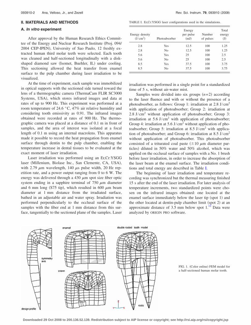
FIG. 1. �Color online� FEM model fora half-sectioned human molar tooth.
TABLE I. Er,Cr:YSGG laser configurations used in the simulations.
Energy density�J /cm2� Photoabsorber
Energyper pulse
�mJ�Numberof pulses
Totalenergy
�J�
2.8 Yes 12.5 100 1.252.8 No 12.5 100 1.255.6 Yes 25 100 2.55.6 No 25 100 2.58.5 Yes 37.5 100 3.758.5 No 37.5 100 3.75
093910-2 Ana, Velloso, Jr., and Zezell Rev. Sci. Instrum. 79, 093910 �2008�
Downloaded 29 Oct 2008 to 200.136.52.139. Redistribution subject to AIP license or copyright; see http://rsi.aip.org/rsi/copyright.jsp

B. Computer calculation
A geometric FEM model of a half-sectioned doublerooted molar tooth was constructed, as shown in Fig. 1. Inorder to obtain the geometric model, a typical profile of atooth root was used, i.e., planar elements were designed to fitthe profile and then a three-dimensional root structure wascreated revolving these planar elements around the verticalaxis. Both roots are symmetrical with respect to the toothaxis and the geometric shape of the crown was also designedas axially symmetric. This is an acceptable approximationsince the model was conceived only to be used in simula-tions of the thermal behavior of dental tissues. Stress andother static and dynamic characteristics that depend on theexact shape of the body under analysis are not objects ofstudy in this work.
The dimensions of the model tooth are typical, corre-sponding to a total volume of 0.6 cm3 and total weightof 1.47 g. The enamel �r=2.8 g /cm3� and the dentin�r=1.96 g /cm3� are both represented in Fig. 1.
1. The thermal equations
Since the goal was to calculate the temperature distribu-tion on the surface and inside the tooth, the FEM model wasused to set up the equations of heat transfer through dentaltissues.
In each element the total heat is given by the internalheat, determined by the material density ��� and specific heat�c�, and the heat flux, determined by the element materialthermal conductivity �k�. The power density Qi ��J /m3 s��corresponds to the heat Hi absorbed or lost by each elementin each second is
Qi =�Hi
�t= �c
dT�r�,t��t
. �1�
The heat flux ��J /m2 s�� in direction r� is
Fi = k�T�r�,t�
�r= k · �� T�r�,t� . �2�
Finally, the laser beam can be considered as an externalenergy source, represented by the function Qext, which, ineach second �s�, adds an amount of heat by volume �m3� toeach irradiated element �Qext �J /m3 s��.
Therefore, considering that the divergence of flux �� ·F=k ·�2T�r� , t� ��J /m3 s�� corresponds to an amount of heat,lost or acquired, in each element, the energy balance in thetooth model can be represented by a matrix differential equa-tion �the classical Poisson equation for the heat propagationalso considering the laser energy as an external source�, in-volving all the elements of the model.
�Qi� = � · �Fi� + �Qext� , �3�
�c�T�r�,t�
�t= k · �2T�r�,t� + Qext. �4�
TABLE II. Physical characteristics of dental hard tissues.
Physical characteristicEnamel�typical�
Dentin�typical�
Specific heat �cal /g °C� 0.17 0.38Thermal conductivity �cal /s cm °C� 2.33�10−3 1.36�10−3
Density �g /cm3� 2.80 1.96
FIG. 2. The dependence between theuncertainty in specific heat and theuncertainty in maximum temperaturereached.
093910-3 3D-finite element analysis of teeth Rev. Sci. Instrum. 79, 093910 �2008�
Downloaded 29 Oct 2008 to 200.136.52.139. Redistribution subject to AIP license or copyright; see http://rsi.aip.org/rsi/copyright.jsp
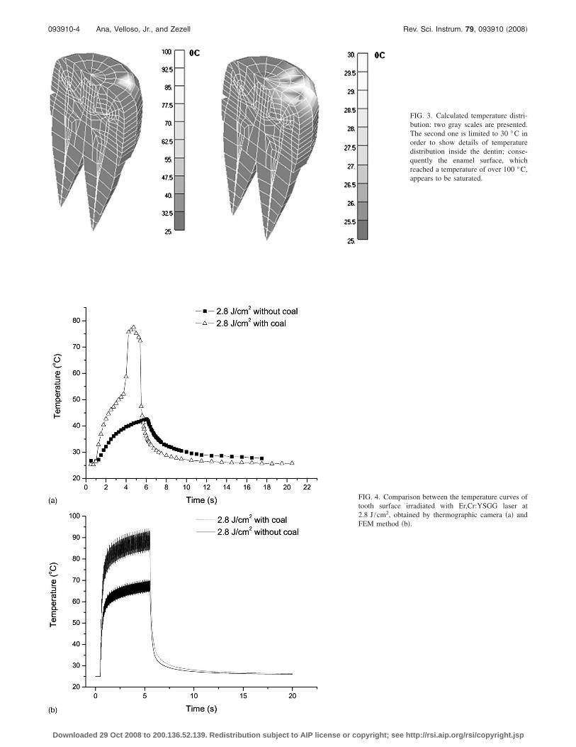
FIG. 3. Calculated temperature distri-bution: two gray scales are presented.The second one is limited to 30 °C inorder to show details of temperaturedistribution inside the dentin; conse-quently the enamel surface, whichreached a temperature of over 100 °C,appears to be saturated.
FIG. 4. Comparison between the temperature curves oftooth surface irradiated with Er,Cr:YSGG laser at2.8 J /cm2, obtained by thermographic camera �a� andFEM method �b�.
093910-4 Ana, Velloso, Jr., and Zezell Rev. Sci. Instrum. 79, 093910 �2008�
Downloaded 29 Oct 2008 to 200.136.52.139. Redistribution subject to AIP license or copyright; see http://rsi.aip.org/rsi/copyright.jsp

Initial and boundary conditions are necessary in order tosolve these differential equations. These conditions are givenby the supposition that at t=0, all the elements haveT=25 °C and that the surrounding ambient has constant tem-perature T=25 °C. Therefore, as the laser beam begins toadd energy to the dental material, the temperatures rise andthe elements on external surface begin to irradiate, followingthe classical Stefan’s law for the emission. These equationswere solved using an educational version of the softwareNASTRAN FEM SOLVER �Santa Ana, CA, USA�.
This model is used based on the fact that the opticalheating source was considered purely superficial, as thetemperature exhibits no dependence on thermal diffusivity.The relaxation time �B�1 /�2�2 �Mandelis and Royce�13
was calculated as 7.9 �s, which is much smaller than thelaser pulse �140 �s�. The absorption coefficient was consid-ered 7000 cm−1 �Stock et al.�14 and � as 2.04�10−3 cm2 /s�Minesaky�.15
2. The model analysis
In Table II some of the elastic and thermal characteristicsof both enamel and dentin used in the numerical essay are
listed as given in some previous studies.16–19 There is con-siderable uncertainty involving these values since they referto biological tissues. Thus, the values in the table must beconsidered as typical.20
In order to estimate the effect of this uncertainty on theresults of FEM models, a large number of calculations weremade, changing the physical characteristics of dental materi-als with regard to their typical values. In a band of 10%around the typical value, ten values were considered. Sincethere are three physical constants interfering in the thermalbehavior �specific heat, conductivity, and density for two ma-terials�, 2000 combinations �2�10�10�10� were ob-tained. Using a random numerical process, 50 of thesecombinations were drawn to run the FEM model. Consider-ing, for example, the maximum temperature reached,the results formed a type of cluster around the mean value.The standard deviation of these values gives an idea aboutthe uncertainty of FEM calculations. Figure 2 showsthe results of this analysis with respect to specific heat.Similar results were obtained for all physical characteristicsused. Based on these results it was considered that the
FIG. 5. Comparison between the temperature curves oftooth surface irradiated with Er,Cr:YSGG laser at5.6 J /cm2, obtained by thermographic camera �a� andFEM method �b�. Inside the circle: detail of temperaturecurve showing the surface temperature change accord-ing to the laser pulses, which agrees with FEM data.
093910-5 3D-finite element analysis of teeth Rev. Sci. Instrum. 79, 093910 �2008�
Downloaded 29 Oct 2008 to 200.136.52.139. Redistribution subject to AIP license or copyright; see http://rsi.aip.org/rsi/copyright.jsp

calculations could be affected by an uncertainty of about�6% and therefore, this error was attributed to all numericaldata.
As a simplicity premise, the absorption of energy by thetissues has been supposed without any loss. In other words,the spectral response of the materials to the laser light hasnot been considered, mainly because of the high enamel ab-sorption of the 2.79 �m from the Er:Cr:YSGG laser used inthis study.
In practical situations, the laser beam is applied to thetooth in sharp pulses in order to avoid high temperatures thatcould represent damage to the dental tissues �pulpal necrosis�or uncomfortable conditions �pulpitis� to the patients. Thus,it is important to simulate the transient response of the dentaltissues when exposed to these sharp laser pulses. All experi-mental conditions were reproduced in the model, such asthree different values of total laser energy �1.25, 2.5, and3.75 J�, considering 100 pulses during 5 s with pulse lengthof 140 �s. For each of these energies two conditions wereused: with or without application of a photoabsorber to theocclusal surface of the tooth. All these conditions were used
in the simulation �see Table I�. The transient analysis wasperformed for each of these configurations. Figure 3 showsthe temperature distribution immediately after the 100th laserpulse was applied to the tooth.
III. RESULTS
A. In vitro experiment analysis
When analyzing the infrared images produced by ther-mographic camera, it was possible to observe the pattern oftemperature change along the tooth and its gradual spread todentin. The variations in the temperature of spot 1 point areshown in Figs. 4–6.
The maximum increases in spot 1 temperatures were79.6�0.8 and 42.7�1.9 °C in samples irradiated at2.8 J /cm2 with and without photoabsorber, respectively;when lased with 5.6 J /cm2, the maximum surface tempera-tures were 184.1�29.3 and 136.1�13.0 °C in sampleswith and without photoabsorber, and 247.6�30.2 and211.8�22.6 °C in samples irradiated with 8.5 J /cm2 withand without photoabsorber, respectively. After the end of ir-
FIG. 6. Comparison between the temperature curves oftooth surface irradiated with Er,Cr:YSGG laser at8.5 J /cm2, obtained by thermographic camera �a� andFEM method �b�.
093910-6 Ana, Velloso, Jr., and Zezell Rev. Sci. Instrum. 79, 093910 �2008�
Downloaded 29 Oct 2008 to 200.136.52.139. Redistribution subject to AIP license or copyright; see http://rsi.aip.org/rsi/copyright.jsp

radiation, the temperatures fitted into an exponential decay,almost reaching their initial temperature value �10 s afterlaser was turned off.
The curves of spot 2 temperature rises showed a delay intemperature increase in comparison with the surface tem-perature, which corresponds to the time required for heatpropagation. The maximum spot 2 temperature variationswere 0.5 and 1.0 °C in samples irradiated with 2.8 J /cm2
�with and without photoabsorber�. In samples irradiated with5.6 J /cm2, the maximum variations were 1.5 and 1.2 °Cwith and without photoabsorber, respectively, and 2.1 and1.9 °C in samples irradiated with 8.5 J /cm2 with and with-out photoabsorber.
B. Computer simulation analysis
The numerical analysis was performed for all pulse con-figurations of Table III. Figure 4�b� shows the transient re-sponse results when pulses were applied directly on theenamel surface in order to transfer 1.25, 2.5, and 3.75 J tothe tooth. The curves corresponded to a node near to the laserapplication point where the temperature reached its maxi-mum value.
The model was able to reproduce the high frequencyvariation of tooth temperature imposed by the pulsed laserbeam with good precision. Figures 7 and 8 show the resultsobtained when the laser spot was projected over a tooth re-gion covered with a thin layer of photoabsorber. The data
correspond to the maximum increase in temperature on thetooth surface and in the pulpal chamber wall, respectively.
The experimental and numerical data are almost coinci-dent if the error bars are considered. The agreement occursboth in the surface temperature �enamel� and pulpal chamberwall temperature �dentin�. Figures 9 and 10 show the sameresults for the case when the laser was projected directly ontothe enamel surface without any photoabsorber.
IV. DISCUSSION
Irradiation of a laser wavelength on dental hard tissues isa critical procedure if the temperature increase in enamel,dentin, pulp, and periodontal tissues is not considered. Anexcessive increase in temperature can cause pulpitis, pulpnecrosis, damage to enamel morphology and structure, andalso injuries to the periodontal ligament and bone.4,21 There-fore, investigating the temperature, particularly inside thepulp chamber, is the first step in choosing an irradiation pa-rameter for a specific application in dentistry.4,5,7 The in vitroevaluation of temperature changes is conventionally per-formed and widely accepted by researchers, considering thatin some cases it is not possible to use vital teeth for ethicalreasons. However, it must be pointed out that the experimen-tal methods are sometimes unable to reproduce the in vivoconditions, such as the influence of the oral environment,presence of saliva, body temperature, blood flow, vital toothconditions, and others.
TABLE III. Comparison of means of temperature rise on surface and in pulp obtained by FEM model and bythermographic camera.
Energydensity�J /cm2�
Presence ofphotoabsorber
Thermographic camera Finite element method model
Surface �°C� Pulp �°C� Surface �°C� Pulp �°C�
2.8 Yes 79.6 0.5 94.2 0.95No 42.7 1.0 66.0 0.9
5.6 Yes 184.1 1.5 178.0 1.9No 136.1 1.2 136.0 1.4
8.5 Yes 247.6 2.1 231.0 2.1No 211.8 1.9 188.0 2.3
FIG. 7. �T reached at enamel sur-face in samples covered with photo-absorber. Experimental and calculateddata.
093910-7 3D-finite element analysis of teeth Rev. Sci. Instrum. 79, 093910 �2008�
Downloaded 29 Oct 2008 to 200.136.52.139. Redistribution subject to AIP license or copyright; see http://rsi.aip.org/rsi/copyright.jsp

In the experimental study, the use of longitudinally sec-tioned teeth immobilized in optical supports, and the laserhandpiece, allowed the visual evaluation of temperaturechanges and heat diffusion from enamel into dentin and thepulp chamber by using thermographic imaging.12 Taking intoaccount that the variation in temperature is also dependent onthe volume and weight of a tooth, the use of sectioned teethdoes not correspond to an in vivo condition inside the mouth,but gives an idea of heat transfer inside the tooth and givesthe exact value of surface temperature during laser irradia-tion. Moreover, in a clinical protocol, laser irradiation is per-formed by scanning all over the enamel surface, and it wasnot possible to reproduce this situation in the present study.To estimate pulpal temperature for therapeutical purposes,the use of thermocouples is the most accurate methodology,allowing the use of entire teeth, simulating the scanningmode of irradiation and immersion of the teeth in a thermalbath to simulate the body temperature of periodontal tissues.
Numerical mathematical methods do not present thistype of problem. By using the FEM model, it was possible toanalyze the temperature increase and distribution due to laserirradiation in a half tooth model. On the other hand, consid-ering the higher heterogeneity of dental tissues, some of thephysical characteristics were based on literature �conductiv-
ity, emissivity, density, and specific heat� and had to be as-sumed, and consequently, these assumptions sometimescause a limitation in generalizing conclusions.
The FEM model has been progressively used in dentistryin studies of stress, load distribution, and temperaturechanges in restored teeth.8–11,16–20 In the present study, theexternal heat source is due to instantaneous light absorptionconverted into heat �Poisson equation �4��. Moreover, inspite of not considering wavelength dependence with absorp-tion, reflection, transmission, and scattering, the FEM modelpredicts accurate values for temperatures inside the teeth,mainly because 2.79 �m is strongly absorbed by the dentalhard tissue.1,6 The optical penetration is very small �to theorder of few micrometers� since the optical absorption coef-ficient of enamel is about 7000 cm−1.14
As calculated in the present model and verified in theexperimental procedure, the temperature increase in the pulpchamber would induce no severe pulpal trauma, consideringthe threshold of 5.5 °C for pulpitis.21 The results of thepresent study are similar to those of a previous study per-formed with thermocouples, using the same wavelength,which found pulpal temperature increments of 0.8, 1.7, and2.1 °C when laser was irradiated at 2.8, 5.6, and 8.5 J /cm2,respectively, without the use of any photoabsorber.12 The
FIG. 9. �T reached at enamel surfacein samples without photoabsorbercovering. Experimental and calculateddata.
FIG. 8. Maximum �T reached at pul-pal chamber wall in samples coveredwith photoabsorber. Experimental andcalculated data compared.
093910-8 Ana, Velloso, Jr., and Zezell Rev. Sci. Instrum. 79, 093910 �2008�
Downloaded 29 Oct 2008 to 200.136.52.139. Redistribution subject to AIP license or copyright; see http://rsi.aip.org/rsi/copyright.jsp

above-mentioned study, in which thermocouples were placedinside the pulp chamber, also corroborated the strong decayof the thermal field in this location.
As regards the enamel surface temperatures, it was pos-sible to observe that they changed according to the laserfluence, and the temperature rise of 247.6 °C found whenteeth were irradiated with 8.5 J /cm2 was lower than the tem-perature reported by Fried et al.,6 who found approximately400 °C measured by an elliptical mirror and a HgCdZnTedetector with a time resolution of 1 �s. Considering that thepulse width of Er,Cr:YSGG laser is 140 �s, even the 900 Hzrecording rate of thermographic camera seemed to be unableto detect the highest temperature peaks during laser irradia-tion. Therefore, the thermographic camera gives an idea oftemperature increments when teeth are irradiated with highintensity lasers; however, more accurate systems are requiredto precisely determine the maximum temperature peaks.
The results of the FEM analysis in this study revealedimportant agreement with the experimental data, estimatingthe temperature rises on the enamel surface as well as insidethe pulp chamber according to the laser energies. It mustbe pointed out that this FEM model does not reproduce allthe interferences of the oral environment in the photothermalresponse of enamel and dentin tissues, such as the influenceof surrounding saliva, pulpal and periodontal tissues, pres-ence of biofilm, body temperature, and other factors. How-ever, under the given conditions, it can be concluded thatthe simulated model was shown to have a good approxima-tion to the physical reality. The FEM model seems to givegood results that can help to understand the thermal behaviorof dental hard tissues under laser irradiation for preventivepurposes.
ACKNOWLEDGMENTS
Special thanks to Dr. Mara T. Meliani for her invaluableencouragement. This work was partially supported by grantsfrom FAPESP �Proc. No. 04/02229-6�.
1 P. A. Ana, L. Bachmann, and D. M. Zezell, Laser Phys. 16, 865 �2006�.2 J. Kinoshita, Y. Kimura, and K. Matsumoto, J. Clin. Laser Med. Surg. 21,307 �2003�.
3 C. Apel, L. Birker, J. Meister, C. Weiss, and N. Gutknecht, Photomed.Laser Surg. 22, 312 �2004�.
4 H. E. Goodis, D. Fried, S. Gansky, P. Rechmann, and J. D. B.Featherstone, Lasers Surg. Med. 35, 104 �2004�.
5 D. M. Zezell, S. C. M. Cecchini, M. Pinotti, and C. P. Eduardo, Proc. SPIE2672, 34 �1996�.
6 D. Fried, J. D. B. Feathestone, S. R. Visuri, W. Seka, and J. T. Walsh,Proc. SPIE 2672, 73 �1996�.
7 M. Lipski, J. Endod. 32, 438 �2006�.8 F. Zarone, R. Sorrentino, D. Apicella, B. Valentino, M. Ferrari, R. Aversa,and A. Apicella, Dent. Mater. 22, 1035 �2006�.
9 C. L. Lin, S. H. Chang, J. C. Wang, and W. J. Chang, Dent. Mater. 34, 682�2006�.
10 M. Toparli and S. Sasaki, J. Oral Rehabil. 30, 921 �2003�.11 M. Toparli, N. Gokay, and T. Aksoy, J. Oral Rehabil. 27, 1077 �2000�.12 P. A. Ana, A. Blay, W. Miyakawa, and D. M. Zezell, Laser Phys. Lett. 4,
827 �2007�.13 A. Mandelis and B. S. H. Royce, J. Appl. Phys. 50, 4330 �1979�.14 J. Stock, R. Hibst, and U. Keller, Proc. SPIE 3192, 88 �1997�.15 Y. Minesaki, Shika Zairyo Kikai 4, 633 �1990�.16 P. Ausiello, A. Apicella, C. L. Davidson, and S. Rengo, J. Biomech. 34,
1269 �2001�.17 C. E. Vilhena Paiva, M.S. thesis, UNIVAP 2002.18 J. G. Ironside and M. V. Swain, J. Australas. Ceram. Soc. 34, 78 �1998�.19 M. Tanaka, T. Naito, M. Yokota, and M. Kohno, J. Oral Rehabil. 30, 60
�2003�.20 W. F. Velloso Jr., Proceedings of the International Congress on Computa-
tional Bioengineering, Spain, 2003 �unpublished�.21 L. Zach and G. Cohen, Oral Surg., Oral Med., Oral Pathol. 19, 515 �1965�.
FIG. 10. Maximum �T reached at pul-pal chamber wall in samples withoutphotoabsorber covering. Experimentaland calculated data compared.
093910-9 3D-finite element analysis of teeth Rev. Sci. Instrum. 79, 093910 �2008�
Downloaded 29 Oct 2008 to 200.136.52.139. Redistribution subject to AIP license or copyright; see http://rsi.aip.org/rsi/copyright.jsp
Related Documents