Thopic of lecture: Thopic of lecture: Symptoms and syndromes Symptoms and syndromes in diseases of in diseases of respiratory organs respiratory organs based on data of based on data of inquiry and general inquiry and general inspection of a inspection of a patient, palpation and patient, palpation and percussion of a chest percussion of a chest

Welcome message from author
This document is posted to help you gain knowledge. Please leave a comment to let me know what you think about it! Share it to your friends and learn new things together.
Transcript

Thopic of lecture: Thopic of lecture: Symptoms and Symptoms and
syndromes in diseases of syndromes in diseases of respiratory organs based respiratory organs based
on data of inquiry and on data of inquiry and general inspection of a general inspection of a patient, palpation and patient, palpation and percussion of a chestpercussion of a chest

The most typical complaints of The most typical complaints of the patient with respiratory the patient with respiratory pathologypathology
• dyspnoea,dyspnoea,• cough, cough, • bloody expectorations, bloody expectorations, • pain in the chest. pain in the chest. • Fever, asthenia, indisposition and Fever, asthenia, indisposition and
loss of appetiteloss of appetite (secondary (secondary complaints)complaints)

• DyspnoeaDyspnoea in its manifestation can be in its manifestation can be subjectivesubjective, , objectiveobjective, or , or subjective and subjective and objective simultaneously.objective simultaneously.
• Subjective dyspnoea - the subjective feeling of Subjective dyspnoea - the subjective feeling of difficult or laboured breathing.difficult or laboured breathing.
• Objective dyspnoea is determined by objective Objective dyspnoea is determined by objective examination and is characterized by changes in examination and is characterized by changes in the respiration rate, depth, or rhythm, and also the respiration rate, depth, or rhythm, and also the duration of the inspiration or expiration. the duration of the inspiration or expiration.
• Diseases of the respiratory system are often accompanied by Diseases of the respiratory system are often accompanied by mixed (i.e. subjective and objective) dyspnoea. It is often mixed (i.e. subjective and objective) dyspnoea. It is often associated with rapid breathing (tachypnoea). These associated with rapid breathing (tachypnoea). These symptoms occur in pneumonia, bronchogenic cancer, and in symptoms occur in pneumonia, bronchogenic cancer, and in tuberculosis. Cases with purely subjective dyspnoea (in tuberculosis. Cases with purely subjective dyspnoea (in hysteria, thoracic radiculitis) or purely objective dyspnoea (in hysteria, thoracic radiculitis) or purely objective dyspnoea (in pulmonary emphysema or pleural obliteration) occur less pulmonary emphysema or pleural obliteration) occur less frequently. frequently.

Three types of dyspnoea are Three types of dyspnoea are differentiated by the prevalent differentiated by the prevalent breathing phase: breathing phase: inspiratory inspiratory dyspnoea, expiratory dyspnoea and dyspnoea, expiratory dyspnoea and mixed dyspnoeamixed dyspnoea when both expiration when both expiration and inspiration become difficult.and inspiration become difficult.
Dyspnoea may be Dyspnoea may be physiologicalphysiological (caused by heavy exercise) and (caused by heavy exercise) and pathological pathological (associated with (associated with pathology of the respiratory organs, pathology of the respiratory organs, diseases of the cardiovascular and diseases of the cardiovascular and haemopoietic systems, and poisoning).haemopoietic systems, and poisoning).

Aetiology of dyspnoea in Aetiology of dyspnoea in respiratory pathology respiratory pathology
obstruction of the respiratory ducts obstruction of the respiratory ducts (expiratory) due to (expiratory) due to inflammatory oedema and inflammatory oedema and swelling of fine bronchi and bronchioles swelling of fine bronchi and bronchioles mucosa, or else in spasms in the smooth mucosa, or else in spasms in the smooth muscles (bronchial asthma), mechanical muscles (bronchial asthma), mechanical obstruction in the upper respiratory ducts obstruction in the upper respiratory ducts (larynx, trachea) (larynx, trachea)
contraction of the respiratory surface of the contraction of the respiratory surface of the lungs due tolungs due to their compression by liquid or air accumulated in their compression by liquid or air accumulated in
the pleural cavity,the pleural cavity, decreased pneumatization of the lung in decreased pneumatization of the lung in
pneumonia, atelectasis, infarctionpneumonia, atelectasis, infarction decreased elasticity of the lungs.decreased elasticity of the lungs.

Pronounced dyspnoea which Pronounced dyspnoea which develops suddenly is called develops suddenly is called asphyxiaasphyxia. Paroxysmal attacks of . Paroxysmal attacks of dyspnoea are called dyspnoea are called asthmaasthma..
Bronchial asthma, in which an Bronchial asthma, in which an attack of dyspnoea occurs as a attack of dyspnoea occurs as a result of spasms of smaller bronchi result of spasms of smaller bronchi and is accompanied by difficult, and is accompanied by difficult, lengthy and noisy expiration, is lengthy and noisy expiration, is differentiated from cardiac asthma differentiated from cardiac asthma which is secondary to left heart which is secondary to left heart failure and is often accompanied by failure and is often accompanied by lung oedema with very difficult lung oedema with very difficult expiration.expiration.

CoughCough is a complicated reflex act which is is a complicated reflex act which is actually a defence reaction aimed at clearing actually a defence reaction aimed at clearing the larynx, trachea, or bronchi from mucus or the larynx, trachea, or bronchi from mucus or foreign material. An inflamed bronchial mucosa foreign material. An inflamed bronchial mucosa produces a secretion which acts on the sensitive produces a secretion which acts on the sensitive reflexogenic zones in the respiratory mucosa to reflexogenic zones in the respiratory mucosa to stimulate the nerve endings and to activate the stimulate the nerve endings and to activate the coughing reflex. coughing reflex.
Cough may be Cough may be drydry, without sputum, and , without sputum, and moist moist which various amounts of sputum of which various amounts of sputum of different quality are expected. Some diseases different quality are expected. Some diseases are attended only by dry cough, e.g. laryngitis, are attended only by dry cough, e.g. laryngitis, dry pleurisy or compression of the main bronchi dry pleurisy or compression of the main bronchi by the lymph nodes. Bronchitis, pulmonary by the lymph nodes. Bronchitis, pulmonary tuberculosis, pneumosclerosis, abscess, or tuberculosis, pneumosclerosis, abscess, or bronchogenic cancer of the lungs can be first bronchogenic cancer of the lungs can be first attended by dry cough, which will then turn into attended by dry cough, which will then turn into moist one with expectoration of the sputum.moist one with expectoration of the sputum.

MorningMorning cough is characteristic of patients with cough is characteristic of patients with chronic bronchitis, bronchiectasis, lung abscess, chronic bronchitis, bronchiectasis, lung abscess, and cavernous tuberculosis of the lungs. The and cavernous tuberculosis of the lungs. The sputum accumulates during the night sleep in the sputum accumulates during the night sleep in the lungs and the bronchi, but as the patient gets up, lungs and the bronchi, but as the patient gets up, the sputum moves to the neighbouring parts of the the sputum moves to the neighbouring parts of the bronchi to stimulate the reflexogenic zones of the bronchi to stimulate the reflexogenic zones of the bronchial mucosa. This causes cough and bronchial mucosa. This causes cough and expectoration of the sputum. Patients with expectoration of the sputum. Patients with pneumonia and bronchitis may complain of cough pneumonia and bronchitis may complain of cough attacks during the entire day, but attacks may attacks during the entire day, but attacks may intensify by night ("evening" cough). intensify by night ("evening" cough). "Night""Night" cough cough is characteristic of tuberculosis, is characteristic of tuberculosis, lymphogranulomatosis, or malignant newgrowths. lymphogranulomatosis, or malignant newgrowths. Enlarged mediastinal lymph nodes in these diseases Enlarged mediastinal lymph nodes in these diseases stimulate the reflexogenic zone of the bifurcation, stimulate the reflexogenic zone of the bifurcation, especially during night when the tone of the vagus especially during night when the tone of the vagus nerve increases, to produce the coughing reflex.nerve increases, to produce the coughing reflex.

Cough is differentiated by its length. It may Cough is differentiated by its length. It may be be permanentpermanent and and periodicperiodic.. Permanent Permanent cough is rarer and occurs in laryngitis, cough is rarer and occurs in laryngitis, bronchitis, bronchogenic cancer of the lungs or bronchitis, bronchogenic cancer of the lungs or metastases into the mediastinal lymph nodes, metastases into the mediastinal lymph nodes, and in certain forms of pulmonary tuberculosis. and in certain forms of pulmonary tuberculosis. Periodic cough occurs more frequently.Periodic cough occurs more frequently.
Cough is also classified by its loudness and Cough is also classified by its loudness and timbre. Loud barking cough is characteristic of timbre. Loud barking cough is characteristic of whooping cough, compressed trachea, whooping cough, compressed trachea, affection of the larynx and swelling of the vocal affection of the larynx and swelling of the vocal cords, and in hysteria; soft cough is cords, and in hysteria; soft cough is characteristic of the first stage of acute lobar characteristic of the first stage of acute lobar pneumonia, dry pleurisy and the early stage of pneumonia, dry pleurisy and the early stage of pulmonary tuberculosis. pulmonary tuberculosis.

HaemoptysisHaemoptysis is expectoration of blood is expectoration of blood with sputum during cough. The physician with sputum during cough. The physician must determine the origin of haemoptysis must determine the origin of haemoptysis and the amount and character of blood and the amount and character of blood expectorated with sputum. Haemoptysis can expectorated with sputum. Haemoptysis can develop in diseases of the lungs and air ways develop in diseases of the lungs and air ways (bronchi, trachea or larynx), as well as in (bronchi, trachea or larynx), as well as in diseases of the cardiovascular system. diseases of the cardiovascular system. Pulmonary tuberculosis and cancer, virus Pulmonary tuberculosis and cancer, virus pneumonia, bronchiectasis, abscess and pneumonia, bronchiectasis, abscess and gangrene of the lung, ac-tinomycosis, gangrene of the lung, ac-tinomycosis, tracheitis and laryngitis associated with virus tracheitis and laryngitis associated with virus influenza are often attended by haemoptysis. influenza are often attended by haemoptysis. This symptom is also characteristic of some This symptom is also characteristic of some heart defects, thrombosis or embolism of the heart defects, thrombosis or embolism of the pulmonary arteries and subsequent pulmonary arteries and subsequent pulmonary infarction.pulmonary infarction.

The amount of blood expectorated with sputum is The amount of blood expectorated with sputum is mostly scant. Blood appears in the form of thin mostly scant. Blood appears in the form of thin streaks, or it may give diffuse colouration to the streaks, or it may give diffuse colouration to the sputum, which can be jelly-like or foamy. Cavernous sputum, which can be jelly-like or foamy. Cavernous tuberculosis, bronchiectases, degrading tumor and tuberculosis, bronchiectases, degrading tumor and pulmonary infarction may be attended by lung pulmonary infarction may be attended by lung haemorrhage, which is usually accompanied with haemorrhage, which is usually accompanied with strong cough.strong cough.
Blood expectorated with sputum can be fresh and Blood expectorated with sputum can be fresh and scarlet, or altered. Scarlet blood in the sputum is scarlet, or altered. Scarlet blood in the sputum is characteristic of pulmonary tuberculosis, characteristic of pulmonary tuberculosis, bronchogenic cancer, bronchiectasis, and bronchogenic cancer, bronchiectasis, and actinomycosis of the lungs. Blood expectorated with actinomycosis of the lungs. Blood expectorated with sputum in acute lobar pneumonia (second stage) has sputum in acute lobar pneumonia (second stage) has the colour of rust (rusty sputum) due to the colour of rust (rusty sputum) due to decomposition of the red blood cells and formation of decomposition of the red blood cells and formation of the pigment haemosiderin. Blood in the sputum is the pigment haemosiderin. Blood in the sputum is fresh and scarlet during the first 2-3 days in lung fresh and scarlet during the first 2-3 days in lung infarction while in subsequent 7-10 days it becomes infarction while in subsequent 7-10 days it becomes altered.altered.

PainPain inin the chest the chest may arise during the may arise during the development of a pathological condition in the development of a pathological condition in the thoracic wall, the pleura, heart, and the aorta, thoracic wall, the pleura, heart, and the aorta, and in diseases of the abdominal organs (by and in diseases of the abdominal organs (by irradiation). irradiation).
Pain in the chest in diseases of the respiratory Pain in the chest in diseases of the respiratory organs depends on irritation of the pleura, organs depends on irritation of the pleura, especially of the costal and diaphragmal parts especially of the costal and diaphragmal parts where sensitive nerve endings are found. where sensitive nerve endings are found. Pleura may be injured during its inflammation Pleura may be injured during its inflammation (dry pleurisy), in subpleural pneumonia (acute (dry pleurisy), in subpleural pneumonia (acute lobar pneumonia, lung abscess, pulmonary lobar pneumonia, lung abscess, pulmonary tuberculosis), in lung infarction, tumour tuberculosis), in lung infarction, tumour metastasis into the pleura or development in metastasis into the pleura or development in it of the primary tumour, in injury it of the primary tumour, in injury (spontaneous pneumothorax, wound, rib (spontaneous pneumothorax, wound, rib fracture), in subdiaphragmal abscess, and in fracture), in subdiaphragmal abscess, and in acute pancreatitis.acute pancreatitis.

Localization of pain depends on the Localization of pain depends on the pathological focus. Pain in the left or right pathological focus. Pain in the left or right inferior part of the chest (pain in the side) is inferior part of the chest (pain in the side) is characteristic of dry pleurisy. Inflammation of characteristic of dry pleurisy. Inflammation of the diaphragmal pleura may be manifested by the diaphragmal pleura may be manifested by pain in the abdomen to simulate acute pain in the abdomen to simulate acute cholecystitis, pancreatitis, or appendicitis.cholecystitis, pancreatitis, or appendicitis.
Pleural pain is often piercing, while in Pleural pain is often piercing, while in diaphragmal pleurisy and spontaneous diaphragmal pleurisy and spontaneous pneumothorax it is acute and intense. pneumothorax it is acute and intense. Pain is Pain is intensified in deep breathing, coughing, or intensified in deep breathing, coughing, or when the patient lies on the healthy side when the patient lies on the healthy side (t(the respiration movements in this position become more he respiration movements in this position become more intense in the affected side of the chest to strengthen friction intense in the affected side of the chest to strengthen friction of the inflamed pleura (rough from deposited fibrin).of the inflamed pleura (rough from deposited fibrin).
Pain lessens when the patient lies on the affected Pain lessens when the patient lies on the affected side. Pleural pain is also lessened when the side. Pleural pain is also lessened when the chest is compressed to decrease the respiratory chest is compressed to decrease the respiratory excursions.excursions.

General weaknessGeneral weakness
TuberculosisTuberculosis – 93 % – 93 % of patientsof patients.. Cancer -Cancer - 92 % 92 % of patientsof patients.. Purulent lung diseasesPurulent lung diseases – 90 % – 90 % of of
patients.patients.

SweatingSweating ((sudatio, sudatio, hyperhydrosis)hyperhydrosis)
Symptom of wet pillow with Symptom of wet pillow with smell of smell of rotten hayrotten hay ((tuberculosistuberculosis).).
Exaggerated sweating with chillsExaggerated sweating with chills ((abscess, gangroeneabscess, gangroene).).

History of present illnessHistory of present illness ((anamnesis morbi)anamnesis morbi)
When and under which When and under which circumstances did the circumstances did the disease developdisease develop, ,
Course of the diseaseCourse of the disease,, Past examinations and Past examinations and
treatment, their efficacytreatment, their efficacy ((in chronic diseasein chronic disease).).

Life historyLife history ( (anamnesis vitae)anamnesis vitae)
Living conditions in childhoodLiving conditions in childhood.. Living and working conditions in the Living and working conditions in the
past and nowpast and now.. Diseases on the pastDiseases on the past Harmful habitsHarmful habits.. HeredityHeredity.. Allergy.Allergy.

Objective examinationObjective examination. . General General inspectioninspection (inspectio)(inspectio)
General condition of General condition of the patientthe patient..
State of State of conscioussnessconscioussness..
Bearing and gareBearing and gare.. WoiceWoice.. Skin and visible Skin and visible
mucosa.mucosa.

Data of objective examination of the Data of objective examination of the patients with respiratory pathology. patients with respiratory pathology.
The patient should be better examined in the upright The patient should be better examined in the upright (standing or sitting) position with the chest being (standing or sitting) position with the chest being naked. naked.
Examination of the chest should be done Examination of the chest should be done according to a definite plan:according to a definite plan:
Static inspection:Static inspection: • general configuration of the chest (position of the general configuration of the chest (position of the
clavicles, supra- and subclavicular fossae, clavicles, supra- and subclavicular fossae, shoulder blades); shoulder blades);
• Chest symmetryChest symmetryDynamic inspection:Dynamic inspection: the type, rhythm and frequency of breathing, the type, rhythm and frequency of breathing, respiratory movements of the left and right shoulder respiratory movements of the left and right shoulder
blades, and of the shoulder girdle, blades, and of the shoulder girdle, involvement of the accessory respiratory muscles in involvement of the accessory respiratory muscles in
the breathing act. the breathing act.

The shape of the chest may be The shape of the chest may be normal normal or or pathologicalpathological. .
A A normalnormal chest is characteristic of healthy chest is characteristic of healthy persons with regular body built. persons with regular body built. Its right and left Its right and left sides are symmetrical, the clavicles and the sides are symmetrical, the clavicles and the shoulder blades should be at one level and the shoulder blades should be at one level and the supraclavicular fossae equally pronounced on supraclavicular fossae equally pronounced on both sides. Since all people with normal both sides. Since all people with normal constitution are conventionally divided into three constitution are conventionally divided into three types, the chest has different shape in accordance types, the chest has different shape in accordance with its constitutional type.with its constitutional type.
Pathological Pathological shape of the chest may be the shape of the chest may be the result of congenital bone defects and of various result of congenital bone defects and of various chronic diseases (emphysema of the lungs, chronic diseases (emphysema of the lungs, rickets, tuberculosis).rickets, tuberculosis).


Normal form of the chest.Normal form of the chest. 1.1. Normosthenic (conical) chestNormosthenic (conical) chest in subjects in subjects
with normosthenic constitution resembles a with normosthenic constitution resembles a truncated cone whose bottom is formed by truncated cone whose bottom is formed by well-developed muscles of the shoulder girdle well-developed muscles of the shoulder girdle and is directed upward. The anteroposterior and is directed upward. The anteroposterior (sterno vertebral) diameter of the chest is (sterno vertebral) diameter of the chest is smaller than the lateral (transverse) one, and smaller than the lateral (transverse) one, and the supraclavicular fossae are slightly the supraclavicular fossae are slightly pronounced. pronounced. ТТhe epigastric angle nears 90°. he epigastric angle nears 90°. The ribs are moderately inclined as viewed The ribs are moderately inclined as viewed from the side; the shoulder blades closely fit from the side; the shoulder blades closely fit to the chest and are at the same level; the to the chest and are at the same level; the chest is about the same height as the chest is about the same height as the abdominal part of the trunk.abdominal part of the trunk.

2. 2. Hypersthenic chestHypersthenic chest in persons in persons with hypersthenic constitution has the with hypersthenic constitution has the shape of a cylinder. The shape of a cylinder. The anteroposterior diameter is about the anteroposterior diameter is about the same as the transverse one; the same as the transverse one; the supraclavicular fossae are absent supraclavicular fossae are absent (level with the chest). The epigastric (level with the chest). The epigastric angle exceeds 90°; the ribs in the angle exceeds 90°; the ribs in the lateral parts of the chest are nearly lateral parts of the chest are nearly horizontal, the intercostal space is horizontal, the intercostal space is narrow, the shoulder blades closely fit narrow, the shoulder blades closely fit to the chest, the thoracic part of the to the chest, the thoracic part of the trunk is smaller than the abdominal trunk is smaller than the abdominal one.one.

3. 3. Asthenic chestAsthenic chest in persons with in persons with asthenic constitution is elongated, asthenic constitution is elongated, narrow (both the anteroposterior narrow (both the anteroposterior and transverse diameters are and transverse diameters are smaller than normal); the chest is smaller than normal); the chest is flat. The supra- and subclavicular flat. The supra- and subclavicular fossae are distinctly pronounced. fossae are distinctly pronounced. The epigastric angle is less than The epigastric angle is less than 90°. The ribs are more vertical at 90°. The ribs are more vertical at the sides, the tenth ribs are not the sides, the tenth ribs are not attached to the costal arch (costa attached to the costal arch (costa decima fluctuens); the intercostal decima fluctuens); the intercostal spaces are wide, the shoulder spaces are wide, the shoulder blades are winged (separated blades are winged (separated from the chest), the muscles of from the chest), the muscles of the shoulder girdle are the shoulder girdle are underdeveloped, the shoulders underdeveloped, the shoulders are sloping, the chest is longer are sloping, the chest is longer than the abdominal part of the than the abdominal part of the trunk.trunk.

Pathological chest.Pathological chest. 11. . Emphysematous (barrel-like) chestEmphysematous (barrel-like) chest resembles a hypersthenic chest in its shape, resembles a hypersthenic chest in its shape,
but differs from it by a barrel-like but differs from it by a barrel-like configuration, prominence of the chest wall, configuration, prominence of the chest wall, especially in the posterolateral regions, the especially in the posterolateral regions, the intercostal spaces are enlarged. This type intercostal spaces are enlarged. This type of chest is found in chronic emphysema of of chest is found in chronic emphysema of the lungs. Active participation of accessory the lungs. Active participation of accessory respiratory muscles in the respiratory act respiratory muscles in the respiratory act (especially m. sternocleidomastoideus and (especially m. sternocleidomastoideus and m. trapezius), depression of the intercostal m. trapezius), depression of the intercostal space, elevation of the entire chest during space, elevation of the entire chest during inspiration and relaxation of the respiratory inspiration and relaxation of the respiratory muscles and lowering of the chest to the muscles and lowering of the chest to the initial position during expiration become initial position during expiration become evident during examination of emphysema evident during examination of emphysema patients.patients.

2. 2. Paralytic chestParalytic chest
resembles the asthenic chest. It is found in resembles the asthenic chest. It is found in emaciated patients, in general asthenia and emaciated patients, in general asthenia and constitutional underdevelopment; it often occurs constitutional underdevelopment; it often occurs in grave chronic diseases, more commonly in in grave chronic diseases, more commonly in pulmonary tuberculosis and pneumosclerosis. pulmonary tuberculosis and pneumosclerosis. During examination of patients with paralytic During examination of patients with paralytic chest, marked atrophy of the chest muscles and chest, marked atrophy of the chest muscles and asymmetry of the clavicles and dissimilar asymmetry of the clavicles and dissimilar depression of the supraclavicular fossae can be depression of the supraclavicular fossae can be observed along with typical signs of aslhenic observed along with typical signs of aslhenic chest. The shoulder blades are not at one level chest. The shoulder blades are not at one level either, and their movements during breathing either, and their movements during breathing are asynchronous.are asynchronous.

3. 3. Rachitic chest (keeled or pigeon chest).Rachitic chest (keeled or pigeon chest). It is It is characterized by a markedly greater anteroposterior characterized by a markedly greater anteroposterior diameter (compared with the transverse diameter) due diameter (compared with the transverse diameter) due to the prominence of the sternum (which resembles to the prominence of the sternum (which resembles the keel of a boat.) The anterolateral surfaces of the the keel of a boat.) The anterolateral surfaces of the chest are as if pressed on both sides and therefore the chest are as if pressed on both sides and therefore the ribs meet at an acute angle at the sternal bone, while ribs meet at an acute angle at the sternal bone, while the costal cartilages thicken like beads at points of the costal cartilages thicken like beads at points of their transition to bones (rachitic beads). As a rule, their transition to bones (rachitic beads). As a rule, these beads can be palpated after rickets only in these beads can be palpated after rickets only in children and youths.children and youths.
4. Funnel chest4. Funnel chest has a funnel-shaped depression in the has a funnel-shaped depression in the lower part of the sternum. This deformity can be lower part of the sternum. This deformity can be regarded as a result of abnormal development of the regarded as a result of abnormal development of the sternum or prolonged compressing effect. In older sternum or prolonged compressing effect. In older times this chest would be found in shoemaker times this chest would be found in shoemaker adolescents. adolescents.
5. 5. Foveated chestFoveated chest is almost the same as the funnel is almost the same as the funnel chest except that the depression is found mostly in the chest except that the depression is found mostly in the upper and the middle parts of the anterior surface of upper and the middle parts of the anterior surface of the chest. This abnormality occurs in syringomyelia, a the chest. This abnormality occurs in syringomyelia, a rare disease of the spinal cord.rare disease of the spinal cord.

4. Funnel and 5. 4. Funnel and 5. Foveated chestFoveated chest
Funnel chest Funnel chest has a funnel-shaped depression in has a funnel-shaped depression in the lower part of the sternum. This deformity the lower part of the sternum. This deformity can be regarded as a result of abnormal can be regarded as a result of abnormal development of the sternum or prolonged development of the sternum or prolonged compressing effect. In older times this chest compressing effect. In older times this chest would be found in shoemaker adolescents. would be found in shoemaker adolescents.
Foveated chestFoveated chest is almost the same as the funnel is almost the same as the funnel chest except that the depression is found chest except that the depression is found mostly in the upper and the middle parts of mostly in the upper and the middle parts of the anterior surface of the chest. This the anterior surface of the chest. This abnormality occurs in syringomyelia, a rare abnormality occurs in syringomyelia, a rare disease of the spinal cord.disease of the spinal cord.


The shape of the chest can readily The shape of the chest can readily change due to enlargement or change due to enlargement or diminution of one half of the chest diminution of one half of the chest (asymmetry of the chest). These changes (asymmetry of the chest). These changes can be transient or permanent.can be transient or permanent.
The The enlargementenlargement of the volume of one of the volume of one half of the chest can be due to escape of half of the chest can be due to escape of considerable amounts of fluid as the considerable amounts of fluid as the result of result of accumulation of accumulation of fluid in the fluid in the pleural cavity, or due to penetration of pleural cavity, or due to penetration of air inside the chest in injuries air inside the chest in injuries (pneumothorax). (pneumothorax).
One part of the chest may One part of the chest may diminishdiminish due to due to• pleural adhesion or complete closure of the pleural adhesion or complete closure of the
pleural slit after resorption of effusion (after pleural slit after resorption of effusion (after prolonged presence of the fluid in the pleural prolonged presence of the fluid in the pleural cavity); cavity);
• contraction of a considerable portion of the contraction of a considerable portion of the lung (pneumosclerosis);lung (pneumosclerosis);
• resection of a pan or the entire lung;resection of a pan or the entire lung;• atelectasis (collapse of the lung or its portion) atelectasis (collapse of the lung or its portion) The chest becomes asymmetrical, the shoulder The chest becomes asymmetrical, the shoulder
of the affected side lowers, the clavicle and the of the affected side lowers, the clavicle and the scapula lower as well, and their movements scapula lower as well, and their movements during deep respiration become slower and during deep respiration become slower and limited; the supra- and subclavicular fossae limited; the supra- and subclavicular fossae become more depressed, the intercostal spaces become more depressed, the intercostal spaces decrease in size or become invisible. The decrease in size or become invisible. The marked depres sion of the supraclavicular fossa marked depres sion of the supraclavicular fossa on one side often depends on the diminution of on one side often depends on the diminution of the apex of a fibrosis-affected lung.the apex of a fibrosis-affected lung.

Respiratory movements of the chestRespiratory movements of the chest should be examined during inspection of should be examined during inspection of the patient. In physiological conditions the patient. In physiological conditions they are performed by the contraction of they are performed by the contraction of the main respiratory muscles: intercostal the main respiratory muscles: intercostal muscles, muscles of the diaphragm, and muscles, muscles of the diaphragm, and partly the abdominal wall muscles. The partly the abdominal wall muscles. The so-called accessory respiratory muscles so-called accessory respiratory muscles (mm. sternocleidomastoideus, trapezius, (mm. sternocleidomastoideus, trapezius, pectoralis major et minor, etc.) are pectoralis major et minor, etc.) are actively involved in the respiratory actively involved in the respiratory movements in pathological conditions movements in pathological conditions associated with difficult breathing.associated with difficult breathing.

The type, The type, frequency, depth frequency, depth and rhythm of and rhythm of respiration can be respiration can be determined by determined by carefully observing carefully observing the chest and the the chest and the abdomen. abdomen. Respiration can be Respiration can be costal (thoracic), costal (thoracic), abdominal, or abdominal, or mixed typemixed type::
a – female typea – female typeb – male typeb – male type

Thoracic (costal) respiratioThoracic (costal) respiratio. Respiratory . Respiratory movements are carried out mainly by the movements are carried out mainly by the contraction of the intercostal muscles. The chest contraction of the intercostal muscles. The chest markedly broadens and slightly rises during markedly broadens and slightly rises during inspiration, while during expiration it narrows inspiration, while during expiration it narrows and slightly lowers. This type of breathing is and slightly lowers. This type of breathing is known as costal and is mostly characteristic of known as costal and is mostly characteristic of women.women.
Abdominal respiration.Abdominal respiration. Breathing is Breathing is mainly accomplished by the diaphragmatic mainly accomplished by the diaphragmatic muscles; during the inspiration phase the muscles; during the inspiration phase the diaphragm contracts and lowers to increase diaphragm contracts and lowers to increase rarefaction in the chest and to suck in air into rarefaction in the chest and to suck in air into the lungs. The intra-abdominal pressure the lungs. The intra-abdominal pressure increases accordingly to displace anteriorly the increases accordingly to displace anteriorly the abdominal wall. During expiration the muscles abdominal wall. During expiration the muscles are relaxed, the diaphragm rises, and the are relaxed, the diaphragm rises, and the abdominal wall returns to the initial position. abdominal wall returns to the initial position. This type of respiration is also called This type of respiration is also called diaphragmatic and is mostly characteristic of diaphragmatic and is mostly characteristic of men.men.

Respiration rateRespiration rate may be may be determined by counting the determined by counting the movements of the chest or the movements of the chest or the abdominal wall, while the abdominal wall, while the patient is being unaware of the patient is being unaware of the procedure (during examination procedure (during examination of his pulse, for example). In of his pulse, for example). In norm the respiration rate is norm the respiration rate is within 16-20 breathing within 16-20 breathing movements a min. It is increased movements a min. It is increased in dyspnea and rises in the case in dyspnea and rises in the case of inhibition of respiratory of inhibition of respiratory center. center.

Pathological changes of rhythm and depth of Pathological changes of rhythm and depth of respiration are as followsrespiration are as follows::
NN The type of respiration The type of respiration disorder disorder
In which pathological In which pathological conditions it takes place conditions it takes place
1.1. Kussmaul’s respiration Kussmaul’s respiration Deep comaDeep coma
2.2. Cheyne-Stoke’s Cheyne-Stoke’s respirationrespiration
Acute and chronic Acute and chronic insufficiency of cerebral insufficiency of cerebral circulation and brain circulation and brain hypoxia, heavy poisoninghypoxia, heavy poisoning
3.3. Biot’s respiration Biot’s respiration Meningitis, agony with Meningitis, agony with disorders of cerebral disorders of cerebral circulation circulation
4.4. Grocco’s respiration Grocco’s respiration Early stages of the same Early stages of the same pathological conditions as (2)pathological conditions as (2)


Palpation of a chestPalpation of a chest
It is used for assessment of:It is used for assessment of: PainPain Elasticity of the chestElasticity of the chest Assessment of vocal fremitusAssessment of vocal fremitus Assessment of epigastric angleAssessment of epigastric angle

Assessment of vocal fremitusAssessment of vocal fremitus Intensifies on affected side:Intensifies on affected side:
Pulmonary tissue consolidation syndromePulmonary tissue consolidation syndrome Lessens on affected side:Lessens on affected side:
PneumosclerosisPneumosclerosis Bronchial tumor with partial obstruction of Bronchial tumor with partial obstruction of
bronchial lumenbronchial lumen Accumulation of small amount of fluid or Accumulation of small amount of fluid or
air in pleural cavityair in pleural cavity Pleural adhesionsPleural adhesions
Disappears on affected side:Disappears on affected side: Hydro- or pneumothoraxHydro- or pneumothorax
Lessens on both sides:Lessens on both sides: Pulmonary emphysemaPulmonary emphysema

Percussion of lungs Percussion of lungs
ComparativeComparative TopographicTopographic
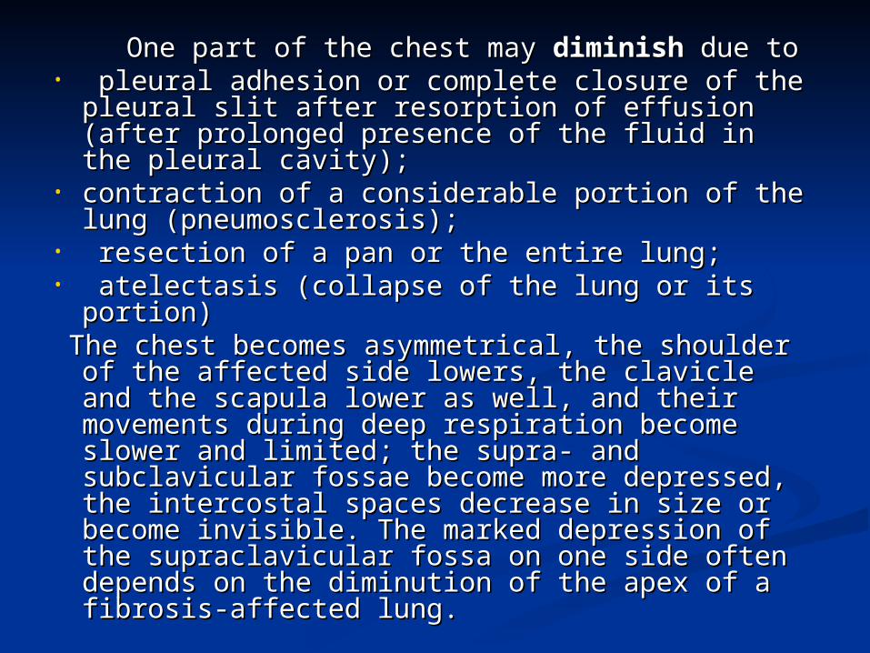
Comparative percussionComparative percussion
Normal percussion sound is Normal percussion sound is resonantresonant (clear pulmonary sound) (clear pulmonary sound)
Pathological sounds:Pathological sounds: DullDull sound (pulmonary tissue sound (pulmonary tissue
consolidation, hydrothorax)consolidation, hydrothorax) ThympanicThympanic sound (abscess, cavern, sound (abscess, cavern,
pneumothorax)pneumothorax) HyperresonanceHyperresonance (bundbox sound): (bundbox sound):
pulmonary emphysemapulmonary emphysema

Topographic percussion Topographic percussion of lungsof lungs
Lower lung border positionLower lung border position Lower lung border respiratory Lower lung border respiratory
mobilitymobility Lungs apexes height and width Lungs apexes height and width
(Kroenig’s area).(Kroenig’s area).

Lower lung border Lower lung border positionposition
Elevated on the affected side:Elevated on the affected side: PneumosclerosisPneumosclerosis AthelectasisAthelectasis Hepato- or splenomegalyHepato- or splenomegaly PneumoniaPneumonia HydrothoraxHydrothorax LobectomiaLobectomia
Elevated on both sides:Elevated on both sides: PregnancyPregnancy MeteorismMeteorism AscitesAscites

Lower lung border Lower lung border positionposition
Displaced downward on the affected Displaced downward on the affected side:side: PneumothoraxPneumothorax
Displaced downward on both sides:Displaced downward on both sides: Pulmonary emphysemaPulmonary emphysema SplanchnoptosisSplanchnoptosis

Sizes of lungs apexesSizes of lungs apexes
Diminish (on the affected side):Diminish (on the affected side): Pneumosclerosis of upper lobe Pneumosclerosis of upper lobe
(tuberculosis)(tuberculosis) LobectomiaLobectomia
Increase:Increase: Pulmonary emphysemaPulmonary emphysema


Topic of the lecture:Topic of the lecture: Symptoms of diseases of Symptoms of diseases of
respiratory organs based o respiratory organs based o data of auscultaion of lungsdata of auscultaion of lungs

1.1.It is important that you try to create a quiet environment It is important that you try to create a quiet environment as much as possible.as much as possible.
22.. Put on your stethoscope so that the ear pieces are Put on your stethoscope so that the ear pieces are directed away from you. Adjust the head of the scope so that directed away from you. Adjust the head of the scope so that
the the
diaphragm is engaged. If you're not sure, scratch lightly on diaphragm is engaged. If you're not sure, scratch lightly on the diaphragm, which should produce a noise. If not, twist the diaphragm, which should produce a noise. If not, twist
the head and try again. Gently rub the head of the the head and try again. Gently rub the head of the stethoscope on your shirt so that it is not too cold prior to stethoscope on your shirt so that it is not too cold prior to
placing it on the patient's skin. placing it on the patient's skin. 3.3. The upper aspect of the posterior fields (i.e. towards the The upper aspect of the posterior fields (i.e. towards the
top of the patient's back) are examined first. Listen over one top of the patient's back) are examined first. Listen over one spot and then move the stethoscope to the same position on spot and then move the stethoscope to the same position on the opposite side and repeat. This again makes use of one the opposite side and repeat. This again makes use of one
lung as a source of comparison for the otherlung as a source of comparison for the other4.4. Then, move around to the front and listen to the anterior Then, move around to the front and listen to the anterior
fields in the same fashion. fields in the same fashion. 5. Don't get in the habit of performing auscultation through 5. Don't get in the habit of performing auscultation through
clothing.clothing.

5.5. Ask the patient to take slow, deep breaths through Ask the patient to take slow, deep breaths through their their
mouths while you are performing your exam. This mouths while you are performing your exam. This forces the patient to move greater volumes of air with forces the patient to move greater volumes of air with each breath, increasing the duration, intensity, and each breath, increasing the duration, intensity, and
thus detectability of any abnormal breath sounds that thus detectability of any abnormal breath sounds that might be present. might be present.
6. 6. Sometimes it's helpful to have the patient cough a Sometimes it's helpful to have the patient cough a few times prior to beginning auscultation. This clears few times prior to beginning auscultation. This clears
airway secretions and opens small atelectatic (i.e. airway secretions and opens small atelectatic (i.e. collapsed) areas at the lung bases. collapsed) areas at the lung bases.
7.7. If the patient cannot sit up (e.g. in cases of If the patient cannot sit up (e.g. in cases of neurologic disease, post-operative states, etc.), neurologic disease, post-operative states, etc.),
auscultation can be performed while the patient is lying auscultation can be performed while the patient is lying on their side. on their side.
8.8. Requesting that the patient exhale forcibly will Requesting that the patient exhale forcibly will occasionally help to accentuate abnormal breath occasionally help to accentuate abnormal breath
sounds (in particular, wheezing) that might not be sounds (in particular, wheezing) that might not be heard when they are breathing at normal flow rates. heard when they are breathing at normal flow rates.


1.1. Apices of lungs extend above clavicles.Apices of lungs extend above clavicles.22. Horizontal fissure follows right 4th rib. Horizontal fissure follows right 4th rib
3.3. Oblique fissures on both sides extend to 6th rib anteriorly Oblique fissures on both sides extend to 6th rib anteriorly4.4. Left lung has large deficit anteriorly extending from 4th to Left lung has large deficit anteriorly extending from 4th to
6th rib and from sternum to costochondral joint – cardiac notch6th rib and from sternum to costochondral joint – cardiac notch
55. Both lungs extend to 8th rib laterally. Both lungs extend to 8th rib laterally66. Parietal pleura extends down to 9th rib laterally. Parietal pleura extends down to 9th rib laterally

Posterior AspectPosterior Aspect7.7. Much of left and right upper portions of lungs are covered Much of left and right upper portions of lungs are covered
by scapulae.by scapulae.8.8. Oblique fissures extend from spinous processes of T2. Oblique fissures extend from spinous processes of T2.
9.9. Lungs extend down to T11 medially and 9th rib laterally Lungs extend down to T11 medially and 9th rib laterally10.10. Parietal pleura extend to T12 medially and 10th rib Parietal pleura extend to T12 medially and 10th rib
laterallylaterally


The vesicular breath sound is the major normal breath The vesicular breath sound is the major normal breath
sound and is heard over most of thesound and is heard over most of the lungs.lungs. The inspiratory sounds are longer than the expiratoryThe inspiratory sounds are longer than the expiratory
sounds.sounds.
Inspiration
Expiration
heard only during the first third of the expiration phase

The vesicular breath sound The vesicular breath sound can be simulated by can be simulated by pronouncing the sound “f” during inspiration, pronouncing the sound “f” during inspiration,
or by drawing tea from saucer.or by drawing tea from saucer. Alveolar walls still vibrateat the initial Alveolar walls still vibrateat the initial
expiration phase to give a shorter second expiration phase to give a shorter second phase of the phase of the vesicular breathing, which is vesicular breathing, which is
heard only during the first third of the heard only during the first third of the expirationexpiration phase because vibrations of elastic phase because vibrations of elastic
alveolar walls are quickly dampened by the alveolar walls are quickly dampened by the decreasing tension of the alveolar walls.decreasing tension of the alveolar walls.

Vesicular breathing may be louder or softer for both Vesicular breathing may be louder or softer for both physiological and pathological reasons.physiological and pathological reasons.
1.1.Vesicular breathVesicular breath sounds may be harsher and slightly sounds may be harsher and slightly
longer if there is rapid deep ventilation or in children longer if there is rapid deep ventilation or in children (“ puerile respiration”(“ puerile respiration” ).).
2.2.Vesicular breathVesicular breath sounds may be softer if the patient sounds may be softer if the patient is frail, elderly, obese, or very muscular.is frail, elderly, obese, or very muscular.
!!Physiological changes in vesicular Physiological changes in vesicular respiration always involve both parts of respiration always involve both parts of the chest, and the chest, and respiratory sounds are respiratory sounds are equally intensified at the symmetrical equally intensified at the symmetrical
points of the chest.points of the chest.
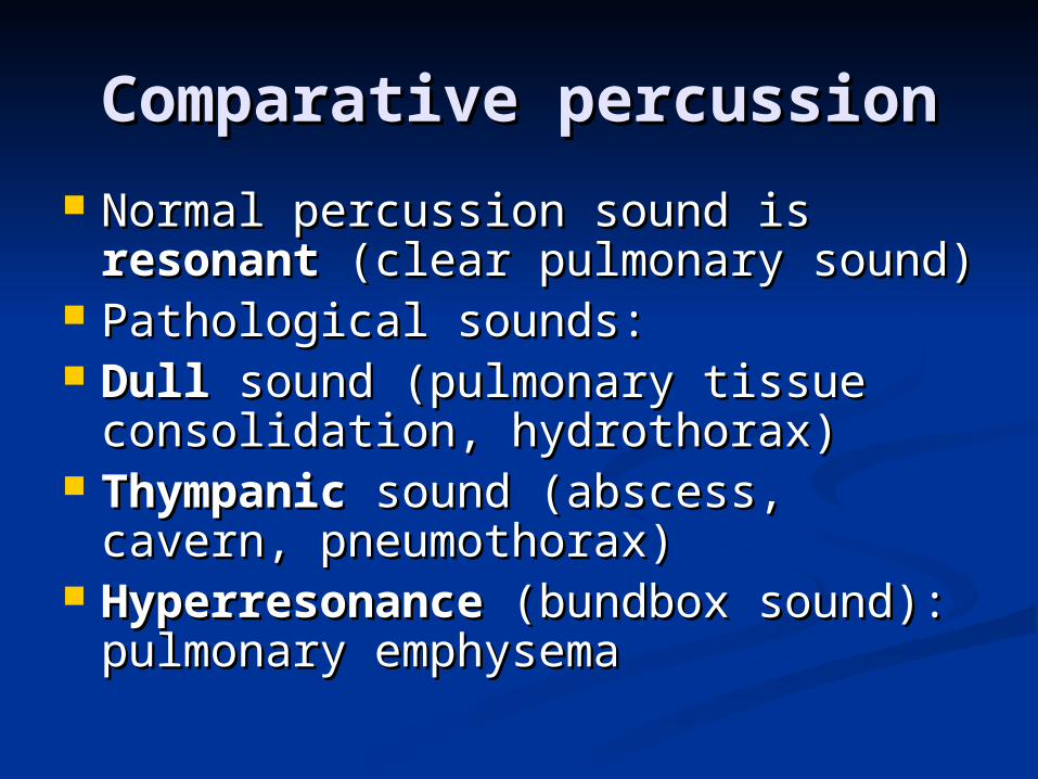
• Alterations in Alterations in vesicular respiration vesicular respiration in in
pathologypathology depend on:depend on: ―― the amount of intact alveoli; the amount of intact alveoli; ―― the properties of their walls; the properties of their walls;
―― the amount of air contained in them; the amount of air contained in them; ― ― the length and strength of the the length and strength of the expiration and inspiration phases;expiration and inspiration phases;
―― the conditions of sound conduction the conditions of sound conduction from the vibrating elastic elements of the from the vibrating elastic elements of the
pulmonary tissue to the surface of the pulmonary tissue to the surface of the chest.chest.

Pathologically decreased vesicular respirationPathologically decreased vesicular respiration
can be: can be:~~ due to a significantly diminished number of due to a significantly diminished number of
the alveoli; the alveoli; ~~ due to inflammation and swelling of the due to inflammation and swelling of the
alveoli walls in a part of the lung; alveoli walls in a part of the lung; ~~ decreased also in insuff icient delivery of decreased also in insuff icient delivery of air air
to the alveoli through the air ways; to the alveoli through the air ways; ~~ due to obstructed conduction of sound due to obstructed conduction of sound
waves from the source of vibration (alveolar waves from the source of vibration (alveolar
walls) to the chest surface.walls) to the chest surface.

1. 1. Abnormally increased Abnormally increased vesicular breathingvesicular breathing depends on obstruction to the air passage through small depends on obstruction to the air passage through small
bronchi or their contracted lumen (increased expiration). bronchi or their contracted lumen (increased expiration). 2. 2. Harsh Harsh vesicular breathingvesicular breathing occurs in marked and occurs in marked and
nonuniform narrowing of the lumen in small bronchi and nonuniform narrowing of the lumen in small bronchi and bronchioles due to| inflammatory oedema of their mucosa (the bronchioles due to| inflammatory oedema of their mucosa (the
inspiration and expiration phases are intensified).inspiration and expiration phases are intensified). 3. 3. Interrupted or cogwheel Interrupted or cogwheel vesicular respiration vesicular respiration is is
characterized by short jerky inspiration efforts interrupted by characterized by short jerky inspiration efforts interrupted by short pauses between them; the expiration is usually normalshort pauses between them; the expiration is usually normal
(occurs in non-uniform contraction of the respiratory muscles, when a patient (occurs in non-uniform contraction of the respiratory muscles, when a patient is auscultated in a cold room, or when he has nervous trembling, or diseases of is auscultated in a cold room, or when he has nervous trembling, or diseases of the respiratory the respiratory musclesmuscles, Interrupted breathing over a limited part of the lung , Interrupted breathing over a limited part of the lung
indicates pathology in fine bronchi (their tuberculous infiltration)indicates pathology in fine bronchi (their tuberculous infiltration)

Bronchial breathing.Bronchial breathing. Respiratory sounds known as Respiratory sounds known as bronchial or tubular breathing bronchial or tubular breathing arise in the larynx and the arise in the larynx and the
trachea as air passes through the vocal slit. As air is inhaled, trachea as air passes through the vocal slit. As air is inhaled, it passes through the vocal slit to enter wider trachea where it passes through the vocal slit to enter wider trachea where it is set in vortex-type motion. Sound waves thus generated it is set in vortex-type motion. Sound waves thus generated
propagate along the air column throughout the entire propagate along the air column throughout the entire bronchial tree. Sounds generated by the vibration of these bronchial tree. Sounds generated by the vibration of these waves are harsh. During expiration, air also passes through waves are harsh. During expiration, air also passes through the vocal slit to enter a wider spase of the larynx where it is the vocal slit to enter a wider spase of the larynx where it is set in a vortex motion. But since the vocal slit is narrower set in a vortex motion. But since the vocal slit is narrower during expiration, the respiratory sound becomes longer, during expiration, the respiratory sound becomes longer,
harsher and longer. This type of breathing is called harsher and longer. This type of breathing is called laryngotracheallaryngotracheal
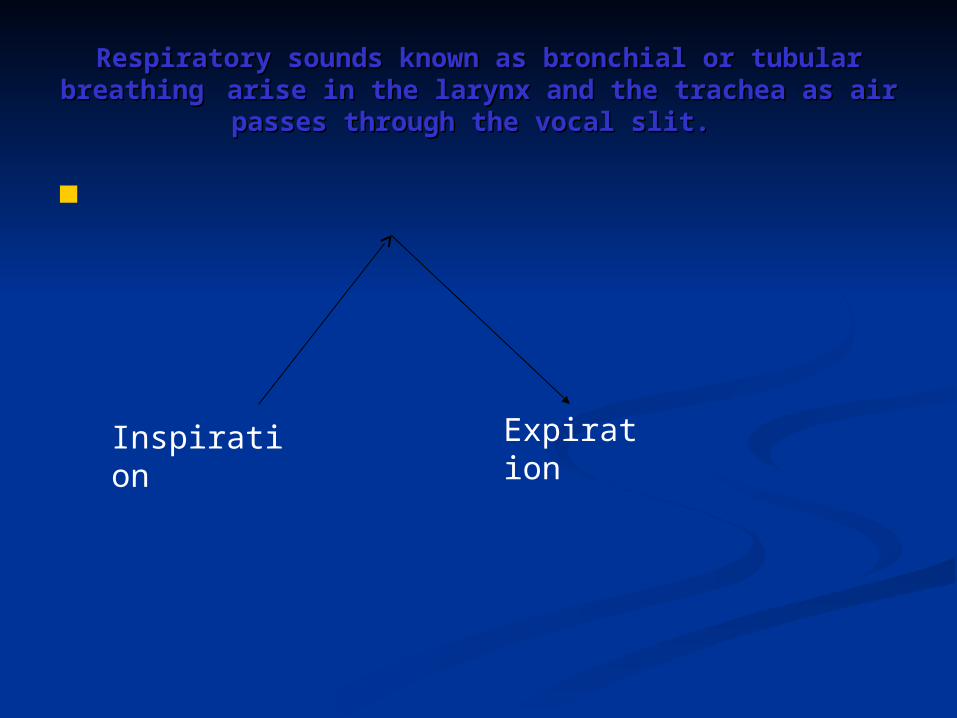
Respiratory sounds known as bronchial or tubular Respiratory sounds known as bronchial or tubular breathing breathing arise in the larynx and the trachea as air arise in the larynx and the trachea as air
passes through the vocal slit.passes through the vocal slit.
Еxpiration
Inspiration

Bronchial breathing can Bronchial breathing can be heard instead of be heard instead of
vesiculai ( or in addition vesiculai ( or in addition to the vesicular to the vesicular
breathing) over the chest breathing) over the chest in pulmonary pathology. in pulmonary pathology. This breathing is called This breathing is called pathological bronchial pathological bronchial
respirationrespiration..

Amphoric respirationAmphoric respiration arises in the presence of a smooth-arises in the presence of a smooth-wall cavity( non less than 5-6 cm in diameter) wall cavity( non less than 5-6 cm in diameter)
communicated with a large bronchuscommunicated with a large bronchus
Metallic respirationMetallic respiration differs from both bronchial and differs from both bronchial and amphoric. It is loud and high, and resembles the sound amphoric. It is loud and high, and resembles the sound
produced when a piece of metal is struck. Metallic produced when a piece of metal is struck. Metallic respiration is heard in open pneumothorax when the air respiration is heard in open pneumothorax when the air of if pleural cavity communicates with the external air. of if pleural cavity communicates with the external air.
Stenotic respirationStenotic respiration is heard in cases with narrowed is heard in cases with narrowed trachea or a large bronchus (due to a tumor); trachea or a large bronchus (due to a tumor);
Bronchovesicular Bronchovesicular or or mixed respirationmixed respiration is heard in lobular is heard in lobular pneumonia or infiltrative tuberculosis, and also in pneumonia or infiltrative tuberculosis, and also in
pneumosclerosis, with foci of con solidated tissue being pneumosclerosis, with foci of con solidated tissue being seated deeply in the pulmonary tissue and far from one seated deeply in the pulmonary tissue and far from one
another.another.

Adventitious soundsAdventitious sounds
are rales, crepitation, and pleural are rales, crepitation, and pleural friction.friction.

RalesRales arise in pathology arise in pathology of the trachea, bronchi, of the trachea, bronchi, or if a cavern is form ed or if a cavern is form ed
in the affected lung. in the affected lung. Rales are classified as Rales are classified as dry (rhonchi) and moist dry (rhonchi) and moist
rales.rales.

Dry rales can be due toDry rales can be due to (1)(1) spasms of smooth muscles of the bronchi during spasms of smooth muscles of the bronchi during
fits of bronchial asthma; fits of bronchial asthma;
(2)(2) swelling of the bronchial mucosa during its swelling of the bronchial mucosa during its inflammation; inflammation;
(3)(3) accumulation of viscous sputum in the bronchi accumulation of viscous sputum in the bronchi which adheres to the wall of the bronchus and narrows which adheres to the wall of the bronchus and narrows
its lumen; its lumen; (4)(4) formation of fibrous tissue in the walls of separate formation of fibrous tissue in the walls of separate bronchi and in the pulmonary tissue with subsequent bronchi and in the pulmonary tissue with subsequent
alteration of their architectonics (bronchiectasis, alteration of their architectonics (bronchiectasis, pneumosclerosis);pneumosclerosis);
(5)(5) vibration of viscous sputum in the lumen of large vibration of viscous sputum in the lumen of large and medium size bronchi during inspiration and and medium size bronchi during inspiration and
expiration: being viscous, the sputum can be drawn (by expiration: being viscous, the sputum can be drawn (by the air stream) into threads which adhere to the the air stream) into threads which adhere to the
opposite walls of the bronchi and vibrate like strings.opposite walls of the bronchi and vibrate like strings.

Dry rales are heard during inspiration and expirationDry rales are heard during inspiration and expiration and vary greatly in their loudness, tone and pitch. and vary greatly in their loudness, tone and pitch. According to the quality and pitch of the sounds According to the quality and pitch of the sounds
produced, produced, dry rales are divided into:dry rales are divided into:
sibilantsibilant (high-pitched and whistling sounds (high-pitched and whistling sounds are produced when the lumen of the small are produced when the lumen of the small
bronchi is narrowed) bronchi is narrowed) sonorous ralessonorous rales (low-pitched and sonoring (low-pitched and sonoring rales are generated in stenosis of medium rales are generated in stenosis of medium calibre and large calibre bronchi or when calibre and large calibre bronchi or when viscous sputum is accumulated in their viscous sputum is accumulated in their
lumen). lumen).

Moist ralesMoist rales are generated because of accumulation of liquid are generated because of accumulation of liquid secretion (sputum, oedematous fluid, blood) in the bronchi secretion (sputum, oedematous fluid, blood) in the bronchi
through which air passes. Air bubbles pass through the liquid through which air passes. Air bubbles pass through the liquid secretion of the bronchial lumen and collapse to produce the secretion of the bronchial lumen and collapse to produce the
specific cracking sound. specific cracking sound. Moist rales are heard during both the inspiration and Moist rales are heard during both the inspiration and expiration, but since the air velocity is higher during expiration, but since the air velocity is higher during
inspiration, moist rales will be better heard at this respiratory inspiration, moist rales will be better heard at this respiratory phasephase..

Depending on the calibre of bronchi where rales are Depending on the calibre of bronchi where rales are generated, generated, moist rales are classified asmoist rales are classified as fine, medium fine, medium and and
coarse bubbling coarse bubbling rales.rales. FineFine bubbling rales bubbling rales are generated in fine bronchi and are generated in fine bronchi and are percepted by the ear as short multiple sounds; are percepted by the ear as short multiple sounds; MediumMedium bubbling rales bubbling rales are produced in bronchi of a are produced in bronchi of a
medium size; medium size; CoarseCoarse bubbling rales bubbling rales in large calibre bronchi, in large in large calibre bronchi, in large
bronchiectases, and in pulmonary cavities (abscess, bronchiectases, and in pulmonary cavities (abscess, cavern) containing liquid secretions and cavern) containing liquid secretions and
communicating with the large bronchus.communicating with the large bronchus.

Depending on the character of the pathology in the lungs, Depending on the character of the pathology in the lungs, moistmoist rales are subdivided into: rales are subdivided into:
~~ consonating consonating or or cracklingcrackling, , ~~ non-consonating non-consonating or or bub bling bub bling rales.rales.
Consonating moist rales are heard in the presence of liquid Consonating moist rales are heard in the presence of liquid secretions in the bronchi surrounded by airless (consolidated) secretions in the bronchi surrounded by airless (consolidated)
pulmonary tissue or in lung cavities with smooth walls pulmonary tissue or in lung cavities with smooth walls surrounded by consolidated pulmonary tissue. The cavity surrounded by consolidated pulmonary tissue. The cavity
itself acts as a resonator to intensify moist rales. itself acts as a resonator to intensify moist rales. Non-consonating rales Non-consonating rales are heard in inflammation of bronchial are heard in inflammation of bronchial mucosa (bronchitis) or acute oedema of the lung due to the mucosa (bronchitis) or acute oedema of the lung due to the
failure of the left chambers of the heart. The sounds produced failure of the left chambers of the heart. The sounds produced by collapsing air bubbles in the bronchi are dampened by the by collapsing air bubbles in the bronchi are dampened by the "air cushion" of the lungs as they are conducted to the chest "air cushion" of the lungs as they are conducted to the chest
surface.surface.

CrepitationCrepitation originates in the alveoli. Crepitation is a originates in the alveoli. Crepitation is a slight crackling sound that can be imitated by rubbing a lock slight crackling sound that can be imitated by rubbing a lock of hair. of hair. The main condition for generation of crepitation is The main condition for generation of crepitation is accumulation of a small amount of liquid secretion in the accumulation of a small amount of liquid secretion in the alveoli.alveoli. During expiration, the alveoli stick together, while During expiration, the alveoli stick together, while during inspiration the alveolar walls are separated with during inspiration the alveolar walls are separated with
difficulty and only at the end of the inspiratoryn movement. difficulty and only at the end of the inspiratoryn movement. Crepitation is therefore only heard during the heighi of Crepitation is therefore only heard during the heighi of
inspiration.inspiration. In other words, crepitation is the sound produced In other words, crepitation is the sound produced by many alveoli during their simultaneous reinflation.by many alveoli during their simultaneous reinflation.


Pleural friction sound.Pleural friction sound.
Pleural friction sound Pleural friction sound are heard during both are heard during both
inspiration and inspiration and expiration.expiration.
The sounds are The sounds are differentiated by differentiated by
intensity, or loudness, intensity, or loudness, length, and site over length, and site over
which they are heard.which they are heard.

Pleural frictionPleural friction sounds can be differentiated from fine sounds can be differentiated from fine bubbling rales and crepitation by the following signs: bubbling rales and crepitation by the following signs:
(1)(1) the character of rales is altered or rales can disappear for the character of rales is altered or rales can disappear for a short time after coughing, while pleural frictionsounds does a short time after coughing, while pleural frictionsounds does
not change in these conditions; not change in these conditions; (2)(2) when a stethoscope is pressed tighter against the chest, when a stethoscope is pressed tighter against the chest, the pleural friction sound is intensified, while rales do not the pleural friction sound is intensified, while rales do not
change;change; (3) (3) crepitation is only heard at the height of inspiration, crepitation is only heard at the height of inspiration,
while pleural friction sound is heard during both inspiration while pleural friction sound is heard during both inspiration and expiration; and expiration;
(4)(4) if a patient moves his diaphragm in and out while his if a patient moves his diaphragm in and out while his mouth and nose are closed, the sound produced by the mouth and nose are closed, the sound produced by the
friction of the pleura due to the movement of the diaphragm friction of the pleura due to the movement of the diaphragm can be heard, while rales and crepitation cannot because can be heard, while rales and crepitation cannot because
there is no air movement in the bronchi.there is no air movement in the bronchi.

Common errors of auscultation:Common errors of auscultation:
____Auscultating one entire lung, and then Auscultating one entire lung, and then
moving to the other lung moving to the other lung ____Auscultating over a patient’s gown or Auscultating over a patient’s gown or
article of clothing article of clothing ____Beginning auscultation inferiorly at the Beginning auscultation inferiorly at the
lower lung fields lower lung fields ____Moving your stethoscope before each Moving your stethoscope before each
exhalation is complete exhalation is complete ____Examiner does not make the room quiet Examiner does not make the room quiet
enough to hear breath sounds. enough to hear breath sounds.

Thank you!Thank you!
Related Documents