01-Mar-18 1 SPIROMETRY Alfian Nur Rosyid, Arief Bakhtiar, Daniel Maranatha, Muhammad Amin Dept./SMF/KSM Pulmonologi dan Ilmu Kedokteran Respirasi RS Unair FK Unair – RSUD Dr.Soetomo Surabaya, Indonesia Menu 2 Introduction Definition Devices Indication Contra indication Procedure

Welcome message from author
This document is posted to help you gain knowledge. Please leave a comment to let me know what you think about it! Share it to your friends and learn new things together.
Transcript
01-Mar-18
1
SPIROMETRY
Alfian Nur Rosyid, Arief Bakhtiar,
Daniel Maranatha, Muhammad Amin
Dept./SMF/KSM Pulmonologi dan Ilmu Kedokteran Respirasi RS Unair
FK Unair – RSUD Dr.Soetomo Surabaya, Indonesia
Menu
2
Introduction
Definition
Devices
Indication
Contra indication
Procedure

01-Mar-18
2
History
3
* English Surgeon
*
VC
4
The First Spirometer

01-Mar-18
3
History
5
1950: Dr. Tiffeneau of France introduced the forced measurement
of air volume during a given time frame, i.e., FEV1.
▪1959: Wright B.M. & McKerrow C.B. introduced peak flow meter
▪2008: Advanced Medical Engineering developed the world's first
wireless spirometer with 3D Tilt-Sensing for far greater quality
control in the testing environment.
FEV1
PEFR
Introduction Pulmonary Function Test
6
Types :
Ventilation
Diffusion

01-Mar-18
4
Definition
7
Definition
8

01-Mar-18
5
“
volume flow
9
(mL of air) = volume / time
(mL/s)
“
10
Physiology of Breath
▪Inspiration
▪Expiration

01-Mar-18
6
Devices
11
Indication "What is the use of spirometry?”
12

01-Mar-18
7
Indication "What is the use of spirometry?”
Spirometry is the best way of detecting the
presence of airway obstruction and making a
definitive diagnosis of asthma and COPD
13
Measure airflow obstruction to help make a definitive diagnosis of COPD.
Confirm presence of airway obstruction. Assess severity of airflow obstruction in COPD. Detect airflow obstruction in smokers who may have few or
no symptoms. Monitor disease progression in COPD. Assess one aspect of response to therapy. Assess prognosis (FEV1) in COPD. Perform pre-operative assessment.
Indication "What is the use of spirometry?”
Spirometry is the best way of detecting the
presence of airway obstruction and making a
definitive diagnosis of asthma and COPD
14
Make a diagnosis and assess severity in a range of other respiratory conditions
Distinguish between obstruction and restriction as causes of breathlessness Screen workforces in occupational environments Assess fitness to dive Perform pre-employment screening in certain
professions

01-Mar-18
8
Contra indication
▪Don’t perform spirometry for them with:
15
Bloody Cough Pneumothorax Unstable Angina Pectoris Acute Myocardial Infarction Brain aneurysm Post surgery: eye, thorax, abdomen in the
healing periode Asthma / COPD excacerbation
Spirometry Value :
1. TV
2. IRV
3. ERV
4. VC or SVC and FVC
5. FEV1
6. FEV1/FVC ratio
7. FRC
16
Value and Graph
Graph:
1. VC per time
2. Flow per time
01-Mar-18
9
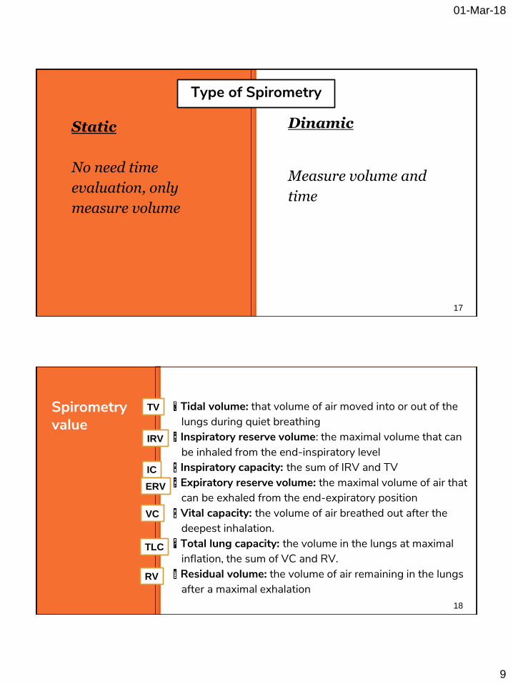
Static
No need time
evaluation, only
measure volume
17
Dinamic
Measure volume and
time
Type of Spirometry
Spirometry value
Tidal volume: that volume of air moved into or out of the lungs during quiet breathing
Inspiratory reserve volume: the maximal volume that can be inhaled from the end-inspiratory level
Inspiratory capacity: the sum of IRV and TV Expiratory reserve volume: the maximal volume of air that can be exhaled from the end-expiratory position
Vital capacity: the volume of air breathed out after the deepest inhalation.
Total lung capacity: the volume in the lungs at maximal inflation, the sum of VC and RV.
Residual volume: the volume of air remaining in the lungs after a maximal exhalation
18
TV
IRV
IC
ERV
VC
TLC
RV

01-Mar-18
10
Spirometry graph
Static
19
Spirometry graph
20
3000
500
2000
1000
01-Mar-18
11
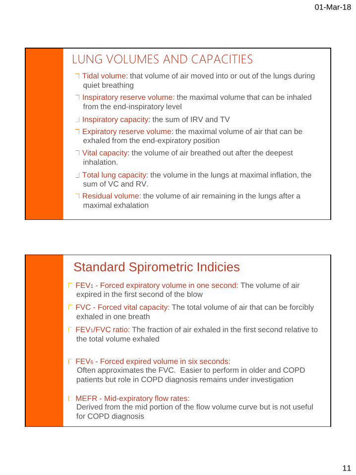
LUNG VOLUMES AND CAPACITIES Tidal volume: that volume of air moved into or out of the lungs during
quiet breathing
Inspiratory reserve volume: the maximal volume that can be inhaled
from the end-inspiratory level
Inspiratory capacity: the sum of IRV and TV Expiratory reserve volume: the maximal volume of air that can be
exhaled from the end-expiratory position Vital capacity: the volume of air breathed out after the deepest
inhalation.
Total lung capacity: the volume in the lungs at maximal inflation, the
sum of VC and RV. Residual volume: the volume of air remaining in the lungs after a
maximal exhalation
Standard Spirometric Indicies
FEV1 - Forced expiratory volume in one second: The volume of air expired in the first second of the blow
FVC - Forced vital capacity: The total volume of air that can be forcibly
exhaled in one breath
FEV1/FVC ratio: The fraction of air exhaled in the first second relative to
the total volume exhaled
FEV6 - Forced expired volume in six seconds: Often approximates the FVC. Easier to perform in older and COPD
patients but role in COPD diagnosis remains under investigation
MEFR - Mid-expiratory flow rates: Derived from the mid portion of the flow volume curve but is not useful
for COPD diagnosis

01-Mar-18
12
Spirometry graph
Dinamic
23
FVC FORCED VITAL CAPACITY (FVC)
■ VC is measured after the pt has blown as
hard and as fast as possible into the spirometry
■ In normal lung VC is equal to FVC
■ In COPD compression of lungs during
forced expiration leads to closure of
airway earlier than usual
■ FVC maybe less than VC
24

01-Mar-18
13
25
Spirometry graph
FEV1 Forced expiratory volume 1 second (FEV1)
• FEV1 is the volume of air in the first
second of a forced expiration
• In a normal lung it is more than 70% of the FVC
• In obstruction as seen in COPD the time
taken to expire is longer thus the ration of
FEV1 to FVC is reduced
• Seen the graph next
26
01-Mar-18
14
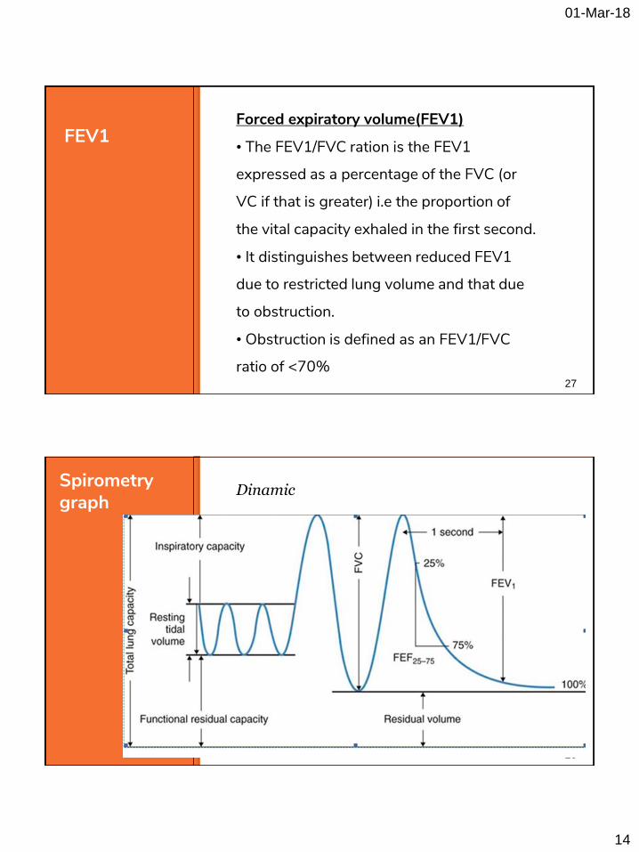
FEV1 Forced expiratory volume(FEV1)
• The FEV1/FVC ration is the FEV1
expressed as a percentage of the FVC (or
VC if that is greater) i.e the proportion of
the vital capacity exhaled in the first second.
• It distinguishes between reduced FEV1
due to restricted lung volume and that due
to obstruction.
• Obstruction is defined as an FEV1/FVC
ratio of <70% 27
28
Spirometry graph
Dinamic

01-Mar-18
15
PROCEDURE
Withholding Medications
Before performing spirometry, withhold: Short acting β2-agonists for 6 hours
Long acting β2-agonists for 12 hours
Ipratropium for 6 hours
Tiotropium for 24 hours
Optimally, subjects should avoid caffeine and cigarette smoking for 30 minutes before performing
spirometry

01-Mar-18
16
Performing Spirometry - Preparation
1. Explain the purpose of the test and demonstrate
the procedure 2. Record the patient’s age, height and gender and
enter on the spirometer 3. Note when bronchodilator was last used 4. Have the patient sitting comfortably 5. Loosen any tight clothing 6. Empty the bladder beforehand if needed
Breath in until the lungs are full
Hold the breath and seal the lips tightly
around a clean mouthpiece
Blast the air out as forcibly and fast as
possible. Provide lots of encouragement!
Continue blowing until the lungs feel empty

01-Mar-18
17
Watch the patient during the blow to assure
the lips are sealed around the mouthpiece
Check to determine if an adequate trace
has been achieved
Repeat the procedure at least twice more
until ideally 3 readings within 100 ml or 5%
of each other are obtained
Spirometry - Possible Side Effects Feeling light-headed
Headache
Facial redness
Fainting: reduced venous return or vasovagal
attack (reflex)
Transient urinary incontinence
Spirometry should be avoided after recent heart attack or stroke
01-Mar-18
18
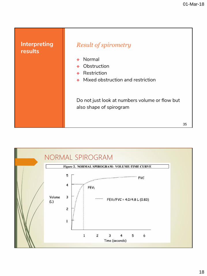
Interpreting results
Result of spirometry
35
Normal Obstruction Restriction Mixed obstruction and restriction
Do not just look at numbers volume or flow but also shape of spirogram
NORMAL SPIROGRAM

01-Mar-18
19
NORMAL FLOW-VOLUME CURVE

01-Mar-18
20
ABNORMAL FLOW-VOLUME PATTERNS
OBSTRUCTIVE DISEASE
01-Mar-18
21
41
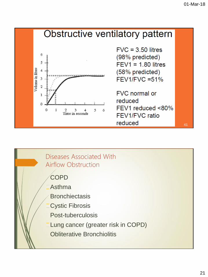
Diseases Associated With
Airflow Obstruction
COPD
Asthma
Bronchiectasis
Cystic Fibrosis
Post-tuberculosis
Lung cancer (greater risk in COPD)
Obliterative Bronchiolitis
01-Mar-18
22
Spirometric Diagnosis of COPD
COPD is confirmed by post-bronchodilator FEV1/FVC < 0.7
Post-bronchodilator FEV1/FVC measured 15
minutes after 400µg salbutamol or equivalent
Bronchodilator Reversibility Testing
Provides the best achievable FEV1
(and FVC) Helps to differentiate COPD from
asthma Must be interpreted with clinical history
- neither asthma nor COPD are diagnosed on spirometry alone
01-Mar-18
23
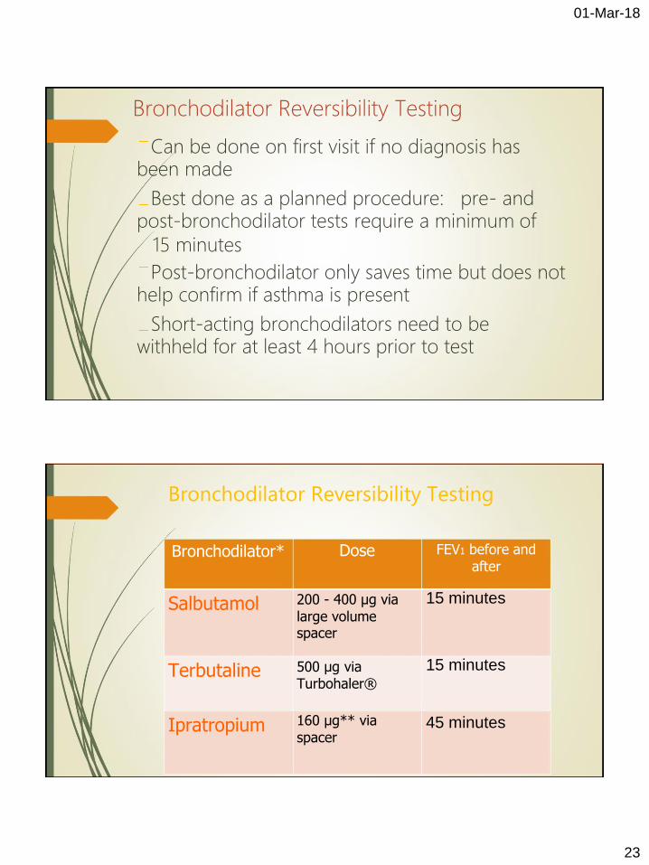
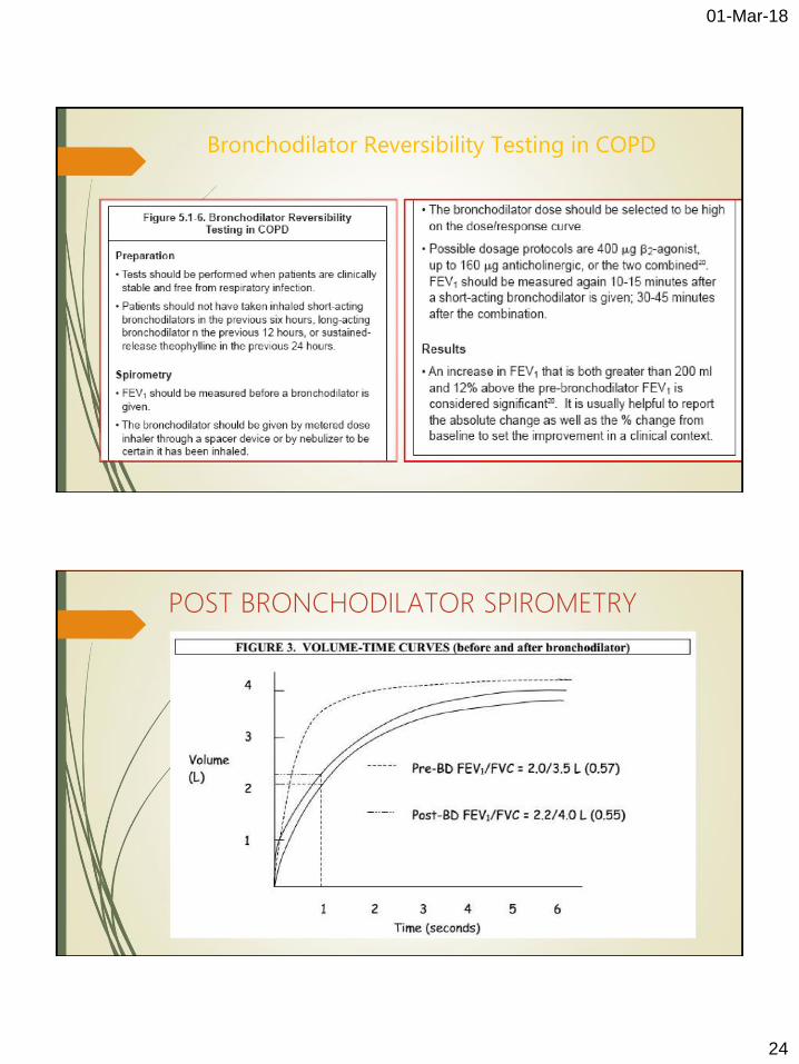
Bronchodilator Reversibility Testing
Can be done on first visit if no diagnosis has
been made Best done as a planned procedure: pre- and
post-bronchodilator tests require a minimum of 15 minutes
Post-bronchodilator only saves time but does not help confirm if asthma is present Short-acting bronchodilators need to be
withheld for at least 4 hours prior to test
Bronchodilator Reversibility Testing
Bronchodilator*
Dose
FEV1 before and after
Salbutamol
Terbutaline
Ipratropium
200 - 400 µg via
large volume
spacer
500 µg via Turbohaler®
160 µg** via
spacer
15 minutes
15 minutes
45 minutes
01-Mar-18
24
Bronchodilator Reversibility Testing in COPD
POST BRONCHODILATOR SPIROMETRY

01-Mar-18
25
RESTRICTIVE DISEASE
Criteria: Restrictive Disease
FEV1: normal or mildly reduced
FVC:< 80% predicted
FEV1/FVC: > 0.7

01-Mar-18
26
51
Diseases Associated with a Restrictive Defect Pulmonary
Fibrosing lung diseases
Pneumoconioses
Pulmonary edema
Parenchymal lung
tumors Lobectomy or
pneumonectomy
Extrapulmonary
Thoracic cage deformity
Obesity
Pregnancy Neuromuscular disorders
Fibrothorax
01-Mar-18
27
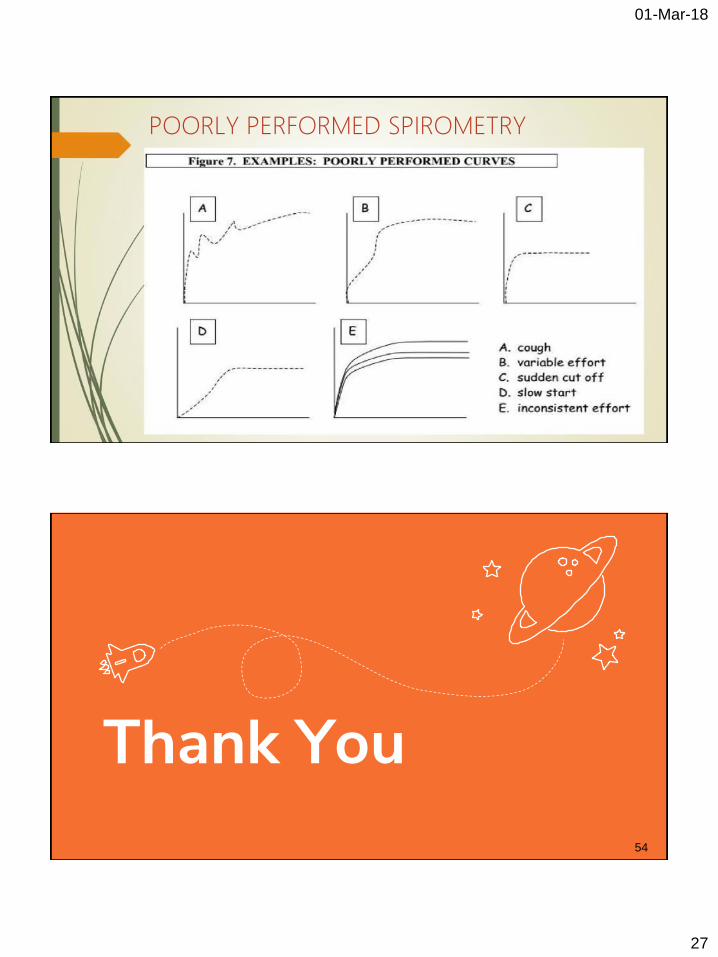
POORLY PERFORMED SPIROMETRY
Thank You 54
Related Documents