University of Plymouth PEARL https://pearl.plymouth.ac.uk Faculty of Health: Medicine, Dentistry and Human Sciences School of Health Professions 2016-05-01 Intra-rater reliability of clinical measures of leg function, in typically developing children aged 1-4 years. Marsden, JF http://hdl.handle.net/10026.1/4993 Association of Paediatric Chartered Physiotherapist Journal All content in PEARL is protected by copyright law. Author manuscripts are made available in accordance with publisher policies. Please cite only the published version using the details provided on the item record or document. In the absence of an open licence (e.g. Creative Commons), permissions for further reuse of content should be sought from the publisher or author.

Welcome message from author
This document is posted to help you gain knowledge. Please leave a comment to let me know what you think about it! Share it to your friends and learn new things together.
Transcript
University of Plymouth
PEARL https://pearl.plymouth.ac.uk
Faculty of Health: Medicine, Dentistry and Human Sciences School of Health Professions
2016-05-01
Intra-rater reliability of clinical measures
of leg function, in typically developing
children aged 1-4 years.
Marsden, JF
http://hdl.handle.net/10026.1/4993
Association of Paediatric Chartered Physiotherapist Journal
All content in PEARL is protected by copyright law. Author manuscripts are made available in accordance with
publisher policies. Please cite only the published version using the details provided on the item record or
document. In the absence of an open licence (e.g. Creative Commons), permissions for further reuse of content
should be sought from the publisher or author.
1
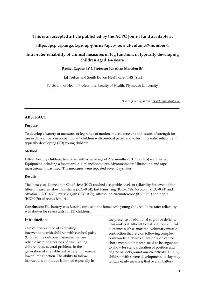
This is an accepted article published by the ACPC Journal and available at
http://apcp.csp.org.uk/group-journal/apcp-journal-volume-7-number-1
Intra-rater reliability of clinical measures of leg function, in typically developing
children aged 1-4 years.
Rachel Rapson [a*], Professor Jonathan Marsden [b]
[a] Torbay and South Devon Healthcare NHS Trust
[b] School of Health Professions, Faculty of Health, Plymouth University
*Corresponding author: [email protected]
ABSTRACT
Purpose
To develop a battery of measures of leg range of motion, muscle tone and indicators of strength for
use in clinical trials in non-ambulant children with cerebral palsy, and to test intra-rater reliability in
typically developing (TD) young children.
Method
Fifteen healthy children, five boys, with a mean age of 29.6 months (SD 9 months) were tested.
Equipment including a footboard, digital inclinometers, Myotonometer, Ultrasound and tape
measurement was used. The measures were repeated seven days later.
Results
The Intra-class Correlation Coefficient (ICC) reached acceptable levels of reliability for seven of the
fifteen measures; slow hamstring (ICC=0.84), fast hamstring (ICC=0.79), Myoton F (ICC=0.74) and
Myoton S (ICC=0.73), muscle girth (ICC=0.95), ultrasound circumference (ICC=0.71) and depth
(ICC=0.76) of rectus femoris.
Conclusion: The battery was feasible for use in the home with young children. Intra-rater reliability
was shown for seven tests for TD children.
Introduction
Clinical trials aimed at evaluating
interventions with children with cerebral palsy
(CP), require outcome measures that are
reliable over long periods of time. Young
children pose several problems in the
generation of a reliable test battery to measure
lower limb function. The ability to follow
instructions at this age is limited especially in
the presence of additional cognitive deficits.
This makes it difficult to test common clinical
outcomes such as maximal voluntary muscle
contraction that rely on following complex
commands. A child’s attention span can be
short, meaning that tests need to be engaging
to allow for standardisation of position and
degree of background muscle activity. Finally,
children with severe developmental delay may
fatigue easily meaning that overall battery

2
duration should be short and position changes
between tests should be minimised.
Studies of normal ranges of movement (ROM)
in typically developing (TD) children highlight
that range of movement changes with age
(Kilgour et al., 2002, Soucie et al., 2011),
particularly between birth and five years.
There is minimal data on the reliability of
measures of ROM in TD young children.
Traditional goniometry has been shown to be
reliable in older non-ambulant children with
CP (Fosang et al., 2003, Bartlett et al., 1985)
with levels of measurement error of between
10-28˚ (Kilgour et al., 2003, Stuberg et al., 1988).
The greatest variances were found when
measuring bi-articular muscles (McDowell et
al., 2000) where there is an increased incidence
of spasticity and contracture.
Reliability can be reported using the Intra-
class correlation coefficient (Shrout and Fleiss,
1979) . The use of digital inclinometers was
reported in measurement of hip abduction,
producing good levels (ICC >0.85) of intra-
rater reliability (Herrero et al., 2011). A factor
that may contribute to the low reliability when
measuring range of motion is the variability in
the duration and size of the applied force used
to move a joint to its end range. Maas et al
(2012) suggested standardising the applied
force when measuring range of motion in the
ankle by using a hand held dynamometer,
which includes a torque wrench and
goniometer attached to a footboard. Variations
in the resistive torque are significant for
movements about the knee and hip, due to the
increased length and weight of the limb
moving in relation to gravity. Therefore, for
the tests of hamstring and hip flexor
extensibility the application of force are
manually determined in the clinical setting.
Hypertonia in children with CP is caused by
both changes in passive musculo-tendinous
properties and enhanced stretch reflexes
(spasticity). The different components of
muscle tone can be difficult to assess (Pandyan
et al., 1999). Clinical tests such as the Tardieu
scale aim to differentiate between the
components by scoring the resistance to
movements above and below the stretch reflex
threshold (Scholtes et al., 2006, Boyd and
Graham, 1999, Gracies et al., 2000). More
recently, ultrasound has been used to
determine factors such as muscle fascicle
length and stiffness in the presence of
spasticity(Kwah et al., 2013). These techniques
require access to computer controlled motors
(e.g. dynamometry) and make the measure
unfeasible for clinical trials when measures
may be taken outside of the laboratory setting.
The Myotonometer is a portable device that
measures tissue compliance. It applies a
standardised perturbation via a probe and the
subsequent motion of the probe is determined
using an accelerometer; from this a range of
measures of tissue compliance are provided
such as tissue stiffness and creep (Lidstrom et
al., 2009). It is a highly reliable tool in healthy
adult subjects (ICC> 0.84) (Leonard et al., 2003).
In children with CP over four years old the
test-retest reliability was substantial in the
relaxed medial gastrocnemius
(ICC>0.89)(Aarrestad et al., 2004). It is portable
and feasible to use in this population.
Muscle cross-sectional area or thickness can be
measured by ultrasound and may be a useful
quantitative measure when evaluating
strengthening interventions in children who
cannot comply with conventional strength
tests (Ohata et al., 2008). Muscle thickness, of
rectus femoris and vastus lateralis were shown
to be a good predictor of muscle strength in
older non-ambulant children with CP (Moreau
et al., 2010). These tests provide a useful
indicator of muscle strength; however
reliability of these tests in TD young children
has not been assessed to date.
A protocol was developed in line with the
literature to assess lower limb function; range
of movement, muscle tone and strength.
The battery of tests were trialled with a group
of TD children aged 1-4 years to assess the
ease of application and to test the intra-rater
reliability for this group. Intra-rater reliability
only was of interest as the battery of measures
was to be used by a single rater in a
subsequent study.
Method

3
Ethical approval was provided via the UK
South West NHS Research Ethics Committees
and the Faculty of Health and Human sciences,
Plymouth University.
A power calculation indicated that thirteen
children would be needed to demonstrate an
ICC of > 0.7(power=0.85; α=0.05). This is in line
with other reliability studies in this
population(Aarrestad et al., 2004, McDowell et
al., 2000).
Children were recruited via adverts at local
nurseries and play centres. Children were
included if they did not have any orthopaedic
or neurological symptoms that could affect
lower limb movement. They were excluded if
they showed signs of infection and illness
lasting more than one day in the week before
the study or in the inter-measurement period.
Children participated following the informed
written consent of the guardians. The child
was first familiarised with the tests by
demonstrating them on a teddy bear and
assent gained where possible.
Development of the battery of tests
The battery of nine tests was developed giving
fifteen outcome measures. A Physiotherapist
with fifteen years of paediatric experience in
clinical examinations, such as range of motion,
was trained in using the tests. The rater had no
prior experience using the equipment;
therefore a training period was undertaken for
4 weeks prior to beginning the trial.
The tests were carried out in the families’
home using portable equipment. The testing
protocol is summarised in Table 1. One leg
was tested as results from both legs may well
have been statistically similar, therefore only
the left leg was tested for each child. The child
was made comfortable with a small pillow and
the parent was encouraged to comfort the
child and help to stabilise the starting position.
When prone, the head was turned to the same
side as the tested leg to standardise influence
on lower limb stretch reflexes(Aiello et al.,
1992). At the beginning of the session a tape
measure determined the distance between
bony points on the shank and thigh and skin
markers were used to indicate the
measurement points for the ultrasound and
myotonometer using an eye liner pencil. A
compliance score was given using a simple
four point scale. This was used to be able to
compare whether the child’s performance was
similar between tests or between the two
testing sessions as changes in test compliance
may particularly be a factor affecting test
reliability in this age group. The tests were
repeated a week later by the same rater, at the
same time of day and in the same setting.
Measurement of range of movement Each range of movement test involved a warm
up of three movements and three test
movements were carried out with a
metronome (1Hz tone) to pace the speed of the
test so that movements were performed at
~5o/s. The starting position, equipment and
test movement for each measurement are
shown in Table 1.
4
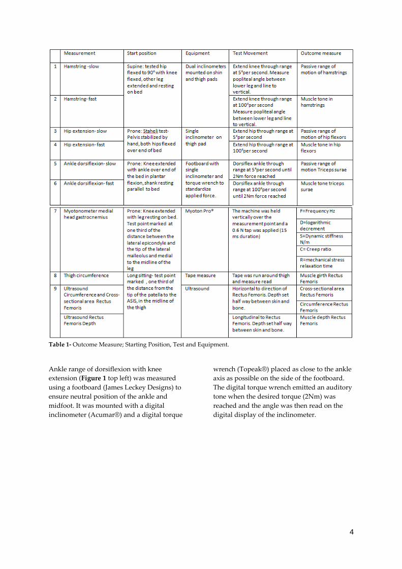
Table 1- Outcome Measure; Starting Position, Test and Equipment.
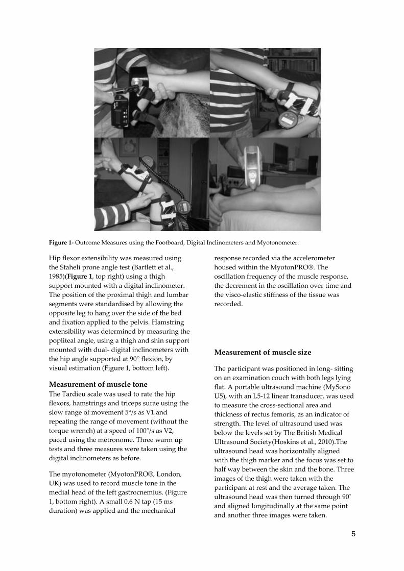
Ankle range of dorsiflexion with knee
extension (Figure 1 top left) was measured
using a footboard (James Leckey Designs) to
ensure neutral position of the ankle and
midfoot. It was mounted with a digital
inclinometer (Acumar®) and a digital torque
wrench (Topeak®) placed as close to the ankle
axis as possible on the side of the footboard.
The digital torque wrench emitted an auditory
tone when the desired torque (2Nm) was
reached and the angle was then read on the
digital display of the inclinometer.
5
Figure 1- Outcome Measures using the Footboard, Digital Inclinometers and Myotonometer.
Hip flexor extensibility was measured using
the Staheli prone angle test (Bartlett et al.,
1985)(Figure 1, top right) using a thigh
support mounted with a digital inclinometer.
The position of the proximal thigh and lumbar
segments were standardised by allowing the
opposite leg to hang over the side of the bed
and fixation applied to the pelvis. Hamstring
extensibility was determined by measuring the
popliteal angle, using a thigh and shin support
mounted with dual- digital inclinometers with
the hip angle supported at 90° flexion, by
visual estimation (Figure 1, bottom left).
Measurement of muscle tone The Tardieu scale was used to rate the hip
flexors, hamstrings and triceps surae using the
slow range of movement 5°/s as V1 and
repeating the range of movement (without the
torque wrench) at a speed of 100°/s as V2,
paced using the metronome. Three warm up
tests and three measures were taken using the
digital inclinometers as before.
The myotonometer (MyotonPRO®, London,
UK) was used to record muscle tone in the
medial head of the left gastrocnemius. (Figure
1, bottom right). A small 0.6 N tap (15 ms
duration) was applied and the mechanical
response recorded via the accelerometer
housed within the MyotonPRO®. The
oscillation frequency of the muscle response,
the decrement in the oscillation over time and
the visco-elastic stiffness of the tissue was
recorded.
Measurement of muscle size
The participant was positioned in long- sitting
on an examination couch with both legs lying
flat. A portable ultrasound machine (MySono
U5), with an L5-12 linear transducer, was used
to measure the cross-sectional area and
thickness of rectus femoris, as an indicator of
strength. The level of ultrasound used was
below the levels set by The British Medical
Ultrasound Society(Hoskins et al., 2010).The
ultrasound head was horizontally aligned
with the thigh marker and the focus was set to
half way between the skin and the bone. Three
images of the thigh were taken with the
participant at rest and the average taken. The
ultrasound head was then turned through 90˚
and aligned longitudinally at the same point
and another three images were taken.
6
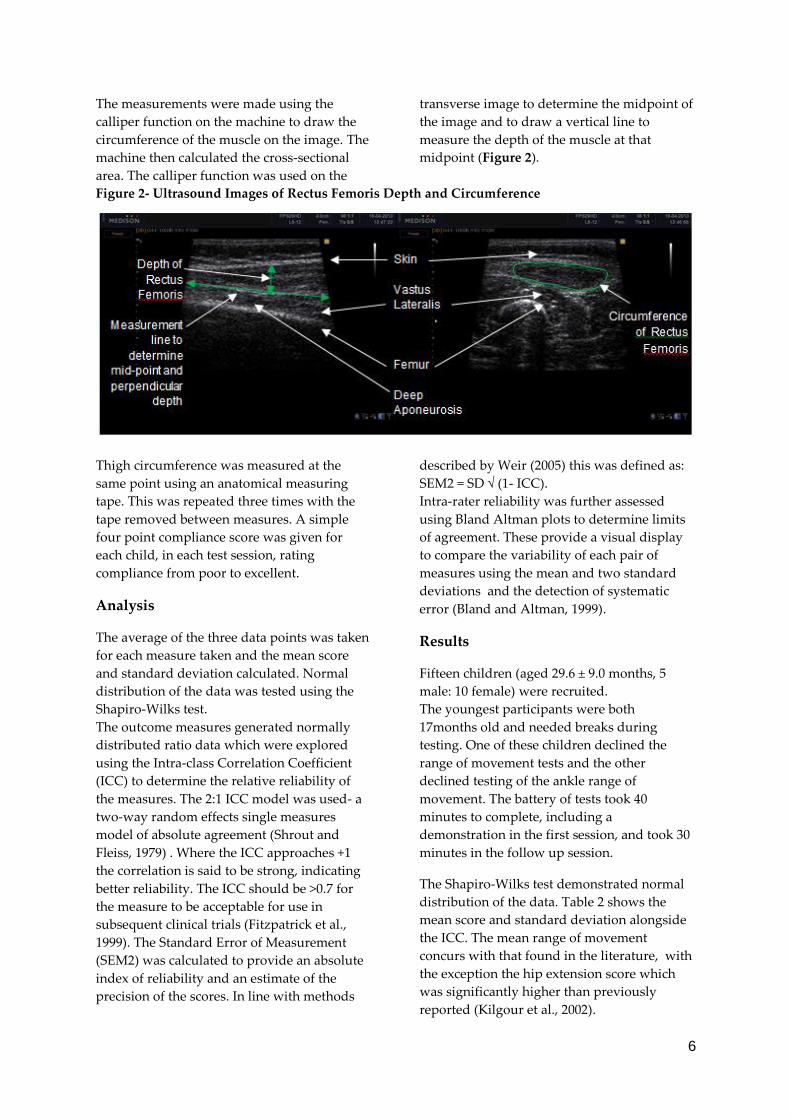
The measurements were made using the
calliper function on the machine to draw the
circumference of the muscle on the image. The
machine then calculated the cross-sectional
area. The calliper function was used on the
transverse image to determine the midpoint of
the image and to draw a vertical line to
measure the depth of the muscle at that
midpoint (Figure 2).
Figure 2- Ultrasound Images of Rectus Femoris Depth and Circumference
Thigh circumference was measured at the
same point using an anatomical measuring
tape. This was repeated three times with the
tape removed between measures. A simple
four point compliance score was given for
each child, in each test session, rating
compliance from poor to excellent.
Analysis
The average of the three data points was taken
for each measure taken and the mean score
and standard deviation calculated. Normal
distribution of the data was tested using the
Shapiro-Wilks test.
The outcome measures generated normally
distributed ratio data which were explored
using the Intra-class Correlation Coefficient
(ICC) to determine the relative reliability of
the measures. The 2:1 ICC model was used- a
two-way random effects single measures
model of absolute agreement (Shrout and
Fleiss, 1979) . Where the ICC approaches +1
the correlation is said to be strong, indicating
better reliability. The ICC should be >0.7 for
the measure to be acceptable for use in
subsequent clinical trials (Fitzpatrick et al.,
1999). The Standard Error of Measurement
(SEM2) was calculated to provide an absolute
index of reliability and an estimate of the
precision of the scores. In line with methods
described by Weir (2005) this was defined as:
SEM2 = SD √ (1- ICC).
Intra-rater reliability was further assessed
using Bland Altman plots to determine limits
of agreement. These provide a visual display
to compare the variability of each pair of
measures using the mean and two standard
deviations and the detection of systematic
error (Bland and Altman, 1999).
Results
Fifteen children (aged 29.6 ± 9.0 months, 5
male: 10 female) were recruited.
The youngest participants were both
17months old and needed breaks during
testing. One of these children declined the
range of movement tests and the other
declined testing of the ankle range of
movement. The battery of tests took 40
minutes to complete, including a
demonstration in the first session, and took 30
minutes in the follow up session.
The Shapiro-Wilks test demonstrated normal
distribution of the data. Table 2 shows the
mean score and standard deviation alongside
the ICC. The mean range of movement
concurs with that found in the literature, with
the exception the hip extension score which
was significantly higher than previously
reported (Kilgour et al., 2002).

7
The reliability testing of the fifteen outcome
measures demonstrated that seven measures
reached acceptable levels of reliability greater
than 0.7 (Fitzpatrick et al., 1998). Dual
inclinometers produced good reliable results
(ICC=0.84-0.87) whereas the use of single
inclinometers showed poor reliability (ICC= -
0.07-0.29). The hip extension measure showed
no correlation between test and re-test
measures (ICC=-0.07).
The myotonometry results showed that the
frequency of the muscle response (Myoton F)
and the stiffness (Myoton S) were reliable.
Outcome Measure N Mean
Score
Standard
Deviation
ICC SEM2
Hamstring –Slow (°) 14 19.13 11.88 0.84 12.61
Hamstring –Fast (°) 14 22.45 10.91 0.79 11.83
Hip extension – Slow (°) 14 27.1 4.8 0.07 _
Hip extension –Fast (°) 14 28.18 4.48 0.24 _
Ankle dorsiflexion –Slow (°) 13 25.24 4.25 0.24 _
Ankle dorsiflexion –Fast (°) 13 27 4.83 0.29 _
Myoton F -Oscillation Frequency (Hz) 15 14.72 1.12 0.74 1.25
Myoton D -Logarithmic Decrement (ratio) 15 1.07 0.11 0.21 _
Myoton S -Dynamic Stiffness (N/m) 15 234 29.04 0.73 32.28
Myoton C -Creep (ratio) 15 1.22 0.1 0.37 _
Myoton R -Mechanical Stress Relaxation Time (ms) 15 20.76 1.81 0.45 _
Thigh Muscle Girth (cm) 15 27.3 1.82 0.95 1.84
Ultrasound CSA Rectus Femoris(cm2) 15 1.19 0.32 0.63 _
Ultrasound Circumference Rectus Femoris (cm) 15 4.62 0.71 0.71 0.81
Ultrasound Depth Rectus Femoris (cm) 15 0.81 0.14 0.76 0.15
Table 2- Intra-Class Correlation Coefficient and Standard Error of Measurement
The SEM2 was calculated for those outcomes with acceptable reliability (ICC ≥0.7).
8
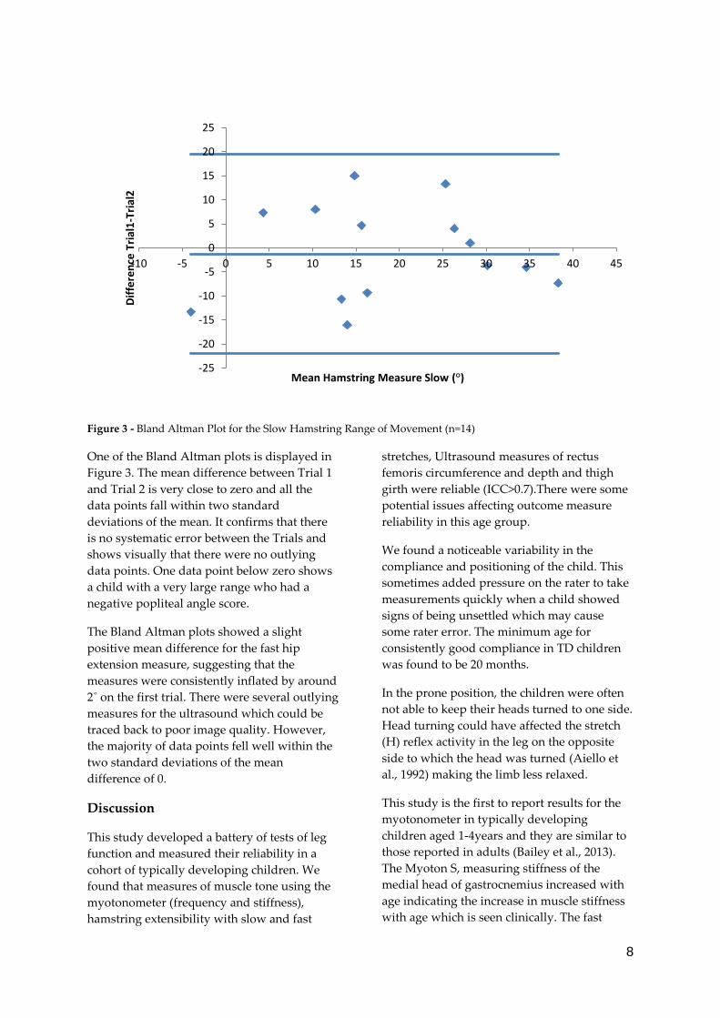
Figure 3 - Bland Altman Plot for the Slow Hamstring Range of Movement (n=14)
One of the Bland Altman plots is displayed in
Figure 3. The mean difference between Trial 1
and Trial 2 is very close to zero and all the
data points fall within two standard
deviations of the mean. It confirms that there
is no systematic error between the Trials and
shows visually that there were no outlying
data points. One data point below zero shows
a child with a very large range who had a
negative popliteal angle score.
The Bland Altman plots showed a slight
positive mean difference for the fast hip
extension measure, suggesting that the
measures were consistently inflated by around
2˚ on the first trial. There were several outlying
measures for the ultrasound which could be
traced back to poor image quality. However,
the majority of data points fell well within the
two standard deviations of the mean
difference of 0.
Discussion
This study developed a battery of tests of leg
function and measured their reliability in a
cohort of typically developing children. We
found that measures of muscle tone using the
myotonometer (frequency and stiffness),
hamstring extensibility with slow and fast
stretches, Ultrasound measures of rectus
femoris circumference and depth and thigh
girth were reliable (ICC>0.7).There were some
potential issues affecting outcome measure
reliability in this age group.
We found a noticeable variability in the
compliance and positioning of the child. This
sometimes added pressure on the rater to take
measurements quickly when a child showed
signs of being unsettled which may cause
some rater error. The minimum age for
consistently good compliance in TD children
was found to be 20 months.
In the prone position, the children were often
not able to keep their heads turned to one side.
Head turning could have affected the stretch
(H) reflex activity in the leg on the opposite
side to which the head was turned (Aiello et
al., 1992) making the limb less relaxed.
This study is the first to report results for the
myotonometer in typically developing
children aged 1-4years and they are similar to
those reported in adults (Bailey et al., 2013).
The Myoton S, measuring stiffness of the
medial head of gastrocnemius increased with
age indicating the increase in muscle stiffness
with age which is seen clinically. The fast
-25
-20
-15
-10
-5
0
5
10
15
20
25
-10 -5 0 5 10 15 20 25 30 35 40 45
Dif
fere
nce
Tri
al1
-Tri
al2
Mean Hamstring Measure Slow (°)
9
range of movement was measured as part of
the Tardieu scale (V3) and in the absence of
increased muscle tone the angle at the end of
range was consistently larger at the faster
speed than the slower. In children with CP,
smaller ranges of overall movement would be
expected at the fast speed due to the ‘catch’ of
the abnormal stretch reflex in the presence of
spasticity.
The study design assumed that the pelvis and
shank were supported in a horizontal plane
for the hip extension and ankle dorsiflexion
measures. During testing it became apparent
that the angle of the shank varied depending
on the bulk of the muscles on the anterior of
the tibia. The pelvis may not have remained
horizontal during the hip extension measure
and may have anteriorly tilted at the end of
range, causing a larger angle to be consistently
recorded. This resulted in poor reliability for
both measures. The digital inclinometer
measures exact angles to two decimal places in
relation to either a horizontal or vertical axis,
whereas the traditional goniometer measures
the angle between two arms using a visual
scale, often rounded to the nearest 5˚. The
hamstring measure included dual
inclinometers where the exact angle was
measured between the two devices, producing
good accuracy and reliability. The positioning
for further reliability of ankle dorsiflexion and
hip extension testing should be changed to
allow a second inclinometer to be used.
The measurements of the hamstrings and
ankle dorsiflexion did not measure the exact
anatomical axes. When measures were taken
with the inclinometers, the axes were along the
shin pads and the footplate. The ankle
measure used a fixed fulcrum on the footplate
with the actual anatomical fulcrum varying
slightly between participants. These factors
need to be considered as they affect the
construct validity of the measurement device
as it could be argued that while they are more
accurate, they are not measuring the true joint
range.
At the outset of the study an attempt was
made to apply a standardised torque. One
limitation to this method was the difficulty in
applying the torque at a standard distance
from the fulcrum due to varying leg lengths.
Secondly, the weight of the limb varied for
each child and changed throughout range with
the effect of gravity and an opposing torque
lessening as the limb approached the vertical.
It may be possible to use an on-line computer
generated algorithm, taking into account the
length of the limb and angle in relation to
gravity in order to apply a standardised
torque, but this was beyond the remit of this
study.
The dimensions of the leg differed greatly
from the youngest to the oldest child. This was
adjusted for with two different length and
width elasticated straps for the footboard, as
the wider strap limited dorsiflexion in the
smallest ankles. It was difficult to apply an
effective force at the ankle to keep the smallest
feet from moving in the footboard laterally
and also to keep the heel down during
dorsiflexion. Ultrasound imaging was more
difficult for the children with the shortest
femurs. The rectus femoris muscle in the
shortest children tapered across the width of
the image due to a shorter overall muscle
length. The measure was taken at a midpoint
on the captured image at 90˚ to the deep
aponeurosis. In three cases this midpoint was
close to the section which steeply graduated
from broad to narrow, making the
measurement more variable. The ultrasound
measures should more accurately represent
muscle size as it is possible to distinguish
between skin, subcutaneous fat and muscle,
whereas the thigh girth gives an indication of
the bulk of both tissues combined. Previous
studies have developed equations for
estimated lean muscle mass based on
measures of limb circumference and skin-fold
thickness (Moritani, 1979) that are correlated
with computerised tomography based
measures of muscle circumference (Defreitas
et al, 2010). Given the high reliability of thigh
circumference the addition of skinfold
measurements, to estimate lean muscle bulk in
the age group, may be warranted. However,
aside from the subcutaneous fat, it should also
be noted that intramuscular fat and fibrosis
10
could also have contributed to the ultrasound
measurement taken.
A limitation of this trial was testing only one
leg, which may have artificially reduced the
time needed to undertake the battery if data
from both legs were needed. Additionally the
study only tested intra-rater reliability, which
limits the ability to generalise to studies that
require inter-rater reliability.
While the results of this trial do not directly
translate to the population of children with CP,
some issues for consideration have arisen from
this trial. Non-ambulant children with CP of
the same age might be more compliant as they
are used to assuming these positions and
being passively moved during therapy.
Conversely, some children with cognitive
difficulties may find engagement with some of
the tests more challenging, especially where
they are required to wear some of the
measurement devices.
Children with CP frequently have persistent
asymmetrical tonic neck reflex, causing
increase in flexor muscle tone on the side to
which the head is turned and increased
extensor tone in the opposite side of the body.
The Thomas test, undertaken in supine with
head in midline, would be preferable for
future battery in children with CP to control
for the influence of head turning in prone and
has been found elsewhere to have similar
reliability to the Staheli test (Glanzman et al.,
2008, Mutlu et al., 2007).
Conclusion
A battery of tests was designed to measure
neuromuscular leg function in typically
developing young children (1-4years). The test
battery included measures of range of
movement, muscle tone and indicators of
muscle strength at the hip, hamstrings and
ankle.
The battery of tests was shown to be feasible to
carry out in the home with young children.
Novel testing methods were developed and
trialled in an attempt to improve accuracy in
clinical measurement. In particular the use of
dual digital inclinometers, and the
myotonometer provided excellent reliability
results and should be considered for use in
future trials.
As the ultimate target group are children with
developmental delay and CP who are non-
ambulant, previous work investigating the
reliability of outcome measures in these
groups, as well as studies in TD children
informed the selection of the tests used. This
battery of tests will need further development
to improve the reliability of those tests that
have not achieved acceptable levels and
reliability testing with children with CP prior
to use in clinical trials.
References
Aarrestad D, Williams M.D, Fehrer S, Mikhailenok
E and Leonard C.T (2004) Intra-and interrater
reliabilities of the myotonometer when assessing
the spastic condition of children with cerebral palsy.
Journal of child neurology, 19, 894-901.
Aiello I, Rosati G, Sau G, Lentinu M, Tidore B,
Sotgiu S, Cacciotto R, Posadinu D, Muzzu S and
Manca I (1992) Interaction of tonic labyrinth and
neck reflexes in man. The Italian Journal of
Neurological Sciences, 13, 195-201.
Bailey L, Samuel D, Warner M and Stokes M (2013)
Parameters representing muscle tone, elasticity and
stiffness of biceps brachii in healthy older males:
symmetry and within-session reliability using the
MyotonPRO. Journal of Neurological Disorders, 1, 1-7.
Bartlett M.D, Wolf L.S, Shurtleff D and Stahell L.T
(1985) Hip flexion contractures: a comparison of
measurement methods. Archives Of Physical
Medicine And Rehabilitation, 66, 620-625.
Bland J.M and Altman D.G (1999) Measuring
agreement in method comparison studies. Stat
Methods Med Res, 8, 135-60.
Boyd R.N and Graham H.K (1999) Objective
measurement of clinical findings in the use of
botulinum toxin type A for the management of
children with cerebral palsy. European Journal of
Neurology, 6, s23-s35.
Defreitas J.M, B.T, Stock M.S, Dillon M.A, Sherk
V.D, Stout J.R and Cramer J.T (2010) A comparison
of techniques for estimating training-induced
11
changes in muscle cross-sectional area. J Strength
Cond Res. , Sept 24, 2383-9.
Fitzpatrick R, Davey C, Buxton, M and Jones D
(1998) Evaluating patient-based outcome measures
for use in clinical trials: a review. Health Technology
Assessment, 2, 74.
Fitzpatrick R, Shortall E, Sculper M, Murray D and
Morris R (1999) Primary total hip replacement
surgery: a structured review of outcomes and
modelling of cost-effectiveness associated with
different prostheses. Health Technology Assessment, 2,
64.
Fosang A.L, Galea M.P, Mccoy A.T, Reddihough
D.S and Story I (2013) Measures of muscle and joint
performance in the lower limb of children with
cerebral palsy. Developmental Medicine & Child
Neurology, 45, 664-670.
Glanzman A.M, Swenson A.E and Kim H (2008)
Intrarater range of motion reliability in cerebral
palsy: a comparison of assessment methods. Pediatr
Phys Ther, 20, 369-72.
Gracies J.M, Marosszeky J.E, Renton R, Sandanam J,
Gandevia S.C and Burke D (2000) Short-term effects
of dynamic Lycra splints on upper limb in
hemiplegic patients. Archives of Physical Medicine
and Rehabilitation, 81, 1547-1555.
Herrero P, Carrera P, García E, Gómez-Trullén E.M
and Oliván-Blázquez B (2011) Reliability of
goniometric measurements in children with
cerebral palsy: a comparative analysis of universal
goniometer and electronic inclinometer. A pilot
study. BMC Musculoskeletal Disorders, 12, 155-155.
Hoskins P.R, Martin K and Thrush A (2010)
Diagnostic ultrasound: physics and equipment,
Cambridge University Press.
Kilgour G, McNair P and Stott N.S (2003) Intrarater
reliability of lower limb sagittal range-of-motion
measures in children with spastic diplegia.
Developmental Medicine & Child Neurology, 45, 391-
399.
Kilgour G.M, McNair P.J and Stott N.S (2002) Lower
limb sagittal range of motion: reliability of
measures and normative values. New Zealand
Journal of Physiotherapy, 30, 8-24.
Kwah L.K, Pinto R.Z, Diong J and Herbert R.D
(2013) Reliability and validity of ultrasound
measurements of muscle fascicle length and
pennation in humans: a systematic review. Journal of
Applied Physiology, 114, 761-769.
Leonard C.T, Deshner W.P, Romo J.W, Suoja E.S,
Fehrer S.C and Mikhailenok E.L (2003)
Myotonometer intra- and interrater reliabilities.
Arch Phys Med Rehabil, 84, 928-32
Lidstrom A, Ahlsten G, Hirchfeld H and Norrlin S
(2009) Intrarater and interrater reliability of
Myotonometer measurements of muscle tone in
children. J Child Neurol, 24, 267-74.
Maas J. C, Dallmeijer A. J, Huijing P. A, Brunstrom-
Hernandez J. E, Van Kampen P. J, Jaspers R. T and
Becher J. G (2012) Splint: the efficacy of orthotic
management in rest to prevent equinus in children
with cerebral palsy, a randomised controlled trial.
BMC Pediatr, 12, 38.
Mcdowell B. C, Hewitt V, Nurse A, Weston T. and
Baker R (2000) The variability of goniometric
measurements in ambulatory children with spastic
cerebral palsy. Gait & posture, 12, 114-121.
Moreau N. G, Simpson K. N, Teefey S. A and
Damiano D. L (2010) Muscle architecture predicts
maximum strength and is related to activity levels
in cerebral palsy. Phys Ther, 90, 1619-30.
Moritani T. A. D., Ha (1979) Neural factors versus
hypertrophy in the time course of muscle strength
gain . Am J Phys Med 58, 115-130.
Mutlu A, Livanelioglu A and Gunel M. K (2007)
Reliability of goniometric measurements in children
with spastic cerebral palsy. Med Sci Monit, 13,
Cr323-9.
Ohata K, Tsuboyama T, Haruta T, Ichihashi N, Kato
T and Nakamura T (2008) Relation between muscle
thickness, spasticity, and activity limitations in
children and adolescents with cerebral palsy. Dev
Med Child Neurol, 50, 152-6.
12
Pandyan A. D, Johnson G. R, Price C. I. M, Curless
R. H, Barnes M. P. and Rodgers H (1999) A review
of the properties and limitations of the Ashworth
and modified Ashworth Scales as measures of
spasticity. Clinical Rehabilitation, 13, 373-383.
Scholtes V. A, Becher J. G, Beelen A and Lankhorst
G. J (2006) Clinical assessment of spasticity in
children with cerebral palsy: a critical review of
available instruments. Dev Med Child Neurol, 48, 64-
73.
Shrout Pe F. J (1979) Intraclass correlations: Uses in
assessing rater reliability. Psychological Bulletin, 86,
420-8.
Soucie J. M, Wang C, Forsyth A, Funk S, Denny M,
Roach K. E and Boone, D (2011) Range of motion
measurements: reference values and a database for
comparison studies. Haemophilia, 17, 500-7.
Stuberg W. A, Fuchs R. H and Miedaner J. A (1988)
Reliability of goniometric measurements of children
with cerebral palsy. Dev Med Child Neurol, 30, 657-66.
Weir J. P (2005) Quantifying test-retest reliability
using the intraclass correlation coefficient and the
SEM. J Strength Cond Res, 19, 231-40.
Related Documents