ORIGINAL ARTICLE: Experimental Endoscopy Third-generation cholecystectomy by natural orifices: transgastric and transvesical combined approach (with video) Carla Rolanda, MD, Este ˆva ˜o Lima, MD, Jose ´ M. Pe ˆgo, MD, Tiago Henriques-Coelho, MD, David Silva, MD, Ivone Moreira, Guilherme Macedo, MD, PhD, Jose ´ L. Carvalho, MD, Jorge Correia-Pinto, MD, PhD Braga, Portugal Background: An isolated transgastric port has some limitations in performing transluminal endoscopic chole- cystectomy. However, transvesical access to the peritoneal cavity has recently been reported to be feasible and safe. Objective: To assess the feasibility and the technical benefits of transgastric and transvesical combined approach to overcome the limitations of isolated transgastric ports. Design: We created a transgastric and transvesical combined approach to perform cholecystectomy in 7 consec- utive anesthetized female pigs. The transgastric access was achieved after perforation and dilation of the gastric wall with a needle knife and with a balloon, respectively. Under cystoscopic control, an ureteral catheter, a guide- wire, and a dilator of the ureteral sheath were used to place a transvesical 5-mm overtube into the peritoneal cavity. By using a gastroscope positioned transgastrically and a ureteroscope positioned transvesically, we carried out cholecystectomy in all animals. Results: Establishment of transvesical and transgastric accesses took place without complications. Under a car- bon dioxide pneumoperitoneum controlled by the transvesical port, gallbladder identification, cystic duct, and artery exposure were easily achieved in all cases. Transvesical gallbladder grasping and manipulation proved to be particularly valuable to enhance gastroscope-guided dissection. With the exclusion of 2 cases where mild liver-surface hemorrhage and bile leak secondary to the sliding of cystic clips occurred, all remaining cholecys- tectomies were carried out without incidents. Limitations: Once closure of the gastric hole proved to be unreliable when using endoclips, the animals were euthanized; necropsy was performed immediately after the surgical procedure. Conclusions: A transgastric and transvesical combined approach is feasible, and it was particularly useful to perform a cholecystectomy through exclusive natural orifices. (Gastrointest Endosc 2007;65:111-7.) Since the first reports in the late 1980s, laparoscopy has progressively become the criterion standard for cholecys- tectomy, one of the most widely performed abdominal in- terventions in developed countries. In fact, minimally invasive surgery is now associated with many proven ad- vantages over traditional open procedures, such as mini- mal scarring, reduced pain, and faster patient recovery. 1 In parallel with the progression of minimally invasive surgery, improvements in endoluminal endoscopy have made it an indispensable and multifaceted instrument for diagnosis and, definitively, for therapy. 2 Recently, Reddy and Rao (N. Reddy,V. G. Rao, oral communications, May 2005; N. Reddy, oral communication, May 2004), in human beings, and Kalloo et al, 3 in a porcine model, de- scribed a new port to the peritoneal cavity through a trans- gastric approach. Subsequently, various investigators described more complex intra-abdominal procedures in porcine model, 4-12 opening a new era in the surgical field in what seems to be the third-generation surgery: natural orifice transluminal endoscopic surgery (NOTES). 13 Park et al 5 conducted the first pilot study in pigs by applying NOTES to perform transgastric cholecystectomy. By using 2 endoscopes or a single endoscope conjugated with a transabdominal trocar, cholecystectomy was feasi- ble, but important limitations were identified. These were related to difficulty in controlling the pneumoperito- neum and in obtaining a stable platform for anatomy ex- posure, organ retraction, secure grasping, and adequate Copyright ª 2007 by the American Society for Gastrointestinal Endoscopy 0016-5107/$32.00 doi:10.1016/j.gie.2006.07.050 www.giejournal.org Volume 65, No. 1 : 2007 GASTROINTESTINAL ENDOSCOPY 111

Welcome message from author
This document is posted to help you gain knowledge. Please leave a comment to let me know what you think about it! Share it to your friends and learn new things together.
Transcript
ORIGINAL ARTICLE: Experimental Endoscopy
Third-generation cholecystectomy by natural orifices: transgastricand transvesical combined approach (with video)
Carla Rolanda, MD, Estevao Lima, MD, Jose M. Pego, MD, Tiago Henriques-Coelho, MD, David Silva, MD,Ivone Moreira, Guilherme Macedo, MD, PhD, Jose L. Carvalho, MD, Jorge Correia-Pinto, MD, PhD
Braga, Portugal
Background: An isolated transgastric port has some limitations in performing transluminal endoscopic chole-cystectomy. However, transvesical access to the peritoneal cavity has recently been reported to be feasible andsafe.
Objective: To assess the feasibility and the technical benefits of transgastric and transvesical combinedapproach to overcome the limitations of isolated transgastric ports.
Design: We created a transgastric and transvesical combined approach to perform cholecystectomy in 7 consec-utive anesthetized female pigs. The transgastric access was achieved after perforation and dilation of the gastricwall with a needle knife and with a balloon, respectively. Under cystoscopic control, an ureteral catheter, a guide-wire, and a dilator of the ureteral sheath were used to place a transvesical 5-mm overtube into the peritonealcavity. By using a gastroscope positioned transgastrically and a ureteroscope positioned transvesically, we carriedout cholecystectomy in all animals.
Results: Establishment of transvesical and transgastric accesses took place without complications. Under a car-bon dioxide pneumoperitoneum controlled by the transvesical port, gallbladder identification, cystic duct, andartery exposure were easily achieved in all cases. Transvesical gallbladder grasping and manipulation proved tobe particularly valuable to enhance gastroscope-guided dissection. With the exclusion of 2 cases where mildliver-surface hemorrhage and bile leak secondary to the sliding of cystic clips occurred, all remaining cholecys-tectomies were carried out without incidents.
Limitations: Once closure of the gastric hole proved to be unreliable when using endoclips, the animals wereeuthanized; necropsy was performed immediately after the surgical procedure.
Conclusions: A transgastric and transvesical combined approach is feasible, and it was particularly useful toperform a cholecystectomy through exclusive natural orifices. (Gastrointest Endosc 2007;65:111-7.)
Since the first reports in the late 1980s, laparoscopy hasprogressively become the criterion standard for cholecys-tectomy, one of the most widely performed abdominal in-terventions in developed countries. In fact, minimallyinvasive surgery is now associated with many proven ad-vantages over traditional open procedures, such as mini-mal scarring, reduced pain, and faster patient recovery.1
In parallel with the progression of minimally invasivesurgery, improvements in endoluminal endoscopy havemade it an indispensable and multifaceted instrumentfor diagnosis and, definitively, for therapy.2 Recently,Reddy and Rao (N. Reddy, V. G. Rao, oral communications,
Copyright ª 2007 by the American Society for Gastrointestinal Endoscopy
0016-5107/$32.00
doi:10.1016/j.gie.2006.07.050
www.giejournal.org
May 2005; N. Reddy, oral communication, May 2004), inhuman beings, and Kalloo et al,3 in a porcine model, de-scribed a new port to the peritoneal cavity through a trans-gastric approach. Subsequently, various investigatorsdescribed more complex intra-abdominal procedures inporcine model,4-12 opening a new era in the surgical fieldin what seems to be the third-generation surgery: naturalorifice transluminal endoscopic surgery (NOTES).13
Park et al5 conducted the first pilot study in pigs byapplying NOTES to perform transgastric cholecystectomy.By using 2 endoscopes or a single endoscope conjugatedwith a transabdominal trocar, cholecystectomy was feasi-ble, but important limitations were identified. Thesewere related to difficulty in controlling the pneumoperito-neum and in obtaining a stable platform for anatomy ex-posure, organ retraction, secure grasping, and adequate
Volume 65, No. 1 : 2007 GASTROINTESTINAL ENDOSCOPY 111
Cholecystectomy by natural orifice transluminal endoscopic surgery Rolanda et al
triangulation of instruments. Swanstrom et al12 attemptedto overcome these limitations by using ShapeLock tech-nology (USGI Medical, San Clement, Calif) as a new over-tube for transgastric surgery. However, even with thisequipment, an isolated transgastric approach for gallblad-der manipulation remained a challenge, with only a 33.3%success rate.12
These studies were able to demonstrate that cholecys-tectomy may one day be performed without skin incisions.The development of other natural orifice accesses, how-ever, may play an important role in overcoming some ofthe limitations identified for those who performed ab-dominal surgery through isolated transgastric surgery.13
By applying these concepts, Lima et al14 demonstratedthat transvesical endoscopic peritoneoscopy was techni-cally feasible and could be safely performed in a porcinemodel. By using a transvesical port, it was possible to in-troduce 5-mm rigid instruments, such as graspers, scis-sors, and telescopes. We hypothesized that a transvesicalport could be useful to perform abdominal proceduresin combination with a transgastric pathway.
The purpose of this experimental study was to assessthe feasibility and the technical benefits of a combinedtransgastric and transvesical approach for cholecystec-tomy in a porcine model.
MATERIALS AND METHODS
This was a nonsurvival study approved by the ethicalreview boards of Minho University (Braga, Portugal). Forthis study, we included 15 small (15-25 kg) female pigs(Sus scrofus domesticus) so that the current ureteroscopelength could easily achieve the upper-abdominal organs.After a significant surgical and anesthetic learning curve,the results of which are not included in this report (9animals), we performed the cholecystectomy exclusivelythrough natural orifices (transgastric and transvesical com-bined approach) in 7 consecutive animals. After the surgi-cal procedures, the animals were euthanized, andnecropsies were performed.
Pig preparationThe animals were fed liquids for 3 days and then were
restrained from food (24 hours) and water (6 hours) be-fore the surgical intervention. All procedures were perfor-med with the pigs under general anesthesia, with 5.0-mmendotracheal intubation and mechanical ventilation.Preanesthesia medication consisted of an intramuscularinjection of 32 mg/mL azaperone (Stressnil; Esteve Farma,Barcelona, Spain) reconstituted with 1 mg/mL midazolam(Dormicum; Roche, Amadora, Portugal) at a dose of 0.15to 0.2 mL/kg.
Venous access was obtained through an intravenousline placed in the marginal ear vein. Anesthesia was in-duced with 3 mg/kg fentanyl (Fentanest; Janssen-Cilag,
112 GASTROINTESTINAL ENDOSCOPY Volume 65, No. 1 : 2007
Capsule Summary
What is already known on this topic
d Transgastric cholecystectomy is feasible in a porcinemodel, but it has limitations that jeopardize its clinicalapplication, including difficulty in controlling thepneumoperitoneum and in securing a stable platform foranatomy exposure, organ retraction, secure grasping,and adequate instrument triangulation.
What this study adds to our knowledge
d In 7 pigs, gallbladder identification and cystic duct andartery exposure were achieved by using transvesicaland transgastric access; transvesical gallbladder graspingand manipulation was particularly useful in gastroscope-guided dissection.
Queluz, Portugal), 10 mg/kg thiopental sodium (Pento-thal; Abbott, Alfragide, Portugal), and 1 mg/kg vecuronium(Norcuron; Organon, Oss, The Netherlands). Anesthesiawas maintained with 1.5% to 2% sevoflurane (Sevorane;Abbott) and a perfusion of 1 mg/kg per hour of vecuro-nium. All animals received an intramuscular injection of1 g ceftriaxone (Rocephin; Roche) before beginning thesurgical procedures.
Surgical techniqueTransvesical access. Creation of a transvesical access
is illustrated in Video 1 (available online at http://www.giejournal.org). An ureteroscope (A2942A; Olympus, To-kyo, Japan) was introduced through the urogenital sinusand the urethra into the bladder with hydrodistension. Be-fore any further procedure, the bladder was emptied ofurine and refilled with saline solution. The vesicotomysite was carefully selected on the ventral bladder wall, pos-terior to the bladder dome. A mucosal incision was madewith a scissors (A2576; Olympus) introduced by the work-ing channel of the ureteroscope. Subsequently, a 5F open-end ureteral catheter (Selectip, 62450200; Angiomed,Bard, Murray Hill, NJ) was pushed forward through the in-cision into the peritoneal cavity. A 0.035-inch flexible-tipguidewire (RF*GA35153M, Terumo Corp, Somerset, NJ)was then inserted into the peritoneal cavity through thelumen of the ureteral catheter. Guided by the flexible-tipguidewire, the vesical hole was enlarged with a dilator ofan ureteroscope sheath (250-105; Microvasive Endoscopy,Boston Scientific Corp, Natick, Mass), which was envel-oped with an overtube designed by us (25-cm length,5.5-mm internal diameter, and 1-mm wall thickness). Arigid ureteroscope was introduced into the peritoneal cav-ity within the overtube and allowed the creation of a pres-sure-controlled carbon dioxide (CO2) pneumoperitoneumup to 12 mmHg. Through the overtube, we could insertinto the peritoneal cavity either a video telescope with
www.giejournal.org
Rolanda et al Cholecystectomy by natural orifice transluminal endoscopic surgery
5-mm diameter, chip-on-the-tip, and 0-degree view direc-tion, normally used as a laparoscope (EndoEye 50021A;Olympus) or a rigid ureteroscope that includes a workingchannel. The peritoneal cavity was thoroughly examined,with particular emphasis on the stomach, the liver, andthe gallbladder.
Transgastric access. An adult forward-viewing, dou-ble-channel endoscope (GIF-2T160; Olympus) was ad-vanced into the esophagus and the stomach. Thestomach was lavaged with instilled water and was aspi-rated through the gastroscope until free of food particles.Subsequently, it was decompressed and a cefazolin solu-tion (1 g in 200 mL saline solution) was instilled. The an-tibiotic solution was left in the stomach for 10 minutesbefore also being aspirated. For the gastrotomy-site selec-tion and to avoid damage of the gastric vessels or sur-rounding organs, the gastroscope operator was workingwith both internal (provided by the gastroscope observingthe internal stomach indentation produced by externalabdominal palpation) and external (provided by theEndoEye or ureteroscope) gastric-wall images. The gastro-tomy site was carefully chosen on the anterior wall (body-antrum transition). A gastric-wall incision was made bypushing forward a needle knife, followed by its sheath(KD-11Q-1; Olympus) with cautery (PSD 20; Olympus) un-der a 12 mmHg CO2 pneumoperitoneum (inducedthrough the transvesical port). The needle-knife sheathwas then used for positioning a guidewire (5156-01; Micro-vasive Endoscopy). The puncture dilation was performedwith an 18-mm through-the-scope balloon (5837; Microva-sive Endoscopy) over the guidewire. On balloon semide-flection, the gastroscope was pushed forward andpassed into the peritoneal cavity. All these procedureswere monitored by the scope positioned through thetransvesical port.
Cholecystectomy surgical procedureAfter establishment of the 2 diametrically opposed
ports (transgastric and transvesical) and CO2-pneumoper-itoneum creation, the ureteroscope easily identified thegallbladder. A forceps was then passed through the ure-teroscope working channel, and the gallbladder funduswas grasped and upward retracted. This maneuver nicelyexposed the gallbladder infundibulum and the cysticduct. Subsequently, the gastroscope was moved on retro-flexion toward the gallbladder. The exposure achieved bythe transvesical gallbladder grasping allowed the gastro-scope operator to quickly identify the cystic duct. Thisallowed us to start careful dissection, handling eithera grasping forceps (FG-6L-1, FG-47L-1; Olympus) ora 2.8-mm ball coagulation electrode (CD-1U; Olympus)passed through the working channels of the gastroscope.For this purpose, we also had available a pre-cut needleknife (KD-11Q-1; Olympus) and a scissors (FS-5L-1; Olym-pus). Both sides of cystic duct were completely dissectedby alternating the working channels of the grasping
www.giejournal.org
forceps and the coagulation electrode. When the cysticduct and artery were dissected and free, 3 clips (HX-200L-135; Olympus) were applied: 2 on the gallbladderextremity and the other proximally. Sectioning of pediclewas then carried out with a needle knife. The graspingprovided by the ureteroscope allowed us to retract thegallbladder body in the major axis (up and down; rightand left), looking for the most appropriate anatomicalexposure for dissection. Small position adjustments werealso possible when using the forceps introduced by thegastroscope. Blunt dissection was taken progressively byelectrocautery of the gallbladder bed. When cholecystec-tomy was completed, the gastroscopic forceps held thecystic-duct extremity distally to clips, and the gallbladderwas removed via the esophagus and the mouth.
RESULTS
All procedures involved in the creation of the vesicalhole (cystoscopy, bladder mucosal incision, vesicotomy,transvesical overtube passage) were performed withoutcomplications. The ureteroscope was easily introducedinto the peritoneal cavity, and insufflation of CO2 was per-formed without incident. By using the transvesical port,we could obtain a perfect view of the upper-abdominal or-gans (liver, gallbladder, stomach, spleen, and diaphragm).This was particularly useful in helping the gastroscopeoperator to safely perform the gastrotomy.
Under a CO2 pneumoperitoneum and with the viewprovided by the transvesical port, we created the gastro-tomy in a rapid, safe, and easy way. In fact, with our ap-proach, neither gastric vessels nor surrounding organswere damaged. In addition, the ureteroscope was ex-tremely useful many times in assisting the passage of thegastroscope through the gastrotomy.
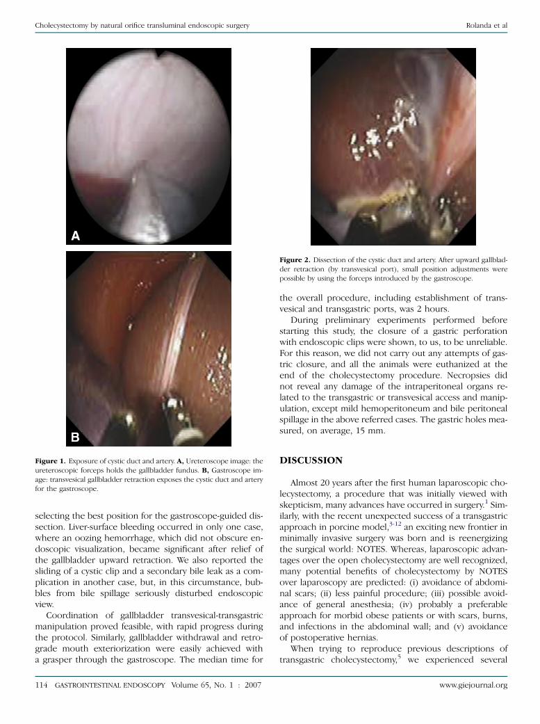
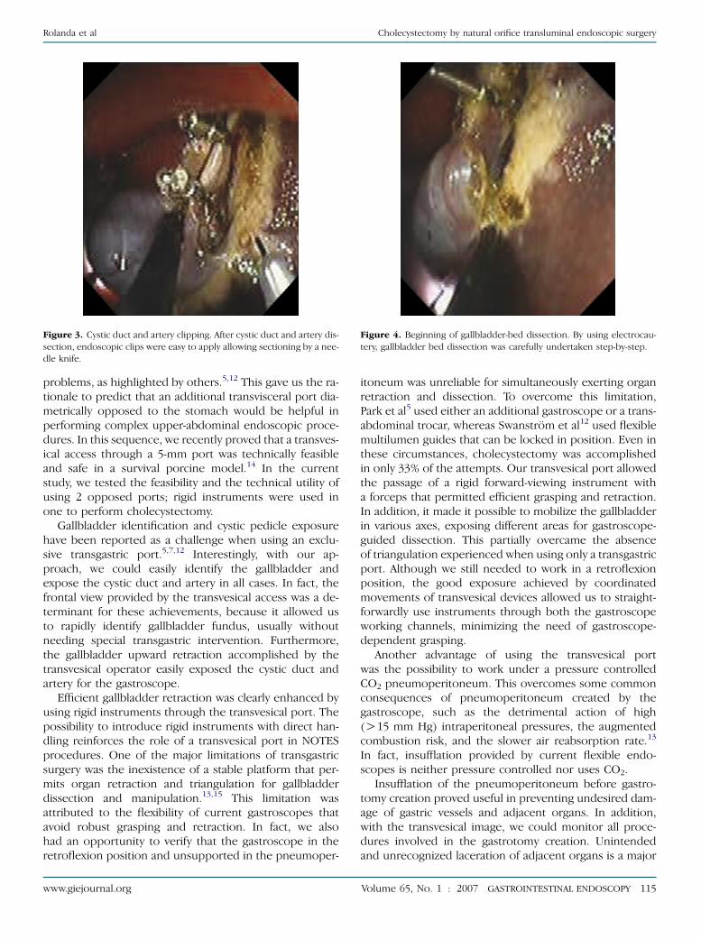
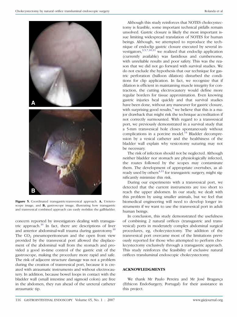
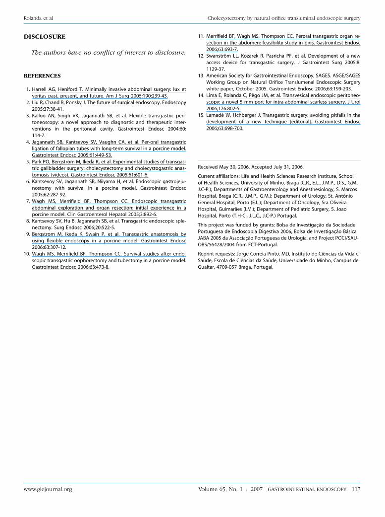
Gallbladder identification was easily achieved in allexperiments, first, by the ureteroscope. After gallbladderfundus grasping with transvesical instruments, the gastro-scope easily identified the cystic duct in all cases (Fig. 1).Under transvesical gallbladder retraction, gastroscopic dis-section (Fig. 2), isolation, clipping, and sectioning (Fig. 3)of the cystic duct and artery was feasible and, therefore,was performed in a precise way in all cases. For these pur-poses, we successfully used a grasping forceps and a coag-ulation electrode inserted through the gastroscopeworking channels. A pre-cut needle knife and scissorswere rarely applied in these procedures. After sectioningof the clipped cystic duct and artery, we began a dissectionof the gallbladder from its bed by using the coagulationelectrode (Fig. 4). This proved to be the most fastidiousand, simultaneously, the most challenging part of the sur-gery, because the gallbladder body is commonly hidden inthe liver parenchyma in pigs. Nevertheless, the transvesi-cal grasping allowed significant manipulation of the gall-bladder (Fig. 5), which was particularly useful in
Volume 65, No. 1 : 2007 GASTROINTESTINAL ENDOSCOPY 113
Cholecystectomy by natural orifice transluminal endoscopic surgery Rolanda et al
selecting the best position for the gastroscope-guided dis-section. Liver-surface bleeding occurred in only one case,where an oozing hemorrhage, which did not obscure en-doscopic visualization, became significant after relief ofthe gallbladder upward retraction. We also reported thesliding of a cystic clip and a secondary bile leak as a com-plication in another case, but, in this circumstance, bub-bles from bile spillage seriously disturbed endoscopicview.
Coordination of gallbladder transvesical-transgastricmanipulation proved feasible, with rapid progress duringthe protocol. Similarly, gallbladder withdrawal and retro-grade mouth exteriorization were easily achieved witha grasper through the gastroscope. The median time for
Figure 1. Exposure of cystic duct and artery. A, Ureteroscope image: the
ureteroscopic forceps holds the gallbladder fundus. B, Gastroscope im-
age: transvesical gallbladder retraction exposes the cystic duct and artery
for the gastroscope.
114 GASTROINTESTINAL ENDOSCOPY Volume 65, No. 1 : 2007
the overall procedure, including establishment of trans-vesical and transgastric ports, was 2 hours.
During preliminary experiments performed beforestarting this study, the closure of a gastric perforationwith endoscopic clips were shown, to us, to be unreliable.For this reason, we did not carry out any attempts of gas-tric closure, and all the animals were euthanized at theend of the cholecystectomy procedure. Necropsies didnot reveal any damage of the intraperitoneal organs re-lated to the transgastric or transvesical access and manip-ulation, except mild hemoperitoneum and bile peritonealspillage in the above referred cases. The gastric holes mea-sured, on average, 15 mm.
DISCUSSION
Almost 20 years after the first human laparoscopic cho-lecystectomy, a procedure that was initially viewed withskepticism, many advances have occurred in surgery.1 Sim-ilarly, with the recent unexpected success of a transgastricapproach in porcine model,3-12 an exciting new frontier inminimally invasive surgery was born and is reenergizingthe surgical world: NOTES. Whereas, laparoscopic advan-tages over the open cholecystectomy are well recognized,many potential benefits of cholecystectomy by NOTESover laparoscopy are predicted: (i) avoidance of abdomi-nal scars; (ii) less painful procedure; (iii) possible avoid-ance of general anesthesia; (iv) probably a preferableapproach for morbid obese patients or with scars, burns,and infections in the abdominal wall; and (v) avoidanceof postoperative hernias.
When trying to reproduce previous descriptions oftransgastric cholecystectomy,5 we experienced several
Figure 2. Dissection of the cystic duct and artery. After upward gallblad-
der retraction (by transvesical port), small position adjustments were
possible by using the forceps introduced by the gastroscope.
www.giejournal.org
Rolanda et al Cholecystectomy by natural orifice transluminal endoscopic surgery
problems, as highlighted by others.5,12 This gave us the ra-tionale to predict that an additional transvisceral port dia-metrically opposed to the stomach would be helpful inperforming complex upper-abdominal endoscopic proce-dures. In this sequence, we recently proved that a transves-ical access through a 5-mm port was technically feasibleand safe in a survival porcine model.14 In the currentstudy, we tested the feasibility and the technical utility ofusing 2 opposed ports; rigid instruments were used inone to perform cholecystectomy.
Gallbladder identification and cystic pedicle exposurehave been reported as a challenge when using an exclu-sive transgastric port.5,7,12 Interestingly, with our ap-proach, we could easily identify the gallbladder andexpose the cystic duct and artery in all cases. In fact, thefrontal view provided by the transvesical access was a de-terminant for these achievements, because it allowed usto rapidly identify gallbladder fundus, usually withoutneeding special transgastric intervention. Furthermore,the gallbladder upward retraction accomplished by thetransvesical operator easily exposed the cystic duct andartery for the gastroscope.
Efficient gallbladder retraction was clearly enhanced byusing rigid instruments through the transvesical port. Thepossibility to introduce rigid instruments with direct han-dling reinforces the role of a transvesical port in NOTESprocedures. One of the major limitations of transgastricsurgery was the inexistence of a stable platform that per-mits organ retraction and triangulation for gallbladderdissection and manipulation.13,15 This limitation wasattributed to the flexibility of current gastroscopes thatavoid robust grasping and retraction. In fact, we alsohad an opportunity to verify that the gastroscope in theretroflexion position and unsupported in the pneumoper-
Figure 3. Cystic duct and artery clipping. After cystic duct and artery dis-
section, endoscopic clips were easy to apply allowing sectioning by a nee-
dle knife.
www.giejournal.org
itoneum was unreliable for simultaneously exerting organretraction and dissection. To overcome this limitation,Park et al5 used either an additional gastroscope or a trans-abdominal trocar, whereas Swanstrom et al12 used flexiblemultilumen guides that can be locked in position. Even inthese circumstances, cholecystectomy was accomplishedin only 33% of the attempts. Our transvesical port allowedthe passage of a rigid forward-viewing instrument witha forceps that permitted efficient grasping and retraction.In addition, it made it possible to mobilize the gallbladderin various axes, exposing different areas for gastroscope-guided dissection. This partially overcame the absenceof triangulation experienced when using only a transgastricport. Although we still needed to work in a retroflexionposition, the good exposure achieved by coordinatedmovements of transvesical devices allowed us to straight-forwardly use instruments through both the gastroscopeworking channels, minimizing the need of gastroscope-dependent grasping.
Another advantage of using the transvesical portwas the possibility to work under a pressure controlledCO2 pneumoperitoneum. This overcomes some commonconsequences of pneumoperitoneum created by thegastroscope, such as the detrimental action of high(O15 mm Hg) intraperitoneal pressures, the augmentedcombustion risk, and the slower air reabsorption rate.13
In fact, insufflation provided by current flexible endo-scopes is neither pressure controlled nor uses CO2.
Insufflation of the pneumoperitoneum before gastro-tomy creation proved useful in preventing undesired dam-age of gastric vessels and adjacent organs. In addition,with the transvesical image, we could monitor all proce-dures involved in the gastrotomy creation. Unintendedand unrecognized laceration of adjacent organs is a major
Figure 4. Beginning of gallbladder-bed dissection. By using electrocau-
tery, gallbladder bed dissection was carefully undertaken step-by-step.
Volume 65, No. 1 : 2007 GASTROINTESTINAL ENDOSCOPY 115
Cholecystectomy by natural orifice transluminal endoscopic surgery Rolanda et al
concern reported by investigators dealing with transgas-tric approach.16 In fact, there are descriptions of liverand anterior abdominal-wall trauma during gastrotomy.10
The CO2 pneumoperitoneum and the open front viewprovided by the transvesical port allowed the displace-ment of the abdominal wall from the stomach and pro-vided a good in-time control of the gastric exit of thegastroscope, making the procedure more rapid and safe.The risk of adjacent structure damage was not a problemduring the creation of transvesical port, because it is cre-ated with atraumatic instruments and without electrocau-tery. In addition, because bowel loops in contact with thebladder wall (small intestine and sigmoid colon) are freein the abdomen, they run ahead of the ureteral catheteratraumatic tip.
Figure 5. Coordinated transgastric-transvesical approach. A, Uretero-
scope image, and B, gastroscope image, illustrating how transgastric
and transvesical combined approach can easily mobilize the gallbladder.
116 GASTROINTESTINAL ENDOSCOPY Volume 65, No. 1 : 2007
Although this study reinforces that NOTES cholecystec-tomy is feasible, some important technical pitfalls remainunsolved. Gastric closure is likely the most important is-sue limiting widespread translation of NOTES for humanbeings. Although, we attempted to reproduce the tech-nique of endoclip gastric closure executed by several in-vestigators,3,5,7,10,11 we realized that endoclip application(currently available) was fastidious and cumbersome,with unreliable results and poor safety. This was the rea-son that we did not go forward with survival studies. Wedo not exclude the hypothesis that our technique for gas-tric perforation (balloon dilation) disturbed the condi-tions for clip application. In fact, we recognize that ifdilation is efficient in maintaining muscle integrity for con-traction, the cutting electrocautery would define moreregular borders for tissue approximation. Even knowinggastric injuries heal quickly and that survival studieshave been done, without any maneuver for gastric closure,with surprising good results,4 we believe that this is a ma-jor drawback that might risk the technique accreditation ifnot correctly surmounted. With regard to a transvesicalport, we previously demonstrated in a survival study thata 5-mm transvesical hole closes spontaneously withoutcomplications in a porcine model.14 Bladder decompre-ssion by a vesical catheter and the healthiness of thebladder wall explain why vesicotomy suturing may notbe necessary.
The risk of infection should not be neglected. Althoughneither bladder nor stomach are physiologically infected,the routes followed by the scopes may contaminatethem. The development of appropriate overtubes, as al-ready used by others4,11 for transgastric surgery, might sig-nificantly minimize this risk.
During our experiments with a transvesical port, wedetected that the current instruments are too short toreach the upper abdomen. In our study, we dealt withthis problem by using smaller animals, but we feel thatbiomedical engineering will need to develop longer in-struments if we want to use the transvesical port in adulthuman beings.
In conclusion, this study demonstrated the usefulnessof combining 2 natural orifices (transgastric and trans-vesical) ports in moderately complex abdominal surgicalprocedures, eg, cholecystectomy. The addition of thetransvesical port overcame most of the limitations previ-ously reported for those who attempted to perform cho-lecystectomy exclusively through a transgastric approach.This study reinforces the feasibility of exclusive naturalorifices transluminal endoscopic cholecystectomy.
ACKNOWLEDGMENTS
We thank Mr Paulo Pereira and Mr Jose Braganca(Ethicon EndoSurgery, Portugal) for their assistance inthis project.
www.giejournal.org
Rolanda et al Cholecystectomy by natural orifice transluminal endoscopic surgery
DISCLOSURE
The authors have no conflict of interest to disclosure.
REFERENCES
1. Harrell AG, Heniford T. Minimally invasive abdominal surgery: lux et
veritas past, present, and future. Am J Surg 2005;190:239-43.
2. Liu R, Chand B, Ponsky J. The future of surgical endoscopy. Endoscopy
2005;37:38-41.
3. Kalloo AN, Singh VK, Jagannath SB, et al. Flexible transgastric peri-
toneoscopy: a novel approach to diagnostic and therapeutic inter-
ventions in the peritoneal cavity. Gastrointest Endosc 2004;60:
114-7.
4. Jagannath SB, Kantsevoy SV, Vaughn CA, et al. Per-oral transgastric
ligation of fallopian tubes with long-term survival in a porcine model.
Gastrointest Endosc 2005;61:449-53.
5. Park PO, Bergstrom M, Ikeda K, et al. Experimental studies of transgas-
tric gallbladder surgery: cholecystectomy and cholecystogastric anas-
tomosis (videos). Gastrointest Endosc 2005;61:601-6.
6. Kantsevoy SV, Jagannath SB, Niiyama H, et al. Endoscopic gastrojeju-
nostomy with survival in a porcine model. Gastrointest Endosc
2005;62:287-92.
7. Wagh MS, Merrifield BF, Thompson CC. Endoscopic transgastric
abdominal exploration and organ resection: initial experience in a
porcine model. Clin Gastroenterol Hepatol 2005;3:892-6.
8. Kantsevoy SV, Hu B, Jagannath SB, et al. Transgastric endoscopic sple-
nectomy. Surg Endosc 2006;20:522-5.
9. Bergstrom M, Ikeda K, Swain P, et al. Transgastric anastomosis by
using flexible endoscopy in a porcine model. Gastrointest Endosc
2006;63:307-12.
10. Wagh MS, Merrifield BF, Thompson CC. Survival studies after endo-
scopic transgastric oophorectomy and tubectomy in a porcine model.
Gastrointest Endosc 2006;63:473-8.
www.giejournal.org
11. Merrifield BF, Wagh MS, Thompson CC. Peroral transgastric organ re-
section in the abdomen: feasibility study in pigs. Gastrointest Endosc
2006;63:693-7.
12. Swanstrom LL, Kozarek R, Pasricha PF, et al. Development of a new
access device for transgastric surgery. J Gastrointest Surg 2005;8:
1129-37.
13. American Society for Gastrointestinal Endoscopy, SAGES. ASGE/SAGES
Working Group on Natural Orifice Translumenal Endoscopic Surgery
white paper, October 2005. Gastrointest Endosc 2006;63:199-203.
14. Lima E, Rolanda C, Pego JM, et al. Transvesical endoscopic peritoneo-
scopy: a novel 5 mm port for intra-abdominal scarless surgery. J Urol
2006;176:802-5.
15. Lamade W, Hchberger J. Transgastric surgery: avoiding pitfalls in the
development of a new technique [editorial]. Gastrointest Endosc
2006;63:698-700.
Received May 30, 2006. Accepted July 31, 2006.
Current affiliations: Life and Health Sciences Research Institute, School
of Health Sciences, University of Minho, Braga (C.R., E.L., J.M.P., D.S., G.M.,
J.C-P.); Departments of Gastroenterology and Anesthesiology, S. Marcos
Hospital, Braga (C.R., J.M.P., G.M.); Department of Urology, St. Antonio
General Hospital, Porto (E.L.); Department of Oncology, Sra Oliveira
Hospital, Guimaraes (I.M.); Department of Pediatric Surgery, S. Joao
Hospital, Porto (T.H-C., J.L.C., J.C-P.) Portugal.
This project was funded by grants: Bolsa de Investigacao da Sociedade
Portuguesa de Endoscopia Digestiva 2006, Bolsa de Investigacao Basica
JABA 2005 da Associacao Portuguesa de Urologia, and Project POCI/SAU-
OBS/56428/2004 from FCT-Portugal.
Reprint requests: Jorge Correia-Pinto, MD, Instituto de Ciencias da Vida e
Saude, Escola de Ciencias da Saude, Universidade do Minho, Campus de
Gualtar, 4709-057 Braga, Portugal.
Volume 65, No. 1 : 2007 GASTROINTESTINAL ENDOSCOPY 117
Related Documents