Thigh Strength and Activation as Predictors of Knee Biomechanics during a Drop Jump Task By: SANDRA J. SHULTZ , ANH-DUNG NGUYEN , MICHAEL D. LEONARD, and RANDY J. SCHMITZ SHULTZ, S. J., A. NGUYEN, M. D. LEONARD, and R. J. SCHMITZ. Thigh Strength and Activation as Predictors of Knee Biomechanics during a Drop Jump Task. Med. Sci. Sports Exerc. , Vol. 41, No. 4, pp. 857– 866, 2009. DOI: 10.1249/MSS.0b013e3181e3b3f Made available courtesy of the American College of Sports Medicine: http://www.acsm.org/ ***Reprinted with permission. No further reproduction is authorized without written permission from the American College of Sports Medicine. This version of the document is not the version of record. Figures and/or pictures may be missing from this format of the document.*** Abstract: Purpose: To examine whether normalized quadriceps and hamstring strength would predict quadriceps and hamstring muscle activation amplitudes and whether these neuromuscular factors would predict knee kinematics and kinetics during a drop jump task. Methods: Thirty-nine females and 39 males were measured for isometric quadriceps and hamstring strength and were instrumented to obtain surface electromyography, kinematic, and kinetic measures during the initial landing of a drop jump. Multiple linear regressions first examined the relationship between thigh strength and activation then examined whether these neuromuscular variables were predictive of hip and knee flexion excursions, knee extensor moments (KEM), and anterior knee shear forces during the deceleration phase of the drop jump. Results: Females versus males produced lower normalized thigh strength and demonstrated greater quadriceps and hamstring activation amplitudes during the drop jump. Lower thigh muscle strength was a weak (males) to moderate (females) predictor of greater quadriceps activation amplitudes. However, thigh strength and activation were poor predictors of hip and knee joint excursions and KEM. Regardless of sex and thigh strength, anterior shear forces were greater in individuals who demonstrated less hip flexion and greater knee flexion excursions and greater peak quadriceps activation and internal KEM during the landing. Conclusions: Although thigh muscle strength explained some of the variance in quadriceps and hamstring activation levels as measured with surface electromyography, we failed to support the hypothesis that these neuromuscular factors are strong predictors of sagittal plane hip and knee flexion excursions or KEM. Although greater quadriceps activation amplitude was a significant predictor of greater anterior tibial shear forces, its contribution was relatively small compared with kinematic and kinetic variables. Key Words: QUADRICEPS DOMINANCE, ACL RISK FACTORS, LANDING BIOMECHANICS, PEAK TORQUE TO BODY WEIGHT Article: The greater risk of noncontact anterior cruciate ligament (ACL) injury in physically active females compared with males continues to be an important health concern. To understand the causes for the greater risk in females, extensive research over the past decade has examined sex differences in neuromuscular and biomechanical patterns during landing and cutting. On the basis of available literature, expert consensus in 2006 suggested that females have quadriceps dominant activation strategies (11) based on studies where females compared with males were reported to activate their quadriceps muscles earlier relative to the hamstrings muscles (13,30) and land and cut with greater quadriceps activation both preground (4,23) and postground (21,31) contact. This quadriceps dominant activation pattern is thought to be a major contributing factor to ACL injury because high levels of quadriceps activation and low levels of hamstring activation during a concentric contraction are thought to produce significant anterior displacement of the tibia relative to the femur (11). This is supported by cadaveric studies that demonstrate unopposed quadriceps forces result in greater loads on the ACL (1,9,20,22),

Welcome message from author
This document is posted to help you gain knowledge. Please leave a comment to let me know what you think about it! Share it to your friends and learn new things together.
Transcript
Thigh Strength and Activation as Predictors of Knee Biomechanics during a Drop Jump Task
By: SANDRA J. SHULTZ, ANH-DUNG NGUYEN, MICHAEL D. LEONARD, and RANDY J. SCHMITZ
SHULTZ, S. J., A. NGUYEN, M. D. LEONARD, and R. J. SCHMITZ. Thigh Strength and Activation as
Predictors of Knee Biomechanics during a Drop Jump Task. Med. Sci. Sports Exerc., Vol. 41, No. 4, pp. 857–
866, 2009. DOI: 10.1249/MSS.0b013e3181e3b3f
Made available courtesy of the American College of Sports Medicine: http://www.acsm.org/
***Reprinted with permission. No further reproduction is authorized without written permission from
the American College of Sports Medicine. This version of the document is not the version of record.
Figures and/or pictures may be missing from this format of the document.***
Abstract:
Purpose: To examine whether normalized quadriceps and hamstring strength would predict quadriceps and
hamstring muscle activation amplitudes and whether these neuromuscular factors would predict knee
kinematics and kinetics during a drop jump task. Methods: Thirty-nine females and 39 males were measured for
isometric quadriceps and hamstring strength and were instrumented to obtain surface electromyography,
kinematic, and kinetic measures during the initial landing of a drop jump. Multiple linear regressions first
examined the relationship between thigh strength and activation then examined whether these neuromuscular
variables were predictive of hip and knee flexion excursions, knee extensor moments (KEM), and anterior knee
shear forces during the deceleration phase of the drop jump. Results: Females versus males produced lower
normalized thigh strength and demonstrated greater quadriceps and hamstring activation amplitudes during the
drop jump. Lower thigh muscle strength was a weak (males) to moderate (females) predictor of greater
quadriceps activation amplitudes. However, thigh strength and activation were poor predictors of hip and knee
joint excursions and KEM. Regardless of sex and thigh strength, anterior shear forces were greater in
individuals who demonstrated less hip flexion and greater knee flexion excursions and greater peak quadriceps
activation and internal KEM during the landing. Conclusions: Although thigh muscle strength explained some
of the variance in quadriceps and hamstring activation levels as measured with surface electromyography, we
failed to support the hypothesis that these neuromuscular factors are strong predictors of sagittal plane hip and
knee flexion excursions or KEM. Although greater quadriceps activation amplitude was a significant predictor
of greater anterior tibial shear forces, its contribution was relatively small compared with kinematic and kinetic
variables.
Key Words:
QUADRICEPS DOMINANCE, ACL RISK FACTORS, LANDING BIOMECHANICS, PEAK TORQUE TO
BODY WEIGHT
Article:
The greater risk of noncontact anterior cruciate ligament (ACL) injury in physically active females compared
with males continues to be an important health concern. To understand the causes for the greater risk in females,
extensive research over the past decade has examined sex differences in neuromuscular and biomechanical
patterns during landing and cutting. On the basis of available literature, expert consensus in 2006 suggested that
females have quadriceps dominant activation strategies (11) based on studies where females compared with
males were reported to activate their quadriceps muscles earlier relative to the hamstrings muscles (13,30) and
land and cut with greater quadriceps activation both preground (4,23) and postground (21,31) contact. This
quadriceps dominant activation pattern is thought to be a major contributing factor to ACL injury because high
levels of quadriceps activation and low levels of hamstring activation during a concentric contraction are
thought to produce significant anterior displacement of the tibia relative to the femur (11). This is supported by
cadaveric studies that demonstrate unopposed quadriceps forces result in greater loads on the ACL (1,9,20,22),
which are sufficient to strain (in vivo and in vitro) (2,35) and to injure (in vitro) (8) the ACL. Because females
have also been reported to land and cut with lower knee flexion angles (12,19,21), greater quadriceps activation
at these smaller knee flexion angles is thought to contribute to the greater normalized anterior knee shear forces
(5,39) and knee extensor moments (KEM) (5,28,31) observed in females compared with males.
Few studies have collectively examined surface electromyography (sEMG), kinematic, and kinetic data to
directly make the connection between greater quadriceps activation, decreased knee flexion, and greater KEM
and knee joint forces. Sigward and Powers (31) recently compared 15 male and 15 female soccer athletes on
muscle activation and sagittal plane knee kinematic and kinetics during the early deceleration of a side-step cut
and reported that females demonstrated greater quadriceps activation, smaller net knee flexor moments, but no
difference in knee flexion angles. Although they suggested that greater quadriceps activation in females may
explain their smaller net knee flexor moment, they did not directly examine this relationship. Sell et al. (28)
lends some support to this theory, examining seven predictors of anterior tibial shear force in 36 subjects during
a stop jump task. They reported that greater integrated EMG of the vastus lateralis along with greater peak
posterior ground reaction force, external knee flexion moment, knee flexion angle, and sex (female) were
significant predictors of greater anterior shear force (ASF).
An important consideration of this body of work (28,31) is that quadriceps activation has been based on sEMG
recordings, which fails to incorporate a quantification of muscle force. It is well accepted that muscle activation
amplitude as measured by EMG is not always linearly related with the force of the muscle contraction (38), and
this becomes even more difficult to interpret during ballistic activities (25). Further, because males compared
with females have a greater proportion of muscle mass to total body mass, lending to greater average strength to
body mass (19,32), the forces exerted during a maximal voluntary isometric contraction (MVIC) by which these
sEMG data are typically normalized are not the same for each sex. As a result, the greater quadriceps activation
observed in females during dynamic tasks may reflect these sex differences in body composition and strength,
with females having to use more of their available muscle force producing capabilities to control the same
amount of absolute body weight during a given task. Because similar demands are not placed on the hamstring
muscles during these tasks, greater quadriceps activation may not necessarily be accompanied by greater
hamstring activation. Whether greater quadriceps activation observed in females during dynamic movements
simply represents a relative quadriceps weakness (resulting in no appreciable effect on dynamic knee control) or
is indicative of greater KEM and ASF is an important distinction in our approach to injury prevention strategies.
Therefore, our purpose was to examine the relationships between body weight normalized strength and
neuromuscular and biomechanical variables during the initial landing of a drop jump. Our first goal was to
determine whether sex differences in the level of quadriceps and hamstring muscle activation during the drop
jump could be explained by sex differences in isometric strength normalized to body mass. Our hypothesis was
that lower relative strength to body weight of the quadriceps and hamstring muscle groups would be strong
predictors of greater quadriceps and hamstring muscle activation amplitudes. Once we understood these
strength–muscle activation relationships, our second goal was to examine the extent to which muscle strength
and activation contributed to sagittal plane knee joint kinematics and kinetics once accounting for other sex-
dependent factors. Our expectation was that the combination of muscle strength and activation would be
stronger predictors of knee and hip flexion motion, KEM, and anterior tibial shear forces during the drop jump
than when muscle activation levels were considered alone.
MATERIALS AND METHODS
As part of a larger ongoing project, 39 females (22.2 ± 2.9 yr, 162.9 ± 6.8 cm, 58.8 ± 7.8 kg) and 39 males (22.6
± 2.6 yr, 177.8 ± 10.1 cm, 81.7 ± 14.0 kg) were measured for body mass index (BMI) and isometric quadriceps
and hamstring strength and were fully instrumented to obtain sEMG, kinematic, and kinetic measures during a
double leg drop jump. Height and weight were obtained during the initial intake session, and participants were
evaluated for strength and landing neuromechanics after first being familiarized to all testing procedures
approximately 2 wk before actual testing. All females were tested during the first 6 d of menses to control for

any potential hormone effects on strength (26) or resulting knee joint neuromechanics. The dominant stance
limb (defined as the stance leg when kicking a soccer ball) was measured on all participants. Before
participation, subjects were informed of all study procedures and signed a consent form approved by the
Institution’s Review Board for the Protection of Human Subjects.
A Biodex System 3 isokinetic dynamometer (Biodex Medical Systems Inc., Shirley, NY) was used to resist
maximal voluntary isometric contractions (MVIC) and record peak knee extension and flexion torques (N-m).
Subjects were seated and positioned at a fixed knee flexion angle of 25° (to best mimic the flexion angle at
initial contact position [7]). The dynamometer axis was aligned with the lateral femoral epicondyle, and the
resistance pad was placed at the distal tibia approximately two fingers breath proximal to the medial malleolus.
Knee extension and flexion torque were recorded while asking subjects to kick out (extend the knee) or flex the
knee, respectively, as hard as possible. Subjects were asked to keep their arms crossed over their chest while
consistent verbal encouragement was provided. Three 3-s MVIC trials were obtained for both knee extension
and knee flexion with a 30-s rest period separating each trial. A coefficient of variation of less than 10% across
trials was confirmed.
For normalization of the sEMG data during the landing task, sEMG data were simultaneously collected during
the MVIC trials using a 16-channel Myopac telemetric system (Run Technologies, Mission Viejo, CA) with an
amplification of 1 mV-Vj 1, a frequency bandwidth of 10 to 1000 Hz, a common mode rejection ratio of 90 dB
min at 60 Hz, an input resistance of 1 Mfl, and an internal sampling rate of 8 KHz. The sEMG signals were
detected with 10 mm bipolar Ag–AgCl surface electrodes (Blue Sensor N-00-S; Ambu Products, Ølstykke,
Denmark) with a center-to-center distance of 20 mm. Myoelectric data were acquired, stored, and analyzed
using DataPac 2K2 lab application software (Version 3.13; Run Technologies). The skin was shaved and
thoroughly cleaned with isopropyl alcohol, and the
electrodes were then placed midway between the motor point and the distal tendon of the lateral quadriceps
(LQ), the medial quadriceps (MQ), the medial hamstrings (MH), and the lateral hamstrings (LH), oriented
perpendicular to the length of the muscle fibers. The reference electrode was attached over the flat portion of
the anteromedial aspect of the tibia. Absence of crosstalk between sampled muscles was visually confirmed
during manual muscle testing using the scope mode of the data acquisition software.
With the sEMG electrodes still firmly attached, six degree-of-freedom position sensors (Ascension
Technologies, Burlington, VT) were attached with double-sided tape and elastic wrap over the anterior midshaft
of the third metatarsal, the midshaft of the medial tibia, and the lateral aspect of the midshaft of the femur of the
dominant stance limb. Two additional sensors were placed on the sacrum and over the C7 spinous process. Hip
joint centers were calculated using the Leardini et al. (18) method. Knee joint centers were calculated as the
centroid of the medial and the lateral femoral epicondyles, and ankle joint centers were calculated as the
centroid of the medial and the lateral malleoli. All kinematic data were collected at 100 Hz using the Motion
Monitor software (Innovative Sports Training, Chicago, IL).
Once instrumented and digitized, five drop jumps were performed with the subject barefoot, dropping from a
wooden platform measuring 0.45 m in height and placed 0.1 m behind the rear edge of the force plate (Type
4060- nonconducting; Bertec Corporation, Columbus, OH). For all trials, subjects began in a standardized
takeoff position in which the toes were aligned along the leading edge of the wooden platform and the hands
were placed at the level of the ears. Subjects were then instructed to drop off the platform with both feet and
perform a maximal vertical jump upon landing. Subjects were not given any special instructions with regard to
their drop jump mechanics to prevent experimenter bias. The hands remained at ear level throughout the task to
eliminate variability in jumping mechanics due to arm swing. In addition to the familiarization session, practice
repetitions (typically three) were allowed before test trials to insure the subject remained comfortable with the
task (both visually and subjectively). Kinematic data sampled at 100 Hz and sEMG and kinetic data sampled at
1000 Hz were then collected during the initial landing phase of five successful drop jumps. All data were
synchronized using the software’s trigger sweep acquisition mode, using a foot contact threshold of 10 N to
trigger data collection. A trial was discarded, and subjects were asked to repeat the trial if we observed them to
step or jump off the box, if they lost their balance, if they did not land bilaterally, if their hands dropped below
the level of the ears, or if they failed to land back onto the force plate after the maximal vertical jump.

Data reduction and analyses.
Quadriceps and hamstring torque data were recorded as the mean of the peak torques obtained over the three
MVIC trials for each muscle group and normalized to the subject’s body mass and reported in newton-meters
per kilogram of body mass (N•m•kg-1
). To estimate body composition (34), we calculated body mass index
(BMI) as the body weight in kilograms divided by the square height in meters. To analyze muscle activation
amplitude, we band-pass filtered the sEMG signal of the LQ, MQ, LH, and MH from 10 to 350 Hz, using a
fourth-order, zero-lag Butterworth filter (16) then processed using a centered root mean square algorithm using
a 100-ms time constant for MVIC trials and a 25-ms time constant for the drop jump trials. sEMG data from the
five landing trials were ensemble averaged, and the peak RMS amplitude obtained from each muscle during the
150-ms immediate before (preactivation) and after (postactivation) initial ground contact of the first landing
phase was obtained. These amplitudes were then normalized using the average of the peak sEMG amplitudes
obtained over the three MVIC trials (%MVIC). Normalized activation amplitudes obtained from the medial and
the lateral aspects of each muscle were then averaged and used to represent activation of the quadriceps and
hamstring muscles, respectively.
All biomechanical data were processed using MotionMonitor Software (InnSport, Chicago, IL). Kinematic
signals from the position sensors were linearly interpolated to force plate data and were subsequently low-pass
filtered at 12 Hz using a fourth-order, zero-lag Butterworth filter. A segmental reference system was defined for
all body segments, with the positive Z-axis defined as the medial to lateral axis, the positive Y-axis defined as
the distal to proximal longitudinal axis, and the positive X-axis defined as the posterior to anterior axis. Knee
angles were calculated using Euler angle definitions with a rotational sequence of Z Y' X" (14). Hip and knee
flexion angles were each extracted at initial ground contact and at maximum knee flexion angle (coinciding
with the maximum center of mass displacement) of the initial landing phase, and the excursion values were
calculated (peak – initial) and averaged across the five drop jump trials. Kinetic data were low-pass filtered at
60 Hz using a fourth-order, zero-lag Butterworth filter, and peak KEM and anterior tibiofemoral shear force
data were obtained between the point of initial ground contact and the maximum knee flexion angle.
Intersegmental kinetic data were calculated via an inverse dynamics model (10) and were normalized to each
participant’s height and weight (N•m x BW-¹ x Ht-1
), and shear force data were normalized to weight (%BW).

Independent-samples t-tests compared males and females on BMI, initial hip (HFLEXINIT) and knee
(KFLEXINIT) flexion angles, hip (HFLEXEXC) and knee flexion (KFLEXEXC) excursions, height and weight
normalized peak knee extensor moments (KEM), and weight normalized peak anterior shear force (ASF) during
the deceleration phase of the drop landing. A 2 x 2 repeated-measures ANOVA examined sex differences in
quadriceps (QUADTRQ) and hamstring (HAMTRQ) muscle peak torque relative to body mass. A 2 x 2 x 2
repeated-measures ANOVA compared males and females on quadriceps and hamstring prelanding (QUADPRE,
HAMPRE) and postlanding (QUADPOST, HAMPOST) activation during the drop jump. Post hoc testing for
significant interactions consisted of main effects testing. After confirming sex differences in strength and
landing activation strategies, separate multiple linear regression analyses examined the extent to which quadri-
ceps and hamstring peak torque normalized to body mass predicted the amount of normalized quadriceps and
hamstring pre- and postlanding activation once accounting for BMI and reciprocal muscle activation (e.g.,
accounting for postlanding hamstring activation when predicting postlanding quadriceps activation). Because
the means and the distributions of the muscle activation variables differed so widely by sex and because of the
known sex differences in BMI, we ran separate regression models for males and females because we did not
feel it would be sufficient to simply control or adjust for sex when examining these relationships. All analyses
were evaluated at P < 0.05. Power calculations determined that with a sample of 39 subjects for each analyses
and with a maximum of four independent variables, we had 80% power to detect a multiple R2 of 0.25 (6). This
criterion was considered acceptable because a large effect would be required to establish thigh strength as a
meaningful and an accurate predictor of quadriceps activation.
To address our second goal, we constructed separate planned stepwise linear regression models to examine the
extent to which muscle strength and activation contributed to sagittal plane kinematics (HFLEXEXC,
KFLEXEXC) and kinetics (KEM, ASF) once accounting for other sex- dependent factors. To parse out the
contributions of muscle strength and activation to HFLEXEXC and KFLEXEXC during the drop jump, we entered
sex on the first step, strength variables (QUADTRQ and HAMTRQ) on the second step, and muscle activation
amplitudes (QUADPRE, HAMPRE QUADPOST, and HAMPOST) on the third step. This allowed us to examine the
contribution of quadriceps and hamstring activation to the dependent variables once the individual’s sex and

strength were accounted for. A similar approach was taken for KEM, with the exception that we also accounted
for HFLEXEXC and KFLEXEXC in the model, and these variables were included in the first step along with sex.
To examine the neuromuscular contributions to ASF, we first controlled for and entered the individual’s sex,
HFLEXEXC, KFLEXEXC, and KEM on the first step, followed by strength (QUADTRQ and HAMTRQ) on the
second step, and muscle activation (QUADPRE, HAMPRE QUADPOST, and HAMPOST) on the third step. On the
basis of a sample size of 78 and a maximum of 10 predictor variables (ASF analysis), we determined we had
over 90% power to detect a multiple R2 of 0.25 (6).
RESULTS
Means and SD for thigh muscle strength are provided in Table 1. When comparing males and females on
quadriceps and hamstring muscle torque, a significant main effect for sex (P = 0.001) but no interaction
between sex and muscle (P = 0.739) indicated that females produced 15.6% lower knee extensor and flexor
torque (11.8% and 17.2% for the quadriceps and the hamstring, respectively) for the same relative body mass
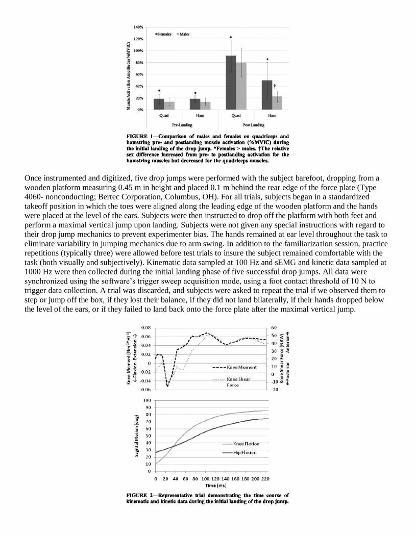
compared with males. When comparing males and females on quadriceps and hamstring muscle activation
during the initial landing of the drop jump, significant effects for sex (P < 0.001), sex x muscle (P = 0.047), and
sex x muscle x landing phase (P = 0.016) interactions were revealed. Post hoc analyses indicated that females
had greater quadriceps and hamstring activation amplitude both pre- and postlanding compared with males.
However, the three-way interaction revealed that whereas females had 27% and 29% more QUADPRE and
HAMPRE during the preactivation phase, the relative sex difference
decreased for QUADPOST (females 13% > males) but increased for HAMPOST (females 54% > males) during the
postlanding phase (Fig. 1). Table 1 also presents the means and SD and the results of the independent-samples t-
tests comparing males and females on BMI and each of the biomechanical variables. In addition to the muscle
strength and activation differences observed, females were also observed to have a lower BMI and land with
greater hip and knee flexion angular excursions and greater peak KEM. However, despite these differences, no
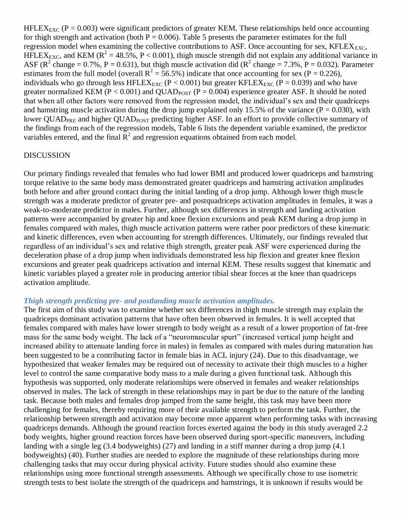
sex differences in peak ASF were observed. Figure 2 demonstrates the kinematic and the kinetic time course of
a representative trial.
Tables 2 and 3 present the parameter estimates for the full regression model separated by sex when predicting
quadriceps pre- and postlanding activation and hamstring pre- and postlanding activation, respectively. When
examining the extent to which an individual’s muscle strength was associated with their quadriceps pre- and
postlanding activation amplitudes during the drop jump, QUADTRQ and HAMTRQ explained an additional 17.2%
(sign R2 change, P = 0.032; overall R
2 = 23.7%, P = 0.050) and 22.2% (sign R
2 change, P = 0.006; overall R
2 =
38.0%, P = 0.002) of the variance in females for pre- and postlanding, respectively, and 11.4% (R2 change, P =
0.120; overall R2 = 14.3%, P = 0.247) and 13.7% (R
2 change, P = 0.079; overall R
2 = 14.7%, P = 0.233) of the
variance in males for pre- and postlanding, respectively, once controlling for individual differences in BMI and
hamstring activation levels. However, only the parameter estimate for QUADTRQ was significant for QUADPRE

(-0.370, P = 0.038) and QUADPOST (-0.406 P = 0.012) in females and QUADPOST (-0.405, P = 0.032) in males.
In each case, these estimates indicate that lower quadriceps torque to body mass predicted greater quadriceps
activation amplitude. When predicting pre- and postlanding hamstring activation amplitudes once controlling
for individual differences in BMI and hamstring activation levels, QUADTRQ and HAMTRQ explained only
12.9% (R2 change, P = 0.080; overall R
2 = 19.2%, P = 0.116) and 2.6% (R
2 change, P = 0.589; overall R
2 =
17.7%, P = 0.146) of the variance in females for pre- and postlanding, respectively, and essentially none of the
variance in males (HAMPRE: R2 change = 0%, P = 0.984; overall R
2 = 4.7%, P = 0.790) (HAMPOST: R
2 change =
0.8%, P = 0.875; overall R2 = 3.3%, P = 0.884). The parameter estimate for HAMTRQ was only significant (-
0.388, P = 0.034) when predicting HAMPRE in females, indicating that lower hamstring torque to body mass
was related to greater hamstring preactivation before the landing.
Results for the prediction of HFLEXEXC during the drop jump reveal that once accounting for sex (R2 = 6.1%, P
= 0.029) and quadriceps and hamstring strength (R2 change = 2.5%, P = 0.370), pre- and postlanding activation
explained an additional 7.8% of the variance (F change, P = 0.170; overall R2 = 16.5, P = 0.071). Although the
overall model was not significant, the parameter estimate for QUADPRE was significant (0.347, P = 0.024) once
controlling for these other variables, indicating that
greater quadriceps preactivation was a significant but weak predictor of greater hip flexion excursion. Results
for knee joint flexion excursion revealed no significant contributions of muscle strength and activation. Once
accounting for sex (R2 = 7.1%, P = 0.0 18), neither quadriceps and hamstring strength (R
2 change = 2.5%, P =
0.370) nor pre- and postlanding activation (R2 change = 2.9%, P = 0.674) contributed significantly to
KFLEXEXC (overall R2 = 12.5, P = 0.206).
Table 4 presents the parameter estimates for the full regression model when examining the neuromuscular and
kinematic contributions to KEM. Once accounting for sex and individual differences in KFLEXEXC and
HFLEXEXC (R2 = 24.4%, P < 0.001), neither thigh muscle strength (R
2 change = 1.9%, P = 0.407) nor pre- and
postlanding activation (R2 change = 1.5%, P = 0.836) was significant predictor of KEM. On the basis of the
prediction equation from the first step in the model, being a female (P = 0.001) and going through less
HFLEXEXC (P = 0.003) were significant predictors of greater KEM. These relationships held once accounting
for thigh strength and activation (both P = 0.006). Table 5 presents the parameter estimates for the full
regression model when examining the collective contributions to ASF. Once accounting for sex, KFLEXEXC,
HFLEXEXC, and KEM (R2 = 48.5%, P < 0.001), thigh muscle strength did not explain any additional variance in
ASF (R2 change = 0.7%, P = 0.631), but thigh muscle activation did (R
2 change = 7.3%, P = 0.032). Parameter
estimates from the full model (overall R2 = 56.5%) indicate that once accounting for sex (P = 0.226),
individuals who go through less HFLEXEXC (P < 0.001) but greater KFLEXEXC (P = 0.039) and who have
greater normalized KEM (P < 0.001) and QUADPOST (P = 0.004) experience greater ASF. It should be noted
that when all other factors were removed from the regression model, the individual’s sex and their quadriceps
and hamstring muscle activation during the drop jump explained only 15.5% of the variance (P = 0.030), with
lower QUADPRE and higher QUADPOST predicting higher ASF. In an effort to provide collective summary of
the findings from each of the regression models, Table 6 lists the dependent variable examined, the predictor
variables entered, and the final R2 and regression equations obtained from each model.
DISCUSSION
Our primary findings revealed that females who had lower BMI and produced lower quadriceps and hamstring
torque relative to the same body mass demonstrated greater quadriceps and hamstring activation amplitudes
both before and after ground contact during the initial landing of a drop jump. Although lower thigh muscle
strength was a moderate predictor of greater pre- and postquadriceps activation amplitudes in females, it was a
weak-to-moderate predictor in males. Further, although sex differences in strength and landing activation
patterns were accompanied by greater hip and knee flexion excursions and peak KEM during a drop jump in
females compared with males, thigh muscle activation patterns were rather poor predictors of these kinematic
and kinetic differences, even when accounting for strength differences. Ultimately, our findings revealed that
regardless of an individual’s sex and relative thigh strength, greater peak ASF were experienced during the
deceleration phase of a drop jump when individuals demonstrated less hip flexion and greater knee flexion
excursions and greater peak quadriceps activation and internal KEM. These results suggest that kinematic and
kinetic variables played a greater role in producing anterior tibial shear forces at the knee than quadriceps
activation amplitude.
Thigh strength predicting pre- and postlanding muscle activation amplitudes.
The first aim of this study was to examine whether sex differences in thigh muscle strength may explain the
quadriceps dominant activation patterns that have often been observed in females. It is well accepted that
females compared with males have lower strength to body weight as a result of a lower proportion of fat-free
mass for the same body weight. The lack of a “neuromuscular spurt” (increased vertical jump height and
increased ability to attenuate landing force in males) in females as compared with males during maturation has
been suggested to be a contributing factor in female bias in ACL injury (24). Due to this disadvantage, we
hypothesized that weaker females may be required out of necessity to activate their thigh muscles to a higher
level to control the same comparative body mass to a male during a given functional task. Although this
hypothesis was supported, only moderate relationships were observed in females and weaker relationships
observed in males. The lack of strength in these relationships may in part be due to the nature of the landing
task. Because both males and females drop jumped from the same height, this task may have been more
challenging for females, thereby requiring more of their available strength to perform the task. Further, the
relationship between strength and activation may become more apparent when performing tasks with increasing
quadriceps demands. Although the ground reaction forces exerted against the body in this study averaged 2.2
body weights, higher ground reaction forces have been observed during sport-specific maneuvers, including
landing with a single leg (3.4 bodyweights) (27) and landing in a stiff manner during a drop jump (4.1
bodyweights) (40). Further studies are needed to explore the magnitude of these relationships during more
challenging tasks that may occur during physical activity. Future studies should also examine these
relationships using more functional strength assessments. Although we specifically chose to use isometric
strength tests to best isolate the strength of the quadriceps and hamstrings, it is unknown if results would be

different using more dynamic, field-based measures of strength. Continued evaluations in this area may lead us
to developing more appropriate tasks for risk factor screening and identification of muscular deficiencies.
Future studies should also explore the role that body composition plays in the relationships between isometric
strength and dynamic muscle activation. Although BMI was used in this study and is considered as a good
estimate of body composition and relative body fat (34), this value is simply based on the overall weight of the
individual compared with their height. Therefore, individuals with a greater than average weight would have a
higher calculated BMI, whether this be due to a higher than average amount of body fat versus a higher than
average amount of lean muscle mass. A more precise assessment of body composition that allows for a more
accurate estimation of available lean mass to total body weight may yield stronger relationships between
strength and muscle activation during a dynamic task.
Thigh strength and activation as predictors of sagittal plane kinematics and kinetics.
Previous studies have reported that females demonstrate greater quadriceps activation patterns during landing
(4,23) and cutting tasks (21,31), which are not always accompanied by greater hamstring activation. Females
are also reported to have decreased knee flexion angles (4,12,19,21) and greater KEM (5,28,31) and ASF (5,39)
during similar landing and cutting tasks compared with males. These finding are often combined to suggest that
females who land with greater quadriceps activation and lower knee flexion angles may experience stiffer
landings leading to greater KEM and shear forces at the knee, thus placing the ACL at greater risk for injury.
However, the direct relationships between quadriceps activation and these kinematic and kinetic variables have
rarely been examined. Of the studies that report both hip and knee flexion excursions along with muscle
activation amplitude during landing or cutting tasks, they consistently report greater quadriceps activation in
females compared with males, but some observe less hip (4) and knee flexion angles (4,21) whereas others
observe equivalent knee flexion angles in females (23,28,31). With regard to the amount of hamstring activation
in females versus males, these studies have noted lower (21), equal (23,31), or greater (4,28) hamstring
activation in females compared with males. Therefore, our second goal was to directly examine the
relationships between neuromuscular, kinematic, and kinetic variables during the drop jump task while
accounting for individual thigh strength differences.
Our findings revealed that the quadriceps dominant activation pattern we observed in females, once controlling
for individual differences in thigh strength and hamstring activation patterns, was not related to sagittal plane
knee and hip kinematics. Although we were unable to compare these findings to similar tasks, our results are
consistent with Wojtys et al. (36,37) who observed lower thigh strength to body weight and lower sagittal plane
and torsional knee stiffness in females compared with males during maximal muscle activations, but no
relationship between the strength and activation levels and the ability to resist knee motions. However, our
findings are limited to thigh strength and activation, and future studies should account for potential differences
in gastrocnemius or posterior hip strength and activation, which also contribute in controlling sagittal plane
motions.
Given the lack of relationships between sagittal plane hip and knee kinematics and thigh strength and activation,
we then accounted for both neuromuscular (quadriceps and hamstring strength and pre- and postlanding
activation amplitudes) and kinematic variables (KFLEXEXC and HFLEXEXC) when examining potential
predictors of adverse knee kinetics (i.e., greater KEM and ASF). As in previous studies (4,28,31), we observed
a greater peak internal KEM in females compared with males but no differences in ASF. Although females had
a greater relative increase in hamstring versus quadriceps activation from pre- to postlanding, neither thigh
muscle strength nor activation amplitude significantly predicted KEM. The strongest predictors of greater KEM
during the landing were being female and less HFLEXEXC, suggesting that sex differences in body position
rather than thigh muscle control may be the driving force behind larger peak KEM during the deceleration
phase of landing. This is supported by recent studies that indicate a forward lean of the trunk (i.e., moving the
center of mass more anterior) results in increased hip and knee flexion (3), decreased knee extensor and
increased plantar flexor and hip extensor moments (15,29), and greater hamstring activation relative to the
quadriceps (15,33) when compared with more upright or backward leaning postures. However, it should be

noted that we did not account for the activation of the rectus femoris in this analysis. Although a smaller muscle
than the two vasti muscles, accounting for this two joint muscle may have yielded a stronger relationship with
KEM.
When we examined the collective contributions to ASF, both kinematic and neuromuscular variables were
significant predictors in the model, although the contribution of strength and activation was relatively small
compared with biomechanical factors. Our prediction model for ASF in large part agrees with the work of Sell
et al. (28), who found that greater integrated EMG activity of the vastus lateralis along with sex (female),
greater peak postground reaction force, decreased external knee flexion moment, and greater knee flexion angle
were significant predictors of greater ASF. As was found in our model, the coefficients in the final model
similarly suggest that the unique contribution of quadriceps activation to ASF, although significant, is relatively
small compared with kinematic and kinetic contributions. Although we did not account for the posterior ground
reaction force in our model, we did account for hip flexion excursion, which again would suggest a more
upright (vs forward) position of the trunk may be an important contribution to adverse knee forces.
An upright trunk has been associated with changes in distal function. When investigating adaptations in
response to an added mass to the trunk during drop jumps, results revealed that subjects adapted by either
landing in a position of trunk extension or trunk flexion (~10° difference) (17). Specifically, those subjects
landing in a more upright or trunk extended position demonstrated 1 1% less hip angular impulses and 18% less
hip energy absorption. Thus, a more upright or extended position of the trunk may place greater energy
dissipation demands on the knee and ankle. Similarly, in a study of sex differences in single leg landing
mechanics, it was reported that females used a more upright, higher peak vertical GRF ankle-dominated
strategy during landing that was theorized to put the noncontractile structures of the more proximal lower
extremity joints (such as the ACL) at risk for injury as the large extensor muscles absorbed less energy (27).
These studies along with the current investigation provides further evidence that the joints of the lower
extremity interact in a kinetic chain to maintain postural control during athletic tasks, suggesting that a
multifactorial approach is needed when attempting to determine when an individual joint may be at risk of
injury.
In summary, our findings suggest that individual differences in thigh muscle strength explained some of the
variance in quadriceps and hamstring activation levels as measured with sEMG during a functional task.
However, even when accounting for strength differences, we did not support the long-held theory that greater
quadriceps activation in females contributes to lower hip and knee flexion angles or greater peak KEM.
Although postlanding quadriceps activation was a small but significant contribution to the prediction model for
knee ASF, the observed predictors for both KEM and ASF indicate that multiple factors determine movement
patterns that result in potentially adverse knee forces. When considering current risk factor screening and
prevention strategies, these findings would suggest that 1) more focus should be placed on positional or postural
differences of the trunk, hip, and knee during landing for their potential to increase sagittal plane knee joint
loads contributing to ACL strain, and 2) evidence of greater quadriceps activation amplitude in females may
simply reflect the presence of muscle weakness rather than increased knee extensor forces, and therefore
strategies to improve overall thigh muscle strength (i.e., both quadriceps and hamstrings) should be considered.
This study was funded by NIH-NIAMS #R01 AR053172. The results of this study do not constitute
endorsement by the ACSM.
REFERENCES
[1] Arms S, Pope MH, Johnson RJ, Fischer RA, Arvidsson I, Eriksson E. The biomechanics of anterior cruciate
ligament rehabilitation and reconstruction. Am JSports Med. 1984;12(1): 8–18.
[2] Beynnon BD, Johnson RJ, Fleming BC, Stankewich CJ, Nichols CE. The strain behavior of the anterior
cruciate ligament during squatting and active flexion-extension: a comparison of open and closed kinetic
chain exercise. Am JSports Med. 1997; 25(6):823–9.

[3] Blackburn JT, Padua DA. Influence of trunk flexion on hip and knee joint kinematics during. Clin Biomech.
2008;23(3):313–9.
[4] Chappell JD, Creighton A, Giuliani C, Yu B, Garrett WE. Kinematics and electromyography of landing
preparation in vertical stopjump: risks for noncontact anterior cruciate ligament injury. Am JSports Med.
2007;35(2):235–41.
[5] Chappell JD, Yu B, Kirkendall DT, Garrett WE. A comparison of knee kinetics between male and female
recreational athletes in stopjump tasks. Am J Sports Med. 2002;30(2):261–7.
[6] Cohen J. Statistical Power Analysis for Behavioral Sciences. 2nd ed. Hillsdale: Laurence Erlbaum Assoc.;
1988. p. 567.
[7] Decker MJ, Torry MR, Wyland DJ, Sterett WI, Richard Steadman J. Gender differences in lower extremity
kinematics, kinetics, and energy absorption during landing. Clin Biomech. 2003;18:662–9.
[8] DeMorat G, Weinhold P, Blackburn T, Chudik S, Garrett W. Aggressive quadriceps loading can induce
noncontact anterior cruciate ligament injury. Am JSports Med. 2004;32(2):477–83.
[9] Draganich LF, Vahey JW. An in-vitro study of anterior cruciate ligament strain induced by quadriceps and
hamstring forces. J Orthop Res. 1990;8:57–63.
[10] Gagnon D, Gagnon M. The influence of dynamic factors on triaxial net muscular moments at the L5/S1
joint during asymmetrical lifting and lowering. JBiomech. 1992;25:891–901.
[11] Griffin LY, Albohm MJ, Arendt EA, et al. Update on ACL prevention: theoretical and practical guidelines.
Am J Sports Med. 2006;34(9):1512–32.
[12] Huston LJ, Vibert B, Ashton-Miller JA, Wojtys EM. Gender differences in knee angle when landing from
a drop jump. Am J Knee Surg. 2001;14:215–20.
[13] Huston LJ, Wojtys EM. Neuromuscular performance characteristics in elite female athletes. Am J Sports
Med. 1996;24(4): 427–36.
[14] Kadaba MP, Ramakrishnan HK, Wootten ME, Gainey J, Gorton G, Cochran GV. Repeatability of
kinematic, kinetic, and electromyographic data in normal adult gait. J Orthop Res. 1989;7: 849–6.
[15] Koyanagi M, Shino K, Yoshimoto Y, Inoue S, Sato M, Nakata K. Effects of changes in skiing posture on
the kinetics of the knee joint. Knee Surg Sports Traumatol Arthrosc. 2006;14: 88–93.
[16] Kulas AS, Schmitz RJ, Shultz SJ, Henning JM, Perrin DH. Sex- specific abdominal activation strategies
during landing. J Athl Train. 2006;41(4):381–6.
[17] Kulas AS, Zalewski P, Hortobagyi T, DeVita P. Effects of added trunk load and corresponding trunk
position adaptations on lower extremity biomechanics during drop-landings. J Biomech. 2008; 41:180–5.
[18] Leardini A, Cappozzo A, Cantani F, et al. Validation of a functional method for the estimation of hip joint
centre location. J Biomech. 1999;32:33–103.
[19] Lephart SM, Ferris CM, Riemann BL, Myers JB, Fu FH. Gender differences in strength and lower
extremity kinematics during landing. Clin Orthop. 2002;401:162–9.
[20] Li G, Rudy TW, Sakane M, Kanamori A, Ma CB, Woo SL. The importance of quadriceps and hamstring
muscle loading on knee kinematics and in-situ forces in the ACL. J Biomech. 1999;32: 395–400.
[21] Malinzak RA, Colby SM, Kirkendall DT, Yu B, Garrett WE. A comparison of knee joint motion patterns
between men and women in selected athletic tasks. Clin Biomech. 2001;16: 438–45.
[22] Markolf KL, O’Neil G, Jackson SR, McAllister DR. Effects of applied quadriceps and hamstrings muscle
loads on forces in the anterior and posterior cruciate ligaments. Am J Sports Med. 2004; 32(5):1144–9.
[23] Nagano Y, Hirofumi I, Akai M, Fukubayashi T. Gender differences in knee kinematics and muscle activity
during single limb drop landings. Knee. 2007;14:218–23.
[24] Quatman CE, Ford KR, Myer GD, Hewett TE. Maturation leads to gender differences in landing force and
vertical jump performance: a longitudinal study. Am J Sports Med. 2006;34: 806–13.
[25] Redfern M. Functional muscle: effects on electromyographic output. In: Soderberg GL, editors. Selected
Topics in Surface Electromyography for Use in the Occupational Setting: Expert Perspectives. National
Institute for Occupational Safety and Health, 1992. pp. 103–20.
[26] Sarwar R, Beltran NB, Rutherford OM. Changes in muscle strength, relaxation rate and fatigability during
the human menstrual cycle. JPhysiol. 1996;493(1):267–72.
[27] Schmitz RJ, Kulas AS, Perrin DH, Riemann BL, Shultz SJ. Sex differences in lower extremity
biomechanics during single leg landings. Clin Biomech. 2007;22(6):681–8.

[28] Sell TC, Ferris CM, Abt JP, et al. Predictors of proximal tibial anterior shear force during a vertical stop
jump. J Orthop Res. 2007;25(12):1589–97.
[29] Shimokochi Y, Lee SY, Shultz SJ, Schmitz RJ. The relationships between sagittal plane lower extremity
moments: implications for landing strategy in ACL injury prevention. J Athl Train. 2008; 43(4):396–408.
[30] Shultz SJ, Perrin DH, Adams JM, Arnold BL, Gansneder BM, Granata KP. Neuromuscular response
characteristics in men and women after knee perturbation in a single-leg weight-bearing stance. JAthl Train.
2001;36(1):37–43.
[31] Sigward SM, Powers CM. The influence of gender on knee kinematics, kinetics and muscle activation
patterns during sidestep cutting. Clin Biomech. 2006;21:41–8.
[32] Uhorchak JM, Scoville CR, Williams GN, Arciero RA, St Pierre P, Taylor DC. Risk factors associated
with non-contact injury of the anterior cruciate ligament. Am J Sports Med. 2003;31(6): 831–42.
[33] Wilk KE, Escamilla RF, Flesig GS, Barrentine SW, Andrews JR, Boyd ML. A comparison of tibiofemoral
joint forces and electromyographic activity during open and closed kinetic chain exercises. Am J Sports
Med. 1996;24(4):518–27.
[34] Wilmore JH, Costill DL. Physiology of Sport and Exercise. 2nd ed. Champaign: Human Kinetics; 1999.
pp. 665–6.
[35] Withrow TJ, Huston LJ, Woytys EM, Ashton-Miller JA. The relationship between quadriceps muscle
force, knee flexion, and anterior cruciate ligament strain in an in-vitro simulated jump landing. Am J Sports
Med. 2006;34(2):269–74.
[36] Wojtys EM, Ashton-Miller JA, Huston LJ. A gender-related difference in contribution of the knee
musculature to sagittal-plane shear stiffness in subjects with similar knee laxity. J Bone Joint Surg. 2002;
84-A(1):10–6.
[37] Wojtys EM, Huston L, Schock HJ, Boylan JP, Ashton-Miller JA. Gender differences in muscular
protection of the knee in torsion in size-matched athletes. J Bone Joint Surg. 2003;85-A(5):782–9.
[38] Woods JJ, Bigland-Ritchie B. Linear and non-linear surface EMG/ force relationships in human muscles.
An anatomical/functional argument for the existence of both. Am J Phys Med Rehabil. 1983;62(6):287–99.
[39] Yu B, Lin CF, Garrett WE. Lower extremity biomechanics during the landing of a stop jump task. Clin
Biomech. 2006;21:297–305.
[40] Zhang S-N, Bates BT, Dufek JS. Contributions of lower extremity joints to energy dissipation during
landings. Med Sci Sports Exerc. 2000;32(4):812–9.
Related Documents