J. Neurol. Neurosurg. Psychiat., 1958, 21, 195. THE SURGICAL SEPARATION OF SIAMESE TWINS CONJOINED BY THE HEADS (CEPHALOPAGUS FRONTALIS) FOLLOWED BY NORMAL DEVELOPMENT BY MAITLAND BALDWIN and ANATOLE DEKABAN From the National Institute of Neurological Diseases and Blindness, National Institutes of Health, Public Health Service, U.S. Department of Health, Education and Welfare, Bethesda 14, Maryland Human conjoined twins are rare. Various authors (Mortimer and Kirshbaum, 1942; Potter, 1952) give figures ranging from one in 60,000 to one in 85,000 births for various types of Siamese twins either alive or stillborn. In 117 conjoined twins, a series of which was compiled by Robertson (1953) from Taruffi's eight-volume study on heratology (Storia della Teratologia, Bologna, Regia Tipografia, 1881- 94), thoracopagus was present in 86, pygopagus in 22, ischiopagus in seven, and cephalopagus in two only. From the analysis of these data it appears that the incidence of cephalopagus would be in the range of one in every two to four million births. The only satisfactory treatment of cephalopagous twins is surgical separation. This, however, presents enormous technical difficulties and the number of surviving infants after such procedures are few. In the present communication we are therefore report- ing an instance of Siamese twins who were conjoined by the frontal regions of the heads and who continue to develop normally after separation. Case Report Virginia and Tresea, 2-month-old female twins con- joined by the heads, were admitted to the National Institute of Neurological Diseases and Blindness for study and possible treatment in October, 1956. The mother was 21 years of age at the time of delivery. She has always been healthy. There were no multiple births in her family nor any evidence of neurological or developmental abnormalities. Her menstruation has always been regular. She has had three pregnancies, all resulting in live births. The first child, a girl 4 years of age, and the second child, a boy, 28 months of age, are normal in all respects. The father died of rheumatic heart disease at the age of 39 years three months before the twins were born. There were no known instances of congenital malforma- tion or any neurological condition in his family. One of the paternal uncles has dissimilar twins. Present Pregnancy.-The mother received regular prenatal care from the second month of gestation. During the first two trimesters she was subject to nausea and minor headaches but no vomiting. About three weeks before the delivery the membranes ruptured sponta- neously and approximately a quart of fluid escaped. She was admitted to hospital for a few days and then remained in bed at home having periodic minor uterine con- tractions. She was re-admitted to the hospital in Elizabethton, Tennessee, on August 9, 1956, and three hours later female Siamese twins were delivered. The total birth weight of the conjoined twins was 3-5 kg. The mother had general anaesthesia and does not know the details of the delivery nor the immediate state of the infants. She was told by the doctor that the second twin, who was slightly smaller (subsequently named Tresea), had to be resuscitated. According to the physician attending the delivery there were two umbilical cords, one placenta, one chorion, and two amnions. After two weeks in the hospital the twins were dis- charged home and for the next two weeks the mother fed them using a dropper. From the age of 1 month they were put on a bottle. In spite of great difficulty with feeding and general care, the mother had them at home until they were 2 months old. Examination of the Twins at 2 Months of Age.-The infants were joined by the frontal and vertical regions in such a fashion that the predominantly right side of the head of one twin was in union with the predominantly right side of the other twin. In this way they appeared in mirror position to each other although slightly to the left (3 cm.) from the mid-sagittal planes (Figs. 1 and 2). The anterior fontanelles were partly open and partly involved in the bony union. The right orbital region and right eye were in close apposition, and when the infants were put in a parallel position the eyes were slightly compressed. The joined areas were in firm bony union. Tresea's joining part of the head appeared to telescope into Virginia's head forming a sort of coning (Figs. 3 and 4). The circumference of the conjoined area was 24 cm. The total weight of both twins was 4 kg. The state of nourishment and general physical conditions were good. They both took their feeds well. There was no evidence of other congenital abnormalities and their internal organs were normal. It was obvious that most 195 4 Protected by copyright. on May 6, 2020 by guest. http://jnnp.bmj.com/ J Neurol Neurosurg Psychiatry: first published as 10.1136/jnnp.21.3.195 on 1 August 1958. Downloaded from

Welcome message from author
This document is posted to help you gain knowledge. Please leave a comment to let me know what you think about it! Share it to your friends and learn new things together.
Transcript
J. Neurol. Neurosurg. Psychiat., 1958, 21, 195.
THE SURGICAL SEPARATION OF SIAMESE TWINS CONJOINEDBY THE HEADS (CEPHALOPAGUS FRONTALIS) FOLLOWED
BY NORMAL DEVELOPMENTBY
MAITLAND BALDWIN and ANATOLE DEKABANFrom the National Institute of Neurological Diseases and Blindness, National Institutes of Health, Public Health
Service, U.S. Department of Health, Education and Welfare, Bethesda 14, Maryland
Human conjoined twins are rare. Various authors(Mortimer and Kirshbaum, 1942; Potter, 1952) givefigures ranging from one in 60,000 to one in 85,000births for various types of Siamese twins either aliveor stillborn. In 117 conjoined twins, a series ofwhich was compiled by Robertson (1953) fromTaruffi's eight-volume study on heratology (Storiadella Teratologia, Bologna, Regia Tipografia, 1881-94), thoracopagus was present in 86, pygopagus in22, ischiopagus in seven, and cephalopagus in twoonly. From the analysis of these data it appearsthat the incidence of cephalopagus would be in therange of one in every two to four million births.The only satisfactory treatment of cephalopagous
twins is surgical separation. This, however, presentsenormous technical difficulties and the number ofsurviving infants after such procedures are few. Inthe present communication we are therefore report-ing an instance of Siamese twins who were conjoinedby the frontal regions of the heads and who continueto develop normally after separation.
Case ReportVirginia and Tresea, 2-month-old female twins con-
joined by the heads, were admitted to the NationalInstitute of Neurological Diseases and Blindness forstudy and possible treatment in October, 1956. Themother was 21 years of age at the time of delivery. Shehas always been healthy. There were no multiple birthsin her family nor any evidence of neurological ordevelopmental abnormalities. Her menstruation hasalways been regular. She has had three pregnancies, allresulting in live births. The first child, a girl 4 years ofage, and the second child, a boy, 28 months of age, arenormal in all respects.The father died of rheumatic heart disease at the age of
39 years three months before the twins were born.There were no known instances of congenital malforma-tion or any neurological condition in his family. Oneof the paternal uncles has dissimilar twins.
Present Pregnancy.-The mother received regularprenatal care from the second month of gestation. During
the first two trimesters she was subject to nausea andminor headaches but no vomiting. About three weeksbefore the delivery the membranes ruptured sponta-neously and approximately a quart of fluid escaped. Shewas admitted to hospital for a few days and then remainedin bed at home having periodic minor uterine con-tractions. She was re-admitted to the hospital inElizabethton, Tennessee, on August 9, 1956, and threehours later female Siamese twins were delivered. Thetotal birth weight of the conjoined twins was 3-5 kg.The mother had general anaesthesia and does not knowthe details of the delivery nor the immediate state of theinfants. She was told by the doctor that the second twin,who was slightly smaller (subsequently named Tresea),had to be resuscitated.
According to the physician attending the delivery therewere two umbilical cords, one placenta, one chorion, andtwo amnions.
After two weeks in the hospital the twins were dis-charged home and for the next two weeks the mother fedthem using a dropper. From the age of 1 month theywere put on a bottle. In spite of great difficulty withfeeding and general care, the mother had them at homeuntil they were 2 months old.
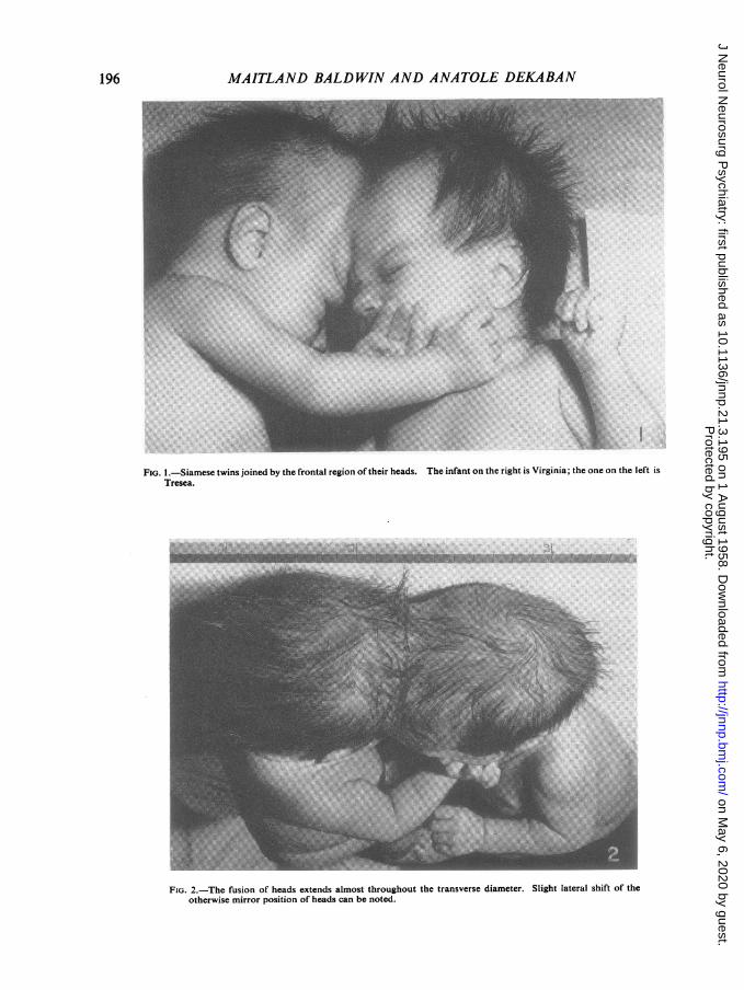
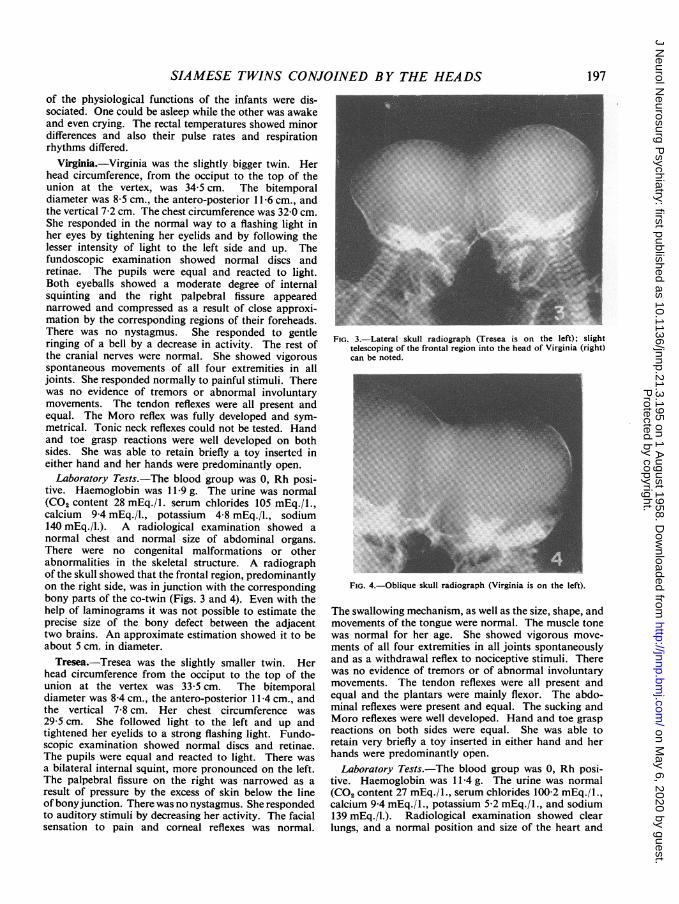
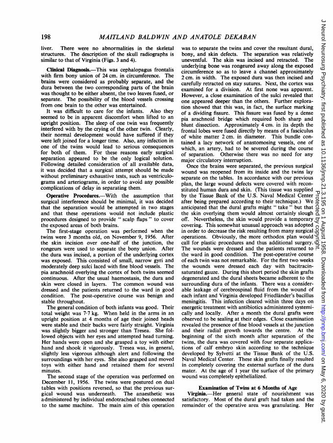
Examination of the Twins at 2 Months of Age.-Theinfants were joined by the frontal and vertical regionsin such a fashion that the predominantly right side of thehead of one twin was in union with the predominantlyright side of the other twin. In this way they appearedin mirror position to each other although slightly to theleft (3 cm.) from the mid-sagittal planes (Figs. 1 and 2).The anterior fontanelles were partly open and partlyinvolved in the bony union. The right orbital region andright eye were in close apposition, and when the infantswere put in a parallel position the eyes were slightlycompressed. The joined areas were in firm bony union.Tresea's joining part of the head appeared to telescopeinto Virginia's head forming a sort of coning (Figs. 3and 4). The circumference of the conjoined area was24 cm. The total weight of both twins was 4 kg. Thestate of nourishment and general physical conditionswere good. They both took their feeds well. There wasno evidence of other congenital abnormalities and theirinternal organs were normal. It was obvious that most
1954
Protected by copyright.
on May 6, 2020 by guest.
http://jnnp.bmj.com
/J N
eurol Neurosurg P
sychiatry: first published as 10.1136/jnnp.21.3.195 on 1 August 1958. D
ownloaded from
MAITLAND BALDWIN AND ANATOLE DEKABAN
FIG. 1.-Siamese twins joined by the frontal region of their heads. The infant on the right is Virginia; the one on the left isTresea.
FIG. 2.-The fusion of heads extends almost throughout the transverse diameter. Slight lateral shift of theotherwise mirror position of heads can be noted.
Wo ...... ..MX.
196
Protected by copyright.
on May 6, 2020 by guest.
http://jnnp.bmj.com
/J N
eurol Neurosurg P
sychiatry: first published as 10.1136/jnnp.21.3.195 on 1 August 1958. D
ownloaded from
SIAMESE TWINS CONJOINED BY THE HEADS
of the physiological functions of the infants were dis-sociated. One could be asleep while the other was awakeand even crying. The rectal temperatures showed minordifferences and also their pulse rates and respirationrhythms differed.
Virginia.-Virginia was the slightly bigger twin. Herhead circumference, from the occiput to the top of theunion at the vertex, was 34-5 cm. The bitemporaldiameter was 8-5 cm., the antero-posterior 11 -6 cm., andthe vertical 7-2 cm. The chest circumference was 32-0 cm.She responded in the normal way to a flashing light inher eyes by tightening her eyelids and by following thelesser intensity of light to the left side and up. Thefundoscopic examination showed normal discs andretinae. The pupils were equal and reacted to light.Both eyeballs showed a moderate degree of internalsquinting and the right palpebral fissure appearednarrowed and compressed as a result of close approxi-mation by the corresponding regions of their foreheads.There was no nystagmus. She responded to gentleringing of a bell by a decrease in activity. The rest ofthe cranial nerves were normal. She showed vigorousspontaneous movements of all four extremities in alljoints. She responded normally to painful stimuli. Therewas no evidence of tremors or abnormal involuntarymovements. The tendon reflexes were all present andequal. The Moro reflex was fully developed and sym-metrical. Tonic neck reflexes could not be tested. Handand toe grasp reactions were well developed on bothsides. She was able to retain briefly a toy inserted ineither hand and her hands were predominantly open.
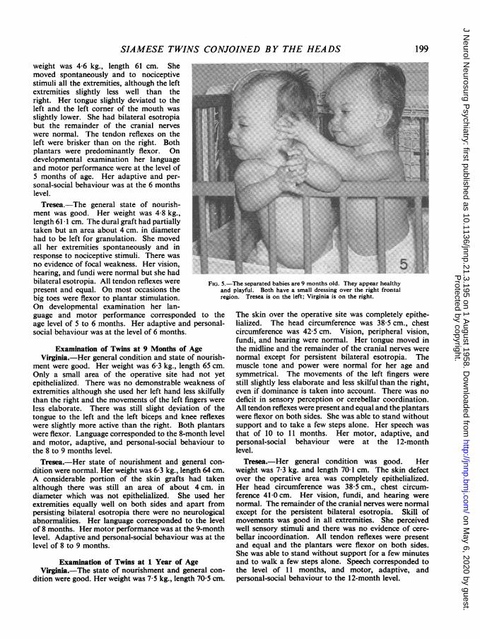
Laboratory Tests.-The blood group was 0, Rh posi-tive. Haemoglobin was 11 9 g. The urine was normal(CO2 content 28 mEq./l. serum chlorides 105 mEq./l.,calcium 9-4 mEq./l., potassium 4-8 mEq./l., sodium140 mEq./l.). A radiological examination showed anormal chest and normal size of abdominal organs.There were no congenital malformations or otherabnormalities in the skeletal structure. A radiographof the skull showed that the frontal region, predominantlyon the right side, was in junction with the correspondingbony parts of the co-twin (Figs. 3 and 4). Even with thehelp of laminograms it was not possible to estimate theprecise size of the bony defect between the adjacenttwo brains. An approximate estimation showed it to beabout 5 cm. in diameter.
Tresea.-Tresea was the slightly smaller twin. Herhead circumference from the occiput to the top of theunion at the vertex was 33-5 cm. The bitemporaldiameter was 8-4 cm., the antero-posterior 1I-4 cm., andthe vertical 7-8 cm. Her chest circumference was29-5 cm. She followed light to the left and up andtightened her eyelids to a strong flashing light. Fundo-scopic examination showed normal discs and retinae.The pupils were equal and reacted to light. There wasa bilateral internal squint, more pronounced on the left.The palpebral fissure on the right was narrowed as aresult of pressure by the excess of skin below the lineof bonyjunction. There was no nystagmus. She respondedto auditory stimuli by decreasing her activity. The facialsensation to pain and corneal reflexes was normal.
FIG. 3.-Lateral skull radiograph (Tresea is on the left); slighttelescoping of the frontal region into the head of Virginia (right)can be noted.
Feas,
FIG. 4.-Oblique skull radiograph (Virginia is on the left).
The swallowing mechanism, as well as the size, shape, andmovements of the tongue were normal. The muscle tonewas normal for her age. She showed vigorous move-ments of all four extremities in all joints spontaneouslyand as a withdrawal reflex to nociceptive stimuli. Therewas no evidence of tremors or of abnormal involuntarymovements. The tendon reflexes were all present andequal and the plantars were mainly flexor. The abdo-minal reflexes were present and equal. The sucking andMoro reflexes were well developed. Hand and toe graspreactions on both sides were equal. She was able toretain very briefly a toy inserted in either hand and herhands were predominantly open.
Laboratory Tests.-The blood group was 0, Rh posi-tive. Haemoglobin was 11-4 g. The urine was normal(CO2 content 27 mEq./1., serum chlorides 100-2 mEq./l.,calcium 9 4 mEq./1., potassium 5-2 mEq./1., and sodium139 mEq./l.). Radiological examination showed clearlungs, and a normal position and size of the heart and
197
Protected by copyright.
on May 6, 2020 by guest.
http://jnnp.bmj.com
/J N
eurol Neurosurg P
sychiatry: first published as 10.1136/jnnp.21.3.195 on 1 August 1958. D
ownloaded from
MAITLAND BALDWIN AND ANATOLE DEKABAN
liver. There were no abnormalities in the skeletalstructures. The description of the skull radiographs issimilar to that of Virginia (Figs. 3 and 4).
Clinical Diagnosis.-This was cephalopagus frontaliswith firm bony union of 24 cm. in circumference. Thebrains were considered as probably separate, and thedura between the two corresponding parts of the brainwas thought to be either absent, the two leaves fused, orseparate. The possibility of the blood vessels crossingfrom one brain to the other was entertained.
It was difficult to care for the infants. Also theyseemed to be in apparent discomfort when lifted to anupright position. The sleep of one twin was frequentlyinterfered with by the crying of the other twin. Clearly,their normal development would have suffered if theywere left joined for a longer time. Also, any infection inone of the twins would lead to serious consequencesfor both of them. For these reasons early surgicalseparation appeared to be the only logical solution.Following detailed consideration of all available data,it was decided that a surgical attempt should be madewithout preliminary exhaustive tests, such as ventriculo-grams and arteriograms, in order to avoid any possiblecomplications of delay in separating them.
Operative Procedures.-With the assumption thatsurgical interference should be minimal, it was decidedthat the separation would be attempted in two stagesand that these operations would not include plasticprocedures designed to provide " scalp flaps" to coverthe exposed areas of both brains.The first-stage operation was performed when the
twins were 3 months old, on November 9, 1956. Afterthe skin incision over one-half of the junction, therongeurs were used to separate the bony union. Afterthe dura was incised, a portion of the underlying cortexwas exposed. This consisted of small, narrow gyri andmoderately deep sulci laced with fine blood vessels. Thepia arachnoid overlying the cortex of both twins seemedcontinuous. After the usual haemostasis, the dura andskin were closed in layers. The common wound wasdressed and the patients returned to the ward in goodcondition. The post-operative course was benign andstable throughout.The general condition of both infants was good. Their
total weight was 7-7 kg. When held in the arms in anupright position at 4 months of age their joined headswere stable and their backs were fairly straight. Virginiawas slightly bigger and stronger than Tresea. She fol-lowed objects with her eyes and attempted head turning.Her hands were open and she grasped a toy with eitherhand and shook it vigorously. Tresea was, in general,slightly less vigorous although alert and following thesurroundings with her eyes. She also grasped and movedtoys with either hand and retained them for severalminutes.The second stage of the operation was performed on
December 11, 1956. The twins were postured on dualtables with positions reversed, so that the previous sur-gical wound was underneath. The anaesthetic wasadministered by individual endotracheal tubes connectedto the same machine. The main aim of this operation
was to separate the twins and cover the resultant dural,bony, and skin defects. The separation was relativelyuneventful. The skin was incised and retracted. Theunderlying bone was rongeured away along the exposedcircumference so as to leave a channel approximately2 cm. in width. The exposed dura was then incised andcarefully retracted on stay sutures. Next, the cortex wasexamined for a division. At first none was apparent.However, a close examination of the sulci revealed thatone appeared deeper than the others. Further explora-tion showed that this was, in fact, the surface markingof a dividing fissure. This fissure was fused by a densepia arachnoid bridge which required both sharp andblunt dissection. Approximately 4 cm. in its depth thefrontal lobes were fused directly by means of a fasciculusof white matter 2 cm. in diameter. This bundle con-tained a lacy network of anastomosing vessels, one ofwhich, an artery, had to be severed during the courseof separation. Otherwise there was no need for anymajor circulatory interruption.Once the brains were separated, the previous surgical
wound was reopened from its inside and the twins layseparate on the tables. In accordance with our previousplan, the large wound defects were covered with recon-stituted human dura and skin. (This tissue was suppliedby the Tissue Bank of the U.S. Naval Medical Centerafter being prepared according to their technique.) Weanticipated that the dural grafts might " take" but thatthe skin overlying them would almost certainly sloughoff. Nevertheless, the skin would provide a temporarycovering. This somewhat unusual approach was adoptedin order to decrease the risk resulting from many surgicaloperations. Obviously, the more orthodox plan wouldcall for plastic procedures and thus additional surgery.The wounds were dressed and the patients returned tothe ward in good condition. The post-operative courseof each twin was not remarkable. For the first two weeksthe wounds were dressed each day with bacitracin-saturated gauze. During this short period the skin graftsdegenerated and the dural sheets became adherent to thesurrounding dura of the infants. There was a consider-able leakage of cerebrospinal fluid from the wound ofeach infant and Virginia developed Friedlander's bacillusmeningitis. This infection cleared within three days onenergetic treatment with antibiotics administered systemi-cally and locally. After a month the dural grafts wereobserved to be sealing at their edges. Close examinationrevealed the presence of fine blood vessels at the junctionand their radial growth towards the centre. At thebeginning of the sixth month after separation of thetwins, the dura was covered with four separate applica-tions of calf embryo skin according to the techniquedeveloped by Sylvetti at the Tissue Bank of the U.S.Naval Medical Center. These skin grafts finally resultedin completely covering the external surface of the duramater. At the age of 1 year the surface of the primarywound was completely epithelialized.
Examination of Twins at 6 Months of AgeVirginia.-Her general state of nourishment was
satisfactory. Most of the dural graft had taken and theremainder of the operative area was granulating. Her
198
Protected by copyright.
on May 6, 2020 by guest.
http://jnnp.bmj.com
/J N
eurol Neurosurg P
sychiatry: first published as 10.1136/jnnp.21.3.195 on 1 August 1958. D
ownloaded from
SIAMESE TWINS CONJOINED BY THE HEADS
weight was 4-6 kg., length 61 cm. Shemoved spontaneously and to nociceptivestimuli all the extremities, although the leftextremities slightly less well than theright. Her tongue slightly deviated to theleft and the left corner of the mouth wasslightly lower. She had bilateral esotropiabut the remainder of the cranial nerveswere normal. The tendon reflexes on theleft were brisker than on the right. Bothplantars were predominantly flexor. Ondevelopmental examination her languageand motor performance were at the level of5 months of age. Her adaptive and per-sonal-social behaviour was at the 6 months -77level.Tresea.-The general state of nourish-
ment was good. Her weight was 4-8 kg.,length 61 1 cm. The dural graft had partiallytaken but an area about 4 cm. in diameterhad to be left for granulation. She movedall her extremities spontaneously and inresponse to nociceptive stimuli. There wasno evidence of focal weakness. Her vision,hearing, and fundi were normal but she hadbilateral esotropia. All tendon reflexes were FIG. 5.-present and equal. On most occasions the an(big toes were flexor to plantar stimulation. regOn developmental examination her lan-guage and motor performance corresponded to theage level of 5 to 6 months. Her adaptive and personal-social behaviour was at the level of 6 months.
Examination of Twins at 9 Months of AgeVirginia.-Her general condition and state of nourish-
ment were good. Her weight was 6-3 kg., length 65 cm.Only a small area of the operative site had not yetepithelialized. There was no demonstrable weakness ofextremities although she used her left hand less skilfullythan the right and the movements of the left fingers wereless elaborate. There was still slight deviation of thetongue to the left and the left biceps and knee reflexeswere slightly more active than the right. Both plantarswere flexor. Language corresponded to the 8-month leveland motor, adaptive, and personal-social behaviour tothe 8 to 9 months level.Tresea.-Her state of nourishment and general con-
dition were normal. Her weight was 6-3 kg., length 64 cm.A considerable portion of the skin grafts had takenalthough there was still an area of about 4 cm. indiameter which was not epithelialized. She used herextremities equally well on both sides and apart frompersisting bilateral esotropia there were no neurologicalabnormalities. Her language corresponded to the levelof 8 months. Her motor performance was at the 9-monthlevel. Adaptive and personal-social behaviour was at thelevel of 8 to 9 months.
Examination of Twins at 1 Year of AgeVirginia.-The state of nourishment and general con-
dition were good. Her weight was 7-5 kg., length 70 5 cm.
ii:..t.-t::...:-The separated babies are 9 months old. They appear healthyId playful. Both have a small dressing over the right frontalgion. Tresea is on the left; Virginia is on the right.
The skin over the operative site was completely epithe-lialized. The head circumference was 38-5 cm., chestcircumference was 42-5 cm. Vision, peripheral vision,fundi, and hearing were normal. Her tongue moved inthe midline and the remainder of the cranial nerves werenormal except for persistent bilateral esotropia. Themuscle tone and power were normal for her age andsymmetrical. The movements of the left fingers werestill slightly less elaborate and less skilful than the right,even if dominance is taken into account. There was nodeficit in sensory perception or cerebellar coordination.All tendon reflexes were present and equal and the plantarswere flexor on both sides. She was able to stand withoutsupport and to take a few steps alone. Her speech wasthat of 10 to 11 months. Her motor, adaptive, andpersonal-social behaviour were at the 12-monthlevel.Tresea.-Her general condition was good. Her
weight was 7-3 kg. and length 70-1 cm. The skin defectover the operative area was completely epithelialized.Her head circumference was 38-5 cm., chest circum-ference 41-0 cm. Her vision, fundi, and hearing werenormal. The remainder of the cranial nerves were normalexcept for the persistent bilateral esotropia. Skill ofmovements was good in all extremities. She perceivedwell sensory stimuli and there was no evidence of cere-bellar incoordination. All tendon reflexes were presentand equal and the plantars were flexor on both sides.She was able to stand without support for a few minutesand to walk a few steps alone. Speech corresponded tothe level of 11 months, and motor, adaptive, andpersonal-social behaviour to the 12-month level.
Protected by copyright.
on May 6, 2020 by guest.
http://jnnp.bmj.com
/J N
eurol Neurosurg P
sychiatry: first published as 10.1136/jnnp.21.3.195 on 1 August 1958. D
ownloaded from
MAITLAND BALDWIN AND ANATOLE DEKABAN
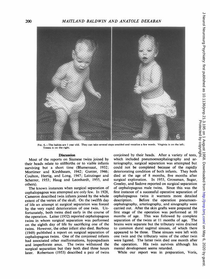
FIG. 6.-The babies are 1 year old. They can take several steps unaided and vocalize a few words. Virginia is on the left;Tresea is on the right.
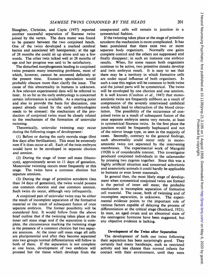
DiscussionMost of the reports on Siamese twins joined by
their heads relate to stillbirths or to viable infantssurviving but a short time (Blumensaat, 1932;Mortimer and Kirshbaum, 1942; Gunter, 1946;Coulton, Hertig, and Long, 1947; Leinzinger andScherrer, 1953; Haug and Leonhardt, 1955, andothers).The known instances when surgical separation of
cephalopagous was attempted are only few. In 1928,Cameron described twin infants joined by the wholeextent of the vertex of the skull. On the twelfth dayof life an attempt at surgical separation was forcedby the very rapid deterioration of one twin. Un-fortunately, both twins died early in the course ofthe operation. Leiter (1932) reported cephalopagoustwins in whom surgical separation was performedon the eighth day of life by sacrificing one of thetwins. However, the other infant also died. Barbosa(1949) published a report on surgical separation ofcephalopagous twins. One of the conjoined infantshad associated other malformations, hypospadiasisand imperforate anus. The twins withstood thesurgical separation but they both died three hourslater. Robertson (1953) described a pair of twins
conjoined by their heads. After a variety of tests,which included pneumoencephalography and ar-teriography, surgical separation was attempted butcould not be completed because of the rapidlydeteriorating condition of both infants. They bothdied at the age of 8 months, five months aftersurgical exploration. In 1953, Grossman, Sugar,Greeley, and Sadove reported on surgical separationof cephalopagous male twins. Since this was thefirst instance of a successful operative separation ofcephalopagous twins it warrants more detaileddescription. Before the operation pneumoen-cephalography, arteriography, and sinography werecarried out. After the skin grafts were prepared thefirst stage of the operation was performed at 10months of age. This was followed by completeseparation of the twins at 11 months of age. Thebrains were separate but the tributary veins drainedto common dural sagittal sinuses, of which thereappeared to be three. These sinuses were left withone twin and the tributary veins of the other twinwere ligated. The latter twin died one month afterthe operation. His twin survives although hismental development seems retarded.
While our report was in preparation, Voris,
200
Protected by copyright.
on May 6, 2020 by guest.
http://jnnp.bmj.com
/J N
eurol Neurosurg P
sychiatry: first published as 10.1136/jnnp.21.3.195 on 1 August 1958. D
ownloaded from
SIAMESE TWINS CONJOINED BY THE HEADS
Slaughter, Christian, and Cayia (1957) reportedanother successful separation of Siamese twinsjoined by the vertex. The dura mater was foundto be present between the two conjoined heads.One of the twins developed a marked cerebralhernia and associated left hemiparesis; at the ageof 28 months she could sit up alone and say a fewwords. The other twin talked well at 28 months ofage and her progress was said to be satisfactory.The disturbed morphogenesis leading to conjoined
twins presents many interesting problems, most ofwhich, however, cannot be answered definitely atthe present time. Excessive speculation wouldprobably obscure more than clarify the issue. Thecause of this abnormality in humans is unknown.A few relevant experimental data will be referred tolater. In so far as the early embryology is concernedcertain information is available. As a starting pointand also to provide the basis for discussion, oneaspect already raised by the early embryologistsneeds to be stressed: the mechanism of the pro-duction of conjoined twins must be closely relatedto the mechanism of the formation of uniovulartwinning.
Theoretically, uniovular twinning may occurduring the following stages of development:
(1) Before or during the early morula stage (firstfew days after fertilization). This must be extremelyrare if it does occur at all. Each of the twin embryoswould have to be enveloped in separate chorionand amnion.
(2) During the stage of inner cell mass (blasto-cyst), approximately seven to 11 days of gestation.Monovular twinning occurs most frequently at thisstage. The twins have a common chorion butseparate amnions.
(3) During the stage of primitive ectoderm (lessthan 14 days of gestation), the twins would possessone common chorion and one common amnion.Such twins do occur, although very infrequently.A conjoined pair of uniovular twins may be either
the result of incomplete separation of the formativematerial or the result of subsequent fusion of onceseparate embryos. The former possibility will beconsidered first. It would follow from the abovebrief outline that if the twinning takes place at theinner cell mass stage and if the separation is com-plete, the circumstance marking the twinning stageis the presence of a common chorion but two separ-ate amnions. At the inner cell mass stage all cellsare pluripotential and after they become separatedinto two groups normal differentiation will follow inboth of them. If the separation is not completeat one locus, development of two embryos willproceed but the tissue which develops from the
unseparated cells will remain in junction in asymmetrical fashion.
If the twinning takes place at the stage of primitiveectoderm the mechanism is more complicated. It hasbeen postulated that there exist two or moreseparate body organizers. Normally one gainscomplete control and the others are suppressed andfinally disappear; in such an instance one embryoresults. When, for some reason both organizerscontinue to be active, two primitive streaks developand twin embryos result. It is easy to see thatthere may be a territory in which formative cellsare under equal influence of both organizers. Insuch a case this region will be common to both twinsand the joined parts will be symmetrical. The twinswill be enveloped by one chorion and one amnion.It is well known (Coulton et al., 1947) that mono-amniotic twins are frequently stillborn as a result ofcompression of the severely intertwined umbilicalcords which lead to obstruction of the blood circu-lation. The possibility of the occurrence of con-joined twins as a result of subsequent fusion of theonce separate embryos seems very remote, at leastin symmetrical Siamese twins. In the first place thejunction would be frequently haphazard and notof the mirror image type, as seen in the majority ofcases. Secondly, contrary to the general findings,such abnormality could occur only in mono-amniotic twins not separated by the interveningmembranes. The experimental work of Mangold(1920) is of considerable interest. This investigatorproduced conjoined individuals in the salamanderby pressing two zygotes together. Since this was ahighly artificial situation and concerned achorionicand anamniotic animals it could hardly be applicableto humans or even lower mammals.
In general then, the most likely stage of develop-ment when symmetrical conjoined twins are formedis the period of inner cell mass; the probablemechanism is incomplete separation of formativecell material. The cause, both of twinning and ofincomplete separation, is unknown. The experi-mental evidence points to the important role ofvarious factors capable of delaying the process ofdifferentiation at the critical stage (Stockard, 1921).In man, an aged ovum and an abnormal state ofthe oestrogenic hormone have been suggested, butany objective evidence is lacking.
Development of the Twins after SeparationThe development of both our twins following
their separation has been surprisingly good. Theycertainly had many handicaps, such as restrictedactivity and less chance than normal infants forcontact with their environment, until they were
201
Protected by copyright.
on May 6, 2020 by guest.
http://jnnp.bmj.com
/J N
eurol Neurosurg P
sychiatry: first published as 10.1136/jnnp.21.3.195 on 1 August 1958. D
ownloaded from
MAITLAND BALDWIN AND ANATOLE DEKABAN
separated at the age of 4 months. Then a long periodof about four months of complete confinement firstin isolates, then in cribs, when the operative woundswere in a dangerous state with leakage of the cerebro-spinal fluid. It was not until they were 11 monthsold that the operative sites in the frontal regionswere completely epithelialized. At the age of1 year their general and mental development can beconsidered as being within the normal range. Treseahas been free of any abnormal neurological findings.Virginia shows a minimal decrease in skilled move-
ments of the left hand but power is normal.
SummarySiamese twins conjoined by their heads are rare.
This malformation requires surgical separation if thepatients are to survive beyond the stage of infancy.A detailed description of cephalopagous twins isgiven. The infants were born by natural means andwere normal in all respects except for union of theheads. Their development and neurological statusbefore the final surgical separation at the age of4 months and during their subsequent course isdescribed. Homeo- and hetero- grafts were used tocover the defect of soft tissue at the site of separation.Development of the infants at 1 year of age was
normal.
The pathogenesis of Siamese twins is discussedin some detail.
Our thanks are due to the neurological and neuro-
surgical residents and nursing staff who cared for bothpatients. In addition we wish to express our appreciationfor their generous help to Dr. Thomas Cone, chiefpediatrician at the National Naval Medical Center, andto the tissue bank, National Naval Medical School.Details of the anaesthesia were described by Dr. Hallin a separate publication (1957). Miss S. Lewis, R.N.,designed a dual table which considerably aided thesurgical procedure.
REFERENCESBarbosa, A. (1949). Rev. brasil. Cirurg., 18, 1047.Blumensaat, C. (1932). Virchows Arch. path. Anat., 285, 140.Cameron, H. C. (1928). Lancet, 1, 284.Coulton, D., Hertig, A. T., and Long, W. N. (1947). Amer. J. Obstet.
Gynec., 54, 119.Grossman, H. J., Sugar, O., Greeley, P. W., and Sadove, M. S. (1953).
J. Amer. med. Ass., 153, 201.Gunter, J. U. (1946). Amer. J. Path., 22, 855.Hall, K. D., Merzig, J., and Norris, F. H. (1957). Anesthesiology,
18, 908.
Haug, H., and Leonhardt, H. (1955). Anat. Anz., 101, 281.Leinzinger, E., and Scherrer, H. (1953). Geburtsh. Frauenheilk.,
13, 543.Leiter, K. (1932). Zbl. Gynak., 56, 1644.Mangold, 0. (1920). Arch. Entwickl.-Mech. Org., Berlin, 47, 249.Mortimer, B., and Kirshbaum, J. D. (1942). Amer. J. Dis. Child.,
64, 697.Potter, E. L. (1952). Pathology of the Fetus and the Newborn. Year
Book Publishers, Chicago.Robertson, E. G. (1953). A.M.A. Arch. Neurol. Psychiat., 70, 189.Stockard, C. R. (1921). Amer. J. Anat., 28, 115.Voris, H. C., Slaughter, W. B., Christian, J. R., and Cayia, E. R.
(1957). J. Neurosurg., 14, 548.
202
Protected by copyright.
on May 6, 2020 by guest.
http://jnnp.bmj.com
/J N
eurol Neurosurg P
sychiatry: first published as 10.1136/jnnp.21.3.195 on 1 August 1958. D
ownloaded from
Related Documents