University of Cape Town ASPECTS OF PHOSPHORUS NUTRITION IN ENDOMYCORRHIZAL FUNGI OF THE ERICACEAE BY COLIN JOHN STRAKER A thesis presented for the degree of Doctor of Philosophy in the Faculty of Science, University of Cape Town Department of Botany January 1986 .. J ..... ........ ,., ------ ...... l Th· •·' 1 ""'" qi>Mn . .rt,;•!t 1'' rf ;'• · the i...:. in ,r in C,·· • I·,·'' •·, th·; authclr, ' . __ ., ...... _,;_ ...... _ ASPECTS OF PHOSPHORUS NUTRITION IN ENDOMYCORRHIZAL FUNG OF THE RICACEAE BY COLIN JOHN STRAKER A thesis presented for the degree of Doctor of Philoso in the Faculty of Science, univ rsi of Cape Town Department of Botany January 1986

Welcome message from author
This document is posted to help you gain knowledge. Please leave a comment to let me know what you think about it! Share it to your friends and learn new things together.
Transcript
Univers
ity of
Cap
e Tow
n
ASPECTS OF PHOSPHORUS NUTRITION
IN ENDOMYCORRHIZAL FUNGI OF THE
ERICACEAE
BY
COLIN JOHN STRAKER
A thesis presented for the degree of Doctor of Philosophy
in the Faculty of Science,
University of Cape Town
Department of Botany
January 1986
.. J ..... ~ ........ ,., ------......
lTh· llni'-:•.:r~i··, •·' 1 ""'" "t,,.,~r, ·-~'"11 qi>Mn th~: . .rt,;•!t 1'' rf ;'• ,,~'l(J~ t~'; · the i...:. in ~~,~,.,f'!e
,r in p~<t C,·· ,C~·_:!•! • I·,·'' •·, th·; authclr,
--~~,.. -~ ' . '~· __ ., ...... _,;_ ...... _
ASPECTS OF PHOSPHORUS NUTRITION
IN ENDOMYCORRHIZAL FUNG OF THE
RICACEAE
BY
COLIN JOHN STRAKER
A thesis presented for the degree of Doctor of Philoso
in the Faculty of Science,
univ rsi of Cape Town
Department of Botany
January 1986
The copyright of this thesis vests in the author. No quotation from it or information derived from it is to be published without full acknowledgement of the source. The thesis is to be used for private study or non-commercial research purposes only.
Published by the University of Cape Town (UCT) in terms of the non-exclusive license granted to UCT by the author.
Univers
ity of
Cap
e Tow
n
(i)
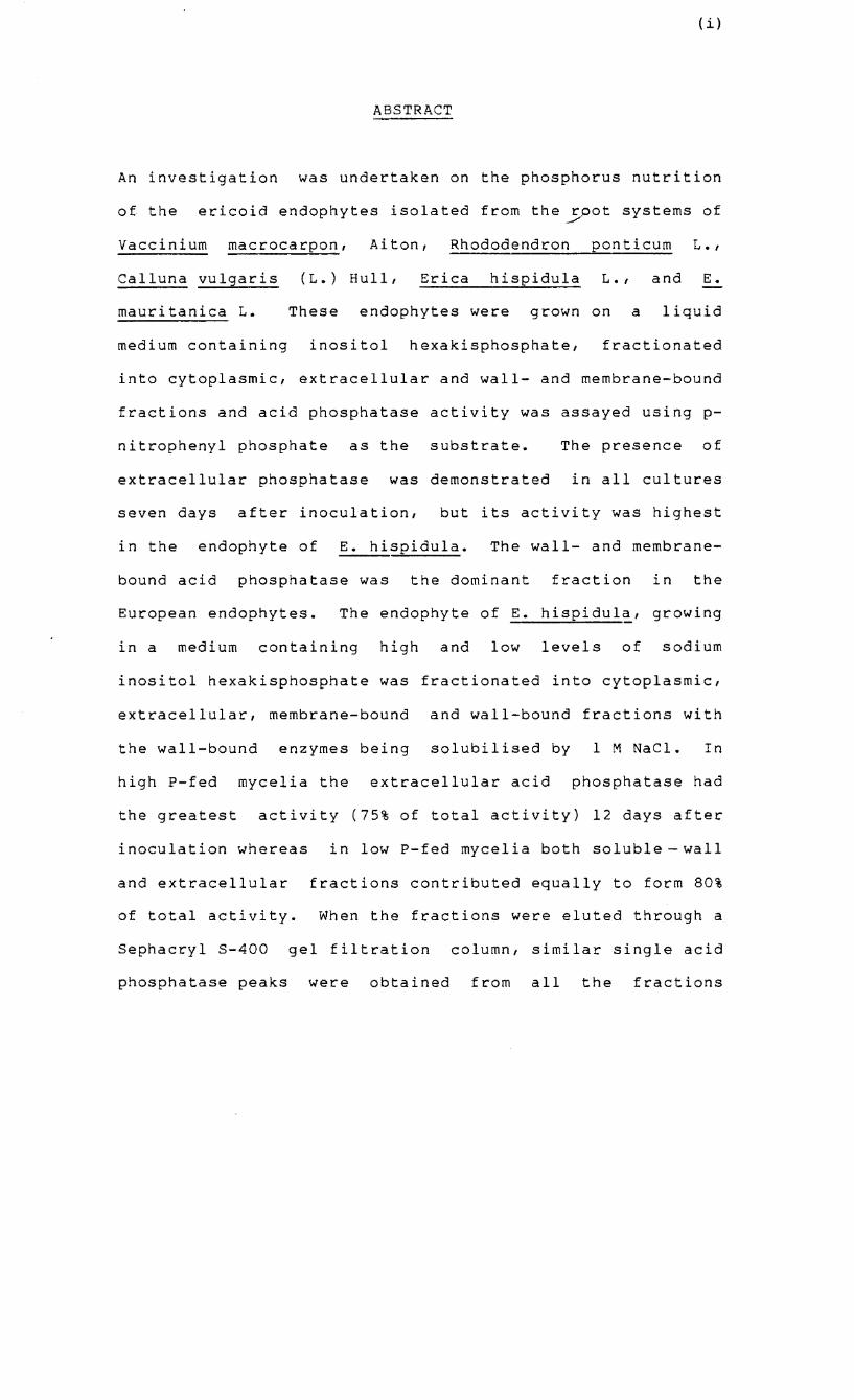
ABSTRACT
An investigation was undertaken on the phosphorus nutrition
of the ericoid endophytes isolated from the rpot systems of ~
vaccinium macrocarpon, Aiton, Rhododendron ponticum L.,
Calluna vulgaris (L.) Hull, Erica hispidula L., and E.
mauritanica L. These endophytes were grown on a liquid
medium containing inositol hexakisphosphate, fractionated
into cytoplasmic, extracellular and wall- and membrane-bound
fractions and acid phosphatase activity was assayed using p-
nitrophenyl phosphate as the substrate. The presence of
extracellular phosphatase was demonstrated in all cultures
seven days after inoculation, but its activity was highest
in the endophyte of E. hispidula. The wall- and membrane-
bound acid phosphatase was the dominant fraction in the
European endophytes. The endophyte of E. hispidula, growing
in a medium containing high and low levels of sodium
inositol hexakisphosphate was fractionated into cytoplasmic,
extracellular, membrane-bound and wall-bound fractions with
the wall-bound enzymes being solubilised by 1 M NaCl. In
high P-fed mycelia the extracellular acid phosphatase had
the greatest activity (75% of total activity) 12 days after
inoculation whereas in low P-fed mycelia both soluble wall
and extracellular fractions contributed equally to form 80%
of total activity. When the fractions were eluted through a
Sephacryl S-400 gel filtration column, similar single acid
phosphatase peaks were obtained from all the fractions
(ii)
except the soluble-wall fraction from low P-fed mycelia
which produced an additional peak representing a lower
molecular weight phosphatase. The molecular weights
corresponding to the main peak common to all fractions and
the lower molecular weight peak were 173 858 and 68 028
respectively. The pH optimum of the low molecular weight
phosphatase was pH 6.5 whereas the high molecular weight
phosphatases showed a broad optimal range between pH 2.0 and
6.0. The enzymes did not show a specific requirement for
metal ions and there were variations in response to
different compounds. All the enzymes were inhibited by
fluoride, molybdenum, arsenate, cyanide, mercury and
phosphate but stimulated by EDTA, citrate and the ferric ion
in low concentrations. The phosphatases showed a wide
substrate specificity with their maximum affinity for
inorganic pyrophosphate and high affinities for a -
and i3 - naphthyl phosphate, phenyl phosphate and a-g lycero-
phosphate but a low affinity for phytic acid. The low
molecular weight enzyme was the only one with the ability to
hydrolyse the organic anhydrides, ATP, ADP and AMP.
Mycelia of the endophyte of E. hispidula were grown in
liquid culture media at high and low levels of
orthophosphate and phosphate uptake rates were measured over
a wide concentration range. The kinetic constants, (i.e.
Vmax and K) were estimated by the Direct Linear Plot m
method using a computer based analysis and a dual uptake
(iii)
system was demonstrated. In the low-affinity system,
V max values of low P-fed and high P-fed mycelia were
similar whereas the Km of high P-fed mycelia was lower than
that of low P-fed mycelia. In the high-affinity
system, the Km values of high P-fed and low P-fed mycelia
were similar whereas high P-fed mycelia showed a higher
V max value than low P-fed mycelia. In low P-fed mycelia,
the low-affinity contribution to total uptake was 93% at 500
pM and 25% at 1 ~M external phosphate. The uptake systems
were sensitive to the pH of the incubation medium and were
inhibited by 2,4-dinitrophenol. The endophyte showed
linearity of absorption for only 1 min and absorption rates
were higher in mycelia grown on very low levels of
orthophosphate.
Metachromatic staining demonstrated the presence of
polyphosphate granules in endophytes isolated from root
systems of V. macrocarpon, R. ponticum, c. vulgaris, E.
his idula and E. mauritanica. The granules accumulated in
response to high concentrations of phosphorus in the
external medium and during the lag phase of growth. Nucleic
acid-polyphosphate co-precipitates prepared from endophytes
were separated by means of polyacrylamide gel
electrophoresis and the molecular weights of polyphosphate
of the endophytes of E. hispidula, E. mauritanica and R.
ponticum were between 3000 and 4700. Inoculated root
systems of V. macrocarpon had significantly more acid-labile
polyphosphate than non-mycorrhizal roots.
(iv)
Mycelia of the endophyte of E. hispidula, grown on high
levels of organic P accumulated high amounts of acid-soluble
and acid-insoluble polyphosphate precipitated by BaCl 2
Under conditions of P deprivation the polyphosphate
fractions declined whereas the orthophosphate fractions
increased. Negligible amounts of polyphosphates accumulated
in low P-fed mycelia. 32 f . . d" d' d P ractlonatlon stu les ln lcate
the acid-insoluble polyphosphate fraction to be higher than
the acid-soluble one.
The results of these investigations are discussed in
relation to the phosphorus nutrition of ectomycorrhizas and
VA mycorrhizas and the importance of ericoid mycorrhizas in
the phosphorus nutrition of heathl~nds.
I would
people
like to
who in
(v)
ACKNOWLEDGEMENTS
express my appreciation to the following
various ways have contributed to the
completion of this thesis.
My supervisor, Professor Derek Mitchell for initially
suggesting the project and for his guidance, interest and
support and thorough scrutiny of the manuscript.
Professor O.A.M. Lewis for permission to use the facilities
of the Botany department.
Professor Eugene Moll for allowing me desk space in the Eco
lab.
Mr Eric O'Neill of the Department of Microbiology for much
appreciated assistance with the gel electrophoresis of the
acid phosphatases and Professor D. Woods for permission to
use the department's equipment.
Dr Mike Picker of the Zoology department for initial advice
on the gel electrophoresis of polyphosphates.
Nicholette Allsopp and Dr Karl Schutte for useful
discussions and advice at various stages of the project.
(v)
Dr Donald Burns of the Department of Scientific and
Industrial Research, Aukland, New Zealand, for allowing me
to use his Double-Hyperbola Curve Fit computer programme.
Hilton Nye for so efficiently modifying the Double-Hyperbola
Curve Fit programme.
Dr Tim Dunne for performing the Correspondence A~alysis of
polyphosphate granule size classes.
Dr D.J. Read of Sheffield University for allowing me the use
of the European isolates.
My post-graduate colleagues, Pat Beeston, Sue Brown, Tony
Cunningham, Craig
Romoff and
friendship.
Willy
Hilton-Taylor,
Stock for
Clive
their
McDowell, Natasha
'camaraderie' and
Tony Walsh for proof-reading some chapters of the manuscript
and assisting with the arduous task of the reference list
and my parents for their support and financial assistance
during lean times.
I gratefully acknowledge financial support from the
University of Cape Town and CSIR, South Africa.
CONTENTS
ABSTRACT
ACKNOWLEDGEMENTS
CONTENTS
PREFACE
LIST OF TABLES
LIST OF FIGURES
CHAPTER 1 INTRODUCTION
1.1 NITROGEN NUTRITION
1.2 PHOSPHORUS NUTRITION
1.2.1 Access to unavailable sources
Phosphorus
1.2.2 Phosphate absorption
1.2.3 Phosphate storage
CHAPTER 2 MATERIALS AND METHODS
2.1 CULTURAL PROCEDURES
2.1.1 Isolation of endophytes
2.1.2 Synthesis of mycorrhizal root
systems
2.1.3 Preparation of liquid cultures
2.1.4 Growth of the endophytes of
Erica hispidula and E. mauri
tanica in culture
of
(vii)
PAGE
(i)
(v)
(vii)
(xiv)
(xvi)
(xviii)
1
6
7
9
12
17
24
24
24
24
26
27
2.2 THE EXTRACTION, FRACTIONATION AND
CHARACTERIZATION OF THE ACID
PHOSPHATASES OF ISOLATED ENDOPHYTES
2.2.1 Fractionation of mycelia of
2.3
endophytes to yield extra
cellular, wall/membrane-bound
and cytoplasmic factions
2.2.2 Fractionation of mycelia of the
endophyte of Erica hispidula
prior to partial purification and
characterization of acid phos
p~atases
2.2.3 Gel filtration
2.2.4 Acid phosphatase assay
2.2.5 Protein determinations
2.2.6 Phosphorus assay
2.2.7 Gel electrophoresis
THE KINETICS OF PHOSPHATE UPTAKE BY
THE ENDOPHYTE OF ERICA HISPIDULA
2.3.1 General incubation and radio-
isotope counting procedures-
2.3.2 Efflux studies
2.3.3 Derivation of kinetic parameters
2.4 IDENTIFICATION, EXTRACTION AND ESTIMAT
ION OF POLYPHOSPHATES AND PHYTIC ACID
(viii)
PAGE
28
28
29
30
31
33
33
35
37
37
40
40
44
(ix)
PAGE
2.4.1 Cytochemical methods for the 44
identification of polyphosphates
2.4.2 Phenol-detergent extraction of 44
undegraded nucleic acid-polyP
co-precipitates
2.4.3 Polyacrylamide gel electro- 46
phoresis
2.4.4 Total P and acid-labile polyP 46
determinations
2.4.5 Statistical analysis 47
2.4.6 Phosphorus extraction and 48
fractionation procedures
2 4 7 32 b t' d' 1 . t' •• P a sorp 10n an ut1 1sa 10n 48
by mycelia in culture
2.4.8 Total P determination of non- 48
radioactive fractions
2.4.9 The effect of activated charcoal 50
on the adsorption of P
contaminants of polyP fractions
2.4.10 Phytic acid determination 50
CHAPTER 3 ACID PHOSPHATASE ACIVITY IN ISOLATED 51
ENDOPHYTES
3.1 INTRODUCTION 51
3.2 RESULTS 52
(x)
PAGE
3.2.1 Growth curves of the South 52
African endophytes
3.2.2 Acid phosphatase activity and 52
protein content of mycelial
fractions of European and a
South African endophyte
3.2.3 Phosphatase activity in 57
fractions of mycelia of the
endophyte of E. hispidula
3.2.4 Gel filtration 60
3.2.5 pH scans 63
3.2.6 The action of effectors 63
3.2.7 Substrate specificity 66
3.2.8 Gel electrophoresis 69
3.3 DISCUSSION 69
CHAPTER 4 KINETICS OF PHOSPHATE UPTAKE BY THE 76
ISOLATED MYCORRHIZAL ENDOPHYTE OF
E. HISPIDULA
4.1 INTRODUCTION 76
4.2 RESULTS 77
4 2 1 U t k of 32p by l' •• p a e myce la grown 77
on different concentrations of P
32 4.2.2 The rate of uptake of P by
mycelia
4.2.3 Efflux studies
4.2.4 Preliminary experiments on the
rate of uptake by mycelia in
relation to P concentration
4.2.5 The effect of pH on P uptake
kinetics
4.2.6 The effect of a metabolic
inhibitor on uptake
4.2.7 The kinetic parameters of dual-
system uptake in high P- and low
P-fed mycelia
4.3 DISCUSSION
CHAPTER 5 THE IDENTIFICATION, EXTRACTION AND
FRACTIONATION OF POLYPHOSPHATES AND
PHYTIC ACID
5.1 INTRODUCTION
5.2 RESULTS
5.2.1 Cytochemical observations of
polyP granules
5.2.2 Polyacrylamide gel electro-
phoresis for separation of
(xi)
PAGE
77
81
83
83
88
88
94
1 01
101
102
102
106
(xii)
PAGE
nucleic acid-polyP
co-precipitates
5.2.3 P and acid-labile polyP content 110
in phenol-detergent extracts
5.2.4 The effect of P starvation on
the endogenous P status of
isolated endophytes of ~.
hispidula grown on high and low
levels of orthophosphate
112
5.2.5 Activated charcoal adsorption of 114
possible contaminants of BaCl2
precipitates
5.2.6 Phytic acid content of mycelia 114
of the endophyte of E. hispidula
5.2.7 Fractionation of 32p in mycelia 116
of the endophyte of E. hispidula
after incubation in KH232p04 and
various concentrations of P
5.2.8 Fractionation of 32 p over time
in mycelia of the endophyte of
E. hispidula grown on high and
low levels of orthophosphate
119
5.3 DISCUSSION 121
CHAPTER 6 GENERAL DISCUSSION 127

REFERENCES
APPENDICES
APPENDIX I DOUBLE-HYPERBOLA CURVE FIT
COMPUTER PROGRAMME
APPENDIX II REPRINT OF PAPER ENTITLED 'THE
CHARACTERIZATION AND ESTIMATION
OF POLYPHOSPHATES IN
ENDOMYCORRHIZAS OF THE
ERICACEAE'. THE NEW PHYTOLOGIST
(1985) 99, 431-440
(xiii)
PAGE
133
145
145
147
(xiv)
PREFACE
soil nutrient levels and the nutrient cycling processes of
the heathlands of the south-western Cape, South Africa have
recently been the subject of an intensive investigation
under the auspices of the Fynbos Biome Programme, CSIR. A
major interest has been the phosphorus status of fynbos
soils, phosphorus distribution patterns within plants,
litter production and decomposition studies and the response
of the vegetation to the addition of nutrients. Little is
known of the phosphorus nutrition of ericaceous plants or
their associated mycorrhizas in the fynbos biome. This
project is a study which investigates aspects of the
phosphorus nutrition of endophytes isolated from root
systems of indigenous ericas. On the basis of other
mycorrhizal studies, three main aspects were selected for
investigation:
1) to investigate the acid phosphatase activity of isolated
endophytes with a view to assessing the role of these
enzymes in the utilization of organic P substrates in the
soil
2) to investigate the phosphorus uptake processes of the
endophyte and establish its efficiency in absorbing free
phosphate ions from the external medium
(xv)
3) to establish the potential of the mycorrhizas to store
phosphorus in the form of polyphosphates, especially in
times of excess P availability.
In view of the differences both in climate and soil nutrient
levels between South African and European heathlands and the
availability of isolated European endophytes from Dr. D.J.
Read of Sheffield University, some comparative studies were
undertaken between South African and European endophytes.
This thesis is structured into six chapters. The
Introduction comprises a review of the literature and states
the aims and objectives of the study. This is followed by
the Materials and Methods and then three chapters dealing
with one aspect of the study, each forming the basis of one
paper. Part of Chapter 5 has already been published as "The
Characterization and Estimation of Polyphosphates in
Endomycorrhizas of
Mitchell in The New
the Ericaceae" by C.J. Straker and
Phytologist (1985) 99, 431-440.
thesis concludes with a General Discussion in Chapter 6.
D.T.
The
LIST OF TABLES
1.1 Kinetic constants of the phosphate uptake
systems of excised mycorrhizas, fungi and
excised plant roots.
3.1 Acid phosphatase activity in fractions of
mycelia of the endophyte of Erica hispidula.
3.2 The influence of various reagents on the acid
phosphatase activity of fractions of mycelia
of the endophyte of Erica hispidula.
3.3 The affinity of acid phosphatase enzymes of
fractions of mycelia of the endophyte of
Erica hispidula towards various substrates.
4.1 The effect of incubation in solutions of
increasing concentration of orthophosphate on
the flux of 32p absorbed by mycelia of the
endophyte of Erica hispidula.
4.2 The influence of pH on the dual-system kinetic
parameter estimates of the P uptake systems
of mycelia
hispidula.
of the endophyte of Erica
(xvi)
PAGE
16
59
65
67
82
87
(xvii)
PAGE
4.3 Dual-system kinetic parameter estimates of the 93
P uptake systems of high P-fed and low P-fed mycelia of
the endophyte of Erica hispidula.
5.1 Acid-labile poly P content of phenol-
detergent extracts and total P of isolated
endophytes and aseptically infected root
systems of Vaccinium macrocarpon.
5.2 The effect of the addition of activated
charcoal to TeA-soluble and -insoluble
extracts on P fraction levels of mycelia of
the endophyte of Erica hispidula.
111
115
LIST OF FIGURES
1.1 Diagram of the polyphosphate cycle
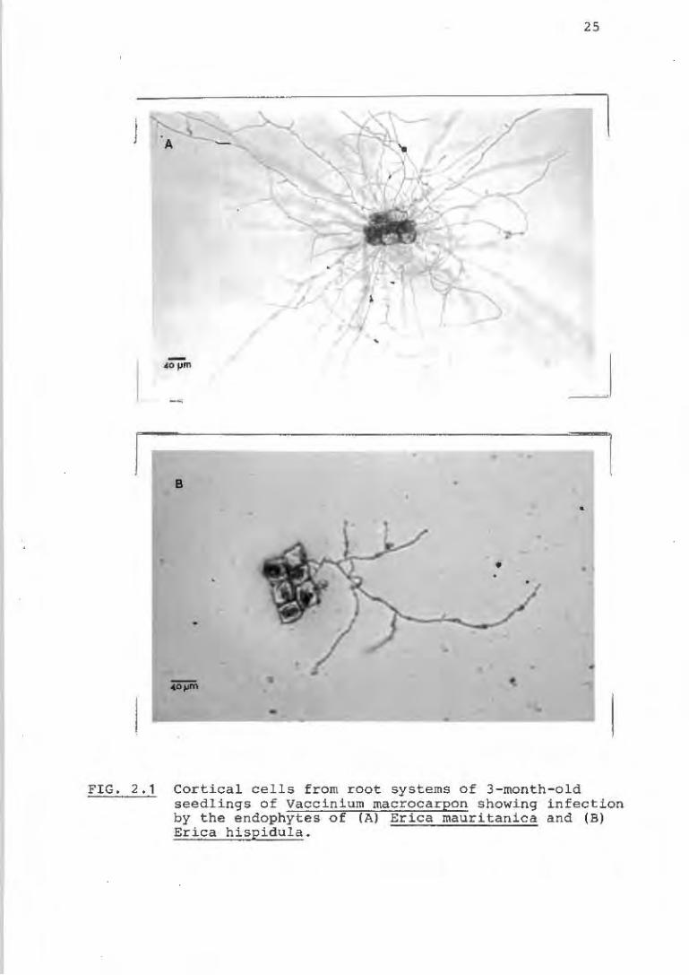
2.1 Cortical cells of seedlings of Vaccinium
macrocarpon infected with the endophytes of
Erica mauritanica and Erica hispidula
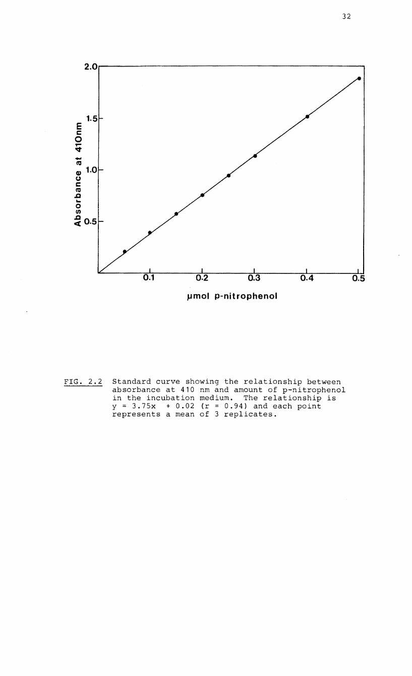
2.2 Standard curve showing the relationship
between absorbance at 410 nm and pmol p
nitrophenol
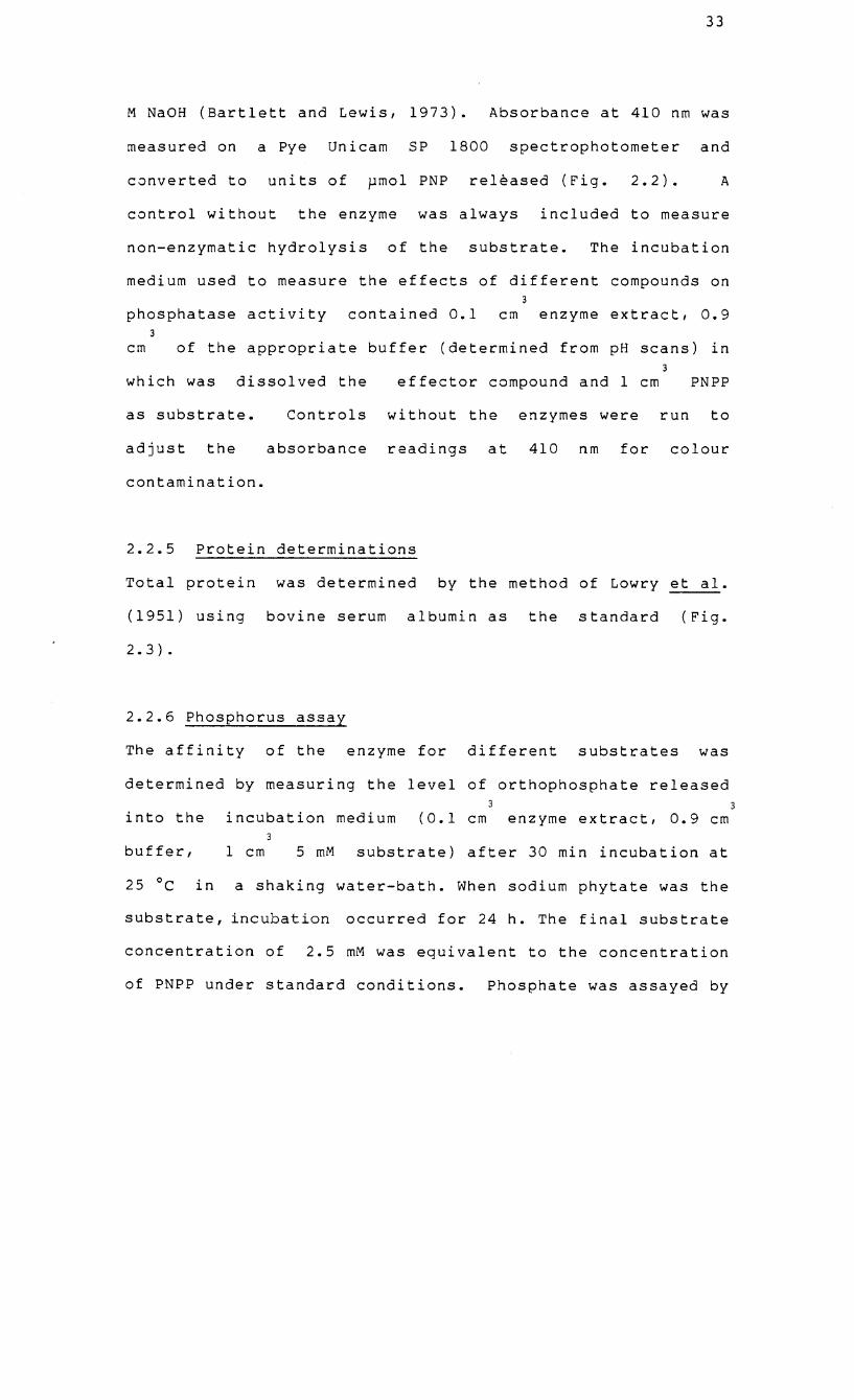
2.3 Standard curve showing the relationship
between absorbance at 500 nm and 750 nm and
~g protein (Lowry et al., 1951)
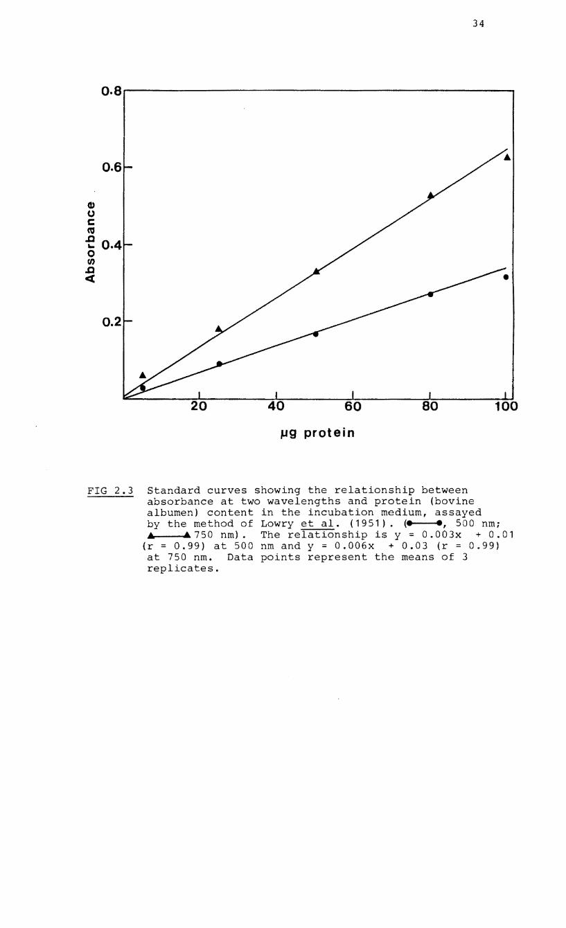
2.4 Standard curve showing the relationship
between absorbance at 882 nm and ~mol
phosphorus (Murphy & Riley, 1962)
2.5 Curves used for the correction of radioactive
counts (cpm) due to quenching
2.6 Standard curve showing the
between absorbance at 689
phosphorus (Kempers, 1975).
relationship
nm and I-lmol
(xviii)
PAGE
21
25
32
34
36
39
41
2.7 Generalised plot of the Hofstee linear
transformation
equation
of the Michaelis-Menten
2.8 Summary of the phenol-detergent extraction
procedure for nucleic acid-polyP co-
precipitates
2.9 Summary of the phosphorus extraction
fractionation procedure based on that
Aitchison & Butt (1973)
and
of
3.1 Growth curves of cultures of the endophyte of
Erica hispidula and Erica mauritanica on
inorganic and organic P
3.2 Protein levels in cytoplasmic, extracellular
and wall/membrane-bound fractions of four
endophytes
3.3 Acid phosphatase
extracellular and
four endophytes
activity of cytoplasmic,
wall/membrane fractions of
3.4 Effect of NaCI on the release of acid
phosphatase from the cell wall of the
endophyte of Erica hispidula
(xix)
PAGE
43
45
49
53
55
56
58
3.5 Elution profiles of protein and acid
phosphatase following gel filtration
(Sephacryl S-400) of fractions of the
endophyte of Erica hispidula
3.6 Molecular weight estimation by gel filtration
of the acid phosphatase enzymes of the
endophyte of Erica hispidula
3.7 pH scans of phosphatase activity in fractions
of the endophyte of Erica hispidula
3.8 Electrophoretic mobility of the acid phos-
phatases of fractions of the endophyte of
Erica hispidula
4.1 Uptake of 32p by mycelia of the endophyte of
Erica hispidula grown on a basal medium
containing various concentrations of
orthophosphate
4.2 Phosphate uptake by mycelia of the endophyte
of Erica hispidula with time
4.3 The rate of uptake of KH 32p04 and glucose-2
32 6-phosphate ( P) over time by mycelia of the
endophyte of Erica hispidula
(xx)
PAGE
61
62
64
68
78
79
80
4.4 Double reciprocal plots of rate of uptake vs
concentration by mycelia of the endophyte of
Erica hispidula
4.5 Hofstee plots showing the effect of pH on
high-affinity and low-affinity P uptake
systems in mycelia of the endophyte of Erica
hispidula
4.6 Hofstee plot of rate of uptake vs P
concentration for mycelia of the endophyte of
Erica hispidula grown on 6 mM orthophosphate
4.7 Hofstee plot of rate of uptake vs P
concentration for mycelia of the endophyte of
Erica hispidula grown on 0.06 mM
orthophosphate
4.8 Diagram showing the relative contributions of
the high- and low-affinity uptake systems in
6 mM- and 0.06 m~1 P-fed mycelia of the
endophyte of Erica hispidula
5.1 A hypha of the mycelium of the endophyte of
Rhododendron ponticum stained with toluidine
blue (pH 1.0) to show vacuolar granules of
polyphosphate
(xxi)
PAGE
84
86
90
91
92
103
5.2 The percentage of metachromatic granules in
relation to different size classes in 10-d
old mycelia of isolated endophytes
5.3 Numbers of metachromatic granules in the
endophyte of Vaccinium macrocarpon grown on
various sources of P
5.4 Frequency of
growth of
macrocarpon
metachromatic
the endophyte
granules during
of Vaccinium
5.5 Absorption spectra
polyacrylamide gels
of segments of
(stained with
8.5%
0.1%
toluidine blue) on which nucleic acid-polyP
phenol-detergent extracts had been run
5.6 Scans of nucleic acid-polyP phenol-detergent
extracts separated on 8.5% polyacrylamide gel
and stained with 0.1% toluidine blue
5.7 Total P in fractions of 8-d-old mycelia of
endophyte of Erica hispidula grown on basal
medium containing either 3.23 mM or 0.16 mM
KH2POq then incubated for 4 d in basal
medium without P
(xxii)
PAGE
104
105
107
108
109
113
5.8 Levels of 32
P in P fractions of 7-d-old
mycelia of the endophyte of Erica hispidula
32 incubated with P and various concentrat-
ions of orthophosphate
5.9 Percentage of 32p incorporated into P
fractions of 7-d-old mycelia of the endophyte 32
of Erica hispidula incubated with P and
various concentrations of orthophosphate
32p 5.10 in fractions of 8-d-old mycelia of the
endophyte of Erica hispidula grown on basal
medium with either 3.23 mM or 0.16 mM KH z P0 4f
then incubated for 4 d in fresh basal medium
32 with P orthophosphate
(xxiii)
PAGE
117
118
120
CHAPTER 1
INTRODUCTION
Heathlands are found in a broad range of climatic zones,
stretching from the Tundra and cool climatic regions of the
northern hemisphere to the warm and seasonally arid heaths
in the mediterranean climatic regions of South Africa and
Australia.
geography,
Despite
heaths are
these variations in climate and
generally characterised by their
evergreen, sclerophyllous vegetation restricted mainly to
acidic soils of a low nutrient status (Specht, 1979). Many
of these heath plants belong to closely related families of
the Grubbiaceae, Empetraceae, Pyrolaceae, Epacridaceae and
Ericaceae in the Ericales. The heaths of South Africa may
be domi na ted by Er i ca sPP. belong i ng to the Er i caceae whereas
the South Australian heaths contain members of the
Epacridaceae.
Nutrient cycling studies in heathlands have concentrated o~
nitrogen (N) and phosphorus (p) because of their high level
of interaction in nutrition and their importance in the
control of nutrient cycling processes (Groves,
South Africa, heathlands form part of the
Kingdom (Takhtajan, 1969; Good, 1974) known
'fynbos' (defined by Moll and Jarman, 1984).
1983). In
Cape Floral
locally as
Mountain
fynbos is found exclusively o~ highly leached acid soils
with low nutrient status whereas coastal fynbos often occurs
2
on well-drained sands of aeolian origin with high base
saturation levels or on lowland limestone (Kruger, 1979).
Mitchell, Brown and Jongens-Roberts (1984) showed that
coastal fynbos soils of the Clovelly form are very low in
total P with the major inorganic form being iron-bound and a
considerable proportion as organic P (27 - 60%) These soils
are also low in both total and available N and ammonium
(NH4+ ) predominates over nitrate (N03-) especially in the
later stages of succession of the plant community (Stock and
Lewis, in press). In South Australia, levels of soil P and N
and organic matter are comparable to those found in the
fynbos (Read and Mitchell, 1983). Levels of organic matter
and soil P and N in Calluna heathlands in Britain are
generally higher than those of the southern hemisphere
heathlands (Read and Mitchell, 1983) but nutrient levels may
also drop to low levels similar to those of Australia and
South Africa (Gimingham, 1972).
The characteristic infertility (or seasonal unavailability
of nutrients) in heathland soils and the universal presence
of ericaceous plants on them are features accompanied by
root systems which are strikingly uniform in structure. The
ultimate, fine, hair roots consist of a central stele
surrounded by o~e to three rows of cortical cells (Read,
1983) which may be characteristically infected with
endomycorrhizal fungi in an assocation known as 'ericoid'
(Harley, 1969). Under the light microscope, the hair roots
3
may be commonly surrounded with a loose weft of septate
mycelium which penetrates the cortical cell walls at points
and within the cell proliferates to form dense, hyphal coils
(Harley and Smith, 1983: Read, 1983). These features have
been observed
Vaccinioideae,
most commonly in
Rhododendroideae and
the sub-families
Ericoideae of the
Ericaceae. Other members of the Ericaceae (Arbutus,
Arctostaphylos) form mycorrhizas of the ecto-endo type
(arbutoid) as do the pyrolaceae (Read, 1983) while the
Monotropaceae are characterised by a distinctive association
called monotropoid (Ouddridge and Read, 1982a). Members of
the Epacridaceae also form mycorrhizas of the ericoid type
McNabb, 1961). (McLennan, 1935:
In recent years, ultrastructural analyses of ericoid
mycorrhizas have enhanced our knowledge of the biological
nature of the symbiosis and its nutritional and ecological
significance. Intracellular hyphae show little evidence of
branching, resemble each other morphologically and have
simple septa and Woronin b~dies characteristic of
ascomycetous fungi. The host plasmalemma invaginates to
surround individual, penetrating hyphae and is separated
from them by a fibrillar, interfacial matrix of varying
thickness (Bonfante-Fasola and Gianinazzi-Pearson, 1979;
Peterson, Mueller and Englander, 1980: Bonfante-Fasola,
Berta and Gianinazzi-Pearson, 1981). Ouddridge and Read
(1982b) followed the sequence of events involved in the
4
initiation, establishment and degeneration of the ericoid
mycorrhizas of Rhododendron ponticum L., both under natural
and sterile conditions and observed that the host cytoplasm
degenerates before that of the fungus. As the functional
life of the association within a cell lasted no more than
seven weeks, it was suggested that if major exchanges of
nutrients between fungus and host occur, they would have to
take place within a relatively short period. In axenic
culture, the infection cycle is even more short-lived with
an association of intense metabolic activity (Bonfante-
Fasola and Gianinazzi-Pearson, 1982). In Vaccinium
myrtillus L. (which is deciduous) infection levels vary
throughout the year with peaks in late summer and autumn
while in Calluna vulgaris (L.) Hull (an evergreen plant)
infection levels are sustained throughout winter but reach a
peak in spring when new hair roots form (Bonfante-Fasola,
Berta and Gianinazzi-Pearson, 1981). In the seasonally-arid
heathlands of the Western Cape however, hair roots are
moribund with little or no infection during the dry summer
season but are growing actively and highly infected in
spring when soil moisture levels are higher (Read, 1978).
It appears that the amounts of mineral P and N which enter
heath land ecosystems are low and that the major reserve of
these elements is in the soil organic matter (Groves, 1983~
Read and Mitchell, 1983). The sclerophyllous vegetation of
heathlands can contain high levels of lignin and phenolics
5
which render the litter resistant to microbial degradation.
Microbial activity is further inhibited by the high C : N
ratios of the fallen litter and the low mobility of leaf N
may also contribute to slow rates of litter decomposition.
In Australia and South Africa, the mediterranean-type
climate causes seasonal moisture stress which may reduce
leaching of minerals from fallen litter, retard microbial
decomposition and promote seasonal peaks
acti vi ty. I n general, mi neral i si ng rates
heath land ecosystems are low (Read and
Harley and Smith, 1983; Stock and Lewis,
in mineralising
of P and N in
Mitchell, 1983;
in press; Stock,
Lewis and Allsopp, in press). Ultrastructural studies of
ericoid mycorrhizas suggest that the host root systems have
an obligatory requirement for infection by the endophyte
which has probably evolved in response to low nutrients.
Phenology, climate and features of decomposition processes
would determine periods of peak activity of the association
when the absorption and exchange of nutrients is maximal and
the association of most benefit to both partners.
Ultrastructural investigations have substantiated
information on the nutritional and biochemical nature of the
ericoid mycorrhizal symbiosis obtained from physiological
studies. There is strong evidence (Stribley and Read, 1974ai
Pearson and Read, 1973b; Harley and Smith, 1983) to indicate
that carbon products flow from autotroph to heterotroph and
since the endophytes possess only a limited cellulolytic
6
ability (Pearson and Read, 1975; Mitchell and Read, 1985)
the mycobiont is probably dependent on its host for the bulk
of its carbon supply. However, the flow of nutrients from
fungus to host is of greater ecological importance,
especially with respect to Nand P.
1.1 NITROGEN NUTRITION
In a typical acid, mor-humus soil of a mature European
+ heathland NH predominates over NO and only 0,4% of the
4 3
+ total N is present as free NH while 71% is in the form of ~
hydrolysable organic N (Stribley and Read, 1980). These
limiting soil N conditions have stimulated research into
the N nutrition of ericoid mycorrhizas, recently reviewed
by Harley and Smith (1983) and Read (1983).
It appears from these studies that the mycobiont is able to
enhance the N nutrition of its host in two important ways.
Firstly, sand-culture studies indicated that the
extramatrical hyphae directly aid the uptake of NH + ions by 4
their ability to cross depletion zones around the roots and
explore a greater volume of soil (Strib1ey and Read, 1974b:
1976). This is a facility which is probably enhanced by the
possible
NH 4
+
high affinity of fungal absorption systems for
ions (Harley and Smith, 1983). Secondly, the
mycorrhizal root systems appear to have access to sources of
N unavailable to non-mycorrhizal roots. Stribley and Read
(1980) established that mycorrhizal plants of V. macrocarpon
7
Aiton can utilize amino-acids as sole N sources, an ability
which was less developed in aseptically-grown plants or
plants infected with common soil saprotrophic fungi.
(1978) has also suggested that under the conditions
winter rainfall and summer drought experienced in
Read
of
the
southern heathlands of the Cape, the endophyte absorbs N
efficiently at periods when soil moisture levels are high,
and N stored in intracellular hypha 1 complexes is made
available to the host during the critical periods of
flowering and seed production. However, in view of the
unlikelihood of digestion of the endophyte by the host
(Dudjridge and Read, 1982b) and the lack of any empirical
substantiation, this hypothesis should be treated as
speculation.
1.2 PHOSPHORUS NUTRITION
Phosphorus is also very limiting in heathland ecosystems and
there is no reason to suppose that the endophyte's ability
to increase the supply of N to the host does not extend to
other elements as well. Moreover, vesicular-arbuscular (VA)
endomycorrhizas and ectomycorrhizas contribute to increased
growth in their hosts by way of supplementing the supply of
P to them (see Harley and Smith, 1983). It seems unlikely
that ericoid endophytes do not contribute to their hosts'
survival in a similar way. The work on N nutrition of
ericoid mycorrhizas and the studies on P nutrition in other
8
mycorrhizal systems emphasize four main areas of potential
research into ericoid mycorrhizal associations.
(1) Does the endophyte have access to forms of P which the
host roots do not?
(2) Does the endophyte possess more efficient mechanisms for
locating and absorbing P than the host roots?
(3) Is the endophyte able to store P which is later
transferred to the host?
(4) What are the transfer mechanisms involved in the
movement of P from fungus to host?
The remainder of this review reflects the field of
information which has motivated this project. The field
includes studies on VA and ectomycorrhizas as well as
investigations into individual aspects of the physiology of
other soil fungi. The review article of Beever and Burns
(1980) suggests that aspects of the P physiology of soil
fungi, including those which form mycorrhizas, are similar.
from studies on other soil fungi may Empirical evidence
therefore pertain to investigations into the P nutrition of
ericoid mycorrhizal
transfer of P at
endophytes. Since the question of
the host/fungus interface was not
9
approached in this study it will be discussed only in
passing.
1.2.1 Access to unavailable sources of Phosphorus
The idea that mycorrhizas are able to tap bound sources of P
unavailable to plant roots has been an appealing one for
some time~ this facility would depend upon the
microorganism's accessibility to external substrates by way
of active enzyme systems. The low orthophosphate (Pi)
availability and high organic P levels (especially soluble
and insoluble inositol phosphates) in northern hemisphere
heathland soils is now well documented (Anderson, 1967:
Cosgrave, 1967: Martin and Cartwright, 1971: Anderson,
Williams and Moir, 1974: Cheshire and Anderson, 1975:
Tinker, 1975). Acid phosphatase (E.C. 3.1.3.2.), an enzyme
of broad specificity has been implicated in the process of
making P available from organic soil molecules. Other
areas of interest are the responses of acid phosphatase
activity to high and low P conditions and the localization
of activity in the cell under different P conditions.
In ectomycorrhizas, an active acid phosphatase localised in
the plasmalemma and cell walls of the sheath of beech roots
was able to hydrolyse a wide range of organic esters and
inorganic P substrates (Woolhouse, 1969; Bartlett and
Lewis, 1973: Williamson and Alexander, 1975). Beech
mycorrhizas are also able to use sodium and calcium
10
phytates in pure culture (Theodorou, 1968: 1971). Ho and
Zak (1979) have found large differences in the acid
phosphatase activity of six ectomycorrhizal fungi.
Alexander and Hardy (1981) found the acid phosphatase
activity of mycorrhizal roots of Sitka spruce from a P
deficient site to be inversely proportional to the litter P
concentration, a fact which suggested a derepression of
phosphatase activity under low Pi conditions. When
fractions of three ectomycorrhizal isolates were compared
for phosphatase activity under low and high pi conditions,
a large increase in total phosphatase activity (localised
mainly in the cell wall) appeared when Pi was deficient
(Calleja et al., 1980). Under these conditions, an active
extracellular phosphatase was also secreted into the
external medium accompanied by a decline in the activity of
the soluble (cytoplasmic) enzyme fraction. Calleja and
D'Auzac (1983) confirmed these findings and attempted to
separate the ectomycorrhizal isolates
saprotrophic fungi on the basis of their
phosphatase activities, but were unable
from three
comparative
to establish
satisfactory parameters for discrimination. Although these
experiments demonstrated for the first time that mycorrhizal
fungi are able to secrete an extracellular phosphatase, this
is not unknown in other fungi (San BIas and Cunningham,
1974: Arnold and Garrison, 1979: Beever and Burns, 1980:
Bojovic-cvetic and Vujicic, 1982).
1 1
Detailed information on the phosphatases of endomycorrhizas
is limited to VA mycorrhizas. The levels of soluble acid
phosphatase activity of onion roots were not significantly
increased by infection with VA endophytes or by the addition
of soluble P to the soil (Gianinazzi-Pearson and Gianinazzi,
1976). In a cytochemical study of infected onion roots
significant acid phosphatase activity was only observed in
immature, terminal arbuscles of the endophyte while alkaline
phosphatase activity, which could be repressed at Pi
levels above -4
10 M was detected in mature arbuscles and
intercellular hyphae (Gianinazzi-Pearson and Gianinazzi,
1978; Gianinazzi, Gianinazzi-Pearson and Dexheimer, 1979).
It appears that the differences in activity of the
phosphatases was a function of the age of the endophyte and
that in mature arbuscles the alkaline phosphatases may be
involved in polyphosphate (polyP) metabolism and P
transfer to the host.
Pearson and Read (1975) have detected acid phosphatase
activity which is inhibited by high concentrations of
external Pi in the ericoid endophyte Pezizella ericae
Read . In addition, the endophytes of R. ponticum and V.
of using soluble and insoluble macrocarpon are capable
phytate salts in culture (Mitchell and Read, 1981).
However, whether this utilization is made possible through
the mediation of surface-bound or extracellular acid
phosphatase is unknown.
12
One cannot assume thi:t the localisation, function and level
of activity of acid phosphatases in all mycorrhizas is going
to be similar, especially when these parameters are strongly
influenced by experimental conditions. However, in
principal, the synthesis of active wall-bound and
extracellular phosphatases under conditions of P deficiency
may well give mycorrhizal fungi greater access to b~und soil
P complexes, than other soil saprotrophs or the host
itself. Whether these enzymes under cultural conditions are
synthesized isoenzymes of functional significance or the
products of cell lysis needs to be investigated.
1.2.2 Phosphate Absorption
Soil Exploration
Free orthophosphate ions, being b~und in soil complexes, may
ba very low in soil solutions and the slow diffusion rates
of the ions accompanied by rapid absorption of them by roots
leads to the formation of depletion zones around roots (Nye
and Tinker, 1977). Harley and smith (1983) reported that _3 _1
inflow of Pi (expressed as mol cm s ) into onion and
clover roots infected with VA endophytes is on the average 3
to 4 times greater than into uninfected controls. The
ability of the extramatrical hyphae to continually colonise
beyond depletion zones is proposed as the main reason for
this greater inflow. A more rapid translocation of Pi
along hyphae than diffusion of Pi through depletion zones
13
also contributes to making the mycobiont an effective
physical extension of the root system. Thomas et al. (1982)
compared the growth of ectomycorrhizal and non-mycorrhizal
tree seedlings on 32p-labelled soil and found that although
mycorrhizal seedlings showed a greater P uptake, the
proportion of non-labile P absorbed was less than by non-
mycorrhizal seedlings. It appears that mycorrhizal roots
are able to tap labile P sources beyond root depletion zones
whereas non-mycorrhizal roots start using non-labile sources
sooner. The only research into by ericoid
mycorrhizal roots showed that the external mycelium of c~
vulgaris and V. oxycoccus L. mycorrhizas is able to absorb
and trans locate P from a source, across a diffusion
barrier and to the host plant which acts as the sink
(Pearson and Read, 1973b).
Phosphate Uptake
If P uptake rates are measured over a narrow range of
external pi concentrations they usually indicate a simple
hyperbolic relationship with concentration and can be
described by the Micha1is-Menten constants of Vmax and
K (Beever and m
the external Pi
Burns, 1980}. The Km values depending o~
concentrations used, fall into 1 of 2
ranges, either 1 to 10 or 100 to 1000 ~M. This indicates
the existence of two separate uptake systems, one with a
greater affinity for P (lower Km) than the other. Two such
separate systems exist in Neurospora crassa Shear and Dodge.
14
(Lowendorff, Slayman and Slayman, 1974~ Lowendorff, Bazinet
and SlaYl.lan, 1975). The relationship between the two systems
has been clarified by Beever and Burns (1977) and Burns and
Beever (1977) by studies in which P uptake was monitored at
concentrations which encompassed the ranges of both systems.
On a transformation plot, the data could no longer be
described by a straight line but by two linear portions
joined by a distinct curve. Moreover, the growth and Pi
udtake rates remained constant during exponential growth
over the broad concentration range used. These authors
interpreted their results in terms of a dual uptake system
acting simultaneously across the plasmalemma so that
compensating changes in the kinetic constants occurred wi th
changes in Pi concentration. Many fungi appear to possess
such systems (Beever and Burns, 1980).
There is some evidence for the operation of dual uptake
systems in mycorrhizal fungi. At selected external Pi
concentrations there was a greater rate of uptake by VA
mycorrhizal roots than non-mycorrhizal roots when expressed
on a mass basis (Bowen, Bevege and Moss, 1975). Cress,
Throneberry and Lindsey (1979) examined in more detail the
kinetics of P uptake in mycorrhizal and non-mycorrhizal
tomato roots. Initial rates of P absorption reached a
maximum over the
transformation plots
with the transition
concentration range used and Hofstee
fitted the data into two linear p~ases
occurring between 20 pM and 30 pM
15
The K m
values for mycorrhizal plants were lower
than for uninfected roots for b~th the high and low
concentration ranges with an increase in V in the high max
range. In the low range, the increased uptake was a result
of an increased site affinity (lower
range it was due to an increase in V max
K ) but at the high m
(i.e. an increase
in number of absorbing sites provided by the mycelium
external to the roots) (Cress et al., 1979).
There are n~ detailed comparative kinetic studies on
ectomycorrhizal and non-mycorrhizal roots of the same
species although Harley and McCready (1950, 1952) have shown
that infected beech mycorrhizas incubated in solutions of
specific concentrations of external Pi absorb five times
as much P on a surface area basis and twice as much on a
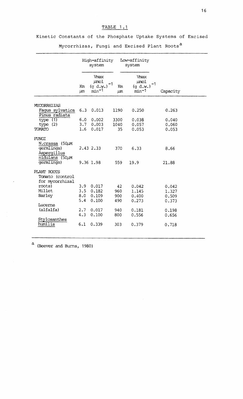
mass basis than uninfected roots. Beever and Burns (1980)
have compared the kinetic constants of ecto- and VA
e~domycorrhizas and some plant roots. It is clear from
Table 1.1 that conclusive data on the uptake kinetics of
mycorrhizas is scanty. Mycorrhizal fungi may n~t depend
exclusively on the exploration of a large soil volume for
increased ion absorption but may also rely on a greater
efficiency in uptake at the sites of transfer themselves,
especially when the nutrient s~pply is infrequent or very
dilute.
16
TABLE 1.1
Kinetic Constants of the Phosphate Uptake Systems of Excised
Mycorrhizas, Fungi and Excised Plant Roots a
High-affinity system
Low-affinity system
Vrnax ,um:>l -1
(q d.w.) Krn
Vrnax }.lITOl -1
(g d.w.) rom -1 }.lID rnin-1 capacity
MYCORRHIZAS Fagus sylvatica Pinus radiata type (1) type (2)
'IDMA'ID
FUNGI N.crassa (50f,lM
6.3 0.013
6.0 0.002 3.7 0.003 1.6 0.017
gerrnlings) 2.43 2.33 Aspergillus nidulans (50f,lM gerrnlings) 9.36 1.98
PLAN!' ROOl'S Tanato (control for mycorrhizal
a
roots) 3 . 9 0 .017 Millet 3.5 0.182 Barley 8.0 0.109
5.4 0.100 Lucerne (alfalfa)
Sty10santhes hurni1is
2.7 0.017 4.3 0.100
6.1 0.339
(Beever and Burns, 1980 )
1190
3300 1040
35
0.250
0.038 0.057 0.053
370 6.33
559 19.9
42 960 900 490
940 800
303
0.042 1.145 0.400 0.273
0.181 0.556
0.379
0.263
0.040 0.060 0.053
8.66
21.88
0.042 1.327 0.509 0.373
0.198 0.656
0.718
17
1.2.3 Phosphate storage - the role of polyphosphates
The pioneering, histochemical staining techniques of Ebel
and Muller (1958); Ebel, Colas and Muller (1958a & b);
Muller and Ebel (1958) h~ve been used to demonstrate the
presence of metachromatic, vacuolar granules in
e~tomycorrhizas (Ashford, Ling-Lee and
VA mycorrhizas (Cox et al., 1980).
transmission electron microscope
Chilvers, 1975) and
In addition, the
(T~M) and X-ray
microanalysis have shown that these granules in
ectomycorrhizas (Strullu et al., 1981: Strullu et al.,
1982, 1983) and VA mycorrhizas (White and Brown, 1979: Cox
et al., 1980) contain high concentrations of P. The P
occurs in the form of inorganic polyphosphate (polyP)
chains (Harold, 1966). PolyP granules have in fact been
observed in a wide range of fungi (Ebel and Muller, 1958:
Ebelet a 1., 1958b: Muller and Ebel, 1958: Chi 1 vers,
Lapeyrie and Douglass, 1985) and may be regarded as
representing a common strategy whereby P is stored in a
condensed form, especially when freely available (Beever and
Burns, 1980). Considerable work is now being done to
clarify the characteristics of polyP metabolism in
mycorrhizal fungi and hence its role in the P nutrition of
the host.
The early 32p radioactive work of Harley and his associates
on P uptake by beech mycorrhizas showed that P reaches the
host cells via the fungal protoplast with uptake and

18
transfer to the host being active processes (see Harley,
1969). The exchanges are mediated by a small pool of Pi
and most P which enters the fungus is shunted into a large,
inorganic pool. When P is not available from the external
environment, it can be mobilised from the large pool to
supplement the smaller, labile pool and hence supply the
host tissues (Harley, 1969). It became clear later
(Chilvers and Harley, 1980) that the site of accumulation of
the reserve inorganic P was in the vacuoles, in the form of
polyP
levels.
granules which accumulated as a result of external P
Lapeyrie, Chilvers and Douglass (1984) have
recently observed a similar trend in pure cultures of the
ectomycorrhizal fungus, Paxillus involutus (Batsch) Fr.
However, quantification of polyP levels in relation to the
levels of other P pools under controlled conditions
requires elaborate fractionation procedures. Harley and
McCready (1981) used the fractionation technique of
Aitchison and Butt (1973) which is based on the traditional
distinction between acid-soluble (short-chain) and acid-
insoluble (long-chain) polyP both
BaC1 2 (See Harold, 1966) to establish that
precipitated by
32 P absorbed by
Fagus mycorrhizas accumulates initially as pi but over 2.5
hours is converted to high levels of polyP compounds (40%
of the total absorbed 32p ). In comparison pure cultures of
ectomycorrhizal fungi store o~ly low levels of polyP even
19
under conditions of P surplus (Martin et al., 1983: Rolin,Le
Tacon and Larher, 1984) •
Callow et al. (1978), critical of fractionation techniques
based on BaClz precipitation of polyP with its risk of
contamination with nucleic acids, employed a method of
phenol-detergent extraction of undegraded polyP-nucleic acid
co-precipitates which were finally separated by
polyacrylamide gel electrophoresis. They estimated a VA
endophyte in onion roots to contain 40% of its total P as
polyP. Since uninfected roots did not contain polyP and
host cells did not stain metachromatically they assumed the
polyP to be fungal in origin. Later the polyP content of
isolated, intact VA endophytes was measured more accurately
to be 16% of the total P (Capaccio and Callow, 1982).
Although polyP might be an important storage form of pi it
may also fulfil an intricate function in the overall P
metabolism of the fungal cell and be linked to pi
transport to the host and sugar transport from the host.
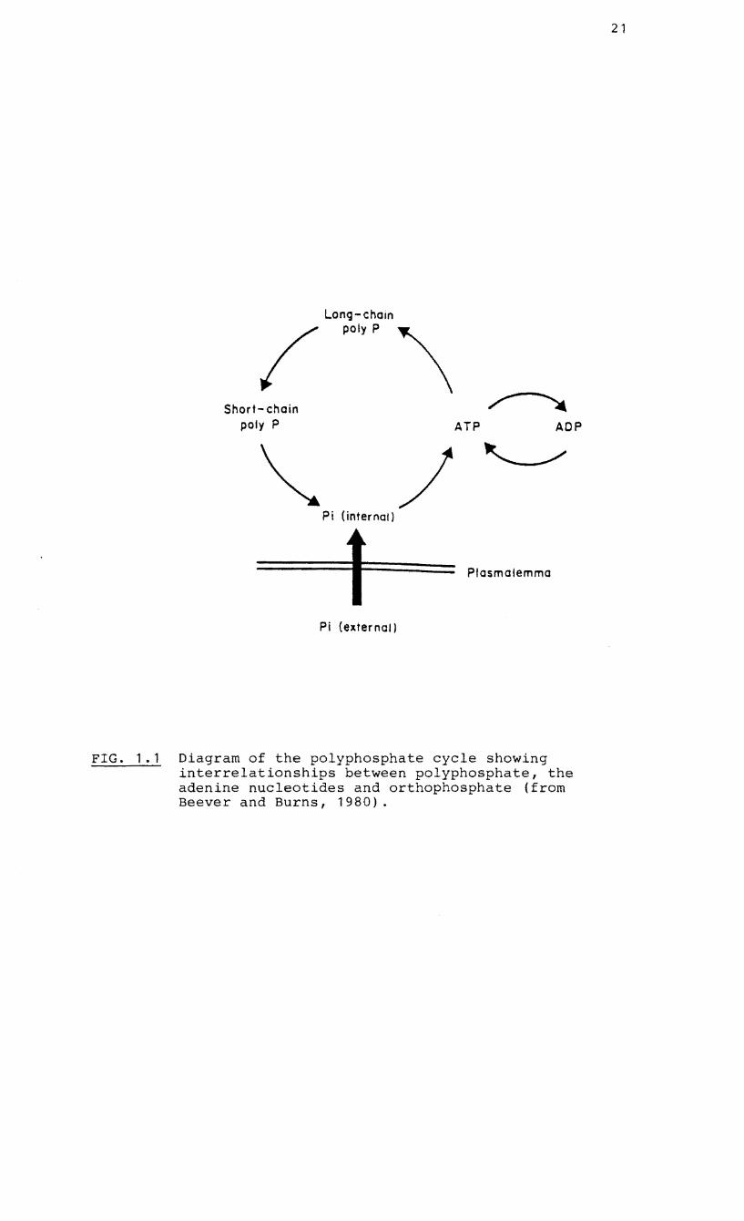
Capaccio and Callow (1982) suggest that the polyP cycle in
VA mycorrhizas probably resembles that proposed for other
microorganisms (Harold, 1966: Beever and Burns, 1980). pi
levels in the cell remain relatively constant by virtue of a
net synthesis of long-chain polyP via ATP (catalysed by
polyP kinase) in times of P excess and a stepwise breakdown
of long-chain polyP to shorter chains and eventually Pi
20
(catalysed by the polyphosphatases) in times of P deficiency
(Fig. 1.1). Hydrolysis of polyP and release of pi across
the tonoplast would maintain a high pi concentration in the
cytoplasm adjacent to the fungal/host interface,
facilitating passive movement into the interfacial apoplast
for active uptake by the host (Harley and Smith, 1983).
Cappacio and Callow (1982) have shown that extracts of
internal VA endophytes and infected onion roots can catalyse
the transfer of terminal P from ATP to a high molecular
weight form with the characteristics of polyP, a reaction
mediated by polyP kinase. PolyP kinase activities were also
greatly increased in infected roots when transferred from a _5
solution of 10 M pi _3
to 10 M Pi and a stronger
activity of exo- and endopolyphosphatases in infected than
un infected roots was detected (Cappacio and Callow, 1982).
In association with sugar uptake, it is proposed that polyP
breakdown is linked to hexose uptake into the fungus by the
synthesis of hexose-p~osphate, catalysed by polyP hexokinase
(Woolhouse, 1975; Harley and Smith, 1983). The
phosphorylated hexoses could be used in metabolism or the
formation of oligo- or polysaccharides and the pi be
released into the labile pi pool. PolyP hexokinase is
active in both internal and external VA hyphae (Cappacio and
Callow, 1982). Alternatively, polyP kinase in its reverse
reaction could generate ATP at a site of transfer~ the ATP
Long-chain I polyP \
Short- chain poly P ATP ADP
Pi (inter nat)
Plasmalemma
Pi (exter nat)
FIG. 1.1 Diagram of the polyphosphate cycle showing interrelationships between polyphosphate, the adenine nucleotides and orthophosphate (from Beever and Burns, 1980).
21
22
would then be used in the active uptake of hexose to form
hexose phosphate (Harley and Smith, 1983).
PolyP has been further implicated in the P nutrition of VA
mycorrhizas through its prospective role in P translocation
(Cox and Tinker, 1976: Cooper and Tinker, 1981; Cox et aI,
1980). It is apparent that an elaboration of some or all of
the potential functions of the molecule is essential to an
investigation into the P nutrition of mycorrhizal fungi.
1.3 AIMS AND OBJECTIVES
The objectives of this project were to investigate the
phosphorus nutrition of ericoid mycorrhizas with a view to
clarifying their importance in the nutrition of their host
plants. On the basis of the information reviewed three
aspects were selected for investigation:
(1) to determine the activity and character of the acid
phosphatases of the endophyte in culture with special
interest in an extracellular fraction.
(2) to investigate the P uptake kinetics of a South African
endophyte in culture with emphasis on the possible operation
of a dual uptake system. This would provide preliminary
evidence for indicating the potential of ericoid mycorrhizas
to facilitate P uptake under conditions of erratic P supply
23
(3) to identify, characterise and estimate the levels of
polyphosphates in endophytes and synthesized mycorrhizal
seedlings and establish the importance of the molecule as a
P storage form under conditions of excess P availability.
24
CHAPTER 2
MATERIALS AND METHODS
2.1 CULTURAL PROCEDURES
2.1.1 Isolation of endophytes
Mycorrhizal endophytes of v. macrocarpon, R. ponticum and
C. vulgaris from the United Kingdom and E. hispidula and
E. mauritanica from South Africa were isolated from root
systems using the serial washing and maceration techniques
described by Pearson and Read (1973~). Cultures of V.
macrocarpon and R. ponticum were those used by Mitchell and
Read (1981) whereas the endophyte of C. vulgaris was
isolated from seedlings taken from Parys Mountain, Anglesey,
United Kingdom. Seedlings of E. hispidula and E.
mauritanica were growing in acid Table Mountain sandstone
soils at the National Botanical Gardens, Kirstenbosch and
Tokai forest respectively, 15 to 18 km S.E. of Cape Town.
All the endophytes were grown on 2% malt extract agar. The
South African isolates were successfully back-inoculated
into seedlings of V. macrocarpon and re-isolated (Fig. 2.1).
2.1.2 Synthesis of mycorrhizal root systems
Seeds extracted from fresh fruits of V. macrocarpon were
surface sterilized in 3% sodium hypochlorite for 5 min,
washed thoroughly with sterile distilled water and
25
j ~, A :/
/'
/
•
I •
• •
•
• •
, •
FIG. 2.1 Cortical cells from root systems of 3-month - o l d seedlings of Vaccinium macrocarpon showing infection by the endophytes of (Al Erica mauritanie a and (B) Erica hispidula.
26
transferred to sterile plates containing 1% agar. After
three weeks, seedlings were transferred to McCartney bottles
containing 20 cm 3 of the following autoclaved medium
(Robbins and White, 1936): MgSO .7H 0, 10 mg; KH PO , 10
mg;
4 2 2 4
FeCI. 6H 0, 2 mg; NH Cl, 32 mg; CaCl • 6H 0, 33.5 32422
mg; agar, 10.0 g with distilled water to 1 dm 3,
supplemented with 0.5 g dm- 3 glucose, 1 g dm- 3 activated
charcoal (Duclos and Fortin, 1983) and covered with a thin
layer of autoclaved acid-washed sand when set. The uncapped
bottles were placed in autoclaved glass boxes (47 cm x
32.6 cm x 20 cm high) standing in stainless steel trays,
which were placed in growth cabinets with 16 h daylight
at 20°C and 8 h darkness at 15°C and an irradiance of
After six weeks, the lightly infected seedlings were
transferred to moist, sterile Clovelly soil and grown for
another six weeks under the same conditions by which time
infection of the root system was sufficiently developed to
permit harvesting of the seedlings. All root systems were
thoroughly washed under tap water and in a number of changes
of distilled water prior to either extraction and digestion
procedures or re-isolation of the endophytes.
2.1.3 Preparation of liquid cultures
The basal liquid nutrient medium used for all experiments
was similar to that used by Mitchell and Read (1981) and
29
fraction. These fractions were assayed for phosphatase
activity and total protein. Further purification and
characterisation of these fractions were undertaken using
methods in sections 2.2.2 and 2.2.3.
2.2.2 Procedure for the fractionation of mycelia of the
endophyte of E. hispidula prior to partial purification and
characterisation of acid phosphatase
The extraction and fractionation procedure was based upon
that of Hasegawa, Lynn and Brockbank (1976).
Extracellular fraction
Cultures of the endophyte of E. hispidula, growing on 3.23
mM sodium inositol hexakisphosphate were harvested at 13 days,
i.e. during the exponential growth phase (Fig. 3.1). Each 3
mycelium, filtered under suction, was rinsed in 10 cm ice-
cold, distilled water. The filtered liquid media of 20
cultures together with the washings were passed through a
"Millipore" filter (0.45 flM), dialysed against distilled
water at 10°C, lyophilised in a New Brunswick freeze-drier
and dissolved in 20 cm 3 0.1 M acetate buffer, (pH 4.5) to
form the crude extracellular fraction.
Cytoplasmic fraction
Mycelia were bulked, thoroughly washed in ice-cold, distilled
water, homogenised in distilled water and centrifuged twice
at 12 OQO x ~ for 15 min. The supernatant was dialysed
30
against distilled water, concentrated by lyophilisation and
dissolved in 20 cm 3 0.1 M acetate buffer (pH 4.5) to form a
crude cytoplasmic fraction.
Wall- and membrane-bound fractions
The residue was suspended in cold 0.2% Triton X-IOO solution
(a non-ionic detergent which dislodges cytoplasmic enzymes
attached to cell membranes) for 2 h, strained through a
number of layers of muslin cloth and washed repeatedly with
Triton X-IOO solution until negligible phosphatase activity
was detected in the washing medium. The Triton X-IOO
filtrate was dialysed against distilled water, concentrated
by lyophilisation and dissolved in 0.1 M acetate buffer (pH
4.5) to form a membrane-bound fraction. The residue was
rinsed with distilled water and incubated in 1 M NaCl at
o °c overnight to release
3.2). The suspension was
wall-bound proteins (see Fig.
centrifuged at 12 000 x ~ for 15
min, the supernatant dialysed against distilled water,
lyophilised and dissolved in 20 cm 3 0.1 M acetate buffer (pH
4.5) to give a soluble, wall-bound fraction. The residue
debris formed the insoluble, wall-bound fraction.
2.2.3 Gel filtration
The acid phosphatase activity of the crude enzyme
fractions was determined immediately (section 2.1.4) and
then stored at -20°C for a maximum of two weeks. The crude
enzymes were further purified by eluting 3 cm 3 volumes
32
2.0~--------------------------------------------~
E c o ,... ~ -m
1.5
Q) 1.0 u c m ..a .. o o ..a ct 0.5
0.1 0.2 0.3 0.4
)Jmol p-nitrophenol
FIG. 2.2 Standard curve showing the relationship between absorbance at 410 nm and amount of p-nitrophenol in the incubation medium. The relationship is y = 3.7Sx + 0.02 (r = 0.94) and each point represents a mean of 3 replicates.
0.5
33
M NaOH (Bartlett and Lewis, 1973). Absorbance at 410 nm was
measured on a pye Unicam SP 1800 spectrophotometer and
converted to units of ~mol PNP released (Fig. 2. 2 ) • A
control without the enzyme was always included to measure
non-enzymatic hydrolysis of the substrate. The incubation
medium used to measure the effects of different compounds on
phosphatase activity contained 0.1 cm enzyme extract, 0.9
cm of the appropriate buffer (determined from pH scans) in
which was dissolved the effector compound and 1 cm PNPP
as substrate. Controls without the enzymes were run to
adjust the absorbance readings at 410 nm for colour
contamination.
2.2.5 Protein determinations
Total protein was determined by the method of Lowry et al.
(1951) using bovine serum albumin as the standard (Fig.
2.3 ) .
2.2.6 Phosphorus assay
The affinity of the enzyme for different substrates was
determined by measuring the level of orthophosphate released 3 3
into the incubation medium (0.1 cm enzyme extract, 0.9 cm
buffer, 3
1 cm 5 mM substrate) after 30 min incubation at
25 °c in a shaking water-bath. When sodium phytate was the
substrate, incubation occurred for 24 h. The final substrate
concentration of 2.5 mM was equivalent to the concentration
of PNPP under standard conditions. Phosphate was assayed by
34
0.8r-------------------------------------------~
0.6
Q) (J c ca .c .. 0.4 o U)
.c «
0.2
40 60 80 100
,.,g prot e i n
FIG 2.3 Standard curves showing the relationship between absorbance at two wavelengths and protein (bovine albumen) content in the incubation medium, assayed by the method of Lowry et ale (1951). (e ., 500 nmi .& .&750nm). Therelationshipisy=0.003x +0.01
(r = 0.99) at 500 nm and y = 0.006x + 0.03 (r = 0.99) at 750 nm. Data points represent the means of 3 replicates.
35
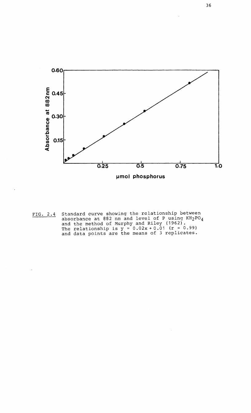
the method of Murphy and Riley (1962) (Fig. 2.4) and
controls without the enzymes were run to measure non-
enzymatic hydrolysis or free orthophosphate contamination of
the substrates. All substrates were obtained from either
Sigma or Boehringer Mannheim.
2.2.7 Gel electrophoresis
Electrophoresis was initially performed according to Davis
(1964) with the use of vertical, flat-bed gels of 5%, 7.5%
or 10% polyacrylamide. No migration of the proteins
occurred either under basic (0.05 M tris-glycine running
buffer, pH 8.3) or acidic (O.OS M S-alanine-acetic acid
running buffer, pH 4.5) conditions during 24 h incubation at
30 mA constant current. The enzymes finally migrated after
more than 24 h at 30 mA constant current in horizontal flat-
beds containing 0.5% agarose with a 0.1 M tris-acetate-EDTA
running buffer adjusted to pH 4.4 with acetic acid. Acid
phosphatase was identified by incubating the gels for 2 h at 3
room temperature in 250 cm 0.2 M acetate buffer (pH 4.0),
containing 250 m3 B-naphthylphosphate and 250 mg fast garnet
GBe salt.
36
0.60.----------------------.
E c 0.45
C\I co co ... m (i) 0.30 (J c m .Q ... o f/) .Q <C
0.25 0.5 0.75
J.lmol phosphorus
FIG. 2.4 Standard curve showing the relationship between absorbance at 882 nm and level of P using KH2P04 and the method of Murphy and Riley (1962). The relationship is y = 0.02x + 0.01 (r = 0.99) and data points are the means of 3 replicates.
1.0
37
2~3 THE KINETICS OF PHOSPHATE UPTAKE BY THE ENDOPHYTE
ISOLATED FROM ROOT SYSTEMS OF ERICA HISPIDULA
2.3.1 General incubation and radioisotope counting
procedures
Active mycelia, growing on the basal liquid medium (Section
2.1.3) were isolated during the logarithmic phase of growth,
washed in ice-cold, distilled water, blotted dry and 3
incubated for 15 min in 3 cm 0.5 mH CaSO at room t;
temperature. The incubation in CaSO t;
is supposed to
increase membrane permeability and reduce efflux of
absorbed isotope (Jennings, 1964; Harrison and 3
Helliwell, 1979). Mycelia were blotted, transferred to 3 cm
of the incubation medium and incubated in a shaking water
b h 2 5 0 lb' d' . d f 32 . at at C. ncu atlon me la conslste 0 P lsotope
dissolved in either 0.5 mM CaSO t;
(pH 4.6) or selected
buffers. In the experiment to test the effect of pH on
uptake kinetics, the pH buffers were 0.1 M acetate (pH 4.6
and 5.6), 0.1 M maleate (pH 6.8) and 0.1 M barbital (pH
7.9). The effect of a metabolic inhibitor on uptake was
tested by using 0.5 mM 2,4-dinitrophenol in 0.1 M acetate
buffer (pH 5.6). Solutions for the experiments to determine
the kinetic constants of high-P and low P-fed mycelia were -3
prepared as follows: 500 pCi KH 32po dm (equivalent to
1. 64 pM)
(KH PO ) 2 t;
2 t;
was added to a stock solution of orthophosphate
to give a final phosphate concentration of 0.5 mM
in 0.1 M acetate buffer (pH 5.6). The stock solution was
diluted to give feeding solutions ranging from 1 pM to
38
0.5 mM phosphate and -3 -3 32
1 pCi dm to 500 pCi dm P-ortho-
phosphate.
After incubation, the mycelia were removed, washed in 10 cm 3
distilled water, oven-dried at 70°C overnight and weighed.
Dried mycelia were solubilised in 0.6 cm 3 of a 40% hydrogen
peroxide/60% perchloric acid mixture (in a ratio of 2 : 1)
for 2 h after which was added 10 cm 3 scintillation cocktail
containing a chemiluminescence inhibitor (Packard Dimilume-
30). Samples were counted for up to 10 min in a Beckman LS-
150 liquid scintillation spectrometer at maximum channel
window width with automatic quench compensation (AQC)
calibrated on an External Standard ratio value of 0.744 with
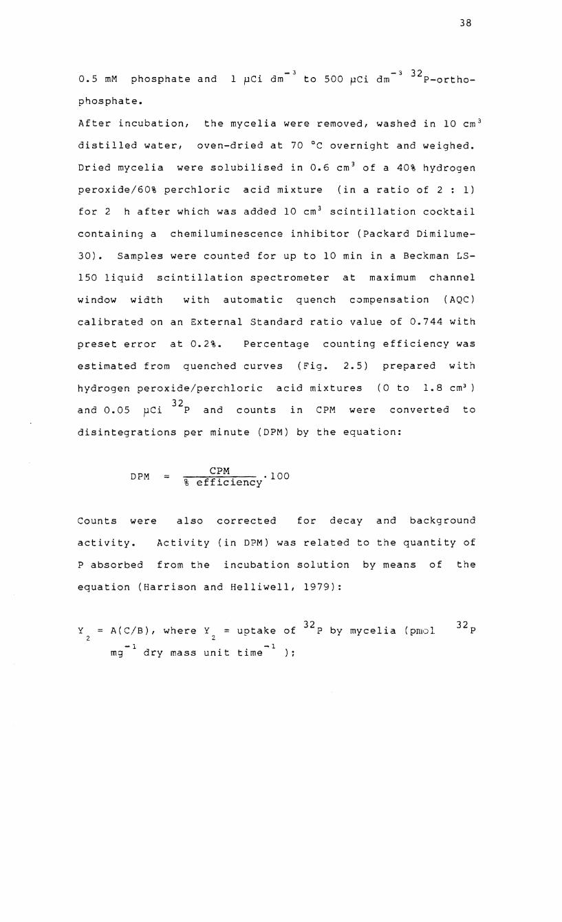
preset error at 0.2%. Percentage counting efficiency was
estimated from quenched curves (Fig. 2.5) prepared with
hydrogen peroxide/perchloric acid mixtures (0 to 1. 8 cm3 )
5 . 32
and 0.0 pCl P and counts in CPM were converted to
disintegrations per minute (DPM) by the equation:
DPM = C~M. .100 % eff~c~ency
Counts were also corrected for decay and background
activity. Activity (in DPM) was related to the quantity of
P absorbed from the incubation solution by means of the
equation (Harrison and Helliwell, 1979):
y 2
A(C/B), where Y = uptake of 32p by mycelia (proal 2
-1 -1 mg dry mass unit time ):
39
~~~~--------------------------------------~100
a
0 -0
>-(J
C Q)
(J
::: 100 Q)
CI b C -C ::J 0
(,J 75
50--~~~~~~~~~~~~~~~~~~~~~~~
0.600 0.450 0.300 0.150 o External standard ratio
FIG. 2.5 Curves used for the correction of radioactive counts (cpm) from (a) KH232P04 and (b) glucose-6-32 p due to quenching. The curves fitted the straight-line equations, y = 32.5x + 83.2, with r = 0.90 (a) and y = 65.6x + 68.3, with r = 0.99 (b). Data points are the means of 3 replicates.
(") 0 c ::J -::J
u::t CD --(') CD ::J (')
'<
0 -.... 0
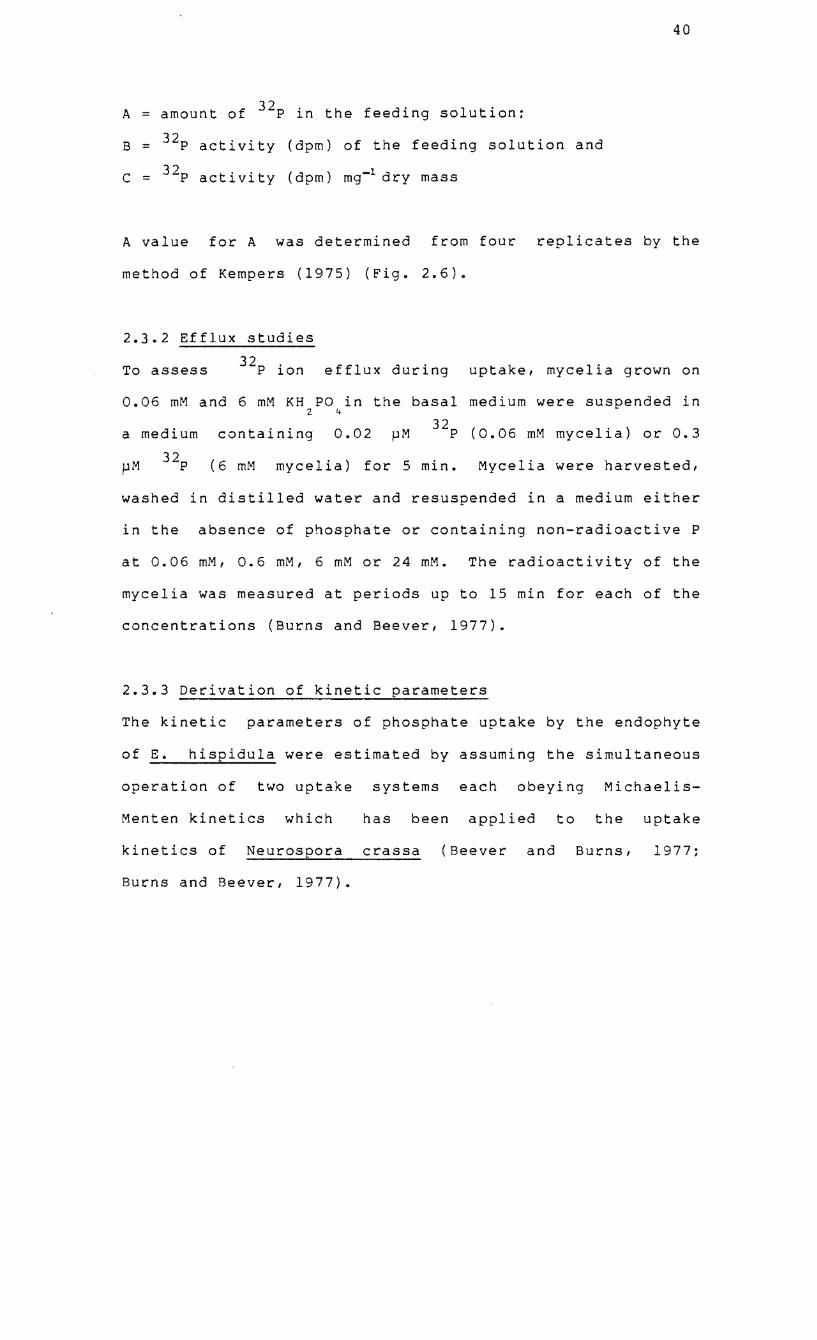
A amount of 32p in the feeding solution;
B = 32p activity (dpm) of the feeding solution and
C 32p activity (dpm) mg-1 dry mass
40
A value for A was determined from four replicates by the
method of Kempers (1975) (Fig. 2.6).
2.3.2 Efflux studies
To assess 32p ion efflux during uptake, mycelia grown on
0.06 mM and 6 mM KH PO in the basal medium were suspended in 2 4
a medium containing 0.02 ~M 32p (0.06 mM mycelia) or 0.3
pM 32p (6 mM mycelia) for 5 min. Mycelia were harvested,
washed in distilled water and resuspended in a medium either
in the absence of phosphate or containing non-radioactive P
at 0.06 mM, 0.6 mM, 6 mM or 24 mM. The radioactivity of the
mycelia was measured at periods up to 15 min for each of the
concentrations (Burns and Beever, 1977).
2.3.3 Derivation of kinetic parameters
The kinetic parameters of phosphate uptake by the endophyte
of E. hispidula were estimated by assuming the simultaneous
operation of two uptake systems each obeying Michaelis-
Menten kinetics which has been applied to the uptake
kinetics of Neurospora crassa (Beever and Burns, 1977;
Burns and Beever, 1977).
41
1.5..-----------------------,
E c 0')
CO 1.0 co
-ct'J
Q) U C ct'J .c ... o 0.5 en .c «
0.1 0.2 0.3 0.4
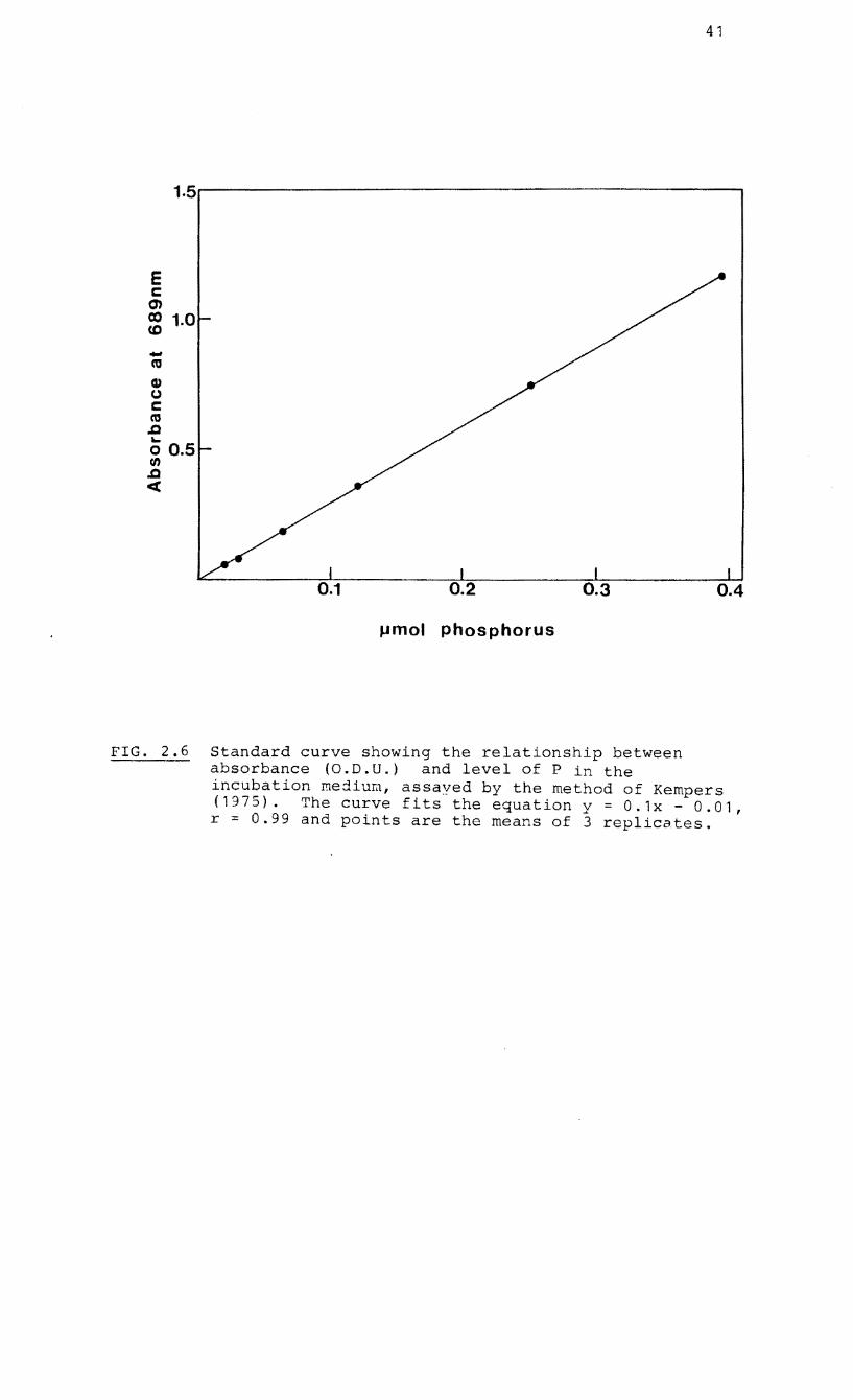
J,lmol phosphorus
FIG. 2.6 Standard curve showing the relationship between absorbance (O.D.U.) and level of P in the incubation mejiu~, assayed by the method of Kempers (1975). The curve fits the equation y = 0.1x - 0.01, r = 0.99 and points are the means of 3 replicates.
42
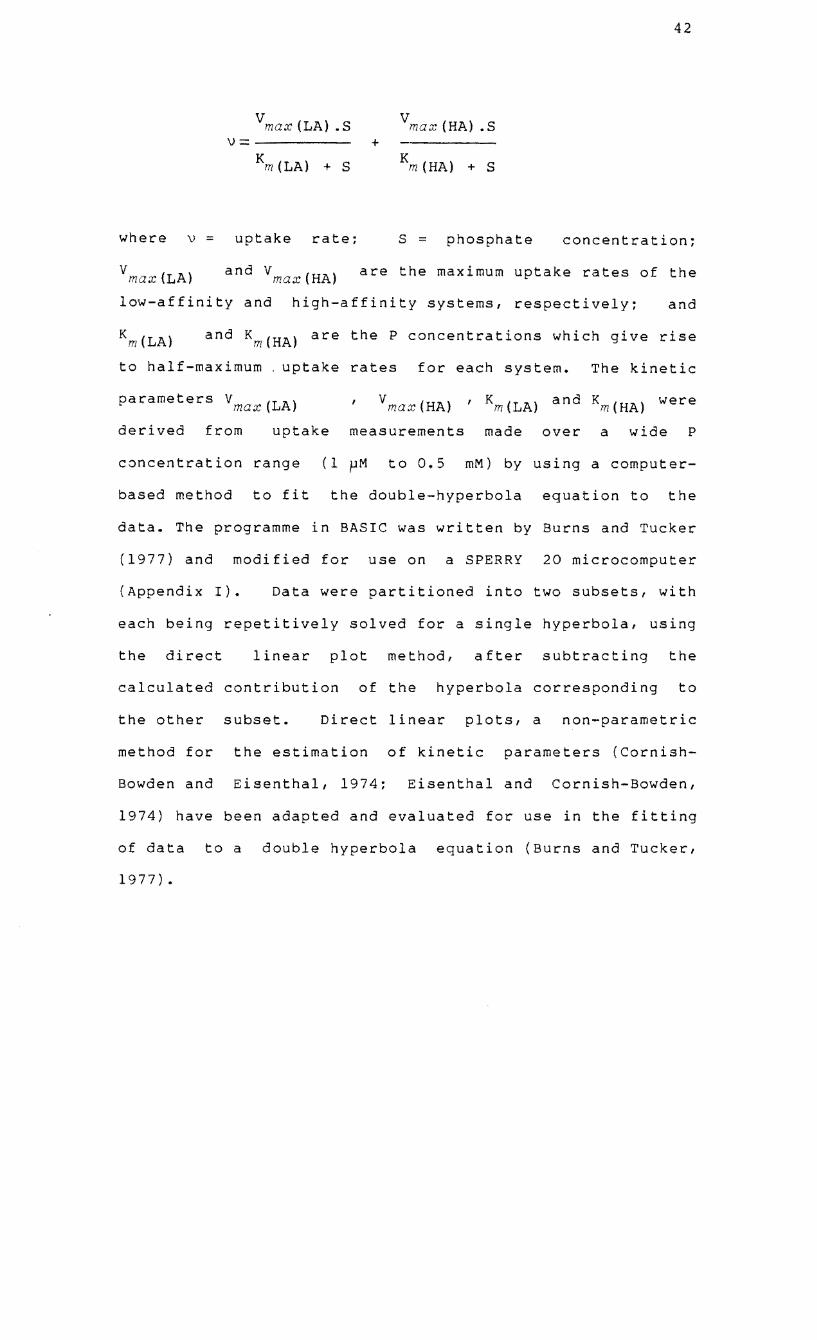
vmax(LA) .S v max (HA) .S v= +
Km(LA) + S K m(HA) + S
where v = uptake rate; S phosphate concentration;
v max (LA) and Vmax(HA) are the maximum uptake rates of the
low-affinity and high-affinity systems, respectively; and
and Km(HA) are the P concentrations which give rise
to half-maximum. uptake rates for each system. The kinetic
parameters Vmax(LA) vmax(HA) , Km(LA) and Km(HA) were
derived from uptake measurements made over a wide P
concentration range (1 ~M to 0.5 mM) by using a computer
based method to fit the double-hyperbola equation to the
data. The programme in BASIC was written by Burns and Tucker
(1977) and modified for use on a SPERRY 20 microcomputer
(Appendix I). Data were partitioned into two subsets, with
each being repetitively solved for a single hyperbola, using
the direct linear plot method, after subtracting the
calculated contribution of the hyperbola corresponding to
the other subset. Direct linear plots, a non-parametric
method for the estimation of kinetic parameters (Cornish-
Bowden and Eisenthal, 1974; Eisenthal and Cornish-Bowden,
1974) have been adapted and evaluated for use in the fitting
of data to a double hyperbola equation (Burns and Tucker,
1977).
43
L.---Vmax
v
v/[s] Vmax/Km
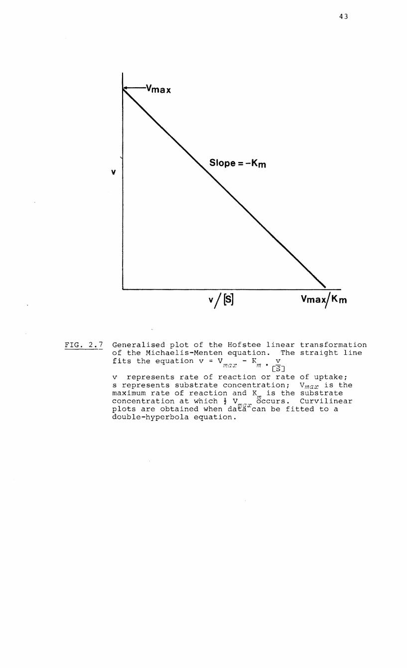
FIG. 2.7 Generalised plot of the Hofstee linear transformation of the Michaelis-Menten equation. The straight line fits the equation v = V - K v
max m • [S]
v represents rate of reaction or rate of uptake; s represents substrate concentration; Vmax is the maximum rate of reaction and K is the substrate concentration at which; V ~ccurs. Curvilinear plots are obtained when dat~Xcan be fitted to a double-hyperbola equation.
44
The relationship between P concentration and initial uptake
rates as calculated by the double-hyperbola curve fit
programme is presented in the form of a Hofstee
transformation (Hofstee, 1959~ Fig. 2.7).
2.4 IDENTIFICATION, EXTRACTION AND ESTn1ATIO~ OF POLY-
PHOSPHAT~ AND PHYTIC ACID
2.4.1 Cytochemical methods for the identification of
polyphosphates
PolyP granules were observed in the hyphae of endophytes by
using the staining and extraction techniques of Ashford et
al. (1975) and Ling-Lee et al. (1975). Hyphae were teased
from mycelial mats, stained for 5 min in a solution of 0.05%
toluidine blue adjusted to pH 1.0 with 10 M HCl, rinsed
briefly in 0.1 M HCl and mounted in glycerine. Hyphae were
also incubated for 15 min in 20% lead nitrate (pH 3.4),
rinsed thoroughly in water for 15 min and then stained in
10% ammonium sulphide for 5 min.
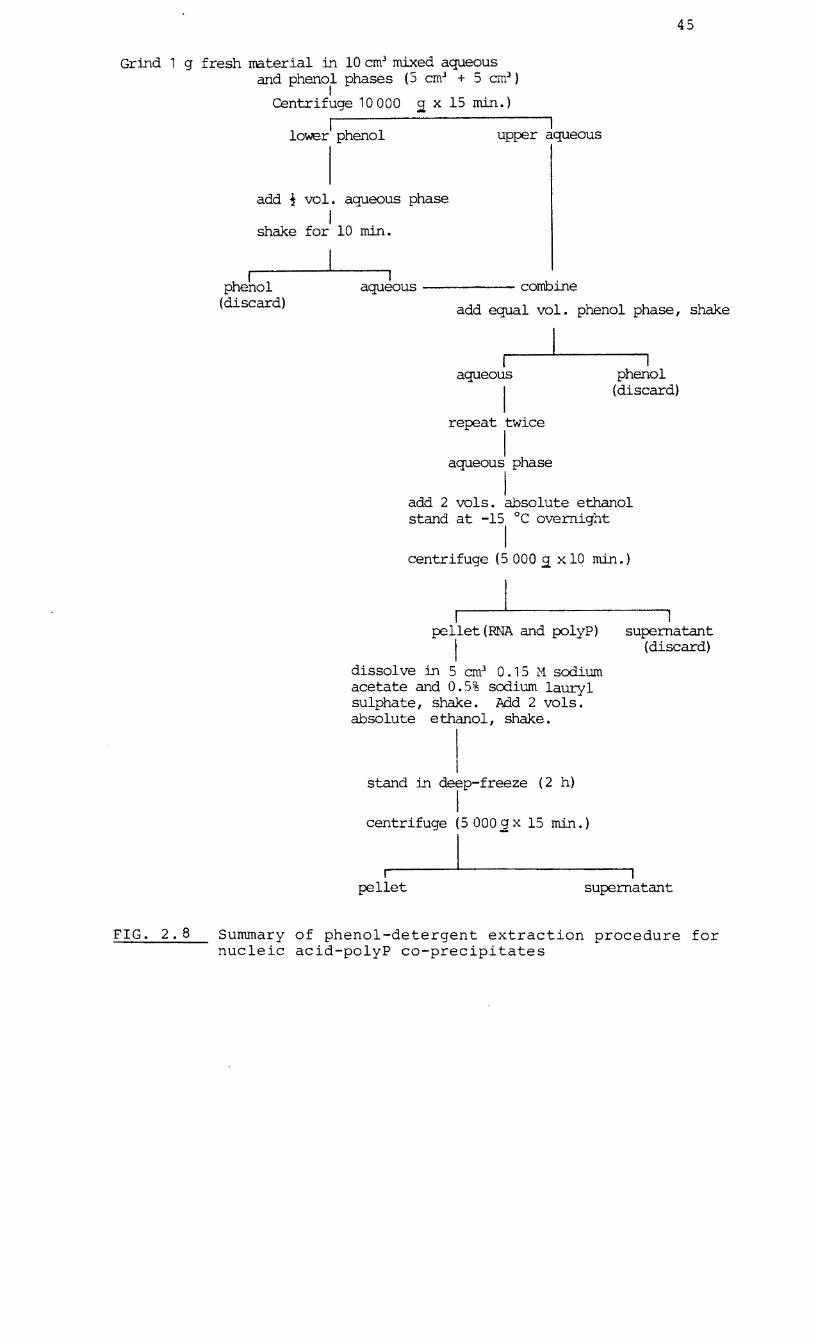
2.4.2 Phenol-detergent extraction of undegraded nucleic
acid-polyP co-precipitates
Extracts of mycelial cultures and seedlings were obtained by
the method described by Callow et al. (1978) and summarised
in Fig.2.8. The final pellet contained polyP and ribonucleic 3
acid and was dissolved in 0.5 cm
Tris-HCl buffer (pH 7.8).
10% sucrose in 0.01 M
45
Grind 1 g fresh material in 10cm3 mixed aqueous and phenol phases (S cm3 + 5 em3
)
I
FIG. 2. 8
Centrifuge 10000 9: x 15 min.)
I lolt.1E!r phenol
I upper aqueous
I add ! vol. aqueous phase
I shake for 10 min.
I phenol
(discard)
I aqueous ----- combine
add equal vol. phenol phase, shake
aqueous
I repeat twice
I aqueous phase
I
I phenol
(discard)
add 2 vols. absolute ethanol stand at -15°C overnight
I centrifuge (5 000 9: x 10 min.)
I pellet (RNA and polyP)
I dissol ve in 5 em] O. 1 5 ;;1 sodium acetate and 0.5% sodium lauryl sulphate, shake. Add 2 vols. absolute ethanol, shake.
stand in deep-freeze (2 h)
I centrifuge (5 0005!. x 15 min.)
I supernatant
(discard)
r pellet
I I
supernatant
Summary of phenol-detergent extraction procedure for nucleic acid-polyP co-precipitates
46
2.4.3 Polyacrylamide gel electrophoresis
Polyphosphates and nucleic acids were separated by gel
electrophoresis. The method was similar to that used by
Callow et ale ..;;...;.--;.;.....,...
(1978) except that electrophoresis was
performed on 8.5% (w/v) acrylamide gels using a vertical
flat-bed apparatus similar to that described by Reid and
Bieleski (1968). After pre-electrophoresing for 2 h at
10 rnA constant current, 30 pI samples were loaded on to the
gel and run for 15 min at 15 rnA followed by up to 2 h at 30
rnA. Gels were run at 10°C, then stained by immersion in
0.1% toluidine blue in 1% acetic acid. After staining and
destaining, individual pink (polyP) and blue (nucleic acid)
bands were scanned between 500 and 700 nm in a pye Unicam
SP1800 spectrophotometer. Gels were scanned using a
Vitatron densitometer at fixed wavelengths closest to
absorption maxima. An extract (30 pI) was run on an 8.5% gel
with a range of synthetic sodium polyP compounds (sodium
phosphate glasses,
Sigma Chemical
Na 2 n+
Co.) of
P 0 n 3n+1
Types 35, 45, 65, 135:
known molecular weights. The
logarithmic relationship between the distance of migration
(determined from densitometer scans) and molecular weight of
the markers was expressed in the form of a power curve
equation.
2.4.4 Total P and acid-labile polyP determinations
Mycelial cultures, seedlings and pink-staining gel segments
were digested with a tri-acid mixture (10 parts HNO : 1 3
47
part H SO: 4 parts HClO ) at 150 to 180°C. Orthophosphate 243
was assayed by the colorimetric method of Kempers (1975)
(Fig. 2.6).
Ribonucleic acid (RNA) in samples of the extracts were
adsorbed onto activated charcoal by the method of Bennett
and Scott (1971) before the hydrolysis of the acid-labile
polyP in 1 M HCl at 100°C for 10 min. Orthophosphate was
then assayed by the method of Kempers (1975).
2.4.5 Statistical analysis
Size classes of metachromatic granules were obtained from
three separate cultures/ each culture representing 40 to 80
random measurements from younger/ marginal hyphae.
Percentages were converted to arcsin transformations. The
original measurements were subjected to a computer-based
programme of Correspondence Analysis (Greenacre/ 1984). All
other granule values were obtained from 3 separate cultures/
each culture representing 10 to 20 random counts. Values
given for the phenol-detergent extraction of the E.
hispidula endophyte represent three separate extractions;
all other values represent at least three replicates from a
single extraction. Molecular weights of polyP molecules
were determined from four separate gels for each of the
three endophytes used.
48
2.4.6 Phosphorus extraction and fractionation procedures
A modified version of the Aitchison and Butt (1973)
fractionation procedure (Fig. 2.9) was used in experiments
which involved the extraction and fractionation of P.
2.4.7 32p absorption and utilization by mycelia in culture
Fresh mycelia were washed in ice-cold, distilled waterl
blotted dry and weighed immediately. When the incubation
medium did not contain fresh basal mediuml mycelia were
incubated in 3 cm 3 of 5 x 10- 4 M CaSO at room temperature 4
for 15 min before transfer to the radioactive incubation
medium. Incubation took place in a shaking water-bath at
25°C and mycelia were washed in 10 cm 3 distilled waterl
blotted dry and fractionated. The procedures for
solubilization of non-aqueous samplesl liquid scintillation
counting and the analysis of data are described in section
2.3.1
nmol
of this chapter. Final results are expressed as
32 -1 P g fresh mass.
2.4.8 Total P determination of non-radioactive fractions
In the experiment to assess the endogenous P status of
fractions of fresh mycelial total P of the fractions was
determined by the method of Kempers (1975) after digestion
(section 2.4.4).
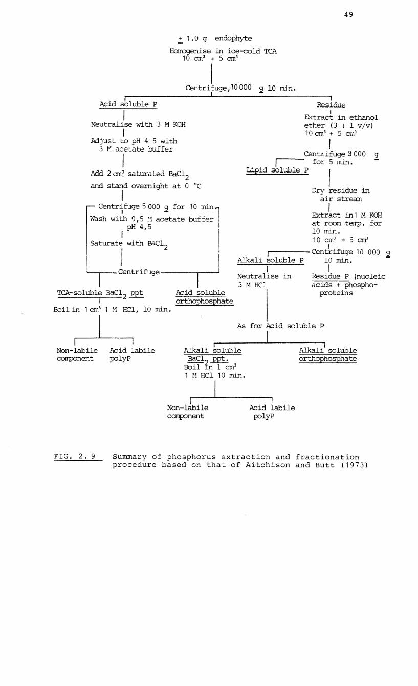
49
..!. 1. a g endophyte
Homogenise in ice-cold TCA 10 em3 + 5 em3
I Centrifuge, 10 000
I 2 10 llL1!"1'
1 Residue Acid s~luble P
I Neutralise with 3 M KOH
I Adjust to pH 4 5 with
3 r1 acetate buffer
I Add 2 em~ saturated BaC1
2 and stand overnight at a °c
I Centrifuge 5000 2 for 10 min.
I
Wash with 0,5 11 acetate buffer pH 4,5
I Saturate with BaC12
I '---..,.--- Centrifuge ----,.----'
TCA-soluble BaC12 ppt I -
Boil in 1 cm3 1 M HC1, 10 min.
Acid soluble orthophosphate
I Extract in ethanol ether (3 : 1 v/v) 10 cm3 + 5 cw3
I Centrifuge 8 000 g
,F"" --- for 5 min. Lipid soluble P
I Dry residue in
air stream
I . Extract .ill 1 M KOH at room temp. for 10 min. 10 cm3 + :3 em3
I . f ,F"" ----Centr~ uge 10 000 2
Alkali soluble P 10 min. I I
Neutralise in Residue P (nucleic 3 M RCl acids + phospho-
proteins
As for Acid soluble P
I Non-labile Acid labile
. I Alkal~ soluble Alkali' soluble
orthophosphate component polyP BaC12
ppt.
FIG. 2. 9
Boil .ill 1 em3
1 r1 Hel 1 0 min.
I I I
Non-labile Acid labile component polyP
Summary of phosphorus extraction and fractionation procedure based on that of Aitchison and Butt (1973)
50
2.4.9 The effect of activated charcoal on the adsorption of
P contaminants of polyP fractions
To assess the potential for contamination of polyP fractions
cultures of the same batch were
and fractionated. In one set of
by unknown P compounds,
separated into two groups
extracts, suspensions of the TCA-soluble and -insoluble
activated charcoal (0.3g per 5 cm 3) supernatants containing
were covered with 0.01 cm 3 of 10% Triton X-IOO, centrifuged
and filtered (Crane, 1958). Final P levels of the fractions
were then compared with those of the set of untreated
extracts.
2.4.10 Phytic acid determination
The level of phytic acid in mycelia from the same batch of
cultures used in part 2.4.9 was determined by the method of
Allen et ale (1974). An acidified extract dissolved any
phytin present and phytic acid was isolated as ferric
phytate which was recovered, digested and estimated as
phytate-phosphorus (see section 2.4.4).
CHAPTER 3
ACID PHOSPHATASE ACTIVITY IN ISOLATED ERICOID ENDOPHYTES
3.1 INTRODUCTION
Acid phosphatases
microorganisms and
are universally
their activity
found in plants
in the soil may
51
and
be
correlated with high amounts of organic carbon and organic
phosphorus and low levels of free phosphate ions (Spiers and
McGill, 1979~ Appiah and Thomas, 1982). These enzymes of
wide specificity, facilitate the hydrolytic cleavage of
phosphate-ester bonds and have been accorded an important
role in the mineralization of the organic P fraction in
soils. Mycorrhizal
phosphatase activity
roots
may
which
be more
possess a
efficient
high
in
acid
the
utilization of soil phosphorus than non-mycorrhizal roots.
High acid phosphatase activity has been measured in
ectomycorrhizal fungi in pure culture (Theodorou, 1971: Ho
and Zak, 1979: Calleja et al., 1980) excised
ectomycorrhizal roots (Woolhouse, 1969: Bartlett and Lewis,
1973) and onion roots infected with vesicular-arbuscular
mycorrhizas (Gianinazzi et al., 1979). The only report of
acid phosphatase activity in ericoid mycorrhizas is that of
Pearson and Read (1975) who found high levels of the enzyme
in the endophyte of C. vulgaris. This chapter investigates
acid phosphatases
endophytes. The
in mycelia of South African and European
high activity of an acid phosphatase
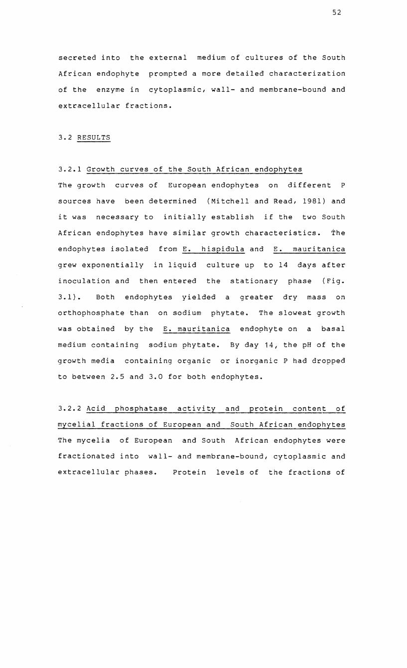
52
secreted into the external medium of cultures of the South
African endophyte
of the enzyme in
prompted a more detailed characterization
cytoplasmic, wall- and membrane-bound and
extracellular fractions.
3.2 RESULTS
3.2.1 Growth curves of the South African endophytes
The growth curves of European endophytes on different P
sources have been determined (Mitchell and Read, 1981) and
it was necessary to initially establish if the two South
African endophytes have similar growth characteristics. The
endophytes isolated from E. hispidula and E. mauritanica
grew exponentially in liquid culture up to 14 days after
inoculation and then entered the stationary phase (Fig.
3.1). Both endophytes yielded a greater dry mass on
orthophosphate than on sodium phytate. The slowest growth
was obtained by the E. mauritanica endophyte on a basal
medium containing sodium phytate. By day 14, the pH of the
growth media containing organic or inorganic P had dropped
to between 2.5 and 3.0 for both endophytes.
3.2.2 Acid phosphatase activity and protein content of
mycelial fractions of European and South African endophytes
The mycelia of European and South African endophytes were
fractionated into wall- and membrane-bound, cytoplasmic and
extracellular phases. Protein levels of the fractions of
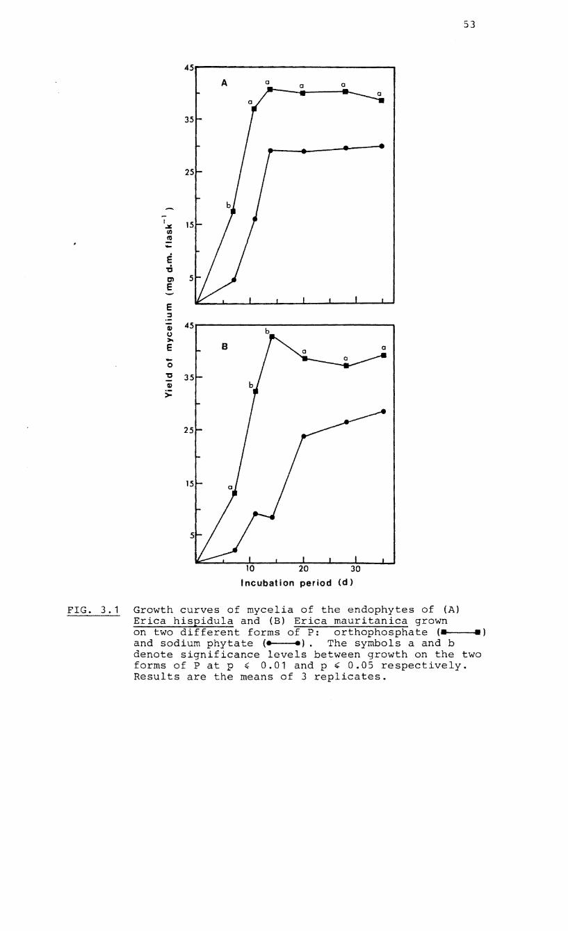
45r-------------------------------~ A a
-I .¥ til «S -E -d en E
E :::I
fI) b (,) >. E -0
'C fI)
>
Incubation period
FIG. 3.1 Growth curves of mycelia of the endophytes of (A) Erica hispidula and (B) Erica mauritanica grown
53
on two different forms of P: orthophosphate (. .) and sodium phytate (e e). The symbols a and b denote significance levels between growth on the two forms of P at p ~ 0.01 and p " 0.05 respectively. Results are the means of 3 replicates.
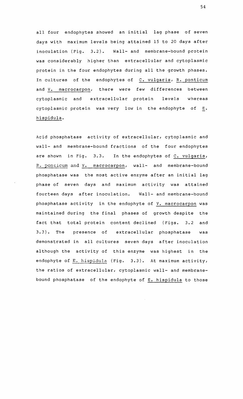
54
all four endophytes showed an initial lag phase of seven
days with maximum levels being attained 15 to 20 days after
inoculation (Fig. 3.2). Wall- and membrane-bound protein
was considerably higher than extracellular and cytoplasmic
protein in the four endophytes during all the growth phases.
In cultures of the endophytes of c. vulgaris, R. ponticum
and v. macrocarpon, there were few differences between
cytoplasmic and extracellular protein levels whereas
cytoplasmic protein was very low in the endophyte of E.
hispidula.
Acid phosphatase activity of extracellular, cytoplasmic and
wall- and membrane-bound fractions of the four endophytes
are shown in Fig. 3.3. In the endophytes of c. vulgaris,
R. ponticum and v. macrocarpon, wall- and membrane-bound
phosphatase was the most active enzyme after an initial lag
phase of seven days and maximum activity was attained
fourteen days after inoculation. Wall- and membrane-bound
phosphatase activity in the endophyte of V. macrocarpon was
maintained during the final phases of growth despite the
fact that total protein content declined (Figs. 3.2 and
3.3). The presence of extracellular phosphatase was
demonstrated in all cultures seven days after inoculation
although the activity of this enzyme was highest in the
endophyte of E. hispidula (Fig. 3.3). At maximum activity,
the ratios of extracellular, cytoplasmic wall- and membrane
bound phosphatase of the endophyte of E. hispidula to those
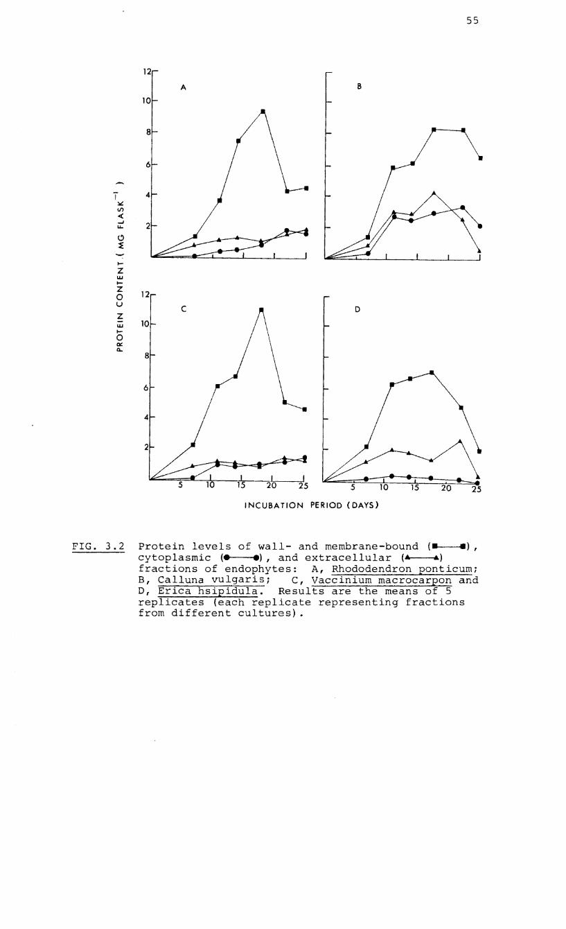
55
12
A B
10
8
6
T 4 :w: \I)
« ~
2 u..
C)
~
I-
Z W I-
Z 12 0 u
C D Z w 10 I-
0 ex CI..
8
6
4
2
.1 I I 15 20 25 15 20 25
INCUBATION PERIOD (DAYS)
FIG. 3.2 Protein levels of wall- and membrane-bound (- -), cytoplasmic (. .), and extracellular (4 &) fractions of endophytes: A, Rhododendron ponticum; B, Calluna vulgaris; C, Vaccinium macrocarpon and D, Erica hsipidula. Results are the means of 5 replicates (each replicate representing fractions from different cultures) .
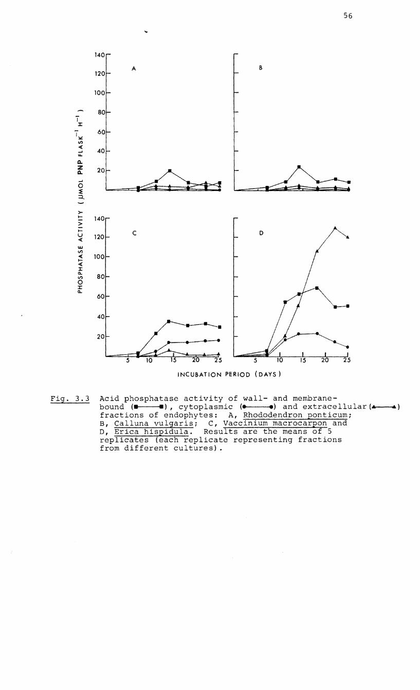
56
140
A B 120
100
80 T J:
I~ 60
!,/)
« ..... ..... Q,.
Z Q,.
..... 0 ~ :::::t..
>-..... 140 > ..... C 0 u 120 « w !,/)
« 100 ..... « J: 0..
80 !,/)
0 J: 0..
60
40
20
5 20 25 5
INCUBATION PERIOD (DAYS)
Fig. 3.3 Acid phosphatase activity of wall- and membrane-bound (_ -), cytoplasmic (e e) and extracellular (.. ...) fractions of endophytes: A, Rhododendron ponticum; B, Calluna vulgaris; C, Vaccinium macrocarpon and D, Erica hispidula. Results are the means of 5 replicates (each replicate representing fractions from different cultures) .
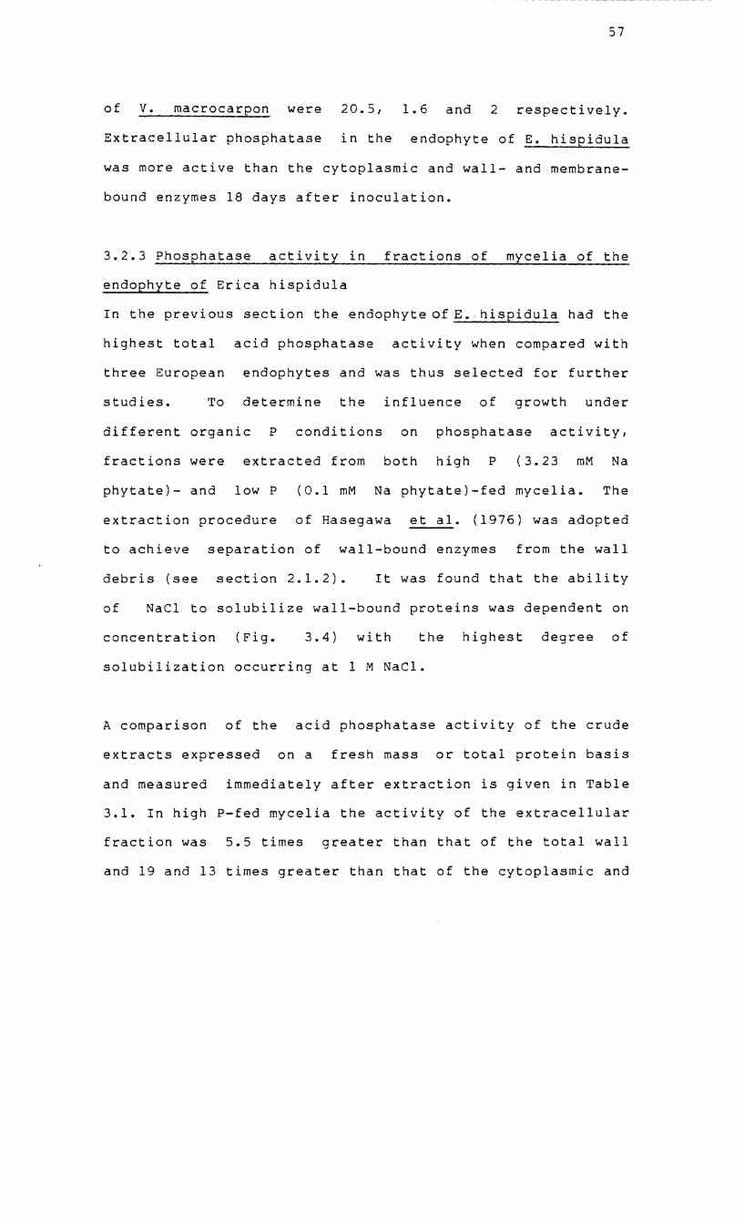
57
of v. macrocarpon were 20.5, 1.6 and 2 respectively.
Extracellular phosphatase in the endophyte of E. hispidula
was more active than the cytoplasmic and wall- and membrane
bound enzymes 18 days after inoculation.
3.2.3 Phosphatase activity in fractions of mycelia of the
endophyte of Erica hispidula
In the previous section the endophyte ofE.hispidula had the
highest total acid phosphatase activity when compared with
three European
studies. To
endophytes and was thus selected for further
determine the influence of growth under
different organic P conditions on phosphatase activity,
fractions were extracted from both high P (3.23 mM Na
phytate)- and low P (0.1 mM Na phytate)-fed mycelia. The
extraction procedure of Hasegawa et ala (1976) was adopted
to achieve separation of wall-bound enzymes from the wall
debris (see section 2.1.2). It was found that the ability
of NaCI to solubilize wall-bound proteins was dependent on
concentration (Fig. 3.4) with the highest degree of
solubilization occurring at 1 M NaCl.
A comparison of the acid phosphatase activity of the crude
extracts expressed on a fresh mass or total protein basis
and measured immediately after extraction is given in Table
3.1. In high P-fed mycelia the activity of the extracellular
fraction was 5.5 times greater than that of the total wall
and 19 and 13 times greater than that of the cytoplasmic and
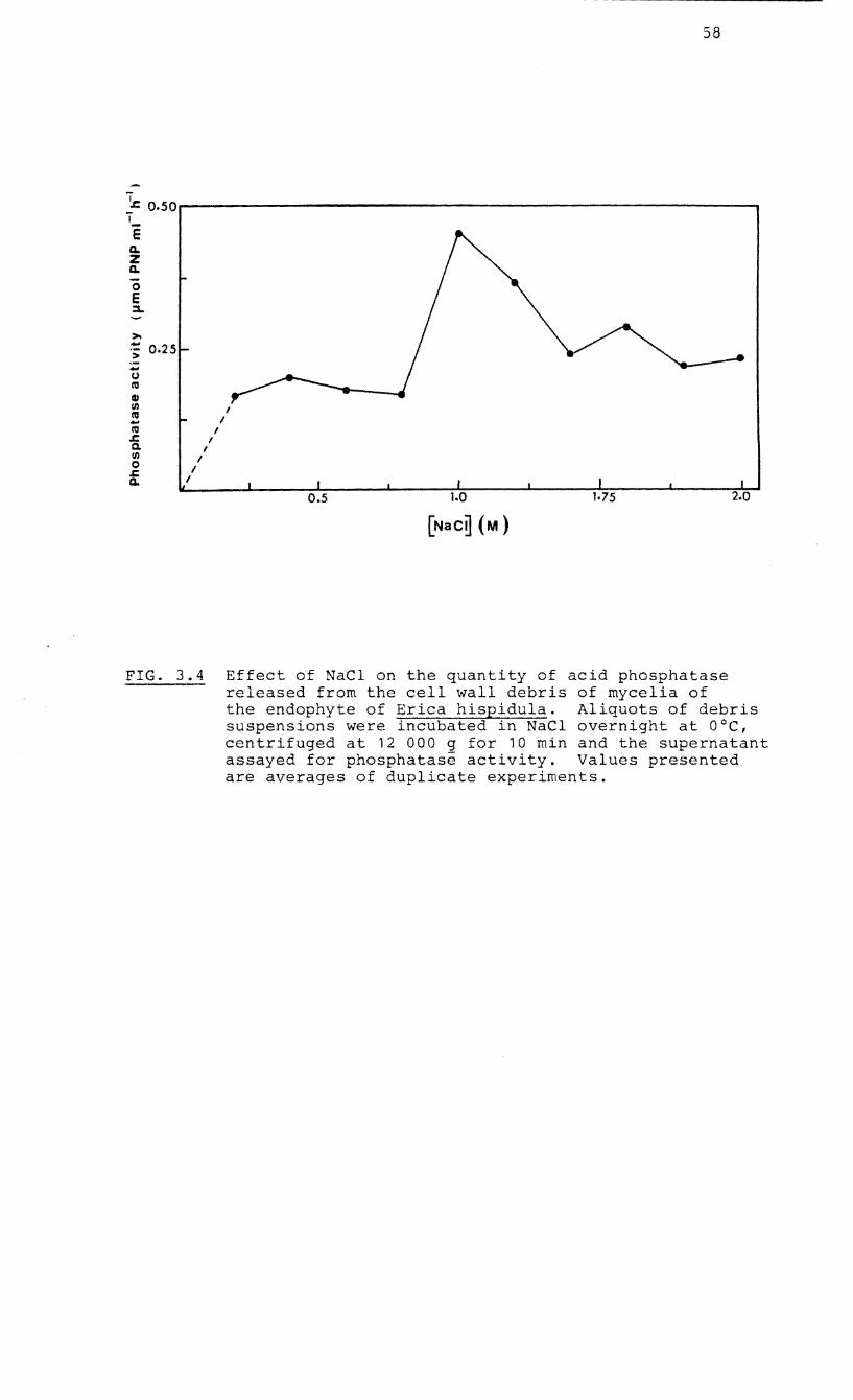
58
-~ 0.50~--------------------------------------------------------------~ I_ E a. Z a. o E ::a..
I I
I
I I
I I
I
1.0 1·75 2.0
FIG. 3.4 Effect of NaCl on the quantity of acid phosphatase released from the cell wall debris of mycelia of the endophyte of Erica hispidula. Aliquots of debris suspensions were incubated in NaCl overnight at OoC, centrifuged at 12 000 g for 10 min and the supernatant assayed for phosphatase activity. Values presented are averages of duplicate experiments.
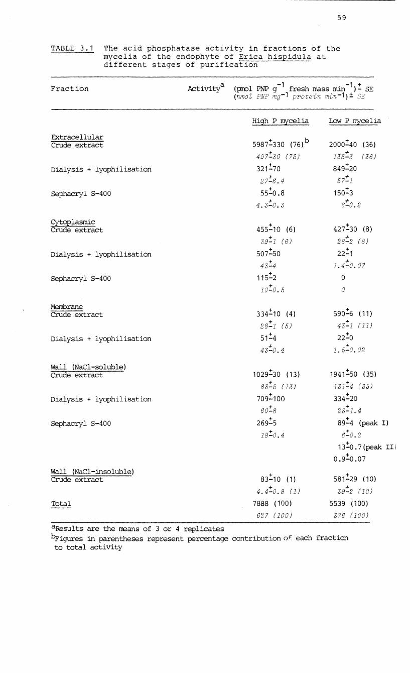
59
TABLE 3.1 The acid phosphatase activity in fractions of the mycelia of the endophyte of Erica hispidula at different stages of purification
Fraction
Extracellular Crude extract
Dialysis + lyophilisation
Sephacryl S-400
Cytoplasmic Crude extract
Dialysis + lyophilisation
8ephacryl 8-400
Membrane Crude extract
Dialysis + lyophilisation
Wall (NaCI-soluble) Crude extract
Dialysis + lyophilisation
8ephacryl 8-400
Wall (NaCl-insoluble) Crude extract
Total
Activitya
aResults are the means of 3 or 4 replicates
(pmol PNP g-1 fresh mass min-1): SE (nmoZ PNP mg-1 protein min-1)± Sf:,'
High P rrucelia
5987:330 (76)b
497:30 (75)
321:70
27:6.4
55:0.8 + 4.3-0.3
455:10 (6)
39:1 (6)
507:50 +
43-4
115:2
10:0.5
334!10 (4) + 28-1 (5)
51:4
43-:'0.4
1029!30 (13)
83-:'5 (13)
709:100
60-:'8
269:5
18:0.4
83:10 (1) + 4.4-0.8 (1)
7888 (100)
627 (100)
IDw P rrucelia
2000:40 (36)
135-:'3 (36)
849:20 + 57-1
150:3
8:0.2
427:30 (8)
28:2 (8) + 22-1 + 1.4-0.07
o o
590!6 (11) + 43-1 (11)
22!0
1.5:0.02
1941!50 (35)
131:4 (35)
334:20
23-:'1.4 + 89-4 (peak I) + 6-0.2
13:0.7 (peak II) +
0.9-0.07
581:29 (10)
39-:'2 (10)
5539 (100)
376 (100)
brigures in parentheses represent percentage contribution of each fraction to total activity
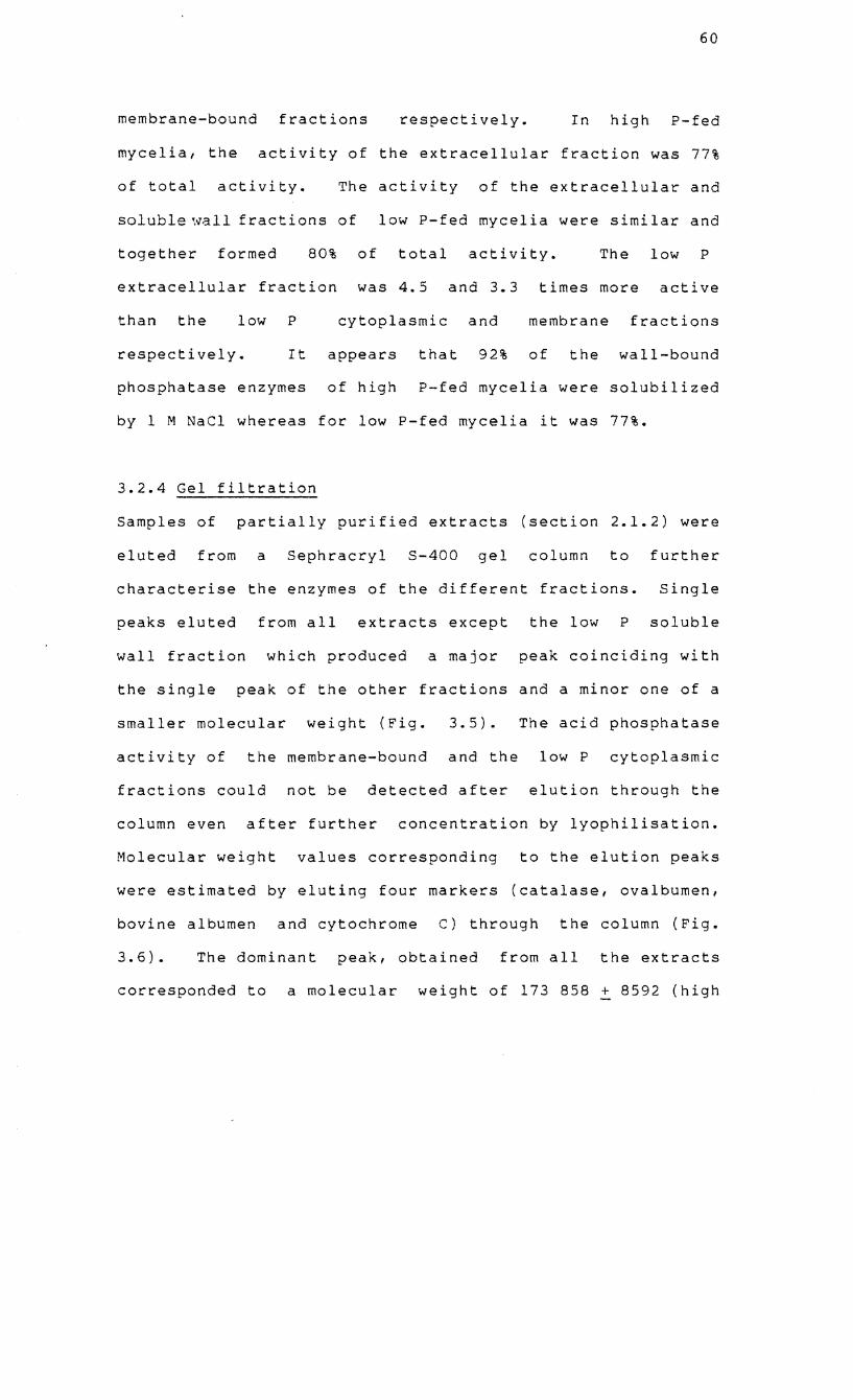
60
membrane-bound fractions respectively. In high P-fed
mycelia, the activity of the extracellular fraction was 77%
of total activity. The activity of the extracellular and
soluble ',vall fractions of
together formed 80%
extracellular fraction
low P-fed mycelia were similar and
of total activity. The low P
was 4.5 and 3.3 times more active
than the low
respectively.
P
It
cytoplasmic and
appears that 92%
membrane fractions
of the wall-bound
phosphatase enzymes of high P-fed mycelia were solubilized
by 1 M NaCI whereas for low P-fed mycelia it was 77%.
3.2.4 Gel filtration
Samples of partially purified extracts (section 2.1.2) were
eluted from a Sephracryl S-400 gel column to further
characterise the enzymes of the different fractions. Single
peaks eluted from all extracts except the low P soluble
wall fraction which produced a major peak coinciding with
the single peak of the other fractions and a minor one of a
smaller molecular weight (Fig. 3.5). The acid phosphatase
activity of the membrane-bound and the low P cytoplasmic
fractions could not be detected after elution through the
column even after further concentration by lyophilisation.
Molecular weight values corresponding to the elution peaks
were estimated by eluting four markers (catalase, ovalbumen,
bovine albumen and cytochrome C) through the column (Fig.
3.6) • The dominant peak, obtained from all the extracts
corresponded to a molecular weight of 173 858 + 8592 (high
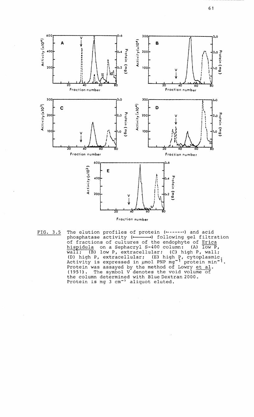
61
600 V I
0.6 300 3.0 .., A ~
.., B 0 0 .... ....
)( )(
~ 0.4 "'0
200 2.0 =l' ~ >- f\ , 2- -~ f \ 0
> h I'D > .... "
, I , (1) .- ::l .... I • \J II 1\ \J I t ::l « II II ...... «
" 'i 0.2 3 100 I ,
" " , I 1.0 3
" I' , Ul , I Ul
\I I W: , ...... I, I
" : t, I t
20 80 Fraction number Fraction number
300 3.0 300 ~
3.0
M C
.., 0 " 0 0
" .... " "'0 )( , \
"'0 ~ V ' I 2.0 ;:; 2- >- , I - ~
, . .... > I'D > , I I'D
::l ,
I ::l .... - I \J \J , , ...... « 3 « , \
1.0 ~ , I
V Ul I 1
~ I , ...... I I t
, "U
f
20 4 80
Fraction number fraction number
600 0.6 ...... M .. E 0
)( ~ "'0 ...... 400- I! -0.4 >- ~
,I 0 - 'I
II ....
> I'D I- .. -" .... I I ::l
\J f : ..... « 200 I 1 - 0.2 3 V I \ . Ul
~ , .~' ...... t "\-
j \.. ,.
V \ 20 40 60 130
fraction number
FIG. 3.5 The elution profiles of protein ( ______ 0) and acid phosphatase activity (0 .) following gel filtration of fractions of cultures of the endophyte of Erica hispidula on a 5ephacryl 5-400 column: (A) low P, wall; (B) low P, extracellular; (C) high P, wall; (D) high P, extracellular; (E) high P, cytoplasmic. Activity is expressed in pmol PNP mg- 1 protein min- 1 Protein was assayed by the method of Lowry et ale (1951). The symbol V denotes the void volume of the column determined with Blue Dextran 2000. Protein is mg 3 cm- 3 aliquot eluted.
62
10 9 8
7
- 6 0
1£'5 0 -' - 4 .... .c Ol CI) 3 3: .. .! ::I U CI) 2 '0
::&
300 400 500 600
Volume of elution from 5ephacryl 5-400 colu'mn (cm3 )
FIG. 3.6 Molecular weight estimation of acid phosphatase peaks eluted from 8ephacryl 8-400 column. Molecular weight I (dominant peak, representative of all fractions) and II (subsidiary peak, present only in low P mycelia) was estimated from a standard curve (y = 0.01x + 7.17, r = 0.91) prepared using the following standards: (1) catalase (232 000); (2) bovine albumin (66200); (3) ovalbumin (45 000); (4) cytochrome C (13096). Volumes of standards were the means of dUplicate elutions and molecular weight estimates are the means of at least 3 replicates. Protein was assayed by the method of Lowry et al. (1951). The technique of molecular weight estimation is that used by Basha (1984).
63
molecular weight enzymes) whereas the minor peak only eluted
from the low P soluble wall extract had a molecular weight
of 68 028 + 3348 (low molecular weight enzyme).
3.2.5 pH scans
The buffer routinely used for the assay of acid phosphatase
was pH 4.5 (Pearson and Read, 1975) but this might not have
been the optimum pH for all the enzymes extracted from
mycelia of the endophyte of E. hispidula. Moreover, pH
serves as a basis for distinguishing the properties of the
extracellular, soluble wall and cytoplasmic fractions.
All the fractions, except the low P soluble wall II enzyme
hydrolysed PNPP optimally between pH 2.0 and 6.0 with a peak
in activity at pH 2.0 (Fig. 3.7). The activity of these
enzymes declined rapidly above pH 5.5 to negligible rates at
pH 7.0. The low P, soluble wall II enzyme showed an optimum
activity at pH 6.5 with a rapid decline at pH 8.0. (Fig. 3.7)
3.2.6 The action of effectors
The action of different compounds as either possible
activators or inhibitors of the acid phosphatases are shown
in Table 3.2. Although variations between enzymes in the
degree of response to different agents was observed, the
clearest distinction is between the low m~lecular weight
enzyme and
The agents
the high molecular weight enzymes as
which stimulated the activity of
a group.
the high
molecular weight enzymes most were ferric ions in low
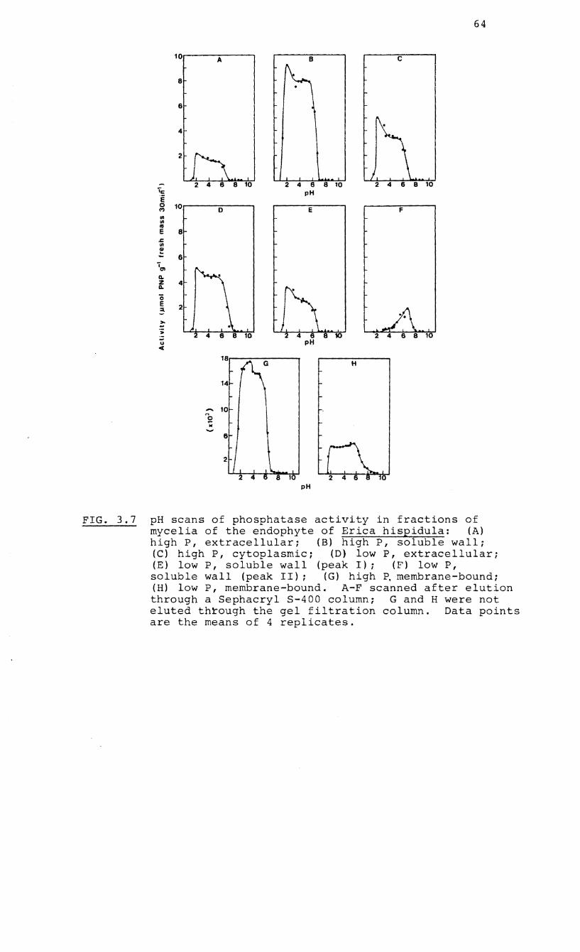
10
8
6
4
2
-~ i 0 10 M ~ ~ ~
E 8 ~ w ~ ~ 6
~ ~ Z 4 ~
0 E 2 ~ ~ ~
; -u C
A
D
18
14
... 10
~ M
'-' 6
2
2 4 6 8 ro pH
E
pH
H
64
c
F
0
FIG. 3.7 pH scans of phosphatase activity in fractions of mycelia of the endophyte of Erica hispidula: (A) high P, extracellular; (B) high P, soluble wall; (e) high P, cytoplasmic; (D) low P, extracellular; (E) low P, soluble wall (peak I); (F) low P, soluble wall (peak II); (G) high ~ membrane-bound; (H) low P, membrane-bound. A-F scanned after elution through a 5ephacryl 5-400 column; G and H were not eluted th~ough the gel filtration column. Data points are the means of 4 replicates.
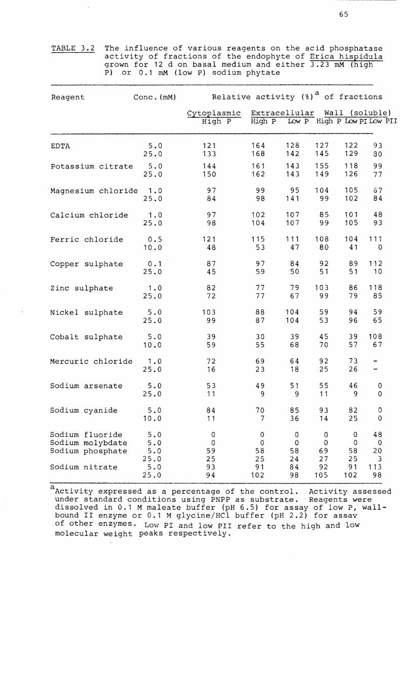
65
TABLE 3.2 The influence of various reagents on the acid phosphatase activity of fractions of the endophyte of Erica hispidula grown for 12 d on basal medium and either 3.23 roM (high P) or 0.1 roM (low P) sodium phytate
Reagent Conc. (roM)
EDTA 5.0 25.0
Potassium citrate 5.0 25.0
Magnesium chloride 1.0 25.0
Calcium chloride 1.0 25.0
Ferric chloride 0.5 10.0
Copper sulphate 0.1 25.0
Zinc sulphate 1.0 25.0
Nickel sulphate 5.0 25.0
Cobalt sulphate 5.0 10.0
Mercuric chloride 1.0 25.0
Sodium arsenate
Sodium cyanide
Sodium fluoride Sodium molybdate Sodium phosphate
Sodium nitrate
5.0 25.0
5.0 10.0
5.0 5.0 5.0
25.0 5.0
25.0
Relative activity (%)a of fractions
Cytoplasmic High P
121 133
144 150
97 84
97 98
121 48
87 45
82 72
103 99
39 59
72 16
53 11
84 11
o o
59 25 93 94
Extracellular Wall (soluble) High P lJ:)w P High P Low PI lJ:)w PH
164 168
161 162
99 98
102 104
115 53
97 59
77 77
88 87
30 55
69 23
49 9
70 7
o o
58 25 91
102
128 142
143 143
95 141
107 107
111 47
84 50
79 67
104 104
39 68
64 18
51 9
85 36
o o
58 24 84 98
127 145
155 149
104 99
85 99
108 80
92 51
103 99
59 53
45 70
92 25
55 11
93 14
o o
69 27 92
105
122 93 129 30
118 99 126 77
105 67 102 84
101 48 105 93
104 111 41 0
89 112 51 10
86 118 79 85
94 59 96 65
39 108 57 67
73 26
46 0 9 0
82 0 25 0
o 48 o 0
58 20 25 3 91 113
102 98
aActivity expressed as a percentage of the control. Activity assessed under standard conditions using PNPP as substrate. Reagents were dissolved in 0.1 M maleate buffer (pH 6.5) for assay of low P, wallbound II enzyme or 0.1 M glycine/HCl buffer (pH 2.2) for assay of other enzymes. Lmv PI and low PII refer to the high and low molecular weight peaks respectively.
66
concentrations, EDTA and citrate whereas ferric and cupric
ions in low concentrations stimulated the activity of the
low molecular weight enzyme. Fluoride and molybdenum
inhibited all the enzymes whereas the degree of inhibition
by phosphate, cyanide, arsenate, mercury, cobalt, copper and
ferric ions was dependent on concentration. Magnesium,
calcium and nickel ions caused little variation in the
activity of the high molecular weight enzymes but in low
concentrations inhibited the activity of the low molecular
weight enzyme.
3.2.7 Substrate specificity
Acid phosphatases generally show affinities for a broad
range of substrates but may differ substantially in their
affinities for specific substrates. The activities of six
acid phosphatase fractions from mycelia of the endophyte, ~.
hispidula, towards various substrates are shown in Table
3.3. The substrate most efficiently hydrolysed by
all the fractions was inorganic pyrophosphate with a
glycerophosphate, a- and 8-naphthyl phosphate, phenol
phosphate, PNPP and S-glycerophosphate also showing high
rates of hydrolysis. All the fractions except the low P,
soluble wall II fraction showed a high affinity for glucose
I-phosphate but not for glucose-6-phosphate or fructose 6-
p~osphate which proved successful substrates for the low P,
soluble wall II enzyme. Fructose-I,6-bisphosphate and the
organic anhydrides, especially ATP, proved suitable
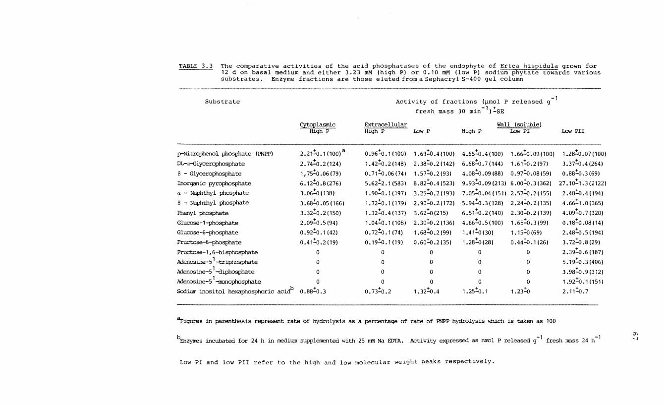
TABLE 3.3 The comparative activities of the acid phosphatases of the endophyte of Erica hispidula grown for 12 d on basal medium and either 3.23 roM (high P) or 0.10 roM (low PI sodium phytate towards various substrates. Enzyme fractions are those eluted from a Sephacryl S-400 gel column
Substrate Activity of fractions (J.lmol P released -1
g
fresh -1 +
mass 30 min I -SE
Cytoplasmic Extracellular Wall (soluble) High P High P Low P High P I£M PI I£M PIr
p-Nitrophenol phosphate (PNPPI 2.21:0.1 (100)a 0.96:0.1 (100) 1.69;:'0.4 (100) 4.65:0.4 (100) 1.66:0 .09 (100) 1.28~0 .07 (100)
DL-a-Glycerophosphate 2.74:0.2(124) 1.42:0.2(148) 2.38;:'0.2(142) 6.68:0.7(144) 1.61:0.2(97) 3.37:0.4(264)
8 - Glycerophosphate 1,75;:'0.06(79) O. 71~.06 (74) 1.57:0.2(93) 4.08~.09(88) 0.97~.08(59) 0.88~.3(69)
Inorganic pyrophosphate 6.12:0.8(276) 5.62;:'2.1(583) 8.82:0.4(523) 9 .93~.09 (213) 6.00:0.3(362) 27.1O~1.3(2122) a - Naphthyl phosphate 3.06:0 (138) 1.90:0.1 (197) 3.25:0.2 (193) 7.05;:'0.04(151) 2.57:0.2 (155) 2.48~.4(194) 8 - Naphthyl phosphate 3.68~.05(166) 1.72;:'0.1 (179) 2. 90~.2 (172) 5.94:0.3(128) 2.24:0.2(135) 4.66;:'1.0 (365)
Phenyl phosphate 3.32;:'0.2(150) 1.32~.4(137) 3.62:0(215) 6.51:0.2(140) 2.30:::.0.2 (139) 4.09;:'0.7(320)
Glucose-l-phosphate 2.09~.5(94) 1.04;:'0.1 (108) 2.30!o.2(136) 4.66~.5(100) 1.65;:'0.3(99) 0.18;:'0.08 (14)
Glucose-6-phosphate 0.92:.0.1 (42) O. 72~. 1 (74) 1.68:0.2(99) 1.41:.0 (30) 1. 15:.0 (69) 2 .48:::.0 .5 (194 )
Fructose-6-phosphate 0.4':0.2(19) 0.'9!o.1 (19) 0.60:.0.2 (35) 1.28:0(28) 0.44:.0.1 (26) 3.72:::.0.8(29)
Fructose-l,6-bisphosphate 0 0 0 0 0 +
2.39-0.6(187)
AdenoSine-5'-triphosphate 0 0 0 0 0 5.19:.0.3 (406)
Adenosine-51-diphosphate 0 0 0 0 0 3.98:.0.9(312)
AdenoSine-5'-monOphosphate 0 0 0 0 0 1.92:::.0.1(151)
Sodium inositol hexaphosphoric acidb 0.88:0.3 0.73:0.2 1.32:.0 .4 1.25:.0.1 1.23;:'0 2.'1~. 7
'Tigures in parenthesis represent rate of hydrolysis as a percentage of rate of PNPP hydrolysis which is taken as 100
bEnzyttes incubated for 24 h in medium supplerrented with 25 Jtt.l. Na EDI'A, .Activity expressed as ruml P released g -1 fresh mass 24 h-1
Low PI and low PII refer to the high and low molecular weight peaks respectively.
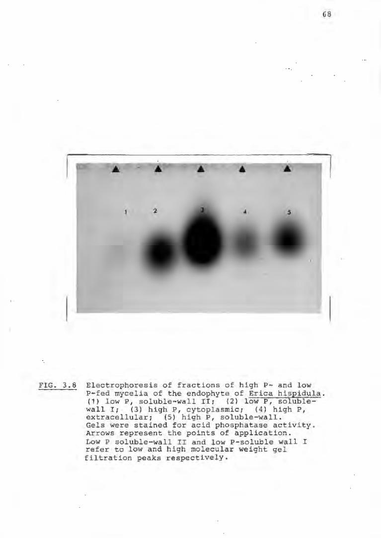
68
; • ' . ,
FIG. 3.8 Electrophoresis of fractions of high P- and low P-fed mycelia of the endophyte of Erica hispidula. (1) low P, soluble-wall IIt (2) low P, solublewall II (3) h:igh P, cytoplasmic; (4) high P, extracellular; (5) high P, soluble-wall. Gels were stained for acid phosphatase activity. Arrows represent the pOints of application. Low P soluble-wall II and low P-soluble wall I refer to low and high molecular weight gel filtration peaks respectively.
71
high P-fed mycelia was 1.5 times greater than that of low P
fed mycelia which can be attributed entirely to the greater
activity of the high P extracellular enzyme. In many plants
pi deficiency has been found to invoke an increase in acid
phosphatase activity, part of which is due to the appearance
of a cell wall-bound enzyme (Bieleski, 1973; Zink and
Veliky, 1979). The studies of Calleja et ale (1980) and
Calleja and d'Auzac (1983) have confirmed this for isolated
ectomycorrhizal fungi. Under conditions of low pi
availability, these organisms showed a large increase (75%)
in the total accessible phosphatase activity which was due
to an increased contribution by the cell wall and
extracellular enzymes. When the acid phosphatases of o~ion
root systems grown with and without the addition of pi to
the soil and infected with VA mycorrhizas were compared with
uninfected roots under similar conditions, no significant
differences in activity were found (Gianinazzi - Pearson and
Gianinazzi, 1976). A surfeit of organic P in the growth
medium did not repress the acid phosphatase activity of the
endophyte of E. hispidula but stimulated the further
synthesis of an already active externally-released enzyme.
In Canadian and Ghanaian soils, high soil phosphatase
activity has been correlated with high organic P contents in
the soil (Appiah and Thomas, 1982).
In this study, the phosphatase enzymes were partially
purified and characterised according to molecular weight,
75
growth when cell lysis would be expected to be minimal.
Thus, in the soil environment this enzyme may well give the
endophyte access to substrates normally inaccessible to the
host roots. The activities of the extracellular enzymes of
the European endophytes were low with the wall- and
membrane-bound fraction being the dominant one. It has
already been shown that the South African and European
endophytes also appear to differ in their ability to utilise
phytate salts (Mitchell and Read, 1985). Although both
European heathland and South African fynbos soils are low in
nutrients, the organic matter and total P content of a
typical fynbos soil may be as low as 2% of that of an upland
heath soil in the United Kingdom (Read and Mitchell, 1983).
Thus the secretion of an extracellular phosphatase by the
endophytes may be more important in the phosphorus nutrition
of the South African ericas than in the European examples of
the Ericales.
76
CHAPTER 4
KINETICS OF PHOSPHATE UPTAKE BY THE ISOLATED MYCORRHIZAL
ENDOPHYTE OF ERICA HISPIDULA
4.1 INTRODUCTION
Free phosphate ions in the soil and those made available
from bound complexes may be taken up by the external hyphae
of ericoid mycorrhizas and either used in metabolism or
transformed into stored P compounds such as polyphosphates.
Ultimatel~ some of the absorbed P may be transferred from
the internal, matrical hyphae to the cortical cells of the
host roots. In this way, the external mycorrhizal hyphae
would act as a physical extension of the host root by
increasing the absorptive surface available for uptake. The
endophyte might also facilitate an efficient P absorption
through the nature of its uptake system(s). Since P
transport across the plasmalemma has been regarded as a
carrier-mediated process, the enzyme kinetic equation of
Michaelis-Menten can be applied to estimate the kinetic
constants
Woolhouse,
of K m
1975;
and V max (Epstein and
Beever and Burns, 1980).
Hagen, 1952;
These constants
give valuable information on the efficiency of ion uptake
under defined conditions.
This chapter describes investigations into the simple uptake
kinetics of an ericoid endophyte as well as giving estimates
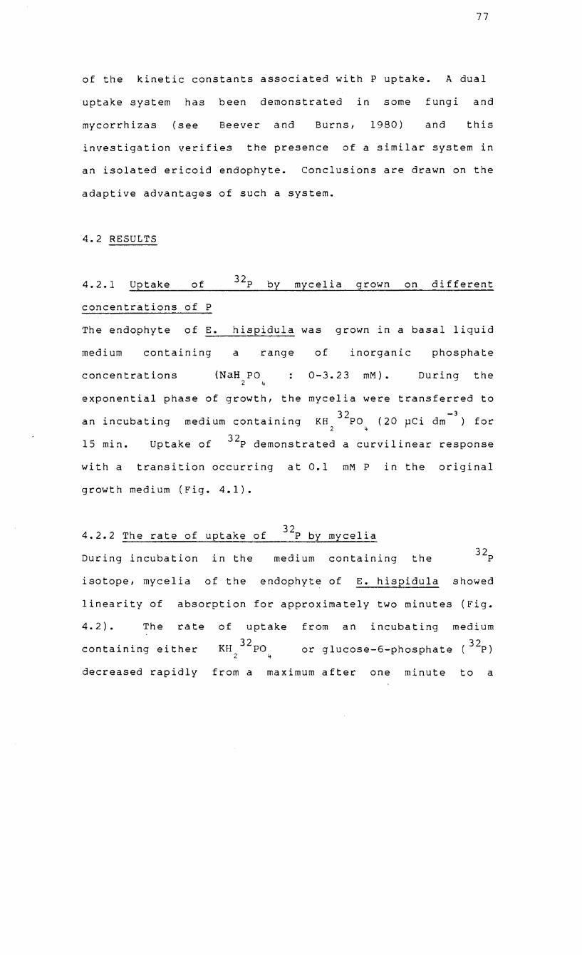
77
of the kinetic constants associated with P uptake. A dual
uptake system has been demonstrated in some fungi and
mycorrhizas (see Beever and Burns, 1980) and this
investigation verifies the presence of a similar system in
an isolated ericoid endophyte. Conclusions are drawn on the
adaptive advantages of such a system.
4.2 RESULTS
4.2.1 ~u~p~t~a~k~e~ __ ~o~f~ ____ 3_2_p~~b~y~_m~y~c~e~1~l~'a~~g~r~o~w~n __ ~o~n~~d~i~f~f~e~r~e~n~t
concentrations of P
The endophyte of E. hispidula was grown in a basal liquid
medium containing a range of inorganic phosphate
concentrations (NaH PO 2 If
0-3.23 mM). During the
exponential phase of growth, the mycelia were transferred to
32 -3 an incubating medium containing KH PO (20 ~Ci dm ) for
2 If
15 min. uptake of 32p demonstrated a curvilinear response
with a transition occurring at 0.1 mM P in the original
growth medium (Fig. 4.1).
4.2.2 The rate of uptake of 32p by mlcelia
During incubation in the medium containing the 32p
isotope, mycelia of the endophyte of E. hispidula showed
linearity of absorption for approximately two minutes (Fig.
4.2). The rate
containing either
of uptake
KH 32po 2 1+
from an incubating medium
glucose-6-phosphate (32p ) or
decreased rapidly from a maximum after one minute to a
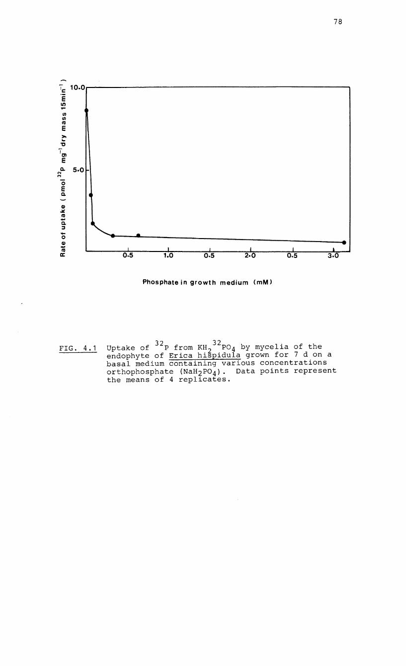
78
Ie: 10.0
E It) ... III III cu E >-... 'tI
TO) E
a.. 5.0 -N M
0 E Q.
Q) .:t:. cu -Q. ~ .... .... ... 0 Q) - I I cu I L I
a:: 0.5 1.0 0.5 2·0 0.5
Phosphate in growth medium (mM)
FIG. 4.1 Uptake of 32p from KH232p04 by mycelia of the endophyte of Erica hispidula grown for 7 d on a basal medium containing various concentrations orthophosphate (NaH2P04). Data points represent the means of 4 replicates.
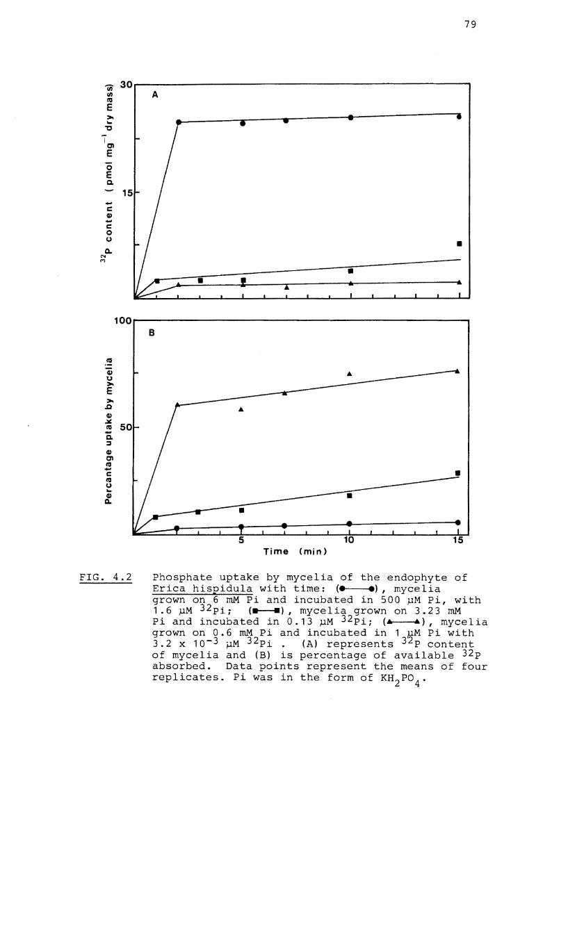
79
(j) 30 en A IV E >-...
"C
I en E 0 E Q.
-C II,) -c 0 u • 0-
N M
• •
100~------------------------------------------------~
FIG. 4.2
B
• •
• 5
Time (min)
Phosphate uptake by mycelia of the endophyte of Erica hispidula with time: (e .), mycelia grown on 6 mM Pi and incubated in 500 MM Pi, with 1.6 MM 32pi; <.__.) I mycelia grown on 3.23 mM Pi and incubated in 0.13 MM 32pi; (& .), mycelia grown on 0.6 mM Pi and incubated in 1 ~M Pi with 3.2 x 10⁻³ ~M 32pi. (A) represents 32p content of mycelia and (B) is percentage of available 32p absorbed. Data points represent the means of four replicates. Pi was in the form of KH 2P0
4"
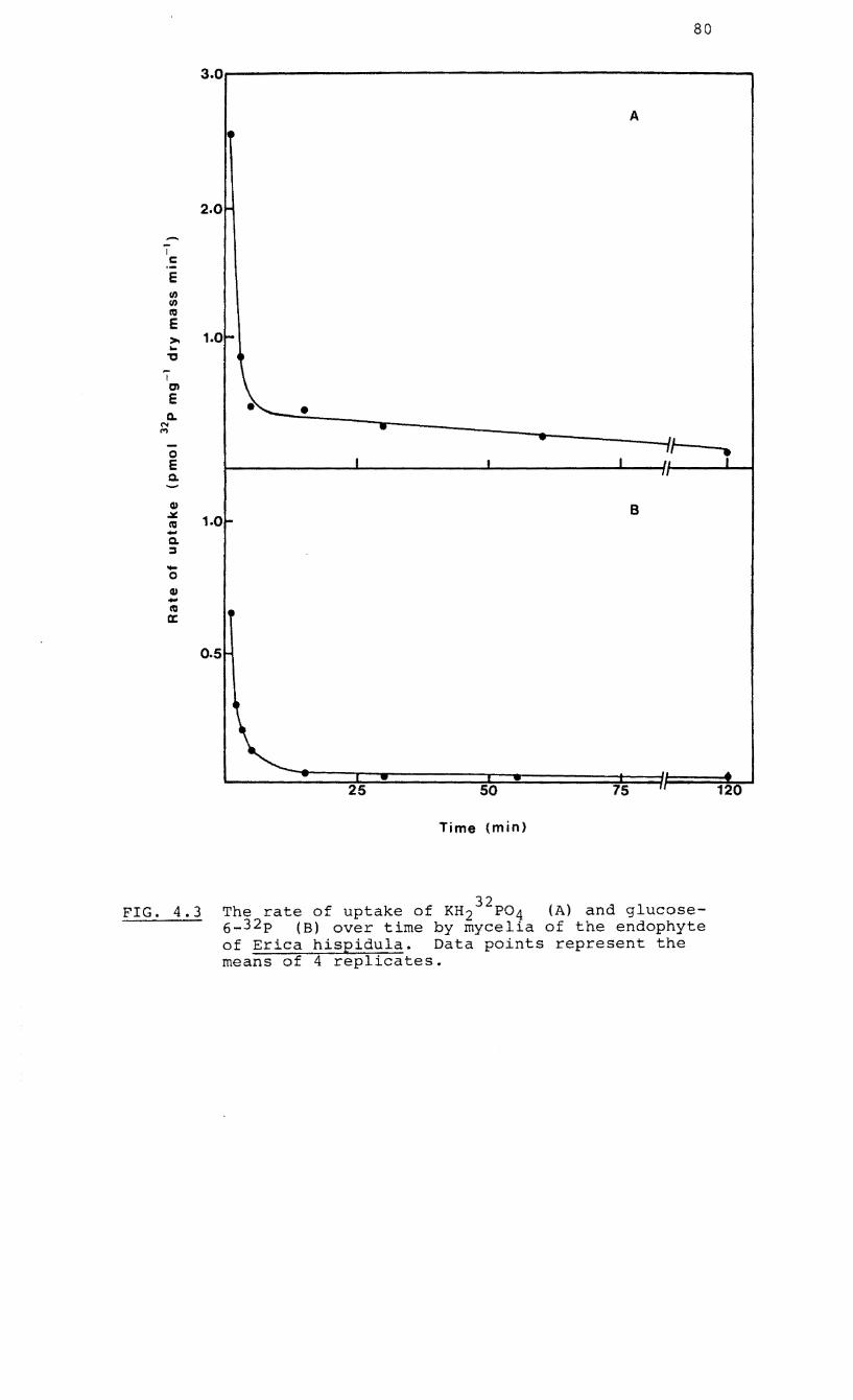
80
3.0r------------------------------------------------A
2.0
i c E en en 1'0 E >- 1.0-.. " ~
I en E • Q. •
N ... ,., 'L
0 J I
II 1 E I II
Q. II
Q) B .iIt 1.0-1'0 -Q. :::J -0 Q) -1'0 • ex:
0.5
I II
25 50 75 120
Time (min)
FIG. 4.3 32 The rate of uptake of KH2 P04 (A) and glucose-
6_ 32 p (B) over time by mycelia of the endophyte of Erica hispidula. Data points represent the means of 4 replicates.
81
steady-state rate at five minutes (Fig. 4.3). When results
were expressed as a percentage of available 32p absorbed by
mycelia against time it appears that the concentration of P
in the growth medium influenced P absorption (Fig. 4.2).
From these studies, the time chosen for further estimation
of kinetic constants was five minutes since an incubation of
one or two minutes would have caused difficulties in
sampling and inaccuracies in counting.
4.2.3 Efflux studies
In P uptake studies of fungi, significant efflux of the
radioisotope during incubation has been observed (Lowendorff
et al., 1974~ Beever and Burns, 1977) and this may
complicate the derivation of kinetic parameters (Neame and
Richards, 1972). Efflux may occur either through some
alteration of the plasmamembrane during the experimental
procedure or in response to "osmotic shock" (Burns and
Beever, 1977). Membrane permeability of the endophyte of
E. hispidula was tested when mycelia, 32 exposed to P
orthophosphate, were incubated in a range of non-radioactive
P solutions and 32p levels were monitored over 15 minutes.
It appears that no significant efflux of 32p occurred with
time even in salt concentrations well above those used in
the kinetic studies (Table 4.1). When data were fitted to
linear regression equations, all the correlation co-
efficients were statistically insignificant.
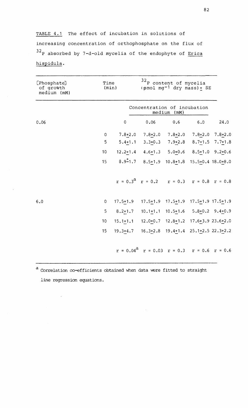
82
TABLE 4.1 The effect of incubation in solutions of
increasing concentration of orthophosphate on the flux of
32 P absorbed by 7-d-old mycelia of the endophyte of Erica
hispidula.
[Phosphate] Time 32 mycelia P content of of growth (min) ( pmol mg- 1 dry mass)! SE medium (roM)
Concentration of incubation medium (roM)
0.06 0 0.06 0.6 6.0 24.0
0 7.8+2.0 7.8+2.0 7.8+2.0 7.8+2.0 7.8+2.0
5 5.4+1.1 3.3+0.3 7.9+2.8 8.7+1.5 7.7+1.8
10 12.2+1.4 4.6+1.3 5.0+0.6 8.5+1.0 9.2+0.6
15 +
8.9-1.7 8.5+1.9 10.8+1.8 15.5+0.4 18.0+8.0
r = 0.3a r = 0.2 r = 0.3 r = 0.8 r 0.8
6.0 o 17.5+1.9 17.5+1.9 17.5+1.9 17.5+1.9 17.5+1.9
5 8.2+1.7 10.1+1.1 10.5+1.6 5.8+0.2 9.4+0.9
10 15.1+1.1 12.0+0.7 12.8+1.2 17.6+3.9 23.6+2.0
15 19.3+4.7 16.3+2.8 19.4+1.4 25.1+2.5 22.3+2.2
r = 0.04a r = 0.03 r = 0.3 r = 0.6 r = 0.6
a Correlation co-efficients obtained when data were fitted to straight
line regression equations.
83
4.2.4 Preliminary experiments on the rate of uptake by
mycelia in relation to P concentration.
When mycelia were incubated in solutions
constant concentration of 32p but a wide
containing a
range of non-
radioactive P concentrations, uptake rate (as represented
by 32p uptake) formed a curvilinear relationship with P
concentration when results were transformed to double-
reciprocal (Lineweaver-Burk) plots (Fig. 4.4). The
transformation in Fig. 4.4 indicated the operation of a
dual-uptake system but kinetic parameters (i.e. Km and Vmax )
were not determined from these results since the ratio
of 32p to non-radioactive P in the incubation medium had
not been kept constant.
4.2.5 The effect of pH on P uptake kinetics
Since the pH of the external medium may influence uptake
rates and thus the values of kinetic parameters (Lowendorff
et al., 1974), it was necessary to determine optimal pH
conditions for the estimation of kinetic parameters for the
endophyte. When mycelia were incubated in a wide range of
P orthophosphate (0.01 ~M to 1.68 ~M) dissolved in buffers
of different pH values (4.6, 5 • 6 , 6.8 and 7.9),
transformations to Hofstee plots indicated curvilinear
responses at pH 5.6, 6.8 and 7.9 but not at 4.6 (Fig. 4.5).
The parameter estimates of the high-affinity system at pH
4.6 were not determinable. At pH 4.6, uptake rates from the
four lower concentrations differed slightly so that the
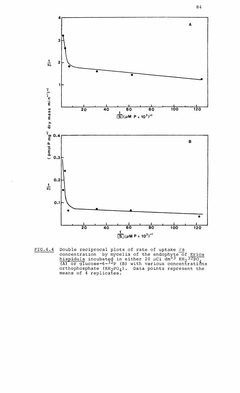
~ Vo
'i ..... I c::
E
... 'C
84
4~---------------------------------------------------' A
• • • ..
1 -
I I I I I I
20 40 60 80 100 120
rID CuM P x 103 )·1
.. ~0.4~-----------------------------------------------------' E
C1.
o E a.
1 YO
0.31-
• 0.2
•
0.11-
•
I 20
B
• • •
I , I I I I I 40 60 80 100 120
1 [S](pM P x 103 )-1
FIG.4.4 Double reciprocal plots of rate of uptake VS concentration by mycelia of the endophyte of Erica hispidula incubated in either 20 pei dm- 3 KH23Tpo (A) or glucose-6-32p (B) with various concentrati~ns orthophosphate (KH2P04)' Data paints represent the means of 4 replicates.
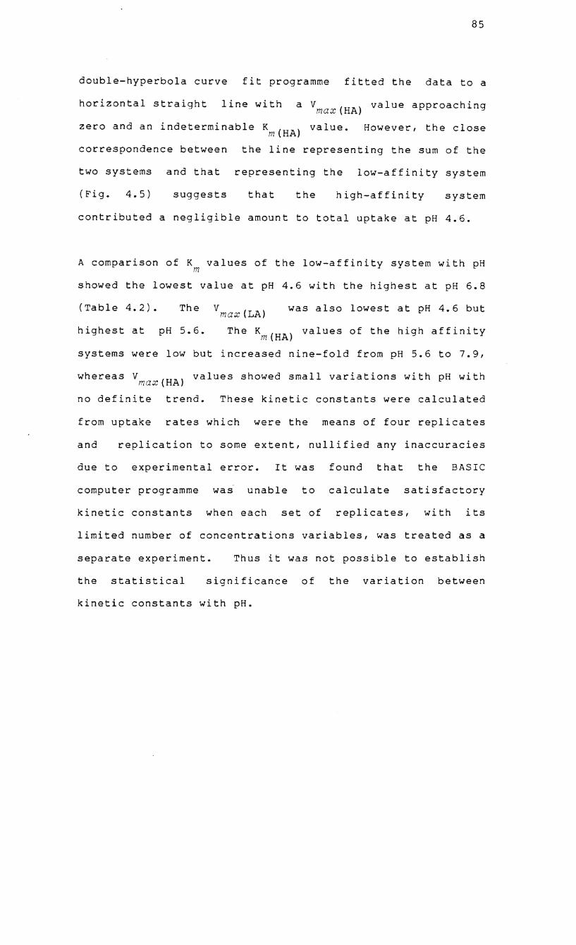
85
double-hyperbola curve fit programme fitted the data to a
horizontal straight line with a Vmax(HA) value approaching
zero and an indeterminable Km(HA) value. However, the close
correspondence between the line representing the sum of the
two systems and that representing the low-affinity system
(Fig. 4.5) suggests that the high-affinity system
contributed a negligible amount to total uptake at pH 4.6.
A comparison of K values of the low-affinity system with pH m
showed the lowest value at pH 4.6 with the highest at pH 6.8
(Table 4.2). The v max (LA) was also lowest at pH 4.6 but
highest at pH 5.6. The Km(HA) values of the high affinity
systems were low but increased nine-fold from pH 5.6 to 7.9,
whereas V values showed small variations with pH with max(HA)
no definite trend. These kinetic constants were calculated
from uptake rates which were the means of four replicates
and replication to some extent, nullified any inaccuracies
due to experimental error. It was found that the BASIC
computer programme was unable to calculate satisfactory
kinetic constants when each set of replicates, with its
limited number of concentrations variables, was treated as a
separate experiment. Thus it was not possible to establish
the statistical significance of the variation between
kinetic constants with pH.
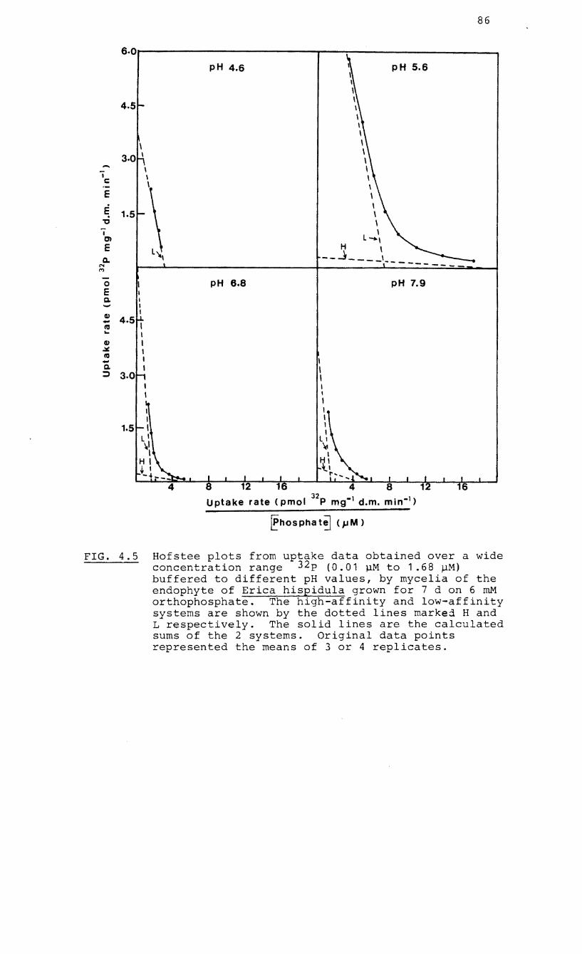
86
6.0,------------,---.------------, pH 4.6 pH 5.6
4.5
\ 3.0 \
-. i c
E
E , . 1.5 \
" \ i ,
c:n L_, E H ,
Q. -_J ----,----'" M
0 PH 6.8 pH 7.9 E I Q. I -- I
Q) I - 4.5 ,
ctI ... I Q) I .:.: I ctI I -Q. I
::::>
4 8 1 Uptake rate (pmol 32p mg-) d.m. min-I)
~hOS pha t!] (}.I M )
FIG. 4.5 Hofstee plots from uptake data obtained over a wide concentration range 32p (0.01 pM to 1.68 ~M) buffered to different pH values, by mycelia of the endophyte of Erica hispidula grown for 7 d on 6 roM orthophosphate. The high-affinity and low-affinity systems are shown by the dotted lines marked Hand L respectively. The solid lines are the calculated sums of the 2 systems. Original data points represented the means of 3 or 4 replicates.
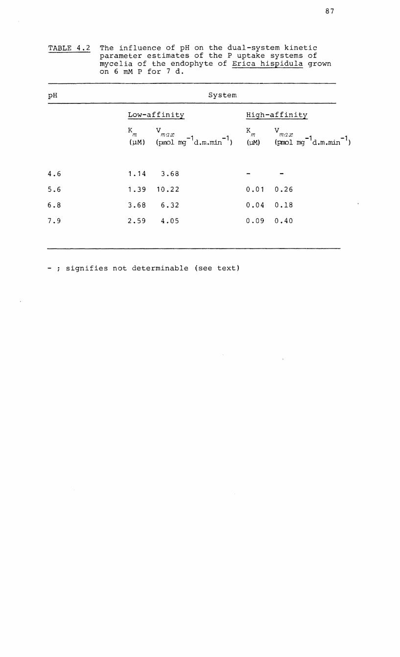
87
TABLE 4.2 The influence of pH on the dual-system kinetic parameter estimates of the P uptake systems of mycelia of the endophyte of Erica hispidula grown on 6 roM P for 7 d.
pH System
4.6
5.6
6.8
7.9
Low-affinity
K m
(j..lM)
1. 14
V max -1 -1
(pu'Ol rrg d.m.min )
3.68
1.39 10.22
3.68 6.32
2.59 4.05
- ; signifies not determinable (see text)
High-affinity
K m
(j..lM)
V max -1 -1
(p:rol rrg d.m.min )
0.01 0.26
0.04 0.18
0.09 0.40
88
4.2.6 The effect of a metabolic inhibitor on uptake
2,4-Dinitrophenol (DNP) inhibits the oxidative
phosphorylation of ATP synthesis and is commonly used as an
inhibitor of active ion transport across membranes. The
kinetic parameters of the low-affinity system of mycelia
growing in high P (6 mM) media were estimated at pH 5.6 in
the presence and absence of 2,4 dinitrophenol.
in the medium the V max
value was 9.76 pmol
-1 mass min and was reduced by 90% to 1.0 pmol
Without DNP
32 -1 P mg dry
32 -1 P mg dry
-1 mass min when DNP was present. The addition of DNP caused
a small increase in the K value from 2.0 to 2.9 pM P. m
4.2.7 The kinetic parameters of dual-system uptake in high P
and low P-fed mycelia
Further experiments were designed to determine more precise
estimates of the kinetic parameters of high-and low-affinity
systems in the endophyte of E. hispidula at pH 5.6. To
allow for the possibility that kinetic parameters "might
vary according to the physiological status of the fungus"
(Burns and Beever, 1977), mycelia were grown in a basal
medium containing either high (6 mM) or low (0.06 mM) levels
of orthophosphate (KH PO ). 2 4
Seven days after inoculation,
mycelia were incubated for five minutes in a wide range of
orthophosphate solutions (1 pM to 0.5 mM) and the levels
of 32p absorbed by mycelia were measured. The range of
incubation concentrations used was assessed from preliminary
results (Fig. 4.4).
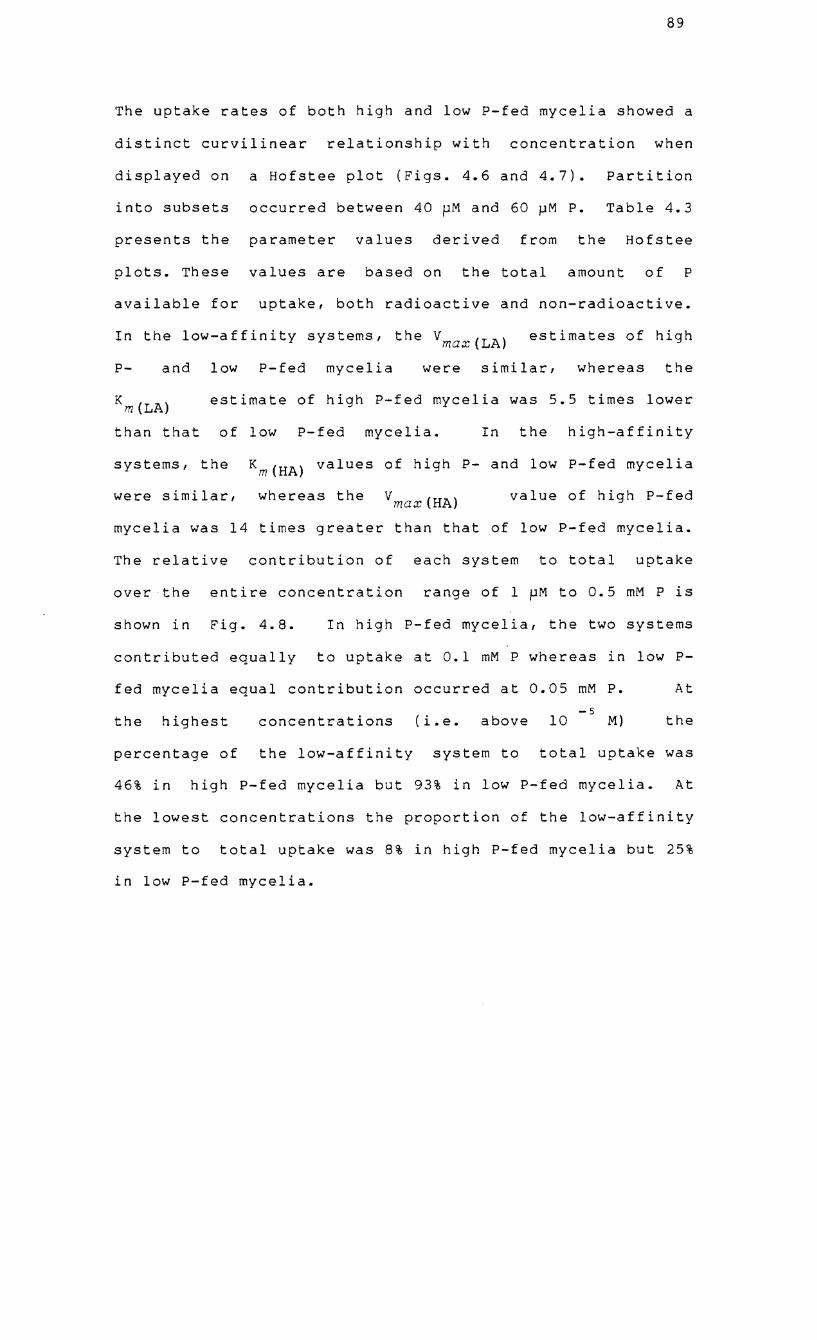
89
The uptake rates of both high and low P-fed mycelia showed a
distinct curvilinear relationship with concentration when
displayed on a Hofstee plot (Figs. 4.6 and 4.7). Partition
into subsets occurred between 40 pM and 60 pM P. Table 4.3
presents the parameter values derived from the Hofstee
plots. These values are based on the total amount of P
available for uptake, both radioactive and non-radioactive.
In the low-affinity systems, the Vmax(LA) estimates of high
P- and low P-fed mycelia were similar, whereas the
estimate of high P-fed mycelia was 5.5 times lower
than that of low P-fed mycelia. In the high-affinity
systems, the Km(HA) values of high P- and low P-fed mycelia
were similar, whereas the Vmax(HA) value of high P-fed
mycelia was 14 times greater than that of low P-fed mycelia.
The relative contribution of each system to total uptake
over the entire concentration range of 1 pM to 0.5 mM P is
shown in Fig. 4.8. In high P-fed mycelia, the two systems
contributed equally to uptake at 0.1 mM P whereas in low P
fed mycelia equal contribution occurred at 0.05 mM P. At
-5 the highest concentrations (i.e. above 10 M) the
percentage of the low-affinity system to total uptake was
46% in high P-fed mycelia but 93% in low P-fed mycelia. At
the lowest concentrations the proportion of the low-affinity
system to total uptake was 8% in high P-fed mycelia but 25%
in low P-fed mycelia.
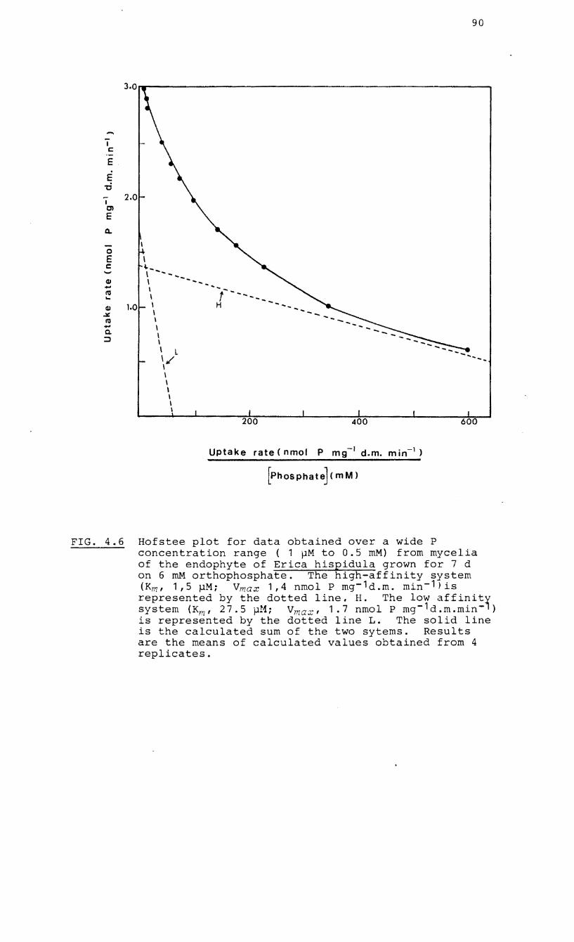
...... I C
E
E -c I CJ)
E
a.
o E c
2.0
1.0
\ ~ ... \ -......... \ ..... - .............. I ~ ...
\ ,- ...... -......... I H - ............ I \ I \ \ \ L 1/ \ I \ \ \ I \
200 400
Uptake rate (nmol P mg -I d.m. m in-I)
[PhOSPhate] (m M)
90
600
FIG. 4.6 Hofstee plot for data obtained over a wide P concentration range ( 1 pM to 0.5 roM) from mycelia of the endophyte of Erica hispidula grown for 7 d on 6 roM orthophosphate. The high-affinity system (Km, 1,5 MMi Vmax 1,4 nmol P mg- 1d.m. min- 1 )is represen:ed by the~ dotted line. H. The l~r affi~i~1 system (Km, 27.5 p.1; V max , 1.7 nmol P mg d.m.mln ) is represented by the dotted line L. The solid line is the calculated sum of the two sytems. Results are the means of calculated values obtained from 4 replicates.
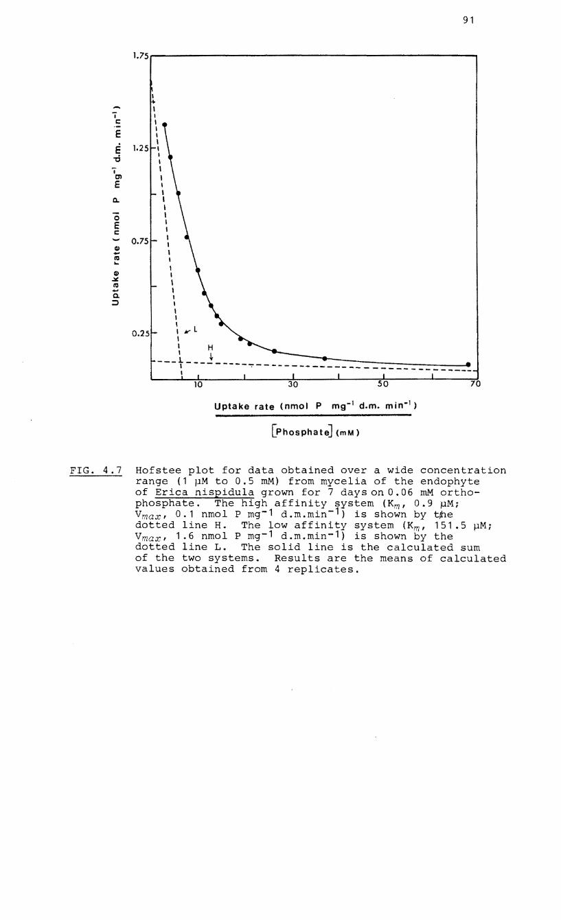
.-i I:
E . E -d I en E
c..
0 E I: ..... Q) .... 10 .... Q)
.:t:. 10 .... a. ::>
91
1.75.----------------------------,
1.25
0.75
0.25
I I , I I I I I I I I , I I I I I I I I I I , I I I I I I , I I k" L I , H
-__ t ___ .! ________ =:-=:-::~_=::::_:_-----_I ~ . ----------------------------
70
Uptake rate (nmol P mg- 1 d.m. min-1 )
[PhoSPhate] (mM)
FIG. 4.7 Hofstee plot for data obtained over a wide concentration range (1 pM to 0.5 mM) from mycelia of the endophyte of Erica nispidula grown for 7 days on 0.06 roM orthophosphate. The high affinity system (Kmf 0.9 ~M; V maxf 0.1 nmol P mg- 1 d.m.min- 1) is shown by t~e dotted line H. The low affinity system (Kmf 151.5 pM; V maxf 1.6 nmol P mg- 1 d.m.min- 1 ) is shown by the dotted line L. The solid line is the calculated sum of the two systems. Results are the means of calculated values obtained from 4 replicates.
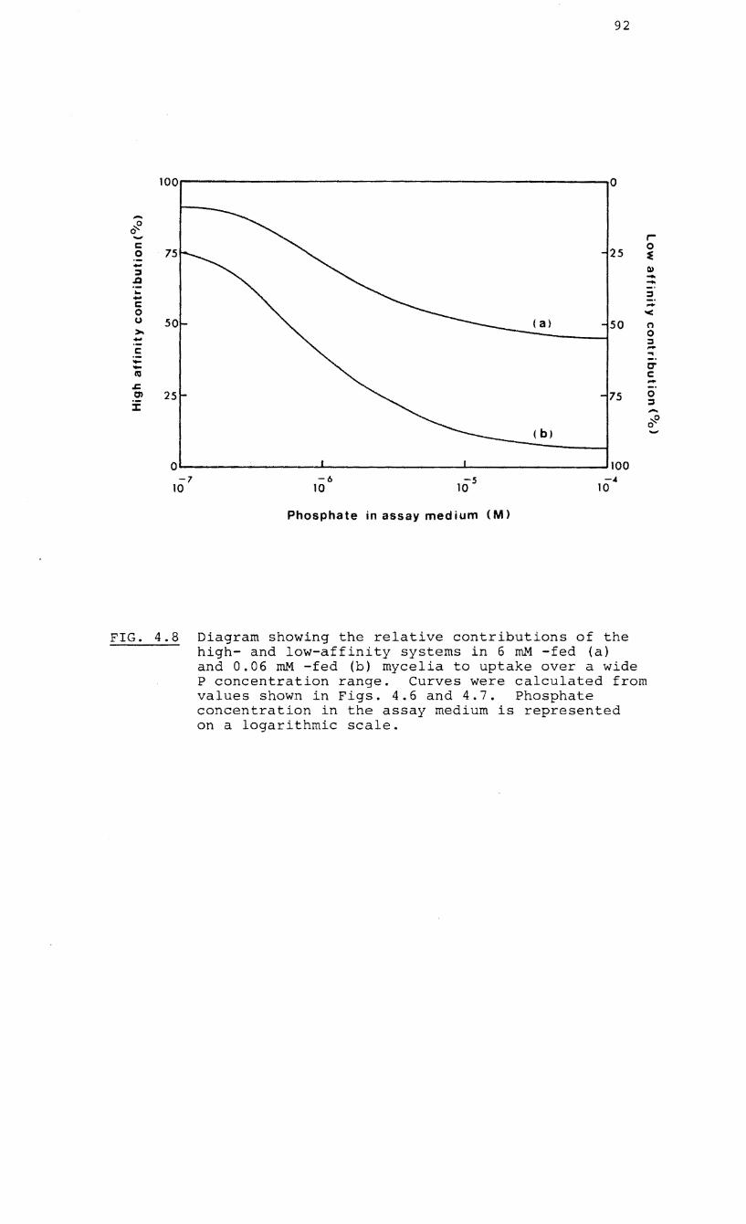
92
100 a
-~ 0 r-c 0 0 25 ~ ....
Q) :::lI ..Q --... :::lI .... C 0 '< () 50 n >- 0 - :::J C -.. - tr -1\'1 C -.z:; 0) 25 75 0
J: :::J -~o 0
( b)
a 100 7 -6 -s -4
10 10 10 10
Phosphate in assay medium (M)
FIG. 4.8 Diagram showing the relative contributions of the high- and low-affinity systems in 6 roM -fed (a) and 0.06 roM -fed (b) mycelia to uptake over a wide P concentration range. Curves were calculated from values shown in Figs. 4.6 and 4.7. Phosphate concentration in the assay medium is represented on a logarithmic scale.
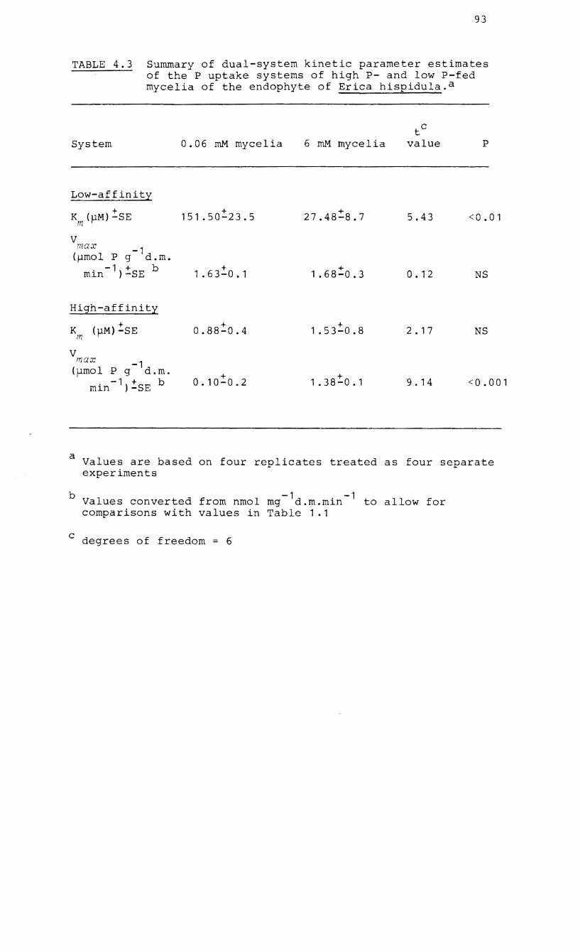
93
TABLE 4.3 Summary of dual-system kinetic parameter estimates of the P uptake systems of high P- and low P-fed mycelia of the endophyte of Erica hispidula. a
System
Low-affinity
+ K (j.1M)-SE m
V max -1 (pmol P g d.m.
-1 + min )-SE b
High-affinity
+ K (j.1M)-SE m
V max -1 ( pmol P g d.m.
-1 + min )-SE b
0.06 roM mycelia 6 roM mycelia
151.50:!:23.5 + 27.48-8.7
+ 1.63-0.1 + 1 .68-0.3
+ 0.88-0.4 + 1.53-0.8
+ 0.10-0.2 + 1.38-0.1
tC
value
5.43
O. 12
2. 17
9. 14
P
<0.01
NS
NS
<0.001
a Values are based on four replicates treated as four separate experiments
b -1 . -1 Values converted from nmol mg d.m.m~n to allow for comparisons with values in Table 1.1
c degrees of freedom = 6
94
4.3 DISCUSSION
The uptake kinetics of a number of ions in both higher
plants (Epstein and Elzam, 1963: Epstein, 1966) and fungi
(Beever and Burns, 1980) has been based upon a dual uptake
system which assumes that two systems, of different kinetic
parameters, operate simultaneously with each contributing to
uptake at all concentrations. Thus uptake at anyone
concentration is the sum of two Michaelis-Menten equations.
When uptake rates in relation to substrate concentration
are transformed dual-system uptake will form a continuous
and curvilinear plot (Nissen, 1973: 1974). However, the
dual-system interpretation is one of several which can be
applied to uptake data. Nissen (1973: 1974) has shown that
ionic uptake data (including that of P uptake) from higher
plants, which had originally been analysed to show a dual
uptake system could be reinterpreted with more detailed and
precise data to indicate a multiphasic system of uptake.
Transformations of such data show discontinuous curves,
indicating transitions from one phase to another. Other
alternatives to dual uptake mechanisms which have similar
kinetic data to those of dual systems, have been proposed
(Nissen, 1974: Borst-Pauwels, 1976). Borst-Pauwels (1976)
established statistical criteria for distinguishing dual
uptake systems from other mechanisms such as non-carrier
transport, two-site carrier transport
transport with multiple binding sites.
and single-site
However, these
95
methods require detailed and very accurate data and Beever
and Burns (1980) have cited direct evidence for the
existence of dual uptake systems. This evidence includes
mutants of N. crassa containing only one system and also the
differential response of the systems in Saccharomyces
cerevisiae Meyen ex Hansen to external ions.
Preliminary experiments on the endophyte of E. hispidula
indicated the possible operation of a dual system of uptake
which was further investigated by using the statistical
procedures of Burns and Tucker (1977). Beever and Burns
(1980) have indicated the operation of dual-system uptake in
ectomycorrhizas and soil saprotrophs (Table 1.1, p.16) and
these values can be compared with those obtained for low P-
fed mycelia of the endophyte of E. hispidula. The
Km(HA) value (0.88 ~M) of the ericoid endophyte is between
two and eleven times lower than any of the values listed in
Table 1.1. The
although ten
v max(HA) -1 -1
value (O.l nmol g d.m.min ),
times higher than those of intact
ectomycorrhizas is twenty times lower than those of N.
crassa and Aspergillus nidulans (Eidam) Winter. Similarly
the Km(LA) value (151.5 pM) of the endophyte is between
seven and twenty-one times lower than those of
ectomycorrhizas and two and four times lower than that of
N. crassa and A. nidulans respectively. The Vmax(LA) value
(1.6 nmol _1 -1
g d.m.min ) of the endophyte, although between
six and thirty times higher than those of ectomycorrhizas
97
comparison of the other kinetic constants of the high
affinity and low-affinity systems does show differences
between the two organisms. The VmaX(LA) and Vmax(HA) values
of low P-fed germlings of N. crassa were significantly
higher than corresponding values of high P-fed germlings and
the Km(LA) value of low P-fed germlings was significantly
lower than that of high P-fed germlings. It was concluded
that low P-fed germlings achieved a greater efficiency at
taking up P from dilute solutions by increasing the
efficiency of their low-affinity system. In contrast, there
was no clear enhancement of the efficiency of the low
affinity system of low P-fed mycelia of the endophyte.
Under conditions of limited P availability, there was a
repression of the high-affinity system with the endophyte
relying more on its low-affinity system for uptake. The
high P-fed mycelia "indulged" in high-affinity uptake even
at high concentrations of P in the external medium (54% of
total uptake at 0.5 mM p). If the adaptive importance of
the low-affinity system lies in its reduced energy demands
on the cell (Beever and Burns, 1980), it would appear that
the differences in the uptake kinetics between high P- and
low P-fed mycelia of the endophyte might be explained by the
importance of P in the energetics of cell metabolism.
These investigations on ericoid mycorrhizas, show that
although the high-affinity system appears to be the dominant
98
one at low phosphorus concentrations, the low - affinity
system plays a significant role in phosphorus uptake,
particularly when the organism is growing in a low
phosphorus environment. This observation is confirmed by the
pH studies (Fig. 4.5; Table 4.2) in which low-affinity
uptake was detectable at external concentrations below 2 pM
P. Levels of available P in many soils may be as low as 0.01
pM to 2 pM (Woolhouse, 1975) and the levels of resin-
extractable P in fynbos soils were shown to vary between 13
pM and 81 pM P (Mitchell, Brown and Jongens-Roberts, 1984).
Phosphate uptake by fungi is carrier-mediated and
accompanied by ATP-dependent proton co-transport against an
electrochemical gradient (Woolhouse, 1975; Beever and
Burns, 1980). The results of this investigation confirm
that when ATP synthesis is interrupted by 0.5 mM DNP, an
inhibitor of oxidative phosphorylation, the rate of uptake
by mycelia of the endophyte of E. hispidula is severely
reduced. optimum uptake of P usually occurs at pH values
around 5 (see Goodman and Rothstein, 1957) when H po is the Z 4
predominant Pion When the uptake systems of mycelia were
operating at different pH values, optimal activity of the
low-affinity and possibly high-affinity systems occurred at
pH 5.6. In both systems of the endophyte of E. hispidula,
there was a trend towards an increase in K m although only
three-fold in the low-affinity and nine-fold in the high-
affinity system. The V max
values followed no particular
99
pattern within each system but the ratio of v • max(LAr
V decreased from pH 4.6 to 7.9. There thus appeared max (HA)
to be a relationship between pH and the contribution of the
high-affinity system to uptake with pH. In pH studies on N.
crassa, the effect of pH on the high-affinity system was
negligible whereas the low-affinity one was strongly
affected; V max (LA) values remained fairly constant but
K m(LA)
values increased 300-fold between pH 4.0 and 7.3
(Lowendorff et al., 1974, 1975). These results were not
explained in terms of two completely separate transport
systems (one with a low Km and low pH optimum and the other
with a high Km and high pH optimum) and did not support the
hypothesis that only H PO and not HPo 2-
2 ~- 4 was the substrate
for the transport system. A model was proposed in which
(or OH served as modifiers of the uptake
systems by altering their affinities for the substrate 2_
(either H PO or HPO ). 244
The absorption of P from solutions, similar in concentration
to those of the soil, has been shown to be linear with time
in beech mycorrhizas with a rate decrease after only
very long periods of time (Harley, 1969). At higher
concentrations of external p, however, uptake was not linear
with time. Cress eta 1. (1979) establi shed tha tat land
60 ~M P, uptake by tomato roots infected with VA mycorrhizas
was linear for up to 25 min. Linearity of absorption by
mycelia of the endophyte of E. hispidula occurred within one
100
to two minutes but the rate of uptake declined dramatically
between 0 and 5 min (Fig. 4.3). After 5 minutes a steady
state decline occurred which could not be attributed to any
efflux of phosphate (Table 4.1). These results indicate
that after an initial rapid influx of P, the endophyte
adjusts its uptake rate so that P is absorbed only according
to requirements, especially when the phosphate ion is
abundant. The pattern of uptake appears to be dependent
upon both external and internal phosphorus levels (Fig.
4.1). N. crassa appears to exert a similar tight control
over the activity of its uptake systems to maintain a
constancy of P uptake over a wide P concentration range
(Beever and Burns, 1977). The constancy of uptake and the
maintenance of a constant internal P content was adequately
explained by changes in the kinetic constants of the high
and low-affinity uptake systems.
This investigation indicates that the ericoid endophyte may
well improve the P status of its host through its ability to
use an energetically inexpensive low-affinity uptake system
at very low soil phosphorus concentrations and to bring
about rapid adjustments in uptake in response to both soil
and host P levels.
101
CHAPTER 5
THE IDENTIFICATION, EXTRACTION AND FRACTIONATION OF
POLYPHOSPHATES AND PHYTIC ACID
5.1 INTRODUCTION
Ultrastructural analyses of
that the P absorbed by
ericoid mycorrhizas indicate
extracellular hyphae may be
transformed into electron-dense, osmiophilicl vacuolar
granules of polyphosphate (polyP) which in dual culture only
persist in the extracellular mycelium (Bonfante-Fasola and
Gianinazzi-Pearson, 1982). In natural ericoid mycorrhizas,
polyP granules have been found in both the intra- and
extracellular mycelium (Bonfante-Fasola et al., 1981). It
appears therefore that the synthesis of polyP is dependent
on the nutrient and metabolic status of the endophyte in
relation to the host and the external environment. Apart
from ultrastructural studies no information is available on
the role of polyP either in the P metabolism of ericoid
mycorrhizas or its function as a potential storage form of
P. PolyP may be of fundamental importance in the survival
of ericaceous plants especially when situated under
conditions of low phosphorus status. This chapter
describes an investigation into the identification,
characterisation and estimation of polyP in cultured
endophytes and
mycorrhizal seedlings.
ation of phosphorus
synthesized mycorrhizal and non-
Experiments involving th fraction
were designed to establish the
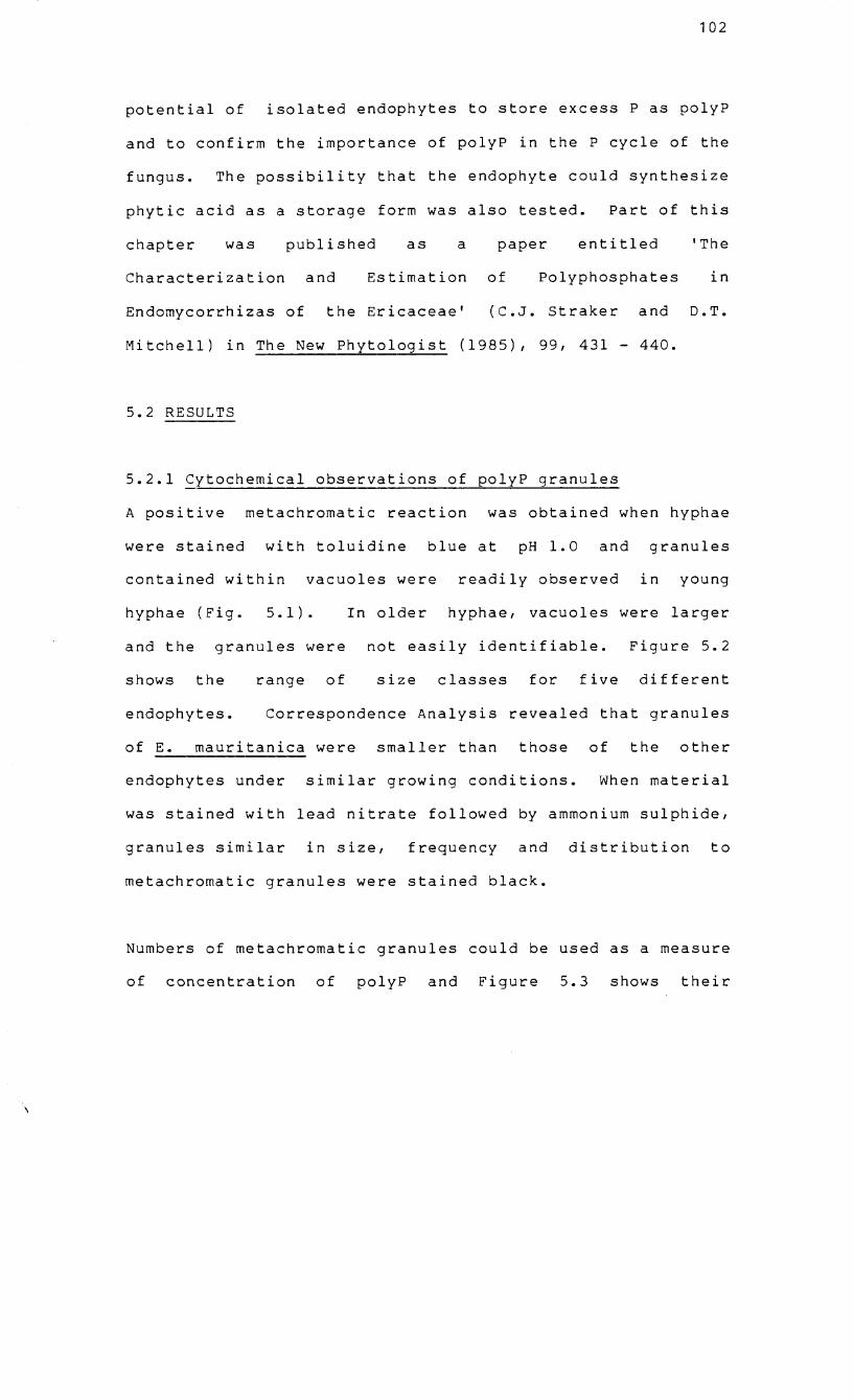
102
potential of isolated endophytes to store excess P as polyP
and to confirm the importance of polyP in the P cycle of the
fungus. The possibility that the endophyte could synthesize
phytic acid as a storage form was also tested. Part of this
chapter was published as a paper entitled 'The
Characterization and Estimation of Polyphosphates in
Endomycorrhizas of the Ericaceae' (C.J. Straker and D.T.
Mitchell) in The New Phytologist (1985), 99, 431 - 440.
5.2 RESULTS
5.2.1 Cytochemical observations of polyP granules
A positive metachromatic reaction was obtained when hyphae
were stained with toluidine blue at pH 1.0 and granules
contained within vacuoles were readily observed in young
hyphae (Fig. 5. 1 ) .
and the granules were
shows the range of
In older hyphae, vacuoles were larger
not easily identifiable. Figure 5.2
size classes for five different
endophytes. Correspondence Analysis revealed that granules
of E. mauritanica were smaller than those of the other
endophytes under similar growing conditions. When material
was stained with lead nitrate followed by ammonium sulphide,
granules similar in size, frequency and distribution to
metachromatic granules were stained black.
Numbers of metachromatic granules could be used as a measure
of concentration of polyP and Figure 5.3 shows their
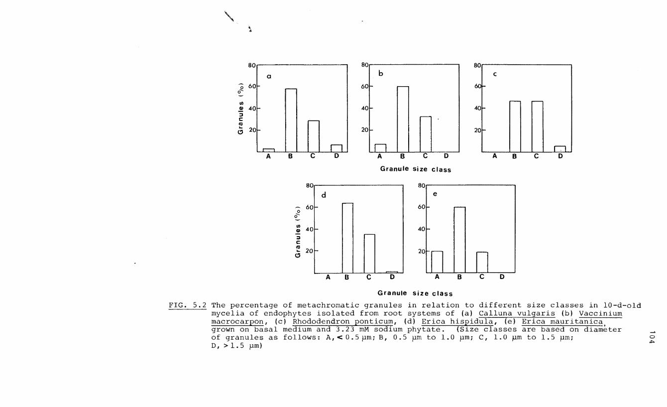
80 80 80
a b c
0 0 .....
60 ~ r- 60 I- r- 60 I-
rJ) ,...-- -
CD 40 I- 40 ... 401-:::J C - r--
til ... 20
" I- l- 20 I-20
,...--, n n I.l A B c o A B c o A B c o
Granule size class
80 80
d e -60 I- 60 r-
0 0 .....
rJ) 40 CD - -40
:::J :--
C co
20 ... "
- :-- :--20
A B c o A B c o
Granule size class
FIG. 5.2 The percentage of metachromatic granules in relation to different size classes in 10-d-old mycelia of endophytes isolated from root systems of (a) Calluna vulgaris (b) Vaccinium macrocarpon, (c) Rhododendron ponticum, (d) Erica hispidula, (e) Erica mauritanica, grown on basal medium and 3.23 mM sodium phytate. (Size classes are based on diameter of granules as follows: A, < 0.5pm; B, 0.5 pm to 1.0 pm; C, 1.0 pm to 1.5 pm; D I > 1 . 5 pm)
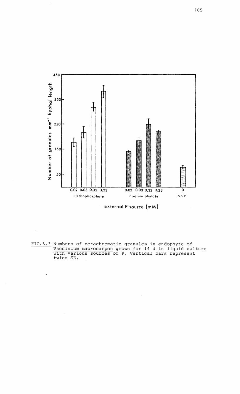
450
..r::. -0> c <V
- 350 0
..r::. a. >..
..r::.
I
E 250 E \1\
<V :::')
C 0 .... 15 0> -0 .... <V
..c E
50 :::')
Z
0.02 0.03 0.32 3.23
Orthophosphate
0.02 0.03 0.32 3.23
Sodium phytate
External P source (mM)
105
No P
FIG.5.3 Numbers of metachromatic granules in endophyte of Vaccinium macrocarpon grown for 14 d in liquid culture with various sources of P. Vertical bars represent twice SE.
106
relationship to the external concentration of phosphorus in
the medium. When the P source was orthophosphate, there was
a direct power curve relationship between these variables (y o .11 2
= 155.6x with r = 0.94). However, when P was supplied
in an organic form, granule numbers increased up to an
external concentration of 0.32 mM but thereafter they
declined. Granules in hyphae during the growth of the
endophyte in culture accumulated rapidly during the lag
phase of growth (Fig. 5.4).
5.2.2.Polyacrylamide gel electrophoresis for separation of
nucleic acid-polyP co-precipitates
The pink staining bands obtained from mycelial extracts of a
South African and European isolate as well as commercial
polyP (sodium phosphate glass Type 65) have absorption
maxima at 530 nm (y-metachromasy) (Fig. 5.5). Blue
metachromatic gel bands from mycelial extracts and
commercial RNA showed a shift to an absorption maximum of
580 nm (B-metachromasy) (Fig. 5.5). The Y-metachromasy of
the pink bands produced absorption peaks at 523 nm whereas
the major blue bands were enhanced at 577 nm (Fig. 5.6).
Molecular weights of polyP molecules
Electrophoretic bands of a range of synthetic polyP markers
were run on the same gel as the mycelial extracts (Fig.
5.6). The relationship between distance of migration and
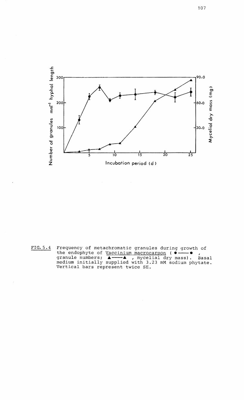
..s::. .... 0> c QJ
0 ..s::. a. >..
..s::.
IE E
II)
QJ
::l C 0 I-
0> ..... 0 I-QJ
...0 E ::::l
Z
107
300
200
Incubation period <d)
0> E ..... II) II)
o E >-I-
""0
o QJ v >-~
FIG.S.4 Frequency of metachromatic granules during growth of the endophyte of Vaccinium macrocarpon ( • --. , granule numbersi .---. ,mycelial dry mass). Basal medium initially supplied with 3.23 mM sodium phytate. Vertical bars represent twice SEe
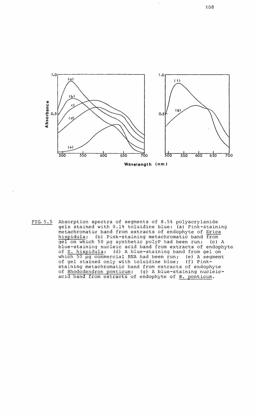
108
1.0.------------------. 1.0..------------.,
Q) (J c C'O -e 0.5 o If)
.Q
<
(0) (f)
700
Wavelength (nm)
FIG.5.S Absorption spectra of segments of 8.5% polyacrylamide gels stained with 0.1% toluidine blue: (a) Pink-staining metachromatic band from extracts of endophyte of Erica hispidulai (b) Pink-staining metachromatic band from gel on which 50 ~g synthetic polyP had been run; (c) A blue-staining nucleic acid band from extracts of endophyte of E. hispidula; (d) A blue-staining band from gel on which 50 ~g commercial RNA had been run; (e) A segment of gel stained only with toluidine blue; (f) Pinkstaining metachromatic band from extracts of endophyte of Rhododendron ponticumi (g) A blue-staining nucleicacid band from extracts of endophyte of R. ponticum.
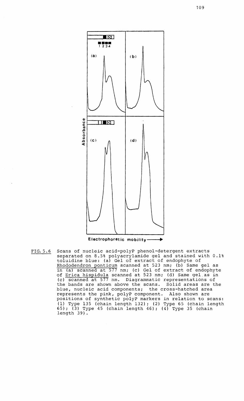
IU (.)
C ltI
,Q .. 0 I/)
,Q
«
(a)
(c)
•••• 1 234
( b)
(d)
Electrophoret ic mobility ..
109
FIG.5.6 Scans of nucleic acid-polyP phenol-detergent extracts separated on 8.5% polyacrylamide gel and stained with 0.1% toluidine blue: (a) Gel of extract of endophyte of Rhododendron ponticum scanned at 523 nmi (b) Same gel as in (a) scanned at 577 nmi (c) Gel of extract of endophyte of Erica hispidula scanned at 523 nmi (d) Same gel.as in (c) scanned at 577 nm. Diagrammatic representations of the bands are shown above the scans. Solid areas are the blue, nucleic acid componentsi the cross-hatched area represents the pink, polyP component. Also shown are positions of synthetic polyP markers in relation to scans: (1) Type 135 (chain length 132); (2) Type 65 (chain length 65); (3) Type 45 (chain length 46); (4) Type 35 (chain length 39).
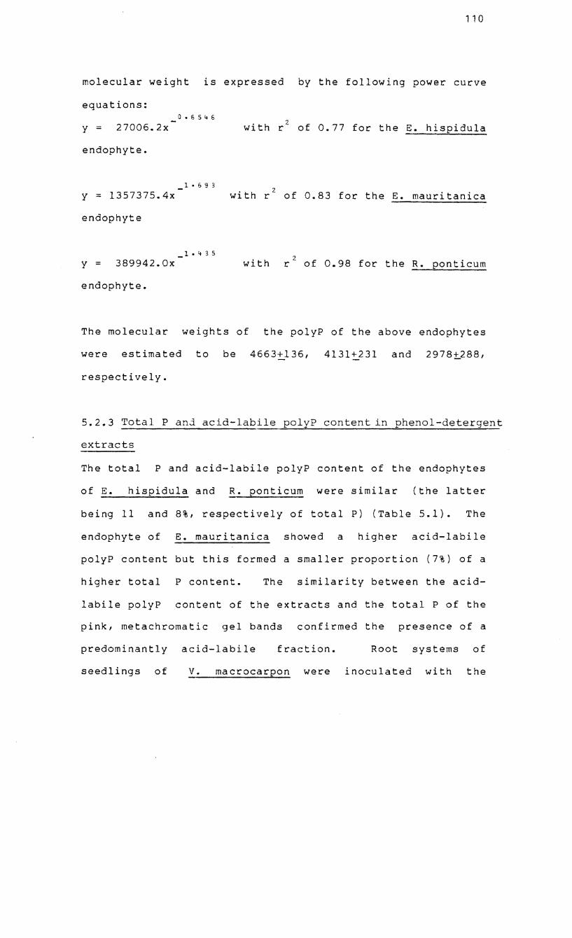
110
molecular weight is expressed by the following power curve
equations: 0.6545
y::: 27006.2x with rZ of 0.77 for the E. hispidula
endophyte.
1 ·69 3
y :: 1357375.4x with r2 of 0.83 for the E. mauritanica
endophyte
1 • 4 3 5
y = 389942.0x with 2
r of 0.98 for the R. ponticum
endophyte.
The molecular weights of the polyP of the above endophytes
were estimated to be 4663~136, 4131+231 and 2978~288,
respectively.
5.2.3 Total P anj acid-labile polyP content in phenol-detergent
extracts
The total P and acid-labile polyP content of the endophytes
of E. hispidula and R. ponticum were similar (the latter
being 11 and 8%, respectively of total P) (Table 5.1). The
endophyte of E. mauritanica showed a higher acid-labile
polyP content but this formed a smaller proportion (7%) of a
higher total P content. The similarity between the acid-
labile polyP content of the extracts and the total P of the
pink, metachromatic gel bands confirmed the presence of a
predominantly acid-labile fraction. Root systems of
seedlings of V. macrocarpon were inoculated with the
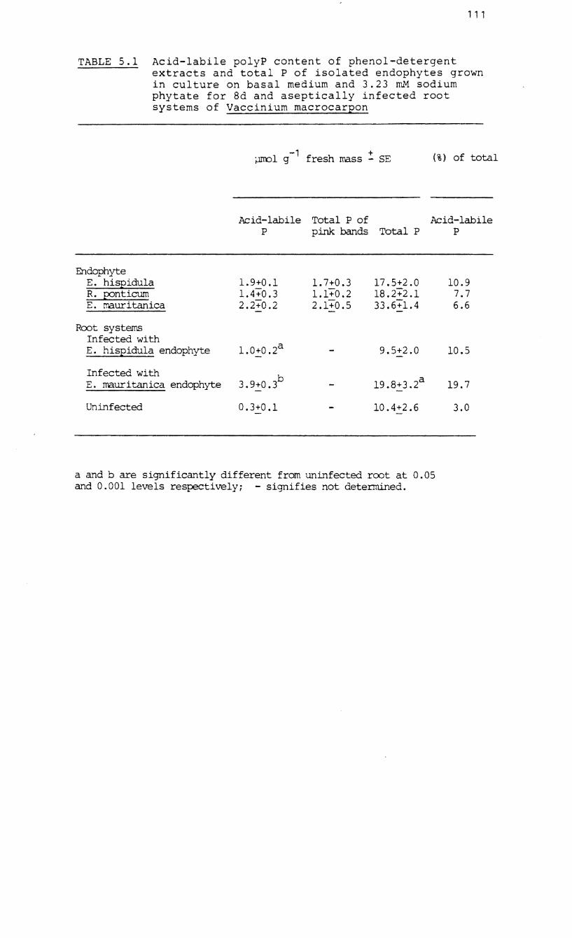
111
TABLE 5.1 Acid-labile polyP content of phenol-detergent extracts and total P of isolated endophytes grown in culture on basal medium and 3.23 roM sodium phytate for 8d and aseptically infected root systems of Vaccinium macrocarpon
~l g-1 fresh mass! SE (%) of total
Acid-labile Total P of Acid-labile P pink bands Total P P
Endophyte E. hispidula 1.9+0.1 1. 7+0.3 17.5+2.0 10.9 R. p:?nticum lA+0.3 1.1+0.2 18.2"+2.1 7.7 E. maur itanica 2.2"+0.2 2.1+0.5 33.6+1.4 6.6
Root systems Infected with E. hispidula endophyte 1.0+0.2 a 9.5+2.0 10.5
Infected with b E. mauritanica endophyte a 19.7 3.9+0.3 19.8+3.2
Un infected 0.3+0.1 10.4+2.6 3.0
a and b are significantly different from uninfected root at 0.05 and 0.001 levels respectivelYi - signifies not determined.
112
endophytes of E. hispidula and E. mauritanica and after
three months incubation, the proportions of acid-labile
polyP of total P were 10 and 20%, respectively. Infection
with E. mauritanica endophyte also resulted in a two-fold
increase in the total P of the root system when compared
with uninfected roots, which had a low acid-labile P content
of 3% of the total P. Infection with the endophyte of E.
hispidula did not result in a similar increase in the total
P of the root system.
5.2.4 The effect of P starvation on the endogenous P status
of isolated endophytes of E. hispidula grown on high and low
levels of orthophosphate
When mycelia grown on high and low levels of KH PO were 2 4
starved of P for four days and total P levels in fractions
assessed, the levels of TCA-soluble orthophosphate rose
rapidly from day one in both sets of mycelia although in the
low P-fed mycelia this fraction declined after three days
(Fig. 5.7). In contrast, negligible amounts of P in the
form of TCA-insoluble orthophosphate were present in either
the high or low P-fed mycelia. A high amount of TCA-soluble
polyP was present .in the high P-fed mycelia at day zero but
then declined. TCA-soluble polyP was negligible in the low
P-fed mycelia. A similar trend was found in the TCA-
insoluble polyP fraction although with a lower content in
the high P-fed mycelia. The TCA-soluble non-labile P
fractions showed low levels but with significant differences
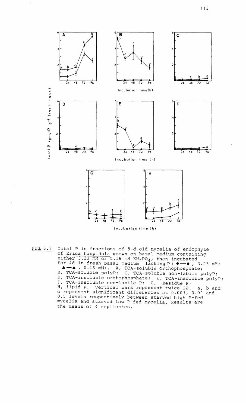
113
6 6 C
C
4
a
2 b 2 t c c ~
24 48 72 96 24 48 72 96 24 48 72 96
VI Incubation time(h) VI
0 E 6 6 6 ~ 0 E F '" G.I
4 4 i b
O'l
a. a
'0 2 2 2 E a =>..
a. a ... --0 24 48 72 96 24 24 48 72 96 -0 I- Incubation time ( h)
6 6 G H
4 4
C 2
~ 1 ~ 1 1 . "T ..i. 1:
24 48 72 96 24 48 72 96
Incubation time(h)
FIG.5.7 Total P in fractions of 8-d-old mycelia of endophyte of Erica hispidula grown on basal medium containing either 3.23 m.a or 0.16 mM KH 2P04 , then incubated for 4d in fresh basal medium lacking P ( • -., 3.23 ITh."1;
A-A I 0.16 roM). A, TCA-solub orthophosphate; B, TCA-soluble polyP: C, TCA-soluble non-labile polyP; D, TCA-insoluble orthophosphate; E, TCA-insoluble polyP; F, TCA-insoluble non-labile P; G, Residue Pi H, lipid P. Vert al bars represent twice 3c. a, band c represent significant differences at 0.001, 0.01 and 0.5 levels respectivelv between starved high P-fed mycelia and starved low P-fed mycelia. Results are the means of 4 replicates.
114
between high and low P-fed mycelia. Levels of the TCA-
insoluble non-labile P fractions were also low. Levels of
residue P were low with little variation over time.
Significantly different amounts of lipid P in high and low
P-fed mycelia were present at day zero but there appeared a
similar pattern of accumulation with time
5.2.5 Activated charcoal adsorption of possible contaminants
of BaC12 precipitates
The addition of activated charcoal to the TCA-soluble and
insoluble filtrates of homogenates prior to precipitation by
BaCl led to a significant increase in TCA-soluble non-2
labile P fractions when compared with untreated samples
(Table 5.2). No significant differences were found between
the levels of labile polyP fractions of treated and
untreated samples which suggests little contamination of
BaCl precipitates by other labile P compounds. 2
5.2.6 Phytic acid content of mycelia of the endophyte of E.
hispidula
Phytic acid in seven-day-old mycelia grown on basal medium
containing 3.23 mM KH PO was determined as 1.26 + 0.21 2 4
-1 umol P 9 fresh mass which represented 6.2% of the total P.
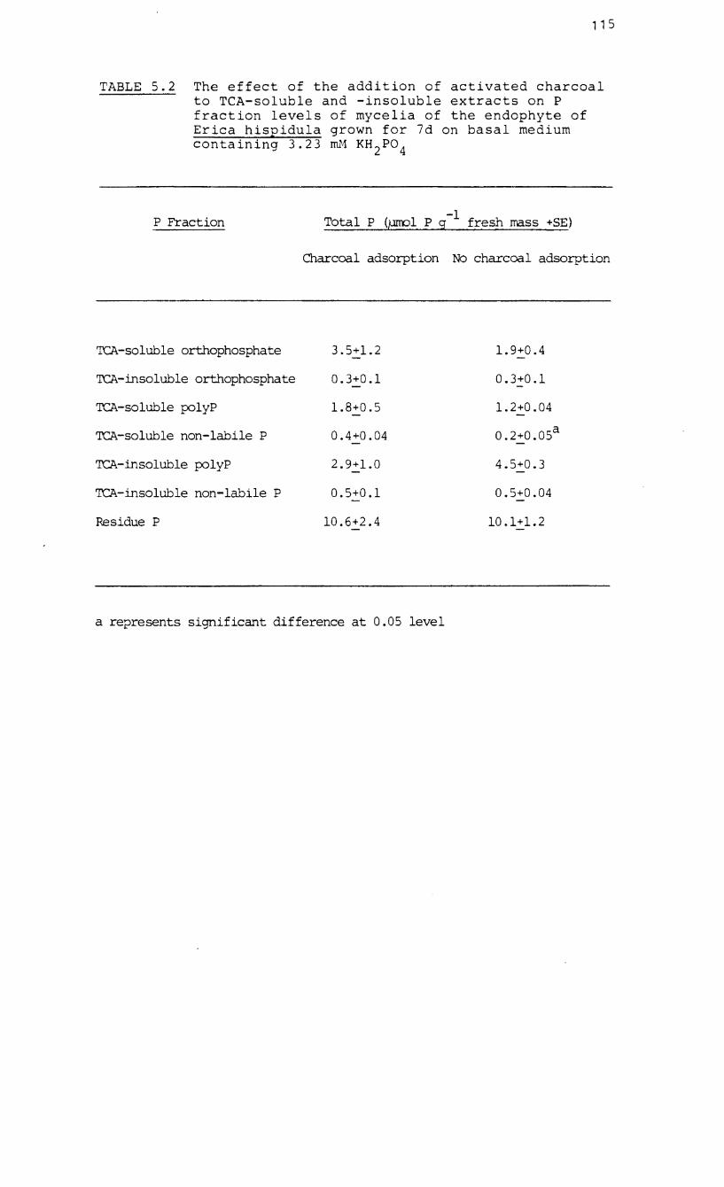
115
TABLE 5.2 The effect of the addition of activated charcoal to TeA-soluble and -insoluble extracts on P fraction levels of mycelia of the endophyte of Erica hispidula grown for 7d on basal medium containing 3.23 mH KH 2P04
P Fraction -1 Total P (ernol P g fresh mass +8E)
Charcoal adsorption No charcoal adsorption
TCA-soluble orthophosphate 3.5+1.2 1. 9+0.4
TCA-insoluble orthophosphate 0.3+0.1 0.3+0.1
TCA-soluble polyP 1.8+0.5 1. 2+0 .04
TCA-soluble non-labile P 0.4+0.04 0.2+0.05 a
TCA-insoluble polyP 2.9+1.0 4.5+0.3
TCA-insoluble non-labile P 0.5+0.1 0.5+0.04
Residue P 10.6+2.4 10.1+1.2
a represents significant difference at 0.05 level
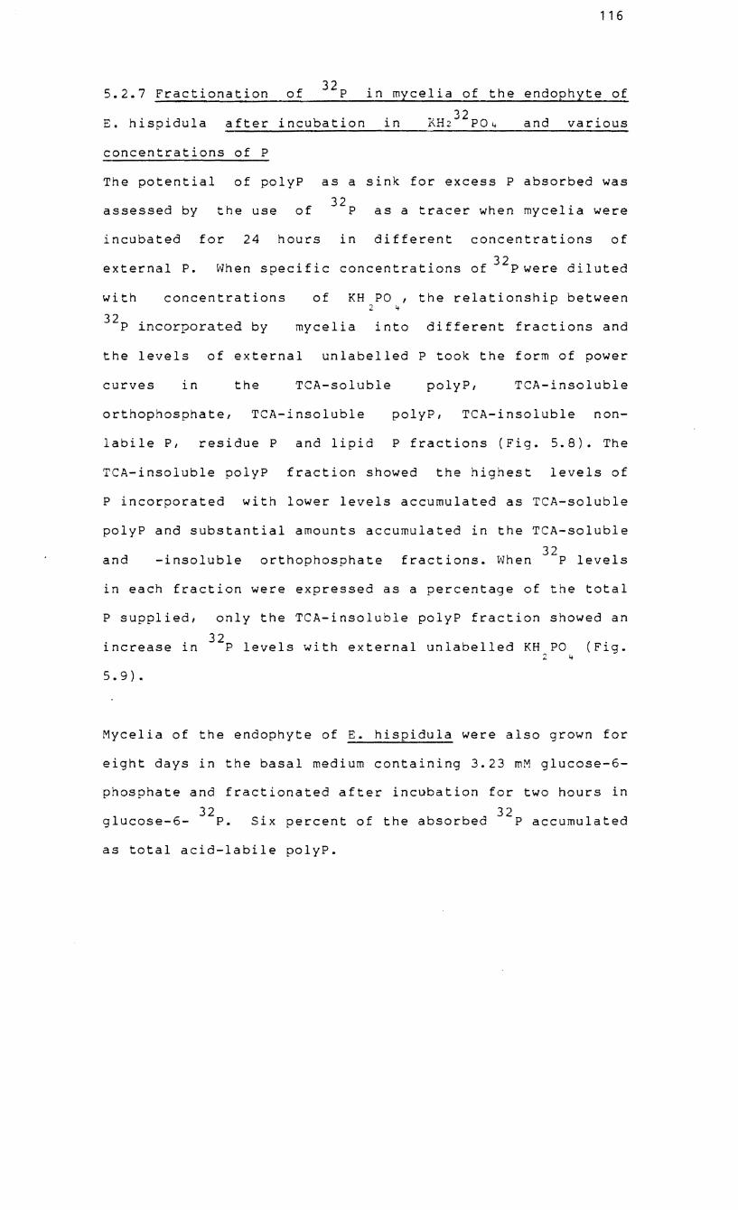
116
5.2.7 Fractionation of 32p in mycelia of the endophyte of
E. hispidula after incubation in RH232
p04 and various
concentrations of P
The potential of polyP as a sink for excess P absorbed was
assessed by the use of 32p as a tracer when mycelia were
incubated for 24 hours in different concentrations of
external P. When specific concentrations of 32pwere diluted
with concentrations of KH PO , the relationship between 2 4
32p incorporated by mycelia into different fractions and
the levels of external unlabelled P took the form of power
curves in the TeA-soluble polyP, TeA-insoluble
orthophosphate, TeA-insoluble polyP, TeA-insoluble non-
labile p, residue P and lipid P fractions (Fig. 5.8). The
TeA-insoluble polyP fraction showed the highest levels of
P incorporated with lower levels accumulated as TeA-soluble
polyP and substantial amounts accumulated in the TeA-soluble
and -insoluble orthophosphate 32
fractions. When P levels
in each fraction were expressed as a percentage of the total
P supplied, only the TeA-insoluble polyP fraction showed an
increase in 32p levels with external unlabelled KH PO 2 4
(Fig.
5.9).
Mycelia of the endophyte of E. hispidula were also grown for
eight days in the basal medium containing 3.23 mM glucose-6-
phosphate and fractionated after incubation for two hours in
32 32 glucose-6- P. Six percent of the absorbed P accumulated
as total acid-labile polyP.
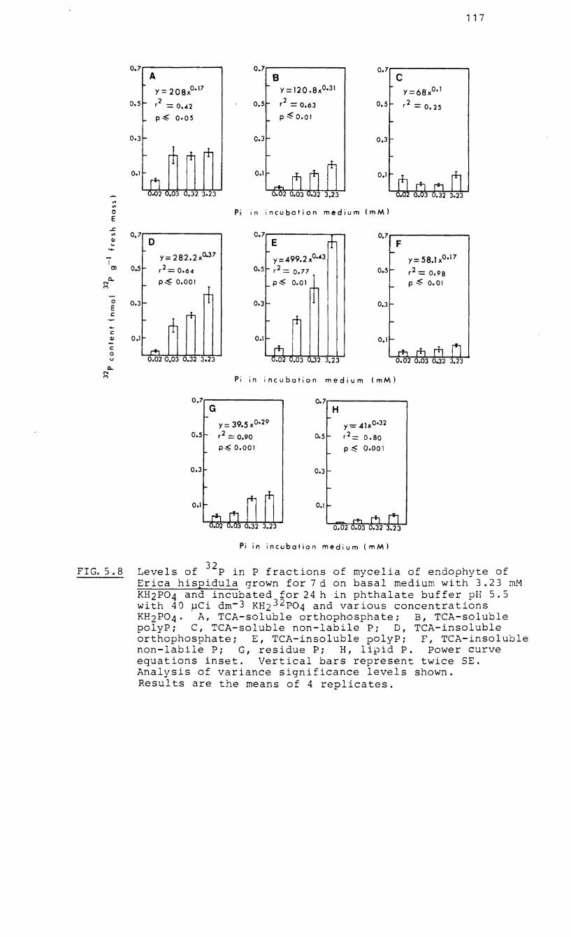
117
0.7 0.7 0.7 A B C
Y = 208xo. 17 Y = 120 .8xO.31 Y=68xO. 1
0.5 2 r = 0.42 0.5 r2 = 0.63 0.5 r2 = 0.25
p~ 0.05 P ~ 0.01
0.3 0.3 0.3
0.1 0.1 0.1
'" '" 0 Pi in incubation medium (mM) E
..t:.
'" 0.7 0.7 + 0.7 ill 0 E F .. ....
1 Y= 282.2xo.J7 y=499.2xO.43 y=58.lx°.17
Cl 0.5 r2 = 0.64 0.5 r2 = 0.77 0.5 r2 == 0.98 Q. p~ 0.001 p~ 0.01 P ~ 0.01 N
<"I
-t 0 0.3 r- 0.3 0.3 E c:; r+ r+ - + c:; ill 0. 1 - 0.1 f- 0.1
r'l c:; ,..., 0 v 0.02 0.03 0.32 3.23 0.02 t .03 .32 3.23
Q. N <"I Pi in incubation medium (mM)
0.7 0.7 G H
y = 39.5 xO.29 y=41xo.32
0.5 r2 == 0.90 0.5 r2 0.80 p~ 0.001 p~ 0.001
0.3 0.3
0.1
Pi in incubation medium (mM)
FIG.5.8 Levels of 32p in P fractions of mycelia of endophyte of Erica hispidula grown for 7 d on basal medium with 3.23 m..rvt KH2P04 and incubated for 24 h in phthalate buffer pH 5.5 with 40 pCi dm- 3 KH232p04 and various concentrations KH2P04' A, TCA-soluble orthophosphate; B, TCA-soluble polyP; C, TCA-soluble non-labile Pi D, TCA-insoluble orthophosphate; E, TCA-insoluble polyP; F, TCA-insoluble non-labile Pi G, residue Pi H, lipid P. Power curve equations inset. Vertical bars represent twice SE. Analysis of variance significance levels shown. Results are the means of 4 replicates.
118
50 A
50.....-------B
50..-------C
40 40 40
30 30 30
20 r- r-
20 20 r0-
10 n 10
0.02 0.03 0.32 3.23
Pi in incubation medium (mM) 0 -
50,.--------. 0 50 -- 0
50 E F
0 40 40 40 -0
30 0- -30 30 r-
'" 0 20 r-- r-- r-- 20 20 r--c;; 10
II> -c;; n 10 10
0 0.02 0.03 0.32 3.23 0.02 0.03 0.32 3.23 v
0... N
Pi in incubation medium (mM) M
50~------- 50,..---------, G H
40 40
30 30
20 20
10 10
Pi in inc u bot ion me diu m (m M )
FIG. 5.9 Percentage of 32p incorporated into P fractions of mycelia of endophyte of Erica hispidula grown for 7 d on basal medium containing 3.23 mM KH 2P0 4 and incubated for 24 h in phthalate buffer pH 5.5 with 40 rCi dm- 3 KH232p04 and various concentrations KH2P04' A, TCA-soluble orthophosphate: B, TCAsoluble polyPi C, TCA-soluble non-labile P; D, TCA-insoluble orthophosphate; E, TeA-insoluble polyP; F, TCA-insoluble non-labile Pi G, residue Pi H, lipid P. The results are the means of 4 replicates.
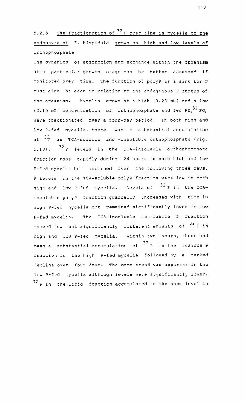
119
5.2.8 The fractionation of 32 P over time in mycelia of the
endophyte of E. hispidula grown on high and low levels of
orthophosphate
The dynamics of absorption and exchange within the organism
at a particular growth stage can be better assessed if
monitored over time. The function of polyP as a sink for P
must also be seen in relation to the endogenous P status of
the organism. Mycelia grown at a high (3.23 mM) and a low
(0.16 mM) concentration of 32 orthophosphate and fed KH2 P04
were fractionated over a four-day period. In both high and
low P-fed mycelia, there was a substantial accumulation
of 3~ as TCA-soluble and -insoluble orthophosphate (Fig.
5.10). 32 P levels in the TCA-insoluble orthophosphate
fraction rose rapidly during 24 hours in both high and low
P-fed mycelia but declined over the following three days.
P levels in the TeA-soluble polyP fraction were low in both
high and low P-fed mycelia. Levels of 32 P in the TCA-
insoluble polyP fraction gradually increased with time in
high P-fed mycelia but remained significantly lower in low
P-fed mycelia. The TCA-insoluble non-labile P fraction
showed low but significantly different amounts of 32 P in
high and low P-fed mycelia. Within two hours, there had
been a substantial accumulation of 32 P in the residue P
fraction in the high P-fed mycelia followed by a marked
decline over four days. The same trend was apparent in the
low P-fed mycelia although levels were significantly lower.
32 P in the lipid fraction accumulated to the same level in
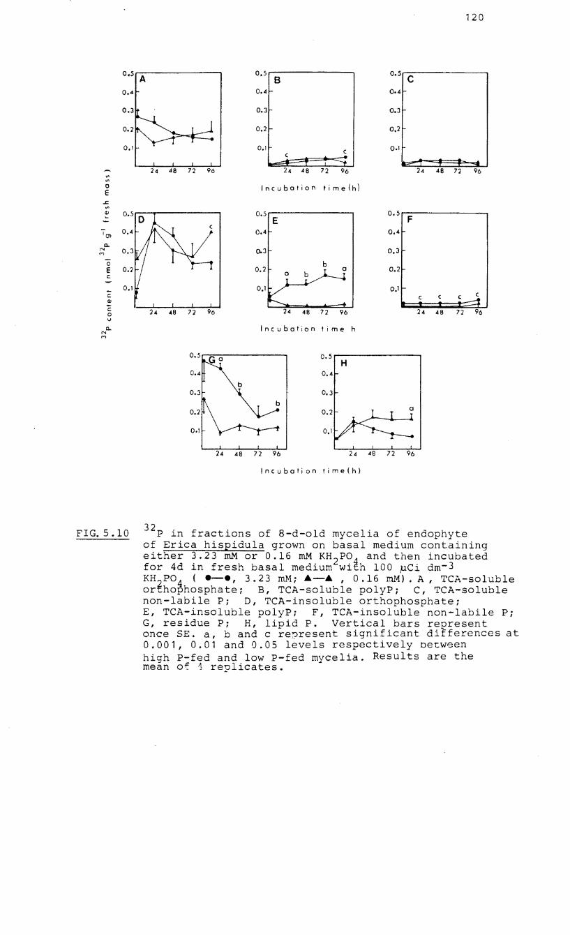
120
0.5 A 0.5 ,.----------, B
0.5 C
0.4 0.4 0.4
0.3 0.3 0.3
0.2 t- 0.2
0.1 0.1 c
l2"":: 24 48 72 96 24 48 72 96 24 48 72 96
'" on 0 E
Incubation time(h)
..r:: '" Q.J ~
0.5...--------, E
0.5 r=-------, F
10) 0.4 0.4
Q.. N M 0.3 0.3
0 E
b 0.2
c:
- 0.1 t-c: Q.J
c c
C 24 <18 72 96 0 48 72 96 24 4B 72 96 I,J
Q.. N
Incubation time h M
0.5,....--------, H
0.4
0.3
24 48 72 96 24 48 72 96
Incubation time(h)
FIG.5.10 32p in fractions of 8-d-old mycelia of endophyte of Erica hispidula grown on basal medium containing either 3.23 mM or 0.16 mM KH 2 P04 and then incubated for 4d in fresh basal medium with 100 pCi dm- 3 KH 2P04 ( .-., 3.23 mM; A-A , 0.16 mM). A, TCA-soluble orthophosphate; B, TCA-soluble polyP; C, TCA-soluble non-labile Pi D, TCA-insoluble orthophosphate; E, TCA-insoluble polyP; F, TCA-insoluble non-labile Pi G, residue Pi H, lipid P. Vertical bars represent once SE. a, band c re?resent significant differences at 0.001, 0.01 and 0.05 levels respectively between high P-fed and low P-fed mycelia. Results are the mean of 1 replicates.
121
high and low P-fed mycelia in 24 hours and thereafter
remained either constant (low P) or declined (high P).
5.3 DISCUSSION
The presence of polyP in pure cultures of the endophyte and
mycorrhizal root systems of ericaceous seedlings has been
confirmed. The metachromatic granules resemble closely
those of ectomycorrhizas of eucalypts and pines (Ashford et
al.! 1975; Ling-Lee et al., 1975; Chilvers and Harley,
1980) and vesicular-arbuscular mycorrhizas (Cox et al.,
1975). The range of granule sizes were similar to those of
beech mycorrhizas incubated in I mM orthophosphate and the
sensitivity of granule numbers to the concentration of the
external P source is similar to the pattern observed in
beech mycorrhizas
culture (Chilvers
The decline in
and an ectomycorrhizal fungus in pure
and Harley, 1980: Lapeyrie et al., 1984)
granule numbers after an organic P
concentration of 0.32 mM may be due to the end-product
inhibition of the phosphatase enzymes (Pearson and Read,
1975) which would have reduced the amount of P available for
uptake. The accumulation of granules in hyphae in media
without the addition of an external P source was most likely
due to the presence of residual P in the yeast extract of
the basal medium. The polyP granules accumulated during the
lag phase of growth. In Corynebacterium xerosis (Neiss. et
Kusch.) Bergey, numbers of granules increased during the lag
122
phase but declined during the exponential phase (Hughes and
Muhammed, 1962).
The phenol-detergent extraction technique of Callow et ale
(1978) was used to isolate polyP and nucleic acid in a
relatively undegraded form and the polyP was finally
characterized by means of polyacrylamide gel
electrophoresis. Callow e tal. (1978) were able to
determine the molecular weight and chain length of the polyP
in VA mycorrhizas as polyphosphates have a constant charge
to mass ratio and their mobility in the gels is a function
of their molecular size. In this study, the molecular
weights of 3 000 to 4 700 from ericoid endophytes were
considerably lower than the figure of 20 800 for the polyP
of vesicular-arbuscular mycorrhizal fungi (Callow et al.,
1978) although the possibility of chain degradation during
extraction should not be discounted. The proportion of acid-
labile polyP to total P in the phenol-detergent extract of
the cultured endophyte was 7 to 11% which is in the same
range estimated by Capaccio and Callow (1982) in phenol
detergent extracts of isolated internal vesicular-arbuscular
endophytes. The polyP content of ericoid mycorrhizal root
systems was significantly greater than that of the non-
mycorrhizal controls indicating the polyP to be largely
fungal in origin. The higher proportion of polyP in the
root systems inoculated with . the endophyte from E.
mauritanica could have been due to a heavier mycorrhizal
infection observed. However, as the higher total P content
123
of these infected seedlings was correlated with a high
concentration of P in the isolated endophyte, the
mycorrhizal fungus of E. mauritanica may also be able to
accumulate more phosphorus.
When high P-fed mycelia of the endophyte of E. hispidula
were starved of P, the initially high levels of TeA-soluble
and -insoluble polyP fell rapidly with a concomitant rise in
TeA-soluble orthophosphate. Low P-fed mycelia contained
negligible polyP and 32p orthophosphate, when supplied, was
not tranformed into polyP. These trends confirm the
potential of the molecule to be a form of P storage when P
is in surplus and to release P into the orthophosphate
fraction under conditions of P deprivation. Mycelia showed
an accumulation and transformation of 32p into various
fractions which was enhanced by external P supply. Only the
TeA-insoluble polyP
increased levels of
(long-chain polyP) fraction showed
32p as a proportion of the total P
(Figs. 5.8 and 5.9). This trend suggests a very rapid
incorporation of P into TeA-insoluble polyP at the expense
of the other fractions. Evidence by Juni et ala (1947),
Wiame (1949), Harold (1962) and Harold (1966) indicated that
acid-insoluble polyP is a more active form with a higher
turnover rate than acid-soluble polyP. However, the rates of
turnover of the polyP pools may be the same but they stand
in a 'precursor-product' relationship with the longer-chain
molecules being formed first (Harold, 1966). When high P-fed
124
mycelia were fed 32p alone, accumulation occurred rapidly in
the residue P fractions (Fig. 5.10) which has been shown to
contain nucleic acids and phosphoproteins (Schmidt and
Thannhauser,1945). This fraction then became depleted in
favour of TCA-insoluble orthophosphate and TCA-insoluble
polyP synthesis. In many microorganisms, the interruption
of nucleic acid synthesis (usually by the absence or
depletion of an essential metabolite) is directly linked to
a concomitant rise in polyP synthesis and vice versa
(Harold, 1962; Harold and Sylvan, 1963; Harold, 1966). It
is unlikely that essential nutrients became exhausted from
fresh basal media after only 24 hours and to suggest reasons
for the cessation of 32p accumulation in the residue
fractions would be speculative in the absence of further
empirical evidence.
High levels of acid-soluble polyP (67% of total P prior to
starvation period) were found in fractions in comparison
with levels determined in the phenol-detergent extracts.
The difference in P source (orthophosphate as opposed to
sodium phytate) may have accounted for part of the
discrepancy since the endophyte does not grow equally well
on both sources (Fig. 3.1). However, it is more likely that
the difference in the extraction techniques was the primary
factor contributing to discrepancies in polyP levels. Levels
of 32p incorporated into acid-labile fractions (23 to 46%
of total 32 ) . h Pare 1n t e same range as those of excised
125
beech mycorrhizas (Harley and McCready, 1981). Martin et
al. (1983) and Rolin et al.(1984) found low levels of polyP
(9 to 17%) in the acid-soluble barium acetate precipitate of
ectomycorrhizal fungi but none in the acid-insoluble
precipitate and polyP levels were found to be unrelated to
external P levels. The extraction procedure used by these
workers included determinations of RNA and DNA levels and
specific identification of polyP by 31p NMR (nuclear
magnetic resonance) and thin-layer chromatography. Acid-
labile nucleotides, nucleic acids or sugar phosphates are
able to contaminate TCA extracts and nucleic acids may form
complexes with the acid-insoluble polyP precipitate (Harold ,
1966). Although the fractionation experiments did not
include adsorption of contaminants by activated charcoal
prior to barium chloride precipitation, a significant
difference was only found in the TCA-soluble non-labile P
fraction when control extractions were made, and thus the
data remained uncorrected. Despite the acid-labile test for
polyP fractions, the extraction and fractionation procedure
of Aitchison and Butt (1973) is not equivocal without
chromatography and metachromasy (Bieleski , 1973) and polyP
estimates in these fractions should not be regarded as
conclusive. Nevertheless, biochemical and histochemical
evidence emphasizes the potential importance of polyP in the
overall P metabolism of ericoid endophytes. The endophyte
also shows an ability to utilise glucose-6-phosphate and
128
be an important inhibiting factor. In the fynbos
vegetation, there is a seasonal pattern in nitrogen
mineralization (Stock et al., Stock and Lewis, in press)
and total phosphorus levels in the Clovelly soil (Mitchell
et al., 1984). Decomposition of the leaf litter does not
show a seasonal pattern but its rate is slow (Mitchell et
al., in press). The slow decomposition rates are attributed
to the poor quality of litter which is due to its high
lignin content and C : N ratios (Mitchell et al., in press).
Although the phosphorus contents of the acidic sandy soils
are low, the bulk of it is organically bound (27-60%) and
thus acid phosphatases secreted by the microorganisms would
be important in the catalytic release of orthophosphate from
bound complexes. In this study, the endophyte was isolated
from the fine hair roots of E. hispidula and E. mauritanica
and grown in culture. The former was shown to possess active
fractions of acid phosphatase, particularly the enzymes
either associated with the cell surface or released into the
external medium (Table 3.1). The optimum pH of phosphatase
activity (i.e. pH 2.0 - 6.0) was within the range for the
fynbos sandy soil (Mitchell et al., 1984), and the enzymes
showed a wide substrate specificity (Table 3.3). Under
field conditionsl the endophyte would be able to achieve
maximal access to organic P substrates during the periodic
cycles of favourable environmental conditions. The
extramatrical hyphae acting as physical extensions of the
130
a 1. I 1983). On these strongly-leached, acidic soils,
winter- spring is probably the main period of nutrient
release from decomposing litter, but this has not been
substantiated. Read (1978) has noted that in the
heathlands of the Cape, mycorrhizal infection of Erica spp.
is most active during the spring but negligible during the
late summer months at a time when many species of Erica spp.
flower and set seed (Pierce, 1984). Duddridge and Read
(1982b) have shown that the longevity of the mycorrhizal
association in cells of Rhododendron ponticum only lasted
approximately seven weeks. If this estimate is true for
Erica spp. in the fynbos, then the advantages gained from
maximal mineralization and absorption of phosphates will be
realised through rapid translocation of P to the
intracellular hyphae and then the storage of excess P for
release to the host plant during the period of both shoot
extension and flower production.
The potential role of polyP as a storage molecule in ericoid
mycorrhizas was confirmed in this study. There were
substantial differences in the levels of total polyP between
mycelia of the endophyte of E. hispidula grown on a high P
and a low P medium (Fig. 5.7). In high P-fed mycelia polyP
levels declined during P starvation with a concomitant rise
in the levels of orthophosphate. If 8-day-old mycelia were
fed increasing concentrations of P I labelled with 32
P
orthophosphate, the acid-insoluble polyP fraction showed the
131
best concomitant increase in P levels (Figs. 5.8 and 5.9).
Mycorrhizal seedlings contained significantly higher levels
of polyP than non-mycorrhizal seedlings (Table 5.1). These
results suggest the operation of a polyP cycle (Fig. 1.1)
whereby absorbed P is initially incorporated into long-chain
acid-insoluble polyP which is broken down to shorter chains
of acid-soluble polyP (Harold, 1966). Finally,
orthophosphate is produced for transfer across the
hyphae/host interface. During the late summer and autumn of
the south western Cape, when mycorrhizal infections of the
root system of Erica spp. diminish and the carbohydrate
drain on the host is reduced, nutrients are probably being
directed to the flowers either from the root system or
internally cycled from the leaves prior to leaf fall.
As well as the South African endophytes, polyP was
identified in the European endophytes with granules
accumulating in the exponential phase of growth and in
response to the concentration of P in the external medium
(Figs. 5.3 and 5.4). However,there appear to be fundamental
differences in the P nutrition between South African and
European endophytes. The South African endophyte of E.
hispidula: (1) possessed higher activities of acid
phosphatases (especially the extracellular fraction), (2)
took a shorter time to reach the stationary phase of growth
in culture, (3) showed a different ability to use phytates
as a P source and (4) has shown a greater ability to use
\
132
inositol as a carbon source (Mitchell and Read, 1985).
These differences may be of an adaptive nature, related to
differences in moisture regimes, soil organic matter and
nutrient levels between northern and
heathlands and further research
validity. The apparent low phytase
southern hemisphere
will establish their
ability of the acid
phosphatases of the ericoid endophyte and the absence of an
alkaline phosphatase suggests that the enzyme physiology of
ericoid mycorrhizas may not be the same as that of either
ectomycorrhizas or VA mycorrhizas. Studies on the enzymes
involved in polyphosphate metabolism have been undertaken on
VA mycorrhizas (Capaccio and Callow, 1982) but research in
this field on ericoid mycorrhizas needs to be pursued. The
studies of Read and Stribley (1973), Stribley and Read
(l974b, 1976, 1980) indicate the ability of ericoid
mycorrhizas to utilise organic sources of nitrogen which the
host cannot directly use and underlines the importance of
the mycorrhizal association in the nitrogen nutrition of
ericaceous plants. This study has concentrated on P
utilization by ericoid endophytes in culture but confirms
their importance in the P nutrition of Erica spp. This will
have to be studied further by synthesising mycorrhizal
root systems of the original host plant which will enable
the distribution patterns of P within the host plant to be
investigated.
133
REFERENCES
Aitchison, R.A. & Butt, V.S. (1973). The relation between the synthesis of inorganic polyphosphate and phosphate uptake by Chlorella vulgaris. Journal of Experimental Botany, 24 (80), 497-510.
Alexander, LJ. & Hardy, K. (1981). Surface phosphatase activity of Sitka spruce mycorrhizas from a serpentine site. Soil Biology and Biochemistry, 13, 301-305.
Allen, S.E., Grimshaw, H.M. Parkinson, J.A. & Quarmby, G. (1974). Chemical Analysis of Ecological Materials. Blackwell Scientific Publications, Oxford.
Anderson, G. (1967). Nucleic acid derivatives and organic phosphates. In Soil Biochemistry (Ed. by A.D. McLaren & G.H. Peterson), pp. 67-89. Marcel Dekker, Inc., N.Y.
Anderson, G., Williams, E.G. comparison of the sorption and inositol hexaphosphate of Soil Science, 25, 51-62.
& Moir, J. (1974). A of inorganic orthophosphate by six acid soils. Journal
Appiah, M.R. & Thomas, R.L. (1982). Inositol phosphate and organic phosphorus contents and phosphatase activity of some Canadian and Ghanaian soils. Canadian Journal of Soil Science, 62, 31-38.
ld . (197 ) 3+. d Arno , W.N. & Garrlson, R.G. 9. An Fe -actlvate acid phosphatase in Saccharomyces rOUXll. Journal of Biological Chemistry, 254 (11), 4919-4925.
Ashford, A.E., Ling-Lee, M. & Chilvers, G.A. (1975). Polyphosphate in Eucalypt mycorrhizas : a cytochemical demonstration. The New Phytologist, 74, 447-453.
Bartlett, E.M. & Lewis, D.H. (1973). Surface activity of mycorrhizal roots of beech. and Biochemistry, 5, 249-257.
phosphatase Soil Biology
Basha, Sheikh Mehboob. (1984). Purification and characterization of (Arachis hypogaea) 62, 385-391.
an acid phosphatase from peanut seed. Canadian Journal of Botany,
Beever, R.E. & Burns, D.J.W. (1977). Adaptive changes in phosphate uptake by the fungus Neurospora crassa in response to phosphate supply. Journal of Bacteriology, 132(2), 520-525.
134
Beever, R.E. & Burns, D.J.W. (1980). Phosphorus uptake, storage and utilization by fungi. In Advances in Botanical Research, vol. 8 (Ed. by H.W. Woolhouse), pp. 127-219. Academic Press, London.
Bennett, J. in the wheat 198.
& Scott, K.J. (1971). Inorganic polyphosphates wheat stem rust fungus and in rust-infected
leaves. Physiological Plant Pathology, I, 185-
Bieleski, R.L. (1973). Phosphate pools, phosphate transport and phosphate availability. Annual Review of Plant fhysiology, 24, 225-252.
Bojovic-Cvetic, D. & Vujicic, R. (1982). Acid phosphatase localisation and distribution in Aspergillus flavus. Transactions of the British Mycological Society, 79(1), 137-141.
Bonfante-Fasola, P. & Gianinazzi-Pearson, V. (1979). Ultrastructural aspects of endomycorrhiza in the Ericaceae. I. Naturally infected hair roots of Calluna vulgaris L. Hull. The New Phytologist, 83, 739-744.
Bonfante-Fasola, P., Berta, G. & Gianinazzi-Pearson, V. (1981). Ultrastructural aspects of endomycorrhizas in the Ericaceae. II. Host-endophyte relationships in Vaccinium myrtillus L. The New Phytologist, 89, 219-224.
Bonfante-Fasola, P. & Gianinazzi-Pearson, V. (1982). Ultrastructural aspects of endomycorrhiza in the Ericaceae. III. Morphology of the dissociated symbionts and modifications occurring during their reassociation in axenic culture. The New Phyto1ogist, 91, 691-704.
Borst-Pauwels, G.W.F.H. (1976). Discrimination criteria for apparent two-site transport models. Journal of Theoretical Biology, 56, 191-204.
Bowen, G.D., Bevege, G.I. & Mosse, B. (1975). Phosphate physiology of vesicular-arbuscu1ar mycorrhizae. In Endomycorrhizas (Ed. by F.E. Sanders, B. Mosse & P.B. Tinker), pp. 241-260. Academic Press, London and New York.
Burns, D.J.W. & Beever, R.E. (1977). Kinetic characterization of the two phosphate uptake systems in the fungus Neurospora crassa. Journal of Bacteriology, 132(2), 511-519.
Burnsl D.J.W. & Tucker, S.A. (1977). fitting methods for the sum application to uptake studies. Biochemistry, 81, 45-52.
An evaluation of two hyperbolas
European Journal
!35
of
of
Callejal M' I Mousain, D., Lecouvreur, B. and d'Auzac, J. (1980). Influence de la carence phosphatee sur les activites phosphatases acides de trois champignons mycorrhiziens. Hebeloma edurum Metrod., suillus ~ranulatus (L ex FR.) O. Kuntze et Pisolithus tinctorius (Pers.) Coker et Couch. Physiologie Vegetale 18(3), 489-504.
Calleja, M. & d'Auzac, J. (1983). Activites phosphatases et carence phosphatee chez des champignons superieurs. Canadian Journal of Botany, 61, 79-86.
Callow, J.A., Capaccio, L.C.M., Parish, G. & Tinker, P.B. (1978). Detection and estimation of polyphosphate in vesicular-arbuscular mycorrhizas. The New Phytologist, 80, 125-134.
Capaccio, L.C.M. & Callow, J.A. (1982). The enzymes of polyphosphate metabolism in vesicular-arbuscular mycorrhizas. !he New Phytologist, 91, 81-91.
Cheshire, M.V. & Andersonl G. (1975). Soil polysaccharides and carbohydrate phosphates. Soil Science, 119, 356-362.
Chilvers, G.A. & Harley, J.L. (1980). Visualization of phosphate accumulation in beech mycorrhizas. The New Phytologist, 84, 319-326.
Chilversl G.A., Lapeyrie, F.F. & Douglass, P.A. (1985). A contrast between Oomycetes and other taxa of mycelial fungi in regard to metachromatic granule formation. The New Phytologist l 99 1 203-210.
Cooper, K.M. & Tinkerl P.B. (1981). Translocation and transfer of nutrients in vesicular-arbuscular mycorrhizas. IV. Effect of environmental variables on movement of phosphorus. The New Phytologist, 88 1 327-339.
Cornish-Bowden, A. & Eisenthal, R. (1974). Statistical considerations in the estimation of enzyme kinetic parameters by the direct linear plot and other methods. Biochemical Journal, 139 1 721-730.
Cosgravel D.J. (1967). Metabolism of organic phosphates in soil. In Soil Biochemistry (Ed. by A.D. McLaren & G.H. Peterson), pp. 216-228. Marcel Dekker, Inc., N.Y.
Cosgrave, D.J. Chemistry, Studies in Publishing
(1980). Inositol phosphates Biochemistry and Physiology. Organic Chemistry. Elsevier
Company, Amsterdam.
136
their Vol. 4.
Scientific
Cox, G. & Tinker, P.B. (1976). Translocation and transfer of nutrients in vesicular-arbuscular mycorrhizas. I. The arbuscle and phosphorus transfer: a guantative ultrastructural study. The New Phyotologist, 77, 371-378.
Cox, G., Moran, K.J., Sanders, P., Nockolds, G. & Tinker, P.B. (1980). Translocation and transfer of nutrients in vesicular-arbuscular mycorrhizas. III. Polyphosphate granules and phosphorus translocation. The New Phytologist, 84, 649-659.
Crane, R.K. (1958). Use of charcoal to separate mixtures of inorganic, ester and nucleotide phosphates. Science, 127, 285-286.
Cress, W.A., Throneberry, G.O. & Lindsay, D.L. (1979). Kinetics of phosphorus absorption by mycorrhizal and non-mycorrhizal tomato roots. Plant Physiology, 64, 484-487.
Davis, B.J. (1964). Disc electrophoresis application to human serum proteins. York Academy of Science, 121, 404-427.
II Method and Annals of New
Duclos, J.L. & Portin, active charcoal mycorrhizas with 94, 95-102.
J.A. (1983). on in vitro Vaccinium spp.
Effect of glucose and synthesis of ericoid
The New Phytologist,
Duddridge, J.A. and Read, D.J. (1982a). An ultrastructural analysis of the development of mycorrhizas in Monotropa hypopitys. L. The New Phytologist, 92, 203-214.
Duddridge, J. and Read, D.J. (1982b). An ultrastructural analysis of the development of mycorrhizas in Rhododendron ponticum. Canadian Journal of Botany, 60, 2345-2356.
EbeL J.P., Colas, J. & Muller, S. (l958a). Recherches cytochimigues sur les polyphosphates inorganigues contenus dans les organismes vivants. II. Mise au point de methodes de detection cytochimigues specifigues des polyphosphates. Experimental Cell Research, 15, 28-42.
Ebel, J.P., Colas, J. & Muller, S. (1958b). cytochimiques sur 1es polyphosphates contenus dans les organismes vivants. III. polyphosphates chez divers organismes Experimental Cell Research, 15, 36-42.
137
Recherches inorganiques Presence de inferieurs.
Ebe1, J.P. & Muller, S. (1958). Recherches cytochimiques sur les polyphosphates inor~aniques contenus dans les organismes vivants. I. Etude de la reaction de metachromasie et des colorations au vert de methyle et a la pyronine en fonction de la longueur de chaine des polyphosphates. Experimental Cell Research, 15, 21-28.
Eisenthal, R. & Cornish-Bowden, A. (1974). The direct linear plot. A new graphical procedure for estimating enzyme kinetic parameters. Biochemical Journal, 139, 715-720.
Epstein, E. (1966). Dual pattern of ion absorption by plant cells and by plants. Nature, 212, 1324-1327.
Epstein, E. & Elzam, O.E. (1963). Resolution of dual mechanisms of potassium absorption by barley roots. Proceedings of the National Academy of Science, U.S.A., 49, 684-692.
Epstein, E. & Hagen, C.E. (1952). absorption of alkali cations Physiology, 27, 457-474.
A kinetic study of the by barley roots. flant
Gianinazzi-Pearson, V. & Gianinazzi, S. (1976). Enzymatic studies on the metabolism of vesicular-arbuscular mycorrhiza. I. Effect of mycorrhiza formation and phosphorus nutrition on soluble phosphatase activities in onion roots. Physiologie Vegetale, 14, 833-841.
Gianinazzi-Pearson, V. & Gianinazzi, S. (1978) Enzymatic studies on the metabolism of vesicular-arbuscular mycorrhiza. II. Soluble alkaline phosphatase specific to mycorrhizal infection in onion roots. Physiological Plant Pathology, 12, 45-53.
Gianinazzi, S., Gianin~zzi-Pearson, V. and Dexheimer, J. (1979). Enzymatic studies on the metabolism of vesicular-arbuscular mycorrhiza. III. ultrastructural localization of acid and alkaline phosphatase in onion roots infected by Glomus mosseae (Nicol. and Gerd.) The New Phytologist, 82, 127-132.
Gi mi ngham, C. H. (1972). Ecology of Hea thlands. Chapman and Ha 11, London.
Good, R. (1974). The Geography of the Flowering Plants, 4th edition, Longman, London.
138
Goodman, J. & Rothstein, A. (1957). The active transport of phosphate into the yeast cell. Journal of General Physiology, 40, 915-921.
Greenacre, M.J. (1984). Correspondence Analysis.
Theory and Application of Academic Press, New York.
Groves, R.H. (1983). Nutrient cycling in Australian Heath and South African Fynbos. In Ecological Studies Vol. 43. Mediterranean-Type Ecosystems The Role of Nutrients (Ed. by F.J. Kruger, D.T. Mitchell & J.U.M. Jarvis), pp. 179-191. Springer-Verlag, Berlin.
Harley, J. L. (1969). The Biology of Mycorrhiza 2nd Edition. Leonard Hill, London.
Harley, J.L. & McCready, C.C. (1950) by excised mycorrhizas of the Phytologist, 49, 388-397.
Uptake beech.
of phosphate I. Th e New
Harley, J.L. & McCready C.C. (1952). Uptake of phosphate by excised mycorrhizas of the beech. III. The effect of the fungal sheath on the availability of phosphate to the core. The New Phytologist, 51, 343-348.
Harley, J.L. & McCready, accumulation in fagus Phytologist, 89, 75-80.
C.C. (1981). mycorrhizas.
Phosphate The New
Harley, J.L. & Smith, S.E. (1983). Mycorrhizal Symbiosis. Academic Press, London.
Harold, F.M. (1962). Depletion and replenishment of the inorganic polyphosphate pool in Neurospora crassa. Journal of Bacteriology, 83, 1047-1057.
Harold, F.M. (1966). Inorganic polyphosphates in biology: structure, metabolism and function. Bacteriological Reviews, 30(4), 772-794.
Harold, F.M. & Sylvan, S. (1963). Accumulation of inorganic polyphosphate in Aerobacter aerogenes. II. Environmental control and the role of sulphur compounds. Journal of Bacteriology, 86, 222-231.
Harrison, A.F. & Helliwell, D.R. (1979). A bioassay for comparing phosphorus availability in soils. Journal of Applied Ecology, 16, 497-505.
Hasegawa, Y., Lynn, K.R. & Brockbank, W.J. (1976). Isolation and partial characterization of cytoplasmic and wall-bound acid phosphatases from wheat roots. Canadian Journal of Botany, 54, 1163-1169.
Ho, r. and Zak, B. (1979). ectomycorrhiza1 fungi. 57,(11), 1203-1205.
139
Acid phosphatase activity of six Canadian Journal of Botany,
Hofstee, B.H.J. (1959). in enzyme kinetics.
Non-inverted versus inverted plots Nature, 184, 1296-1298.
Hughes, D.E. & Muhamned, A. (1962). The metabolism of polyphosphate in bacteria. Colloques International du Centre National de la Recherche Scientifigue (Paris), 106, 591--602.
Jennings, D.H. (1964). Changes in the size of orthophosphate pools in mycorrhizal roots of beech with reference to absorption of the ion from the external medium. The New Phytologist, 63, 181-193.
Juni, E., Kamen, M.D., Spiegelman, S. & Wiame, J.M. (1947). Physiological heterogeneity of metaphosphate in yeast. Nature, 160, 717-718. ---
Kempers, A.J. (1975). Determination of submicro-quantities of plant available phosphate in aqueous soil extracts. Plant and Soil, 42, 423-427.
Kruger, F.J. (1979). South African Heathlands. In Heathlands of the World Vol. 9A. Heathlands and Related Shrub1ands Specht, pp.19-80.
: Descri tive Studies (Ed. by R.L. Elsevier, Amsterdam.
Lapeyrie, F.F., Chilvers, G.A. & Douglass, P.A. (1984). Formation of metachromatic granules following phosphate uptake by mycelial hyphae of an ectomycorrhizal fungus. The New Phytologist, 98, 345-360.
Ling-Lee, M., Chilvers, G.A. & Ashford, A.E. (1975). Polyphosphate granules in three different kinds of tree mycorrhiza. The New Phytologist, 75, 551-554.
Lowendorf, H.S., Slayman, Phosphate transport characterization of system. Biochimica et
C.L. & Slayman, C.W. (1974). in Neurospora. Kinetic
a constitutive, low-affinity Biophysica Acta, 373, 369-382.
Lowendorf, H.S., Bazinet, G.F. & Slayman, C.W. (1975). Phosphate transport in·Neurospora. Derepression of a high-affinity transport system during phosphorus starvation. Biochimica et Biophysica Acta, 389, 541-549.
Lowry, O.H., (1951). reagent. 275.
Roseborough, N.J., Farr, A.L., & Randall, R.J. Protein measurement with the Folin phenol Journal of Biological Chemistry, 193, 265-
140
Martin, J.K. & Cartwright, B. (1971). The comparative plant availability of 32p myo-inositol hexaphosphate and KH 2
32p0 4 added to soils. Communication of Soil
Science and Plant Analysis, 2, 375-381.
Martin, F., Canet, D., Rolin, D., Marchal, J.P. & Larher, F. (1983). Phosphorus -31 nuclear magnetic resonance study of polyphosphate metabolism in intact ectomycorrhizal fungi. ~lant and Soil, 71, 469-476.
McLennan, E.l. (1935). Non-symbiotic development of seedlings of _E~p~a~c~r~l~'s ____ i_m~p_r_e_s_s~a Labill. The New Phytologist, 34, 55-63.
McNabb, R.F.R. (1961). Mycorrhiza in the New Zealand Ericales. Australian Journal of Botany, 9, 57-61.
Mitchell, D.T. and Read, D.J. (1981). Utilization of inorganic and organic phosphates by the mycorrhizal endophytes of Vaccinium macrocarpon and Rhododendron ponticum. Transactions of the British Mycological So:::iety, 76(2), 255-260.
Mitchell, D.T., Brown, G. & Jongens-Ro~erts, S.M. (1984). variation of forms of phosphorus in the sandy soils of coastal fynbos, south-western Cape. The Journal of Ecology, 72, 575-584.
Mitchell,D.T. & Read, D.J. (1985). Growth of African and European ericoid mycorrhizal endophytes on a range of substrates. Transactions of the British Mycological Society, 84, 355-357.
Mitchell,D.T., Coley, P.G.F., Webb, S. & Allsopp, N. (in press). Litter fall and decomposition processes in the coastal fynbos vegetation, south-western Cape, South Africa.
Moll, E.J. & Jarman, M.L. (1984), Clarification of the term fynbos. South African Journal of Science, 80(8), 351-352.
Muller, S. & Ebel, J.P. (1958). Etudes des polyphosphates condenses contenus dans divers champignons. Bulletin de la societe de chimie biologique, 40, 1153-1161.
Murphy, J. & Riley, J.P. (1962). A modified single solution method for the determination of phosphate in natural waters. Analytico Chimica Acta, 27, 31-36.
Neame, K.D. & Richards, T.G. (1972). Elementary Kinetics of Membrane Carrier Transport, pp.80-90. Blackwell Scientific Publications, Oxford.
141
~issen, Per. plants.
(1973). Kinetics of ion uptake in higher Physiologia Plantarum, 28, 113-120.
Nissen, Per. organic.
(1974). Uptake mechanisms inorganic and Annual Review of Plant Physiology, 25, 53-79.
Nye, P.H. & Tinker, P.B. (1977). Soil-Root System. Blackwell
Solute Movement in the Scientific Publications,
Oxford.
Pearson, V. & Read, D.J. (1973a). The biology of mycorrhiza in the Ericaceae. I. The isolation of the endophyte and synthesis of mycorrhizas in aseptic culture. The New Phytologist, 72, 371-379.
Pearson, V.J. & mycorrhiza in carbon and mycorrhiza.
Read, D.J. (1973b). The biology the Ericaceae. II. The transport
p~osphorus by the endophyte and The New Phytologist, 72, 1325-1331.
of of
the
Pearson, V. & Read, D.J. (1975). The physiology of the mycorrhizal endophyte of Calluna vulgaris. Transactions of the British Mycological Society, 64, 1-7.
Peterson, T.A., Mueller, W.C. & Englander, L. (1980). Anatomy and ultrastructure of a Rhododendron rootfungus association. Canadian Journal of Botany, 58(23), 2421-2435.
Pierce, S.M. (1984). A synthesis of plant phenology in the Fynbos Biome. South African National Scientific Programnes Report No. 88.
Read, D.J. (1978). The biology of mycorrhiza in heath land e=osystems with special reference to nitrogen nutrition of the Ericaceae. In Microbial Ecolo y : International Microecology Symposium, New Zeland Ed. by M.W. Loutit & J.A.R. Miles), pp. 324-328. Springer-Verlag, Berlin.
Read, D.J. (1983). The biology of mycorrhiza in the Ericales. Canadian Journal of Botany, 61, 985-1004.
Read, D.J. & Mitchell, D.T. (1983). Decomposition and mineralization processes in mediterranean-type ecosystems and in heathlands of similar structure. In Ecological Studies Vol. 43. Mediterranean-Type Ecosystems: The Role of Nutrients (Ed. by F.J. Kruger, D.T. Mitchell & J.U.M. Jarvis), pp. 208-232. Springer-Verlag, Berlin.
Reid, M.S. & Bieleski, R.L. (1968). A simple apparatus for vertical flat-sheet polyacrylamide gel electrophoresis. Analytical Biochemistry, 22, 374-381.
142
Robbins, W.J. & White, V.B. (1936). abnormalities in excised corn Gazette, 98, 209-242.
Limited root tips.
growth and Botanical
Rolin, D., Le Tacon, F. & Larher, F. (1984). Characterization 6f the different forms of phosphorus in the mycelium of the ectomycorrhizal fungus, Hebeloma cylindrosporum, cultivated in pure culture. The New Phytologist, 98, 335-343.
San BIas, G. & Cunningham, W.L. (1974). Production of extracellular acid phosphatase by the yeast Hansenula holstii NCYC 560. Biochimica et Biophysica Acta, 343, 208-210.
Schmidt, G. (1961). In ~he Enzymes P.O., Lardy, H. Press, New York.
Nonspecific acid phosphomonoesterases. Vol. 5., 2nd edition. (Ed. by Boyer,
& Myrback, K.) I pp. 37-47. Academic
Schmidt, G. & Thannhauser, S.J. (1945). A method for the determination of deoxyriboniacleic acid, ribonucleic acid and phosphoproteins in animal tissues. The Journal of Biological Chemistry, 162, 83-89.
Specht, R.L. (1979). Heathlands and related shrublands of the world. In Heathlands of the World, Vol. 9A. H~at~~~ds and Related Shrublands : Descriptive Studies TEd. by R.L. Specht), pp. 1-18. Elsevier, Amsterdam.
Specht, R.L., Moll, E.J., Pressinger, F. and Sommerville, J. (1983). Moisture regime and nutrient control of seasonal growth in mediterranean ecosystems. In ~col~~~~tudies Vol. 43. Mediterranean-Tyee E£osystems: The Role of Nutrients (Ed. by F.J. Kruger, D.T. Mitchell & J.D.M. Jarvis), pp. 120-132. Springer-Verlag, Berlin.
Spiers, G.A. & McGill, W.B. (1979). Effects of phosphorus addition and energy supply on acid phosphatase production and activity in soils. Soil Biology and Biochemistry, II, 3-8.
Stock, W.O. & Lewis, O.A.M. (1986). Soil nitrogen and the role of fire as a mineralizing agent in a South African coastal fynbos ecosystem. Journal of Ecology (in press) •
Stock, W.D. , Lewis, O.A.M. & Allsopp, N. (1986). Soil nitrogen mineralization in a post-fire coastal fynbos succession at Pella, south-western Cape, South Africa. Oecologia (in press).
143
Stribley, D.P. & Road, D.J. (1974a). The biology of mycorrhiza in the Ericaceae. III. Movement of carbon-14 from host to fungus. The New Phytologist, 73, 731-741.
Stribley, D.P. & Read, D.J. (1974b). The biology of mycorrhiza in the Ericaceae. IV. The effects of mycorrh i za 1 i nf ect ion 0 n uptake of lSN from 1 abe lIed soil by Vaccinium macrocarpon Ait. The New Phytologist, 73, 1149-1155.
Stribley, D.P. & Read, D.J. (1976). The biology of mycorrhiza in the Ericaceae. VI. The effects of mycorrhizal infection and concentration of ammonium nitrogen on growth of cranberry (vaccinium macrocarpon Ait.) in sand culture. The New Phytologist, 77, 63-72.
Stribley, D.P. and Read, D.J. (1980). mycorrhiza in the Ericaceae. VII. between mycorrhizal infection and utilise simple and complex organic The New Phytologist, 86, 365-371.
The biology of The relationship
the capacity to nitrogen sources.
Strullu, D.G., Gourret, J.P., Garrec, J.P. & Fourcy, A. (1981). Ultrastructure and electron-probe microanalysis of the metachromatic vacuolar granules occurring in Taxus mycorrhizas. The New Phytologist, 87, 537-545.
Strullu, D.G., Harley, J.L./ Gourret, J.P. and Garrec, J.P. (1982). Ultrastructure and microanalysis of the polyphosphate granules of the ectomycorrhizas of Fagus sylvatica. The New Phytologist, 92, 417-423.
Strullu, D.G., Harley, J.L., Gourret, J.P. & Garrec, J.P. (1983). A note on the relative phosphorus and calcium contents of metachromatic granules in Fagus mycorrhizas. The New Phytologist, 94, 89-94.
Takhtajan, A. (1969). Flowering Plants Origin and Di spersal. Oliver and Boyd, Edinburgh.
Theodorou, c. (1968). Inositol phosphate in needles of Pinus radiata D. Don and the phytase activity of mycorrhizal fungi. Proceedings of the Congress of Soil Science Adelaide, 3, 483-493.
Theodorou, C. (1971). The phytase activity of the mycorrhizal fungus Rhizopogon roseolus. Soil Biology and Biochemistry, 3, 89-90.
144
Tnomas, G.W., Clarke, C.A., Mosse, B. and Jackson, R.M. (1982). Source of phosphate taken up from two soils by mycorrhizal (Thelephora terrestris) and non-mycorrhizal Picea sitchensis seedlings. Soil Biology and Biochemistry, 14, 73-75.
Tinker, P.B. (1975). The soil chemistry of phosphorus and In
& P.B. mycorrhizal effects on plant growth. Endomycorrhizas (Ed. by F.E. Sanders, B. Mosse Tinker), pp. 353-371. Academic Press, London.
Verjee, Z.H.M. from wheat 439-444.
(1969). Isolation of three acid phosphatases germ. European Journal of Biochemistry, 9,
White, J.A. and Brown, M.F. (1979). Ultrastructure and xray analysis of phosphorus granules in a vesiculararbuscular mycorrhizal fungus. Canadian Journal of Botany, 57(24), 2812-2818.
Wiame, J.M. (1949). The occurrence and physiological behaviour of two metaphosphate fractions in yeast. Journal of Biological Chemistry, 178, 919-929.
Williamson, B. & Alexander, I.J. (1975). Acid phosphatase localised in the sheath of beech mycorrhiza. Soil Biology and Biochemistry, 7, 195-198.
Woo1house, H.W. (1969). Differences in the properties of the acid phosphatases of plant roots and their significance in the evolution of adaptive ecotypes. In Ecological Aspects of Mineral Nutrition of Plants (Ed. by I.H. Robinson). Symposium of the British Ecological Society, 9, 357-380.
Woolhouse, H.W. (1975). Membrane structure and transport problems considered in relation to phosphorus and carbohydrate movements and the regulation of endotrophic mycorrhizal associations. In Endomycorrhizas (Ed. by R.E. Sanders, B. Mosse, and-P:S. Tinker), pp. 209-239. Academic Press, London.
Zink, M.W. and Veliky, LA. (1979). Acid phosphatases of Ipomoea Spa cultured in vitro. I. Influence of pH and inorganic phosphate on the formation of phosphatases. Canadian Journal of Botany, 57, 739-753.
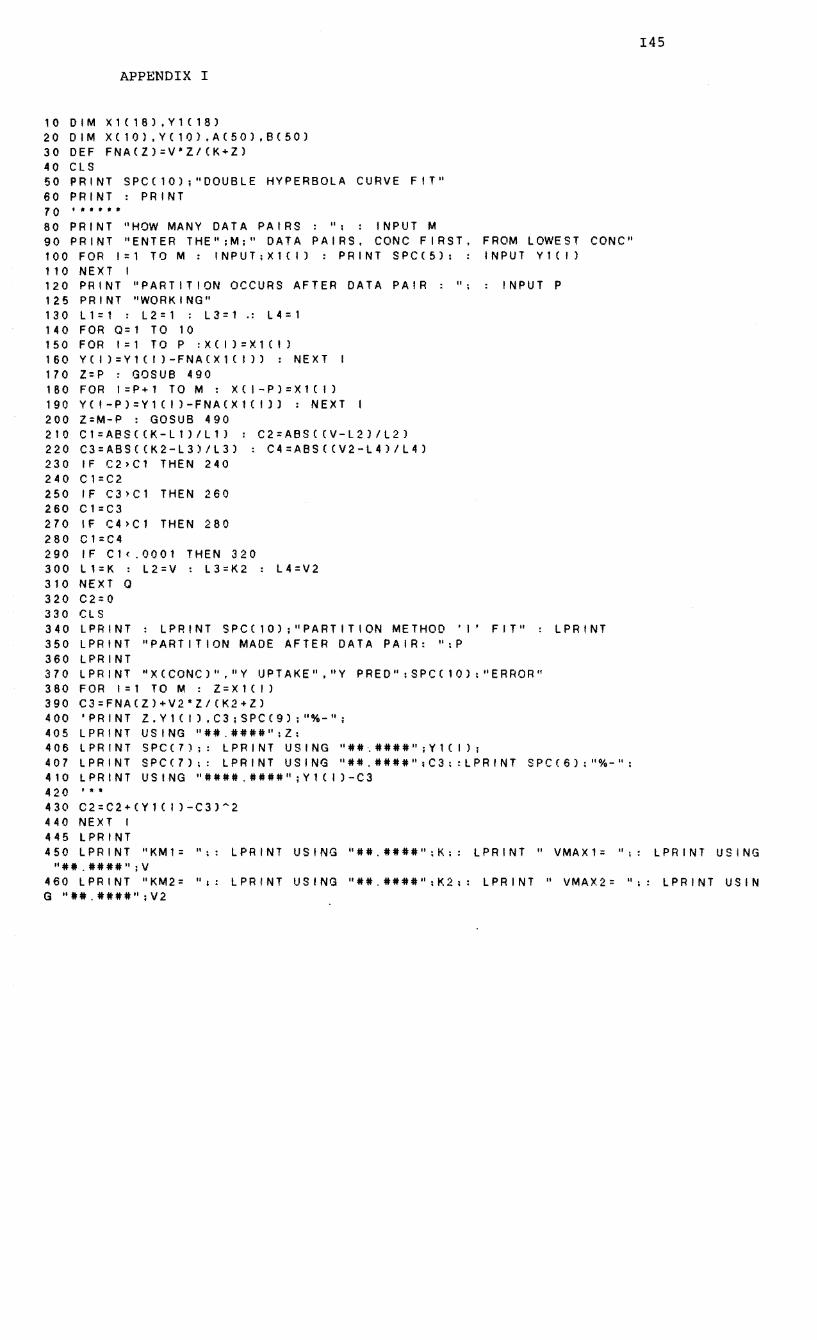
APPENDIX I
10 DIM X1(18LY1C18) 20 DIM XC10LYC10LA(50),BC50) 30 DEF FNA(Z)=V'Z/CK+Z) 40 CLS 50 PRINT SPC(10);"DOUBLE HYPERBOLA CURVE FIT" 60 PRINT: PRINT
70 • * • • • • 80 PRINT "HOW MANY DATA PAIRS: ": INPUT M 90 PRINT "ENTER THE";M;" DATA PAIRS, CONC FIRST. FROM LOWEST CONC" 100 FOR 1=1 TO M : INPUT;X1(/) : PRINT SPC(5); : INPUT VHf)
110 NEXT I 120 PRINT "PARTITION OCCURS AFTER DATA PAIR: "; INPUT P 125 PRINT "WORKING" 130 Ll=l : L2=1 ; L3=1 .: L4=1 140 FOR Q=l TO 10 150 FOR 1=1 TO P :X(I)=X1CI) 160 YCI)=Y1(1)-FNA(X1CI» : NEXT 170 Z=P : GOSUB 490 180 FOR I=P+1 TO M : X(I-P)=X1CI) 190 Y(I-P)=Y1(1)-FNA(X1(1) : NEXT 200 Z=M-P : GOSUB 490 2TO CT=ABS«K-LT)/L1) : C2=ABS«V-L2)/L2) 220 C3=ABS«K2-L3)/L3) : C4=ABS«V2-L4)/L4) 230 IF C2)Cl THEN 240 240 C1=C2 250 IF C3>C1 THEN 260 260 C1=C3 270 IF (4)Cl THEN 280 280 C1=C4 290 IF C1(.0001 THEN 320 300 Ll=K L2=V: L3=K2 : L4=V2 310 NEXT 0 320 C2=0 330 CLS 340 LPRINT : LPRINT SPC(10);"PARTITION METHOD 'I' FIT" LPRINT 350 LPRINT "PARTITION MADE AFTER DATA PAIR: ":P 360 LPRINT 370 LPRINT "XCCONC)","Y IJPTAKE","Y PRED";SPC(10):uERROR" 380 FOR 1=1 TO M : Z=X1CI) 390 C3=FNACZ)+V2*Z/(K2+Z) 400 'PRINT Z,Y1CI),C3;SPC(9);"%-"; 405 LPRINT USING "##.####":Z: 4 0 6 L P R I N T S PC ( 7 ) ;: L P R I N T U SIN G "# # '. # # # #" ; Y 1 C I ) ; 407 LPRINT SPC(7);: LPRINT USING u##.####":C3;:LPRINT SPC(6);"%-": 410 LPRINT USING "####.####";Y1(1)-C3
•• * C2=C2+CY1CI)-C3)~2
NEXT I lPRINT
145
420 430 440 445 450 LPRINT "KM1= ";: LPRINT USING "##.####n;K:: LPRINT II VMAX1= ";: LPRINT USING
"##.####";V 460 LPRINT nKM2= ":: LPRINT USING "##.####":K2;; LPRINT U VMAX2= ";: LPRINT USIN G "1#.####";V2
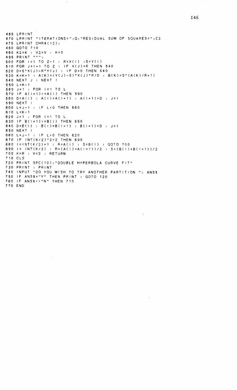
465 LPRINT 470 LPRINT "ITERATIONS=";O:/ORESIDUAl SUM OF SOUARES=";C2 475 lPRINT CHRSCI2); 480 GOTO 710 490 K2=K : V2=V : K=O 495 PRINT "'''; 500 FOR 1=1 TO Z-l : R=XCI) :S=YCI) 510 FOR J=I+1 TO Z IF XCJ1=R THEN 540 520 O=S'XCJ)-R*Y(J) : IF 0=0 THEN 540 530 K=K+l : ACK)=CYCJ)-S)*XCJ)*R/D : BCK1=S*CACK1/R+l) 540 NEXT J : NEXT I 550 L=K-l 560 J=l : FOR 1=1 TO L 570 IF ACI+1»=ACI) THEN 590 580 D=ACI) : ACI l=ACI+l) ; ACI+1)=0 J=I 590 NEXT I 600 l=J-1 IF l>O THEN 560 610 l=K-1 620 J=l FOR 1=1 TO l 630 IF 8(1+1»=8(1) THEN 650 6400=8(1) ; 8(1 )=8(1+1) : 8(1+1)=0 J=! 650 NEXT I 660 l=J-l IF l>O THEN 620 670 IF INTCK/2)*2=2 THEN 690 680 l=tNTCK/2)+1 : R=ACI) : S=8(1) : GOTO 700 690 1= INTCK/2) : R=CAC 1)+ACI+l))/2 : S=C8CI )+BC 1+1))/2 700 K=R : V=S : RETURN 710 ClS 720 PRINT SPC(10):"OOU8lE HYPERBOLA CURVE FIT" 730 PRINT: PRINT 740 INPUT ''~O YOU WISH TO TRY ANOTHER PARTITION "; ANSS 750 IF ANSS="Y" THEN PRINT: GOTO 120 760 IF ANS$(>"N" THEN 710 770 END
146
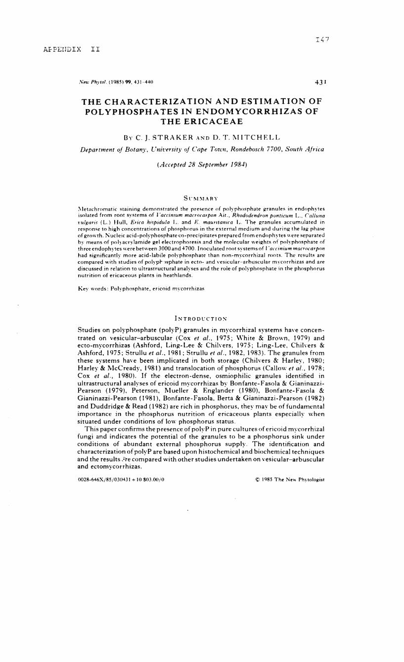
AFPErrDIX II
N"" Phytol. (198S) 99. 431 ~44n 431
THE CHARACTERIZATION AND ESTIMATION OF POLYPHOSPHATES IN ENDOMYCORRHIZAS OF
THE ERICACEAE
By C. J. STRAKER AND D. T. l\.[ITCHELL
Department oj Botany, Uni'L'ersitJ' oj Cape TOtt'n, Rondebosch nOG, South Africa
(Accepted 28 September 198.1)
S l' !\1 :1.1 :\ R Y
i\)etachromatic staining demonstrated the presence of polyphosphate granules in endophYles isolated from root syMems of r'accimum macrocarpon Ait., Rhododendron pontlwm L.. Calluno t'u/pans (L.) Hull, Erica hispidu/o L. and E. mauY/tanica L. The R'ranules accumulated in response 10 high concentrations of phosphorus in the external medium and during the laR' phase of growth. Nucleic acid-polyphosphate co-precipitates prepared from endophytes were separated by means of polyacrylamide Rei electrophoresis and the molecular weight, of polyphosphate of three endophytes were between 3000 and 4- iOO. I noculated root systems of I 'orcin/um macrocarpon had significantly more acid-labile polyphosphate than non-mycorrhizal roots. The results are compared with studies of polypI- ')sphate in ecto- and ,'esicular-arbuscular m\'cnrrhizas and are discussed in relation to ultrastructural analyses and the role of polyphosrhate in the phosphorus nutrition of ericaceous plants in heath lands.
Key words: Polypho'phate. ericoid mycorrhizas
INTRODl'CTlON
Studies on polyphosphate (polyP) granules in mycorrhizal systems ha\'e concentrated on vesicular-arbuscular (Cox et al., 1975; White & Brown, 1979) and ecto-mycorrhizas (Ashford, Ling- Lee & Chilvers, 1975; Ling- Lee, Chil"ers & Ashford, 1975; Strullu et al., 1981; Strullu et al., 1982, 1983). The granules from these systems have been implicated in both storage (Chi1\'ers & Harley, 1980; Harley & McCready, 1981) and translocation of phosphorus (Callow et al., 1978; Cox et aI., 1980). If the electron-dense, osmiophilic granules identified in ultrastructural analyses of ericoid mycorrhizas by Bonfante- Fasola & GianinazziPearson (1979), Peterson, Mueller & Englander (1980), Bonfante- Fasola & Gianinazzi-Pearson (1981), Bonfante-Fasola, Berta & Gianinazzi-Pearson (1982) and Duddridge & Read (1982) are rich in phosphorus, they may be of fundamental importance in the phosphorus nutrition of ericaceous plants especially when situated under conditions of low phosphorus status.
This paper confirms the presence of polyP in pure cultures of ericoid mycorrhizal fungi and indicates the potential of the granules to be a phosphorus sink under conditions of abundant external phosphorus supply. The identification and characterization of polyP are based upon histochemical and biochemical techniques and the results :Ore compared with other studies undertaken on vesicular-arbuscular and ectomycorrhizas.
0028-646X/85/03043I + 101103,00/0 © 1985 Tht' l'it'\\ Phytologist
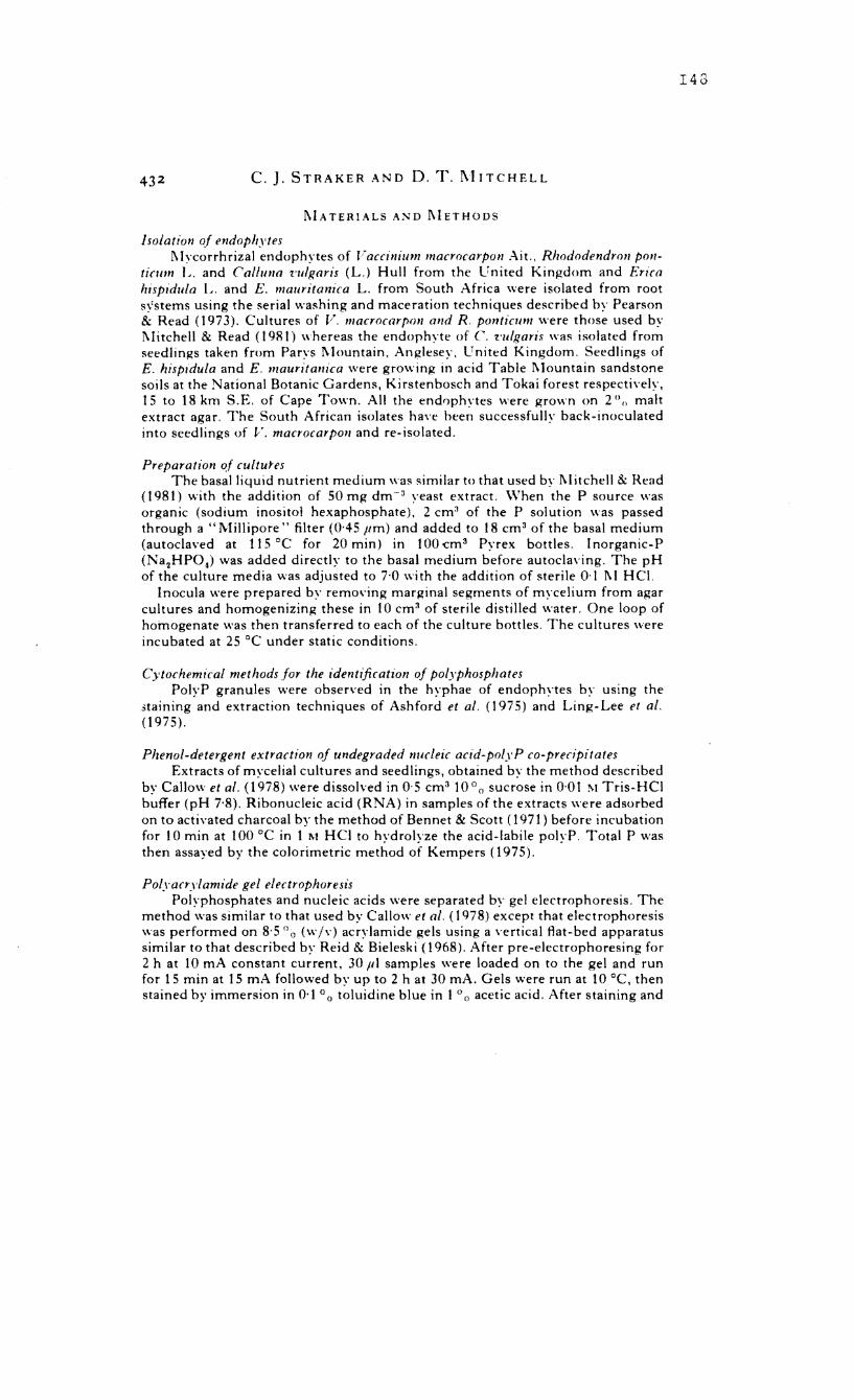
43 2 c. J. S T R A K ERA N D D. T. 1\1 IT C H ELL
I\IATERIALS AND I\IETHODS
I solation of endopltytes l\Iycorrhrizal endophytes of Facclllium macrocarpon Ait .. Rhododendron POI/
ticwn L. and Co/how 'nllgaris (L.) Hull from the United Kingdom and Erica Itispidula L. and E. mallr/tonica L. from South Africa were isolated from root s!'stems using the serial washing and maceration techniques described by Pearson & Read (1973). Cultures of V. macrocarpoll and R. p011ticum were those used by l\litchell & Read (1981) whereas the endophyte of C. 'l:ulgaris was isolated from seedlings taken from Parys Mountain. Anglesey. llnited Kingdom. Seedlings of E. hispldula and E. maurita"ica were growing in acid Table I\lountain sandstone soils at the National Botanic Gardens, Kirstenbosch and Tokai forest respectively, 15 to 18 km S.E. of Cape Town. All the endnphytes were grown on 2 0
" malt extract agar. The South African isolates have been successfully back-inoculated into seedlings of V. macrocarpoll and re-isolated.
Preparation of cultul"es The basal liquid nutrient medium was similar to that used by I\litchell & Read
(1981) with the addition of 50 mg dm- a yeast extract. \Vhen the P source was organic (sodium inositol hexaphosphate), 2 cm3 of the P solution was passed through a "l\lillipore" filter (0'45 11m) and added to t 8 cm 3 of the basal medium (autoclaved at t 15°C for 20 min) in tOO 'Cm 3 Pyrex bottles. I norganic-P (Na 2HP04) was added directly to the basal medium before autocla\'ing. The pH of the culture media was adjusted to 7·0 with the addition of sterile 0·1 1\1 HC!.
Inocula were prepared by remm'ing marginal segments of mycelium from agar cultures and homogenizing these in to cm3 of sterile distilled water. One loop of homogenate was then transferred to each of the culture bottles. The cultures were incubated at 25°C under static conditions.
Cytochemical methods for the identification of polyplzosphates PolyP granules were obsen'ed in the hyphae of endophytes by using the
staining and extraction techniques of Ashford et al. (1975) and Ling-Lee et al. (1975).
Phenol-detergent extractioll of undegraded nucleic acid-polyP co-precipitates Extracts of mycelial cultures and seedlings, obtained by the method described
by Callow et al. (1978) were dissolved in O· 5 cm3 to 0 () sucrose in 0·01 ;\1 Tris-Hel buffer (pH 7'8). Ribonucleic acid (RNA) in samples of the extracts were adsorbed on to activated charcoal by the method of Bennet & Scott (1971) before incubation for to min at 100°C in 1 l\I HCI to hydrolyze the acid-labile polyP. Total P was then assayed by the colorimetric method of Kempers (1975).
Polyacrylamide gel electrophoresis Polyphosphates and nucleic acids "'ere separated by gel electrophoresis. The
method was similar to that used by Callow et al. (1978) except that electrophoresis was performed on 8·5 °0 (wi\') acrylamide gels using a vertical Rat-bed apparatus similar to that described by Reid & Bieleski (1968). After pre-electrophoresing for 2 h at 10 rnA constant current, 30 III samples were loaded on to the gel and run for 15 min at 15 rnA followed by up to 2 hat 30 rnA. Gels were run at 10 °C, then stained by immersion in 0·1 0 0 toluidine blue in 1 "0 acetic acid. After staining and
143
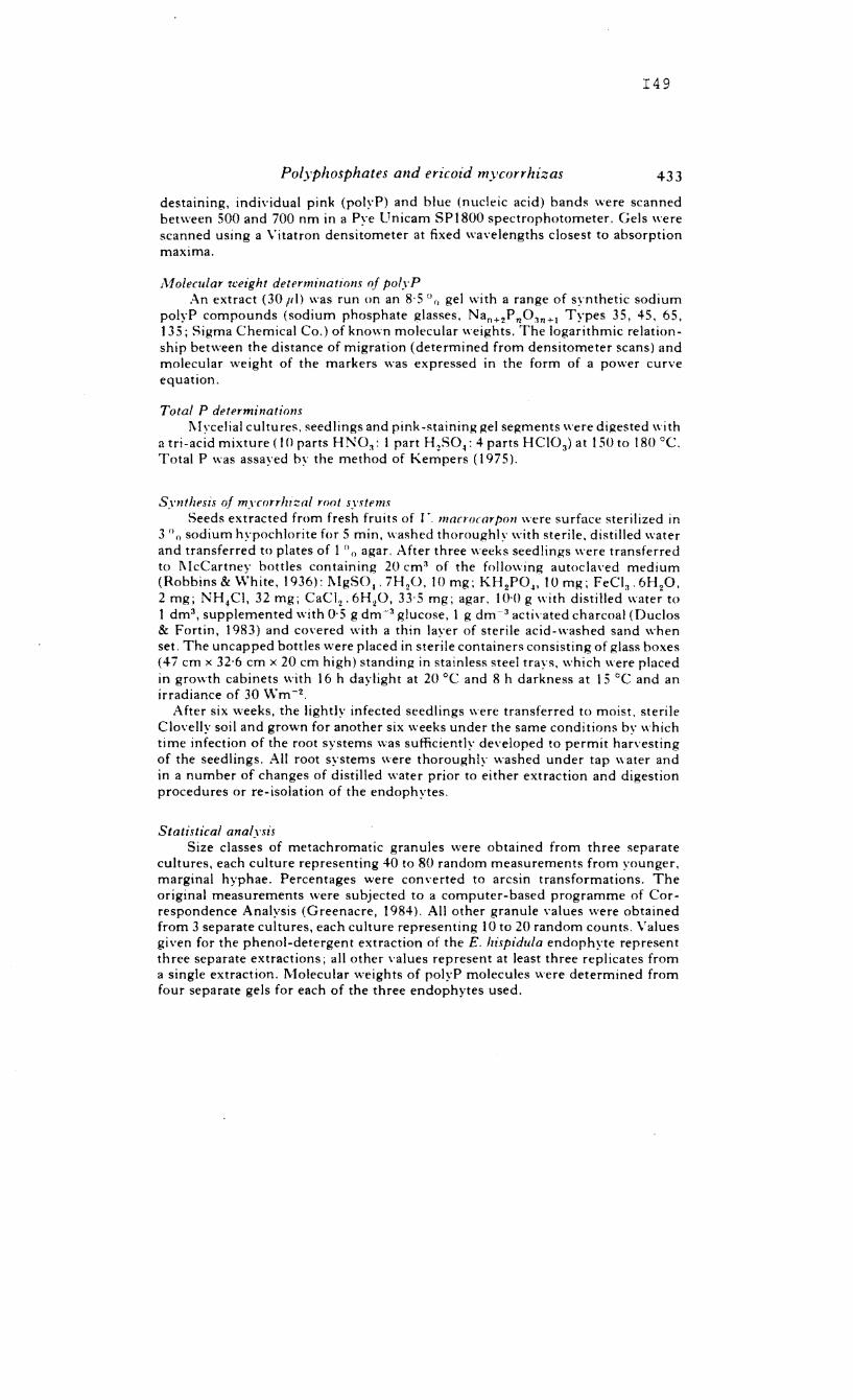
149
Polyphosphates and ericoid mycorririzas 433
destaining, individual pink (polyP) and blue (nucleic acid) bands were scanned between 500 and 700 nm in a Pye llnicam SP1800 spectrophotometer. Gels were scanned using a Vitatron densitometer at fixed wavelengths closest to absorption maxima.
A10lecular 1~'eight determinations of polyP An extract (30 p I) was run on an 8· 5 () () gel \\'ith a range of synthetic sodium
polyP compounds (sodium phosphate glasses, Nan +2P 110311+1 Types 35, 45, 65, 135; Sigma Chemical Co.) of knmm molecular weights. The logarithmic relationship between the distance of migration (determined from densitometer scans) and molecular weight of the markers was expressed in the form of a power curve equation.
Total P determinations 1\1 ycelial cultu res, seed lings and pink-staining gel segments were digested wi th
a tri-acid mixtu re ( 10 parts H N03 : 1 part HzSO ~: 4 parts H C10 3 ) at 1 SO to 180°C. Total P was assayed by the method of Kempers (1975).
Synthesis of mycorrhizal ront systems Seeds extracted from fresh fruits of r'. maao(Grpol! were surface sterilized in
3 () () sodium hypochlorite for 5 min, washed thoroughly with sterile. distilled water and transferred to plates of 1 °0 agar. After three weeks seedlings were transferred to I\lcCartney bottles containing 20 cm 3 of the following autocla\'ed medium (Robbins & White, 1936): I\lgSO,. 7H 20. 10 mg; KH 2P04 , 10 mg; FeCI 3 • 6H
20.
2 mg; NH 4Cl, 32 mg; CaCI 2 • 6H20, 33·5 mg; agar, 10·0 g with distilled water to t dm 3 , supplemented with 0·5 g dm-3 glucose, 1 g dm-3 acti\'ated charcoal (Duclos & Fortin, 1983) and covered with a thin layer of sterile acid-washed sand when set. The uncapped bottles were placed in sterile containers consisting of glass boxes (47 cm x 32'6 cm x 20 cm high) standing in stainless steel trays, which were placed in growth cabinets with 16 h daylight at 20°C and 8 h darkness at 15°C and an irradiance of 30 Wm-2 .
After six weeks, the lightly infected seedlings were transferred to moist, sterile ClovelJy soil and grown for another six weeks under the same conditions by which time infection of the root systems was sufficiently developed to permit harvesting of the seedlings. All root systems were thoroughly washed under tap water and in a number of changes of distilled water prior to either extraction and digestion procedures or re-isolation of the endophytes.
Statistical analysis Size classes of metachromatic granules were obtained from three separate
cultures, each culture representing 40 to 80 random measurements from younger, marginal hyphae. Percentages were con\'erted to arcsin transformations. The original measurements were subjected to a computer-based programme of Correspondence Analysis (Greenacre, 1984). All other granule values were obtained from 3 separate cultures, each culture representing 10 to 20 random counts. Values given for the phenol-detergent extraction of the E. hispidula endophyte represent three separate extractions; all other values represent at least three replicates from a single extraction. Molecular weights of polyP molecules were determined from four separate gels for each of the three endophytes used.
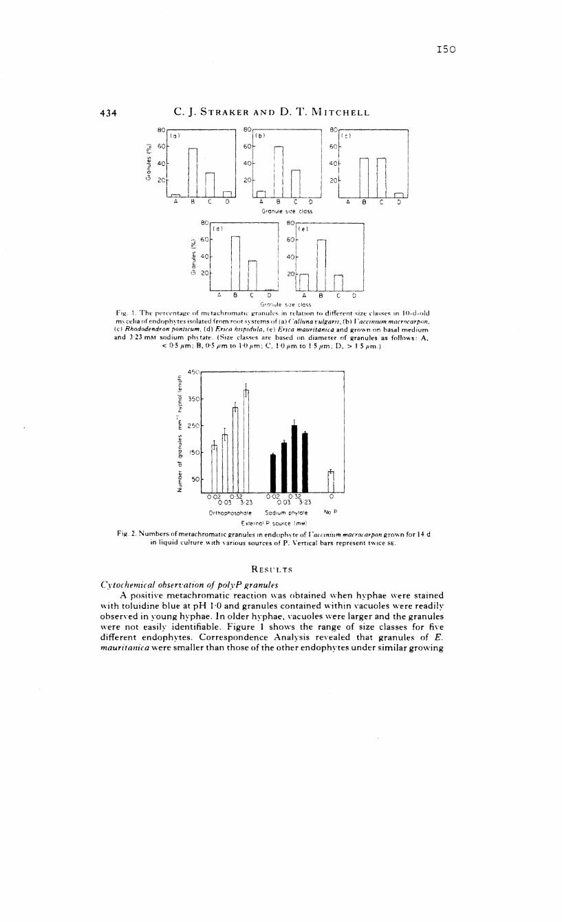
434 C. J. STRAKER AND D. T. MITCHELL
80 80 80 i 0 I lib I Ie}
~ 60 GOr 60
~ I
'" 40 40 40~ :; c ! 0
<5 20 I
J 0
Granule sIze class
80· 80 lid} (e I
~ .of n 60
'" '" '::0 :; n c 0 I ,
<5 20~ I
I I I I
I I I I A B C D
Gr(1r,ule $\ze CIQ')S
I'll(, I, The percentage of metachromatic granuk, in rdatlon to diff,'n'nt size "Iasst's In IO-d-old m\ celia of end()ph~,tt's isolatcd from root systems of (a) Col/llna ,'ulf(oris. (b i "ncC/III"'" macrncorpnll,
(cl Rhododendron ponticum, (d) F.run hispld,tlo, lei ErICn mallritomco and Rrown on basal medium and 3,23 mM sodium phYlatc, (Sizc classes are based nn diameter of Rranules as follows: A.
< O-S 11m; B. 0-5 pm to 1-0 11m; C, HJ 11m to 1'5 I'm; D. > IS,lm,)
1 45C
11 350 ;: "' 'E E 250 ~ ~
.l
I
! Orthop",osphote
002 0":'2 003 323
Sodium D~vl0'e
Ex;ter('lO~ P source ImM)
n o
No P
Fi!/, 2, Numbers of metachromatic !/ranules 10 endophytc of I'occmium mo(r(){nrp()n o:rown for 14 d in liquid culture with various sources of p, Vertical bars represent tWice SE.
RESl' L TS
Cytochemical obsen'ation of polyP granules
150
A positiye metachromatic reaction was obtained when hyphae were stained with toluidine blue at pH 1·0 and granules contained within vacuoles were readily observed in young hyphae. I n older hyphae, vacuoles were larger and the granules were not easily identifiable. Figure I shows the range of size classes for five different endophytes. Correspondence Analysis revealed that granules of E. maun'tallt"ca were smaller than those of the other endophytes under similar growing
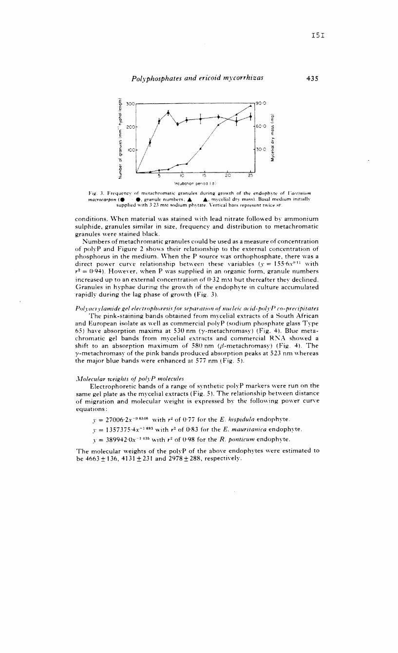
151
Polyphosphates and aicoid mycorrhizas 435
:2 & 300~------------------------------~ c: ~
"6 .c. ~ L:
- 200 'E E
'" '" :; E 100 0'
I 15 20 25
lncuba'fon penOQ (d)
or E
::; o E
Fill. J, Frt'qlH'ncI' of metachromatic granules dunnjl jlfO"th of the endorh\'l(' of /'arrmillm mQcroCQrpon Ie e. granule numhers, • ., m\'cl'iial dry rna,,). IJasal medium initially
supplied with J2J mM ,odium phqatl'. Vertical bars represent twice SF,
conditions, \\'hen material was stained with lead nitrate followed by ammonium sulphide, granules similar in size. frequency and distribution to metachromatic granules were stained black.
Numbers of metachromatic granules could be used as a measure of concentration of polyP and Figure 2 shows their relatIOnship to the external concentration of phosphorus in the medium. \Yhen the P source was orthophosphate. there was a direct power cun'e relationship between these \'ariables (y = 155'6",°11 with r2 = ()·94). Howe\'er, when P was supplied in an organic form. granule numbers increased up to an external concentration of (J'32 ml\1 but thereafter they declined. Granules in hyphae during the growth of the endophyte in culture accumulated rapidly during the lag phase of growth (Fig, 3).
PolY(lCl'ylamide 1(('1 elect ropizoresis for sl'para I iflll of nucl eit at id-poly P (0- preripitales The pink-staining bands obtained from mycelial extracts of a South African
and European isolate as well as commercial polyP (sodium phosphate glass Type (5) han absorption maxima at 530 nm (y-metachromasy) (Fig, 4). Dlue metachromatic gel bands from mycelial extnlcts and commercial R:,\A showed a shift to an absorption maximum of 580 nm (If-metachromasy) (Fig. 4), The y-metachromasy of the pink bands produced absorption peaks at 523 nm whereas the major blue bands were enhanced at 5ii nm (Fig, 5).
;l.lolecular u:eiJ!lzts of polyP molecules Electrophoretic bands of a range of synthetic polyP markers were run on the
same gel plate as the mycelial extracts (Fig. 5). The relationship between distance of migration and molecular weight is expressed by the following power curve equations:
y = 27006·2x-nsiH6 with r2 of O'ii for the E, JlIspldula endophyte .
.\' = 1357375'4x- 1693 with r2 of 0·83 for the E. maurilanica endophyte .
.\' = 389942'OX- 1135 with r2 of 0·98 for the R. ponticum endophyte.
The molecular weights of the pol\'P of the above endophytes were estimated to be 4663 ± t 36. 4131 ± 231 and 2978 ± 288, respectively.

c. J. STRAKER AND D. T. !\llTCHELL
1·0...---------------., I a)
700 500 550 600 650 700
WOIJe1enqlh (:'1ml
Fig. 4. Ahsorption spectra of segments of S'5 nn pO!\'acn lam ide lie Is stained with (l'1 "" toluidmc hlue' (a I Pink-stain in!! metachromal Ie hand from extracts of endorhytr of FII(<I Imp/drd,,; (h) Pink-stalnm!! metachromatic hand from gel on which 50 I'" synthetic pold' had he"n run: I c I .\ hlue-staining nucleic acid hand from extracts of endorhvtc of E. hi;p/dula: (d) A hlue-staming hand from "el on which SO,'!! ("mOll're.al R:"A had heen run; Ie) A sc!!ment of "d stairwd nnl\ \\lIh toluidine blue; (n Pink-staining metachromallc hand from extracts of endorhyte "f Rh"d"derrd,tlI/
ponticum; (ll) A blue-staining nucleic-acid band from extracts of endophne of R. p""tuum.
:- i'~1 .... (0) I (b)
E!e:trop~,oretlc rnob1lfy
Fig 5, Scans of nucleiC acid-polyP rhenol-deter"t'n! eXlract" separated on ~·3", polYacT\ lamid .. gel and stained \\ ith I) 1 "" toluidine hlue' (a) (iel of extract of endophyte of Rlwdndnrdrnn P,,"llfUII!
scanned at 523 nm; (h) Same lIel as in I a) scanned at 5ii nm: Ie) Gel of ~xtract of ~nd"ph\·te of Erica hlspldula scanned at 52) nm; (d) Same l.wl as in (l'l ,canned at 57i nm. Dlalo(rammatil' representations of the bands art' shown aho\'e the scans. Solid areas are th,' hlul·. nucleIC' acrd components; the cross-hatched area represems the pl!1k, polyP componcnt. Also sho\\f1 an' positions of synthetic pol"P markers in relation to scans: (I) '[\pe 1.15 (chain Icnllth 132); (2) T\pe
65 (chain length (5); (3) Type 45 (chain length 46); (411\'pe 35 Icham lenllth 39)
I52
153
Polyphosphates and ericoid mycorrhizas 437
P and acid-soluble polyP content The total P and acid-soluble polyP content of the endophytes of E. hispidula
and R. ponticum were similar (the latter being t 1 and 8 () (J, respectively of total P) (Table t). The endophyte of E. mauritanica showed a higher acid-soluble polyP content but this formed a smaller proportion (7 0
0 ) of a much higher total P content. The similarity between the acid-soluble polyP content of the extracts and the total P of the pink, metachromatic gel bands confirmed the presence of a
Table t. Acid-labile polyP content of phenol-detergent extracts and total P of isolated endophytes grown in culture on basal medium mId 3·23 mM sodium phytatefor 8 d and
aseptically infected root systems of Vaccinium macrocarpon
Endoph\'te E. hispidula R. porr/icum E. mauri/amra
Root systems Infected with E. hlSp,dula endophyte
Infected with E. mauri/amra endophyte
L:ninfected
Fresh mass ± SE (/ImoIIC')
Acid-labile Total P of P pink bands Total P
1·9::,:0'\ 17±03 17'5::t2'0 14±0'3 \.\ ±02 18·2 ::,:21 2'2±02 2\ ::,:0'5 3H±H
Hl±02" 9·5 ± 20
3'9:,: 03" 19·8::tJ·2" 0'3±01 10·4±2'6
(°0) of total
Acid-labile P
109 77 6'6
10·5
19·i 3·0
a and b are significantly different from uninfected root at 0·05 and 0·001 level respectively; - signifies not determined.
predominately acid-soluble fraction. Root systems of seedlings of V. macrocarpoll were inoculated with the endophytes of E. hispidula and E. mauritanica and, after three months incubation, the proportions of acid-labile polyP of total P were 10 and 20 () 0. respectively. Infection with the E. mattritamca endophyte also resulted in a two-fold increase in the total P of the root system when compared with uninfected roots, which had a low acid-labile P content of 3 0
0 of the total P. Infection with the endophyte of E. hispidula did not result in a similar increase in the total P of the root system.
DISCL'SSIO!\l
Phosphate appears to be a limiting element for growth in heathlands. the natural habitat of ericaceous plants. These include the acidic sandy soils with a low organic matter content from S.\V. Cape. South Africa and the acidic mar-humus soils with R high organic matter content from Europe (Read & l\litchell, 1983). An investigation of the phosphate metabolism of these plants is therefore an essential step towards an understanding of their ecology.
The presence of polyP in pure cultures of the endophyte and mycorrhizal root systems of ericaceous seedlings has been confirmed. The metachromatic granules resemble closely those of ectomycorrhizas of eucalypts and pines (Ashford et al.,
154
C. J. STHAKER AND D. T. 1\1 ITCHELL
1975; Ling- Lee et al., 1975; Chi! vers & Harley, 1980) and vesicular-arbuscular mycorrhizas (Cox et al., 1975). The range of granule sizes were similar to those obtained for beech mycorrhizas incubated in I m:\1 orthophosphate (Chilvers & Harley, t 980) and granule numbers were sensitive to both the form and concentration of the external P source (Fig. 2). The decline in granule numbers after an organic P concentration of 0·32 mfl.l may be due to the end-product inhibition of the phosphatase enzymes (Pearson & Read, t 975) which \vould have reduced the amount of P available for uptake. The accumulation of granules in hyphae in media without the addition of an external P source was most likely due to the presence of residual P in the yeast extract of the basal medium. The polyP granules accumulated during the lag phase of growth. In Cor.t'nebacterium xerosis, numbers of granules increased during the lag phase but declined during the exponential phase (Hughes & Muhammed, 1962).
The phenol-detergent extraction technique of Callow et al. (1978) was used to isolate polyP and nucleic acid in a relati\'e1y undegraded form and the polyP was finally characterized by means of polyacrylamide gel electrophoresis. Callow et al. (1978) were able to determine the molecular weight and chain length of the polyP in \'esicular-arbuscular mycorrhizas as poly phosphates ha\'e a constant charge to mass ratio and their mobility in the gels is a function of their molecular "ize. In this study, the molecular \ .... eights of 3000 to 4700 from ericoid endophytes were considerably lower than the figure of 20800 for polyphosphates of \'esiculararbuscular mycorrhizal fungi (Callow et al., 1978) although the possibility of chain degradation during extraction should not be discounted. The proportion of acid-labile polyP to total P was 7 to 11 "" in the cultured endophyte. Isolated internal vesicular-arbuscular endophytes have been estimated to contain 16 (l" total polyP (Capaccio & Callow, 1982). Ecto-mycorrhizas incorporated 40"" of absorbed 32p into total polyP over 2·5 h (Harley & l\IcCready, 1981) although clearly this does not represent a steady state condition. The polyP content of ericoid mycorrhizal root systems was significantly greater than that of the non-mycorrhizal controls indicating the polyP to be largely fungal in origin. The higher proportion of polyP in the root systems inoculated with the endophyte from E. mauritanica could have been due to a heavier mycorrhizal infection obsen'ed, However, as the higher total P content of these infected seedlings was correlated with a high concentration of P in the isolated endophyte. the mycorrhizal fungus of E. mauritanica may also be able to accumulate more phosphorus, Thus, preliminary results suggest that the capacity of the endophyte to store polyP may play an important part in the phosphorus nutrition of ericaceous plants. This is emphasized by the increase in granules of the endoph\'te in culture concomitant with the increase in concentration of the external supply of orthophosphate,
Electron-dense granules are located in the fungal vacuoles in both the cultured endophyte and extra- and intracellular hyphae of natural ericoid mycorrhizal root systems (Bonfante- Fasola & Gianinazzi-Pearson. 1(81), Howe\'er, in dual cultures, they often occur in the external mycelium but rarely in intracellular hyphae (Bontante-Fasola & Gianinazzi-Pearson. 1(82). It appears that the storage of phosphorus as polyP in the endophyte would not persist and transfer from the fungus to the host may im'oh'e an acti\'e process. The mycorrhizal endophyte outli\'es the ericaceous cortical cell in which cytoplasmic contents increase with infection indicating a mycorrhizal association of intense metabolic acti\'ity (Bonfante-Fasola & Gianinazzi-Pearson. 1982; Duddridge &.Hcad, 1982; Read. 1(83). Biochemical and histochemical evidence and ultrastructural studies indicate
155
Polyphosphates and ericoid mycorrhizas 439
that the endophyte exterior to the root exploits the soil for phosphate, part of which accumulates in these external hyphae as vacuolar granules of polyP molecules.
ACK NOV.' LEDG EM ENTS
We wish to thank the CSIR, South Africa and the University of Cape Town for financial assistance, Dr T. T, Dunne for assistance with the Correspondence Analysis and Dr D. J. Read of Sheffield l1niversity for the use of the ericoid endophytes from the 11 K.
REFERENCES
AiiHFORl>, A E., LI"G-LEE. \1. & CHII.\'fR~, {j. A. (1975). Polyphosphate in eucalypt mycorrnizas: a cnocnemical demonstration. The ,Vere Phytolo!?Ht, 74, H7 ~53.
B()"F.~"TF-r"~SOI.A, P. & (il~NI"\ZZI-PEMISON, V (1979). l'ltrastru<.:tural aspects of "ndom,corrh,za In th" Er.caceae. [. :-;aturalh infected hair roots of ('a//u"(1 "u/g(lri' L. Hull. The Sen' Phyrn/"RISI, 83. 7J9 7~~.
BO"F ~ "TT-F~sOI~, P. & (j I.\"I"UZI- PF.~RSO", V (19H I). l'ltrastructural aspects of endomycnrrhizas in the Ertcaceae. II. Ilost-endophyte relationships m l'aatnlUm m.l·rll/lls L. The Se,,' Phylnlogllt. 89,219 2B.
BO"F \"TF.-F "SOI.A, P. & {j IIINI"UZI-PEARSO". \. (1982 L l'ltrastructural aspects of endom\corrh.zas in the Ertcace"e. III. \Inrrholol(\' of the dissociated "'mhionts and modifications ,wcurrinR durlnR their reassociation in axenic culture. Tlu Sft<· Pln·to/IiRlSt. 91, /)91 704
CAI.I.O\\, J. A .. C~P!\{,ClO, L. \1.. P.\RII'Il, (i, & TINKER, P B. (1978). Detection and estimation of polvphosphate in \'esicular~arhuscular mv·corrhlzas. The Xet<· Phylnl()f[HI. 80, 125 1 H
CAPMTIO, L. C. \1. & C.~LI.O\\, J. A (1982) The ennme, of polyphosphate metaholism in vesiculararhuscular mycorrhizas. The Xef<' Ph),l%gl.<l. 91, 81 ~ 9 I.
CHIt.HR5, G. A. & HARLEY. J. L (1980). Visualization of phosphate accumulation in beech mvcorrhizas. The Seu' PirytologlSl. 84, 319 326.
Cox, G., S.~"DERS, F. E., TI"KER, P B. & WilD, J. A. (\975) l·ltrastructural e\'idence relatm" to hOM-endophvte transfer in a vesicular- arhuscularmvcorrniza. In Endomycorrhlzas (Ed. bl' F. E. Sanders. Barbara \1055e & P. B Tinker). pp, 297 J 12. Academic Prl's" :-;'''' York
COl(, (i., \IOR.~S, K. J., SANnERs, F .. r-;()('KOI.l>S, c. & TISKER. P B. (19HO). Translocation and transfer of nutrients In ves!cular-arbuscular ml'corrhlzas. III. Polyphosphate Io1ranules and phosphorus translocation. The Nev.: Phylnlag/st, 84, M9 659.
Dl'CI.OS, J L. & FORT'I", J. A. (1983). Effect of ,,'ueo.e and actll e charcoal on In "Itra synthesis of cricoid n1\Torrhiza with I 'accinium spr. The Sm' Phyt%gHt, 94, 95102.
DnlDRIDGE. J. & RE.~O. D. J. (\ (82), An ultrastructural analySl' of the development of mycorrhizas in Rhododendron pmllicum. Canadian Journa/ of Rolan.\'. 60. 2345- 2356.
(iREE" KRE. \1. J, (l98~ I. Theory a"d Applirat/(m of ('oTTesp,mdenre Ana/ySls' Academic Press, ,\,,,, York. H~RLF.Y. J. L. & \1cCREADY, C. C. (19811. Phosphate accumulation .n Faltw m\'corrhlzas. The Sm'
PhytolaglSl, 84, 75-80. Ht'GHES, D. E. & \It'HAMMEIl. A. (1962). The metabolism of pol~'phosphate in bactena. Colloques
/n/ernatianaux du Cenlre Satr"nal de la Recherche Scie,lflnque (Paris)' 106, 59) (,02. KEMPER5, A. J. (1975). Determination of submicro-quantities of plant aV'aHable phosphate in aqueous soil
extracts. Plant and Soli, 42, 423-~27. Ln';G.LEE. \.1.. CHIlXERS, G. A. & ASHFORD, A E. (1975). Poh'phosphate granules in three different kinds
of tree mv'corrhiza. The Sev: Phylalogist, 75, 551 554. :\IITCIlEI.I., D. T. & RE~Il, D. J. (1981). L'tilization of inorganIc and or!ZanlC phosphates b\ the mycorrhizal
endophytesof Vaceimum macrocarpon and Rhododendron p{'mtlcum. T,ansactlO'lSaf Ihe BY/tlSlt .lfycn/oglCa/ SOCIety, 76(2). 255-260.
PEARSO", \'. & READ, D. J. (\973). The bioloRY of mvcorrhiza in the Erlcaceae. I. The isolatIon of the endophyte and synthesis of mycorrhizas in aseptic culture. The Set" PhyrologlSt, 72, 371-379.
PEARSO .... , V. & READ, D. J. (1975). The physiologl of the mycorrhizal endophyte of Cal/una ['ulgam. TrallSaclin'lS of the Bmi.h Jlycnlogical Snnety, 64. I·· i.
PETERSO", T. A., Ml'EI.I.ER, W. C & ENGL.~"DER, L. (1<)80). Anatomy and ultrastructure of a Rhododendron root-fungus association. Canadian Journal nf Botany, 58. 2421·2~33.
READ, D. J. (1983). The bioloRY of mycorrhiza in the Ericales Canadian Journal of Bn/a"y, 61. 985-1004. RE.IIO, D. J. & MITCHELL, D. T. (1983). Decomposition and mineralisation processes in mediterranean-type
e-cosystems and in heathlands of similar structure. In Ec%gical Studies ['0/. '/.1, ;'I,fediterranean· Type Eco!}'sltms: The Role af Nutrients (Ed. by F. J. K ru"er, D. T. Mitchell & J. L'. M. Jarvis), pp, 208-232. SprinRer-Verlal!, Berlin.
440 C. J. STRAKER AND D. T. MITCHELL
REID, :\1. S. & BIELESKI, R. L, (1968), A simple apparatus for vertical flat-sheet polyacrylamide gel electrophoresis. Analytical Biochemistry, 22, 3H 381.
ROBBINS, W j, & WIflTE, \'. B. (1936). Limned growth and abnormalities in eXCised corn root tips. BotanICal Gazetle, 98,209242.
STRl'LLl', D. G .. GOl:RRET, J P., G.~RRf{" J. p, & FOl'RCY, A. (1981) l'ltrastructure and electron-probe microanah',is of the metachromatic vacuolar IOlranules nccurrinlOl in Taxus mycorrhizas. The Set<' Phylalogist, 87, 537-545,
STRt'LLt', D. G" HARLEY, j, L., GOl'BRET, J. P. & GARREC, J P. (1982). Vltrastructure and microanalysis of the polyphosphate granules of the ectomycorrhizas of Fagus sylt.'atica. The .~'t!W PhYIOlogist, 92, 417-423.
STRl'I.LI" D. G., HARLEY, J. L" GOl'RRET, J. p, & GARREr, J P (1983). A note on the relative phosphorus and calcium contents of metachromatic granules in Fagus m\'cnrrhizss. The Seu' Phyt%grst, 94,89-94,
WHITE, J A. & BROWN, M. F. (1979), Vltrastructure and X-ra\, analysis of phosphorus granules In a \'eslcular-arbuscular mycorrhizal fungus, Canaduln Journal of Botany, 57, 2812-2818.
156
i _ .....
, • .
\Jat. 2+4, No I S g , ?fl. & ( 2L
;r~ f tg I '9=}-3.
Effect of Mycorrhizal Infection on Nitrogen and Phosphorus Nutrition of Ericaceous Plants
,
LARGE ,areas of the northern hemisphere are' covered with nutrient-poor soils which support ericaceous plants. The roots of the most important representatives, Callulla vulgaris 1. Hull. and species of Vaccinium and Erica. are always infected with an endotrophic mycorrhizal fungus. Much early work on these mycorrhizas was devotcd to a controversy surrounding the nature of the infection and there is little known of the physiological role of the mycorrhizal association l •
Techniques, developed for the isolation of the endophyte and for the culture of mycorrhizal and non-mycorrhizal seedlings under aseptic conditionsZ, have been used to e\'aluate the influence of mycorrhizas on the nutrition of C. l'Ulgaris and
. Vaccinium macrocarpon Ail. Small, uniform quantities of sterilized hcathland soil were immersed in 0,5 ~~ water agar and aseptically germinatcd seedlings of the two species were transferred to the medium. Mycorrhizal seedlings were produced by inoculation of the soil with an isolate of the endophyte.
.' .. , ) .. ;:.~''':~'~ ~. --. ,." '';.
.' .
:,
. ,
.\~
t .f
f. ;
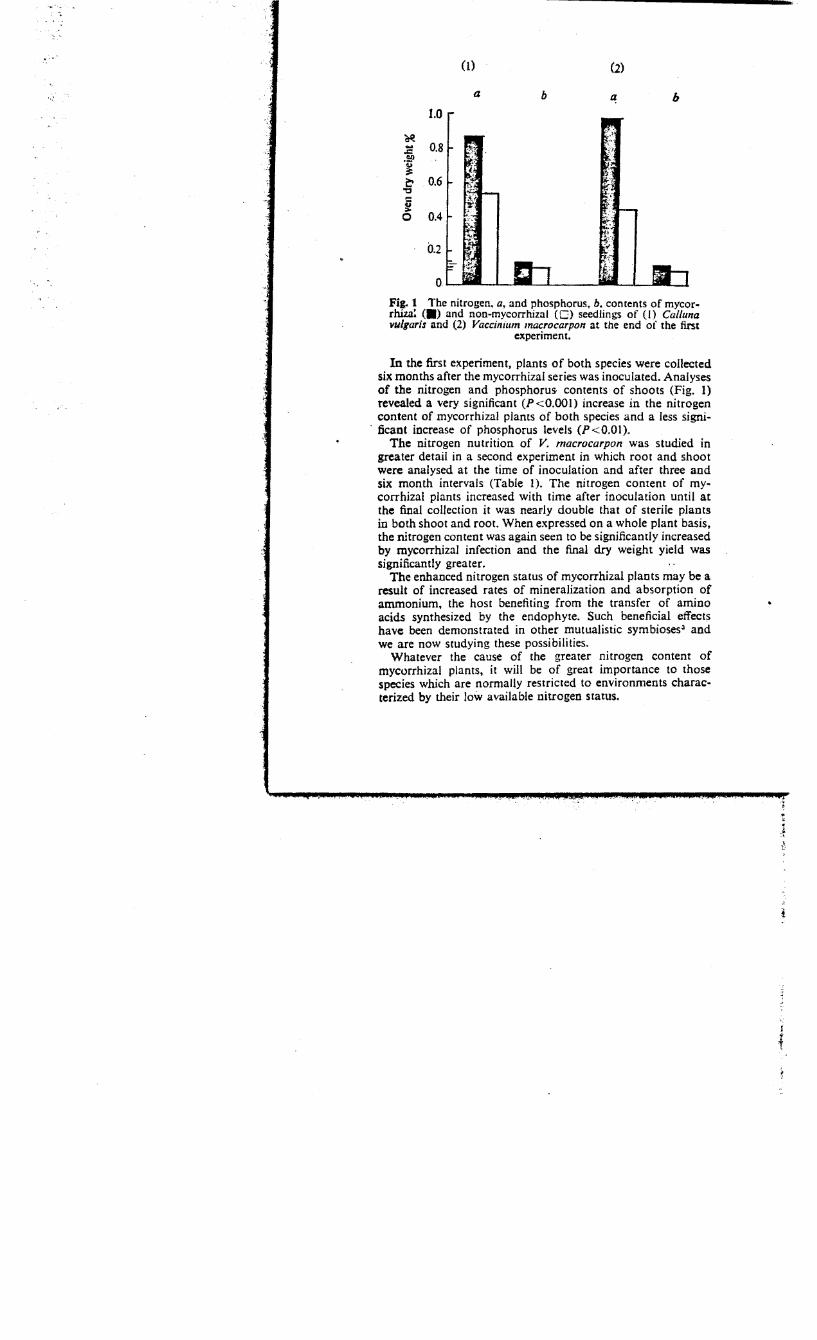
a b b 1.0
'CtI! 1: 0.8 .59 ~ ~ 0.6 'C
~ 0 0.4
0.2
Fig. 1 The nitrogen. a, and phosphorus. b. contents of mycorrhiza: (.) and non-mycorrhizal (0) seedlings of (I) Cal/una vul,aris and (2) Vaccinillm macrocarpon at the end of the first
experiment.
In the first experiment, plants of both species were collected six months after the mycorrhizal series was inoculated. Analyses of the nitrogen and phosphorus- contents of shoots (Fig. 1) revealed a very significant (P <0.001) increase in the nitrogen content of mycorrhizal plants of both species and a less signi-
o Bcant increase of phosphorus levels (P<O.OI). The nitrogen nutrition of V. macrocarpon was studied in
greater detail in a second experiment in which root and shoot were analysed at the time of inoculation and after three and six month intervals (Table O. The nitrogen content of mycorrhizal plants increased with time after inoculation until at the final collection it was nearly double that of sterile plants in both shoot and root. When expressed on a whole plant basis, the nitrogen content was again seen to be significantly increased by mycorrhizal infection and the final dry weight yield was significantly greater.
The enhanced nitrogen status of mycorrhizal plants may be a result of increased rates of mineralization and absorption of ammonium. the host benefiting from the transfer of amino acids synthesized by the endophyte. Such beneficial effects have been demonstrated in other mutualistic symbioses~ and we are now studying these possibilities.
Whatever the cause of the greater nitrogen content of mycorrhizal plants. it will be of great importance to those species which are normally restricted to environments characterized by their low available nitrogen Status.
I
i
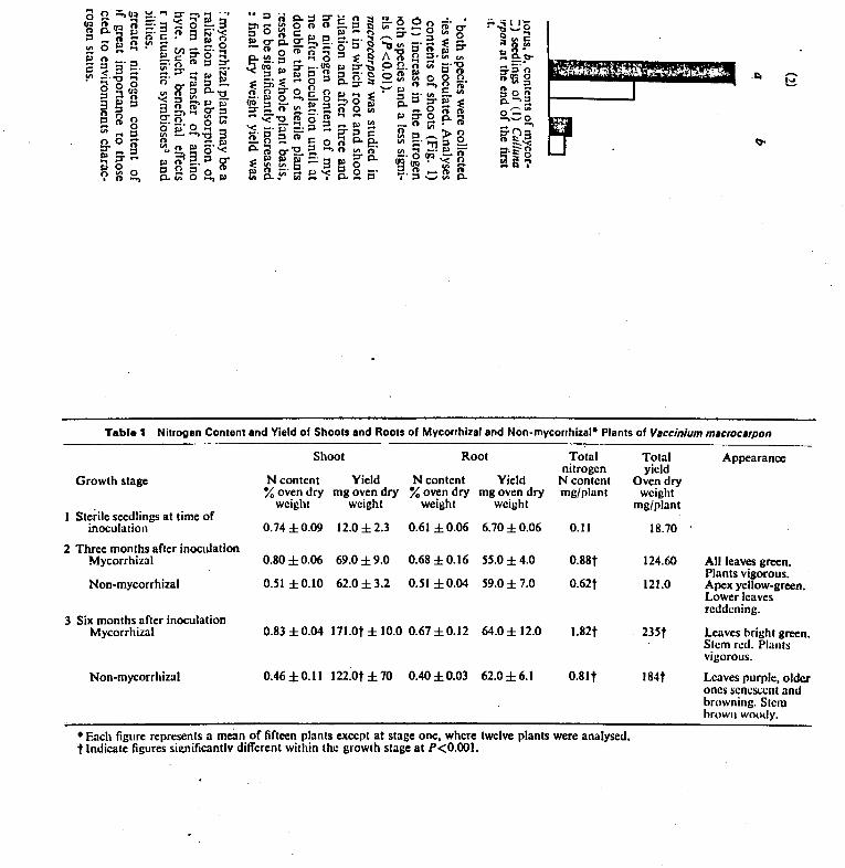
.1:0
Tabla 1 Nitrogen Content and Yield of Shoots and Roots of Mycorrhizal and Non-mycorrhizal- Plants of Vllccinium mllcrocllrpon
Shoot Root Total nitrogen
Growth stage N content Yield N content Yield N content % oven dry mgovendry % oven dry mgoven dry mg/plant
Sterile seedlings at time of weight weight weight weight
inoculation 0.74 ±0.09 12.0 ±2.3 0.61 ±0.06 6.70±0.06 0.11
2 Three monlhs after inoculation Mycorrhizal 0.80 ±0.06 69.0 ±9.0 0.68 ±0.16 55.0 ±4.0 0.88t
Non-mycorrhizal 0.51 ±O.IO 62.0 ±3.2 0.51 ±0.04 59.0± 7.0 0.62t
3 Six months after inoculation Mycorrhizal 0.83±0.04 171.0t±10.0 0.67±0.12 64.0± 12.0 1.82t
Non-mycorrhizal 0.46 ± 0.11 122.0t ± 70 0.40 ±O.oJ 62.0 ± 6.1 0.81t
• Each figure represenls a mean of fifteen plants except at stage one, where twelve plants were analysed. t Indicate figures significantlv different within the growth stage at P<O.OOI.
Total yield
Oven dry weight
mg/plant
18.70
124.60
121.0
235t
184t
Appearance
All leaves green. Plants vigorous. Apex yellow-green. Lower leaves reddening.
Leaves Ilright green. Stem red. Plants vigorous.
Leaves purple, older ones seneSl:cnl and browning. Stem hrown woudy.
We thank the NERC for support. D. J. READ
Department of Botany, D. P. STRIBLEY
The University, Sheffield SIO 2TN
Received March 21,1973.
1 Harley, 1. L., The Biology 0/ Mycorrhira (Leonard Hill, London, 1969).
• Pearson, V., and Read, D. J., New Phytol .. 72.371 (1973). • Melin, E., and Nilsson, H., Sveruk. Bot. Tidskr.,46, :81 (195:!).
Prinled in Greal BriiaiD by Flarep:alb l'rinICrI Lid., 51. AlbaN, Hens.
The FUNGAL COMMUNITY ITS ORGANIZATION AND ROLE IN THE ECOSYSTEM
EDITED BY
Donald I Wicklow Northern Regional Research Center Agricultural Research Science and Education Administration U.s. Department of Agriculture Peoria, Illinois
George C. Carroll Department of Biology University of Oregon Eugene, Oregon
MARCEL DEKKER, INC. New York and Basel
"
Chapter 33
ROLE OF ENDOHYCORRHIZAL FUNGI IN PHOSPHORUS CYCLING IN THE ECOSYSTEM
v. Gianinazzi-Pearson* and S. Gianinazzi*
Station de Ph!lsiopathologie Vegetale Insti tut: National de Ia Recherche Agronomique Dijon, France
I. INTRODUCTION
In natural ecosystems plants depend largely on the activity of soil microorganisms
for the supply of mineral nutrients essential to their growth. It is evident t.hat
microorganisms that form symbiotic associations with plant roots, for example, nitro
gen-fixing bacteria and mycorrhizal fungi, are particularly well placed to intervene
in plant nutrition.
A distinguishing feature of mycorrhizal fungi is that after root infection part
of the mycelium remains active in the soil. Plant roots provide them with an ecolog
ical niche with abundant substrate from which their hyphae extend outward through the
soil and effectively explore a much greater volume than nonmycorrhizal roots. The
two most important groups of mycorrhizal fungi; consist of (l) those forming ectomy
corrhizas which are characterized by mycelial sheaths around the roots and intercel~ . lular hypha I invasion of the root cortex and (2) those forming endomycorrhizas with . a loose extemal hyphal netwo:tX in the soil and extensive intracellular hypha! growth
in the root cortex.
The beneficial effects of ectomycorrhizas on the growth and nutrition of tree
species in soils of low nutrient status have been recognized for a long time (Mitchell
et al., 1937; McComb, 1938), and the role of ectomycorrhiza! fungi in supply~ng min
eral nutrients to the host plant has been conclUsively established (Melin and Nilsson,
1950, 1952, 1953a,b; Melin et al., 1958; Harley, 1969). It is only:within the last
few years, however, that the potential importance of endomycorrhizas in the growth
and mineral nutrition of their host plants has been widely appreciated (Masse, 1973;
Gianinazzi-Pearson, 1976). Two groups of endcmycorrhizas t are able to benefit their
host plants in improving min~ra! nutrient uptake: the ericoid and the vesicular-ar
buscular mycorrhizas. This chapter deals in particular with the role of the fungi
that form these endomycorrhizas in phosphorus uptake by plants and discusses the im
portance of this role in the cycling of phosphorus in the ecosystem.
*Station d'Amelioration des Plantes, Institut National de la Recherche Agronomique, Dijon, France.
tA third group, found in the Orchidaceae and concerned with the carbohydrate nutrition of developing seedlings, is not considered here.
637
638 Gianinazzi-pearson and Gianinazzi
II. ENDOMYCORRHIZAL INFECTIONS: ECOLOGY, MORPHOLOGY, AND ENDOPHYTE TAXONOMY
Ericoid mycorrhizas are restricted to genera of the Ericaceae, of which Call una, Vac
CiniUlll, and Erica are. examples. These plants occur widely as dominant and codominant
members of calcifuge plant cOllllllunities and are nomally associated with mor-humus
soils of low nutrient status. The endomycorrhizal fungi can be isolated and cultured
axenically (Pearson and Read, 1973a), and one true mycorrhizciJ. isolate has been iden
tified as Pezlzella eriea sp. nov. (Read, 1974). It is probable that all ericoid en
dophytes will ultimately be recognized as Ascomycetes of this or a related genus.
Endomycorrhizas are. formed annually with development of the lateral hair roots of the
host plant. Endophytic hyphae present in the soil or originating from previously in
fected roots penetrate the host cells after formation of appressoria and by repeated
branching develop compact intracellular mycelial complexes, or "hyphal coils," in the
outer cortical cells (Fig. la). These intracellular hyphae appear to be separated
from the host cytoplasm by the surrounding intact host plasmalemma (Fig. lbJ. Nutri
ent exchange is thought to take place by lysis of these intracellular hyphae, but this
has not yet been demonstrated. In young seedlings the extent of mycorrhizal infection
can reach up to 70\ of the total root system and the number of hyphal entry points
well over 1000 per centimeter of root (Read and Stribley, 1975). The mycelium spreads
around the root and into the soil, establishing frequent hyphal connections between
infected host cells and soil particles around the,root.
vesicular-arbuscular (VA) mycorrhizas, unlike ericoid mycorrhiza5, are not lim
ited to anyone plant fami.ly and have an exceptionally wide range of hosts and habi
tats. There are only a few families where they are not found, and these include some
fo%llling ectomycorrhizas or non-VA endomycorrhizas and those not forming mycorrhizas
at all (Chenopodiaceae and Cruciferae). VA mycorrhizas are formed by phycomycetous
fungi which cannot be cultured axenically but which, on the basis of their spore IOOr-'
phology, have been identified as members of the genera Glomus, Gigaspora, Acaulospora,
and Selerocystis of the Endogonaceae (Gerdemann and Trappe, 1974). There is a marked
lack of host specificity among the different VA endophyte strains or species. Typical
aspects of VA infections are shown in Figs. lc, d, and e for soybean, onion, and clo
ver roots, respectively. An infecting hypha, originating from a spore or previously
infected root in the soil, enters the root without forming a well-defined appressorium,
then ramifies rapidly and spreads intercellularly along the inner layers of the cortex
(Fig. lc). At intervals hyphae penetrate the cortical cells and form the highly
branched haustoria-like structures known ~s arbuscules (Fig. ld). These intracellular
hyphae do not penetrate into the host cytoplasm hut remain enveloped by the host plas
malemma (see Fig. 6a in Sec. IV), thus creating a large surface area of contact be
tween the fungus and the host cell (Cox and Sanders, 1974; Cox and Tinker, 1976;
Dexheimer et al., 1979). Although there is no direct proof, this is generally regard
ed as the site of transfer of material between the symbionts. Lipid-containing ves
icles, which are probably storage organs, may form as the infe,ction ages (Fig. le).
The extent of VA infection varies considerably according to the host plant, the endo-
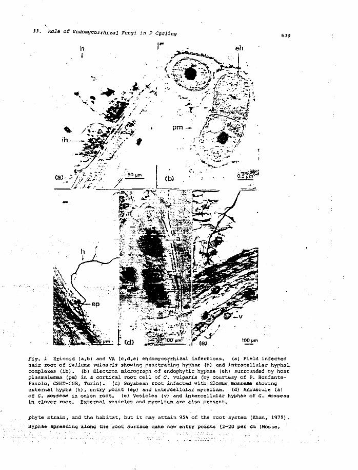
33. Role of Endomycorrhizal Fungi in P Cgcling 639
Fig. 1 Ericoid (a,b) and VA (c,d,e) endomycorrhizal infections. (a) Field infected hair root of Calluna vulgaris showing penetrating hyphae (h) and intracellular hyphal complexes (ih). (b) Electron micrograph of endophytic hyphae (eh) surrounded by host plasmalemma (pm) in a cortical root cell of C. vulgaris (by courtesy of P. BonfanteFasolo, CSHT-CNR, Turin). (c) Soyabean root infected with Glomus mosseae showing external hypha (h), entry point (ep) and intercellular mycelium. (d) Arbuscule (a) of G. mosseae in onion root. (e) Vesicles (v) and intercellular hyphae of G. mosseae in clover root. External vesicles and mycelium are also present.
phyte strain, and the habitat, but it may attain 95\ of the root system (Khan, 1975).
Hyphae spreading along the root surface make new entry points [2-20 per em (Mosse,
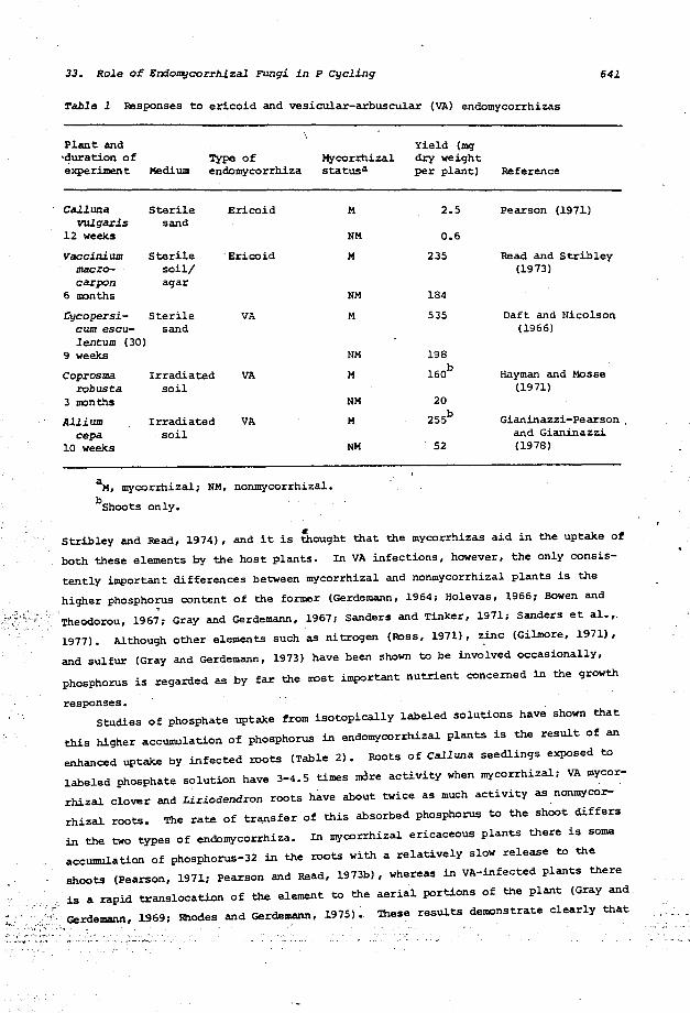
33. Role of Endol1qcorrh.izal Fungi in P Cycling 641
Table 1 Responses to ericoid and vesicular-arbuscular (VA) endomycorrhizas
Plant and '~uration of experiment Medium
Type of endomycorrhiza
Call una Sterile Ericoid vulgaris sand
12 weeks
Vaccinium Sterile " Ericoid macro- soil/ carpon agar
6 IlDnths
Cycopersi- Sterile VA cum escu- sand lentum (30)
9 weeks
Coprosma Irradiated VA robusta soil
3 IlDnths
Allium I=adiated VA cepa soil
10 weeks
Mycorrhizal statusa
M
NM
M
NM
M
NM
M
NM
M
NM
aM, mycorrhizal; NM, nonmycorrhizal.
bShoots only.
Yield (1119' dry weight per plant)
2.5
0.6
235
184
535
198
160b
20
255b
52
Reference
Pearson (1971)
Read and Stribley (1973)
Daft and Nicolson (1966)
Hayman and Masse (1971)
Gianinazzi-pearson. and Gianinazzi (1978)
Stribley and Read, 1974), and it is ~ought that the mycorrhizas aid in the uptake of
both these elements by the host plants. In VA infections, however, the only consis
tently important differences between mycorrhizal and nonmycorrhizal plants is the
higher phospho~ content of the former (Gerdemann, 1964; Holevas, 1966; Bowen and
Theodorou, 1967; Gray and Gerdemann, 1967; Sanders and Tinker, 1971; Sanders et al.,.
1977). Although other elements such as nitrogen (Ross, 1971), zinc (Gilmore, 1971),
and sulfur (Gray and Gerdemann, 1973) have been shown to be involved occasionally,
phosphorus is regarded as by far the IlDst important nutrient concerned in the growth
responses. Studies of phosphate uptake from isotopically labeled solutions have shown that
this higher accumulation of phosphorus in endomycorrhlzal plants is the result of an
enhanced uptake by infected roots (Table 2). Roots of calluna seedlings exposed to
labeled phosphate solution have 3-4.5 times mdre activity when mycorrhizal; VA mycor
rhizal clover and Liriodendron roots have about twice as much activity as nonmycor
rhizal roots. The rate of transfer of this absorbed phosphorus to the shoot differs
in the two types of endomycorrhiza. In mycorrhizal ericaceous plants there is some
accumulation of phosphorus-32 in the roots with a relatively slow release to the
shoots (Pearson, 1971; Pearson and Read, 1973b), whereas in VA-infected plants there
is a rapid translocation of the element to the aerial portions of the plant (Gray and
A,"" ",,: '""" Gerdemann, 1969; Rhodes and Gerdemann, 1975). These results demonstrate clearly that
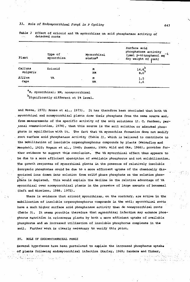
33. Role of Endomycorrhlzal Fungi in P Cycling
Table 3 Effect of ericoid and VA myco=hizas on acid phosphatase activity of detached roots
Surface acid phosphatase activity
643
Type of -1 Mycorrhizal (].!mol p-nitrophenol me; Plant mycorrhiza statusa
Call1llla Ericoid M vulgaris NM
Allium VA M cepa NM
aM, mycorrhizal; NM, nonmycorrhizal
bSignificantly different at 5\ level.
dry weight of foot)
b l4.6
b 8.0
1.5 1.6
and Mosse, 1972; Masse et al., 1973). It has therefore been concluded that both VA
mycorrhizal and nonmycorrhizal plants draw their phosphate from the same source and,
from measurements of the specific activity of the soil solutions (J. C. Fardeau, per
.sonal communication, 1978), that this source is the soil solution or adsorbed phos
phate in equilibrium with it. The fact that VA mycorrhiza formation does not modify
root surface acid phosphatase activity (Table 3), which is believed to contribute to
the mobilization of insoluble organophosphorus compounds by plants (Weissflog and
Mengdehl, 1933; Rogers et al., 1940; Saxena, 1964; wild and ake, 1966), provides fur
ther evidence to support this conclusion. The VA myco=hizal effect thus appears to
be due to a more efficient absorption of available phosphorus and not solubilization.
The growth responses of mycorrhizal plants in the presence of relativelY insoluble
inorganic phosphates could be due to a more efficient uptake of the chemically dis
sociated ions drawn into solution from solid phase phosphate as the solution phos-~.
pit~~ is depleted. This would explain the decline in the relative advantage of VA
mycorrhizal over nonmycorrhizal plants in. the presence of large amounts of bonemeal
(Daft and Nicolson, 1966, 1972).
There is evidence that ericoid mycorrhizas, on the contrary, are active in the
mobilization of insoluble organophosphorus compounds in the soil; mycorrhizal roots
have a much higher surface acid phosphatase activity than do nonmycorrhizal roots
(Table 3). It seems possible therefore that ~corrhiZal infection may enhance phos
phorus nutrition in ericaceous plants by both a more efficient uptake of available
phosphorus and an increased utilization of insoluble phosphorus complexes in the
soil. Further work is clearly necessary to verify this point.
IV. ROLE OF ENDOMYCORRHIZAL FUNGI
Several hypotheses have been postulated to explain the increased phosphorus uptake
of plants following endomycorrhizal infection (Harley, 19691 Sanders and Tinker,
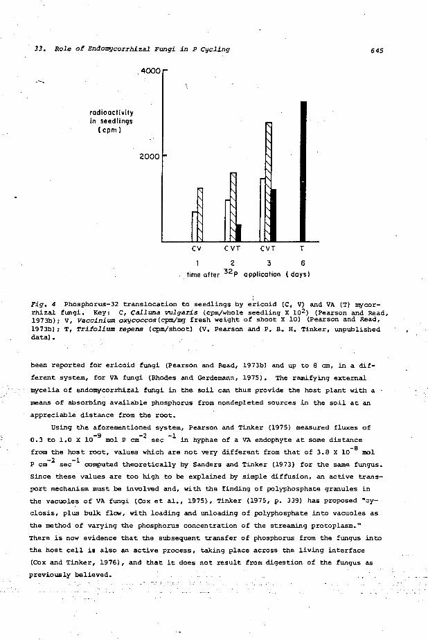
33. Role of Endoll1!lcorrhizal Fungi in P Cycling
.-,
radioactivity In seedlings
(cpm)
,4000 r
2000
CV CVT CVT T
2 3 6
time after 32p application (days)
,
645
Fig. 4 Phosphorus-32 translocation to seedlings by ericoid (C, VI and VA (T) mycorrhizal fungi. Key: C, Calluna vulgaris (cpm/whole seedling X 102 ) (Pearson and Read, 1973b)1 V, vaccinium oxycoccos(cpm!mg fresh weight of shoot X 10) (Pearson and Read, 1973b) 1 T, Trifolium repens (cpm/shoot) (V. Pearson and P. B. H. Tinker, unpublished data) •
been reported for ericoid fungi (Pearson and Read, 1973b) and up to 8 em, in a dif
ferent system, for VA fungi (Rhodes and Gerdemann, 1975). The ramifying external
mycelia of endomycorrhizal fungi in the soil can thus provide the host plant with a
means of absorbing available phosphorus from nondepleted sources in the soil at an
appreciable distance from the root.
Using the aforementioned system, Pearson and Tinker (1975) measured fluxes of
0.3 to 1.0 X 10⁻⁹ mol P cm-2 sec -1 in hyphae of a VA endophyte at some distance
from the host root, values which are not very different from that of 3.8 X 10⁻⁸ mol
P cm -2 sec -1 computed theoretically by Sanders and Tinker (1973) for the same fungus.
Since these values are too high to be explained by simple diffusion, an active trans
port mechanism must be involved and, with the finding of polyphosphate granules in
the vacuoles of VA fungi (COx et al., 1975)., Tinker (1975, p. 339) has proposed "cy
closis, plus bulk. flow, with loading and unloading of polyphosphate into vacuoles as
the method of varying the phosphorus concentration of the streaming protoplasm."
There is now evidence that the subsequent transfer of phosphorus from the fungus into
the host cell is also an active process, taking place across the living interface
(Cox and Tinker, 1976), and that it does not result from digestion of the fungus as
previously believed.
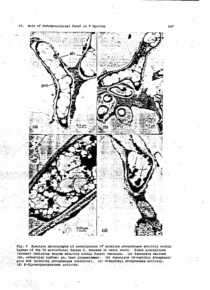
33. Role of Endomycorrhizal Fungi in P Cycling 647
.-, .- ;.
,'-1",-"'" .... _'
.::.:~-:. ~':.:. ·:!1-<\·,\~i·
" :;~'h" ." .. ' ....... ~ ...
-.--~-.~-
..
Cd)
Fig. 6 Electron micrographs of localization of alkaline phosphatase activity within hyphae of the VA mycorrhizal fungus G. mosseae in onion roots. Black precipitate
. (arrows) indicates enzyme activity within fungal vacuoles. (a) Substrate omitted (ah, arbuscular hyphae; pm, host plasmalemma). (b) Substrate (a-naphthyl phosphate)
, . plus KCN {alkaline phosphatase inhibitor). (c) a-Naphthyl phosphatase activity • . ~ (d) ~-Glyceropl1osphatase activity.
33. Role of Endol11l}corrhizal Fungi in P Cycling 649
REFERENCES
Asimi, S., Gianinazzi-pearson, V., Gianina:t:zi, 5., Obaton, M., and Berthau. S. (1978). Interactions entre les endomycorrhizes il vesicules et arbuscules (VA) et Ie RbizoblW11 chez Ie soja. 1036 congr. Nat. Soc. Sav., Vol. 1, pp. 247-256.
Bannister, P., and Norton, W. M. (1974). The response of mycorrhizal rooted cuttings of heather (Calluna vulgaris (L) Hull) to variations in nutrient and water regimes. New Phytol. 73: 81-88.
Bertheau, Y. (1977). Etudes des phosphatases soluhles des endomycorhi:t:es a vesicules et arbuscules. D.E.A. thesis, University of Dijon, France.
Beslow, D. T., Hacskaylo, E., and Melhuish, J. H. (1~70). Effects of environment on beaded root development in red maple. Bull. Torrey Bot. Club 97: 248-252.
Bowen, G. D., and Theodorou, C. (1967). Studies on phosphate uptake by mycorrhizas. Proc. 14th IUFRO Congr. 24: 116-138.
Brook, P. J. (1952). Mycorrhiza of Pernettya macrostigma. New Phytol. 51: 388-397.
Burgeff, H. (1961). Mikrabiologie des Hochmoores. FiSCher, Stuttgart.
Cosgrove, D. J., and Tate, M. E. (1963). The occurrence of neo-inosito1 hexaphosphate in soil. Nature 200: 568-569.
Cox, G., and Sanders, F. (1974). vesicu1ar-arbuscu1ar mycorrhiza.
Ultrastructure of the host-fungus interface in a New Phytol. 73: 901-~12.
Cox, G., and Tinker, P. B. (1976). Translocation and transfer of nutrients in vesicular-arbuscu1ar mycorrhiza. I. The arbuscu1e and phosphorus transfer: A quantitative ultrastructural study. N(f!/Jf Phytol. 77: 371-378.
Cox, G. C., sanders, F. E. T., Tinker, P. B., and Wild, J. (1975). Ultrastructural evidence relating to host-endophyte transfer in a vesicular-arbuscu1ar mycorrhiza. In Endol11l}corrhizas, F. E. sanders, B. Masse, and P. B. Tinker (Eds.). Academic Press, New York, pp. 297-312.
Daft, M. J., and Nicolson, T. H. (1966). Effect of Endogone mycorrhiza on plant growth. New Phytol. 65: 343-350.
Daft, M. J., and Nicolson, T. H. (1972). Effect of Endogene mycorrhiza on plant growth. IV. Quantitative relationships between the growth of the host and the development of the endophyte in tomato and maize. New PhytOl. 71: 287-295.
Dexheimer, J., Gianinazzi, S., and Gianinazzi-Pearson, V. (1979). ultrastructural cytochemistry of the host-fungus interfaces in the endomycorrhizal association Glomus mosseae/Allium cepa. Z. Pflanzenpnysiol. 92: 191-206.
Fitter, A. H. (1977). Influence of mycorrhizal infection on competition for phosphorus and potassium by two grasses. New Phytol. 79: 119-125.
Freis1eben, R. (1936). Jahrb. Wiss. Bot. 82:
Weitere Untersuchungen Uher die Mykotrophie der Ericaceen. 413-459.
Gerdemann, J. W. (1964). The effect of mycorrhiza on the growth of maize. Nycologia 56: 342-349.
Gerdernann, J. W., and Trappe, J. M. (1974). The Endogonaceae of the Pacific Northwest. Mycol. Memoir 5: 75 pp.
Gianinazzi, SOl Gianinaz:t:i-Pearson', V., and Dexneimer, J. (1979). Enzymatic studies on the metabolism of vesicular-arbuscular mycorrhiza. III. Ultrastructural localisation of acid and alkaline phosphatase in onion roots infected by Glomus llDsseae NichOl. & Gerd. New Phytol. 82: 127-132.
Gianinazzi-Pearson, V. (1976). Les mycorhizes endotrophes: etat actue1 des connaissances et possibi1ites d'app1ication dans 1a pratique cu1tura1e. Ann. Phytopath. 8: 249-256.
" 33. Role of Endomycorrhizal Fungi in P Cycling 651
Mitchell, H. L., Finn, R. F., and Rosendhal, R. O. (1937). The relation between mycorrhizae and the growth and nutrient absorption of coniferous seedlings in nursery beds. Black Rock For. Papers 1: 58-73.
Morrison, T. M. (1957). stigma. New Phytol. 56:
Host-endophyte relationship in mycorrhiza of Pernettya macro-247-257.
Mosse, B. (1959). The regular germination of resting spores and some observations on the growth requirements of Endogone species causing vesicul~amuscular mycorrhizas. Trans. Brit. Hycol. Soc. 42: 274-286.
Mosse, B. (1973). Advances in the study of vesicular-arbuscular myco=hiza. Ann. Rev. Phytopath. 11: 171-196.
Mosse, B., and Bowen, G. D., quoted by Bowen anQ fheodorou (1967), above.
Mosse, B., Hayman, D. 5., and Arnold, D. (1973). Plant growth responses to vesicu1ar-arbuscular mycorrhiza. V. Phosphate uptake by three plant species from P-deficient soils labelled with 32p. New Phytol. 72: 809-815.
Murdoch, C. L., Jackobs, J. A., and Gerdemann, J. W. (1967). Utilization of phosphorus sources of different availability by mycorrhizal and non-mycorrhizal maize. Plant Soil 27: 329-334.
Nieuwdorp, P. J. (1969). Some investigations on the mycorrhiza of Calluna, Erica, and Vaccinium. Acta Bot. Neerl. 18: 180-196.
North, J., and Lewis, D. (1971). Phosphatases of Coprinus lagopus: for their production and the genetics of the alkaline phosphatase. (Cambridge) 18: 153-166.
The condi tions Genet. Res.
Nye, P. H. (1969). The soil =de1 and its application to plant nutrition. In Ecological Aspects of the Hineral Nutrition of Plants, Symp. Brit. Ecol. Soc. 9: 201-213.
Pearson, V. (1971). The biology of mycorrhiza in the Ericaceae. Ph.D. thesis, University of Sheffield, England.
Pearson, V., ·and Read, D. J. (1973a). I. The isolation of the endophyte and New Phytol. 72: 371-379.
The biology of mycorrhiza in the Ericaceae. synthesis of mycorrhiza in aseptic culture.
Pearson, V., and Read, D. J. (1973b). The biology of mycorrhiza in Ericaceae. II. The transport of carbon and phosphorus by the endophyte and the mycorrhiza. New Phytol. 72: 1325-1331.
Pearson, V., and Read, D. J. (1975). The physiology of the myco=hizal endophyte of CallUM vulgaris. Trans. Brit. Hycol. Soc. 64: 1-7.
Pearson, V., and Tinker, P. B. H. (1975). Measurement of phosphorus fluxes in the external hyphae of endomycorrhizas. In Endomycorrhizas, F. E. Sanders, B. Masse, and P. B. Tinker (Eds.). Academic Press, New York, pp. 277-287.
Read, D. J. (1974). Pezizella ericae sp. nov. the perfect state of a typical mycorrhizal endophyte of Ericaceae. Trans. Brit. Hycol. Soc. 63: 381-383.
Read, D. J., and Strib1ey, D. P. (1973). Effect of mycorrhizal infection on nitrogen and phosphorus nutrition of ericaceous plants. Nature 244: 81-82.
Read, D. J., and Strib1ey, D. P. (1975). Some mycological aspects of the biology of mycorrhiza in the Ericaceae. In Endomycorrhizas, F. E. Sanders, B. Mosse, and P. B. Tinker (Eds.). Academic Press, New York, pp. 105-117.
Rhodes, L. H., and Gerdemann, J. W. (1975). and non-mycorrhizal onions. New Phytol. 75:
Phosphate uptake zones of mycorrhizal 555-561.
Rogers, H. T., Pearson, R. w., and Pierre, N. H. (1940). Absorption of organophosphates by corn and tomato plants and the mineralizing action of exoenzyme systems of growing roots. Soil. Sci. Soc. Amer. Proc. 5: 285-291.
Ross, J. P. (1971). Effect of phosphate fertilization on yield of myco=hizal and non-mycorrhizal soybeans. Phytopathol. 61: 1400-1403.
652 Gianinazzi-Pearson and Gianinazzi
sanders, F. E., and Tinker, P. B. (1971). Mechanism of absorption of phosphate by Eooogone myconhiza5. Nat.ure 233: 278:-279.
\
sanders, F. E. T., and Tinker, P. B. H. (1973). Phosphate flow into mycorrhizal roots. Pest.ie. Sci. 4: 385-395.
Sanders, F. E. T., Tinker, P. B., Black, R. L. B., and Palmerley, S. M. (1977). The development of endomycorrhizal root systems. I. Spread of infection and growth-promoting effects with four species of vesicular-arbuscular endophyte. New Phyt.ol. 78: 257-268.
Saxena, S. N. (1964). Phytase activity of plant roots. J. Exp. Bot.. 15: 654-658.
Stribley, O. P., and Read, D. J. (1974). The biology of mycorrhiza in the Ericaceae. IV. The effect of mycorrhizal infection on uptake of 15N from labelled soil ~y Vaccinium maerocarpon Ait. New Phytol. 73: 1149-1155.
Stribley, O. P., and Read, D. J. (1976). The biology of mycorrhiza in the Ericaceae. VI. The effects of mycorrhizal infection and concentration of ammonium nitrogen on growth of cranberry (Vaccinium macrocarpon Ait.) in sand culture. New Phytol. 77: 63-72.
Stribley, D. P., Read, D. J., and Hunt, R. (1975). The biology of mycorrhiza in the Ericaceae. V. The effects of mycorrhizal infection, soil type and partial soil sterilization (by gamma-irradiation) on growth of cranberry (Vaccinium macrocarpon Ait.). New Phyt.ol. 75: 119-130.
Tinker, P. B. H. (1975). Effects of vesicular-arbuscular mycorrhizas on higher plants. In Symbiosis, Symp. Soc. Exp. Biol. 29: 325-349.
Weissflog, J., and Mengdehl, H. (1933). Studien zum Phosphorstoffwechsel in den hOheren Pflanzen. III. Aufnahme und Verwertbarkeit organischer Phosphosaureverbindungen durch die Planze. Plant.a 19: 182-241.
Wild, A" and Oke, O. L. (1966). Organic phosphate compounds in calcium chloride extracts of soil: Identification and availability to plants. J. Soil Sci. 17: 356-' 371.
REFERENCES ADDEO IN PROOF
Asimi, S., Gianinazzi-Pearson, V., and Gianinazzi, S. (1980). InflUence of increasing soil phosphorus levels on interactions between vesicular-arbuscular mycorrhizae and Rhizobium in soybeans. Can. J. Bot. 58: 2200-2206.
Bonfante-Fasolo, P., and Gianinazzi-Pearson, V. (1979). Ultrastructural aspects of endomycorrhiza in the Ericaceae. I. Naturally infected hair roots of Calluna vulgaris L. Hull. New Phytol. 83: 739-744.
Callow, J. A~, Capaccio, L. C. M., Parish, G., and Tinker, P. B. (1978). Detection and estimation of polyphosphate in vesicular-arbuscular mycorrhizas. New Phytol. 80: 125-134.
Cooper, K. M., and Tinker, P. B. (1978). vesicular-arbuscular mycorrhizas. II. and sulphur. New Phytol. 81: 43-52.
Translocation and transfer of nu.t.r.ients in Uptake and translocation of phosphorus, zinc
Cox, G., Moran, K. J., Sanders, F., Nockolds, C., and Tinker, P. B. (1980). location and transfer of nutrients in vesicular-arbuscular mycorrhizas. III. phosphate granules and phosphorus translocation. New Phytol. 84: 649-659.
TransPoly-
Cress, W. A., Throneberry, G. 0., and Lindsey, D. L. (1979). Kinetics of phosphorus absorption in mycorrhizal and nonmycorrhizal tomato roots. plant Physiol. 64: 484-487.
9 JUl 1986'
Related Documents