Studies on safety issues in anticoagulant management

Welcome message from author
This document is posted to help you gain knowledge. Please leave a comment to let me know what you think about it! Share it to your friends and learn new things together.
Transcript
Studies on safety issues in
anticoagulant management

Pieter Eijgenraam, Maastricht 2015
Cover: Boris Eijgenraam
Layout: Tiny Wouters
Printed by: Proefschriftmaken.nl || Uitgeverij BOXPress
ISBN: 978‐90‐9029390‐5
Studies on safety issues in
anticoagulant management
PROEFSCHRIFT
Ter verkrijging van de graad van doctor aan de Universiteit Maastricht,
op gezag van de Rector Magnificus, Prof. Dr. L.L.G. Soete,
volgens het besluit van het College van Decanen,
in het openbaar te verdedigen op
donderdag 17 december 2015 om 10.45 uur
door
Pieter Eijgenraam
Geboren op 27 oktober 1963 te Leiden
Promotor
Prof. dr. H. ten Cate
Copromotores
Dr. A.J. ten Cate‐Hoek
Dr. R. van den Ham (Philips Research)
Beoordelingscommissie :
Prof. dr. J.G. Maessen (voorzitter)
Dr. J.F.B.M. Fiolet
Prof. dr. P.W. Kamphuisen (UMC Groningen)
Prof. dr. R.P. Koopmans
Dr. M.J.H.A. Kruip (Erasmus Medisch Centrum Rotterdam)
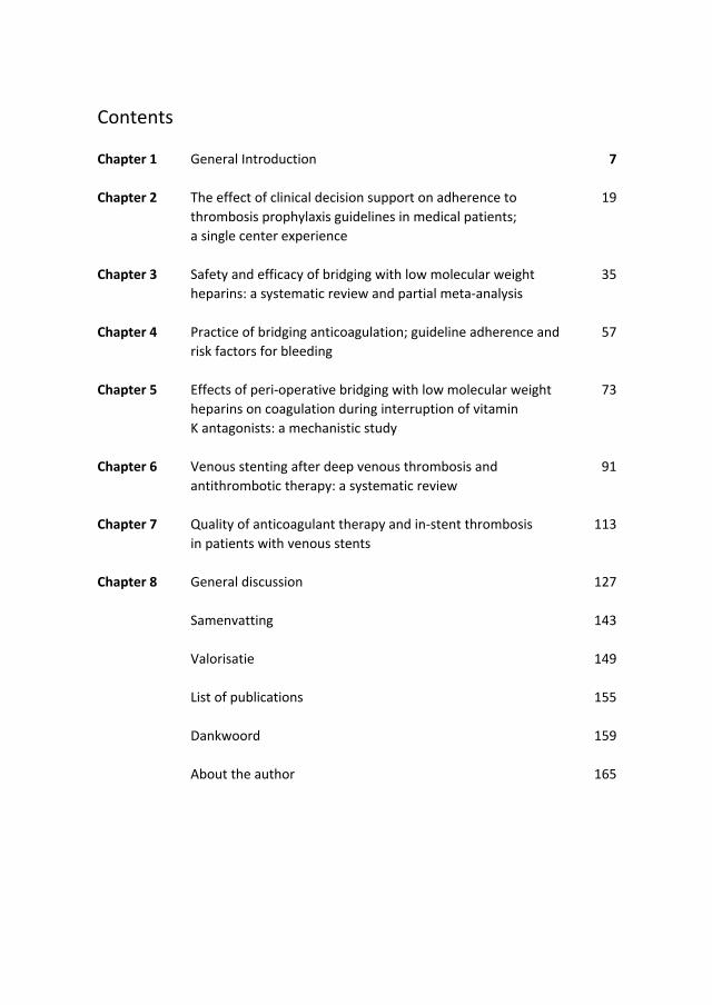
Contents
Chapter 1 General Introduction 7
Chapter 2 The effect of clinical decision support on adherence to 19
thrombosis prophylaxis guidelines in medical patients;
a single center experience
Chapter 3 Safety and efficacy of bridging with low molecular weight 35
heparins: a systematic review and partial meta‐analysis
Chapter 4 Practice of bridging anticoagulation; guideline adherence and 57
risk factors for bleeding
Chapter 5 Effects of peri‐operative bridging with low molecular weight 73
heparins on coagulation during interruption of vitamin
K antagonists: a mechanistic study
Chapter 6 Venous stenting after deep venous thrombosis and 91
antithrombotic therapy: a systematic review
Chapter 7 Quality of anticoagulant therapy and in‐stent thrombosis 113
in patients with venous stents
Chapter 8 General discussion 127
Samenvatting 143
Valorisatie 149
List of publications 155
Dankwoord 159
About the author 165


7
Chapter 1 General introduction

Chapter 1
8
General introduction
9
Hemostasis
Blood coagulation is responsible for the prevention of fatal blood loss but also for the
occurrence of venous and arterial thrombi in the vascular system, leading to a partial
or total interruption of the blood stream. Primary or secondary prevention of venous
or arterial thromboembolism (TE) often encompasses the administration of oral
and/or parenteral anticoagulant agents. All anticoagulant use increases the risk of
bleeding. Bleedings attributable to anticoagulant use can sometimes be fatal. The
determination of the ideal type, dosing and timing of anticoagulant therapy is an
ongoing challenge for patient care; a delicate balance has to be found between the
risk of thromboembolism and bleeding for all individual patients.
The coagulation system is based on well balanced steps regulated by the coagulation
proteases, the vessel wall and platelets. The liver is responsible for the production of
most of the coagulation proteases like factors V, VII, IX, X, XI, XII, prothrombin and
fibrinogen. The coagulation system can be activated in two ways, via the intrinsic and
the extrinsic pathway. Thrombin is the central enzyme in blood coagulation, the
product of a series of protease directed protein cleavages, starting with the exposure
of tissue factor (TF) on the sub‐endothelium or at microparticles in the circulation. The
tenase and prothrombinase complex jointly further catalyze the transformation of
prothrombin into thrombin and result in a burst of thrombin.1
Different mechanisms lead to the inhibition of coagulation. In one of these
mechanisms thrombin in complex with thrombomodulin enables the activation of
protein C into activated protein C (APC). APC associated with protein S in turn
inactivates activated FV (FVa) and FVIIIa, leading to inhibition of thrombin production
via a negative feedback loop.1
Thrombosis and bleeding
Abnormalities in coagulation can lead to the formation of a thrombus. Arterial TE, a
major health problem especially in the elderly population, is associated with
conditions such as atrial fibrillation, arteriosclerosis, or the introduction of mechanical
aortic/mitral valves prosthesis. Deep venous thrombosis (DVT) and subsequently
pulmonary embolism (PE), collectively referred to as venous thromboembolism (VTE)
is caused by diverse risk conditions including malignant neoplasms, hospital or nursing
home confinement, trauma/surgery and neurological disease with extremity paresis.
The primary and secondary prevention of potentially life threatening and invalidating
TE requires adequate antithrombotic treatment, which consists mainly of

Chapter 1
10
anticoagulant and/or antiplatelet therapy.2 The most important adverse effect of
anticoagulant treatment is the risk of bleeding. Ideally, when antithrombotic therapy
is applied a balance is found between the risk of TE and bleeding.
Anticoagulant agents
In day to day practice a wide range of anticoagulants is available for the prevention of
TE.3 In this thesis we discuss safety and efficacy aspects of vitamin K antagonists (VKA)
and low molecular weight heparins (LMWH) in different settings. VKA inhibits the
activity of the vitamin K dependent procoagulant proteins prothrombin, FVII, FIX and
FX, and the anticoagulant proteins C and S. VKA inhibits the process of recycling
vitamin K by blocking VKORC1, resulting in a relative vitamin K deficiency in the liver
cell. This results in the production of impaired, non Υ carboxylated coagulation
factors.4,5 The anticoagulant effect of LMWH is mainly derived from the anti FXa effect
induced by a conformational change of antithrombin (AT). In the presence of LMWH
the anticoagulant effect of AT, particularly against FXa, is accelerated.5
Anticoagulation assays
Several blood tests are developed to monitor the anticoagulation intensity in patients.
The most commonly used test to monitor VKA therapy is the prothrombin time (PT).4
This test responds to changes in concentration of F II, VII and X, which are reduced by
acenocoumarol and fenprocoumon, VKAs commonly used in the Netherlands. This
reduction is proportional to the half‐life of the clotting factor; the first days after the
(re) initiation of VKA therapy PT mainly reflects the changes in F VII, which has a
relatively short half‐life of 6 hours. The PT assay is performed by adding calcium and a
thromboplastin to citrated plasma and is expressed in seconds. Due to for instance
different sensitivity of thromboplastins used the PT initially lacked standardization. In
1982 a calibration model was adapted to standardize reporting by correcting for
different thromboplastins used in different laboratories; International Normalized
Ratio (INR) = (patient PT/mean normal PT) ISI where International Sensitivity Index (ISI)
denotes the thromboplastin used in the local laboratory.6 Currently, the intensity of
VKA is measured by PT and expressed as INR. Frequent measurement of an
individual’s INR value is required in order to manage anticoagulation within a
therapeutic range (internationally, an INR range of 2.0‐3.0 is most common; in this
country 2.5‐3.5 is the most relevant range).
General introduction
11
The anti‐Xa assay is designed to monitor the effect of LMWH and unfractionated
heparin (UFH). There is no recommendation in guidelines for repeated use of an anti‐
Xa assay for dose adjustment in patients using LMWH for prophylaxis or treatment
options. According to experts in the field, the anticoagulant effect of LMWH only
needs to be monitored in obese patients, patients with reduced renal clearance7,8 and
during pregnancy.9 Although there is little firm evidence for appropriate therapeutic
anti‐Xa ranges (also considering the fact that there is little evidence supporting a
strong correlation between anti‐Xa activity and efficacy) the therapeutic peak
(2‐4 hours after subcutaneous injection) window is estimated at 1‐2 units/ml for odd
LMWH and 0.5‐1.0 units/ml for bid dosed LMWH. Renal impairment and multiple
therapeutic doses of LMWH can result in bioaccumulation and therefore increased
risk of bleeding. Testing anti‐Xa level in such patients would seem appropriate.
The thrombin generation (TG) assay offers a global view on hemostasis. This assay can
be performed in both platelet poor and platelet rich plasma. TG measures the
concentration of thrombin over time formed after triggering coagulation with one of
the recommended stimuli: 1pM TF, 1 pM TF + thrombomodulin, and 5 pM TF. TG
results in a curve which describes the variation of the amount of thrombin during the
activation of coagulation cascade in time.10 The 2 most important parameters
obtained from this curve are peak height, the maximum concentration of thrombin at
a certain moment in time and the endogenous thrombin potential which represents
the total amount of thrombin produced over time.10 The position of TG measurement
in practice has not been established yet. Eventually, it is likely that simplified versions
of TG, for instance based on the whole blood point of care prototype assay11, will
allow broad applications, including for predicting TE and bleeding, or to monitor
anticoagulant therapy with VKA, LMWH, UFH and NOAC.10
Thrombosis prophylaxis and CDS
Thrombosis prophylaxis is a major topic, since many hospitalized patients are at
increased risk for VTE, due to for instance increased age, immobility, cancer or
surgery. The yearly incidence of DVT in the Netherlands is 0.6‐1.2 cases per 1000
inhabitants,12 the reported VTE incidence in hospitalized patients is 100 times
greater.13 In case of low VTE risk only early ambulation is advised; for patients at
increased risk, apart from early ambulation, daily doses of LMWH is the preferred
treatment option. Solid evidence of the efficacy of VTE prevention is presented in
numerous studies.14‐16 However, large studies show that antithrombotic prophylaxis is
underused in the hospital setting, leading to avoidable cases of VTE. In different

Chapter 1
12
studies only as little as 30‐50% of the patients received appropriate prophylactic
therapy.17‐19 In medical patients the rates of patients receiving appropriate
prophylaxis are even lower than in surgical patients.20 Reasons for underutilization
include unfamiliarity or disagreement with current guidelines, underestimation of VTE
risk or fear for bleeding complications.
Over the last years the use of clinical decision support (CDS) has gained more
attention. Prospectively validated risk assessment models (RAM) for VTE risk factors
are available for integration in CDS; risk factors for VTE and bleeding are awarded with
a score; simply adding the numbers after establishing a cut‐off point, results in an
advice to whether or not apply prophylactic interventions and in which dose. These
prophylactics include pharmacological interventions (LMWH, fondaparinux or UFH) or
mechanical interventions (graduated compression stockings, intermittent pneumatic
compression). Available RAMs for bleeding risk estimation are not prospectively
validated yet.15 High scores for VTE risk factors and low bleeding scores result in the
advice to apply pharmacological interventions, a high VTE risk score combined with a
high bleeding score results in the advice to apply mechanical measures and in case of
a low VTE and bleeding score only the advice of early ambulation is given. Several
studies in different settings have been performed to evaluate the effectiveness of
different CDS systems. In most studies a positive effect of CDS was established,
translating in increased guideline adherence or even a reduction of the incidence of
VTE.21,22 In some studies the effectiveness of CDS was temporary. Studies evaluating
CDS systems in which a direct link to the ordering system was provided and/or CDS
use was mandatory showed the best results.21,23,24
In chapter 2 of this thesis we present results of a study evaluating a pilot CDS system
(from September 1st to December 1st 2013) in the Maastricht University Medical
Center+ (MUMC+). CDS was introduced in cooperation with Philips Group Innovation
Research, Eindhoven, the Netherlands, on two different wards comprising mainly of
medical patients. The ACCP guidelines 2012, the prevention of VTE in nonsurgical
patients,15 were used to build the CDS. The application was installed on 4 stand‐alone
personal computers in two different wards. The hospital patient data system (SAP,
Germany) menu was extended with a dedicated CDS button on the opening page of
the patient’s record. A direct link to the ordering system of pharmacological or
mechanical prophylaxis was not provided. All attending physicians of the wards were
trained in the use of the CDS system and motivated to use CDS daily for all admitted
patients.
General introduction
13
Bridging therapy
Anticoagulation in patients receiving long‐term VKA undergoing surgery is challenging
in terms of maintaining the delicate balance between bleeding and TE; the patient
using anticoagulants is vulnerable for bleeding in the perioperative period. During a
preferably short periprocedural period the patient should not be anticoagulated, to
avoid bleeding during and after the intervention. The anticoagulation‐free interval is
minimized by “bridging” with LMWH or UFH which replace the longer acting VKA. The
efficacy and safety of bridging with LMWH has however not been unequivocally
established.3 In chapter 3 of this thesis results are presented of a systematic review
analyzing the safety and efficacy aspects of bridging therapy. Several studies show
that the risk of bleeding during bridging therapy is increased, while TE risk is
unknown.25‐32 Furthermore, guidelines for optimal use of bridging anticoagulation
seem poorly adhered to.33 In chapter 4 results of a study analyzing guideline
adherence in a Dutch University hospital are presented.
In 2012 the American College of Chest Physicians (ACCP) presented their latest
guidelines for perioperative management of antithrombotic therapy.34 In the
management of perioperative anticoagulation in patients on VKA, 3 options can be
considered. First, in procedures with a low bleeding risk and the possibility of local
hemostatic measures, such as dental extractions, cataract operations and small
dermatologic procedures, it is considered a safe choice to continue VKA use. Second,
in patients at low risk of TE, ACCP guidelines recommend stopping warfarin
administration 5 days before the intervention and restarting warfarin 12‐24 hours
after the procedure when adequate hemostasis is secured. Finally, in patients at
moderate or high TE risk current ACCP guidelines recommend bridging therapy
consisting of parenteral administration of LMWH or UFH in the periprocedural period,
combined with interruption of VKA use. The decision to apply bridging anticoagulation
should always be based on an assessment of individual patient‐ and surgery‐related
factors.34 The suggested TE risk stratification by the ACCP is based mainly on indirect
evidence from studies outside of the perioperative setting involving patients with a
mechanical heart valve, chronic atrial fibrillation or VTE who either were not receiving
anticoagulation or were receiving less‐effective treatment.34‐37 It is conceivable that
due to the lack of insight in hypo or hypercoagulability during bridging therapy in
individual patients the concept of bridging anticoagulation might be qualified as a
‘black box’. In chapter 5 we present the results of a study in which the (interactive)
effects of the co‐administration of VKA and LMWH and surgery on different
coagulation assays, including thrombin generation assay, INR, anti Xa assays and the
Chapter 1
14
measurement of concentrations of vitamin K dependent coagulation factors, were
assessed.
Venous stenting
Post thrombotic syndrome (PTS) develops in more than 50% of patients with
iliofemoral DVT (IFDVT).38 Conventional treatment regimes, comprising of a
combination of compression therapy, mobilization and oral anticoagulants, up till now
mainly VKA, do not always lead to rapid resolution of symptoms or recanalization of
venous occlusions, but are associated with long‐term disability. Since several years the
use of percutaneous transluminal angioplasty (PTA) and stenting in the venous system
in patients with outflow obstructions of the iliofemoral veins has gained more
attention. PTA and stenting appears to be effective in terms of improvement of PTS
symptoms and has shown to be characterized by good mid‐ to long‐term patency
rates in mainly observational studies.39‐45 In acute IFDVT patients in whom catheter
directed thrombolysis (CDT) is applied, additional venous stenting immediately
following thrombolysis is often deemed indicated in case of underlying venous
pathology, such as iliac vein compression syndromes, which may be the cause of the
thrombosis in these patients.46 Both CDT and stenting procedures are usually followed
by anticoagulant therapy with VKA for at least 3 months; patients with PTS usually
already use VKA prior to the intervention.
Arterial stenting in combination with antiplatelet therapy has been applied in a larger
number of patients for a longer period of time; and as a consequence the body of
evidence concerning antithrombotic therapy has grown over time.47 However, so far
no data evaluating aspects of safety and efficacy for any antithrombotic therapy after
venous stenting have been published, resulting in the application of a wide range of
anticoagulant (VKA and new oral anticoagulants (NOAC)) and antiplatelet therapies)
for different periods of time in day‐to‐day practice. In chapter 6 of this thesis we
present the results of a systematic literature search addressing the issue of
antithrombotic therapy after venous stenting. In chapter 7 the influence of the quality
of anticoagulant treatment with VKA after stent placement was evaluated in terms of
stent re‐ occlusion. Time within therapeutic range (TTR) and the proportion of INR
values <2.0 were assessed as the main determinants of efficacy in this study. Chapter
8 provides a summary and general discussion of the contents of this thesis.
General introduction
15
References
1. Versteeg HH, Heemskerk JW, Levi M, Reitsma PH. New fundamentals in hemostasis. Physiological reviews. 2013;93:327‐58.
2. Heit JA, Silverstein MD, Mohr DN, Petterson TM, O'Fallon WM, Melton LJ, 3rd. Risk factors for deep
vein thrombosis and pulmonary embolism: a population‐based case‐control study. Arch Intern Med. 2000;160:809‐15.
3. Douketis JD, Berger PB, Dunn AS, Jaffer AK, Spyropoulos AC, Becker RC, et al. The perioperative
management of antithrombotic therapy: American College of Chest Physicians Evidence‐Based Clinical Practice Guidelines (8th Edition). Chest. 2008;133(6 Suppl):299S‐339S.
4. Ansell J, Hirsh J, Hylek E, Jacobson A, Crowther M, Palareti G, et al. Pharmacology and management of
the vitamin K antagonists: American College of Chest Physicians Evidence‐Based Clinical Practice Guidelines (8th Edition). Chest. 2008;133(6 Suppl):160S‐98S.
5. Harder S, Klinkhardt U, Alvarez JM. Avoidance of bleeding during surgery in patients receiving
anticoagulant and/or antiplatelet therapy: pharmacokinetic and pharmacodynamic considerations. Clin Pharmacokinet. 2004;43:963‐81.
6. Kirkwood TB. Calibration of reference thromboplastins and standardisation of the prothrombin time
ratio. Thromb Haemost. 1983;49:238‐44. 7. Samama MM, Poller L. Contemporary laboratory monitoring of low molecular weight heparins. Clin
Lab Med. 1995;15:119‐23.
8. Francis CW, Pellegrini VD, Jr., Totterman S, Boyd AD, Jr., Marder VJ, Liebert KM, et al. Prevention of deep‐vein thrombosis after total hip arthroplasty. Comparison of warfarin and dalteparin. The Journal
of bone and joint surgery American volume. 1997;79:1365‐72.
9. Nieuwenhuis HK, Albada J, Banga JD, Sixma JJ. Identification of risk factors for bleeding during treatment of acute venous thromboembolism with heparin or low molecular weight heparin. Blood.
1991;78:2337‐43.
10. Campo G, Pavasini R, Pollina A, Fileti L, Marchesini J, Tebaldi M, et al. Thrombin generation assay: a new tool to predict and optimize clinical outcome in cardiovascular patients? Blood Coagul
Fibrinolysis. 2012;23:680‐7.
11. Ninivaggi M, Apitz‐Castro R, Dargaud Y, de Laat B, Hemker HC, Lindhout T. Whole‐blood thrombin generation monitored with a calibrated automated thrombogram‐based assay. Clin Chem.
2012;58:1252‐9.
12. Linden MWvd, Westert, G.P., Bakker, D. de, Schellevis, F. Tweede Nationale Studie naar ziekten en verrichtingen in de huisartspraktijk: klachten en aandoeningen in de bevolking en in de
huisartspraktijk. Utrecht: NIVEL, 2004.
13. Duff J, Walker K, Omari A, Stratton C. Prevention of venous thromboembolism in hospitalized patients: analysis of reduced cost and improved clinical outcomes. Journal of vascular nursing : official
publication of the Society for Peripheral Vascular Nursing. 2013;31:9‐14.
14. Falck‐Ytter Y, Francis CW, Johanson NA, Curley C, Dahl OE, Schulman S, et al. Prevention of VTE in orthopedic surgery patients: Antithrombotic Therapy and Prevention of Thrombosis, 9th ed:
American College of Chest Physicians Evidence‐Based Clinical Practice Guidelines. Chest. 2012;141(2
Suppl):e278S‐325S. 15. Kahn SR, Lim W, Dunn AS, Cushman M, Dentali F, Akl EA, et al. Prevention of VTE in nonsurgical
patients: Antithrombotic Therapy and Prevention of Thrombosis, 9th ed: American College of Chest
Physicians Evidence‐Based Clinical Practice Guidelines. Chest. 2012;141(2 Suppl):e195S‐226S. 16. Gould MK, Garcia DA, Wren SM, Karanicolas PJ, Arcelus JI, Heit JA, et al. Prevention of VTE in
nonorthopedic surgical patients: Antithrombotic Therapy and Prevention of Thrombosis, 9th ed:
American College of Chest Physicians Evidence‐Based Clinical Practice Guidelines. Chest. 2012;141(2 Suppl):e227S‐77S.
17. Monreal M, Kakkar AK, Caprini JA, Barba R, Uresandi F, Valle R, et al. The outcome after treatment of
venous thromboembolism is different in surgical and acutely ill medical patients. Findings from the RIETE registry. J Thromb Haemost. 2004;2:1892‐8.
Chapter 1
16
18. Goldhaber SZ, Tapson VF, Committee DFS. A prospective registry of 5,451 patients with ultrasound‐
confirmed deep vein thrombosis. Am J Cardiol. 2004;93:259‐62. 19. Tapson VF, Decousus H, Pini M, Chong BH, Froehlich JB, Monreal M, et al. Venous thromboembolism
prophylaxis in acutely ill hospitalized medical patients: findings from the International Medical
Prevention Registry on Venous Thromboembolism. Chest. 2007;132:936‐45. 20. Cohen AT, Tapson VF, Bergmann JF, Goldhaber SZ, Kakkar AK, Deslandes B, et al. Venous
thromboembolism risk and prophylaxis in the acute hospital care setting (ENDORSE study): a
multinational cross‐sectional study. Lancet. 2008;371:387‐94. 21. Maynard GA, Morris TA, Jenkins IH, Stone S, Lee J, Renvall M, et al. Optimizing prevention of hospital‐
acquired venous thromboembolism (VTE): prospective validation of a VTE risk assessment model. J
Hosp Med. 2010;5:10‐8. 22. Umscheid CA, Hanish A, Chittams J, Weiner MG, Hecht TE. Effectiveness of a novel and scalable
clinical decision support intervention to improve venous thromboembolism prophylaxis: a quasi‐
experimental study. BMC medical informatics and decision making. 2012;12:92. 23. Kucher N, Koo S, Quiroz R, Cooper JM, Paterno MD, Soukonnikov B, et al. Electronic alerts to prevent
venous thromboembolism among hospitalized patients. N Engl J Med. 2005;352:969‐77.
24. Kucher N, Puck M, Blaser J, Bucklar G, Eschmann E, Luscher TF. Physician compliance with advanced electronic alerts for preventing venous thromboembolism among hospitalized medical patients. J
Thromb Haemost. 2009;7:1291‐6.
25. Ahmed I, Gertner E, Nelson WB, House CM, Dahiya R, Anderson CP, et al. Continuing warfarin therapy is superior to interrupting warfarin with or without bridging anticoagulation therapy in patients
undergoing pacemaker and defibrillator implantation. Heart Rhythm. 2010;7:745‐9.
26. Cano O, Osca J, Sancho‐Tello MJ, Olague J, Castro JE, Salvador A. Morbidity associated with three different antiplatelet regimens in patients undergoing implantation of cardiac rhythm management
devices. Europace. 2011;13:395‐401.
27. Chow V, Ranasinghe I, Lau J, Stowe H, Bannon P, Hendel N, et al. Peri‐procedural anticoagulation and the incidence of haematoma formation after permanent pacemaker implantation in the elderly. Heart
Lung Circ. 2010;19:706‐12.
28. Ghanbari H, Feldman D, Schmidt M, Ottino J, Machado C, Akoum N, et al. Cardiac resynchronization therapy device implantation in patients with therapeutic international normalized ratios. Pacing Clin
Electrophysiol. 2010;33:400‐6.
29. Krane LS, Laungani R, Satyanarayana R, Kaul S, Bhandari M, Peabody JO, et al. Robotic‐assisted radical prostatectomy in patients receiving chronic anticoagulation therapy: role of perioperative bridging.
Urology. 2008;72:1351‐5.
30. Li HK, Chen FC, Rea RF, Asirvatham SJ, Powell BD, Friedman PA, et al. No increased bleeding events with continuation of oral anticoagulation therapy for patients undergoing cardiac device procedure.
Pacing Clin Electrophysiol. 2011;34:868‐74.
31. Page SP, Siddiqui MS, Finlay M, Hunter RJ, Abrams DJ, Dhinoja M, et al. Catheter ablation for atrial fibrillation on uninterrupted warfarin: can it be done without echo guidance? J Cardiovasc
Electrophysiol. 2011;22:265‐70.
32. Tompkins C, Cheng A, Dalal D, Brinker JA, Leng CT, Marine JE, et al. Dual antiplatelet therapy and heparin "bridging" significantly increase the risk of bleeding complications after pacemaker or
implantable cardioverter‐defibrillator device implantation. J Am Coll Cardiol. 2010;55:2376‐82.
33. Eijgenraam P, ten Cate H, ten Cate‐Hoek AJ. Practice of bridging anticoagulation: guideline adherence and risk factors for bleeding. Neth J Med. 2014;72:157‐64.
34. Douketis JD, Spyropoulos AC, Spencer FA, Mayr M, Jaffer AK, Eckman MH, et al. Perioperative
Management of Antithrombotic Therapy: Antithrombotic Therapy and Prevention of Thrombosis, 9th ed: American College of Chest Physicians Evidence‐Based Clinical Practice Guidelines. Chest.
2012;141(2 Suppl):e326S‐50S.
35. Cannegieter SC, Rosendaal FR, Briet E. Thromboembolic and bleeding complications in patients with mechanical heart valve prostheses. Circulation. 1994;89:635‐41.
36. Hart RG, Benavente O, McBride R, Pearce LA. Antithrombotic therapy to prevent stroke in patients
with atrial fibrillation: a meta‐analysis. Ann Intern Med. 1999;131:492‐501.
General introduction
17
37. Douketis JD, Foster GA, Crowther MA, Prins MH, Ginsberg JS. Clinical risk factors and timing of
recurrent venous thromboembolism during the initial 3 months of anticoagulant therapy. Arch Intern Med. 2000;160:3431‐6.
38. Kahn SR, Shrier I, Julian JA, Ducruet T, Arsenault L, Miron MJ, et al. Determinants and time course of
the postthrombotic syndrome after acute deep venous thrombosis. Ann Intern Med. 2008;149:698‐707.
39. AbuRahma AF, Perkins SE, Wulu JT, Ng HK. Iliofemoral deep vein thrombosis: conventional therapy
versus lysis and percutaneous transluminal angioplasty and stenting. Ann Surg. 2001;233:752‐60. 40. Titus JM, Moise MA, Bena J, Lyden SP, Clair DG. Iliofemoral stenting for venous occlusive disease. J
Vasc Surg. 2011;53:706‐12.
41. Sillesen H, Just S, Jorgensen M, Baekgaard N. Catheter directed thrombolysis for treatment of ilio‐femoral deep venous thrombosis is durable, preserves venous valve function and may prevent
chronic venous insufficiency. Eur J Vasc Endovasc Surg. 2005;30:556‐62.
42. Kolbel T, Lindh M, Holst J, Uher P, Eriksson KF, Sonesson B, et al. Extensive acute deep vein thrombosis of the iliocaval segment: midterm results of thrombolysis and stent placement. J Vasc
Interv Radiol. 2007;18:243‐50.
43. Baekgaard N, Broholm R, Just S, Jorgensen M, Jensen LP. Long‐term results using catheter‐directed thrombolysis in 103 lower limbs with acute iliofemoral venous thrombosis. Eur J Vasc Endovasc Surg.
2010;39:112‐7.
44. Manninen H, Juutilainen A, Kaukanen E, Lehto S. Catheter‐directed thrombolysis of proximal lower extremity deep vein thrombosis: a prospective trial with venographic and clinical follow‐up. Eur J
Radiol. 2012;81:1197‐202.
45. Sharifi M, Mehdipour M, Bay C, Smith G, Sharifi J. Endovenous therapy for deep venous thrombosis: the TORPEDO trial. Catheter Cardiovasc Interv. 2010;76:316‐25.
46. Kim JY, Choi D, Guk Ko Y, Park S, Jang Y, Lee do Y. Percutaneous treatment of deep vein thrombosis in
May‐Thurner syndrome. Cardiovasc Intervent Radiol. 2006;29:571‐5. 47. Vik‐Mo H, Slette M, Hegbom K. [Antithrombotic therapy after percutanous coronary intervention with
stenting]. Tidsskrift for den Norske laegeforening : tidsskrift for praktisk medicin, ny raekke.
2008;128:436‐9.

Chapter 1
18
19
Chapter 2 The effect of clinical decision support on adherence
to thrombosis prophylaxis guidelines in medical
patients; a single center experience
Pieter Eijgenraam, Nathalie Meertens, René van den Ham, Hugo ten Cate,
Arina J ten Cate‐Hoek
Thromb Res. 2015;135: 464‐471
Chapter 2
20
Abstract
Introduction
Venous thromboembolism (VTE) is an underestimated health problem. The administration of
low molecular weight heparins (LMWH) to the appropriate patients dramatically decreases VTE
incidence. Clinical decision support (CDS) might contribute to thrombosis prophylaxis guideline
adherence.
Methods
A computerized integrated risk score program was used to estimate VTE and bleeding risk of
nonsurgical patients. A VTE risk score of ≥4 resulted in an advice to administer LMWH. We
selected 64 medical patients before the introduction of CDS (T0) and 64 patients after the
introduction (T1). We compared guideline compliance between these groups using chi2 tests.
Results
No difference between groups was found; Adherence to the guidelines at T0 was 59.4%, the
same percentage of 59.4% was found at T1. To evaluate the effect of the introduction of CDS in
terms of under and overtreatment we compared the prevalence of over and under treatment at
T1 and T0. The OR for receiving under treatment at T1 compared to T0 is 0.48 (95% CI:
0.18‐1.30), p=0.14. The OR for overtreatment at T1 compared to T0 is 1.66 (95% CI: 0.74‐3.73),
p=0.22
Conclusion
We found no improvement in guideline adherence towards anti thrombotic prophylaxis in
medical patients after the introduction of CDS in this pilot study. There was however a non‐
significant shift towards over treatment. Possible explanations for these results are the
increased awareness of the risk for thromboembolism induced by the study, suboptimal use of
CDS and deviation from CDS advice caused by patient’s preferences.
The effect of clinical decision support on adherence to thrombosis prophylaxis guidelines
21
Introduction
Deep venous thrombosis (DVT) and pulmonary embolism (PE) collectively referred to
as venous thromboembolism (VTE) represent a major health problem for hospitalized
patients. The yearly incidence of DVT in The Netherlands is 0.6‐1.2 cases per 1000
inhabitants.1 The reported VTE incidence in hospitalized patients is 100 times greater.2
Currently, antithrombotic prophylaxis with low molecular weight heparins (LMWH) or
in some cases unfractionated heparin is applied to prevent VTE. The positive effect of
antithrombotic prophylaxis in general surgical, orthopedic surgical and nonsurgical
patients on the incidence of VTE has been firmly established in different studies.3‐5
However, literature provides evidence supporting the thesis that antithrombotic
prophylaxis is underused in the hospital setting, leading to avoidable cases of VTE.
Different studies demonstrated that in only 30‐50% of the patients indicated for
prophylaxis appropriate measures are indeed taken.6‐8 The administration of
appropriate prophylaxis in medical patients is observed to be even less than in surgical
patients.9 Initiatives promoting the use of clinical decision support systems (CDS) or
simple electronic alerts have gained more and more attention and have proven
efficacy in terms of adherence to guidelines and in some cases reduction of VTE
incidence.10‐14 Especially in institutions such as university hospitals, with a high
throughput of inexperienced medical personnel in combination with a complex
patient load, CDS could function as a guide for the management of antithrombotic
prophylaxis.
We assessed whether the introduction of a computer based CDS embedded in the
hospital patient data system might lead to improved adherence to guidelines for
antithrombotic prophylaxis in medical patients.
Methods
CDS
From September 1st to December 1st 2013 a pilot study was performed in the
Maastricht University Medical Centre (MUMC+) on the introduction, use and
evaluation of a computer based CDS. Institutional review board approval was
obtained (METC 13‐5‐034). Before the introduction of CDS the application of
thromboprophylaxis was left to the discretion of the physician, who could consult
locally available web based protocols (ODIN), based on international guidelines. By
using CDS a protocol based VTE prophylaxis advice is generated, avoiding the need of
Chapter 2
22
consulting the underlying protocol by the prescribing physician. A customized
computerized integrated risk score program was used to estimate VTE and bleeding
risk of nonsurgical patients, as described below. All physicians involved in the pilot
study were collectively informed about the use and function of CDS and were
motivated to participate. The first risk assessment for all admitted medical patients
was performed within 24 hours after hospitalization and was hereafter repeated daily.
The CDS was installed on four stand‐alone personal computers on two wards of the
Maastricht University Medical Centre (MUMC+). The following inclusion criteria were
applied: 1) the patient is non‐surgical and admitted to one of the two participating
wards and 2) the (expected time) of admission is at least 48 hours. Excluded were 1)
patients on therapeutic anticoagulants and 2) patients with active bleeding.
The hospital patient data system (SAP, Germany) menu was extended with a
dedicated CDS button on the opening page of the patient’s record. The use of this
button was not mandatory and if CDS generated a recommendation for
antithrombotic prophylaxis, no automatic link to the pharmacotherapeutic ordering
system was provided.
The Padua Prediction Score11 for VTE risk factors, endorsed by the ACCP guidelines
20124, was used to compose the CDS data form. This risk assessment model (RAM),
prospectively validated in a study with patients not receiving prophylaxis15 awards
each risk factor with a maximum of 3 points. Scores of the different risk factors are
computed into a total VTE risk score, by simple addition. VTE risk was considered high
at a score of 4 points or more. The bleeding risk was assessed using a non‐validated
RAM.16 With a bleeding score of 7 or more points the patient was considered at high
risk of bleeding. Both the risk for thrombosis and the risk for bleeding were
dichotomized. A VTE risk score of <4 resulted in an advice not to administer
prophylaxis, a VTE score of ≥4 led to an advice to administer (weight adjusted) LMWH.
A bleeding score of >7 resulted in a warning in CDS that bleeding risk was high, in case
the score ≤7 points the announcement ‘low bleeding risk’ was depicted within CDS.
Assessment of compliance
In the period prior to the introduction of the CDS, when only the MUMC+ protocol,
mainly based on ACCP 2008 guidelines, was available for physicians as a guide to
prescribe the correct antithrombotic prophylaxis, compliance to antithrombotic
guidelines was assessed on two randomly selected dates for baseline measurements
(T0). The measurements were repeated on two randomly selected dates towards the
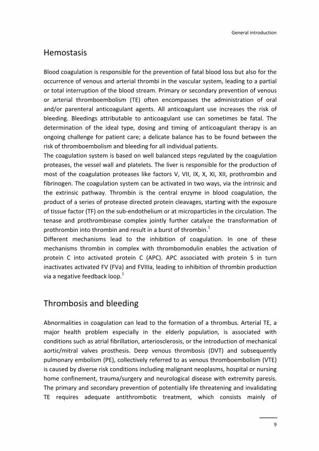
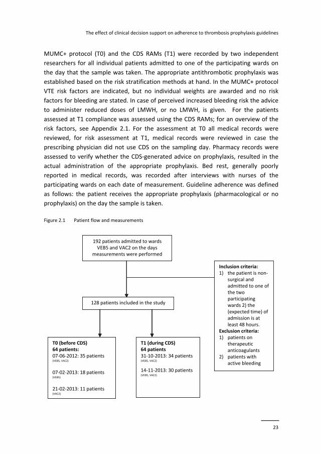
end of the CDS pilot period (T1). For an overview of the patient flow and
measurements see Figure 2.1. All risk factors for VTE and bleeding included in the
The effect of clinical decision support on adherence to thrombosis prophylaxis guidelines
23
MUMC+ protocol (T0) and the CDS RAMs (T1) were recorded by two independent
researchers for all individual patients admitted to one of the participating wards on
the day that the sample was taken. The appropriate antithrombotic prophylaxis was
established based on the risk stratification methods at hand. In the MUMC+ protocol
VTE risk factors are indicated, but no individual weights are awarded and no risk
factors for bleeding are stated. In case of perceived increased bleeding risk the advice
to administer reduced doses of LMWH, or no LMWH, is given. For the patients
assessed at T1 compliance was assessed using the CDS RAMs; for an overview of the
risk factors, see Appendix 2.1. For the assessment at T0 all medical records were
reviewed, for risk assessment at T1, medical records were reviewed in case the
prescribing physician did not use CDS on the sampling day. Pharmacy records were
assessed to verify whether the CDS‐generated advice on prophylaxis, resulted in the
actual administration of the appropriate prophylaxis. Bed rest, generally poorly
reported in medical records, was recorded after interviews with nurses of the
participating wards on each date of measurement. Guideline adherence was defined
as follows: the patient receives the appropriate prophylaxis (pharmacological or no
prophylaxis) on the day the sample is taken.
Figure 2.1 Patient flow and measurements
192 patients admitted to wards VEB5 and VAC2 on the days
measurements were performed
128 patients included in the study
Inclusion criteria: 1) the patient is non‐
surgical and admitted to one of the two participating wards 2) the (expected time) of admission is at least 48 hours.
Exclusion criteria:1) patients on
therapeutic anticoagulants
2) patients with active bleeding
T0 (before CDS)64 patients:07‐06‐2012: 35 patients (VEB5, VAC2)
07‐02‐2013: 18 patients(VEB5)
21‐02‐2013: 11 patients(VAC2)
T1 (during CDS)64 patients31‐10‐2013: 34 patients(VEB5, VAC2)
14‐11‐2013: 30 patients(VEB5, VAC2)
Chapter 2
24
In case guidelines were not followed, the following 2 options were recorded; 1) Under
treatment defined as not receiving LMWH, while an indication was present; 2) over
treatment defined as receiving LMWH without indication.
Evaluation of CDS use
After termination of the pilot we evaluated the use of CDS; a questionnaire consisting
of 4 domains was designed to identify possible barriers for CDS use. The questionnaire
is based on perceived barriers impeding guideline adherence among Dutch general
practitioners.17,18 We identified barriers related to knowledge (e.g. lack of awareness
of CDS, lack of familiarity with CDS), barriers related to attitude (e.g. lack of outcome
expectancy, lack of motivation), barriers related to behaviour (e.g. patients’
preferences not matching recommendations), and environmental factors (e.g. lack of
education, lack of time). See Appendix 2.2 for the complete questionnaire. We asked
the physicians who worked with CDS to complete the questionnaire anonymously.
Statistical analysis
To detect an improvement in guideline adherence from 50% to 75%, with a power of
80%, 128 (T0: 64, T1: 64) participants were needed. Cumulative first use of CDS of the
participating wards is reported. Descriptive statistics were used to determine patient
characteristics. Continuous variables are reported as means and their standard
deviations (SD); categorical data are presented as counts and percentages. Chi2 tests
are used to compare categorical variables including the compliance to the
antithrombotic prophylaxis guidelines before and after the introduction of CDS. To
estimate the strength of the association between the introduction of CDS and
observed compliance Phi statistics are used. Student’s t‐tests were used to compare
continuous variables. Under and over treatment was separately assessed. The
associations are also expressed as odds ratios (OR) with accompanying confidence
intervals (CI). A two sided p‐value <0.05 is considered statistically significant. Data
were analyzed with SPSS version 22.
Results
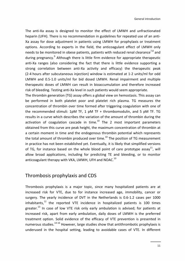
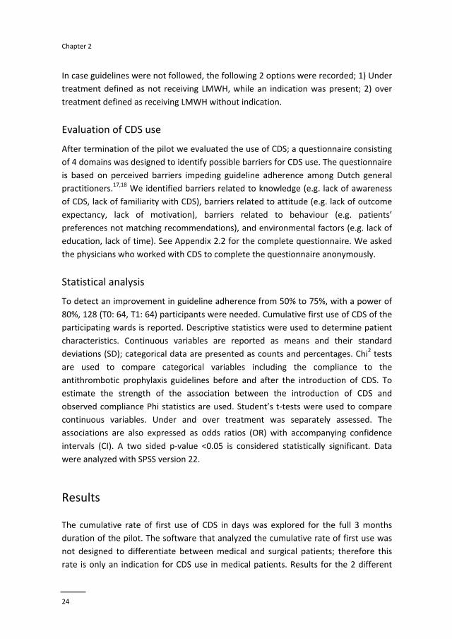
The cumulative rate of first use of CDS in days was explored for the full 3 months
duration of the pilot. The software that analyzed the cumulative rate of first use was
not designed to differentiate between medical and surgical patients; therefore this
rate is only an indication for CDS use in medical patients. Results for the 2 different
The effect of clinical decision support on adherence to thrombosis prophylaxis guidelines
25
wards (VEB5 and VAC2) are depicted in Figure 2.2. Ward VEB5 consists of
predominantly medical patients; VAC2 is a ward populated by both medical and
surgical patients, only medical patients were assessed.
Figure 2.2 Cumulative rate of first CDS use in time (days); ward VEB5 and VAC2
We included 128 patients; 64 patients were included in the baseline measurement
(T0), and 64 patients were included in the post CDS introduction measurement (T1).
For detailed patient characteristics of included patients at T0 and T1 see Table 2.1.
Anticipated bed rest with bathroom privileges for at least 3 days, a major risk factor,
was prescribed to 17.2% at T 0 and 3.1% at T1. In our two random samples the
medical records of in total 96 patients were reviewed (all 64 patients on T0, and 64
minus 32 CDS assessed patients at T1). The CDS score lists were completed on the day
the sample was taken in 50.0% of the cases; the remaining patients were evaluated
without CDS (17.2%), or on the basis of a CDS advice which was calculated on previous
days (32.8%).
According to the local protocol at T0, 45.3% of the patients should have received
pharmacological prophylaxis, 6.3% mechanical prophylaxis and 48.4% of patients were
not eligible for any thromboprophylaxis. Pharmacological prophylaxis was
administered in 50.0% of the cases; mechanical prophylaxis was rarely used, in only
1.6% of the patients.
According to the RAMs for VTE and bleeding risk assessment at T1 21.9% of the
patients should have received pharmacological prophylaxis, 6.3% mechanical
prophylaxis and 71.9% were not eligible for any antithrombotic prophylaxis. The
percentage of patients that received pharmacologic prophylaxis was more than twice
as high (45.3%), and mechanical prophylaxis was not used. For an overview of the
prescribed antithrombotic treatment modalities see Table 2.2. Adherence to the
34,3637,95
42,0545,64
50,77 52,3157,44
28,72
0,00
50,00
<1 <2 <3 <4 <5 <6 <7 >=7
Dagen na opname op afd.
Pe
rce
nta
ge
Cumulatief eerste gebruik CDSS
34,3637,95
42,0545,64
50,77 52,3157,44
28,72
0,00
50,00
<1 <2 <3 <4 <5 <6 <7 >=7
Dagen na opname op afd.
Pe
rce
nta
ge
Cumulatief eerste gebruik CDSS
Chapter 2
26
guidelines at T0 was 59.4%, the same percentage of 59.4% was found at T1, resulting
in a Pearson’s 2 of 0.00; p‐value=1.00.
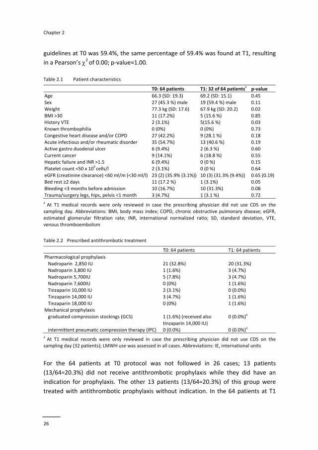
Table 2.1 Patient characteristics
T0: 64 patients T1: 32 of 64 patientsa p‐value
Age 66.3 (SD: 19.3) 69.2 (SD: 15.1) 0.45
Sex 27 (45.3 %) male 19 (59.4 %) male 0.11
Weight 77.3 kg (SD: 17.6) 67.9 kg (SD: 20.2) 0.02
BMI >30 11 (17.2%) 5 (15.6 %) 0.85 History VTE 2 (3.1%) 5(15.6 %) 0.03
Known thrombophilia 0 (0%) 0 (0%) 0.73
Congestive heart disease and/or COPD 27 (42.2%) 9 (28.1 %) 0.18 Acute infectious and/or rheumatic disorder 35 (54.7%) 13 (40.6 %) 0.19
Active gastro duodenal ulcer 6 (9.4%) 2 (6.3 %) 0.60
Current cancer 9 (14.1%) 6 (18.8 %) 0.55 Hepatic failure and INR >1.5 6 (9.4%) 0 (0 %) 0.15
Platelet count <50 x 109 cells/l 2 (3.1%) 0 (0 %) 0.64
eGFR (creatinine clearance) <60 ml/m (<30 ml/l) 23 (2) (35.9% (3.1%)) 10 (3) (31.3% (9.4%)) 0.65 (0.19) Bed rest ≥2 days 11 (17.2 %) 1 (3.1%) 0.05
Bleeding <3 months before admission 10 (16.7%) 10 (31.3%) 0.08
Trauma/surgery legs, hips, pelvis <1 month 3 (4.7%) 1 (3.1 %) 0.72
a At T1 medical records were only reviewed in case the prescribing physician did not use CDS on the
sampling day. Abbreviations: BMI, body mass index; COPD, chronic obstructive pulmonary disease; eGFR,
estimated glomerular filtration rate; INR, international normalized ratio; SD, standard deviation, VTE, venous thromboembolism
Table 2.2 Prescribed antithrombotic treatment T0: 64 patients T1: 64 patients
Pharmacological prophylaxis
Nadroparin 2,850 IU 21 (32.8%) 20 (31.3%) Nadroparin 3,800 IU 1 (1.6%) 3 (4.7%)
Nadroparin 5,700IU 5 (7.8%) 3 (4.7%)
Nadroparin 7,600IU 0 (0%) 1 (1.6%) Tinzaparin 10,000 IU 2 (3.1%) 0 (0.0%)
Tinzaparin 14,000 IU 3 (4.7%) 1 (1.6%)
Tinzaparin 18,000 IU 0 (0%) 1 (1.6%) Mechanical prophylaxis
graduated compression stockings (GCS) 1 (1.6%) (received also
tinzaparin 14,000 IU)
0 (0.0%)a
intermittent pneumatic compression therapy (IPC) 0 (0.0%) 0 (0.0%)a
a At T1 medical records were only reviewed in case the prescribing physician did not use CDS on the
sampling day (32 patients); LMWH use was assessed in all cases. Abbreviations: IE, international units
For the 64 patients at T0 protocol was not followed in 26 cases; 13 patients
(13/64=20.3%) did not receive antithrombotic prophylaxis while they did have an
indication for prophylaxis. The other 13 patients (13/64=20.3%) of this group were
treated with antithrombotic prophylaxis without indication. In the 64 patients at T1
The effect of clinical decision support on adherence to thrombosis prophylaxis guidelines
27
also deviation from protocol was found in 26 patients; in this group 7 patients
(7/64=10.9%) were undertreated and 19 patients (19/64=29.7%) were over treated;
see Table 2.3. To evaluate the effect of the introduction of CDS in terms of under and
overtreatment we compared the prevalence of over and under treatment at T1 and
T0. The OR for under treatment at T1 compared to T0 is 0.48 (95% CI: 0.18‐1.30),
p=0.14. The OR for receiving overtreatment at T1 compared to T0 is 1.66 (95% CI:
0.74‐3.73), p=0.22. A summary of the comparison between T0 and T1 in terms of
adherence can be found in Table 3. When the clinicians made use of CDS assistance in
12.5% (4/32) of the cases patients did not receive the actual LMWH dose prescribed
by CDS.
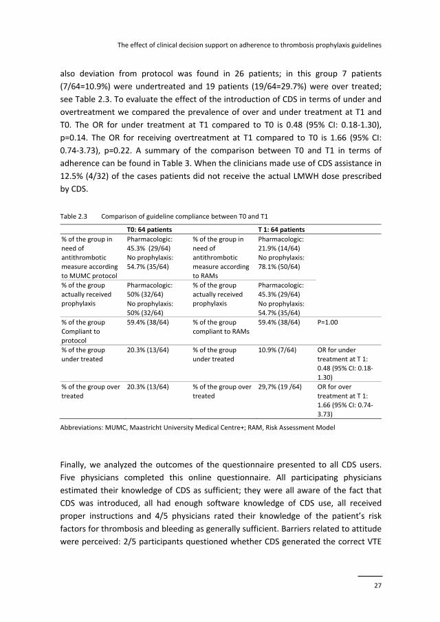
Table 2.3 Comparison of guideline compliance between T0 and T1
T0: 64 patients T 1: 64 patients
Pharmacologic:
45.3% (29/64)
Pharmacologic:
21.9% (14/64)
% of the group in
need of
antithrombotic measure according
to MUMC protocol
No prophylaxis:
54.7% (35/64)
% of the group in
need of
antithrombotic measure according
to RAMs
No prophylaxis:
78.1% (50/64)
Pharmacologic:
50% (32/64)
Pharmacologic:
45.3% (29/64)
% of the group
actually received prophylaxis No prophylaxis:
50% (32/64)
% of the group
actually received prophylaxis No prophylaxis:
54.7% (35/64)
% of the group
Compliant to protocol
59.4% (38/64) % of the group
compliant to RAMs
59.4% (38/64) P=1.00
% of the group
under treated
20.3% (13/64) % of the group
under treated
10.9% (7/64)
OR for under
treatment at T 1: 0.48 (95% CI: 0.18‐
1.30)
% of the group over
treated
20.3% (13/64) % of the group over
treated
29,7% (19 /64)
OR for over
treatment at T 1: 1.66 (95% CI: 0.74‐
3.73)
Abbreviations: MUMC, Maastricht University Medical Centre+; RAM, Risk Assessment Model
Finally, we analyzed the outcomes of the questionnaire presented to all CDS users.
Five physicians completed this online questionnaire. All participating physicians
estimated their knowledge of CDS as sufficient; they were all aware of the fact that
CDS was introduced, all had enough software knowledge of CDS use, all received
proper instructions and 4/5 physicians rated their knowledge of the patient’s risk
factors for thrombosis and bleeding as generally sufficient. Barriers related to attitude
were perceived: 2/5 participants questioned whether CDS generated the correct VTE
Chapter 2
28
prophylaxis advice for complex patients with several co‐morbidities, the average score
for experiencing difficulties due to the introduction of CDS (1: ‘no problem’‐5: ‘very
difficult’) was scored 2.6 (SD: 1.5), 3/5 did not know whether CDS was ‘evidence
based’, 4/5 perceived CDS advices as clear and 4/5 thought that the use of CDS would
lead to better patients outcomes. We also inquired after behavioural aspects towards
CDS; the question whether patient’s preferences influenced the decision to deviate
from the CDS advice, was answered with an average 2.4 (SD: 0.5) on a scale from 1
‘never’ to 5 ‘very often’ and of all participants 2 preferred to use CDS for high‐complex
patients and 3 preferred to use CDS in both high and low‐complex patients. Finally we
inquired after environmental factors regarding the CDS use: all participants judged
that the use of CDS required a substantial additional time investment and 2/5 had the
opinion that a direct link to the ordering system would lead to a decrease in mistakes
with regard to LMWH administration.
Discussion
We found no improvement in guideline adherence towards anti thrombotic
prophylaxis in medical patients after the introduction of CDS in this pilot study.
Guidelines were followed in 59.4% both before and after the introduction of CDS.
There was however a non‐significant shift towards over treatment, which may be
indicative of higher prophylaxis awareness. The finding that CDS did not result in
higher guideline adherence is not coherent with results presented in other studies.
Several studies demonstrated a positive, often temporary effect on adherence caused
merely by the fact that the introduction of CDS was accompanied by increased
awareness of the importance of VTE prevention. The introduction of CDS is associated
with increased rates of per protocol administration of VTE prophylaxis, increased rates
of administration of VTE prophylaxis in general14 and even with reduced rates of
VTE.13 The observed lack of improvement in adherence in this pilot study could, at
least partially, be caused by the suboptimal use of CDS. Final use of CDS varied
between 23.7% and 57.4% for the different wards. A barrier towards implementation
of CDS could have been the additional time investment needed as indicated by
physicians in the questionnaire; moreover, time consuming separate login procedures
were required in order to enter CDS. Doubts whether CDS was based on solid
evidence, uncertainty about the correctness of CDS advices for ‘complex’ patients,
experienced difficulties due to the introduction of CDS and deviation from CDS advice
caused by patient’s preferences as indicated in the questionnaire also might have
attributed to the perceived lack of improvement in adherence.
The effect of clinical decision support on adherence to thrombosis prophylaxis guidelines
29
Our study has several weaknesses. In the first place the sample size was small; only
128 patients participated in this pilot study and no follow‐up VTE incidences were
assessed. Therefore no association with the incidence of VTE could be made.
Secondly, VTE risks for patients at T0 were determined using a local protocol that did
not include a risk score for bleeding. The RAM used at T1 included a calculated score
for bleeding risk. This could have led to a difference in risk perception. The fact that
use of CDS required time consuming login procedures and the fact that no link was
provided to the ordering system may have been the most important barrier, resulting
in the limited impact of CDS in this study.
In contrast to findings in other studies we conclude that introduction of CDS did not
have a positive impact on guideline adherence. A non‐significant shift towards over
treatment was observed following the introduction of CDS. An easily accessible and
mandatory CDS linked to the electronic pharmacy system might be needed in order to
improve guideline adherence and associated reduction in VTE incidence.
Chapter 2
30
References
1. Linden MWvd, Westert GP, Bakker D de, Schellevis F. Tweede Nationale Studie naar ziekten en verrichtingen in de huisartspraktijk: klachten en aandoeningen in de bevolking en in de
huisartspraktijk. Utrecht: NIVEL, 2004.
2. Duff J, Walker K, Omari A, Stratton C. Prevention of venous thromboembolism in hospitalized patients: analysis of reduced cost and improved clinical outcomes. J Vasc Nurs. 2013;31:9‐14.
3. Falck‐Ytter Y, Francis CW, Johanson NA, Curley C, Dahl OE, Schulman S, et al. Prevention of VTE in
orthopedic surgery patients: Antithrombotic Therapy and Prevention of Thrombosis, 9th ed: American College of Chest Physicians Evidence‐Based Clinical Practice Guidelines. Chest. 2012;141(2
Suppl):278‐325.
4. Kahn SR, Lim W, Dunn AS, Cushman M, Dentali F, Akl EA, et al. Prevention of VTE in nonsurgical patients: Antithrombotic Therapy and Prevention of Thrombosis, 9th ed: American College of Chest
Physicians Evidence‐Based Clinical Practice Guidelines. Chest. 2012;141:195‐226.
5. Gould MK, Garcia DA, Wren SM, Karanicolas PJ, Arcelus JI, Heit JA, et al. Prevention of VTE in nonorthopedic surgical patients: Antithrombotic Therapy and Prevention of Thrombosis, 9th ed:
American College of Chest Physicians Evidence‐Based Clinical Practice Guidelines. Chest.
2012;141:227‐77. 6. Monreal M, Kakkar AK, Caprini JA, Barba R, Uresandi F, Valle R, et al. The outcome after treatment of
venous thromboembolism is different in surgical and acutely ill medical patients. Findings from the
RIETE registry. J Thromb Haemost. 2004;2:1892‐8. 7. Goldhaber SZ, Tapson VF, Committee DFS. A prospective registry of 5,451 patients with ultrasound‐
confirmed deep vein thrombosis. Am J Cardiol. 2004;93:259‐62.
8. Tapson VF, Decousus H, Pini M, Chong BH, Froehlich JB, Monreal M, et al. Venous thromboembolism prophylaxis in acutely ill hospitalized medical patients: findings from the International Medical
Prevention Registry on Venous Thromboembolism. Chest. 2007;132:936‐45.
9. Lecumberri R, Marques M, Panizo E, Alfonso A, Garcia‐Mouriz A, Gil‐Bazo I, et al. High incidence of venous thromboembolism despite electronic alerts for thromboprophylaxis in hospitalised cancer
patients. Thromb Haemost. 2013;110:184‐90.
10. Kucher N, Puck M, Blaser J, Bucklar G, Eschmann E, Luscher TF. Physician compliance with advanced electronic alerts for preventing venous thromboembolism among hospitalized medical patients. J
Thromb Haemost. 2009;7:1291‐6.
11. Kucher N, Koo S, Quiroz R, Cooper JM, Paterno MD, Soukonnikov B, et al. Electronic alerts to prevent venous thromboembolism among hospitalized patients. N Engl J Med. 2005;352:969‐77.
12. Baroletti S, Munz K, Sonis J, Fanikos J, Fiumara K, Paterno M, et al. Electronic alerts for hospitalized
high‐VTE risk patients not receiving prophylaxis: a cohort study. J Thromb Thrombolysis. 2008;25: 146‐50.
13. Maynard GA, Morris TA, Jenkins IH, Stone S, Lee J, Renvall M, et al. Optimizing prevention of hospital‐
acquired venous thromboembolism (VTE): prospective validation of a VTE risk assessment model. J Hosp Med. 2010;5:10‐8.
14. Umscheid CA, Hanish A, Chittams J, Weiner MG, Hecht TE. Effectiveness of a novel and scalable
clinical decision support intervention to improve venous thromboembolism prophylaxis: a quasi‐experimental study. BMC Med Inform Decis Mak. 2012;12:92.
15. Barbar S, Noventa F, Rossetto V, Ferrari A, Brandolin B, Perlati M, et al. A risk assessment model for
the identification of hospitalized medical patients at risk for venous thromboembolism: the Padua Prediction Score. J Thromb Haemost. 2010;8:2450‐7.
16. Decousus H, Tapson VF, Bergmann JF, Chong BH, Froehlich JB, Kakkar AK, et al. Factors at admission
associated with bleeding risk in medical patients: findings from the IMPROVE investigators. Chest. 2011;139:69‐79.
17. Lugtenberg M, Burgers JS, Besters CF, Han D, Westert GP. Perceived barriers to guideline adherence:
a survey among general practitioners. BMC Fam Pract. 2011;12:98.

The effect of clinical decision support on adherence to thrombosis prophylaxis guidelines
31
18. Lugtenberg M, Zegers‐van Schaick JM, Westert GP, Burgers JS. Why don't physicians adhere to
guideline recommendations in practice? An analysis of barriers among Dutch general practitioners. Implement Sci. 2009;4:54.
Chapter 2
32
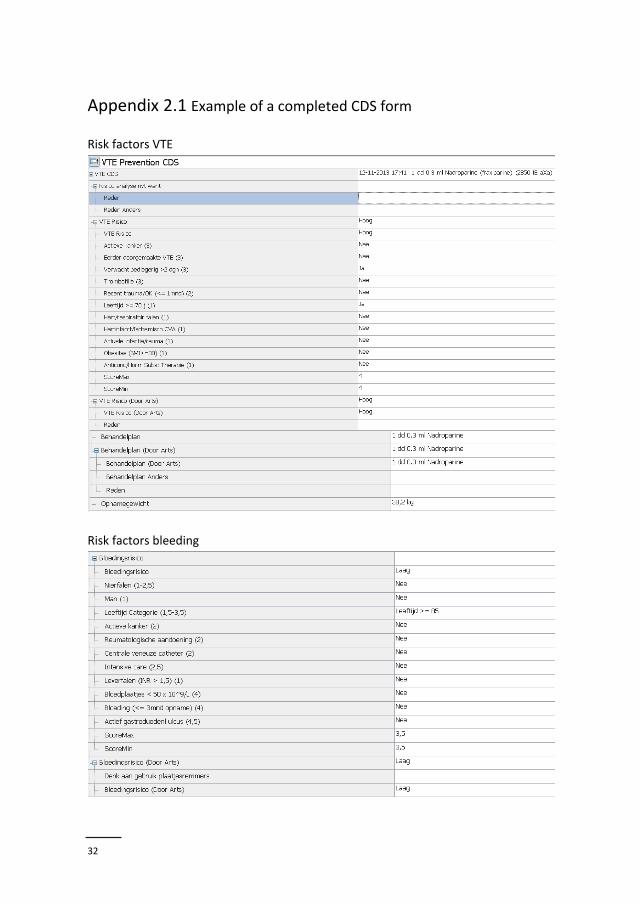
Appendix 2.1 Example of a completed CDS form
Risk factors VTE
Risk factors bleeding
The effect of clinical decision support on adherence to thrombosis prophylaxis guidelines
33
Appendix 2.2 Questionnaire for evaluation of CDS (in Dutch)
Kennis 1) Was u in de periode dat de pilot CDS VTE prevention actief was (1‐9‐13/1‐12‐13) op de hoogte
van het bestaan van CDS op uw afdeling?
a. Ja b. Nee
2) Zo ja, had u voldoende kennis van de software om het programma op de juiste wijze te kunnen
gebruiken? a. Ja
b. Nee
3) Heeft u voldoende instructie over het gebruik van CDS ontvangen? a. Ja
b. Nee
4) Had u bij gebruik van CDS doorgaans voldoende kennis van de patient om alle risicofactoren (trombose/bloeding) op de juiste wijze in het systeem in te voeren?
a. Ja
b. Nee
Attitude
5) Leidt het gebruik van CDS tot een juist behandeladvies voor complexe patienten met veel co‐morbiditeit, zoals die veelvuldig op uw afdeling voorkomen?
a. Ja
b. Nee 6) Maakte u voor de introductie van de pilot gebruik van het ODIN protocol om op de juiste wijze
tromboprofylaxe voor te schrijven?
a. Ja b. Nee
7) Was het moeilijk over te stappen naar een nieuwe vorm (CDS) van inschatting van het
tromboserisico? Geef een score van 1‐5, waarbij 1= geen enkel probleem en 5=zeer lastig.
1 2 3 4 5 (een score omcirkelen)
8) Was CDDS in de gehanteerde vorm (pilot) voldoende ‘evidence based’? a. Ja
b. Nee
c. Weet ik niet 9) De door CDS gegenereerde aanbevelingen: (meerdere alternatieven mogen aangekruist)
a. Zijn duidelijk
b. Wegen alle relevante factoren mee in het behandeladvies c. Zijn ‘up to date’
d. Zijn makkelijk toepasbaar
10) Denkt u dat het gebruik van CDS leidt tot betere patientenuitkomsten m.b.t. DVT‐preventie vergeleken met de ‘oude’ situatie?
a. Ja
b. Nee
Gedrag
11) Speelt de persoonlijke voorkeur van de patient een rol bij het afwijken van volgens protocol voor te schrijven LMWH?
Geef een score van 1‐5, waarbij 1= nooit en 5= zeer vaak.
1 2 3 4 5 (een score omcirkelen)
Chapter 2
34
12) Hoe vaak heeft u ten tijde van de pilot gebruik gemaakt van CDS bij het bepalen of een patient
tromboprofylaxe nodig heeft of niet? a. Nooit
b. In ongeveer 0‐25% van de gevallen
c. In ongeveer 25‐50% van de gevallen d. In ongeveer 50‐75% van de gevallen
e. In ongeveer 75‐100% van de gevallen
f. Altijd 13) Ik heb het meest gebruik gemaakt van CDS voor:
a. Mobiele, laagcomplexe patienten met een op het oog laag DVT risico
b. Meer hoogcomplexe patienten c. Zowel de hoog‐ als laagcomplexe patienten
Omgevingsfactoren 14) Hoe beoordeelt u het gebruiksgemak van CDS:
Geef een score van 1‐5, waarbij 1=zeer gebruiksvriendelijk, 5=zeer gebruiksonvriendelijk
1 2 3 4 5 (een score omcirkelen)
15) Open vraag: Op welke wijze zou de gebruiksvriendelijkheid van CDS kunnen worden verbeterd?
Antwoord:.......................................................................................................................................................................................................................................................................................................
..............................................................................
16) Denkt u dat een directe koppeling tussen CDS en het medicijnbestelsysteem EVS zou leiden tot minder onjuiste voorschrijvingen/onjuiste doseringen van LMWHs.
a. Ja
b. Nee 17) Vergt het gebruik van CDS een belangrijke extra tijdsinvestering?
a. Ja
b. Nee 18) Heeft CDS in deze vorm een toegevoegde waarde?
Geef een score van 1‐5, waarbij 1=zeer zeker, 5=zeer zeker niet
1 2 3 4 5 (een score omcirkelen)
35
Chapter 3 Safety and efficacy of bridging with low molecular
weight heparins: a systematic review and partial
meta‐analysis
Pieter Eijgenraam,Hugo ten Cate, Arina J ten Cate‐Hoek
Curr Pharm Des. 2013;19:4014‐4023
Chapter 3
36
Abstract
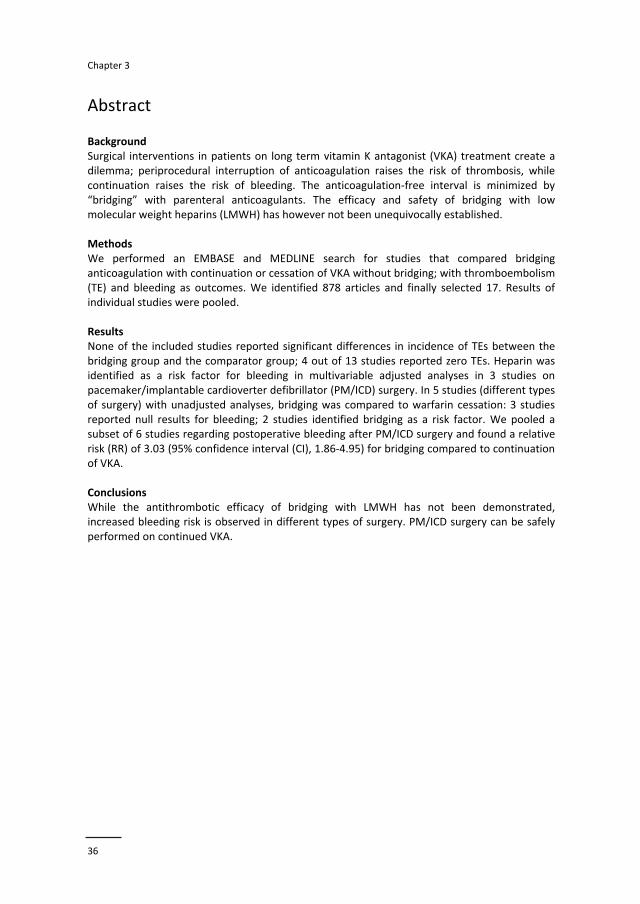
Background Surgical interventions in patients on long term vitamin K antagonist (VKA) treatment create a dilemma; periprocedural interruption of anticoagulation raises the risk of thrombosis, while continuation raises the risk of bleeding. The anticoagulation‐free interval is minimized by “bridging” with parenteral anticoagulants. The efficacy and safety of bridging with low molecular weight heparins (LMWH) has however not been unequivocally established. Methods We performed an EMBASE and MEDLINE search for studies that compared bridging anticoagulation with continuation or cessation of VKA without bridging; with thromboembolism (TE) and bleeding as outcomes. We identified 878 articles and finally selected 17. Results of individual studies were pooled. Results None of the included studies reported significant differences in incidence of TEs between the bridging group and the comparator group; 4 out of 13 studies reported zero TEs. Heparin was identified as a risk factor for bleeding in multivariable adjusted analyses in 3 studies on pacemaker/implantable cardioverter defibrillator (PM/ICD) surgery. In 5 studies (different types of surgery) with unadjusted analyses, bridging was compared to warfarin cessation: 3 studies reported null results for bleeding; 2 studies identified bridging as a risk factor. We pooled a subset of 6 studies regarding postoperative bleeding after PM/ICD surgery and found a relative risk (RR) of 3.03 (95% confidence interval (CI), 1.86‐4.95) for bridging compared to continuation of VKA. Conclusions While the antithrombotic efficacy of bridging with LMWH has not been demonstrated, increased bleeding risk is observed in different types of surgery. PM/ICD surgery can be safely performed on continued VKA.
Safety and efficacy of bridging with low molecular weight heparins
37
Background
Vitamin K antagonists (VKA) still are commonly used agents in the prevention of
venous or arterial thromboembolism (TE). Annually, approximately 10% of the patient
population on VKA undergoes at least 1 invasive procedure.1 The treating physician
faces a challenge: a delicate balance must be maintained between the risk of TE and
the risk of bleeding in the perioperative anticoagulant management. While arterial TE
is associated with a high mortality, or in survivors with major disability,2 perioperative
bleeding contributes to the need for reoperation, transfusion, prolonged
hospitalization, and in some cases death.3 In 2012 the American College of Chest
Physicians (ACCP) presented the latest guidelines for perioperative management of
antithrombotic therapy.1,4
In the management of perioperative anticoagulation 3 options can be considered.
First, in procedures with a low bleeding risk and the possibility of local hemostatic
measures, such as dental extractions, cataract operations and small dermatologic
procedures, it is considered a safe choice to continue VKA use.5‐8 Second, in patients
at low risk of TE, ACCP guidelines recommend stopping warfarin administration 5 days
before the intervention and restarting warfarin 12‐24 hours after the procedure when
adequate hemostasis is secured. Finally, in patients at moderate or high TE risk
current ACCP guidelines recommend bridging therapy consisting of parenteral
administration of LMWH or unfractionated heparin (UFH) in the periprocedural
period, combined with interruption of VKA use. A problem that occurs when
comparing different studies that assess bridging anticoagulation is the fact that there
is no standardized definition of “bridging anticoagulation”; studies assess bridging
protocols in which different kinds and doses of LMWH, different time points of
administration and cessation of VKA and LMWH are advised.2,9‐17
The evidence evaluating these 3 options is not very strong,4 except for VKA
continuation in case of procedures with a low bleeding risk. In general, there is paucity
in randomized trials assessing different strategies, and in most cases the evidence is
based on single armed cohort studies.9,18‐21 Furthermore, most available studies have
small sample sizes and are therefore underpowered to determine whether a certain
management strategy is safe (prevention of bleeding) and efficacious (prevention of
TE).12,14,16,18,22 Recently, more solid evidence is emerging regarding bleeding risk
related to different anticoagulant management options in PM/ICD surgery, indicating
that the continuation of VKA might provide a safer option than bridging.13,23‐26
We systematically reviewed studies assessing different peri‐interventional
management options, compared to bridging therapy. Participants in these studies are
all chronic VKA users who undergo surgery. We aimed to assess both the safety and
Chapter 3
38
efficacy of the different strategies. Furthermore we performed a meta‐analysis
comparing studies with bridging therapy to warfarin continuation in PM/ICD surgery;
pocket hematoma and non‐pocket bleeding risk were assessed. For this review we
considered 4 treatment comparisons 1) bridging therapy versus periprocedural
cessation of VKA 2) bridging therapy versus periprocedural continuation of VKA,
3) low dose LMWH versus high dose LMWH and 4) early, within 24 hours
postoperative restart of LMWH versus late, after 24 hours postoperative restart of
LMWH.
Methods
Study selection
We included only studies wherein at least 2 periprocedural anticoagulant regimens
were assessed, 1 of which could be classified as bridging anticoagulation, while other
regimens consist of periprocedural withdrawal of VKA without administration of
heparins, periprocedural continuation of VKA without heparin administration or
surgery under sub therapeutic INR without heparin administration. We decided to
include studies in which data were assessed of patients with different risk categories
for a TE (low, moderate, high) and surgical procedures with different bleeding risks
(low, high). The primary outcome is objectively confirmed perioperative TE and is
defined as any thromboembolism, death caused by TE, composite score of TE. The
secondary outcome is perioperative bleeding and is defined as: minor bleeding, major
bleeding, and any bleeding. Major bleeding is defined as any bleeding resulting in
death, any intracranial bleeding, and any bleeding that leads to transfusion of packed
red cells and/or treatment in a hospital, or joint bleeds. All other bleedings, including
pocket hematomas in cardiac device surgery are qualified as minor bleedings. Cohort
studies, case control studies, cross sectional studies and controlled trials were eligible.
Our study protocol is based on the third edition of the Centre for Reviews and
Dissemination guidance for undertaking systematic reviews, York, United Kingdom.
Data sources and searches
We searched MEDLINE (2001‐2011, week 24) and EMBASE (2001‐2011, week 36)
databases. The search strategy can be found in appendix. A further selection was
made on title and abstract. The final selection of articles was made after full reading.
Safety and efficacy of bridging with low molecular weight heparins
39
Data extraction and quality assessment
Data from individual studies were collected on case report forms consisting of
3 sections: study eligibility, checklist of items for data collection, and quality
assessment using the Newcastle‐Ottawa Scale (NOS) for cohort studies.27
Data synthesis and analysis
We compared baseline characteristics and type of interventional procedure between
continued warfarin therapy and bridging groups on the basis of Fisher’s exact test and
Student’s t‐test, in order to assess the comparability of these groups with respect to
bleeding risk. In our meta‐analysis on PM/ICD surgery the random effects model as
proposed by DerSimonian and Laird was chosen; individual studies are weighed
according to the method proposed by Mantel‐Haenszel and expressed as RRs.28 RRs
and 95% confidence intervals of each included study and the overall effect were
computed. The primary outcome is total bleeding; bridging therapy is compared to
warfarin continuation. We tested for heterogeneity with the Breslow‐Day test; we
used the method proposed by Higgins et al, expressed as I2 to measure inconsistency
of effects of bridging therapy.29 To assess the possibility of bias of the cumulative
evidence, publication bias was explored by presenting funnel plots. There were no
tests for publication bias carried out, because with the few studies (n<10) included the
power of these tests is too low to distinguish chance from real asymmetry.28 We
conducted sensitivity analyses in a pre‐specified way; the intervention effects were
examined according to a) the quality assessment of the individual studies (cutoff point
6 stars) and b) we excluded the randomized trial (1) and the non randomized trial (1)
from our analyses. For our analyses we used free access Revman 5.1, provided by the
Cochrane collaboration.
Results
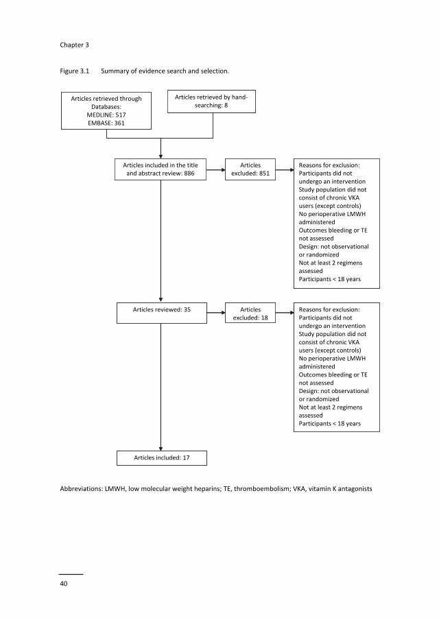
Our database search resulted in a total of 878 studies (including duplications),
whereof 517 articles were extracted from MEDLINE and 361 from EMBASE. A first
selection on title and abstract yielded a total of 35 articles, further restriction after full
reading resulted in a set of 17 studies including a subset of 9 regarding PM/ICD
surgery. Eight articles were retrieved by hand searching. For a summary of the search
see Figure 3.1. All included studies were published in English. For a summary of the
included studies see Table 3.1; for a summary of the effect estimates see Table 3.2.
Chapter 3
40
Figure 3.1 Summary of evidence search and selection.
Abbreviations: LMWH, low molecular weight heparins; TE, thromboembolism; VKA, vitamin K antagonists
Articles included in the title and abstract review: 886
Articles excluded: 851
Reasons for exclusion:Participants did not undergo an interventionStudy population did not consist of chronic VKA users (except controls)No perioperative LMWH administeredOutcomes bleeding or TE not assessedDesign: not observational or randomizedNot at least 2 regimens assessedParticipants < 18 years
Articles reviewed: 35 Articles excluded: 18
Reasons for exclusion:Participants did not undergo an interventionStudy population did not consist of chronic VKA users (except controls)No perioperative LMWH administeredOutcomes bleeding or TE not assessedDesign: not observational or randomized Not at least 2 regimens assessedParticipants < 18 years
Articles included: 17
Articles retrieved by hand‐searching: 8
Articles retrieved throughDatabases:
MEDLINE: 517EMBASE: 361
Safety and efficacy of bridging with low molecular weight heparins
41
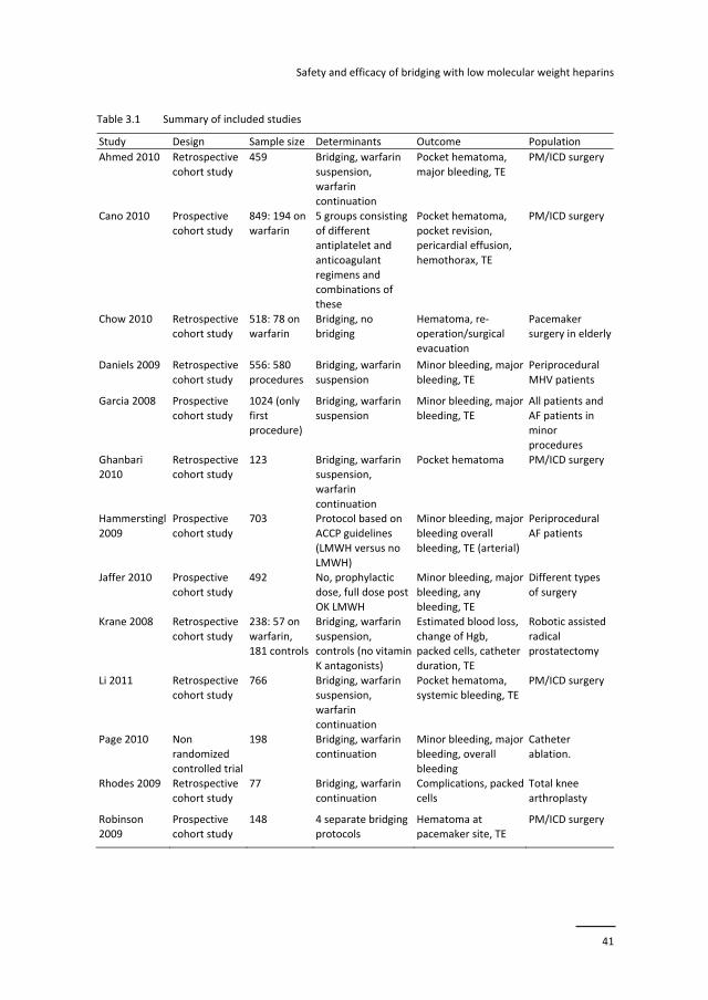
Table 3.1 Summary of included studies
Study Design Sample size Determinants Outcome Population
Ahmed 2010 Retrospective
cohort study
459 Bridging, warfarin
suspension, warfarin
continuation
Pocket hematoma,
major bleeding, TE
PM/ICD surgery
Cano 2010 Prospective
cohort study
849: 194 on
warfarin
5 groups consisting
of different antiplatelet and
anticoagulant
regimens and combinations of
these
Pocket hematoma,
pocket revision, pericardial effusion,
hemothorax, TE
PM/ICD surgery
Chow 2010 Retrospective cohort study
518: 78 on warfarin
Bridging, no bridging
Hematoma, re‐operation/surgical
evacuation
Pacemaker surgery in elderly
Daniels 2009 Retrospective
cohort study
556: 580
procedures
Bridging, warfarin
suspension
Minor bleeding, major
bleeding, TE
Periprocedural
MHV patients
Garcia 2008 Prospective
cohort study
1024 (only
first procedure)
Bridging, warfarin
suspension
Minor bleeding, major
bleeding, TE
All patients and
AF patients in minor
procedures
Ghanbari 2010
Retrospective cohort study
123 Bridging, warfarin suspension,
warfarin
continuation
Pocket hematoma PM/ICD surgery
Hammerstingl
2009
Prospective
cohort study
703 Protocol based on
ACCP guidelines
(LMWH versus no LMWH)
Minor bleeding, major
bleeding overall
bleeding, TE (arterial)
Periprocedural
AF patients
Jaffer 2010 Prospective
cohort study
492 No, prophylactic
dose, full dose post OK LMWH
Minor bleeding, major
bleeding, any bleeding, TE
Different types
of surgery
Krane 2008 Retrospective
cohort study
238: 57 on
warfarin, 181 controls
Bridging, warfarin
suspension, controls (no vitamin
K antagonists)
Estimated blood loss,
change of Hgb, packed cells, catheter
duration, TE
Robotic assisted
radical prostatectomy
Li 2011 Retrospective cohort study
766 Bridging, warfarin suspension,
warfarin
continuation
Pocket hematoma, systemic bleeding, TE
PM/ICD surgery
Page 2010 Non
randomized
controlled trial
198 Bridging, warfarin
continuation
Minor bleeding, major
bleeding, overall
bleeding
Catheter
ablation.
Rhodes 2009 Retrospective
cohort study
77 Bridging, warfarin
continuation
Complications, packed
cells
Total knee
arthroplasty
Robinson 2009
Prospective cohort study
148 4 separate bridging protocols
Hematoma at pacemaker site, TE
PM/ICD surgery
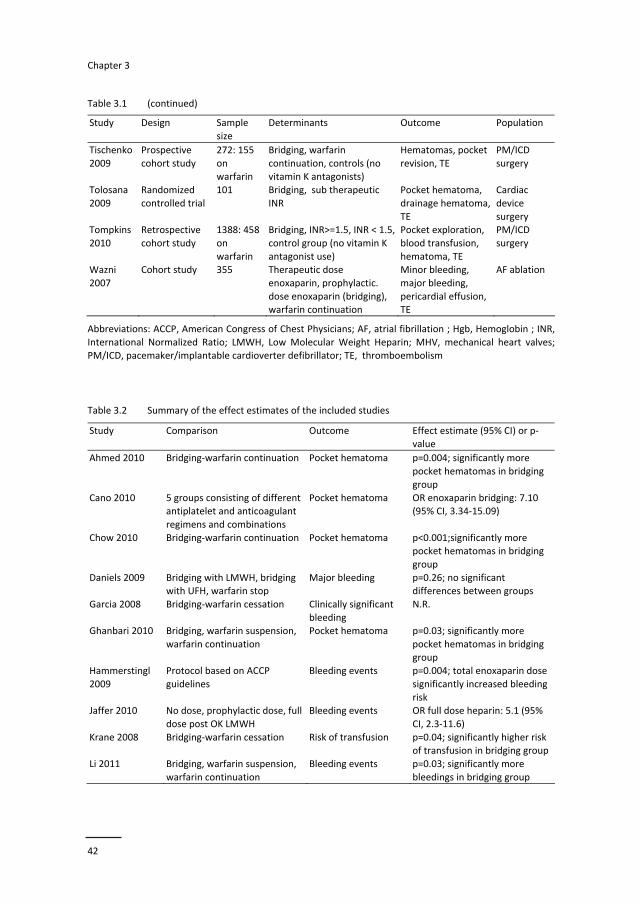
Chapter 3
42
Table 3.1 (continued)
Study Design Sample
size
Determinants Outcome Population
Tischenko
2009
Prospective
cohort study
272: 155
on warfarin
Bridging, warfarin
continuation, controls (no vitamin K antagonists)
Hematomas, pocket
revision, TE
PM/ICD
surgery
Tolosana
2009
Randomized
controlled trial
101 Bridging, sub therapeutic
INR
Pocket hematoma,
drainage hematoma,
TE
Cardiac
device
surgery Tompkins
2010
Retrospective
cohort study
1388: 458
on
warfarin
Bridging, INR>=1.5, INR < 1.5,
control group (no vitamin K
antagonist use)
Pocket exploration,
blood transfusion,
hematoma, TE
PM/ICD
surgery
Wazni
2007
Cohort study 355 Therapeutic dose
enoxaparin, prophylactic.
dose enoxaparin (bridging), warfarin continuation
Minor bleeding,
major bleeding,
pericardial effusion, TE
AF ablation
Abbreviations: ACCP, American Congress of Chest Physicians; AF, atrial fibrillation ; Hgb, Hemoglobin ; INR,
International Normalized Ratio; LMWH, Low Molecular Weight Heparin; MHV, mechanical heart valves; PM/ICD, pacemaker/implantable cardioverter defibrillator; TE, thromboembolism Table 3.2 Summary of the effect estimates of the included studies
Study Comparison Outcome Effect estimate (95% CI) or p‐value
Ahmed 2010 Bridging‐warfarin continuation Pocket hematoma p=0.004; significantly more
pocket hematomas in bridging
group
Cano 2010 5 groups consisting of different antiplatelet and anticoagulant
regimens and combinations
Pocket hematoma OR enoxaparin bridging: 7.10 (95% CI, 3.34‐15.09)
Chow 2010 Bridging‐warfarin continuation Pocket hematoma p<0.001;significantly more pocket hematomas in bridging
group
Daniels 2009 Bridging with LMWH, bridging with UFH, warfarin stop
Major bleeding p=0.26; no significant differences between groups
Garcia 2008 Bridging‐warfarin cessation Clinically significant
bleeding
N.R.
Ghanbari 2010 Bridging, warfarin suspension,
warfarin continuation
Pocket hematoma p=0.03; significantly more
pocket hematomas in bridging
group Hammerstingl
2009
Protocol based on ACCP
guidelines
Bleeding events p=0.004; total enoxaparin dose
significantly increased bleeding
risk Jaffer 2010 No dose, prophylactic dose, full
dose post OK LMWH
Bleeding events OR full dose heparin: 5.1 (95%
CI, 2.3‐11.6)
Krane 2008 Bridging‐warfarin cessation Risk of transfusion p=0.04; significantly higher risk of transfusion in bridging group
Li 2011 Bridging, warfarin suspension,
warfarin continuation
Bleeding events p=0.03; significantly more
bleedings in bridging group
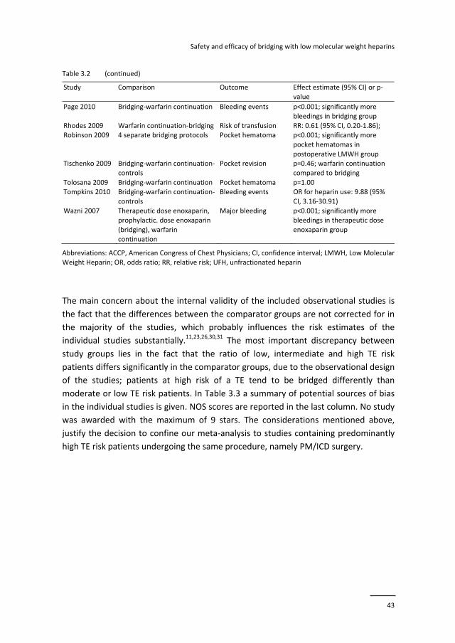
Safety and efficacy of bridging with low molecular weight heparins
43
Table 3.2 (continued)
Study Comparison Outcome Effect estimate (95% CI) or p‐
value
Page 2010 Bridging‐warfarin continuation Bleeding events p<0.001; significantly more
bleedings in bridging group
Rhodes 2009 Warfarin continuation‐bridging Risk of transfusion RR: 0.61 (95% CI, 0.20‐1.86); Robinson 2009 4 separate bridging protocols Pocket hematoma p<0.001; significantly more
pocket hematomas in
postoperative LMWH group Tischenko 2009 Bridging‐warfarin continuation‐
controls
Pocket revision p=0.46; warfarin continuation
compared to bridging
Tolosana 2009 Bridging‐warfarin continuation Pocket hematoma p=1.00 Tompkins 2010 Bridging‐warfarin continuation‐
controls
Bleeding events OR for heparin use: 9.88 (95%
CI, 3.16‐30.91)
Wazni 2007 Therapeutic dose enoxaparin, prophylactic. dose enoxaparin
(bridging), warfarin
continuation
Major bleeding p<0.001; significantly more bleedings in therapeutic dose
enoxaparin group
Abbreviations: ACCP, American Congress of Chest Physicians; CI, confidence interval; LMWH, Low Molecular Weight Heparin; OR, odds ratio; RR, relative risk; UFH, unfractionated heparin The main concern about the internal validity of the included observational studies is
the fact that the differences between the comparator groups are not corrected for in
the majority of the studies, which probably influences the risk estimates of the
individual studies substantially.11,23,26,30,31 The most important discrepancy between
study groups lies in the fact that the ratio of low, intermediate and high TE risk
patients differs significantly in the comparator groups, due to the observational design
of the studies; patients at high risk of a TE tend to be bridged differently than
moderate or low TE risk patients. In Table 3.3 a summary of potential sources of bias
in the individual studies is given. NOS scores are reported in the last column. No study
was awarded with the maximum of 9 stars. The considerations mentioned above,
justify the decision to confine our meta‐analysis to studies containing predominantly
high TE risk patients undergoing the same procedure, namely PM/ICD surgery.
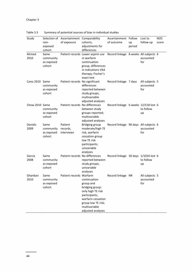
Chapter 3
44
Table 3.3 Summary of potential sources of bias in individual studies
Study Selection of non‐exposed cohort
Ascertainment of exposure
Comparability cohorts, adjustments for differences
Ascertainment of outcome
Follow up period
Lost to follow up
NOS score
Ahmed 2010
Same community as exposed cohort
Patient records Lower aspirin use in warfarin continuation group, differences in indications VKA therapy; Fischer’s exact test
Record linkage 8 weeks All subjects accounted for
6
Cano 2010 Same community as exposed cohort
Patient records No significant differences reported between study groups; multivariable adjusted analyses
Record linkage 7 days All subjects accounted for
5
Chow 2010 Same community as exposed cohort
Patient records No differences between study groups reported; multivariable adjusted analyses
Record linkage 6 weeks 12/518 lost to follow up
6
Daniels 2009
Same community as exposed cohort
Patient records, interviews
Bridging group moderate/high TE risk, warfarin cessation group low TE risk participants; univariable analyses
Record linkage 90 days All subjects accounted for
6
Garcia 2008
Same community as exposed cohort
Patient records No differences reported between study groups; univariable analyses
Record linkage 30 days 1/1024 lost to follow up
6
Ghanbari 2010
Same community as exposed cohort
Patient records Warfarin continuation group and bridging group: only high TE risk participants, warfarin cessation group low TE risk; multivariable adjusted analyses
Record linkage NR All subjects accounted for
5
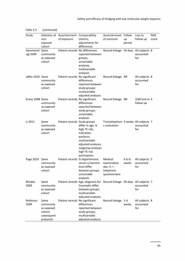
Safety and efficacy of bridging with low molecular weight heparins
45
Table 3.3 (continued)
Study Selection of non‐exposed cohort
Ascertainment of exposure
Comparability cohorts, adjustments for differences
Ascertainment of outcome
Follow up period
Lost to follow up
NOS score
Hammerstingl 2009
Same community as exposed cohort
Patient records No differences reported between groups; univariable analyses, multivariable analyses
Record linkage 30 days All subjects accounted for
8
Jaffer 2010 Same community as exposed cohort
Patient records No significant differences reported between study groups; multivariable adjusted analyses
Record linkage NR All subjects accounted for
8
Krane 2008 Same community as exposed cohort
Patient records No significant differences reported between study groups; univariable analyses
Record linkage NR 3/60 lost to follow up
4
Li 2011 Same community as exposed cohort
Patient records Study groups differ in age, % high TE risk, indication warfarin; multivariable adjusted analyses, subgroup analyses high TE risk participants
Transtelephonic evaluation
4 weeks All subjects accounted for
7
Page 2010 Same community as exposed cohort
Patient records % Hypertension, serum creatinine level differ between groups; univariable analyses
Medical examination day +1 + telephone questionnaire
4 to 6 weeks
All subjects accounted for
5
Rhodes 2009
Same community as exposed cohort
Patient records Age, diagnosis for Coumadin differ between groups; multivariable adjusted analyses
Record linkage 90 days All subjects accounted for
7
Robinson 2009
Same community as exposed cohort; subsequent protocols
Patient records No significant differences reported between study groups; multivariable adjusted analyses
Record linkage 1‐4 weeks
All subjects accounted for
8
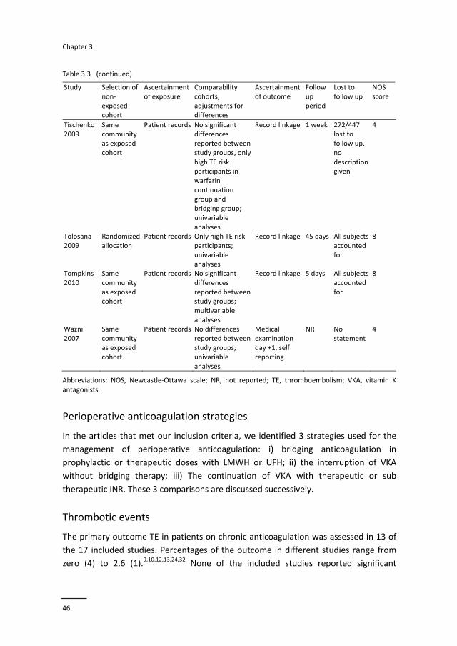
Chapter 3
46
Table 3.3 (continued)
Study Selection of non‐exposed cohort
Ascertainment of exposure
Comparability cohorts, adjustments for differences
Ascertainment of outcome
Follow up period
Lost to follow up
NOS score
Tischenko 2009
Same community as exposed cohort
Patient records No significant differences reported between study groups, only high TE risk participants in warfarin continuation group and bridging group; univariable analyses
Record linkage 1 week 272/447 lost to follow up, no description given
4
Tolosana 2009
Randomized allocation
Patient records Only high TE risk participants; univariable analyses
Record linkage 45 days All subjects accounted for
8
Tompkins 2010
Same community as exposed cohort
Patient records No significant differences reported between study groups; multivariable analyses
Record linkage 5 days All subjects accounted for
8
Wazni 2007
Same community as exposed cohort
Patient records No differences reported between study groups; univariable analyses
Medical examination day +1, self reporting
NR No statement
4
Abbreviations: NOS, Newcastle‐Ottawa scale; NR, not reported; TE, thromboembolism; VKA, vitamin K antagonists
Perioperative anticoagulation strategies
In the articles that met our inclusion criteria, we identified 3 strategies used for the
management of perioperative anticoagulation: i) bridging anticoagulation in
prophylactic or therapeutic doses with LMWH or UFH; ii) the interruption of VKA
without bridging therapy; iii) The continuation of VKA with therapeutic or sub
therapeutic INR. These 3 comparisons are discussed successively.
Thrombotic events
The primary outcome TE in patients on chronic anticoagulation was assessed in 13 of
the 17 included studies. Percentages of the outcome in different studies range from
zero (4) to 2.6 (1).9,10,12,13,24,32 None of the included studies reported significant
Safety and efficacy of bridging with low molecular weight heparins
47
differences in cumulative incidence of TEs with regard to the comparisons made in
this review. For the meta‐analysis of bridging therapy compared to VKA continuation
in PM/ICD surgery; only 3 studies reported on TE and could therefore not be included
in these analyses, this number would be too small to yield relevant results.
Bridging therapy and interruption of anticoagulants
Bridging therapy consisting of different LMWH or UFH in both prophylactic and
therapeutic doses was compared to cessation of VKA in 8 studies.11,23‐25,30,32‐34 All of
these studies had an observational design. In only 2 of these studies authors reported
that in patients in whom warfarin was interrupted TE risk was predominantly low and
when bridging was applied patients had mainly moderate to high TE risk.11,25 This is in
accordance with the recommendation by the current ACCP guidelines. The other
authors did not specify differences in TE risk. The type of procedures differed between
the studies (Table 3.1). All outcomes of interest could be classified as TE, minor
bleeding, major bleeding or overall bleeding. The study by Krane et al, mentioned an
increased risk of transfusion for bridged patients after robotic‐assisted radical
prostatectomy compared to interrupted VKA patients (p=0.04); this analysis was
unadjusted.33 In a retrospective cohort study performed by Daniels et al. the
comparison of LMWH or UFH to no heparin in patients with mechanical heart valves
undergoing different types of surgery resulted in an unadjusted RR of 1.25 (95% CI,
0.73‐2.14) for overall bleeding.11 Pooling the results of 7 individual studies for the
outcome minor bleeding resulted as expected in heterogeneity (p<0.001;
I2=78%).11,16,23‐25,30,34 Analysis of major bleeding, overall bleeding and TE resulted in
heterogeneity as well (results not shown). In 3 studies multivariable adjusted analyses
were presented. First, a study performed by Cano et al. reported an odds ratio (OR) of
7.10 (95% CI, 3.34‐15.09) for pocket hematoma in patients bridged with enoxaparin
who underwent the implantation of cardiac rhythm devices compared to no
enoxaparin administration.32 Secondly, Tompkins et al. reported an OR for heparin use
compared to no heparin use of 9.88 (95% CI, 3.16‐30.91) for bleeding complications in
patients after cardiac device implantation.23 Thirdly, Li et al. computed, after adjusting
for age and gender a p‐value of 0.02 comparing warfarin cessation to bridging in
PM/ICD surgery; the incidence of overall bleeding was higher in the bridging group.24
Ghanbari et al. reported in a study on PM/ICD surgery an unadjusted OR of 6.17 (95%
CI, 1.6‐24) for major device pocket hematoma in a high TE risk group on bridging
therapy.25 Finally, Garcia et al. and Ahmed et al. reported no significant differences in
bleeding risk in their studies of patients undergoing minor procedures and PM/ICD
surgery, respectively.30,34
Chapter 3
48
Bridging therapy and continuation of warfarin
Bridging therapy consisting of different LMWH and UFH in both prophylactic and
therapeutic doses was compared to continuation of VKA in 9 studies12‐14,23‐26,31,34
whereof 6 studies regarding PM/ICD interventions.12,13,23‐25,34 The 3 remaining studies
assessed the comparison in catheter ablation (2) and total knee arthroplasty (TKA) (1).
These 9 studies included 7 cohort studies,13,14,23‐25,31,34 1 randomized trial12 and 1 non
randomized trial.26 See Table 3.1 for a summary of the included studies. All outcomes
of interest could be classified as TE, minor bleeding, major bleeding or overall
bleeding. Again, pooling of these studies resulted in unacceptable heterogeneity
(p<0.001; I2=76%).
Rhodes et al. presented an age adjusted RR of 0.61 (95% CI, 0.20‐1.86) for transfusion
required, when they compared a group that continued coumadin with a bridging
group in TKA surgery.14 A non randomized trial including patients undergoing a
catheter ablation performed by Page et al. resulted in overall bleeding in the bridging
LMWH group of 78% and 56% in the warfarin group (p<0.001).26 A randomized
controlled trial by Tolosana et al. resulted in p values of 1.00 for both pocket
hematoma and drainage hematoma.12
We performed a meta‐analysis assessing the risk of minor bleeding (pocket
hematoma), major bleeding (pocket revision, drainage hematoma, pericardial
tamponnade and hemothorax) and overall bleeding in PM/ICD surgery. We included 6
studies with an observational design (5)13,23‐25,34 and randomized allocation (1),12 all
with unadjusted results. In all studies, except the study performed by Tolosana, the
patients on warfarin had a therapeutic INR. The studies performed by Ghanbari,
Tischenko and Tolosana included only high TE risk patients. For the outcome minor
bleeding we were able to use a subgroup of patients from the study of Li et al, with
high TE risk.24 Differences with respect to known risk factors for bleeding (age, male
sex, diabetes, concomitant antiplatelet drug use, bleeding disorders, kind and extend
of procedure) were assessed between the bridging and continued warfarin group
using Fisher’s exact test and the Student’s t‐test.9,35,36 Significant differences between
these groups are higher proportions of aspirin use in the bridging group in the study
performed by Li,24 higher proportions of patients with diabetes in the bridging group
in the study by Ahmed et al.34 and younger age in the bridging group in studies by Li24
and Tischenko et al.13 (results not shown).
Homogenous results (p=0.29, I2=19%) were observed regarding pocket hematoma,
and a RR of 2.62 (95% CI, 1.41‐4.86) was computed. For major bleeding we observed
homogenous results too (p=0.36, I2=8%) with a borderline insignificant RR of 3.26
(95% CI, 0.99‐10.78). Analyses regarding overall bleeding resulted in a homogenous
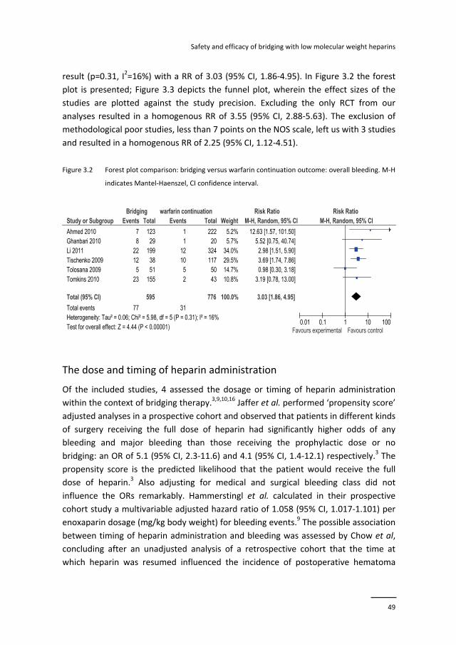
Safety and efficacy of bridging with low molecular weight heparins
49
result (p=0.31, I2=16%) with a RR of 3.03 (95% CI, 1.86‐4.95). In Figure 3.2 the forest
plot is presented; Figure 3.3 depicts the funnel plot, wherein the effect sizes of the
studies are plotted against the study precision. Excluding the only RCT from our
analyses resulted in a homogenous RR of 3.55 (95% CI, 2.88‐5.63). The exclusion of
methodological poor studies, less than 7 points on the NOS scale, left us with 3 studies
and resulted in a homogenous RR of 2.25 (95% CI, 1.12‐4.51).
Figure 3.2 Forest plot comparison: bridging versus warfarin continuation outcome: overall bleeding. M‐H
indicates Mantel‐Haenszel, CI confidence interval.
The dose and timing of heparin administration
Of the included studies, 4 assessed the dosage or timing of heparin administration
within the context of bridging therapy.3,9,10,16 Jaffer et al. performed ‘propensity score’
adjusted analyses in a prospective cohort and observed that patients in different kinds
of surgery receiving the full dose of heparin had significantly higher odds of any
bleeding and major bleeding than those receiving the prophylactic dose or no
bridging: an OR of 5.1 (95% CI, 2.3‐11.6) and 4.1 (95% CI, 1.4‐12.1) respectively.3 The
propensity score is the predicted likelihood that the patient would receive the full
dose of heparin.3 Also adjusting for medical and surgical bleeding class did not
influence the ORs remarkably. Hammerstingl et al. calculated in their prospective
cohort study a multivariable adjusted hazard ratio of 1.058 (95% CI, 1.017‐1.101) per
enoxaparin dosage (mg/kg body weight) for bleeding events.9 The possible association
between timing of heparin administration and bleeding was assessed by Chow et al,
concluding after an unadjusted analysis of a retrospective cohort that the time at
which heparin was resumed influenced the incidence of postoperative hematoma
Study or Subgroup
Ahmed 2010Ghanbari 2010Li 2011Tischenko 2009Tolosana 2009Tomkins 2010
Total (95% CI)
Total eventsHeterogeneity: Tau² = 0.06; Chi² = 5.98, df = 5 (P = 0.31); I² = 16%Test for overall effect: Z = 4.44 (P < 0.00001)
Events
78
2212
523
77
Total
12329
1993851
155
595
Events
11
121052
31
Total
22220
324117
5043
776
Weight
5.2%5.7%
34.0%29.5%14.7%10.8%
100.0%
M-H, Random, 95% CI
12.63 [1.57, 101.50]5.52 [0.75, 40.74]
2.98 [1.51, 5.90]3.69 [1.74, 7.86]0.98 [0.30, 3.18]
3.19 [0.78, 13.00]
3.03 [1.86, 4.95]
Bridging warfarin continuation Risk Ratio Risk RatioM-H, Random, 95% CI
0.01 0.1 1 10 100Favours experimental Favours control
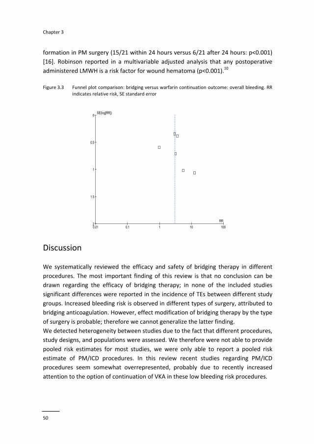
Chapter 3
50
formation in PM surgery (15/21 within 24 hours versus 6/21 after 24 hours: p<0.001)
[16]. Robinson reported in a multivariable adjusted analysis that any postoperative
administered LMWH is a risk factor for wound hematoma (p<0.001).10
Figure 3.3 Funnel plot comparison: bridging versus warfarin continuation outcome: overall bleeding. RR
indicates relative risk, SE standard error
Discussion
We systematically reviewed the efficacy and safety of bridging therapy in different
procedures. The most important finding of this review is that no conclusion can be
drawn regarding the efficacy of bridging therapy; in none of the included studies
significant differences were reported in the incidence of TEs between different study
groups. Increased bleeding risk is observed in different types of surgery, attributed to
bridging anticoagulation. However, effect modification of bridging therapy by the type
of surgery is probable; therefore we cannot generalize the latter finding.
We detected heterogeneity between studies due to the fact that different procedures,
study designs, and populations were assessed. We therefore were not able to provide
pooled risk estimates for most studies, we were only able to report a pooled risk
estimate of PM/ICD procedures. In this review recent studies regarding PM/ICD
procedures seem somewhat overrepresented, probably due to recently increased
attention to the option of continuation of VKA in these low bleeding risk procedures.
0.01 0.1 1 10 100
0
0.5
1
1.5
2RR
SE(log[RR])
Safety and efficacy of bridging with low molecular weight heparins
51
In PM/ICD surgery bridging anticoagulation results in a higher bleeding risk compared
to perioperative continuation of VKA. This finding is novel. To our knowledge this is
the only review in which a pooled RR is presented for the comparison of bridging
therapy to continuation of VKA in PM/ICD surgery.
Most of the 17 included studies that assessed bleeding risk produced biased results. In
4 out of 5 high quality studies (a minimum of 8 stars on the NOS scale) bridging was
identified as a risk factor for bleeding3,9,10,23; only 1 high quality study reported a null
net result.12 The finding that bridging is a risk factor for perioperative bleeding is
supported by 2 reviews regarding low bleeding risk procedures.37,38
The short exposure time to bridging anticoagulation, according to ACCP guidelines 7
to 11 days, might have influenced the detection that no significant differences
occurred in incidence of TEs between different study groups; overall TE incidence was
low, in some studies zero. Similar findings were reported in other reviews.39,40 The net
clinical benefit of bridging is therefore unclear. Nevertheless, the risk of a major
bleeding can be weighed against the risk of TE, when case‐fatality rates are compared.
The case‐fatality rate for major bleeding is 8‐9%. For arterial TE these rates vary from
15% (heart valve thrombosis) to 70 % (stroke), while venous TE carries a 6% rate for
death or major disability and a 25% risk for pulmonary embolism.41 These numbers
suggest that one is more likely to accept major bleedings due to periprocedural
anticoagulant strategies to prevent arterial TE.41
A possible explanation for the bleeding risk associated with bridging therapy is
provided by Gerotziafas et al, who demonstrated that treatment with a combination
of VKA and LMWH induces a more profound inhibition of thrombin generation than
treatment with VKA alone.42 VKA and LMWH administration is usually resumed within
12‐78 hours after surgery; LMWH administration is stopped when the INR reaches a
value of 2. In this way a period in which the patient might be more vulnerable for a
bleeding is established by the early introduction of bridging therapy. Douketis et al.
demonstrated that bridging anticoagulation with LMWH after interruption of warfarin
therapy is associated with a residual anticoagulant effect prior to surgery, which might
lead to a higher bleeding tendency.43
In our meta‐analysis on PM/ICD surgery bridging is a risk factor for bleeding. However,
these analyses should be interpreted with caution, because in none of these, mostly
observational studies, adjusted analyses were presented, and only 2 studies scored 8
points or more on the NOS scale. An earlier meta‐analysis on a similar topic
performed by Jamula et al. reported that a strategy of warfarin continuation appears
to confer approximately one‐half the risk (OR: 0.43; 95% CI, 0.26‐0.73) of experiencing
an access site bleeding complication compared with a strategy of warfarin cessation
or bridging in elective coronary angiography with or without PCI.44
Chapter 3
52
The main limitations of this study are the fact that most of the included studies are of
poor methodological quality. Furthermore most of our analyses yielded
heterogeneous results; hence only 1 pooled risk estimate could be presented.
Based on previous research, as well as on conclusions presented in this article, the
question whether the application of fixed dose LMWH bridging therapy, which may
lead to increased risk of bleeding in certain patients is always warranted, seems
legitimate. Results provided by large randomized trials like the BRIDGE trial, due in
2013, and the BRUISE control trial in PM/ICD surgery will need to shine more light on
the safety and efficacy of bridging. Possibly, a more personalized bridging scheme,
including not only TE risk and bleeding risk, but also biomarkers including the
individual thrombin generating potential may lead to a better safety and efficacy
profile of this intervention. On the other hand, the expected large‐scale use of new
oral anticoagulants like direct thrombin and factor Xa inhibitors with short half‐lives
raises the question whether there will be a need to bridge patients who use these
new agents.
Safety and efficacy of bridging with low molecular weight heparins
53
References
1. Douketis JD, Berger PB, Dunn AS, et al. The perioperative management of antithrombotic therapy: American College of Chest Physicians Evidence‐Based Clinical Practice Guidelines (8th Edition). Chest
2008;133:299‐339.
2. McBane RD, Wysokinski WE, Daniels PR, et al. Periprocedural anticoagulation management of patients with venous thromboembolism. Arterioscler Thromb Vasc Biol 2010;30:442‐8.
3. Jaffer AK, Brotman DJ, Bash LD, Mahmood SK, Lott B, White RH. Variations in perioperative warfarin
management: outcomes and practice patterns at nine hospitals. Am J Med 2010;123:141‐50. 4. Douketis JD, Spyropoulos AC, Spencer FA, et al. Perioperative Management of Antithrombotic
Therapy: Antithrombotic Therapy and Prevention of Thrombosis, 9th ed: American College of Chest
Physicians Evidence‐Based Clinical Practice Guidelines. Chest. 2012;141:326‐50. 5. Bajkin BV, Popovic SL, Selakovic SD. Randomized, prospective trial comparing bridging therapy using
low‐molecular‐weight heparin with maintenance of oral anticoagulation during extraction of teeth. J
Oral Maxillofac Surg 2009;67:990‐5. 6. Sacco R, Sacco M, Carpenedo M, Moia M. Oral surgery in patients on oral anticoagulant therapy: a
randomized comparison of different INR targets. J Thromb Haemost 2006;4:688‐9.
7. Syed S, Adams BB, Liao W, Pipitone M, Gloster H. A prospective assessment of bleeding and international normalized ratio in warfarin‐anticoagulated patients having cutaneous surgery. J Am
Acad Dermatol 2004;51:955‐7.
8. Katz J, Feldman MA, Bass EB, et al. Risks and benefits of anticoagulant and antiplatelet medication use before cataract surgery. Ophthalmology 2003;110:1784‐8.
9. Hammerstingl C, Schmitz A, Fimmers R, Omran H. Bridging of chronic oral anticoagulation with
enoxaparin in patients with atrial fibrillation: results from the prospective BRAVE registry. Cardiovasc Ther 2009;27:230‐8.
10. Robinson M, Healey JS, Eikelboom J, et al. Postoperative low‐molecular‐weight heparin bridging is
associated with an increase in wound hematoma following surgery for pacemakers and implantable defibrillators. Pacing Clin Electrophysiol 2009;32:378‐82.
11. Daniels PR, McBane RD, Litin SC, et al. Peri‐procedural anticoagulation management of mechanical
prosthetic heart valve patients. Thromb Res 2009;124:300‐5. 12. Tolosana JM, Berne P, Mont L, et al. Preparation for pacemaker or implantable cardiac defibrillator
implants in patients with high risk of thrombo‐embolic events: oral anticoagulation or bridging with
intravenous heparin? A prospective randomized trial. Eur Heart J 2009;30:1880‐4. 13. Tischenko A, Gula LJ, Yee R, Klein GJ, Skanes AC, Krahn AD. Implantation of cardiac rhythm devices
without interruption of oral anticoagulation compared with perioperative bridging with low‐
molecular weight heparin. Am Heart J 2009;158:252‐6. 14. Rhodes DA, Severson EP, Hodrick JT, Dunn HK, Hofmann AA. Discontinuation of warfarin is
unnecessary in total knee arthroplasty. Clin Orthop Relat Res 2010;468:120‐6.
15. Ercan M, Bostanci EB, Ozer I, et al. Postoperative hemorrhagic complications after elective laparoscopic cholecystectomy in patients receiving long‐term anticoagulant therapy. Langenbecks
Arch Surg 2010;395:247‐53.
16. Chow V, Ranasinghe I, Lau J, et al. Peri‐procedural anticoagulation and the incidence of haematoma formation after permanent pacemaker implantation in the elderly. Heart Lung Circ 2010;19:706‐12.
17. Klamroth R, Gottstein S, Essers E, Landgraf H. Bridging with enoxaparin using a half‐therapeutic dose
regimen: safety and efficacy. Vasa 2010;39:243‐8. 18. Dotan ZA, Mor Y, Leibovitch I, et al. The efficacy and safety of perioperative low molecular weight
heparin substitution in patients on chronic oral anticoagulant therapy undergoing transurethral
prostatectomy for bladder outlet obstruction. J Urol 2002;168:610‐3. 19. Pengo V, Cucchini U, Denas G, et al. Standardized low‐molecular‐weight heparin bridging regimen in
outpatients on oral anticoagulants undergoing invasive procedure or surgery: an inception cohort
management study. Circulation 2009;119:2920‐7.
Chapter 3
54
20. Dunn AS, Spyropoulos AC, Turpie AG. Bridging therapy in patients on long‐term oral anticoagulants
who require surgery: the Prospective Peri‐operative Enoxaparin Cohort Trial (PROSPECT). J Thromb Haemost 2007;5:2211‐8.
21. Omran H, Hammerstingl C, Paar WD. Perioperative Bridging with Enoxaparin. Results of the
Prospective BRAVE Registry with 779 Patients. Med Klin (Munich) 2007;102:809‐15. 22. Elzayat E, Habib E, Elhilali M. Holmium laser enucleation of the prostate in patients on anticoagulant
therapy or with bleeding disorders. J Urol 2006;175:1428‐32.
23. Tompkins C, Cheng A, Dalal D, et al. Dual antiplatelet therapy and heparin "bridging" significantly increase the risk of bleeding complications after pacemaker or implantable cardioverter‐defibrillator
device implantation. J Am Coll Cardiol 2010;55:2376‐82.
24. Li HK, Chen FC, Rea RF, et al. No increased bleeding events with continuation of oral anticoagulation therapy for patients undergoing cardiac device procedure. Pacing Clin Electrophysiol 2011;34:868‐74.
25. Ghanbari H, Feldman D, Schmidt M, et al. Cardiac resynchronization therapy device implantation in
patients with therapeutic international normalized ratios. Pacing Clin Electrophysiol 2010;33:400‐6. 26. Page SP, Siddiqui MS, Finlay M, et al. Catheter ablation for atrial fibrillation on uninterrupted
warfarin: can it be done without echo guidance? J Cardiovasc Electrophysiol 2011;22:265‐70.
27. Stang A. Critical evaluation of the Newcastle‐Ottawa scale for the assessment of the quality of nonrandomized studies in meta‐analyses. Eur J Epidemiol 2010;25:603‐5.
28. Higgins J, Ed. Cochrane Handbook for Systematic Reviews of Interventions Version 5.1.0 [updated
March 2011]. THe Cochrane Collaboration, 2011 [cited 2012 September 28]. Available from www.cochrane‐handbook.org2011.
29. Higgins JP, Thompson SG, Deeks JJ, Altman DG. Measuring inconsistency in meta‐analyses. BMJ 2003;
327:557‐60. 30. Garcia DA, Regan S, Henault LE, et al. Risk of thromboembolism with short‐term interruption of
warfarin therapy. Arch Intern Med 2008;168:63‐9.
31. Wazni OM, Beheiry S, Fahmy T, et al. Atrial fibrillation ablation in patients with therapeutic international normalized ratio: comparison of strategies of anticoagulation management in the
periprocedural period. Circulation. 2007;116:2531‐4.
32. Cano O, Osca J, Sancho‐Tello MJ, Olague J, Castro JE, Salvador A. Morbidity associated with three different antiplatelet regimens in patients undergoing implantation of cardiac rhythm management
devices. Europace. 2011;13:395‐401.
33. Krane LS, Laungani R, Satyanarayana R, et al. Robotic‐assisted radical prostatectomy in patients receiving chronic anticoagulation therapy: role of perioperative bridging. Urology 2008;72:1351‐5.
34. Ahmed I, Gertner E, Nelson WB, et al. Continuing warfarin therapy is superior to interrupting warfarin
with or without bridging anticoagulation therapy in patients undergoing pacemaker and defibrillator implantation. Heart Rhythm 2010;7:745‐9.
35. Manoukian SV. Predictors and impact of bleeding complications in percutaneous coronary
intervention, acute coronary syndromes, and ST‐segment elevation myocardial infarction. Am J Cardiol 2009;104:9‐15.
36. Promberger R, Ott J, Kober F, et al. Risk factors for postoperative bleeding after thyroid surgery. Br J
Surg 2012;99:373‐9. 37. Birnie D, Healey JS, Krahn A, et al. Bridge or continue Coumadin for device surgery: a randomized
controlled trial rationale and design. Curr Opin Cardiol 2009;24:82‐7.
38. Ramirez A, Wall TS, Schmidt M, Selzman K, Daccarett M. Implantation of cardiac rhythm devices during concomitant anticoagulation or antiplatelet therapy. Expert Rev Cardiovasc Ther 2011;9:
609‐14.
39. Jamula E, Douketis JD, Schulman S. Perioperative anticoagulation in patients having implantation of a cardiac pacemaker or defibrillator: a systematic review and practical management guide. J Thromb
Haemost 2008;6:1615‐21.
40. Levi M, Eerenberg E, Kamphuisen PW. Periprocedural reversal and bridging of anticoagulant treatment. Neth J Med 2011;69:268‐73.

Safety and efficacy of bridging with low molecular weight heparins
55
41. Spyropoulos AC. To bridge or not to bridge: that is the question. The argument FOR bridging therapy
in patients on oral anticoagulants requiring temporary interruption for elective procedures. J Thromb Thrombolysis 2010;29:192‐8.
42. Gerotziafas GT, Dupont C, Spyropoulos AC, et al. Differential inhibition of thrombin generation by
vitamin K antagonists alone and associated with low‐molecular‐weight heparin. Thromb Haemost 2009;102:42‐8.
43. Douketis JD, Woods K, Foster GA, Crowther MA. Bridging anticoagulation with low‐molecular‐weight
heparin after interruption of warfarin therapy is associated with a residual anticoagulant effect prior to surgery. Thromb Haemost 2005;94:528‐31.
44. Jamula E, Lloyd NS, Schwalm JD, Airaksinen KE, Douketis JD. Safety of uninterrupted anticoagulation
in patients requiring elective coronary angiography with or without percutaneous coronary intervention: a systematic review and metaanalysis. Chest 2010;138:840‐7.
Chapter 3
56
Appendix 3.1 The search strategy for MEDLINE and EMBASE
(bridging OR perioperative prophylaxis OR periprocedural anticoagulant management
OR periprocedural anticoagulant treatment) AND (anticoagulants OR antithrombotic
agents OR vitamin k OR vitamin k antagonist OR 4‐Hydroxycoumarins OR warfarin OR
coumadin OR coumatetralyl OR phenprocoumon OR dicoumarol OR tioclomarol OR
brodifacoum OR phenindione OR clorindione OR diphenadione OR lmwh OR low
molecular weight heparin OR ardeparin OR certoparin OR enoxaparin OR parnaparin
OR tinzaparin OR dalteparin OR reviparin OR nadroparin OR fondaparinux OR UFH OR
unfractionated heparin OR heparin) AND (bleeding risk OR bleeding event OR bleeding
complication OR thrombotic event OR TE OR stroke OR cerebrovascular incident OR
CVA OR venous thromboembolism OR VTE OR pulmonary embolism OR myocardial
infarction OR thrombo (wildcard)
Limits Activated MEDLINE: English, German, Dutch, MEDLINE, published in the last 10
years
Limits Activated EMBASE: English, German, Dutch, published in the last 10 years
57
Chapter 4 Practice of bridging anticoagulation; guideline
adherence and risk factors for bleeding
Pieter Eijgenraam,Hugo ten Cate, Arina J ten Cate‐Hoek
Neth J Med. 2014;72:157‐164
Chapter 4
58
Abstract
Background
Perioperative bridging with low molecular weight heparins (LMWH) is applied to minimize the
risk of thromboembolism (TE). Guidelines characterize patients at risk and strategies to be
followed. We assessed guideline adherence of bridging episodes and identified possible risk
factors for bleeding in a retrospective cohort study.
Methods
We searched the electronic patient data system of the Maastricht anticoagulation service, the
Netherlands. We identified 181 patients on chronic anticoagulation who underwent surgery
(222 procedures) and were bridged with LMWH. Guideline adherence was defined in terms of
the relation between TE risk and the dose of LMWH administered, the bleeding risk of the
procedure and the duration of postprocedural administration of LMWH. Logistic regression was
used to identify risk factors for bleeding.
Results
Of all low TE risk patients (n=102) 84.3% was treated with therapeutic doses of LMWH. The
median duration of postprocedural LMWH administration was eight days. The 30‐day incidence
of major bleeding in the entire group (n=222) was 11.3%. Two patients (0.90%) experienced a
deep venous thrombosis. Creatinine clearance ≤40 ml/min (odds ratio (OR) 5.03, 95%
confidence interval (CI) 1.25 to 20.26) and dental procedures (OR 3.32, 95% CI 1.22 to 9.04)
were independent predictors for total bleeding.
Conclusion
Guideline adherence was low, leading to prolonged bridging procedures, excess treatment of
patients and high bleeding rates. The majority of patients had a low thromboembolic risk profile
or underwent low risk procedures. For patients with decreased creatinine clearance reduced
doses of LMWH should be considered to reduce bleeding risk.
Practice of bridging anticoagulation
59
Introduction
Perioperative interruption of chronic anticoagulation harbours the risk of
thromboembolism (TE). To minimize the risk for TE during the anticoagulant free
interval, bridging therapy with low molecular weight heparins (LMWH) is applied. This
introduces the risk of bleeding. Vitamin K antagonists (VKA) are administered to
patients at increased risk of a venous or arterial TE due to for instance venous
thromboembolism, atrial fibrillation, mechanical heart valves, or stroke. According to
current guidelines LMWH or unfractionated heparin (UFH) in therapeutic dosages is
the preferred anticoagulant for high TE risk patients undergoing high bleeding risk
surgery. For patients with an intermediate TE risk profile two options are available:
therapeutic or prophylactic dosages of LMWH. For patients at low TE risk again two
options are available: prophylactic dosages or interruption of VKA without LMWH or
UFH. In low bleeding risk procedures VKA should not be interrupted. The
perioperative administration of UFH or LMWH as a part of bridging therapy possibly
leads to increased bleeding risk associated with sometimes severe consequences like
intracranial bleeding with major disability or even death as a result.1‐4 Although
guidelines characterize patients at risk and advise on strategies to be followed, the
application of bridging therapy is often guideline discordant. Literature reveals that a
wide range of approaches to bridging anticoagulation is in use, possibly due to
unfamiliarity with the current guidelines and the weak evidence on which they are
based.5,6 Evidence from randomized trials is lacking, probably because ethical issues
might arise during the design of such a study; available evidence is therefore mainly
based on observational studies. The incidence of postoperative total bleeding ranges
from 4.1% to 25% in populations subject to diverse bridging regimes undergoing
different surgical interventions.4,7‐9 The incidence of TE ranges from 0% to 2.6% in
different studies.3,4,10‐13 Apart from improvement of adherence to guidelines there is a
need of defining patient and procedure related characteristics which might fine‐tune
the bridging strategy and thereby decrease the incidence of periprocedural bleeding
and TE. Until now, renal insufficiency,1,14 CHADS2 (a composite score of congestive
heart failure, hypertension, age, diabetes and stroke),8,9,13 mitral valve
replacement,4,15,16 thrombocytopenia,8,16 LMWH administration within 24 hours after
the procedure,16,17 increasing age,7,13 and total duration of periprocedural heparin
use18 are the only consistent risk factors for bleeding. Baseline INR in bridging groups
is not associated with postoperative bleeding in different studies.7,19 It is still unclear if
the application of bridging therapy results in decreased TE risk when compared to VKA
cessation alone or VKA continuation without the administration of LMWH.20
Chapter 4
60
Our primary goal was to delineate the features of bridging strategies applied in the
region around Maastricht, the Netherlands. We intended to assess guideline
adherence and to document the incidence of bridging‐related bleeding and TE. The
secondary goal was to identify possible risk factors for bleeding during bridging
therapy, both patient related risk factors and risk factors associated with the bridging
strategy itself.
Methods
Cohort
To determine guideline adherence and identify risk factors for major and total
bleeding, we retrospectively searched the electronic patient data system of the
Maastricht anticoagulation service for the interval of September 2010 until June 2012.
This database contains data of approximately 4200 patients in relation to VKA therapy
and bleeding complications. Additional medical information for these patients was
retrieved from the patient database of the Maastricht University Medical Centre
(MUMC+). Institutional review board approval was obtained (METC 11‐4‐140).
We identified 181 patients (222 procedures) on chronic anticoagulation who received
bridging therapy. A broad definition of bridging therapy was used: any period of
periprocedural cessation of VKA including the day of the intervention and
administration of any dose of periprocedural LMWH or UFH. The following inclusion
criteria were determined: 1) participants were bridged as defined above and 2) were
on chronic VKA treatment, initiated more than three months ago. Participants, who
had 1) additional surgery within 30 days, 2) underwent an emergency procedure, or
3) yielded inconsistent data, (i.e. contradictory information was found in the different
databases), were excluded. We sought to report our study according to the
recommendations for reporting studies in periprocedural antithrombotic and bridging
therapy issued by the ISTH.21
Guideline adherence
We determined guideline adherence of bridging episodes; we ascertained the
proportion of patients bridged according to the ACCP guidelines for perioperative
management of antithrombotic therapy 2008.22 These guidelines are officially
adopted and propagated in the MUMC+. For surgical bleeding risk classification we
made use of a two tier distribution in our descriptive analyses; Dental procedures,
(extraction of 1‐3 teeth, implant placement, surgical extraction of wisdom teeth,
Practice of bridging anticoagulation
61
surgical root canal treatment, incision of an abscess, and dental hygiene treatment),
cataract surgery, small dermatological interventions, and all other procedures with
local anticoagulant options were considered low bleeding risk procedures making it
possible to continue VKA treatment. All other procedures were qualified as high
bleeding risk procedures and therefore warranted bridging anticoagulation according
to ACCP guidelines, providing that elevated TE risk was established in the patient.
Arterial and venous TE risks were defined as low, intermediate, or high. TE risk in
general was defined as a composite score of venous and arterial TE risk. A low,
intermediate, or high arterial/venous TE risk was defined as a low, intermediate, or
high composite risk respectively; in case of exposure to both an arterial and venous
risk the highest score on either risk was expressed as the TE risk. The variable TE risk
in general was composed to be able to estimate the combined effect of arterial and
venous TE risk on the physician’s decision to administer therapeutic doses of LMWH
and on bleeding risk in our univariable and multivariable analyses.
Guideline adherence was defined as low TE risk patients receiving prophylactic doses
LMWH post procedurally and intermediate to high TE risk patients receiving
prophylactic or therapeutic doses post procedurally. Patients without prior surgical
bleedings undergoing low risk dental, cataract, or dermatological procedures should
not be bridged; continuation of VKA is the preferred option, but for patients who have
experienced a prior surgical bleeding bridging is indicated for these low risk
procedures. The period of postoperative administration of LMWH should not exceed
seven days. We determined the proportion of all patients on long‐term
acenocoumarol or phenprocoumon therapy, treated within our institution that
received postprocedural therapeutic doses of LMWH. In the literature this proportion
ranges from 22% to 85% and is associated with major bleeding.6
Complications
We documented the incidence of perioperative total bleeding, major bleeding and TE
from three days prior until 30 days after the procedure. Major bleeding was defined
according to the criteria used by the Federatie van Nederlandse Trombosediensten
(FNT) as any bleeding resulting in death, any intracranial bleeding, and any bleeding
that leads to transfusion of packed red cells and/or treatment in a hospital or joint
bleeds; all other bleedings including hematomas are qualified as minor bleedings.
Thromboembolic complications are defined as any objectively confirmed TE, and
death caused by TE; myocardial infarction and acute coronary syndrome were
excluded due to the difficulty of attributing these events to cardio embolism in the
perioperative setting.21
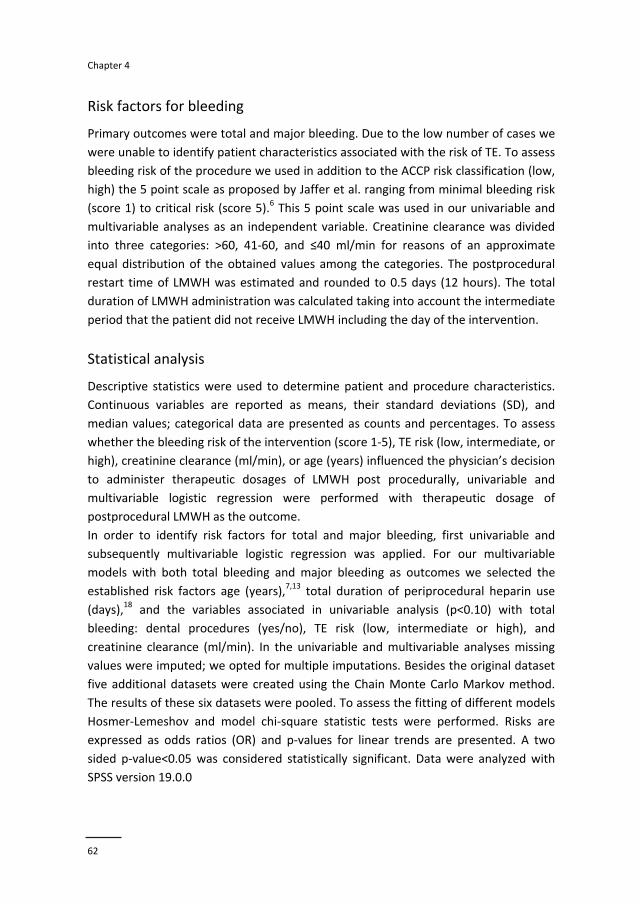
Chapter 4
62
Risk factors for bleeding
Primary outcomes were total and major bleeding. Due to the low number of cases we
were unable to identify patient characteristics associated with the risk of TE. To assess
bleeding risk of the procedure we used in addition to the ACCP risk classification (low,
high) the 5 point scale as proposed by Jaffer et al. ranging from minimal bleeding risk
(score 1) to critical risk (score 5).6 This 5 point scale was used in our univariable and
multivariable analyses as an independent variable. Creatinine clearance was divided
into three categories: >60, 41‐60, and ≤40 ml/min for reasons of an approximate
equal distribution of the obtained values among the categories. The postprocedural
restart time of LMWH was estimated and rounded to 0.5 days (12 hours). The total
duration of LMWH administration was calculated taking into account the intermediate
period that the patient did not receive LMWH including the day of the intervention.
Statistical analysis
Descriptive statistics were used to determine patient and procedure characteristics.
Continuous variables are reported as means, their standard deviations (SD), and
median values; categorical data are presented as counts and percentages. To assess
whether the bleeding risk of the intervention (score 1‐5), TE risk (low, intermediate, or
high), creatinine clearance (ml/min), or age (years) influenced the physician’s decision
to administer therapeutic dosages of LMWH post procedurally, univariable and
multivariable logistic regression were performed with therapeutic dosage of
postprocedural LMWH as the outcome.
In order to identify risk factors for total and major bleeding, first univariable and
subsequently multivariable logistic regression was applied. For our multivariable
models with both total bleeding and major bleeding as outcomes we selected the
established risk factors age (years),7,13 total duration of periprocedural heparin use
(days),18 and the variables associated in univariable analysis (p<0.10) with total
bleeding: dental procedures (yes/no), TE risk (low, intermediate or high), and
creatinine clearance (ml/min). In the univariable and multivariable analyses missing
values were imputed; we opted for multiple imputations. Besides the original dataset
five additional datasets were created using the Chain Monte Carlo Markov method.
The results of these six datasets were pooled. To assess the fitting of different models
Hosmer‐Lemeshov and model chi‐square statistic tests were performed. Risks are
expressed as odds ratios (OR) and p‐values for linear trends are presented. A two
sided p‐value<0.05 was considered statistically significant. Data were analyzed with
SPSS version 19.0.0
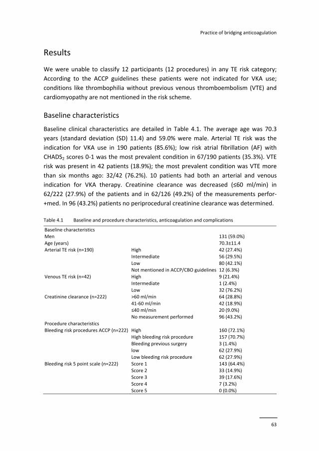
Practice of bridging anticoagulation
63
Results
We were unable to classify 12 participants (12 procedures) in any TE risk category;
According to the ACCP guidelines these patients were not indicated for VKA use;
conditions like thrombophilia without previous venous thromboembolism (VTE) and
cardiomyopathy are not mentioned in the risk scheme.
Baseline characteristics
Baseline clinical characteristics are detailed in Table 4.1. The average age was 70.3
years (standard deviation (SD) 11.4) and 59.0% were male. Arterial TE risk was the
indication for VKA use in 190 patients (85.6%); low risk atrial fibrillation (AF) with
CHADS2 scores 0‐1 was the most prevalent condition in 67/190 patients (35.3%). VTE
risk was present in 42 patients (18.9%); the most prevalent condition was VTE more
than six months ago: 32/42 (76.2%). 10 patients had both an arterial and venous
indication for VKA therapy. Creatinine clearance was decreased (≤60 ml/min) in
62/222 (27.9%) of the patients and in 62/126 (49.2%) of the measurements perfor‐
+med. In 96 (43.2%) patients no periprocedural creatinine clearance was determined.
Table 4.1 Baseline and procedure characteristics, anticoagulation and complications
Baseline characteristics
Men 131 (59.0%)
Age (years) 70.3±11.4
Arterial TE risk (n=190) High 42 (27.4%)
Intermediate 56 (29.5%)
Low 80 (42.1%)
Not mentioned in ACCP/CBO guidelines 12 (6.3%)
Venous TE risk (n=42) High 9 (21.4%)
Intermediate 1 (2.4%)
Low 32 (76.2%)
Creatinine clearance (n=222) >60 ml/min
64 (28.8%)
41‐60 ml/min
42 (18.9%)
≤40 ml/min
20 (9.0%)
No measurement performed 96 (43.2%)
Procedure characteristics
Bleeding risk procedures ACCP (n=222) High 160 (72.1%)
High bleeding risk procedure 157 (70.7%)
Bleeding previous surgery 3 (1.4%)
low 62 (27.9%)
Low bleeding risk procedure 62 (27.9%)
Bleeding risk 5 point scale (n=222) Score 1 143 (64.4%)
Score 2 33 (14.9%)
Score 3 39 (17.6%)
Score 4 7 (3.2%)
Score 5 0 (0.0%)
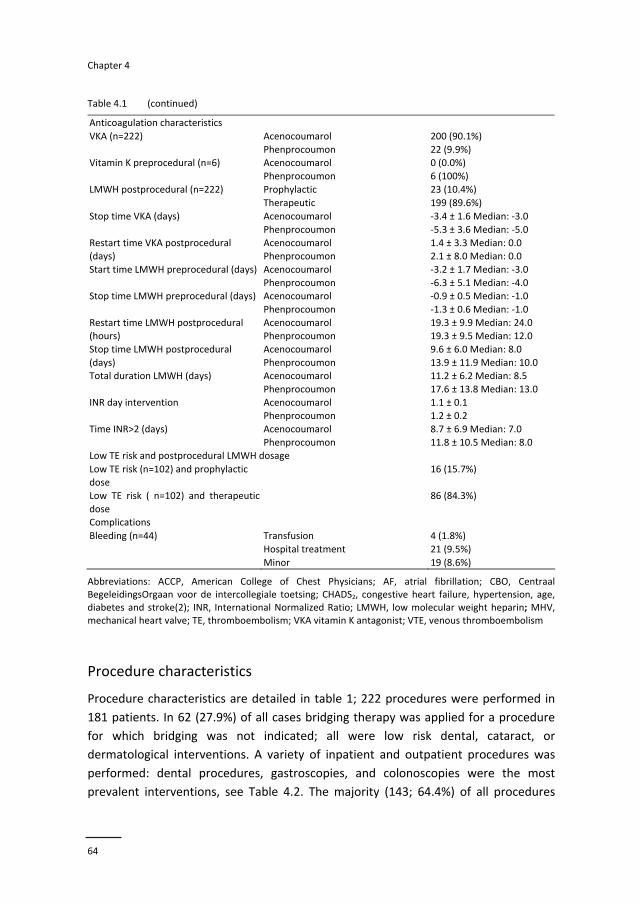
Chapter 4
64
Table 4.1 (continued)
Anticoagulation characteristics
VKA (n=222) Acenocoumarol
Phenprocoumon
200 (90.1%)
22 (9.9%) Vitamin K preprocedural (n=6) Acenocoumarol
Phenprocoumon
0 (0.0%)
6 (100%)
Prophylactic 23 (10.4%) LMWH postprocedural (n=222)
Therapeutic 199 (89.6%)
Stop time VKA (days) Acenocoumarol
Phenprocoumon
‐3.4 ± 1.6 Median: ‐3.0
‐5.3 ± 3.6 Median: ‐5.0
Restart time VKA postprocedural (days)
Acenocoumarol Phenprocoumon
1.4 ± 3.3 Median: 0.0 2.1 ± 8.0 Median: 0.0
Start time LMWH preprocedural (days) Acenocoumarol
Phenprocoumon
‐3.2 ± 1.7 Median: ‐3.0
‐6.3 ± 5.1 Median: ‐4.0 Stop time LMWH preprocedural (days) Acenocoumarol
Phenprocoumon
‐0.9 ± 0.5 Median: ‐1.0
‐1.3 ± 0.6 Median: ‐1.0
Restart time LMWH postprocedural (hours)
Acenocoumarol Phenprocoumon
19.3 ± 9.9 Median: 24.0 19.3 ± 9.5 Median: 12.0
Stop time LMWH postprocedural
(days)
Acenocoumarol
Phenprocoumon
9.6 ± 6.0 Median: 8.0
13.9 ± 11.9 Median: 10.0 Total duration LMWH (days) Acenocoumarol
Phenprocoumon
11.2 ± 6.2 Median: 8.5
17.6 ± 13.8 Median: 13.0
INR day intervention Acenocoumarol Phenprocoumon
1.1 ± 0.1 1.2 ± 0.2
Time INR>2 (days) Acenocoumarol
Phenprocoumon
8.7 ± 6.9 Median: 7.0
11.8 ± 10.5 Median: 8.0 Low TE risk and postprocedural LMWH dosage
Low TE risk (n=102) and prophylactic dose
16 (15.7%)
Low TE risk ( n=102) and therapeutic
dose
86 (84.3%)
Complications
Bleeding (n=44) Transfusion 4 (1.8%)
Hospital treatment 21 (9.5%)
Minor 19 (8.6%)
Abbreviations: ACCP, American College of Chest Physicians; AF, atrial fibrillation; CBO, Centraal BegeleidingsOrgaan voor de intercollegiale toetsing; CHADS2, congestive heart failure, hypertension, age, diabetes and stroke(2); INR, International Normalized Ratio; LMWH, low molecular weight heparin; MHV, mechanical heart valve; TE, thromboembolism; VKA vitamin K antagonist; VTE, venous thromboembolism
Procedure characteristics
Procedure characteristics are detailed in table 1; 222 procedures were performed in
181 patients. In 62 (27.9%) of all cases bridging therapy was applied for a procedure
for which bridging was not indicated; all were low risk dental, cataract, or
dermatological interventions. A variety of inpatient and outpatient procedures was
performed: dental procedures, gastroscopies, and colonoscopies were the most
prevalent interventions, see Table 4.2. The majority (143; 64.4%) of all procedures
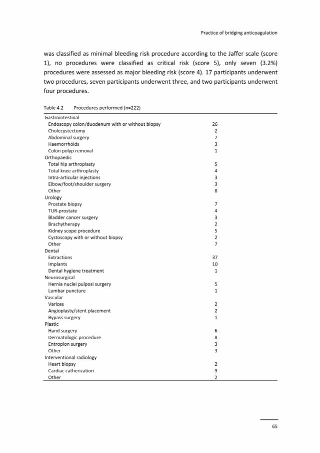
Practice of bridging anticoagulation
65
was classified as minimal bleeding risk procedure according to the Jaffer scale (score
1), no procedures were classified as critical risk (score 5), only seven (3.2%)
procedures were assessed as major bleeding risk (score 4). 17 participants underwent
two procedures, seven participants underwent three, and two participants underwent
four procedures.
Table 4.2 Procedures performed (n=222)
Gastrointestinal
Endoscopy colon/duodenum with or without biopsy 26
Cholecystectomy 2
Abdominal surgery 7 Haemorrhoids 3
Colon polyp removal 1
Orthopaedic Total hip arthroplasty 5
Total knee arthroplasty 4
Intra‐articular injections 3 Elbow/foot/shoulder surgery 3
Other 8
Urology Prostate biopsy 7
TUR‐prostate 4
Bladder cancer surgery 3 Brachytherapy 2
Kidney scope procedure 5
Cystoscopy with or without biopsy 2 Other 7
Dental
Extractions 37 Implants 10
Dental hygiene treatment 1
Neurosurgical Hernia nuclei pulposi surgery 5
Lumbar puncture 1
Vascular Varices 2
Angioplasty/stent placement 2
Bypass surgery 1 Plastic
Hand surgery 6
Dermatologic procedure 8 Entropion surgery 3
Other 3
Interventional radiology Heart biopsy 2
Cardiac catherization 9
Other 2
Chapter 4
66
Table 4.2 (continued)
Other
ENT surgery 2
Neurolysis 11
Cataract 2 Hernia umbilicalis/inguinalis 4
Breast cancer 4
Breast biopsy 6 Bronchoscopy with or without biopsy 4
Other 5
Abbreviations: ENT, ear, nose and throat; TUR, transurethral resection of the prostate
Anticoagulation
Anticoagulation characteristics are detailed in Table 4.1. The majority of the patients
used acenocoumarol as oral anticoagulant: 200 (90.1 %), the remaining 22 (9.9%) used
phenprocoumon. The median preoperative stop time of VKA was day ‐3 (mean ‐3.4,
SD 1.6) and day ‐5 (mean ‐5.3, SD 3.6) for acenocoumarol and phenprocoumon,
respectively. Vitamin K was used to reverse anticoagulation only in six patients (2.7%);
all used phenprocoumon, a VKA with a relative long half‐life of 120‐200 hours.
Acenocoumarol was resumed a median of 0 days (mean 1.4, SD 1.6); phenprocoumon
was resumed a median of 0 days (mean 2.1, SD 8.0). LMWH was used as bridging
agent of first choice in all patients. The proportion of patients at low TE risk (n=102)
treated with therapeutic doses of LMWH post procedurally was 84.3% (n=86). We also
explored periprocedural timing of LMWH administration; LMWH therapy was initiated
a median 3 days (mean 3.2, SD 1.7) and a median 4 days (mean 6.3, SD 5.1) before and
stopped a median of 1 (mean 0.9, SD 0.5 and mean 1.3, SD 0.6) day prior to the
planned procedure in acenocoumarol and phenprocoumon users respectively. The
median time of postoperative restart of LMWH therapy was 24 hours (mean 19.3, SD
9.9) and 12 hours (mean 19.3, SD 9.5) in acenocoumarol and phenprocoumon users
respectively. The median duration of postoperative LMWH administration was 8 days
(mean 11.2, SD 6.2) and 13 days (mean: 17.6, SD 13.8) in acenocoumarol and
phenprocoumon users respectively. Of all patients undergoing bridging therapy 199
(89.6%) were treated with therapeutic dosages of LMWH after the procedure.
Univariable logistic regression with postprocedural therapeutic dosage of LMWH as
the outcome resulted in non significant effects for all variables. No proof was found
that the prescribing physician’s decision to administer therapeutic dosages of LMWHs
was influenced by age, TE risk, surgical bleeding risk, or creatinine clearance. Patients
at high TE risk compared to low risk patients had a non significant higher risk of
exposure to therapeutic doses (OR 4.22, 95% CI 0.93 to 19.24), p for linear trend=0.06.
Practice of bridging anticoagulation
67
Patients with a creatinine clearance within the range of 41‐60 ml/min compared to a
clearance >60 ml/min had a non significant lower risk of exposure to therapeutic
doses (OR 0.43, 95% CI 0.10 to 1.82), p for linear trend=0.88. A high bleeding risk
procedure (score 4) on the Jaffer scale compared to a procedure score of 1 resulted in
a non significant decreased risk of exposure to therapeutic doses of LMWH (OR 0.24,
95% CI 0.04 to 1.36), p for linear trend=0.11. Multivariable analyses including the
aforementioned variables resulted in overall non significant results. Patients at high TE
risk compared to low risk had a borderline non significant higher risk of exposure to
therapeutic dosages after their intervention (OR 4.96, 95% CI 0.97 to 25.26), p for
linear trend=0.05. Patients with a creatinine clearance within the range of 41‐60
ml/min compared to clearance >60 ml/min had a non significant lower risk of
exposure to therapeutic doses (OR 0.38, 95% CI 0.08 to 1.81), p for trend=0.73. Finally,
a bleeding risk score 4 compared to score 1 resulted in a non significant decreased risk
(OR 0.27, 95% CI 0.04 to 1.86), p for linear trend=0.23. The goodness of fit of the
model was assessed, resulting in a p‐value of 0.77 on the Hosmer‐Lemeshov test and
the model chi‐square statistic resulted in a p‐value of 0.75.
Complications
The 30‐day incidence of total bleeding in the entire group of procedures performed
was 44 (19.8%), the incidence of major bleeding 25 (11.3%); there were no deaths, no
intracranial bleedings, four patients required a transfusion, and 21 had to be treated
in a hospital due to postoperative bleeding. Two patients (0.9%) experienced a deep
venous thrombosis and recovered; see Table 4.1.
Risk factors for bleeding
Univariable logistic regression analysis revealed high versus low TE risk (OR 2.59, 95%
CI 1.11 to 6.04), p for linear trend=0.03 and dental procedures (OR 2.98, 95% CI 1.45
to 6.13) as risk factors for total bleeding; all other results are non significant.
Creatinine clearance ≤40 versus >60 ml/min and intermediate versus low TE risk
resulted in non‐significant elevated risks: OR 2.35, 95% (CI 0.93 to 5.90), p for linear
trend=0.07 and OR 1.83, 95% CI 0.77 to 4.33, respectively. To minimize the risk of
reversed causality we excluded 18 cases in which VKA were stopped and LMWH
administration due to a total bleeding was prolonged; the initial significantly increased
risk caused by the total duration of LMWH administration (result not shown)
disappeared: (OR 0.94, 95% CI 0.86 to 1.03). After exclusion of the aforementioned 18
cases, dental procedures (OR 3.32, 95% CI 1.22 to 9.04) and creatinine clearance ≤40
versus >60 ml/min (OR 5.03, 95% CI 1.25 to 20.26), p for linear trend=0.02 were
Chapter 4
68
identified as independent predictors of total bleeding in a model completed with the
variables age, duration of periprocedural use of LMWH, and TE risk. The Hosmer‐
Lemeshov test resulted in a p‐value of 0.47 and the model chi‐square statistic yielded
a significant result: p=0.01.
Finally, we explored major bleeding. Univariable logistic regression revealed
intermediate versus low TE risk as a risk factor (OR 3.40, 95% CI 1.17 to 9.93), p for
linear trend=0.11. No further risk factors were identified. Our dataset contained only
three high TE risk patients due to mitral valve replacements of which one experienced
a major bleeding; hospital treatment was necessary (OR 4.06, 95% CI 0.36 to 46.50). In
multivariable analysis, using the same model, we again excluded the aforementioned
18 cases to avoid differential misclassification and no significant risk factors were
identified. The model as a whole scored a p‐value of 0.83 on the Hosmer‐Lemeshov
test with a p‐value of 0.50 on the model chi‐square statistic.
Discussion
In our study we found that guideline adherence to bridging therapy in the region
around Maastricht, the Netherlands, is not optimal. The most striking finding is that
84.3% of all low TE risk patients was bridged with therapeutic doses LMWH. Low TE
risk does not warrant bridging therapy and certainly not with therapeutic doses of
LMWH.22 Furthermore we found compared to other studies, high rates of total and
major bleedings.6,9,23,24 We were unable to find an association of this observed
aggressive treatment with anticoagulants and the high bleeding rates, possibly due a
lack of contrast in our population. Studies performed by Robinson et al. and Jaffer et
al. identified postprocedural therapeutic doses of LMWH as a risk factor for
bleeding.6,11 In general, bridging therapy exposes the patient to additional risks, also
including a risk of heparin‐induced thrombocytopenia (HIT).25 Interventions for which
no bridging anticoagulation is indicated and VKA administration can simply be
continued, represented 27.9% of the total number of procedures performed in our
cohort; a fairly high proportion. Possibly due to the fact that the majority of the
participants were outpatients, the period of exposure to LMWH was much longer than
necessary according to the ACCP guidelines; in outpatients rigidly performed INR
testing is often not feasible.26 Furthermore, due to a change in the anticoagulant
regime in the outpatient setting the patient’s compliance might be at risk; this might
introduce an additional risk factor for bleeding or TE. Another possible explanation for
prolonged LMWH administration might be the use of too low restart doses of VKA (i.e.
the maintenance dose) instead of 1,5 to 2 times higher doses of acenocoumarol and
Practice of bridging anticoagulation
69
phenprocoumon as advised in guidelines issued by the FNT.27 Overall TE incidence was
low and in concordance with some other studies6,9,23,24; no arterial TE occurred. We
conclude that individual clinicians often do not act according to the current bridging
guidelines; in the observed cohort the decision to administer therapeutic dosages of
LMWH was not or barely influenced by surgical bleeding risk, TE risk, or renal
insufficiency. Krahn et al. and Skolarus et al. report similar findings5,28; Gerson et al. on
the other hand concluded that most people receiving bridging therapy were managed
according to current society guidelines.29 Possible explanations for non adherence are
the lack of familiarity with these guidelines, lack of awareness of the significance of
consistent bridging practices, disagreement with the guidelines, and resistance to
change.30 It is also conceivable that physicians tend to over treat patients because the
threat of a TE is considered more severe than the threat of bleeding.5
Renal insufficiency appeared an independent predictor for total bleeding. Other
studies support this finding1,14; the clearance of LMWH is primarily renal, the plasma
half‐live increases in patients with renal failure and dose reduction is advised in these
patients following the Cockroft Gault formula.8,31 As far as we know only one study
performed by Hammerstingl reported high TE risk as a risk factor for perioperative
bleeding.8 Possibly confounding biased this finding since in our analysis increasing TE
risk was only found to be a risk factor for total bleeding in univariable analyses. An
unexpected, novel finding is that dental treatment inflicts a very high bleeding risk on
patients; most dental treatments do not warrant bridging therapy; instead, VKA
continuation in combination with the oral administration of antifibrinolytic agents like
tranexaminic acid are advised.22 Several studies report that restarting LMWH in close
proximity to the intervention might induce bleeding.16,17 Our study does not support
these findings; the observed high rate (28.8%) of missing values concerning this
variable might have diluted this effect.
Strengths and limitations of study
Our study has some weaknesses; the sample size was small and data were analyzed
retrospectively. We were unable to compare different institutions with respect to
guideline adherence, so only a local view on bridging practices could be provided. The
strengths of our study are: a well defined study population and the observational
design that allowed us to establish guideline adherence and identify risk factors for
bleeding. We allow comparison of our results with other studies because we reported
according to the recommendations for reporting studies in periprocedural anti‐
thrombotic and bridging therapy, issued by the ISTH.

Chapter 4
70
Conclusions
Guideline adherence to bridging therapy is poor in the observed single regional
setting. This results in patients being unnecessarily exposed to LMWH and for too long
periods of time. Since bridging is in general associated with increased bleeding
risks,20,32 it should be avoided in the absence of a good indication. The additional
omission of risk stratification based on assessment of renal function further increased
bleeding rates. Although these observations are confined to a limited region within
one country, there is no reason to expect that this represents a unique and regional
problem. Rather, it illustrates the importance of adhering to guidelines for
antithrombotic management.
Practice of bridging anticoagulation
71
References
1. Cano O, Osca J, Sancho‐Tello MJ, Olague J, Castro JE, Salvador A. Morbidity associated with three different antiplatelet regimens in patients undergoing implantation of cardiac rhythm management
devices. Europace. 2011;13:395‐401.
2. Tompkins C, Cheng A, Dalal D, et al. Dual antiplatelet therapy and heparin "bridging" significantly increase the risk of bleeding complications after pacemaker or implantable cardioverter‐defibrillator
device implantation. J Am Coll Cardiol. 2010;55:2376‐82.
3. Li HK, Chen FC, Rea RF, et al. No increased bleeding events with continuation of oral anticoagulation therapy for patients undergoing cardiac device procedure. Pacing Clin Electrophysiol. 2011;34:868‐74.
4. Ghanbari H, Feldman D, Schmidt M, et al. Cardiac resynchronization therapy device implantation in
patients with therapeutic international normalized ratios. Pacing Clin Electrophysiol. 2010;33:400‐6. 5. Krahn AD, Healey JS, Simpson CS, Essebag V, Sivakumaran S, Birnie DH. Anticoagulation of patients on
chronic warfarin undergoing arrhythmia device surgery: wide variability of perioperative bridging in
Canada. Heart Rhythm. 2009;6:1276‐9. 6. Jaffer AK, Brotman DJ, Bash LD, Mahmood SK, Lott B, White RH. Variations in perioperative warfarin
management: outcomes and practice patterns at nine hospitals. Am J Med. 2010;123:141‐50.
7. Ercan M, Bostanci EB, Ozer I, et al. Postoperative hemorrhagic complications after elective laparoscopic cholecystectomy in patients receiving long‐term anticoagulant therapy. Langenbecks
Arch Surg. 2010;395:247‐53.
8. Hammerstingl C, Omran H. Perioperative bridging of chronic oral anticoagulation in patients undergoing pacemaker implantation‐‐a study in 200 patients. Europace. 2011;13:1304‐10.
9. Hammerstingl C, Omran H. Bridging of oral anticoagulation with low‐molecular‐weight heparin:
experience in 373 patients with renal insufficiency undergoing invasive procedures. Thromb Haemost. 2009;101:1085‐90.
10. Hammerstingl C, Schmitz A, Fimmers R, Omran H. Bridging of chronic oral anticoagulation with
enoxaparin in patients with atrial fibrillation: results from the prospective BRAVE registry. Cardiovasc Ther. 2009;27:230‐8.
11. Robinson M, Healey JS, Eikelboom J, et al. Postoperative low‐molecular‐weight heparin bridging is
associated with an increase in wound hematoma following surgery for pacemakers and implantable defibrillators. Pacing Clin Electrophysiol. 2009;32:378‐82.
12. Tolosana JM, Berne P, Mont L, et al. Preparation for pacemaker or implantable cardiac defibrillator
implants in patients with high risk of thrombo‐embolic events: oral anticoagulation or bridging with intravenous heparin? A prospective randomized trial. Eur Heart J. 2009;30:1880‐4.
13. Tischenko A, Gula LJ, Yee R, Klein GJ, Skanes AC, Krahn AD. Implantation of cardiac rhythm devices
without interruption of oral anticoagulation compared with perioperative bridging with low‐molecular weight heparin. Am Heart J. 2009;158:252‐6.
14. Ahmed I, Gertner E, Nelson WB, House CM, Zhu DW. Chronic kidney disease is an independent
predictor of pocket hematoma after pacemaker and defibrillator implantation. J Interv Card Electrophysiol. 2010;29:203‐7.
15. Steger V, Bail DH, Graf D, Walker T, Rittig K, Ziemer G. A practical approach for bridging
anticoagulation after mechanical heart valve replacement. J Heart Valve Dis. 2008;17:335‐42. 16. Tafur AJ, McBane R, 2nd, Wysokinski WE, et al. Predictors of major bleeding in peri‐procedural
anticoagulation management. J Thromb Haemost. 2012;10:261‐7.
17. Chow V, Ranasinghe I, Lau J, et al. Peri‐procedural anticoagulation and the incidence of haematoma formation after permanent pacemaker implantation in the elderly. Heart Lung Circ. 2010;19:706‐12.
18. Kiviniemi T, Karjalainen P, Pietila M, et al. Comparison of additional versus no additional heparin
during therapeutic oral anticoagulation in patients undergoing percutaneous coronary intervention. Am J Cardiol. 2012;110:30‐5.
19. Lahtela H, Rubboli A, Schlitt A, et al. Heparin bridging vs. uninterrupted oral anticoagulation in
patients with Atrial Fibrillation undergoing Coronary Artery Stenting. Results from the AFCAS registry. Circ J. 2012;76:1363‐8.
Chapter 4
72
20. Eijgenraam P, Ten Cate H, Ten Cate‐Hoek AJ. Safety and Efficacy of Bridging with Low Molecular
Weight Heparins: A Systematic Review and Partial Meta‐Analysis. Curr Pharm Des. 2012. 21. Spyropoulos AC, Douketis JD, Gerotziafas G, Kaatz S, Ortel TL, Schulman S. Periprocedural
antithrombotic and bridging therapy: recommendations for standardized reporting in patients with
arterial indications for chronic oral anticoagulant therapy. J Thromb Haemost. 2012;10:692‐4. 22. Douketis JD, Berger PB, Dunn AS, et al. The perioperative management of antithrombotic therapy:
American College of Chest Physicians Evidence‐Based Clinical Practice Guidelines (8th Edition). Chest.
2008;133:299‐339. 23. Pengo V, Cucchini U, Denas G, et al. Standardized low‐molecular‐weight heparin bridging regimen in
outpatients on oral anticoagulants undergoing invasive procedure or surgery: an inception cohort
management study. Circulation. 2009;119:2920‐7. 24. Malato A, Saccullo G, Lo Coco L, et al. Patients requiring interruption of long‐term oral anticoagulant
therapy: the use of fixed sub‐therapeutic doses of low‐molecular‐weight heparin. J Thromb Haemost.
2010;8:107‐13. 25. Jaffer AK, Brotman DJ, Chukwumerije N. When patients on warfarin need surgery. Cleve Clin J Med.
2003;70:973‐84.
26. Deerhake JP, Merz JC, Cooper JV, Eagle KA, Fay WP. The duration of anticoagulation bridging therapy in clinical practice may significantly exceed that observed in clinical trials. J Thromb Thrombolysis.
2007;23:107‐13.
27. Dolder BDv. De kunst van het doseren. Voorschoten: Federatie van Nederlandse trombosediensten; 2010.
28. Skolarus LE, Morgenstern LB, Froehlich JB, Lisabeth LD, Brown DL. Guideline‐discordant
periprocedural interruptions in warfarin therapy. Circ Cardiovasc Qual Outcomes. 2011;4:206‐10. 29. Gerson LB, Michaels L, Ullah N, Gage B, Williams L. Adverse events associated with anticoagulation
therapy in the periendoscopic period. Gastrointest Endosc. 2010;71:1211‐7.
30. Eisenstein DH. Anticoagulation management in the ambulatory surgical setting. AORN J. 2012;95:510‐21 examination 22‐4.
31. Harder S, Klinkhardt U, Alvarez JM. Avoidance of bleeding during surgery in patients receiving
anticoagulant and/or antiplatelet therapy: pharmacokinetic and pharmacodynamic considerations. Clin Pharmacokinet. 2004;43:963‐81.
32. Spyropoulos AC, Turpie AG. Perioperative bridging interruption with heparin for the patient receiving
long‐term anticoagulation. Curr Opin Pulm Med. 2005;11:373‐9.
73
Chapter 5 Effects of peri‐operative bridging with low molecular
weight heparins on coagulation during interruption
of vitamin K antagonists: a mechanistic study
Pieter Eijgenraam,Hugo ten Cate, Y. Henskens, R. van den Ham, Arina J ten Cate‐Hoek
Submitted
Chapter 5
74
Abstract
Background
Bridging with vitamin K antagonists (VKA) comprises peri‐procedural substitution by low
molecular weight heparins (LMWH) upon interruption of VKA. In this exploratory study we
investigated the interactive effects of the co‐administration of VKA, LMWH and surgery on peri‐
operative coagulation.
Methods
Blood was sampled daily from day ‐3 to day +5 in 13 patients. In addition to measurement of
INR and anti‐Xa activity, thrombin generation (TG) testing and assessment of individual
coagulation factors was performed.
Results
At the time of intervention the mean INR was 1.0 (SD 0.1, range 0.9‐1.2); the mean anti‐Xa at
day 0 was 0.19 units/ml (SD 0.20 units/ml, range <0.05–0.60). The intervention caused a 2 to 3
fold increase in TG at day 0. Of all coagulation factors, factor (F) XI had the strongest correlation
with TG (r=0.629; p=0.02) for peak and endogenous thrombin potential (ETP) (r=0.625; p=0.02).
Thrombomodulin‐induced reduction of ETP increased from 10.0% (SD 9.2) at day ‐3 to 18.2%
(SD 9.5) at day 0, p=0.02. After surgery, FVIII and fibrinogen were significantly increased
(p<0.001).
Conclusions
In spite of residual anti‐Xa activity at the day of the intervention there is a marked increase in
TG related to surgery. Peri‐operatively, 3 prothrombotic mechanisms were exposed: FXI
dependent TG, reduced activity of the activated protein C pathway and postoperative rises in
FVIII and fibrinogen. For the complex perioperative management the value of TG or other global
assays to monitor the coagulation balance merits further study.

Effects of peri‐operative bridging with LMWH on coagulation during interruption of VKA
75
Introduction
Current guidelines advise substitution of longer acting VKA therapy with relatively
short acting LMWH in patients at the highest risk of thrombosis in the periprocedural
period.1,2 Bridging with LMWH therefore is routinely used in patients undergoing
invasive procedures that require interruption of VKA, but are deemed too high risk for
thromboembolism to allow absence of anticoagulation during the post‐intervention
period. However, there is only low quality evidence underlying current practice as
randomized clinical trials (RCT) were up till recently lacking. The available evidence
derived from systematic reviews and meta‐analyses suggests that bridging is
associated with an increased risk of bleeding. While the bleeding risk may be mostly
attributable to the intervention itself, the contribution of fixed, body weight adjusted
doses of LMWH in conjunction with stopping/restarting of VKA, may further impair
hemostasis in an undesirable manner.3 A recent review showed low rates of thrombo‐
embolism in bridged and non‐bridged patients of respectively 0.9 and 0.6 percent, but
associated high rates of bleeding in bridged patients (13.1% vs. 3.4% all bleeds, and
4.2% vs. 0.9% major bleeds.4 Given the observed elevated risk of bleeding associated
with this LMWH bridging policy, the routine application of bridging has been recently
challenged. In the very recently published BRIDGE study, in patients randomized to
placebo, the risk of thromboembolic events was comparably low to the risk in the
LMWH bridged subjects (who had a higher risk of bleeding). Based on these outcomes
the authors also discussed the issue of thrombogenicity related to the procedure,
stating that “The premise that warfarin interruption leads to rebound
hypercoagulability and that the milieu of the procedure confers a prothrombotic
state, which in turn leads to arterial thromboembolism, is not supported by the
results of this trial”.5 However, the BRIDGE study included for the large part low risk
for thromboembolism patients. Notably, another study showed that there is a 7 times
difference in thrombotic risk between more extensive surgery (risks being the highest
for hip‐surgery or cancer‐surgery) compared to less extensive day‐surgery risk.6 The
thromboembolic risks associated with the bridging procedure as well as the
underlying mechanisms have not been explored extensively and are therefore largely
unknown.
In this explorative study we therefore aimed to investigate the combined effects of
cessation of VKA prior to surgery and institution of LMWH in combination with
surgery on peri‐operative coagulation, assessed by thrombin generation and a panel
of its protein determinants.
Chapter 5
76
Methods
Patient selection
During the period from June 2013 until April 2014 13 patients on chronic treatment
with VKA for atrial fibrillation (AF), recurrent venous thromboembolism (VTE), or
aortic valve replacement who visited the Maastricht Anticoagulation Clinic, the
Netherlands, and in whom bridging of anticoagulation was indicated for planned
surgery were invited to participate in the study. Institutional review board approval
was obtained (METC 12‐1‐008). All participants gave informed consent; all patients
participated once.
Blood samples were collected daily within a time slot of 3 hours and before noon
sharp from day ‐3 to day +5, including the day of the intervention. The following
inclusion criteria were applied: 1) At least 3 months acenocoumarol use, a VKA that is
commonly used in the Netherlands 2) planned invasive procedure 3) indication for
peri‐procedural bridging with LMWH 4) mentally competent, and 5) a minimum age of
18 years. Excluded were 1) patients with severe renal failure (MDRD <30 ml/min),
2) patient who were pregnant or breastfeeding and 3) patients undergoing emergency
procedures.
Bridging anticoagulation
In all patients acenocoumarol was stopped on day ‐3, LMWH was installed at day ‐3
(6 patients) or day‐2 (7 patients). The exact bridging regimen was left to the discretion
of the prescribing physician, reflecting routine practice. The last dose of LMWH before
the intervention was administered at day ‐1. Acenocoumarol was re‐installed at day 0
(1 patient), day +1 (5 patients) or later (7 patients). LMWH was restarted at day 0
(6 patients), day +1 (5 patients), day +2 (1 patient), or day +4 (1 patient).
Blood collection and plasma preparation
Venous blood was collected in 3.2% citrate (w/v) citrated vacutainer glass tubes
(Becton Dickinson, Plymouth, UK). Within 1 hour after blood collection platelet‐poor
plasma (PPP) was prepared using two centrifugation steps: the first at 2000xg for 5
minutes and a second step at 10 000xg for 10 minutes. Plasma aliquots were snap‐
frozen, stored at ‐80°C and thawed at 37°C before analysis. The research nurse
responsible for the collection of the blood samples recorded the time the sample was
taken, the time of LMWH and VKA administration, the type and dose of LMWH and
Effects of peri‐operative bridging with LMWH on coagulation during interruption of VKA
77
VKA, and the time of the intervention (day 0) in case report forms (CRF). On day 0
blood was collected before (n=9) or after surgery (n=4).
Thrombin generation measurements
Thrombin generation in platelet‐poor plasma was measured using the Calibrated
Automated Thrombogram method (Thrombinoscope BV, Maastricht, The
Netherlands), as previously published.[7] The following experimental conditions were
used: 0 pM TF with 4 µM PL at 20:20:60 mol% PS:PE:PC, 1 pM TF with 4 µM PL in the
absence and presence of thrombomodulin (TM) (Thrombinoscope BV) with a final
concentration of 0.65 nM, and 5 pM TF with 4 µM PL. The threshold of detectable
thrombin level with this assay is 10 nM; therefore we imputed for undetectable peak
height (PH) values a value of 5 nM, slope=0 and endogenous thrombin potential
(ETP)=0.5*5*(average value of start tail (time)‐lag time (LT) for the concerning
reagent).
Measurement of plasma factor levels, INR and anti‐Xa
The INR was assessed using Neoplastine‐r (STAGO) reagent. Heparin was assessed by
means of the anti‐Xa levels using the liquid Anti‐Xa (STAGO) and a specific LMWH
calibrator; the threshold of detectable anti‐Xa level with this assay is 0.05 IU/ml.
Plasma fibrinogen levels were measured using the Clauss method using liquid Fib as
reagent (STAGO). All other coagulation factor levels were determined by one‐stage
PT‐based (FII, FV, FVII, FX) or aPTT‐based (FVIII, FIX, FXI, FXII) clotting assays using
plasma deficient in factor V, VII, X, XI, or XII, or plasma immune‐depleted for factors
VIII, or IX, as reagents. AT levels (FX‐a inhibitory activity) with Stachrom AT as reagent
and protein C levels (Stachrom prot C) were measured using chromogenic assays. All
plasma factor level measurements employed commercially available standards
(STAGO, Asnières sur Seine, France) calibrated to WHO standards. All assays were
performed on a STA‐R coagulation analyzer (STAGO).
Statistical analysis
Descriptive statistics were used to determine patient and procedure characteristics.
Continuous variables are reported as means, and their standard deviations (SD);
categorical data are presented as counts and percentages. When related samples
were analyzed Wilcoxon signed‐ranks or Friedman tests were applied as appropriate.
Non‐related samples were tested with Mann‐Whitney, Kruskal‐Wallis or Student’s
t‐tests as appropriate. Pre and post interventional comparisons were made: day ‐3 to
Chapter 5
78
day –1 (pre intervention) and day +1 to day +5 (post intervention). To allow linear
modeling using linear regression with time expired since LMWH administration as
independent variable and anti‐Xa levels as dependent variable, anti‐Xa values were
log transformed (ln anti‐Xa) and measurements in which blood sample collection
occurred within 4 hours after LMWH administration were excluded; because LMWH
values peak 4 hours after administration.[8] To account for repeated measurements
we constructed linear models for all patients individually and calculated the time from
LMWH administration to an anti‐Xa level of 0.20 IU/ml using these models; the mean
values are reported. According to the linear regression model time expired since
LMWH administration to reach an anti‐Xa level of 0.20 IU/ml is expressed as (ln 0.2‐β0
(intercept))/β1). Correlations were determined using Pearson’s correlation coefficient.
Mean values of the different variables for each patient were taken into account. A two
sided p‐value <0.05 was considered statistically significant. Data were analyzed with
SPSS version 22.
Study results are reported according to the recommendations for studies in peri‐
procedural antithrombotic and bridging therapy issued by the ISTH.9
Results
Patient characteristics
Patient characteristics are presented in Table 5.1. The average age of the patients was
64.7 (SD 9.5), 8/13 (61.5%) were male. All participants, 13/13 (100%) were on
acenocoumarol treatment for atrial fibrillation (8), DVT (4) and prosthetic aortic valves
(1). All patients but one (dalteparin) received therapeutic subcutaneous doses
(tinzaparin, nadroparin or enoxaparin) of LMWH peri‐operatively: once daily in 12
(92.3%), and twice daily in 1 (7.7%). One of thirteen patients (7.7%) underwent a low
bleeding/ thrombotic risk procedure (skin biopsy) the remaining 12/13 (82.3%)
underwent a high bleeding/thrombotic risk procedure (umbilical hernia repair (2),
ablation for AF (2), orthopedic surgery (4), transurethral resection of the prostate (2),
cholecystectomy (1), and nephrectomy (1)).
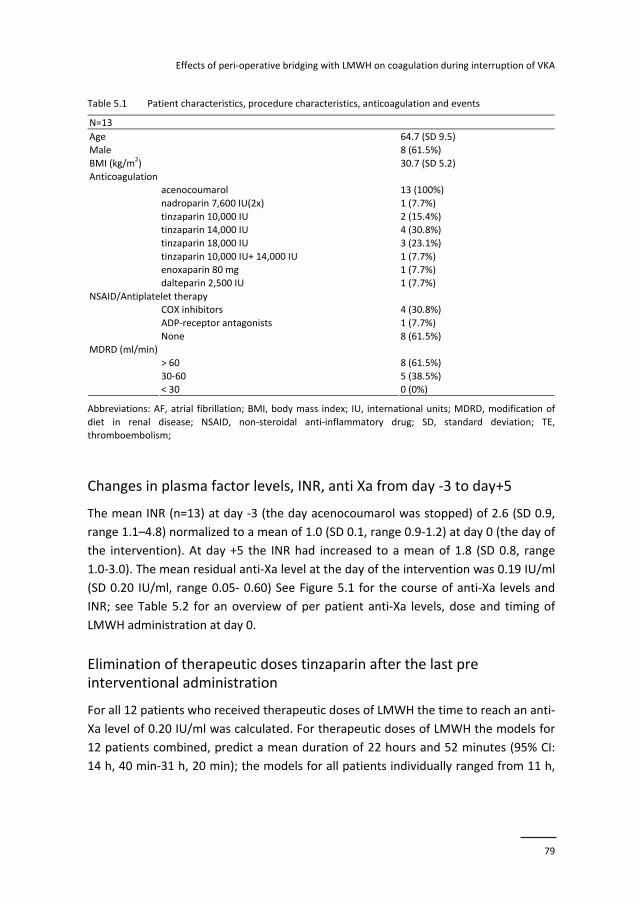
Effects of peri‐operative bridging with LMWH on coagulation during interruption of VKA
79
Table 5.1 Patient characteristics, procedure characteristics, anticoagulation and events
N=13
Age 64.7 (SD 9.5)
Male 8 (61.5%)
BMI (kg/m2) 30.7 (SD 5.2)
Anticoagulation acenocoumarol 13 (100%)
nadroparin 7,600 IU(2x) 1 (7.7%)
tinzaparin 10,000 IU 2 (15.4%) tinzaparin 14,000 IU 4 (30.8%)
tinzaparin 18,000 IU 3 (23.1%)
tinzaparin 10,000 IU+ 14,000 IU 1 (7.7%) enoxaparin 80 mg 1 (7.7%)
dalteparin 2,500 IU 1 (7.7%)
NSAID/Antiplatelet therapy COX inhibitors 4 (30.8%)
ADP‐receptor antagonists 1 (7.7%)
None 8 (61.5%) MDRD (ml/min)
> 60 8 (61.5%)
30‐60 5 (38.5%)
< 30 0 (0%)
Abbreviations: AF, atrial fibrillation; BMI, body mass index; IU, international units; MDRD, modification of
diet in renal disease; NSAID, non‐steroidal anti‐inflammatory drug; SD, standard deviation; TE, thromboembolism;
Changes in plasma factor levels, INR, anti Xa from day ‐3 to day+5
The mean INR (n=13) at day ‐3 (the day acenocoumarol was stopped) of 2.6 (SD 0.9,
range 1.1–4.8) normalized to a mean of 1.0 (SD 0.1, range 0.9‐1.2) at day 0 (the day of
the intervention). At day +5 the INR had increased to a mean of 1.8 (SD 0.8, range
1.0‐3.0). The mean residual anti‐Xa level at the day of the intervention was 0.19 IU/ml
(SD 0.20 IU/ml, range 0.05‐ 0.60) See Figure 5.1 for the course of anti‐Xa levels and
INR; see Table 5.2 for an overview of per patient anti‐Xa levels, dose and timing of
LMWH administration at day 0.
Elimination of therapeutic doses tinzaparin after the last pre interventional administration
For all 12 patients who received therapeutic doses of LMWH the time to reach an anti‐
Xa level of 0.20 IU/ml was calculated. For therapeutic doses of LMWH the models for
12 patients combined, predict a mean duration of 22 hours and 52 minutes (95% CI:
14 h, 40 min‐31 h, 20 min); the models for all patients individually ranged from 11 h,
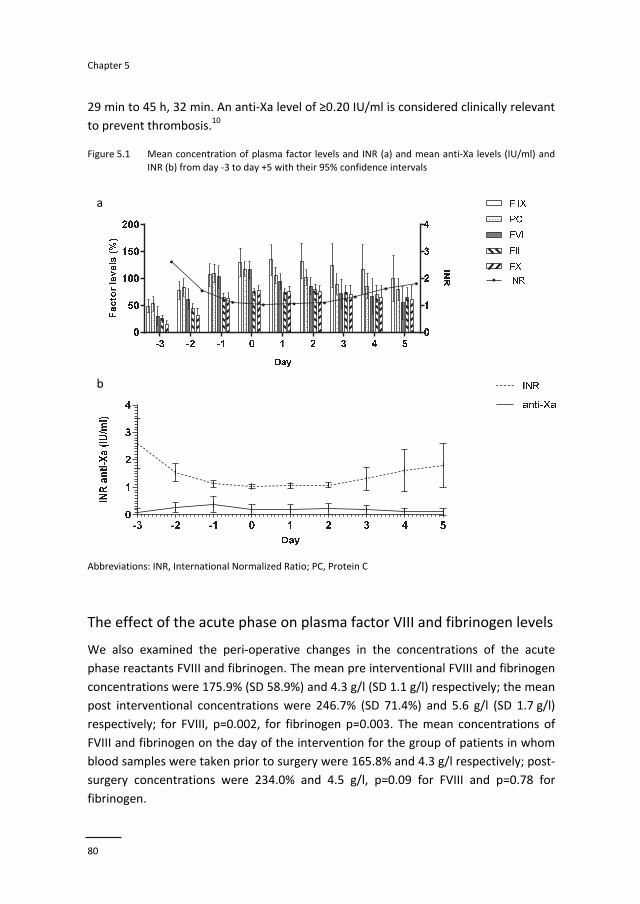
Chapter 5
80
29 min to 45 h, 32 min. An anti‐Xa level of ≥0.20 IU/ml is considered clinically relevant
to prevent thrombosis.10 Figure 5.1 Mean concentration of plasma factor levels and INR (a) and mean anti‐Xa levels (IU/ml) and
INR (b) from day ‐3 to day +5 with their 95% confidence intervals
a
b
Abbreviations: INR, International Normalized Ratio; PC, Protein C
The effect of the acute phase on plasma factor VIII and fibrinogen levels
We also examined the peri‐operative changes in the concentrations of the acute
phase reactants FVIII and fibrinogen. The mean pre interventional FVIII and fibrinogen
concentrations were 175.9% (SD 58.9%) and 4.3 g/l (SD 1.1 g/l) respectively; the mean
post interventional concentrations were 246.7% (SD 71.4%) and 5.6 g/l (SD 1.7 g/l)
respectively; for FVIII, p=0.002, for fibrinogen p=0.003. The mean concentrations of
FVIII and fibrinogen on the day of the intervention for the group of patients in whom
blood samples were taken prior to surgery were 165.8% and 4.3 g/l respectively; post‐
surgery concentrations were 234.0% and 4.5 g/l, p=0.09 for FVIII and p=0.78 for
fibrinogen.
a
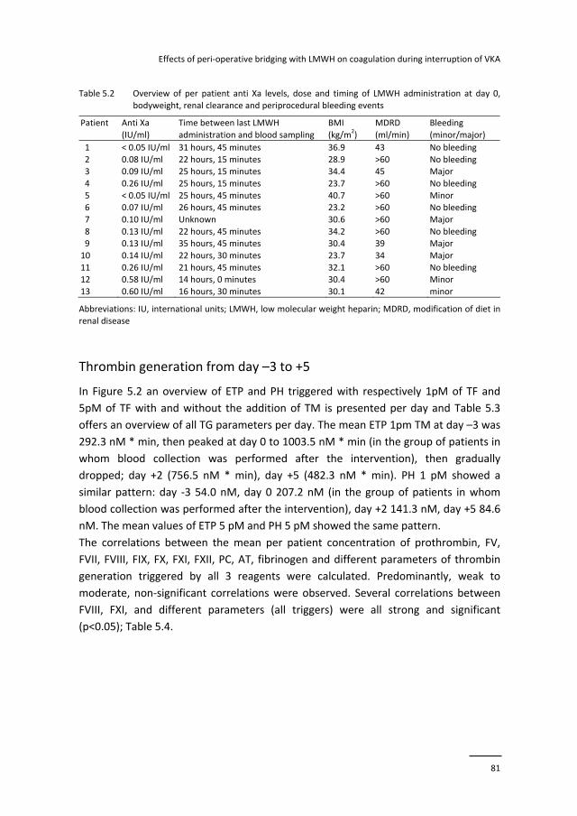
Effects of peri‐operative bridging with LMWH on coagulation during interruption of VKA
81
Table 5.2 Overview of per patient anti Xa levels, dose and timing of LMWH administration at day 0,
bodyweight, renal clearance and periprocedural bleeding events
Patient Anti Xa
(IU/ml)
Time between last LMWH
administration and blood sampling
BMI
(kg/m2)
MDRD
(ml/min)
Bleeding
(minor/major)
1 < 0.05 IU/ml 31 hours, 45 minutes 36.9 43 No bleeding
2 0.08 IU/ml 22 hours, 15 minutes 28.9 >60 No bleeding 3 0.09 IU/ml 25 hours, 15 minutes 34.4 45 Major
4 0.26 IU/ml 25 hours, 15 minutes 23.7 >60 No bleeding
5 < 0.05 IU/ml 25 hours, 45 minutes 40.7 >60 Minor 6 0.07 IU/ml 26 hours, 45 minutes 23.2 >60 No bleeding
7 0.10 IU/ml Unknown 30.6 >60 Major
8 0.13 IU/ml 22 hours, 45 minutes 34.2 >60 No bleeding 9 0.13 IU/ml 35 hours, 45 minutes 30.4 39 Major
10 0.14 IU/ml 22 hours, 30 minutes 23.7 34 Major
11 0.26 IU/ml 21 hours, 45 minutes 32.1 >60 No bleeding 12 0.58 IU/ml 14 hours, 0 minutes 30.4 >60 Minor
13 0.60 IU/ml 16 hours, 30 minutes 30.1 42 minor
Abbreviations: IU, international units; LMWH, low molecular weight heparin; MDRD, modification of diet in renal disease
Thrombin generation from day –3 to +5
In Figure 5.2 an overview of ETP and PH triggered with respectively 1pM of TF and
5pM of TF with and without the addition of TM is presented per day and Table 5.3
offers an overview of all TG parameters per day. The mean ETP 1pm TM at day –3 was
292.3 nM * min, then peaked at day 0 to 1003.5 nM * min (in the group of patients in
whom blood collection was performed after the intervention), then gradually
dropped; day +2 (756.5 nM * min), day +5 (482.3 nM * min). PH 1 pM showed a
similar pattern: day ‐3 54.0 nM, day 0 207.2 nM (in the group of patients in whom
blood collection was performed after the intervention), day +2 141.3 nM, day +5 84.6
nM. The mean values of ETP 5 pM and PH 5 pM showed the same pattern.
The correlations between the mean per patient concentration of prothrombin, FV,
FVII, FVIII, FIX, FX, FXI, FXII, PC, AT, fibrinogen and different parameters of thrombin
generation triggered by all 3 reagents were calculated. Predominantly, weak to
moderate, non‐significant correlations were observed. Several correlations between
FVIII, FXI, and different parameters (all triggers) were all strong and significant
(p<0.05); Table 5.4.
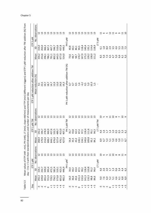
Chapter 5
82
Table 5.3
Mean values of ETP (nM * min), PH (nM), LT (m
in), slope nM/m
in) and TTP
(min) (different triggers) and ETP
1 pM red
uction after TM addition (%) from
day – 3 to + 5 with their standard deviations and number of measuremen
ts per day
Day
ETP 1 pM
ETP 1 pM TM
ETP 1 pM red
uction after addition TM
ETP 5 pM
Mean
SD
No. observations
Mean
SD
No. observations
Mean red
uction (%)
Mean
SD
No. observations
‐3
292,3
154,1
12
268,9
142,2
12
9,1
334,2
154,0
12
‐2
229,8
232,7
13
179,9
194,7
13
23,5
253,6
263,0
13
‐1
265,4
339,0
13
214,0
278,9
13
19,7
296,1
366,3
13
0
721,9
393,4
13
608,2
360,9
13
18,7
786,1
367,1
13
+ 1
729,5
472,5
13
644,1
437,6
13
14,7
791,1
467,7
13
+ 2
756,5
582,4
12
676,0
532,5
12
13,9
802,2
596,7
12
+ 3
573,8
519,1
12
527,4
489,7
12
13,0
643,5
510,6
12
+ 4
601,6
423,3
13
476,1
413,0
13
24,0
677,0
437,8
13
+ 5
482,3
446,9
13
427,0
407,1
13
14,3
514,0
449,5
13
PH 1 pM
* PH 1 pM TM
PH 1 pM red
uction after addition TM (%)
PH 5 pM
‐3
54,0
34,4
12
54,4
35,7
12
0,7
73,6
40,8
12
‐2
36,6
44,2
13
35,5
43,8
13
4,7
48,7
60,1
13
‐1
43,6
63,4
13
43,2
62,3
13
0,5
58,7
86,1
13
0
131,3
87,5
13
130,1
87,4
13
1,4
170,7
99,5
13
+ 1
132,6
99,2
13
134,1
97,5
13
‐4,2
167,9
118,4
13
+ 2
141,3
118,4
12
140,6
118,4
12
0,5
175,3
140,4
12
+ 3
110,3
110,8
12
109,8
111,6
12
5,0
139,2
135,1
12
+ 4
98,8
91,2
13
95,1
95,1
13
10,3
138,0
114,6
13
+ 5
84,6
94,4
13
84,1
94,3
13
1,5
111,2
118,9
13
LT 1 pM
LT 1 pM TM
LT 5 pM
‐3
9,0
4,6
10
8,9
4,6
10
8,5
6,1
11
‐2
7,6
2,6
6
6,7
1,6
5
5,6
1,8
6
‐1
6,0
2,0
5
5,3
1,7
4
7,3
8,0
5
0
5,2
1,7
11
5,1
1,4
11
4,5
2,9
12
+ 1
5,1
1,3
10
5,0
1,0
10
4,7
3,5
11
+ 2
4,6
0,6
8
4,6
0,5
8
5,0
4,6
9
+ 3
6,1
3,4
8
5,0
0,5
7
6,6
5,5
9
+ 4
8,8
4,4
11
7,7
3,3
10
6,1
2,6
11
+ 5
8,9
6,5
9
8,7
6,1
9
‐
9,3
7,5
10
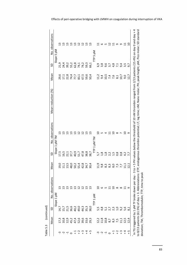
Effects of peri‐operative bridging with LMWH on coagulation during interruption of VKA
83
Table 5.3
(continued
)
Mean
SD
No. observations
Mean
SD
No. observations
Mean red
uction (%)
Mean
SD
No. observations
Slope 1 pM
Slope 1 pM TM
Slope 5 pM
‐3
17,3
14,7
13
19,0
17,5
13
29,6
21,9
13
‐2
10,8
15,7
13
11,1
16,5
13
17,1
24,4
13
‐1
12,9
21,5
13
13,5
22,1
13
22,8
39,2
13
0
45,2
36,6
13
48,0
37,7
13
74,4
55,2
13
+ 1
47,4
40,2
13
50,2
40,9
13
77,7
62,8
13
+ 2
52,6
49,0
12
55,4
51,7
12
83,1
74,1
12
+ 3
39,6
44,3
12
42,8
47,8
12
67,5
74,2
12
+ 4
33,6
35,3
13
34,7
38,8
13
59,6
59,3
13
+ 5
29,3
38,0
13
30,4
38,7
13
50,4
66,1
13
TTP 1 pM
TTP 1 pM TM
TTP 5 pM
‐3
12,2
5,5
10
11,9
5,7
10
11,2
6,9
11
‐2
11,8
4,8
6
9,8
1,8
5
9,4
3,8
6
‐1
10,8
5,1
5
8,3
1,8
4
10,5
9,6
5
0
8,7
2,7
11
8,3
2,2
11
7,4
3,7
12
+ 1
8,3
2,3
10
8,0
1,5
10
7,6
4,8
11
+ 2
7,5
1,0
8
7,3
0,8
8
8,1
7,1
9
+ 3
11,1
9,2
8
7,7
0,8
7
10,7
9,4
9
+ 4
12,5
5,6
11
11,1
4,2
10
9,0
3,5
11
+ 5
12,4
8,1
9
12,1
7,7
9
12,7
9,7
10
* In TG triggered
by 1 pM TF broke down per day ‐ 3 to + 5 PH values below the threshold of 10 nM thrombin ranged from 2/13 patients (15.4%) on day 0 and day + 4
to 8/13 patients (61.5%) of day + 3. Abbreviations: ETP, endogenous thrombin potential; LT, lag tim
e; nM, N
ano m
olar; PH, p
eak height; pM, P
ico m
olar; SD standard
deviation; TM, Thrombomodulin; TTP, tim
e to peak
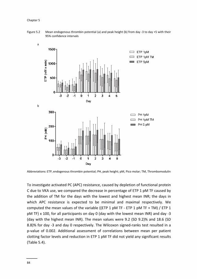
Chapter 5
84
Figure 5.2 Mean endogenous thrombin potential (a) and peak height (b) from day ‐3 to day +5 with their
95% confidence intervals
a
b
Abbreviations: ETP, endogenous thrombin potential; PH, peak height; pM, Pico molar; TM, Thrombomodulin
To investigate activated PC (APC) resistance, caused by depletion of functional protein
C due to VKA use, we compared the decrease in percentage of ETP 1 pM TF caused by
the addition of TM for the days with the lowest and highest mean INR; the days in
which APC resistance is expected to be minimal and maximal respectively. We
computed the mean values of the variable ((ETP 1 pM TF ‐ ETP 1 pM TF + TM) / ETP 1
pM TF) x 100, for all participants on day 0 (day with the lowest mean INR) and day ‐3
(day with the highest mean INR). The mean values were 9.2 (SD 9.2)% and 18.6 (SD
8.8)% for day ‐3 and day 0 respectively. The Wilcoxon signed‐ranks test resulted in a
p‐value of 0.002. Additional assessment of correlations between mean per patient
clotting factor levels and reduction in ETP 1 pM TF did not yield any significant results
(Table 5.4).
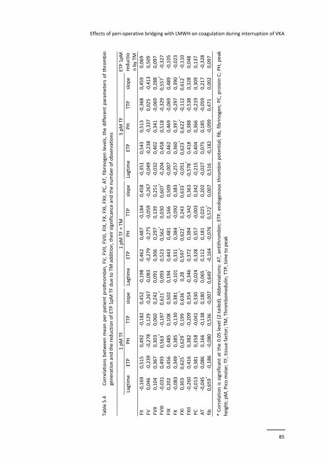
Effects of peri‐operative bridging with LMWH on coagulation during interruption of VKA
85
Table 5.4
Correlations between
mean
per patient prothrombin, FV
, FV
II, FV
III, FIX, FX, FXI, FXII, PC, AT, fibrinogen levels, the different param
eters of thrombin
generation and the red
uction of ETP 1pM TF due to TM addition, their significance and the number of observations
1 pM TF
1 pM TF + TM
5 pM TF
Lagtim
e
ETP
PH
TTP
slope
Lagtim
eETP
PH
TTP
slope
Lagtim
e ETP
PH
TTP
slope
ETP 1pM
reductio
n by TM
FII
‐0,169
0,515
0,492
‐0,182
0,452
‐0,198
0,462
0,487
‐0,184
0,458
‐0,351
0,543
0,513
‐0,368
0,459
0,069
FV
0,046
‐0,239
‐0,278
0,129
‐0,267
‐0,083
‐0,279
‐0,275
‐0,059
‐0,267
‐0,049
‐0,238
‐0,337
0,025
‐0,413
0,509
FVII
0,104
0,367
0,303
0,060
0,242
0,091
0,306
0,297
0,139
0,251
‐0,032
0,402
0,341
‐0,069
0,288
0,097
FVIII
‐0,031
0,493
0,563*
‐0,197
0,621*
0,093
0,523
0,562*
0,030
0,607*
‐0,204
0,458
0,518
‐0,329
0,557*
‐0,327
FIX
0,202
0,456
0,485
0,108
0,502
0,194
0,443
0,481
0,166
0,509
‐0,007
0,462
0,469
‐0,069
0,489
‐0,105
FX
‐0,083
0,349
0,385
‐0,130
0,381
‐0,101
0,331
0,384
‐0,092
0,383
‐0,257
0,360
0,397
‐0,297
0,390
‐0,023
FXI
0,303
0,625*
0,629*
0,199
0,636*
0,28
0,597*
0,622*
0,243
0,633*
‐0,051
0,623*
0,622*
‐0,112
0,612*
‐0,110
FXII
‐0,260
0,416
0,382
‐0,209
0,354
‐0,346
0,372
0,384
‐0,342
0,363
‐0,578*
0,418
0,388
‐0,538
0,328
0,048
PC
‐0,013
0,381
0,358
‐0,041
0,330
‐0,024
0,328
0,357
‐0,003
0,342
‐0,215
0,404
0,366
‐0,219
0,309
0,137
AT
‐0,045
0,086
0,166
‐0,138
0,180
0,065
0,112
0,181
0,025
0,202
‐0,027
0,075
0,185
‐0,059
0,217
‐0,328
fib
0,659*
‐0,186
‐0,080
0,536
‐0,007
0,649*
‐0,164
‐0,076
0,572*
0,007
0,516
‐0,182
‐0,099
0,471
0,002
0,097
* Correlation is significant at the 0.05 level (2‐tailed). Abbreviations: AT, antithrombin; ETP, en
dogenous thrombin potential; fib, fibrinogen; PC, protein C; PH, peak
height; pM, Pico m
olar; TF, tissue factor; TM, Thrombomodulin;TTP, tim
e to peak

Chapter 5
86
Correlations between INR, anti‐Xa, and parameters of thrombin generation
Correlations between mean INR per patient and mean per patient plasma levels of FII,
FVII, FIX, FX and protein C were negative, strong, and significant for FII, FVII and
protein C. Bonferroni corrected significance of 0.01 was reached for all factors, except
the correlations INR‐FIX (r=‐0.36, p=0.22) and INR–FX (r=‐0.65, p=0.02) (Table 5.5).
Correlations between INR and different parameters of thrombin generation triggered
by 1 pM TF with and without the addition of TM, and triggered by 5 pM were
moderate and significant (p<0.05) for LT (all triggers), and for TTP 1 pM TF+TM and
TTP 5 pM TF; weak, inverse correlations were observed between INR and slope, PH
and ETP. For comparison we also calculated the correlations between INR and TG on
days no LMWH was administered; overall we found similar correlations, p‐values
<0.05 were reached for LT (all triggers) (Table 5.5).
Table 5.5 Correlations between mean per patient INR, anti‐Xa and mean per patient factor levels, different parameters of thrombin generation, their significance and the number of
observations
INR INR
(after at least 1 day
no LMWH)
Anti‐Xa Anti‐Xa
(after at least 3 days
no VKA)
FII ‐0,777‡
FVII ‐0,816‡ FIX ‐0,363
FX ‐0,650*
PC ‐0,781‡
‐ ‐
AT ‐
‐
‐0,018 0,009
Lagtime 1pM 0,591* 0,597
* ‐0,039 0,491
ETP 1pM ‐0,424 ‐0,393 ‐0,697† ‐0,793‡ PH 1pM ‐0,350 ‐0,369 ‐0,644
* ‐0,715†
TTP 1pM 0,517 0,423 0.036 0,585*
Slope 1pM ‐0,284 ‐0,370 ‐0,608* ‐0,646
*
Lagtime 1pM+TM 0,601* 0,655
* 0.023 0,425
ETP 1pM+TM ‐0,369 ‐0,374 ‐0,674* ‐0,756‡
PH 1pM+TM ‐0,349 ‐0,372 ‐0,640* ‐0,711†
TTP 1pM+TM 0,559* 0,506 0,076 0,481
Slope 1pM+TM ‐0,289 ‐0,386 ‐0,602* ‐0,642
*
Lagtime 5pM 0,673* 0,662
* 0,394 0,697
*
ETP 5pM ‐0,447 ‐0,433 ‐0,722† ‐0,817‡
PH 5pM ‐0,372 ‐0,337 ‐0,667* ‐0,742†
TTP 5pM 0,646* 0,534 0,386 0,708†
Slope 5pM ‐0,278 ‐0,351 ‐0,630* ‐0,640
*
* Correlation is significant at the 0.05 level (2‐tailed); † Correla on is significant at the 0.01 level (2‐tailed);
‡ Correlation is significant at the 0.003 level (2‐tailed). Abbreviations: ETP, endogenous thrombin potential; INR, International Normalized Ratio; PH, peak height; pM, Pico molar; TM, Thrombomodulin; TTP, time to
peak
Effects of peri‐operative bridging with LMWH on coagulation during interruption of VKA
87
Correlations between the mean anti‐Xa per patient and different parameters of
thrombin generation triggered by all 3 reagents were calculated. A strong correlation
between ETP (all triggers) was found, ETP 1 pM TF and ETP 5 pM TF reached a p‐value
of <0.01. Other TG parameters showed moderate correlations, a significance level of
<0.05 was reached for 9/15 TG parameters. No measurement reached the Bonferroni
corrected p‐value <0.003. Overall stronger correlations were observed between the
mean per patient anti‐Xa and the parameters of TG when only the days that no
acenocoumarol was administered for at least 3 days were included in the analysis. The
observed correlations for ETP (all triggers) were stronger and Bonferroni corrected p‐
values <0.003 were recorded for all three ETP triggers. Compared to the correlations
that ignored VKA activity higher levels of correlation were also found for LT and PH,
slope and ETP (all triggers). A significance level of <0.05 was reached for 12/15 TG
parameters (Table 5.5).
Post procedural bleeding and thromboembolism
Our study was explorative in character and not powered to associate outcomes to
levels of individual coagulation factors or levels of anticoagulants. No TE (stroke, TIA,
systemic embolism) was reported in any patient. However, we observed 4 major
bleeding events (30.8%) and 3 minor bleeding events (23.1%) during the 30‐day post
procedural follow‐up; all patients recovered. During the time of the bleeding event all
patients had INRs close to 1 (1.0‐1.1), but 3 of the patients had residual anti Xa levels
of >0.10 IU/ml and 3 patients had impaired kidney function (MDRD 34 ml/min, 39
ml/min, and 45 ml/min respectively). ETP at 1 pM TF 371,4 nM*min (SD 196.7) and
PH at 1 pM TF 65,8 nM (SD 41,7) were lower than those of patients without major
bleeding; ETP at 1 pM TF 588,5 nM*min (SD 215,0) and PH at 1 pM TF 105,9 nM (SD
50,9), but these differences were not statistically significant (p=0,11 and 0,20
respectively). Similar results were obtained with 5 pM TF activation.
Discussion
Surprisingly, the recently published BRIDGE trial suggests that peri‐operative bridging
with LMWH would no longer be needed, at least in patients undergoing minor surgical
interventions, since the risk of thromboembolism was comparable to the rate
observed in non‐bridged control patients.5 This raises the question whether brief
cessation of VKA is associated with any rebound hypercoagulability due to surgery. In
order to assess peri‐operative coagulation activity in patients undergoing surgical
Chapter 5
88
interventions, we determined the overall impact of cessation of VKA, peri‐procedural
instillation of LMWH and surgery on net coagulation activity, measured by TG. To
understand the nature of any changes in TG, we measured all relevant coagulation
proteins that may determine the TG profile.
The results of our exploratory study shows that surgical intervention quickly induced a
marked increment in TG peak and ETP values, confirming a “rebound” prothrombotic
state. In a further analysis of coagulation proteins we observed that FVIII and FX, but
in particular FXI had significant effects on the rate and height of TG. The effect of FXI
may support the apparent importance of this protein in driving the risk of
postoperative thromboembolism, as illustrated by a recent trial showing better
efficacy of FXI inhibition as compared to LMWH.11 Release of tissue factor may engage
increased thrombin generation, which through a feedback loop amplifies FXI
dependent TG. In this process the contributions of FVIII, FIX and prothrombin are also
evident; FVIII was in this setting observed to be a determinant for thrombin
generation. Interestingly, the rise in FVIII and fibrinogen occurs gradually, due to the
well‐known cytokine dependent, acute phase synthesis and the contribution of FVIII
to TG may therefore only become evident days after the intervention. Several studies
report increased levels of F VIII and fibrinogen until 2 days to 72 hours
postoperatively.12,13 This may in part explain the protracted risk of thrombosis
following surgery. The residual anticoagulant activity may in part be neutralized by the
acute phase thrombogenic response following the intervention. Another contributor
to the thrombosis risk may be the reduced sensitivity to activated protein C (APC
resistance) mimicked in our experiments by adding TM to plasma. While
paradoxically, during VKA treatment due to the reduced level of PC, this
prothrombotic APC resistance effect is already evident, it does not fully correct when
the VKA effect is no longer present as indicated by INR of 1.0 at day 0. The TM
reduction is optimized to give a 50% reduction in PH of a normal plasma, but the
maximum reduction observed in these patients was around 20%; since PC had at that
stage reached normal values, this suggests that other factors contribute to the
apparent TM related APC resistance, adding to the risk of thrombosis. Theoretically,
the unexplained but steady increase in FV levels may have attributed to this APC
resistance effect.
Cessation of VKA and start of LMWH induces marked changes in the coagulation
proteome. Strong, inverse correlations between INR and vitamin K dependent pro and
anticoagulants were observed. Weak to moderate correlations between INR and TG
parameters were recorded; on the other hand, correlations between anti‐Xa levels
and TG were moderate to strong and significant. The correlations between anti‐Xa
and TG were stronger after correction for the effect of VKA. In a study in which the
Effects of peri‐operative bridging with LMWH on coagulation during interruption of VKA
89
concomitant effect of VKA (in vivo administration) and LMWH (added in vitro) on
different parameters of TG was investigated, similar findings were reported.14 It is
possible that the effect on TG of LMWH in combination with AT, which leads to
inactivation of FXa and thrombin and subsequent release of TFPI from the
endothelium, forming a complex with FXa, TF and FVIIa, is stronger than the effect of
the inhibition of prothrombin, FVII, FIX and FX by VKA.
The current study was set up as a mechanistic study, this could be considered as a
limitation as it restricts the potential for the assessment of significant risk
associations. We did however even within this limited sample observe 4 cases of
major bleeding. Three out of 4 had reduced renal function and anti Xa levels
>0.1 IU/ml suggesting that bridging with LMWH, especially in patients with renal
impairment should be monitored closely. TG tended to be lower in patients who
experienced bleeding; no statistically significant difference in levels was observed,
most likely due to the limited sample size.
Our study has several strengths. In the first place, as far as we know this is the first
study that evaluates bridging therapy by means of a wide range of blood coagulation
assays. Results of this exploratory study might offer insight in the so far poorly
understood effects of the complex in vivo interaction between VKA and LMWH
administration, and the effects of surgery on hemostasis. Secondly, we were able to
follow the patients for a 9‐day period, so that the whole perioperative bridging period
could be evaluated.
We conclude that VKA (acenocoumarol) arrest in bridged patients starting 3 days
before surgery gives an effective reduction of INR, but that LMWH may need to be
individually tailored. We found that for several patients the ACCP advised timeframe
of 24 hours between the last preoperative therapeutic dose administration of LMWH
and planned surgery is not long enough to warrant sufficient decline in anti‐Xa levels.
In spite of residual anti‐Xa activity, there was a marked increase in TG related to
surgery. Three prothrombotic mechanisms are exposed: FXI dependent TG, reduced
activity of the APC pathway and postoperative rise in FVIII and fibrinogen. For the
complex perioperative hemostasis management the value of TG or other global
assays, to monitor the hemostatic balance, therefore merits further study.
Chapter 5
90
References
1. Douketis JD, Spyropoulos AC, Spencer FA, et al. Perioperative management of antithrombotic therapy: Antithrombotic Therapy and Prevention of Thrombosis, 9th ed: American College of Chest
Physicians Evidence‐Based Clinical Practice Guidelines. Chest 2012;141:326‐50.
2. Keeling D, Baglin T, Tait C, et al. British Committee for Standards in H. Guidelines on oral anticoagulation with warfarin ‐ fourth edition. Br J Haematol 2011;154:311‐24.
3. Eijgenraam P, Ten Cate H, Ten Cate‐Hoek AJ. Safety and Efficacy of Bridging with low molecular
weight heparins: a systematic review and partial meta‐analysis. Curr Pharm Des 2012;19:4014‐23. 4. Siegal D, Yudin J, Kaatz S, Douketis JD, et al. Periprocedural heparin bridging in patients receiving
vitamin K antagonists: systematic review and meta‐analysis of bleeding and thromboembolic rates.
Circulation 2012;126:1630‐9. 5. Douketis JD, Spyropoulos AC, Kaatz S, et al. Perioperative bridging anticoagulation in patients with
atrial fibrillation. N Engl J Med. 2015;373:823‐33.
6. Sweetland S, Green J, Liu B, et al. Duration and magnitude of the postoperative risk of venous thromboembolism in middle aged women: prospective cohort study. BMJ 2009;339:4575‐83.
7. Dielis AW, Castoldi E, Spronk HM, et al. Coagulation factors and the protein C system as determinants
of thrombin generation in a normal population. J Thromb Haemost 2008;6:125‐31. 8. Garcia DA, Baglin TP, Weitz JI, et al. Parenteral anticoagulants: antithrombotic therapy and prevention
of thrombosis, 9th ed: American College of Chest Physicians Evidence‐Based Clinical Practice
Guidelines. Chest 2012;141:24‐43. 9. Spyropoulos AC, Douketis JD, Gerotziafas G, et al. Periprocedural antithrombotic and bridging
therapy: recommendations for standardized reporting in patients with arterial indications for chronic
oral anticoagulant therapy. J Thromb Haemost 2012;10:692‐4. 10. Harder S, Klinkhardt U, Alvarez JM. Avoidance of bleeding during surgery in patients receiving
anticoagulant and/or antiplatelet therapy: pharmacokinetic and pharmacodynamic considerations.
Clin Pharmacokinet 2004;43:963‐81. 11. Buller HR, Bethune C, Bhanot S, et al. Factor XI antisense oligonucleotide for prevention of venous
thrombosis. N Engl J Med 2015;372:232‐40.
12. Oberweis BS, Cuff G, Rosenberg A, et al. Platelet aggregation and coagulation factors in orthopedic surgery. J Thromb Thrombolysis 2014;38:430‐8.
13. Nygaard OP, Unneberg K, Reikeras O, et al. Thromboplastin activity of blood monocytes after total hip
replacement. Scand J Clin Lab Invest 1990;50:183‐6. 14. Gerotziafas GT, Dupont C, Spyropoulos AC, et al. Differential inhibition of thrombin generation by
vitamin K antagonists alone and associated with low‐molecular‐weight heparin. Thromb Haemost
2009;102:42‐8.
91
Chapter 6 Venous stenting after deep venous thrombosis and
antithrombotic therapy: a systematic review
Pieter Eijgenraam,Hugo ten Cate, Arina J ten Cate‐Hoek
Reviews in Vacular Medicine. 2014;2:88‐97
Chapter 6
92
Abstract
Introduction
Over the last years venous stent placement after deep venous thrombosis (DVT) in the
iliofemoral veins has gained more attention. The majority of studies evaluating the safety and
efficacy of this intervention are of poor methodological quality and the association with
antithrombotic therapy has not been studied explicitly. We performed a systematic review to
summarize the available literature on antithrombotic management in relation to the safety and
efficacy of venous stenting.
Methods
We performed a Medline search to identify studies that addressed anticoagulation and/or
antiplatelet treatment options after venous stenting in patients with a prior DVT in the
iliofemoral area. We identified 192 articles and finally selected 14 articles for use in this review.
Results
In 86% (12/14) of the included studies anticoagulation was administered to all patients who
underwent iliac venous stenting. In 33% of the studies patients received antiplatelet therapy
consisting of aspirin and/or clopidogrel (4/12). The duration of antithrombotic treatment was
not guided by the stenting procedure in 93% (13/14) of studies. The incidence of re‐thrombosis
in (sub) groups of only stented patients, ranged from 5% to 25%. Primary, assisted primary, and
secondary patency rates 12 months after stent placement ranged from 54%, 72%, 83%
respectively to 78%, 83%, 95% in (sub)groups of only stented patients. Rates of major bleedings
during long term follow‐up ranged from 0% to11%.
Conclusion
Antithrombotic therapy does not seem to influence any of the outcomes in patients with
venous stenting after DVT: recurrent DVT, patency, post‐thrombotic syndrome or restenosis
and bleeding.
Venous stenting after deep venous thrombosis and antithrombotic therapy
93
Introduction
For a substantial proportion of patients with deep venous thrombosis (DVT), current
treatment strategies are suboptimal. Especially for the group of patients who are at
the highest risk for post‐thrombotic syndrome (PTS), new treatment modalities are
being investigated. Over the last years the use of stenting of the iliofemoral veins has
gained more attention. Nowadays, stenting is predominantly used in patients with
venous outflow obstructions of the iliac and femoral veins after thrombus removal
with catheter directed thrombolysis (CDT) combined with percutaneous mechanical
thrombectomy (PMT).1
Arterial interventions are much better studied than venous interventions and the
mechanisms of arterial stent failure are therefore better understood. Iliofemoral
venous re‐stenosis after stenting probably results from intraluminal fibrosis in post‐
thrombotic lesions or compression by primary obstructions, while arterial lesions arise
from smooth muscle cell proliferation driven by cytokines and growth factors derived
from platelets and macrophages.2 Platelet adhesion and aggregation likely plays a
more important role in high‐flow, high shear conditions in the arterial system,
coagulation seems more important in the low‐flow, low‐shear conditions of the
venous system.2,3 Finally, both in venous injury and atherosclerotic plaque rupture,
the exposure of procoagulant tissue factor and other proteins to blood, triggers blood
coagulation.3 Therefore the selection of appropriate antithrombotic management is
important.
The efficacy and safety profile of venous stenting is evaluated in several clinical
studies, but not much attention is given to the optimal duration for the post
intervention antithrombotic management.4‐20 In patients receiving venous stents after
a DVT, anticoagulation and possibly antiplatelet therapy may play an important role in
preventing recurrent thrombosis and subsequent embolization into the vasculature of
the lungs. So far, the choice of agent, dosing and duration of anticoagulant and
possibly antiplatelet therapy is unclear for patients with venous stents and prior DVT.
The lack of methodologically well‐designed studies to address this matter is eminent.
While conventional anticoagulants such as vitamin K antagonists and low molecular
weight heparins have proven to reduce the risk of recurrent DVT dramatically, their
role in preventing DVT in patients with venous stents remains unclear. Antiplatelet
therapy has proven to be safe and efficacious in patients with arterial stents, but the
effect in patients with venous stents, with or without prior DVT also remains unclear.
The most recently issued ACCP guidelines (2012) do not advise on extended
anticoagulation/antiplatelet therapy after venous stenting; the possibility of stenting
after thrombolysis in patients presenting with DVT is not even discussed.21 Likewise,
Chapter 6
94
the most recent NICE and ESC guidelines do not discuss venous stenting as a
treatment option for patients after a DVT.
Initially we intended to compare the effect of different antithrombotic agents and
treatment durations in patients undergoing venous stenting in the iliofemoral area
after a first or recurrent DVT, in terms of recurrent DVT, pulmonary embolism (PE),
PTS, patency rates and bleeding risk. Since, after our literature search no comparison
data were available, we decided to describe and summarize the available literature on
different antithrombotic options in relation to these main outcome measures.
Methods
Study selection
Research questions and inclusion criteria of the studies were specified in advance and
documented in a protocol. The population of this review consists of patients who
underwent venous stenting after venous thrombosis, often in combination with
balloon angioplasty preceded by CDT and/or PMT. We only included studies wherein
the use of post‐interventional antithrombotic therapy was addressed. Antithrombotic
therapy consist of: 1) The administration of anticoagulants (vitamin K antagonists,
unfractionated heparin (UFH), fondaparinux, low molecular weight heparin (LMWH),
direct factor Xa inhibitors, direct thrombin (factor IIa) inhibitors, 2) the administration
of antiplatelet drugs: cyclooxygenase (COX) inhibitors: aspirin, ADP‐receptor
antagonists such as clopidogrel, plasugrel and ticagrelor. We decided to also include
studies in which not all participants were stented; a part of the population underwent
CDT and/or PMT without stenting. Most studies assessed, with the use of duplex,
long‐term primary patency rates (i.e. patent stent without re‐intervention), assisted
primary patency rates (i.e. patent stent after re‐intervention without any occlusions)
and secondary patency rates (i.e. patent stent after occlusion, with patency ending at
the moment the occlusion was present). Other reported outcomes were PTS,
recurrent DVT, PE and major bleeding complications. Assessment of PTS was
performed with different methods, including the Villalta scale and Venous Clinical
Severity scores. No restrictions regarding follow‐up period were imposed. In this
systematic review case reports, cohort studies, case‐control studies, cross‐sectional
studies and randomized controlled trials were eligible.

Venous stenting after deep venous thrombosis and antithrombotic therapy
95
Data sources and searches
We searched MEDLINE database up to week 45 of 2013. The search strategy can be
found in appendix 6.1. We used MeSH headings for keyword and text word searching
and free text for word searching only. No publication date restrictions were imposed;
we restricted our search to articles in English. Further selection was made on title and
abstract. The final selection of articles was made after full reading.
Data extraction and quality assessment
As far as we know no quality assessment tools designed for specific use in non‐
comparator, observational studies are available.22 For the quality assessment of
individual studies we made use of the Newcastle‐Ottawa Scale (NOS); this tool is
designed for cohort studies.23 We selected 5 relevant items and prioritized them in
our quality assessment (see appendix 6.2). Each item can be awarded with a star,
resulting in a maximum of 5 stars. The following items were assessed: represen‐
tativeness of the cohort, ascertainment of the exposure, assessment of the outcome,
length and adequacy of the follow‐up. Data collected from individual studies were
reported on case report forms consisting of 3 sections: study eligibility (i.e. type of
intervention and outcome), checklist of items for data collection (i.e. characteristics of
the participants and study design), and quality assessment using the adapted NOS
scale for cohort studies.23 Data extraction was performed by one reviewer (PE),
supervised by the second reviewer (AtC).
Data analysis
Patency rates in most of the included studies were estimated using the method as
proposed by Kaplan‐Meier. Differences between groups were assessed using the
Student’s t‐test; other outcomes were presented as means with their standard
deviation or as percentages. Due to the lack of comparator studies in our review no
meta‐analysis could be performed.
Results
Inclusion and quality assessment
Our MEDLINE database search resulted in a total of 192 studies. For a summary of the
search see Figure 6.1. A first selection on title and abstract yielded a total of
27 articles, further restriction after full reading resulted in a set of 14 studies. We
identified 12 cohort studies (6 prospective,4‐7,9,10 4 retrospective studies,8,11,14,17
Chapter 6
96
2 studies with an unclear character),13,18 1 trial15 and 1 case report.12 In only 1 cohort
study a comparison with another treatment modality was made; stenting was
compared to anticoagulation alone.4 We included 3 studies in which the full
population was stented;8,13,17 one additional study allowed full subgroup analysis of
the patients that underwent stenting.11 In 10 studies between 15%15 and 89%11 of the
population was stented. In all included studies the patients underwent CDT and/or
PMT and/or angioplasty and stenting and were prescribed to anticoagulation and/or
antiplatelet agents and/or graded compression stockings. For a summary of the
included studies see Table 6.1.
Figure 6.1 Summary of evidence search and selection
Abbreviations: DVT, deep venous thrombosis; PE, pulmonary embolism; PTS, post thrombotic syndrome
Articles included in the title and abstract review:
192
Articlesexcluded:
165
Reasons for exclusion:Participants did notundergo venousstentingStent was not placed in the iliofemoral areaStenting without prior DVTNo postoperativeantithrombotic optionsmentionedOutcomes major bleeding, DVT, PE orPTS not assessed
Articles reviewed: 27 Articlesexcluded: 13
Reasons for exclusion:Participants did notundergo venousstentingStent was not placed in the iliofemoral areaStenting without prior DVTNo postoperativeantithrombotic optionsmentionedOutcomes major bleeding, DVT, PE orPTS not assessed
Articles included: 14
Articles retrieved throughDatabase:
MEDLINE: 192
Articles retrieved by hand‐searching: 0
Venous stenting after deep venous thrombosis and antithrombotic therapy
97
Table 6.1
Summary of the included studies
Study
Design
Inclusion criteria
Sample size
Intervention
Outcome
1.AbuRahma
2001
Prospective
cohort study
Iliofemoral D
VT
51 patients; (group 1: 33
anticoagulation alone, group
2: 18 endovascular interven‐
tions (10 patients stented))
Anticoagulation versus anticoagulation+
CDT; balloon venoplasty+stenting
Recurren
t DVT; PE; paten
cy rates;
survival; bleed
ing, CEA
P, m
ortality
2.Dayal 2005
Prospective
cohort study
Acute/chronic critical thrombotic
occlusions at different sites
25 patients; 15 patients
sten
ted
CDT; PMT; balloon venoplasty+stenting;
antithrombotic therapy
Recurren
t DVT; paten
cy rates; survival;
bleeding
3a.Sillesen
2005
Retrospective
cohort study
Iliofemoral D
VT; onset <2
weeks;
open
popliteal veins
45 patients; 30 patients
sten
ted
CDT; balloon venoplasty+ stenting;
antithrombotic therapy
Recurren
t DVT; reflux; patency rates
3b.Bækgaard
2009
Prospective
cohort study
First acute IFVT; <14 days since
diagnosis; age<60; open
popliteal
vein
101 patients(103 limbs); 57
limbs sten
ted
CDT; balloon venoplasty+stenting;
antithrombotic therapy
Complications (including PTS); venous
reflux; patency rates
4.Husm
ann
2007
Retrospective
cohort study
First DVT; sym
ptoms <1
week
11 patients; all stented
Thrombolysis; thrombectomy; stenting;
antithrombotic therapy
Complications; patency rates; valve
function
5.Kölbel
2007
Prospective
cohort study
Acute extensive iliocaval D
VT;
symptoms<3 weeks
37 patients; 36 limbs sten
ted
CDT; balloon venoplasty+stenting;
antithrombotic therapy
Paten
cy rates; clinical outcomes
6.Raju 2009
Cohort study
Confirm
ed DVT
159 patients; all sten
ted
Percutaneo
us recanalization; balloon
venoplasty+stenting; antithrombotic therapy Paten
cy rates; CEA
P; Q
OL; VAS;
swelling; recurrent DVT; PE; m
ortality
7.Grommes
2011
Prospective
cohort study
Confirm
ed DVT; life expectancy
>6 m
onths
12 patients; 3
patients stentedCDT; balloon venoplasty+stenting;
antithrombotic therapy
Recurren
t DVT; PE; paten
cy rates;
bleeding
8.Oguzkurt
2010
Case series
Confirm
ed DVT; Phlegm
ea cerulea
dolens
7 patients; 3
patients stented Manual aspiration thrombectomy; CDT;
balloon venoplasty+stenting; antithrombotic
therapy
Recurren
t DVT; sym
ptoms (for all
participants)
9.Titus 2010
Retrospective
cohort study
Confirm
ed DVT
38 patients; 40 limbs sten
ted
Angioplasty+ stenting; antithrombotic
therapy
Recurren
t DVT; bleeding; PE; paten
cy
rates; CEA
P
10.W
ahlgren
2010
Cohort study
Chronic PTS
50 patients; 20 patients
endovascular surgery
Guide wire recanalization; stenting;
antithrombotic therapy
Paten
cy rates; venous clinical severity
score; recurrent DVT; PE; m
ortality
11.M
anninen
2011
Prospective
cohort study
Acute DVT; sym
ptoms <2
weeks
56 patients; 9
patients stentedCDT; balloon venoplasty+stenting;
antithrombotic therapy
Patency: iliofemoral and crural veins;
PTS; recurrent DVT; PE; m
ortality
12.Sharifi 2012
Trial (RCT)
Confirm
ed DVT; Severe
symptoms; femoral popliteal
veins or more proximal veins
affected
183 patients; 2
groups (92
anticoagulation alone, 91
endovascular interventions
(27 patients stented))
Anticoagulation versus Percutaneo
us
thrombectomy; local low dose thrombolytic
therapy; balloon ven
oplasty+sten
ting;
anticoagulation
Recurren
t DVT; PE; PTS; bleeding;
duration hospitalization; leg edem
a;
skin induration; subjective change;
mortality
13.Nayak 2012
Retrospective
cohort study
PTS; age>18 years; available
follow‐up records
44 patients; 39patients, (45
limbs sten
ted )
Endovenous laser ablation; balloon
venoplasty+stenting; antithrombotic therapy
CEA
P (pre‐post); overall im
provement;
symptoms: pain, swelling, ulcer
Abbreviations: CDT, catheter‐directed thrombolysis; CEA
P, clinical, etiology, anatomy, pathophysiology; DVT, deep ven
ous thrombosis; IFV
T, iliofemoral venous thrombosis; PE, pulm
onary
embolism; PMT, percutaneo
us mechanical thrombectomy; PTS, post‐thrombotic syndrome; Q
OL, quality of life; VAS, visual analogue scale.
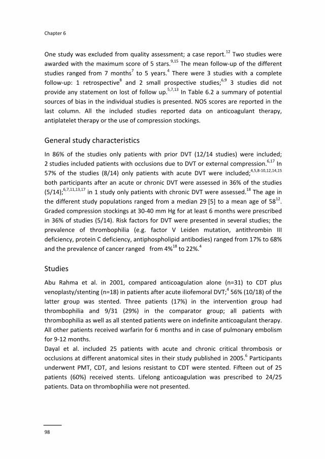
Chapter 6
98
One study was excluded from quality assessment; a case report.12 Two studies were
awarded with the maximum score of 5 stars.9,15 The mean follow‐up of the different
studies ranged from 7 months7 to 5 years.4 There were 3 studies with a complete
follow‐up: 1 retrospective8 and 2 small prospective studies;6,9 3 studies did not
provide any statement on lost of follow up.5,7,13 In Table 6.2 a summary of potential
sources of bias in the individual studies is presented. NOS scores are reported in the
last column. All the included studies reported data on anticoagulant therapy,
antiplatelet therapy or the use of compression stockings.
General study characteristics
In 86% of the studies only patients with prior DVT (12/14 studies) were included;
2 studies included patients with occlusions due to DVT or external compression.6,17 In
57% of the studies (8/14) only patients with acute DVT were included;4,5,8‐10,12,14,15
both participants after an acute or chronic DVT were assessed in 36% of the studies
(5/14);6,7,11,13,17 in 1 study only patients with chronic DVT were assessed.18 The age in
the different study populations ranged from a median 29 [5] to a mean age of 5812.
Graded compression stockings at 30‐40 mm Hg for at least 6 months were prescribed
in 36% of studies (5/14). Risk factors for DVT were presented in several studies; the
prevalence of thrombophilia (e.g. factor V Leiden mutation, antithrombin III
deficiency, protein C deficiency, antiphospholipid antibodies) ranged from 17% to 68%
and the prevalence of cancer ranged from 4%18 to 22%.4
Studies
Abu Rahma et al. in 2001, compared anticoagulation alone (n=31) to CDT plus
venoplasty/stenting (n=18) in patients after acute iliofemoral DVT;4 56% (10/18) of the
latter group was stented. Three patients (17%) in the intervention group had
thrombophilia and 9/31 (29%) in the comparator group; all patients with
thrombophilia as well as all stented patients were on indefinite anticoagulant therapy.
All other patients received warfarin for 6 months and in case of pulmonary embolism
for 9‐12 months.
Dayal et al. included 25 patients with acute and chronic critical thrombosis or
occlusions at different anatomical sites in their study published in 2005.6 Participants
underwent PMT, CDT, and lesions resistant to CDT were stented. Fifteen out of 25
patients (60%) received stents. Lifelong anticoagulation was prescribed to 24/25
patients. Data on thrombophilia were not presented.
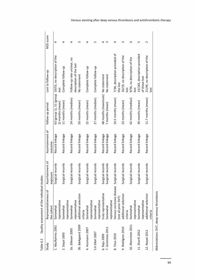
Venous stenting after deep venous thrombosis and antithrombotic therapy
99
Table 6.2
Quality assessment of the individual studies
Study
Representativeness of
the cohort
Ascertainment of
exposure
Ascertainment of
outcome
follow‐up period
Lost to follow‐up
NOS score
1. A
buRahma 2001
Somewhat
representative
Surgical records
Record linkage
63 (group 1), 51 (group
2) months (m
ean
) 19/55, no description of the
lost
4
2. D
ayal 2005
Somewhat
representative
Surgical records
Record linkage
11 m
onths (m
ean)
Complete follow‐up
4
3a. Sillesen
2005
Somewhat
representative
Surgical records
Record linkage
24 m
onths (m
edian)
Follow‐up rate unclear, no
description of the lost
4
3b. B
ækgaard 2009
additional selection
criteria
Surgical records
Record linkage
50 m
onths (m
ean)
No statement
3
4. H
usm
ann 2007
Somewhat
representative
Surgical records
Record linkage
22 m
onths (m
ean)
Complete follow‐up
4
5.K ölbel 2007
Somewhat
representative
Surgical records
Record linkage
27 m
onths (m
edian)
Complete follow‐up
5
6. R
aju 2009
truly rep
resentative
Surgical records
Record linkage
48 m
onths (m
axim
um) No statement
4
7. G
rommes 2011
Somewhat
representative
Surgical records
Record linkage
7 m
onths (m
ean)
No statement
3
8. Titus 2010
Ven
ous occlusive disease
(not all prior DVT)
Surgical records
Record linkage
10.5 m
onths (m
ean
) 7/36, description provided of
the lost
4
9. W
ahlgren 2010
additional selection
criteria
Surgical records
Record linkage
23 m
onths (m
ean)
16/19, no description of the
lost
3
10. M
anninen
2011
Somewhat
representative
Surgical records
Record linkage
42 m
onths (m
edian)
9/56, no description of the
lost
4
11. Sharifi 2012
Somewhat
representative
Surgical records
Record linkage
30 m
onths (m
ean)
14/183, description provided
of those lost
5
12. N
ayak 2012
additional selection
criteria
Surgical records
Record linkage
11.7 m
onths (m
ean
) 21/44, no description of the
lost
2
Abbreviations: DVT, deep
venous thrombosis
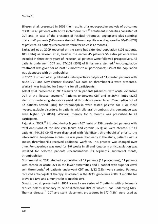
Chapter 6
100
Sillesen et al. presented in 2005 their results of a retrospective analysis of outcomes
of CDT in 45 patients with acute iliofemoral DVT.14 Treatment modalities consisted of
CDT and, in case of the presence of residual thrombus, angioplasty plus stenting;
thirty of 45 patients (67%) were stented. Thrombophilia was diagnosed in 30/45 (67%)
of patients. All patients received warfarin for at least 12 months.
Bækgaard et al. 2009 reported on the same but extended population (101 patients,
103 limbs) as Sillesen et al, besides the earlier 45 patients 56 extra patients were
included in three extra years of inclusion, all patients were followed prospectively. All
patients underwent CDT and 57/103 (55%) of limbs were stented.5 Anticoagulation
treatment was given for at least 12 months to all participants; 54% of the population
was diagnosed with thrombophilia.
In 2007 Husmann et al. published a retrospective analysis of 11 stented patients with
acute DVT and May‐Thurner disease.8 No data on thrombophilia were presented.
Warfarin was installed for 6 months for all participants.
Kölbel et al. presented in 2007 results on 37 patients (44 limbs) with acute, extensive
DVT of the iliocaval segment.9 Patients underwent CDT and in 36/44 limbs (82%)
stents for underlying stenosis or residual thrombosis were placed. Twenty‐five out of
32 patients tested (78%) for thrombophilia were tested positive for 1 or more
hypercoagulable disorders. In patients with bilateral thrombosis this percentage was
even higher 6/7 (86%). Warfarin therapy for 6 months was prescribed to all
participants.
Raju et al, 2009.13 included during 9 years 167 limbs of 159 unselected patients with
total occlusions of the iliac vein (acute and chronic DVT); all were stented. Of all
patients, 44/159 (34%) were diagnosed with ‘significant thrombophilia’ prior to the
intervention. Long‐term aspirin use was prescribed early in the study; patients with a
known thrombophilia received additional warfarin. This practice was changed over
time; Fondaparinux was used for 4‐6 weeks in all and long‐term anticoagulation was
installed for selected patients (recanalizations ≥3 segments, suprarenal stents,
thrombophilia).
Grommes et al, 2011 studied a population of 12 patients (13 procedures), 11 patients
with chronic or acute DVT in the lower extremities and 1 patient with superior caval
vein thrombosis.7 All patients underwent CDT and 3/12 (25%) were stented. Patients
received anticoagulant therapy as advised in the ACCP guidelines 2008: 3 months for
provoked DVT and 6 months for idiopathic DVT.
Oguzkurt et al. presented in 2009 a small case series of 7 patients with phlegmasia
cerulea dolens secondary to acute iliofemoral DVT of which 3 had underlying May‐
Thurner disease.12 CDT and stent placement procedures in 3/7 (43%) were used as
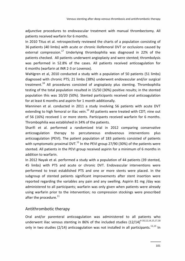
Venous stenting after deep venous thrombosis and antithrombotic therapy
101
adjunctive procedures to endovascular treatment with manual thrombectomy. All
patients received warfarin for 6 months.
In 2010 Titus et al. retrospectively reviewed the charts of a population consisting of
36 patients (40 limbs) with acute or chronic iliofemoral DVT or occlusions caused by
external compression.17 Underlying thrombophilia was diagnosed in 22% of the
patients checked. All patients underwent angioplasty and were stented; thrombolysis
was performed in 52.8% of the cases. All patients received anticoagulation for
6 months (warfarin at INR 2‐3 or Lovenox).
Wahlgren et al, 2010 conducted a study with a population of 50 patients (51 limbs)
diagnosed with chronic PTS; 21 limbs (38%) underwent endovascular and/or surgical
treatment.18 All procedures consisted of angioplasty plus stenting. Thrombophilia
testing of the total population resulted in 15/50 (30%) positive results; in the stented
population this was 10/20 (50%). Stented participants received oral anticoagulation
for at least 6 months and aspirin for 1 month additionally.
Manninen et al. conducted in 2011 a study involving 56 patients with acute DVT
extending to high femoral or iliac vein.10 All patients were treated with CDT; nine out
of 56 (16%) received 1 or more stents. Participants received warfarin for 6 months.
Thrombophilia was established in 34% of the patients.
Sharifi et al. performed a randomized trial in 2012 comparing conservative
anticoagulation therapy to percutaneous endovenous interventions plus
anticoagulation (PEVI). The patient population of 183 patients consisted of patients
with symptomatic proximal DVT.15 In the PEVI group 27/90 (30%) of the patients were
stented. All patients in the PEVI group received aspirin for a minimum of 6 months in
addition to warfarin.
In 2012 Nayak et al. performed a study with a population of 44 patients (39 stented,
45 limbs) with PTS and acute or chronic DVT. Endovascular interventions were
performed to treat established PTS and one or more stents were placed. In the
subgroup of stented patients significant improvements after stent insertion were
reported regarding the variables any pain and any swelling. Aspirin 81 mg /day was
administered to all participants; warfarin was only given when patients were already
using warfarin prior to the intervention; no compression stockings were prescribed
after the procedure.11
Antithrombotic therapy
Oral and/or parenteral anticoagulation was administered to all patients who
underwent iliac venous stenting in 86% of the included studies (12/14)4‐10,12,14,15,17,18
only in two studies (2/14) anticoagulation was not installed in all participants.11,13 In
Chapter 6
102
the study performed by Nayak, only patients who were on warfarin prior to stent
insertion received post‐intervention anticoagulation; all participants, including those
on warfarin received aspirin at daily doses of 81 mg.11 In the study performed by Raju,
only patients with thrombophilia received warfarin. A not specified proportion of the
population received only aspirin to maintain long‐term stent patency.13 This practice
was changed over time (not specified); later fondaparinux for 4 to 6 weeks was
prescribed for all patients and long‐term warfarin for selected patients
(recanalizations ≥3 segments, suprarenal stents, thrombophilia); no reason was
provided for this change of practice and no differentiation was made between these
regimes in terms of outcomes.13 Anticoagulant management in the included studies
therefore can roughly be divided in 3 regimes: 1) Warfarin administration with a
target International Normalized Ratio (INR) of 2‐3 for 3 to 6 months concurrent with
LMWH in the initial phase or in case of renal insufficiency UFH administration until the
INR reaches a value of 2 for 2 consecutive days: 58% of studies (8/12), 2) Warfarin
with an target INR of 2‐3 for at least 12 months concurrent with LMWH or UFH in the
initial phase: 17% of studies (2/12) and 3) warfarin administration, INR range 2‐3
indefinitely concurrent with initial LMWH or UFH: 17% of studies (1/12). One
remaining study could not be classified in one of these categories (8%). These regimes
were supplemented in 33% (4/12) of studies with 4) antiplatelet administration;
aspirin or aspirin plus clopidogrel. For a detailed summary of antithrombotic therapy
in the different studies see Table 6.3.
Outcomes
Recurrent DVT and occlusions
The incidence of recurrent DVT was reported in 12/14 studies;4,5,7‐10,12‐15,17,18 The
incidence of re‐thrombosis in (sub) groups of only stented patients ranged in different
studies from 5%15 to 25% (early re‐thrombosis 6%; late re‐ thrombosis 19%).13 In the
study by AbuRahma et al. recurrent DVT occurred in 3/18 (17%) of the patients in the
endovenous intervention group and in 2/8 (25%) in the comparator group.4 A fairly
low rate of DVT was reported by Titus; 1/36 (3%),17 Sillesen;14 1/45 (2%) and also in
the study by Kölbel 1/44 limbs (2%).9 Bækgaard et al. reported 6/103 (6%) recurrent
DVT (3 early within days and 3 late within 2‐3 years).5 Four patients (31%) experienced
an early recurrent DVT in the study by Grommes et al.7 Recurrent DVT occurred in 2
patients (29%) in the study by Oguzkurt et al.12 Post intervention DVT was diagnosed
in 2/56 (3.8%) of patients by Manninen et al.10 After 6 months recurrent DVT was 2%
in the PEVI group compared to 15% in the anticoagulation alone group (p=0.003).15 In‐
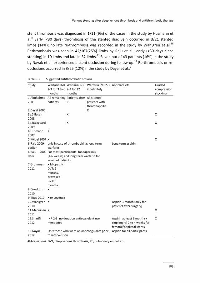
Venous stenting after deep venous thrombosis and antithrombotic therapy
103
stent thrombosis was diagnosed in 1/11 (9%) of the cases in the study by Husmann et
al.8 Early (<30 days) thrombosis of the stented iliac vein occurred in 3/21 stented
limbs (14%); no late re‐thrombosis was recorded in the study by Wahlgren et al.18
Rethrombosis was seen in 42/167(25%) limbs by Raju et al.; early (<30 days since
stenting) in 10 limbs and late in 32 limbs.13 Seven out of 43 patients (16%) in the study
by Nayak et al. experienced a stent occlusion during follow‐up.11 Re‐thrombosis or re‐
occlusions occurred in 3/25 (12%)in the study by Dayal et al..6
Table 6.3 Suggested antithrombotic options
Study Warfarin INR
2‐3 for 3 to 6 months
Warfarin INR
2‐3 for 12 months
Warfarin INR 2‐3
indefinitely
Antiplatelets Graded
compression stockings
1.AbuRahma
2001
All remaining
patients
Patients after
PE
All stented,
patients with
thrombophilia
2.Dayal 2005 X 3a.Sillesen
2005
X X
3b.Bækgaard 2009
X X
4.Husmann
2007
X
5.Kölbel 2007 X X
6.Raju 2009
earlier
only in case of thrombophilia: long term
warfarin
Long term aspirin
6.Raju 2009
later
For most participants: fondaparinux
(4‐6 weeks) and long term warfarin for
selected patients
7.Grommes
2011
X Idiopathic
DVT: 6
months, provoked
DVT: 3
months
8.Oguzkurt
2010
X
9.Titus 2010 X or Lovenox 10.Wahlgren
2010
X Aspirin 1 month (only for
patients after surgery)
11.Manninen 2011
X X
12.Sharifi
2012
INR 2‐3; no duration anticoagulant use
mentioned
Aspirin at least 6 months+
clopidogrel 2 to 4 weeks for femoral/popliteal stents
X
13.Nayak
2012
Only those who were on anticoagulants prior
to intervention
Aspirin for all participants
Abbreviations: DVT, deep venous thrombosis; PE, pulmonary embolism
Chapter 6
104
Patency rates
Long‐term stent patency rates were assessed in 10/15 studies;4‐6,8‐10,13,14,17,18 primary,
assisted primary patency and secondary patency rates 12 months after stent
placement ranged from 54%, 72%, 83%13 respectively to 78%, 83%, 95%17 in
(sub)groups of only stented patients. High patency rates in a (sub) population of only
stented patients were reported by Titus et al. After 12 months of follow‐up primary,
assisted primary and secondary patency rates were 78%, 83% and 95% respectively.
These high patency rates can possibly be explained by the fact that also patients
without a prior DVT were included in this study.17
Pulmonary embolism
In 11/14 studies the incidence of PE was reported. Three studies reported an
incidence of 0% PE in (sub) groups of only stented patients.11,13,17 Nayak et al. reported
PE incidence of a subgroup of stented patients with PTS; the prevalence of inferior
vena cava (IVC) filters was not reported,11 Raju et al. reported zero PE in a population
of stented patients with post‐thrombotic chronic total occlusions; the prevalence of
IVC filters was 10%13 and Titus et al. studied patients undergoing iliofemoral venous
angioplasty and stenting; IVC filters were placed in case of very high PE risk.17 The
population with the highest incidence of PE (7%), studied by Manninen et al. consisted
of patients with acute iliofemoral DVT; in 9% of the patients an IVC filter was placed
during the procedure.10 In the studies, performed by Abu Rahma (in endovascular
intervention group), Grommes, Kölbel, and Oguzkurt 0% PE was reported in partially
stented populations.4,7,9,12 The remaining studies by Bækgaard, Sillesen, and Sharifi (in
endovascular intervention group) reported 6%, 2% and 1% of PE respectively.5,14,15
Post thrombotic syndrome
PTS was assessed in 6/14 studies.8,11,13,15,17,18; in only 2 studies 8,18 the Villalta‐Prandoni
scale or the Venous Clinical Severity Score was used, both validated in different
settings24‐26 and subsequently endorsed by the International Society on Thrombosis
and Hemostasis (ISTH).
One of these studies, a study performed by Husmann et al. assessed PTS in a
population of only venous stented participants with May‐Thurner syndrome and DVT:
10/11 stented patients with a target INR of 2‐3 for the first 6 months had a Villalta
score of 0 after a mean follow‐up of 22 months. Duplex ultrasonography revealed no
post‐thrombotic changes in these patients; 1 patient reported mild PTS symptoms.
Patients did not receive compression stockings after the intervention.8
Venous stenting after deep venous thrombosis and antithrombotic therapy
105
Post‐hoc analysis performed after a trial by Sharifi et al. suggested a protective effect
of aspirin, on top of warfarin treatment (INR 2‐3) in reducing the prevalence of PTS;
relative risk (RR) of 0.37, confidence interval (CI) 0.19 to 0.74. In the endovascular
intervention plus anticoagulation group 7% PTS was reported versus 30% in the
anticoagulation alone group (p<0.001). All patients wear advised to wear compression
stockings; however, 6 months compliance was only 27%.15
A Chronic Venous Insufficiency Questionnaire was completed before and after the
recanalization of 128 limbs in the report presented by Raju; significant improvements
were noted concerning leg pain, sleep disturbance, effect on social activities and
morale after the intervention. The hemodynamic parameters hand‐foot pressure and
ambulatory venous pressure showed no significant improvements. No graded
compression stockings were prescribed post procedurally.13
Nayak et al. reported significant improvements after stent insertion for the variables
any pain and any swelling as proxy for PTS; no compression stockings were prescribed
after the procedure.11 Titus et al assessed by telephone whether symptoms overall
had improved, had not changed, or became progressively worse: a distribution of
83%, 7% and 10% was found. In this study patients did not receive compression
stockings.17
Finally, Wahlgren et al. reported Venous Clinical Severity scores before and after the
intervention; the score dropped from 9.1 points preprocedural to 6.0 points
postprocedural (p<0.0001). Patients were not advised to wear stockings post
procedurally.18
Major bleeding
Finally we explored rates of major bleeding. Five studies reported 0% major bleedings
during long term follow‐up.11‐14,17 In only 1 of these studies, performed by Nayak et al.
all patients received aspirin, and anticoagulation was installed for those prior on
warfarin; in the subgroup of stented patients no major bleeding occurred.11
In another study by Raju et al. in which no major bleeding complications were
reported only a part of the population received aspirin plus anticoagulation; the
remaining part received anticoagulation or aspirin.13
In the remaining studies with 0% major bleeding complications all patients received
warfarin for different time periods. The highest major bleeding scores (11%) were
reported in a population, described by Abu Rahma et al, consisting of patients with
iliofemoral DVT, of which 56% of the participants were stented. All patients were on
warfarin (INR 2‐3) indefinitely, without the addition of aspirin.4 Studies performed by
Chapter 6
106
Bækgaard, Grommes, Kölbel and Manninen reported major bleeding rates of 1%, 8%,
8% and 4% respectively.5,7,9,10 In all these studies patients received only warfarin.
Discussion
Venous stenting is performed in case residual thrombi remain after CDT9,14 or lesions
appear to be resistant to angioplasty.6 Overall, the evidence supporting the use of
venous stents after a DVT in the iliofemoral area for improved short‐term and long‐
term patency is convincing although of limited methodological quality. The lack of
comparator studies is evident: only 2 studies4,15 of which 1 trial15 compared venous
surgery plus anticoagulation to anticoagulation alone. In these studies the venous
surgery group (not all stented) performed significantly better than the anticoagulation
alone group in terms of patency,4 recurrent venous thromboembolism and
development of PTS.15 Interestingly, in many cases thrombophilia screening is
performed, but only on limited occasions type and duration of antithrombotic
treatment are based on the outcomes of its findings.4,13 Post procedural
antithrombotic therapy is not assessed separately as a potential factor for short‐term
and long‐term failure or success of the stenting procedure in any of the presented
studies. Based on this review we conclude that most stented patients receive warfarin
only for the standard duration of the treatment of their underlying DVT; the fact that
venous stents are inserted does not seem to affect this antithrombotic regime. This
practice is in accordance with current ACCP guidelines.21 In only 4 studies patients
received additional antiplatelet therapy after CDT and/or PMT and stent
placement.11,13,16,18 In 1 study clopidogrel at 75 mg per day for 2‐4 weeks was
administered to exclusively stented patients.16 Combination therapy, consisting of
anticoagulant and antiplatelet therapy or dual antiplatelet therapies (aspirin plus
clopidogrel) may theoretically be an interesting option. It is conceivable that
antiplatelet agents could provide additional protection against the formation of
thrombi after venous stenting of the iliac vein.27 Shear forces are a major determinant
of platelet deposition in thrombi; in the venous system usually low‐flow, low‐shear
conditions exist and occurring thrombi are likely platelet poor and fibrin rich. In the
iliac vein however, due to thickening and scarification of the vessel wall after a DVT
and/or external compression, shear forces are in some cases anticipated to be nearly
identical to the arterial system.27 In experimentally induced thrombi in these venous
segments, indeed platelet rich thrombi occurred and these platelets were in an
activated state.27 However, aspirin, clopidogrel, and aspirin plus clopidogrel did not
Venous stenting after deep venous thrombosis and antithrombotic therapy
107
significantly reduce stent thrombosis in an experimental model neither did they
prevent platelet accumulation, while anticoagulant therapy did reduce thrombosis.27
The lowest patency rates were reported by Wahlgren, Dayal and Raju:6,13,18 Low
patency was combined with high rates of recurrent DVT (14%, 12% and25%
respectively). In these studies acute and chronic DVT, and in the case of Dayal also
DVT at unusual sites were included. As far as the antithrombotic regimen that was
applied in these studies is concerned, no beneficial effect was observed by the
additional aspirin administration in those stented.13,18 The low patency rates in these
studies can at least partially be explained by the fact that patients with chronic DVT
and with total occlusions were included. In the study by Nayak aspirin was prescribed
to all, and additional warfarin to those with previous use of warfarin; the recurrence
rate was 16%.11 Besides the antithrombotic effects also anti‐inflammatory effects
could play a role while using antiplatelet agents. There could be an additional effect of
aspirin on top of warfarin treatment for the reduction of the prevalence of PTS, as was
suggested by the post‐hoc analysis performed by Sharifi et al.15 that showed a
significant reduction of the incidence of PTS in patients on aspirin.
The safety of antiplatelet therapy in combination with anticoagulation in patients
after venous stenting is something to be considered. Limited data are provided by 2
studies by Nayak and Raju, wherein aspirin plus warfarin therapy was applied and 0%
major bleedings were reported,11,13 the remaining 2 studies in which the patients
received antiplatelet plus anticoagulant therapy also no major bleedings were
reported.15,18 Therefore in terms of safety, the addition of aspirin to the
antithrombotic regimen does not seem to pose a large problem, at least on the short
term in the patients selected in these studies.
The highest primary patency rates were reported in studies performed by Manninen,
Sillesen and Abu Rahma;4,10,14 all patients received warfarin with a target INR 2‐3 for
the standard duration for the underlying DVT, and no antiplatelet therapy was
administered. In all three studies only patients with acute DVT were included; fresh
thrombi probably resolve better after CDT than sub acute thrombi.28 Also low rates of
recurrent thrombosis were reported in these studies. These 3 studies were all
awarded with 4/5 stars for study quality; we therefore can assume that the study
findings are trustworthy. Kölbel also reported low rates of recurrent DVT and PE.9 A
very high percentage (68%) of the total study population was diagnosed with
thrombophilia; this did however not influence antithrombotic management.
Anticoagulants likely play a more important role than antiplatelet agents in the
prevention of re‐stenosis in venous stented patients with a prior DVT.2,21 Recurrent
venous thrombosis is primarily caused by increased thrombin generation,29 therefore
anticoagulants likely have better antithrombotic properties in patients with venous
Chapter 6
108
stents after a DVT than antiplatelet drugs. The major bleeding risk of long‐term
warfarin use with a target INR of 2‐3 was estimated at 1.3% per year.30 The rates of
major bleeding reported in this review are in a higher range between 8‐11%. The
additional bleeding risk is mainly due to the stenting procedure. This may be an
acceptable risk if stenting would lead to clearly better outcomes.
Based on the available evidence in this review no firm conclusions regarding the
association between the duration of antithrombotic therapy and the study outcomes
can be drawn. A major limitation of this study is the fact that no meta‐analysis could
be performed. The quality of the included studies was sub optimal; single armed
cohort studies with mostly a small sample size formed the majority in this review.
Applicability of the review may be affected by the fact that most studies assessed
patient groups undergoing CDT and/or PMT of which only a part underwent stenting;
subgroups consisting of only stented patients could be assessed in only a few cases.
The most important factors contributing to a favorable outcome, high patency and
low recurrence rate are mainly dependent on the location of the thrombus and the
age of the thrombus. Thrombophilia is not a discriminating factor for success.
Anticoagulant therapy for the duration of the underlying thrombotic lesions seems to
be sufficient.
The conclusion of this systematic literature review is that antithrombotic therapy does
not seem to influence any of the outcomes in patients with venous stenting after DVT:
patency, PTS, PE, recurrent DVT or restenosis and bleeding. The main drivers for the
outcome are the location of the DVT, the age of the thrombus and the stenting
procedure itself.
Venous stenting after deep venous thrombosis and antithrombotic therapy
109
References
1. Jenkins JS. Endovascular therapies to treat iliofemoral deep venous thrombosis. Prog Cardiovasc Dis. 2011;54:70‐6.
2. Meissner MH. Indications for platelet aggregation inhibitors after venous stents. Phlebology. 2013;28
Suppl 1:91‐8. 3. Mackman N. Triggers, targets and treatments for thrombosis. Nature. 2008;451:914‐8.
4. AbuRahma AF, Perkins SE, Wulu JT, Ng HK. Iliofemoral deep vein thrombosis: conventional therapy
versus lysis and percutaneous transluminal angioplasty and stenting. Ann Surg. 2001;233:752‐60. 5. Baekgaard N, Broholm R, Just S, Jorgensen M, Jensen LP. Long‐term results using catheter‐directed
thrombolysis in 103 lower limbs with acute iliofemoral venous thrombosis. Eur J Vasc Endovasc Surg.
2010;39:112‐7. 6. Dayal R, Bernheim J, Clair DG, Mousa AY, Hollenbeck S, DeRubertis B, et al. Multimodal percutaneous
intervention for critical venous occlusive disease. Ann Vasc Surg. 2005;19:235‐40.
7. Grommes J, Strijkers R, Greiner A, Mahnken AH, Wittens CH. Safety and feasibility of ultrasound‐accelerated catheter‐directed thrombolysis in deep vein thrombosis. Eur J Vasc Endovasc Surg.
2011;41:526‐32.
8. Husmann MJ, Heller G, Kalka C, Savolainen H, Do DD, Schmidli J, et al. Stenting of common iliac vein obstructions combined with regional thrombolysis and thrombectomy in acute deep vein thrombosis.
Eur J Vasc Endovasc Surg. 2007;34:87‐91.
9. Kolbel T, Lindh M, Holst J, Uher P, Eriksson KF, Sonesson B, et al. Extensive acute deep vein thrombosis of the iliocaval segment: midterm results of thrombolysis and stent placement. J Vasc
Interv Radiol. 2007;18:243‐50.
10. Manninen H, Juutilainen A, Kaukanen E, Lehto S. Catheter‐directed thrombolysis of proximal lower extremity deep vein thrombosis: a prospective trial with venographic and clinical follow‐up. Eur J
Radiol. 2012;81:1197‐202.
11. Nayak L, Hildebolt CF, Vedantham S. Postthrombotic syndrome: feasibility of a strategy of imaging‐guided endovascular intervention. J Vasc Interv Radiol. 2012;23:1165‐73.
12. Oguzkurt L, Ozkan U, Demirturk OS, Gur S. Endovascular treatment of phlegmasia cerulea dolens with
impending venous gangrene: manual aspiration thrombectomy as the first‐line thrombus removal method. Cardiovasc Intervent Radiol. 2011;34:1214‐21.
13. Raju S, Neglen P. Percutaneous recanalization of total occlusions of the iliac vein. J Vasc Surg.
2009;50:360‐8. 14. Sillesen H, Just S, Jorgensen M, Baekgaard N. Catheter directed thrombolysis for treatment of ilio‐
femoral deep venous thrombosis is durable, preserves venous valve function and may prevent
chronic venous insufficiency. Eur J Vasc Endovasc Surg. 2005;30:556‐62. 15. Sharifi M, Bay C, Mehdipour M, Sharifi J. Thrombus Obliteration by Rapid Percutaneous Endovenous
Intervention in Deep Venous Occlusion (TORPEDO) trial: midterm results. J Endovasc Ther.
2012;19:273‐80. 16. Sharifi M, Mehdipour M, Bay C, Smith G, Sharifi J. Endovenous therapy for deep venous thrombosis:
the TORPEDO trial. Catheter Cardiovasc Interv. 2010;76:316‐25.
17. Titus JM, Moise MA, Bena J, Lyden SP, Clair DG. Iliofemoral stenting for venous occlusive disease. J Vasc Surg. 2011;53:706‐12.
18. Wahlgren CM, Wahlberg E, Olofsson P. Endovascular treatment in postthrombotic syndrome. Vasc
Endovascular Surg. 2010;44:356‐60. 19. Neglen P, Hollis KC, Olivier J, Raju S. Stenting of the venous outflow in chronic venous disease: long‐
term stent‐related outcome, clinical, and hemodynamic result. J Vasc Surg. 2007;46:979‐90.
20. Ye K, Lu X, Li W, Huang Y, Huang X, Lu M, et al. Long‐term outcomes of stent placement for symptomatic nonthrombotic iliac vein compression lesions in chronic venous disease. J Vasc Interv
Radiol. 2012;23:497‐502.
Chapter 6
110
21. Kearon C, Akl EA, Comerota AJ, Prandoni P, Bounameaux H, Goldhaber SZ, et al. Antithrombotic
therapy for VTE disease: Antithrombotic Therapy and Prevention of Thrombosis, 9th ed: American College of Chest Physicians Evidence‐Based Clinical Practice Guidelines. Chest. 2012;141(2
Suppl):e419S‐94S.
22. Sanderson S, Tatt ID, Higgins JP. Tools for assessing quality and susceptibility to bias in observational studies in epidemiology: a systematic review and annotated bibliography. Int J Epidemiol.
2007;36:666‐76.
23. Stang A. Critical evaluation of the Newcastle‐Ottawa scale for the assessment of the quality of nonrandomized studies in meta‐analyses. Eur J Epidemiol. 2010;25:603‐5.
24. Jayaraj A, Meissner MH. A comparison of villalta‐prandoni scale and venous clinical severity score in
the assessment of post thrombotic syndrome. Ann Vasc Surg 2014;28:313‐7. 25. Galanaud JP, Holcroft CA, Rodger MA, Kovacs MJ, Betancourt MT, Wells PS, et al. Comparison of the
Villalta post‐thrombotic syndrome score in the ipsilateral vs. contralateral leg after a first unprovoked
deep vein thrombosis. J Thromb Haemost. 2012;10:1036‐42. 26. Kahn SR. Measurement properties of the Villalta scale to define and classify the severity of the post‐
thrombotic syndrome. J Thromb Haemost. 2009;7:884‐8.
27. McBane RD, 2nd, Leadley RJ, Jr., Baxi SM, Karnicki K, Wysokinski W. Iliac venous stenting: antithrombotic efficacy of PD0348292, an oral direct Factor Xa inhibitor, compared with antiplatelet
agents in pigs. Arterioscler Thromb Vasc Biol. 2008;28:413‐8.
28. Zhang X, Ren Q, Jiang X, Sun J, Gong J, Tang B, et al. A prospective randomized trial of catheter‐directed thrombolysis with additional balloon dilatation for iliofemoral deep venous thrombosis: a
single‐center experience. Cardiovasc Intervent Radiol. 2014;37:958‐68.
29. Geerts WH, Pineo GF, Heit JA, Bergqvist D, Lassen MR, Colwell CW, et al. Prevention of venous thromboembolism: the Seventh ACCP Conference on Antithrombotic and Thrombolytic Therapy.
Chest. 2004;126(3 Suppl):338S‐400S.
30. Levine MN, Raskob G, Landefeld S, Kearon C. Hemorrhagic complications of anticoagulant treatment. Chest. 2001;119(1 Suppl):108‐21.
Venous stenting after deep venous thrombosis and antithrombotic therapy
111
Appendix 6.1 The search strategy for MEDLINE
((stents[MeSH Terms] OR stent[MeSH Terms] OR angioplasty[MeSH Terms] OR
Percutaneous recanalization OR endovascular interventions) AND (venous ulcer[MeSH
Terms] OR venous ulcers[MeSH Terms] OR venous insufficiencies[MeSH Terms] OR
venous insufficiency[MeSH Terms] OR "postthrombotic syndrome"[MeSH Major Topic]
OR deep venous thromboses[MeSH Terms] OR deep venous thrombosis[MeSH Terms])
AND (agents, anticoagulant[MeSH Terms] OR anticoagulant agents[MeSH Terms] OR
anticoagulant drugs[MeSH Terms] OR anticoagulants[MeSH Terms] OR drugs,
anticoagulant[MeSH Terms] OR blood platelet aggregation inhibitors[MeSH Terms] OR
agents, antiplatelet[MeSH Terms] OR antiplatelet agents[MeSH Terms] OR antiplatelet
drugs[MeSH Terms] OR drugs, antiplatelet[MeSH Terms])) OR ((stent OR angioplasty
OR Percutaneous recanalization) AND (venous ulcer[MeSH Terms] OR venous
ulcers[MeSH Terms] OR venous insufficiencies[MeSH Terms] OR venous
insufficiency[MeSH Terms] OR "postthrombotic syndrome"[MeSH Major Topic] OR
deep venous thromboses[MeSH Terms] OR deep venous thrombosis[MeSH Terms])
AND (anticoagulant OR anticoagulant agents OR anticoagulant drugs OR
anticoagulants OR blood platelet aggregation inhibitors OR Antiplatelet OR
antiplatelet agents OR antiplatelet drugs))
Limits Activated MEDLINE: English
Chapter 6
112
Appendix 6.2 Newcastle‐Ottawa quality assessment scale for cohort studies (selection of 5 items
Selection
1) Representativeness of the cohort
a. truly representative of the population of patients undergoing venous
stenting in the iliofemoral area after a first or recurrent DVT; all
participants stented *
b. somewhat representative of the population of patients undergoing
venous stenting in the iliofemoral area after a first or recurrent DVT;
not all participants stented *
c. additional inclusion criteria and not all participants stented.
d. no description of the derivation of the cohort
2) Ascertainment of exposure
a. secure record (e.g. surgical records) *
b. structured interview *
c. written self report
d. no description
Outcome
3) Assessment of outcome
a. independent blind assessment *
b. record linkage *
c. self report
d. no description
4) Was follow‐up long enough for outcomes to occur
a. yes (≥24 months (mean or median)) *
b. no
5) Adequacy of follow‐up of cohorts
a. complete follow‐up ‐ all subjects accounted for *
b. subjects lost to follow‐up (< 5%) unlikely to introduce bias or
description provided of those lost *
c. follow‐up rate <95% and no description of those lost
d. no statement
113
Chapter 7 Quality of anticoagulant therapy and in‐stent
thrombosis in patients with venous stents
Pieter Eijgenraam, Mark A.F. de Wolf, Ralph L.M. Kurstjens, Hugo ten Cate,
Cees H.A. Wittens, Arina J ten Cate‐Hoek
Submitted
Chapter 7
114
Abstract
Introduction
In patients with venous outflow obstruction following Iliofemoral deep vein thrombosis (IFDVT)
stenting of venous tracts to prevent or alleviate post thrombotic syndrome (PTS) is applied with
increasing frequency. Up till now the association of quality of anti‐thrombotic therapy and
effectiveness of stenting has not been studied explicitly. The impact of the quality of
anticoagulant therapy with vitamin K antagonists (VKA) expressed as time within therapeutic
range (TTR) and proportion of INR values <2.0 on in‐stent thrombosis is currently unknown.
Methods
Seventy‐nine patients with venous stent placement after acute or chronic iliofemoral or caval
DVT were included in this study. All patients used VKA after the intervention. TTR was
calculated according to the linear interpolation method. In‐stent thrombosis was assessed by
the use of duplex ultrasound. Survival analysis (Cox regression, Kaplan Meier curves) was used
to analyze the data.
Results
The average TTR was 46.4% (SD 20.8); the average proportion of INR measurements <2.0 was
9.5% (SD 11.6). Increasing TTR was associated with a lower risk of in‐stent‐thrombosis. The
multivariable adjusted analysis resulted in a hazard ratio of 0.940 (95% CI 0.890–0.993) per 1%
increase in TTR. An increase of the proportion of INR values <2.0 was not significantly
associated with the outcome in‐stent thrombosis; HR per 1% increase in proportion of 1.018
(95% CI 0.957–1.084).
Conclusions
We conclude that an increase of TTR offers protection against in‐stent thrombosis. The
proportion of INR <2.0, although expected to be an important predictor for in‐stent thrombosis,
could not be identified as a risk factor in this study. An overall low average TTR of 46.4%
(SD 20.8) was recorded, possibly due to the relative short follow‐up in combination with the
initiation phase of VKA, leading to unstable INR values.
Quality of anticoagulant therapy and in‐stent thrombosis in patients with venous stents
115
Introduction
Post thrombotic syndrome (PTS) develops in over 50% of patients with iliofemoral
deep venous thrombosis (IFDVT).1 Conservative treatment regimens comprising of a
combination of compression therapy, mobilization and oral anticoagulants, up till now
mainly vitamin K antagonists (VKA), result in suboptimal outcomes in these patients.
Over the last 2 decades the use of percutaneous transluminal angioplasty (PTA) and
stenting of venous outflow obstructions of the iliofemoral and caval veins has gained
more attention. PTA and stenting appears to be effective in terms of objective and
subjective clinical improvement of PTS symptoms and has been characterized by good
mid‐ to long‐term patency rates in mainly observational studies.2‐8 The treatment
usually consists of guide‐wire recanalization of the obstructed venous segments and
placement of one or more stents, followed by anticoagulant therapy with VKA for at
least 3 months. In acute DVT patients in whom catheter directed thrombolysis (CDT) is
applied, additional venous stenting immediately following thrombolysis is often
indicated in case of underlying venous pathology, such as iliac vein compression
syndromes, which is thought to be the underlying cause of thrombosis in these
patients.9
So far the contribution of the quality of anticoagulant therapy, to the effectiveness of
venous stenting has not been evaluated.10 The current guidelines issued by the
American College of Chest Physicians (ACCP) do not specifically advise on
anticoagulation therapy after venous stenting mainly for lack of evidence.11 In the
literature a variety of antithrombotic therapies are suggested after venous stent
placement for chronic venous obstruction or directly after CDT in acute thrombosis;
anticoagulant therapy varying from 3 months to 1 year with target range INR of
usually 2 to 3 is prescribed, sometimes in combination with antiplatelet therapy.10
While time within therapeutic range (TTR) of VKA therapy in acute DVT is known to
have substantial impact on recurrent DVT,12,13 the role of the quality of TTR during
VKA therapy in preventing re‐occlusion after venous stenting remains unclear. In
addition INR values <2.0 are an established risk factor for recurrent venous
thromboembolism (VTE),13 as well as for PTS.14,15 Even though the contribution of the
quality of anticoagulation therapy will therefore likely also play a role in the
effectiveness of venous stenting, so far no study has analyzed TTR and proportion of
INR values <2.0 in patients undergoing venous stenting in acute DVT or for chronic
venous obstruction in relation to stent patency.
We therefore evaluated the contribution of the quality of anticoagulation control
during treatment with VKA calculated with the linear interpolation method. In
addition we assessed the impact of the proportion of INR values <2.0 on in‐stent
Chapter 7
116
thrombosis in chronic PTS patients on long‐term anticoagulation with iliofemoral and
or caval venous stents placed in patients with chronic PTS.
Methods
Patient selection
During the period from March 2009 until January 2013 108 patients, referred to the
Department of Vascular Surgery in the Maastricht University Medical Center, the
Netherlands for venous stent placement after acute or chronic iliofemoral or caval
DVT, and who had used VKA for at least 3 months after the intervention and had at
least 1 year of follow up data available were invited to participate in the current
study. Patients with symptomatic deep venous obstruction without a history of DVT;
i.e. cases of iliac vein compression syndrome (such as May‐Thurner syndrome) were
not eligible. Seventy‐nine patients of whom 82.4 % used VKA prior to the intervention,
gave informed consent to retrieve patient data from their anticoagulation clinic
records. All data, except data concerning anticoagulation therapy were collected
prospectively. Institutional review board approval was obtained (METC 14‐4‐081).
Baseline work‐up
All patients were scheduled for a standardized work‐op, consisting of clinical
evaluation, duplex ultrasound (DUS) and magnetic resonance venography (MRV) prior
to the intervention. The primary objective using DUS is the assessment of signs of
recanalization and/or flow impairment, luminal narrowing, vein wall thickening and
any possible external compression on vein segments, such as iliac vein compression
syndrome. During follow‐up DUS was used to assess patency of stented venous tracts
and vein segments proximal and distal of the stents, as well as the configuration of the
stents themselves. The LET score, a classification that stratifies patients into 4 groups
(I‐IV) in accordance to the location of the most proximal thrombus, was used to
classify patients.16 With MRV the extend and severity of intraluminal post thrombotic
changes and collateralization, and the locations of trabeculae, longitudinal vertical
strands attached to the vein wall that indicate PTS, were recorded.17
Stent placement
All procedures were performed under general anesthesia in a dedicated angiosuite. In
patients using VKA at the time of intervention, the procedure was performed under
full anticoagulation; i.e. coumadins were not stopped in the days preceding the
Quality of anticoagulant therapy and in‐stent thrombosis in patients with venous stents
117
stenting procedure if INR was <4. In patients not using anticoagulation at the time of
intervention, 5.000 IU of unfractionated heparin was administered at the start of the
procedure, and another 5.000 IU was given at the discretion of the treating physician
in case of prolonged duration of the intervention. Venous access was obtained via the
femoral vein under ultrasound guidance, generally at least 15 cm distally to the deep
femoral inflow. With the use of various guide wires recanalization into the vena cava
inferior was performed.18 As only stented patients were included in this study, the
success rate for recanalization was set at 100%. Following recanalization pre‐
dilatation, i.e. PTA, of the obstructed vein segments was performed in the iliofemoral
tract. Following pre‐dilatation the affected vein segments were stented. The proximal
and distal landing zones of the stents were placed in “healthy vein”. Self‐expendable
stents, ranging in size of 12‐16mm in the iliofemoral tract, and 25mm in the caval
tract, were used in all cases (sinus XL, sinus Venous and sinus XL‐flex, Optimed,
Ettlingen, Germany and Zilver Vena (Cook, Galway, Ireland). For a small number of
bilaterally affected patients balloon expendable stents were used to reconstruct the
confluens of the vena cava ((AndraStent, Andramed, Reutlingen, Germany).19
In patients using VKA prior to the intervention, anticoagulant therapy was continued
for at least 6 months following the intervention. All other patients received LMWH
directly after the procedure, and were started on VKA on the first day post‐
intervention. Stenting procedures were performed for three different indications: CDT
+ stenting after acute DVT, stenting after (chronic) PTS, and stenting with
endophlebectomy and AV fistula after (chronic) PTS.
Follow up and outcomes
Patients were assessed at 2 weeks, 6 weeks, and 3, 6 and 12 months after the
intervention and annually thereafter, or earlier when necessary, for a regular check‐
up. A standardized clinical assessment by a trained physician was performed and DUS
to assess patency and possible stent configuration problems (kinking, migration,
fracture, changes in orientation). In‐stent thrombosis was defined as thrombosis with
100% occlusion of the treated vein. An endpoint for the study was reached when VKA
administration was stopped, or when the last patency measurement was performed
or when in‐stent thrombosis occurred.
Anticoagulation
Patient records were retrieved from patient’s individual anticoagulation clinics.
Patient records included INR values, start and stop dates of VKA, indications for VKA
therapy, target INR values, type of VKA (acenocoumarol, fenprocoumon) and
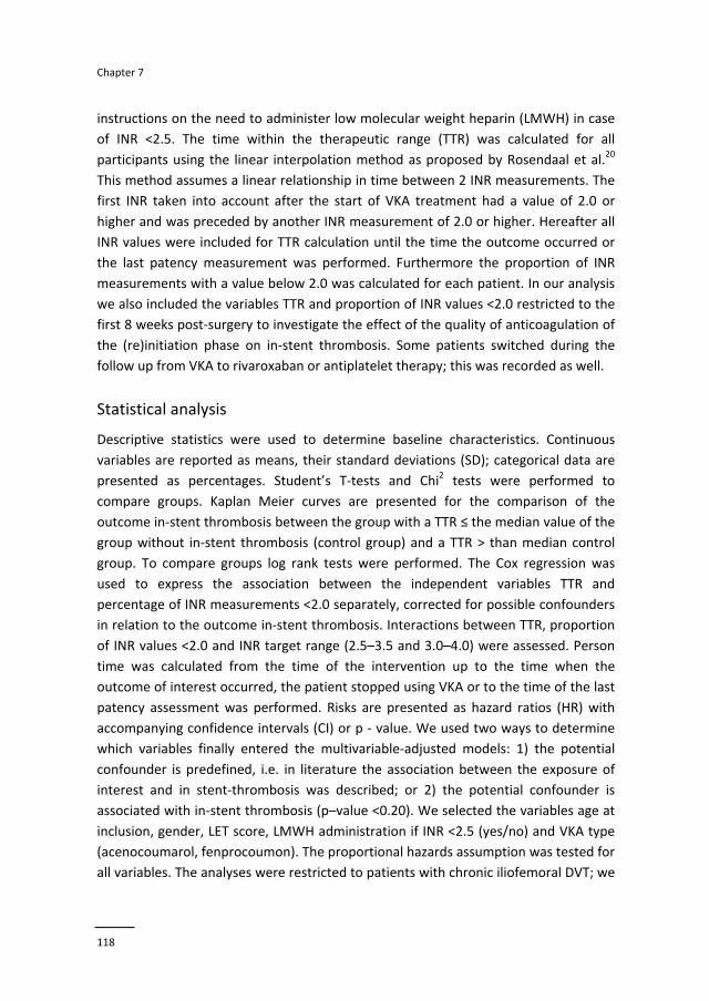
Chapter 7
118
instructions on the need to administer low molecular weight heparin (LMWH) in case
of INR <2.5. The time within the therapeutic range (TTR) was calculated for all
participants using the linear interpolation method as proposed by Rosendaal et al.20
This method assumes a linear relationship in time between 2 INR measurements. The
first INR taken into account after the start of VKA treatment had a value of 2.0 or
higher and was preceded by another INR measurement of 2.0 or higher. Hereafter all
INR values were included for TTR calculation until the time the outcome occurred or
the last patency measurement was performed. Furthermore the proportion of INR
measurements with a value below 2.0 was calculated for each patient. In our analysis
we also included the variables TTR and proportion of INR values <2.0 restricted to the
first 8 weeks post‐surgery to investigate the effect of the quality of anticoagulation of
the (re)initiation phase on in‐stent thrombosis. Some patients switched during the
follow up from VKA to rivaroxaban or antiplatelet therapy; this was recorded as well.
Statistical analysis
Descriptive statistics were used to determine baseline characteristics. Continuous
variables are reported as means, their standard deviations (SD); categorical data are
presented as percentages. Student’s T‐tests and Chi2 tests were performed to
compare groups. Kaplan Meier curves are presented for the comparison of the
outcome in‐stent thrombosis between the group with a TTR ≤ the median value of the
group without in‐stent thrombosis (control group) and a TTR > than median control
group. To compare groups log rank tests were performed. The Cox regression was
used to express the association between the independent variables TTR and
percentage of INR measurements <2.0 separately, corrected for possible confounders
in relation to the outcome in‐stent thrombosis. Interactions between TTR, proportion
of INR values <2.0 and INR target range (2.5–3.5 and 3.0–4.0) were assessed. Person
time was calculated from the time of the intervention up to the time when the
outcome of interest occurred, the patient stopped using VKA or to the time of the last
patency assessment was performed. Risks are presented as hazard ratios (HR) with
accompanying confidence intervals (CI) or p ‐ value. We used two ways to determine
which variables finally entered the multivariable‐adjusted models: 1) the potential
confounder is predefined, i.e. in literature the association between the exposure of
interest and in stent‐thrombosis was described; or 2) the potential confounder is
associated with in‐stent thrombosis (p–value <0.20). We selected the variables age at
inclusion, gender, LET score, LMWH administration if INR <2.5 (yes/no) and VKA type
(acenocoumarol, fenprocoumon). The proportional hazards assumption was tested for
all variables. The analyses were restricted to patients with chronic iliofemoral DVT; we
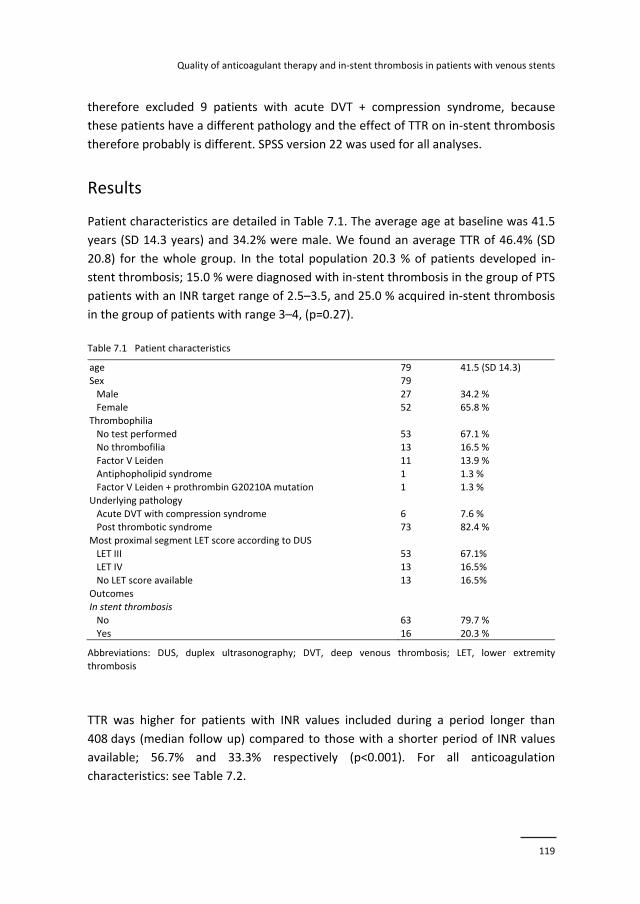
Quality of anticoagulant therapy and in‐stent thrombosis in patients with venous stents
119
therefore excluded 9 patients with acute DVT + compression syndrome, because
these patients have a different pathology and the effect of TTR on in‐stent thrombosis
therefore probably is different. SPSS version 22 was used for all analyses.
Results
Patient characteristics are detailed in Table 7.1. The average age at baseline was 41.5
years (SD 14.3 years) and 34.2% were male. We found an average TTR of 46.4% (SD
20.8) for the whole group. In the total population 20.3 % of patients developed in‐
stent thrombosis; 15.0 % were diagnosed with in‐stent thrombosis in the group of PTS
patients with an INR target range of 2.5–3.5, and 25.0 % acquired in‐stent thrombosis
in the group of patients with range 3–4, (p=0.27).
Table 7.1 Patient characteristics
age 79 41.5 (SD 14.3)
Sex 79
Male 27 34.2 % Female 52 65.8 %
Thrombophilia
No test performed 53 67.1 % No thrombofilia 13 16.5 %
Factor V Leiden 11 13.9 %
Antiphopholipid syndrome 1 1.3 % Factor V Leiden + prothrombin G20210A mutation 1 1.3 %
Underlying pathology
Acute DVT with compression syndrome 6 7.6 % Post thrombotic syndrome 73 82.4 %
Most proximal segment LET score according to DUS
LET III 53 67.1% LET IV 13 16.5%
No LET score available 13 16.5%
Outcomes In stent thrombosis
No 63 79.7 %
Yes 16 20.3 %
Abbreviations: DUS, duplex ultrasonography; DVT, deep venous thrombosis; LET, lower extremity thrombosis
TTR was higher for patients with INR values included during a period longer than
408 days (median follow up) compared to those with a shorter period of INR values
available; 56.7% and 33.3% respectively (p<0.001). For all anticoagulation
characteristics: see Table 7.2.

Chapter 7
120
Table 7.2
Anticoagulation characteristics for all patients, patients with a TTR
below and patients above the m
edian TTR
of control group (47.9%)
Variable
Number
All patients
TTR ≤47.9%
(med
ian controls)
TTR >47.9%
(med
ian controls)
p‐value
INR Range
2.5–3.5
INR Range
3.0–4.0
p‐value
INR range
2.5‐3.5
40
48.1%
16
18
3.0‐4.0
36
43.0%
19
11
3.5‐4.5
3
3.8%
1
1
p=0.43
VKA
acenocoumarol
46
55.7%
22
18
25
20
fenprocoumon
33
40.5%
14
12
p=0.93
15
15
p=0.64
TTR
66
46.4% (SD
20.8)
31.9% (SD
13.0)
63.9% (SD
13.6)
p<0.001
49.1%
43.2%
p=0.27
TTR (in patients with follow up
>408 days (m
edian)
33
56.7%
TTR (in patients with follow up
<408 days (m
edian)
33
33.3%
p<0.001
Mean % IN
R < 2
66
9.5% (SD
11.6)
13.8%(SD 12.0)
4.4% (SD
8.7)
p<0.001
12.0% (SD
12.5)
6.0% (SD
8.5)
p=0.03
INR tests in
range
66
41.9% (SD
18.4)
30.1% (SD
13.7)
54.4%(SD 15.9)
p<0.001
45.0% (SD
20.3)
36.4% (SD
16.5) p=0.07
Average IN
R
66
3.2 (SD
0.47)
3.1 (SD
0.5)
3.2 (SD
0.3)
p=0.52
3.0 (SD
0.4)
3.3 (SD
0.5)
p=0.002
LMWH if IN
R <2.5
No
52
22
18
27
22
yes
27
14
12
p=0.93
12
14
p=0.46
Abbreviations: LMWH, low m
olecular weight hep
arin; TTR, tim
e within therapeu
tic range; V
KA; vitam
in K antagonists; LMWH , low m
olecular weight hep
arins
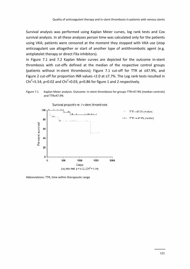
Quality of anticoagulant therapy and in‐stent thrombosis in patients with venous stents
121
Survival analysis was performed using Kaplan Meier curves, log rank tests and Cox
survival analysis. In all these analyses person time was calculated only for the patients
using VKA; patients were censored at the moment they stopped with VKA use (stop
anticoagulant use altogether or start of another type of antithrombotic agent (e.g.
antiplatelet therapy or direct FXa inhibitors).
In Figure 7.1 and 7.2 Kaplan Meier curves are depicted for the outcome in‐stent
thrombosis with cut‐offs defined at the median of the respective control groups
(patients without in‐stent thrombosis); Figure 7.1 cut‐off for TTR at ≤47.9%, and
Figure 2 cut‐off for proportion INR values <2.0 at ≤7.7%. The Log rank tests resulted in
Chi2=5.54, p=0.02 and Chi2=0.03, p=0.86 for figure 1 and 2 respectively.
Figure 7.1 Kaplan Meier analysis. Outcome: in‐stent thrombosis for groups TTR<47.9% (median controls)
and TTR≥47.9%
Abbreviations: TTR, time within therapeutic range
Chapter 7
122
Figure 7.2 Kaplan Meier analysis. Outcome: in‐stent thrombosis for groups proportion of INR<2.0≤7.7%
(median controls) and proportion of INR<2.0>7.7%
The analyses using the Cox regression are presented in Table 7.3. A linear decrease of
the risk of in‐stent‐thrombosis associated with increasing TTR was found in all three
models: the univariable, the age and sex adjusted model and in the multivariable
adjusted analysis; respective HR per 1% increase in TTR were 0.954 (95% CI 0.920‐
0.988), 0.943 (95% CI 0.903‐0.985) and 0.940 (95% CI 0.890‐0.992). For INR values
lower than 2.0 multivariable adjusted analysis resulted in a HR per 1% increase in
proportion of 1.018 (95% CI 0.957‐1.084). In the analysis of both determinants we
entered the same possible confounders in our models. Pearson correlations between
the independent variables TTR and proportion of INR values <2.0 were determined for
the groups with INR ranges 2.5‐3.5 and 3.0‐4.0 (only PTS patients); ‐0.55 (p=0.004)
and ‐0.27 (p=0.17) respectively. Interactions between TTR, proportion of INR values
<2.0 and INR target range (2.5‐3.5 and 3.0‐4.0) were calculated: p=0.08 and 0.47
respectively. To investigate the effect of the (re) initiation phase on in‐stent
thrombosis we calculated the TTR and proportion of INR values <2.0 for the first 8
weeks post‐surgery. Overall non‐significant results were found; in models using the
same confounder set as described earlier the increase of 1% in TTR resulted in a HR of
1.023 (95% CI 0.980‐1.069); an increase of 1% in the proportion of INR values <2.0
resulted in a HR of 0.928 (95% CI 0.848‐1.015). The proportional hazard assumptions
were not violated for TTR and proportions of INR values <2; therefore the effect of
these independent variables may be assumed constant over time; also for all possible
confounders this assumption was not violated.

Quality of anticoagulant therapy and in‐stent thrombosis in patients with venous stents
123
Table 7.3 Univariable, age and sex adjusted and multivariable adjusted hazard ratios for in stent
thrombosis in patients with PTS according to TTR and percentage INR<2
Univariable:
HR (95% CI)
Age sex adjusted:
HR (95% CI)
Multivariable adjusteda:
(HR (95% CI)
TTR (per 1% increase) 0.954 (0.920‐0.988) 0.943 (0.903‐0.985) 0.940 (0.890‐0.992)
TTR (per 10% increase) 0.624 (0.434‐0.886) 0.556 (0.361‐0.860) 0.539 (0.312‐0.922) INR <2 (per 1 % increase 1.024 (0.969‐1.083) 1.033 (0.975‐1.096) 1.018 (0.957‐1.084)
INR <2 (per 10% increase 1.268 (0.730‐2.220) 1.384 (0.776‐2.501) 1.195 (0.664‐2.240)
a Adjusted for: Age at baseline, sex, LET score, LMWH administration if INR <2.5 (yes/no), VKA:
acenocoumarol or fenprocoumon. Abbreviations: HR, hazard ratio; PTS, postthrombotic syndrome; TTR,
time within therapeutic range
Discussion
In this study we found that the quality of anticoagulant therapy with vitamin K
antagonists has a strong effect on in‐stent thrombosis in patients after iliofemoral
stent placement. We identified TTR as the most important predictive variable that
reflects the quality of anticoagulant therapy and its associated risk for in‐stent
thrombosis. A 10% increase in TTR resulted in a risk reduction for in‐stent thrombosis
of 46.1%. In case of in‐stent thrombosis also the proportion of INR values <2.0 are
expected to be of interest. And although not significant, an increase of 10% in
proportion of INR values <2.0 was associated with an 18.4% increase in in‐stent
thrombosis. The reason for this lack of significance may be due to the relatively small
sample size and the lack of contrast in the population. LMWH was prescribed for
INR<2.5 in both patients without in‐stent thrombosis and patients with a thrombotic
event in equal proportions, which may have further obscured possible differences. In
addition the overall percentage of INR values <2.0 was low in this population,
compared to other studies.13‐15
The TTR was significantly higher in patients without in‐stent thrombosis and remained
an independent predictor of thrombotic complications after correction for possible
confounders. Interestingly, the proportion INR <2.0 between the groups with TTR
below and above median of controls differs considerably (13.8% (SD 12.0) versus 4.4%
(SD 8.7) and is highly significant, indicating that the effect of TTR can be explained at
least partially by the fact that a low TTR results in increased proportions of sub
therapeutic INR values. Furthermore, strong correlations between TTR and proportion
of INR values <2.0 were observed, especially in the group with INR range 2.5‐3.5.
Besides the association between TTR and sub therapeutic INR values, another
phenomenon with regard to TTR and in‐stent thrombosis might play a role. Patients
with low TTR experience more periods of increasing/decreasing effects of VKA; during
Chapter 7
124
periods of increasing effects the relatively low concentrations of active procoagulants
protein C and protein S compared to high concentrations of active procoagulant
proteins FX and prothrombin might induce a prothrombotic state irrespective of the
INR value.
Compared to other studies low rates of TTR were found in our, overall young study
population.13 This can be explained at least partially by the fact that the study
population consists of patients with a relatively short treatment period over which
TTR was calculated. This, combined with the fact that in patients with shorter follow‐
up lower TTR values are recorded because (re) initiation after surgery of VKA therapy,
leads to unstable INR values during the first 6 weeks and results in low TTR.21
The sample size of our study was small. This can be explained by the fact that venous
stenting is a relatively new treatment modality, and is not yet performed on a large
scale. There are however also several strengths of our study, including the prospective
design, the clear definition of endpoints, an objectively documented recurrence, and
regular follow up of patients.
Based on the results of our study we conclude that following a stenting procedure in
patients with PTS, the quality of anticoagulant treatment is of paramount importance.
The fact that an overall low average TTR was recorded in this specific peri‐procedural
setting may indicate the need for stricter monitoring of anticoagulant treatment or for
alternative treatment modalities.

Quality of anticoagulant therapy and in‐stent thrombosis in patients with venous stents
125
References
1. Kahn SR, Shrier I, Julian JA, Ducruet T, Arsenault L, Miron MJ, Roussin A, Desmarais S, Joyal F, Kassis J, Solymoss S, Desjardins L, Lamping DL, Johri M, Ginsberg JS. Determinants and time course of the
postthrombotic syndrome after acute deep venous thrombosis. Ann Intern Med. 2008;149:698‐707.
2. AbuRahma AF, Perkins SE, Wulu JT, Ng HK. Iliofemoral deep vein thrombosis: conventional therapy versus lysis and percutaneous transluminal angioplasty and stenting. Ann Surg. 2001;233:752‐60.
3. Titus JM, Moise MA, Bena J, Lyden SP, Clair DG. Iliofemoral stenting for venous occlusive disease. J
Vasc Surg. 2011;53:706‐12. 4. Sillesen H, Just S, Jorgensen M, Baekgaard N. Catheter directed thrombolysis for treatment of ilio‐
femoral deep venous thrombosis is durable, preserves venous valve function and may prevent
chronic venous insufficiency. Eur J Vasc Endovasc Surg. 2005;30:556‐62. 5. Kolbel T, Lindh M, Holst J, Uher P, Eriksson KF, Sonesson B, Malina M, Ivancev K. Extensive acute deep
vein thrombosis of the iliocaval segment: midterm results of thrombolysis and stent placement. J Vasc
Interv Radiol. 2007;18:243‐50. 6. Baekgaard N, Broholm R, Just S, Jorgensen M, Jensen LP. Long‐term results using catheter‐directed
thrombolysis in 103 lower limbs with acute iliofemoral venous thrombosis. Eur J Vasc Endovasc Surg.
2010;39:112‐7. 7. Manninen H, Juutilainen A, Kaukanen E, Lehto S. Catheter‐directed thrombolysis of proximal lower
extremity deep vein thrombosis: a prospective trial with venographic and clinical follow‐up. Eur J
Radiol. 2012;81:1197‐202. 8. Sharifi M, Mehdipour M, Bay C, Smith G, Sharifi J. Endovenous therapy for deep venous thrombosis:
the TORPEDO trial. Catheter Cardiovasc Interv. 2010;76:316‐25.
9. Kim JY, Choi D, Guk Ko Y, Park S, Jang Y, Lee do Y. Percutaneous treatment of deep vein thrombosis in May‐Thurner syndrome. Cardiovasc Intervent Radiol. 2006;29:571‐5.
10. Eijgenraam P, ten Cate H, ten Cate‐Hoek AJ. Venous stenting after deep venous thrombosis and
antithrombotic therapy: a systematic review. Rev Vasc Med. 2014;2:88‐97. 11. Douketis JD, Spyropoulos AC, Spencer FA, Mayr M, Jaffer AK, Eckman MH, Dunn AS, Kunz R.
Perioperative management of antithrombotic therapy: Antithrombotic Therapy and Prevention of
Thrombosis, 9th ed: American College of Chest Physicians Evidence‐Based Clinical Practice Guidelines. Chest. 2012;141:e326S‐50S.
12. Palareti G, Legnani C, Cosmi B, Guazzaloca G, Cini M, Mattarozzi S. Poor anticoagulation quality in the
first 3 months after unprovoked venous thromboembolism is a risk factor for long‐term recurrence. J Thromb Haemost. 2005;3:955‐61.
13. Mearns ES, Kohn CG, Song JS, Hawthorne J, Meng J, White CM, Raut MK, Schein JR, Coleman CI. Meta‐
analysis to assess the quality of international normalized ratio control and associated outcomes in venous thromboembolism patients. Thromb Res. 2014;134:310‐9.
14. van Dongen CJ, Prandoni P, Frulla M, Marchiori A, Prins MH, Hutten BA. Relation between quality of
anticoagulant treatment and the development of the postthrombotic syndrome. J Thromb Haemost. 2005;3:939‐42.
15. Chitsike RS, Rodger MA, Kovacs MJ, Betancourt MT, Wells PS, Anderson DR, Chagnon I, G LEG,
Solymoss S, Crowther MA, Perrier A, White RH, Vickars LM, Ramsay T, Kahn SR. Risk of post‐thrombotic syndrome after subtherapeutic warfarin anticoagulation for a first unprovoked deep vein
thrombosis: results from the REVERSE study. J Thromb Haemost. 2012;10:2039‐44.
16. Strijkers RH, Arnoldussen CW, Wittens CH. Validation of the LET classification. Phlebology. 2015; 30:14‐9.
17. Arnoldussen CW, Toonder I, Wittens CH. A novel scoring system for lower‐extremity venous
pathology analysed using magnetic resonance venography and duplex ultrasound. Phlebology. 2012;27 Suppl 1:163‐70.
18. Neglen P, Hollis KC, Olivier J, Raju S. Stenting of the venous outflow in chronic venous disease: long‐
term stent‐related outcome, clinical, and hemodynamic result. J Vasc Surg. 2007;46:979‐90.

Chapter 7
126
19. de Graaf R, de Wolf M, Sailer AM, van Laanen J, Wittens C, Jalaie H. Iliocaval confluence stenting for
chronic venous obstructions. Cardiovasc Intervent Radiol. 2015;38:1198‐204. 20. Rosendaal FR, Cannegieter SC, van der Meer FJ, Briet E. A method to determine the optimal intensity
of oral anticoagulant therapy. Thromb Haemost. 1993;69:236‐9.
21. Gadisseur AP, van der Meer FJ, Adriaansen HJ, Fihn SD, Rosendaal FR. Therapeutic quality control of oral anticoagulant therapy comparing the short‐acting acenocoumarol and the long‐acting
phenprocoumon. Br J Haematol. 2002;117:940‐6.

127
Chapter 8 General discussion

Chapter 8
128
General discussion
129
In this chapter, the main findings of this thesis will be discussed. Three topics are
discussed: first, a study on clinical decision support (CDS) with regard to
thromboprophylaxis in non‐surgical hospital patients; second, safety and efficacy
issues of bridging anticoagulation and finally, safety and efficacy issues of
anticoagulation after venous stenting. All safety issues concern the risk of minor or
major bleedings; efficacy aspects involve the risk of thromboembolism (TE), both
venous and arterial.
Summary
In Chapter 2 practical aspects of thromboprophylaxis in hospitalized medical patients
were assessed. The adherence to guidelines was measured before and after
introduction of a CDS system. Contrary to expectations from literature no
improvement was observed in guideline adherence towards antithrombotic
prophylaxis.
In Chapter 3 a systematic literature review is presented, evaluating bridging therapy
compared to vitamin K antagonists (VKA) continuation and cessation in terms of
bleeding and TE risk. The review yields 2 findings: 1) no conclusion can be drawn
regarding the efficacy of bridging therapy because the low incidence of
thromboembolic events; 2) bridging anticoagulation results in a higher bleeding risk
compared to perioperative continuation of VKA in surgical interventions involving
pacemaker/defibrillator implantations or replacements.
An observational registry assessing the practice of perioperative bridging of
anticoagulation showed limited guideline adherence resulting in prolonged bridging
procedures, excess treatment of patients and high bleeding rates (Chapter 4).
An explorative study assessing the mechanisms underlying bridging therapy showed
that whereas cessation of VKA normalized the INR value at the day of surgery, in a
number of individuals clinically relevant residual anti‐Xa activity was measured on the
day of the intervention. In spite of this, we did not find a clear‐cut association
between residual anti‐Xa activity and post interventional bleeding in this small series
of patients. A detailed analysis of coagulation proteins yielded a pattern of
procoagulant effects involving 3 distinct elements: a marked effect of FXI in the
thrombin generation assay, a reduced activity of the APC pathway and a
postoperative rise in FVIII and fibrinogen (Chapter 5).
A systematic review of studies evaluating the effectiveness of venous stenting in
relation to postthrombotic complaints exposed an apparent lack of concern for
antithrombotic therapy in relation to the stenting procedures, resulting in a marked
Chapter 8
130
variation in anticoagulant management strategies, both in duration and intensity.
(Chapter 6)
To investigate the importance of anticoagulation in relation to in‐stent thrombosis
after venous stenting a study was performed in patients undergoing venous stenting.
When analyzing time in the therapeutic range (TTR) in patients treated with VKA, we
observed that higher TTR, a measure of quality of anticoagulation, offers better
protection against in‐stent thrombosis (Chapter 7).
Clinical decision support for thromboprophylaxis
Findings in perspective
Deep venous thrombosis (DVT) and pulmonary embolism (PE), collectively referred to
as venous thromboembolism (VTE), is an underestimated health problem. Different
studies demonstrate that in only 30‐50% of the patients indicated for prophylaxis
appropriate measures are indeed taken.1‐3 Thromboprophylaxis is underused in day‐
to‐day practice, while LMWH has proven to be very effective in the prevention of VTE
in surgical and non‐surgical patients.4‐6 CDS, varying from simple electronic alerts to
extensive fill‐in lists of (weighed) risk factors, integrated in digital patient’s records,
are tools that may potentially improve guideline adherence and reduce the risk of in‐
hospital VTE. We found no improvement in guideline adherence towards
antithrombotic prophylaxis in medical patients after the introduction of CDS in our
pilot study, performed in the Maastricht University Medical Center. Guidelines were
followed in 59.4% both before and after the introduction of CDS. There was however
a non‐significant shift towards over‐treatment, which may be indicative of higher
prophylaxis awareness. The finding that CDS did not result in higher guideline
adherence is not coherent with results presented in other studies. The introduction of
CDS is in several studies associated with increased rates of per protocol administration
of VTE prophylaxis, increased rates of administration of VTE prophylaxis in general7
and even with reduced rates of VTE.8 The observed lack of improvement in adherence
in this pilot study could, at least partially, be caused by the suboptimal use of CDS.
Final use of CDS varied between 23.7% and 57.4% for the different wards. A barrier
towards implementation of CDS could have been the additional time investment
needed as indicated by physicians; moreover, time consuming separate login
procedures were required in order to enter CDS. Doubts whether CDS was based on
solid evidence, uncertainty about the correctness of CDS advices for ‘complex’
patients, experienced difficulties due to the introduction of CDS and deviation from
General discussion
131
CDS advice caused by patient’s preferences as indicated in the questionnaire
presented to participating physicians, also might have contributed to the perceived
lack of improvement in adherence. CDS can be a helpful tool in VTE prophylaxis
management if the barriers as described above are broken down. An important
condition for successful implementation is that all parties involved in patient care
(physicians, ICT staff, hospital management) need to be convinced of the importance
of CDS and that sufficient financial resources are available to implement a workable
system.
Methodological issues
Only 128 patients participated in this pilot study and no follow‐up VTE incidences
were assessed. Therefore the possible association with the incidence of VTE could not
be investigated. Secondly, VTE risks for patients at baseline (T0) were determined
using a local protocol that did not include a risk score for bleeding. The risk
assessment model (RAM) used at T1 included a calculated score for bleeding risk. This
could have led to a difference in risk perception. The fact that no link was provided to
the ordering system may have been the most important barrier, resulting in the
limited impact of CDS in this study.
Clinical implications
The use of CDS can, if used consistently lead to improved guideline adherence and
even to reduced incidence of VTE. The long‐term successful effect of CDS requires
training in CDS use on a regular basis to avoid a fade‐out effect; this training should
include an overview of the strengths of available evidence and the validation
procedure of the risk scores. Also the importance of CDS in terms of avoidable VTE
cases should be made clear. Only if the time investment is limited (swift log in
procedures, short lists with risk factors and risk factors like age, BMI, blood results
filled in automatically) a significant long‐term effect of CDS can be expected. In the
context of our study a questionnaire evaluating CDS use was filled in by the
participating physicians: 40 % had the opinion that a direct link to the ordering system
would lead to an improvement; 80% questioned whether the CDS was evidence
based. Bates et al. reviewed studies in which CDS systems used in different fields of
expertise were evaluated and found that “speed” is a key factor for success; slow
systems are ineffective.(9) A successful CDS VTE prevention system in which a direct
link to the ordering system was provided was described in a study performed by
Umscheid et al.7
Chapter 8
132
Future implications
CDS supported prophylaxis is potentially superior to conventional prescription of
thromboprophylaxis. In some studies CDS was effective, other studies failed to
demonstrate the effect of CDS on guideline adherence. Therefore, future research
should focus on the conditions needed for the implementation of an effective CDS
system, such as user‐friendliness and user‐confidence in the correctness of the CDS
generated advice.
Bridging anticoagulation
Findings in perspective
We reviewed the efficacy and safety of bridging therapy systematically.10 The most
important finding of our study is that no conclusion can be drawn regarding the
efficacy of bridging therapy. Overall TE incidence was low, in some studies zero.
Similar findings were reported in other reviews11,12. We performed a meta‐analysis
assessing the risk of minor bleeding, major bleeding and overall bleeding in
pacemaker/implantable defibrillator surgery. Analyses of overall bleeding resulted in a
relative risk (RR) of 3.03 (95% confidence interval (CI), 1.86‐4.95). A recent meta‐
analysis of patients undergoing pacemaker/implantable defibrillator surgery
performed by Yang et al. reported that a strategy of warfarin continuation compared
to bridging anticoagulation actually reduced the risk of bleeding (odds ratio (OR) 0.31
(95 % CI, 0.18‐0.53).13 The recently published BRIDGE study found in patients with
atrial fibrillation undergoing overall low surgical stress inducing interventions that
discontinuation of warfarin therapy was noninferior to LMWH bridging in terms of
arterial TE and superior in terms of major bleeding.14 These data raise several
questions that cannot be easily answered at this stage: 1) does the lack of advantage
in the recent BRIDGE study effectively rule out application of bridging in all surgical
procedures (including high thrombosis risk procedures and high risk patients)? 2) is
LMWH effective, but are we over‐dosing, given the residual anti‐Xa levels on the day
of the intervention and the apparent and in several analyses observed elevated
bleeding risk? Or is bridging started to close to surgery? In an exploratory study on
bridging anticoagulation we tried to get a better insight into the different mechanisms
operational around the intervention. Thus, in a small series of patients we were able
to collect blood samples over 9 consecutive days and found that on the day of the
intervention LMWH associated anti‐Xa levels >0.05 IU/ml, were still detectable in
84.6% of the population. Possible contributors to this residual anticoagulant activity
General discussion
133
were reduced renal clearance and BMI >30 kg/m2.(15) Based on our study one may
speculate that the timeframe of 24 hours between the last dose of LMWH and surgery
should be extended. In a study performed by Douketis et al. residual anti‐Xa effects of
>0.10 IU/ml just before surgery were observed in 16.4% of the population16 This
residual anti‐Xa effect might play a contributory role in postoperative bleeding; a
systematic review showed that patients bridged with therapeutic doses of LMWH had
a significantly increased risk of bleeding compared to patients bridged with
prophylactic/intermediate doses of LMWH.17 These data show the uncertainty that
pertains to both the time interval of bridging to surgery and the actual doses that
should be employed. Moreover, there is uncertainty as to which LMWH preparations
should be used for bridging for which indication. For instance, for bridging patients
with mechanical heart valves there are only few bridging studies done with
enoxaparin,18,19 while in practice all kinds of LMWH preparations are used. One way to
reduce uncertainty might be to monitor LMWH activity around surgery, by anti‐Xa
activity measurements. Optimal anti‐Xa levels are unknown for this indication, hence,
laboratory based optimization is in fact impossible. Overall, one should conclude that
after all, there is no firm basis for the use of LMWH bridging regimens, given the poor
efficacy‐benefit risk ratio and remarkable uncertainty about type and dose of LMWH
preparation. The fact that the administration is simple, allowing ambulant
administration of s.c. injections, is no sufficient argument for LMWH bridging. Thus,
the application of LMWH bridging, if still warranted, such as in patients with
mechanical heart valves undergoing surgery, should be carefully re‐considered.
Unfortunately and probably also driven by ease of treatment, we found that guideline
adherence to bridging therapy is not optimal in a study investigating guideline
adherence performed in the Netherlands.20 In fact, in about half of patients, LMWH
bridging was not indicated and these data are in line with other studies.21,22 We were
unable to find an association of the observed aggressive treatment with
anticoagulants and the high bleeding rates, possibly due a lack of contrast in our
population; in some studies post procedural therapeutic doses of LMWH were
identified as a risk factor for bleeding.23,24 Possibly due to the fact that the majority of
the participants were outpatients, the period of exposure to LMWH was much longer
than necessary according to the ACCP guidelines; in outpatients rigidly performed INR
testing is often not feasible.25 Overall TE incidence was low and in concordance with
some other studies.24,26‐28
In our patient series we also studied the effects of surgery on the different
procoagulant mechanisms. Surgery quickly induced a marked increment in thrombin
generation (TG) peak and endogenous thrombin potential (ETP) values. In a further
analysis of coagulation proteins we observed that in particular FXI had significant
Chapter 8
134
effects on the rate and height of TG. This effect of FXI may support the apparent
importance of this protein in driving the risk of postoperative thromboembolism, as
illustrated by a recent trial showing better efficacy of FXI inhibition as compared to
LMWH.29 The rise in FVIII and fibrinogen occurs gradually. Several studies report
increased levels of F VIII and fibrinogen until 2 days to 72 hours postoperatively.30,31
This may in part explain the protracted risk of thrombosis following surgery. Another
contributor to the thrombosis risk may be the effect of reduced sensitivity to
activated protein C (APC resistance). Likely though, the level of procoagulant changes
will depend on the actual surgical stress inflicted and this may explain why, in spite of
the exposure to these 3 prothrombotic effects during bridging therapy, this does not
immediately result in a clinically evident prothrombotic situation. In the BRIDGE trial,
the vast majority of patients underwent low risk for thrombosis procedures such that
the indication for bridging may have been quite low to begin with. The lesson from
this study may be that the indications for bridging, as we already postulated in the
above, should be revised, while for truly high risk for thrombosis patients, the
question of the optimal drug and dose should also be re‐established.
Methodological issues
The main limitation of our systematic literature review is the fact that most included
studies are of poor methodological quality; the majority of studies have single‐armed,
retrospective, observational designs. Furthermore, due to different patient groups
and interventions most of the meta‐analyses yielded heterogeneous results; hence
only 1 pooled risk estimate could be presented. Our study investigating guideline
adherence had a small sample size and data were analyzed retrospectively. We were
unable to compare different institutions with respect to guideline adherence, so only
a local view on bridging practices could be provided. The strength of this study is the
well‐defined study population that allowed us to establish guideline adherence.
Results of the exploratory study into biochemical mechanisms during bridging
episodes was not sufficiently powered to detect possible associations between
different assays and bleeding and TE.
Clinical implications
Bridging is associated with an increased risk of bleeding, possibly caused by residual
anti‐Xa effects on the day of the intervention. The prescribing physician should closely
monitor the patient’s renal function during treatment and warrant a sufficiently long
interval between the last pre‐operative dose of LMWH and surgery. Frequent INR
checks after VKA resumption, to minimize the period in which the patient is exposed
General discussion
135
to both the anticoagulant effect of LMWH and VKA, might result in a decrease in
postoperative bleeding risk. Physicians should be aware of the potential risks caused
by over‐treatment with anticoagulants during the peri‐procedural period. The role of
bridging anticoagulation in prevention of TE remains unclear, although several
prothrombotic mechanisms caused both by the intervention and bridging seem to
play a role. When the decision to apply bridging is taken, apart from exclusively
weighing the risk of thromboembolism caused by patient’s characteristics, as
advocated by all guidelines (for instance high CHADS2 scores in combination with atrial
fibrillation), the risk of thromboembolism caused by surgery should be weighed also.
More invasive surgery causes an elevated post‐operative TE risk, therefore LMWH
bridging might still be indicated.
In the near future more often patients will be orally anticoagulated with non‐vitamin K
oral anticoagulants (NOAC). In case of interruption for surgery, bridging with LMWH
should not be indicated in such patients. However, also in these patients, timing
between cessation of NOAC and surgery is not self‐evident. Particularly in elderly
subjects with varying renal function, the apparent half‐life of NOACs may be altered
such that significant accumulation of NOAC activity occurs.32,33 In such cases clinically
relevant bleeding risks may occur and routine check of renal (and liver) function prior
to surgery is definitely warranted. In addition, the use of activity measurements like
TG for NOACs may be considered, at least in patients with declining renal function.
Future implications
Sufficiently powered studies into the effects of bridging therapy on the incidence of
perioperative TE should further clarify the role bridging therapy plays in combination
with the prothrombotic effect of surgery in the onset of TE. Further study of the
interaction of LMWH, VKA and surgery on the incidence of bleeding and TE is an
important next step towards a better understanding of the mechanisms involved in
bridging therapy. The possible role of the TG assay in assessing the pro or
anticoagulant status of patients undergoing bridging can be explored in further
studies. These future initiatives might result in more solid evidence with regard to
bridging and even might result in a personalized bridging advice.
Chapter 8
136
Venous stenting and antithrombotic therapy
Findings in perspective
The main conclusions of our systematic review on venous stenting and antithrombotic
therapy are:34 1) post procedural antithrombotic therapy is not assessed separately as
a potential factor for short‐term and long‐term failure or success of the stenting
procedure in any of the presented studies, 2) most stented patients receive warfarin
only for the standard duration of the treatment of their underlying DVT. This practice
is in accordance with current ACCP guidelines.35 In only 4 studies patients received
additional antiplatelet therapy after stent placement.36‐39 Anticoagulants likely play a
more important role than antiplatelet agents in the prevention of re‐stenosis in
venous stented patients with a prior DVT;35,40 in‐stent thrombosis is primarily caused
by increased thrombin generation.41
We identified TTR as the most important predictive variable that reflects the quality of
anticoagulant therapy and its associated risk for in‐stent thrombosis. A 10% increase
in TTR resulted in a risk reduction for in‐stent thrombosis of 46.1%. In case of in‐stent
thrombosis also the proportion of INR values <2.0 are expected to be of interest. And
although not significant, an increase of 10% in proportion of INR values <2.0 was
associated with an 18.4% increase in in‐stent thrombosis. The reason for this lack of
significance may be due to the relatively small sample size and the lack of contrast in
the population. LMWH was prescribed for INR <2.5 in both patients without in‐stent
thrombosis and patients with a thrombotic event in equal proportions, which may
have further obscured possible differences. Besides the association between TTR and
sub therapeutic INR values, another phenomenon with regard to TTR and in‐stent
thrombosis might play a role. Patients with low TTR experience more periods of
increasing/decreasing effects of VKA; during periods of increasing effects the
relatively low concentrations of active anticoagulants protein C and protein S
compared to high concentrations of active procoagulant proteins FX and prothrombin
might induce a prothrombotic state irrespective of the INR value.
In addition, the overall percentage of INR values <2.0 was low in this population,
compared to other studies;42‐44 45.6% of patients had an INR target range of 3‐4.
Compared to other studies low rates of TTR were found. This can be explained at least
partially by the fact that the study population consists of patients with a relatively
short period in which TTR was calculated. In patients with shorter follow‐up lower TTR
values were recorded; (re)initiation after surgery of VKA therapy leads to unstable INR
values during the first 6 weeks and results in a lower TTR.45 A very limited number of
the patients in our study switched from VKA to rivaroxaban, a direct FXa inhibitor.
General discussion
137
Therefore, no comparison in terms of in‐stent thrombosis between rivaroxaban and
VKA could be made. Rivaroxaban has a more stable anticoagulant effect compared to
VKA, caused by the fact that rivaroxaban shows less interactions with concomitant
drug use, genetic factors and food intake.46 Possibly, rivaroxaban or other NOACs
might have a better efficacy profile than VKA in patients with iliofemoral stents, but
this requires new studies on this matter. The strong effect of TTR on in‐stent
thrombosis is an indication that further improvement of anticoagulant therapy in
stented patients should at least be aimed for.
Methodological issues
A major limitation of the systematic review is the fact that no meta‐analysis could be
performed. Applicability of the review may be affected by the fact that most studies
assessed patient groups undergoing catheter directed thrombolysis (CDT) and/or
percutaneous mechanical thrombectomy (PMT) of which only a part underwent
stenting; subgroups consisting of only stented patients could be assessed in only a few
cases. Our study into the effect of quality of anticoagulant therapy had a small sample
size. This can be explained by the fact that venous stenting is a relatively new
treatment modality, not performed on a large scale. The strengths of this study are:
clear definition of endpoints, objectively documented recurrence, and regular follow
up of the patients.
Clinical implications
With increasing application of venous stents in patients that require anticoagulant
treatment for DVT, the management of anticoagulation will deserve more
consideration. Our data suggest that the quality of anticoagulant management is
perhaps an even more important determinant of the risk of re‐thrombosis than in
patients without stents. So far, most patients with venous stents are treated with
VKA, sometimes antiplatelet drugs are added to further reduce the risk of stent
occlusion. The quality of VKA therapy is an important tool to avoid patency failure,
and must be optimized by frequent monitoring, advice on dietary intake and emphasis
of the importance of adherence. Based on results presented in different studies it is
not possible to give an indication of optimal INR target range; in our study the INR
target range did not influence the risk of in‐stent thrombosis, therefore an INR target
range of 2.0‐3.0 is possibly optimal, since this range minimizes the bleeding risk. The
optimal duration of anticoagulant treatment is hard to pinpoint as no time dependent
effects were observed in our study.

Chapter 8
138
Future implications
Future studies in which the optimal length and intensity of VKA therapy is studied may
help to further improve VKA management in patients with venous stents. A
comparison of conventional VKA therapy, stratified according to TTR ranges and NOAC
in terms of in‐stent thrombosis might also be needed to expand the range of
antithrombotic options in these patients.

General discussion
139
References
1. Monreal M, Kakkar AK, Caprini JA, Barba R, Uresandi F, Valle R, et al. The outcome after treatment of venous thromboembolism is different in surgical and acutely ill medical patients. Findings from the
RIETE registry. J Thromb Haemost. 2004;2:1892‐8.
2. Goldhaber SZ, Tapson VF, Committee DFS. A prospective registry of 5,451 patients with ultrasound‐confirmed deep vein thrombosis. Am J Cardiol. 2004;93:259‐62.
3. Tapson VF, Decousus H, Pini M, Chong BH, Froehlich JB, Monreal M, et al. Venous thromboembolism
prophylaxis in acutely ill hospitalized medical patients: findings from the International Medical Prevention Registry on Venous Thromboembolism. Chest. 2007;132:936‐45.
4. Gould MK, Garcia DA, Wren SM, Karanicolas PJ, Arcelus JI, Heit JA, et al. Prevention of VTE in
nonorthopedic surgical patients: Antithrombotic Therapy and Prevention of Thrombosis, 9th ed: American College of Chest Physicians Evidence‐Based Clinical Practice Guidelines. Chest.
2012;141:227‐77.
5. Kahn SR, Lim W, Dunn AS, Cushman M, Dentali F, Akl EA, et al. Prevention of VTE in nonsurgical patients: Antithrombotic Therapy and Prevention of Thrombosis, 9th ed: American College of Chest
Physicians Evidence‐Based Clinical Practice Guidelines. Chest. 2012;141:195‐226.
6. Falck‐Ytter Y, Francis CW, Johanson NA, Curley C, Dahl OE, Schulman S, et al. Prevention of VTE in orthopedic surgery patients: Antithrombotic Therapy and Prevention of Thrombosis, 9th ed:
American College of Chest Physicians Evidence‐Based Clinical Practice Guidelines. Chest. 2012;
141:278‐325. 7. Umscheid CA, Hanish A, Chittams J, Weiner MG, Hecht TE. Effectiveness of a novel and scalable
clinical decision support intervention to improve venous thromboembolism prophylaxis: a quasi‐
experimental study. BMC Med Inform Decis Mak. 2012;12:92. 8. Maynard GA, Morris TA, Jenkins IH, Stone S, Lee J, Renvall M, et al. Optimizing prevention of hospital‐
acquired venous thromboembolism (VTE): prospective validation of a VTE risk assessment model. J
Hosp Med. 2010;5:10‐8. 9. Bates DW, Kuperman GJ, Wang S, Gandhi T, Kittler A, Volk L, et al. Ten commandments for effective
clinical decision support: making the practice of evidence‐based medicine a reality. Journal of the
American Medical Informatics Association : JAMIA. 2003;10:523‐30. 10. Eijgenraam P, Ten Cate H, Ten Cate‐Hoek AJ. Safety and Efficacy of Bridging with Low Molecular
Weight Heparins: A Systematic Review and Partial Meta‐Analysis. Curr Pharm Des. 2013;19:4014‐23.
11. Jamula E, Douketis JD, Schulman S. Perioperative anticoagulation in patients having implantation of a cardiac pacemaker or defibrillator: a systematic review and practical management guide. J Thromb
Haemost. 2008;6:1615‐21.
12. Levi M, Eerenberg E, Kamphuisen PW. Periprocedural reversal and bridging of anticoagulant treatment. Neth J Med. 2011;69:268‐73.
13. Yang X, Wang Z, Zhang Y, Yin X, Hou Y. The safety and efficacy of antithrombotic therapy in patients
undergoing cardiac rhythm device implantation: a meta‐analysis. Europace. 2015. 14. Douketis JD, Spyropoulos AC, Kaatz S, Becker RC, Caprini JA, Dunn AS, et al. Perioperative bridging
anticoagulation in patients with atrial fibrillation. N Engl J Med. 2015;373:823‐33.
15. Clark NP. Low‐molecular‐weight heparin use in the obese, elderly, and in renal insufficiency. Thromb Res. 2008;123:58‐61.
16. Douketis JD, Woods K, Foster GA, Crowther MA. Bridging anticoagulation with low‐molecular‐weight
heparin after interruption of warfarin therapy is associated with a residual anticoagulant effect prior to surgery. Thromb Haemost. 2005;94:528‐31.
17. Siegal D, Yudin J, Kaatz S, Douketis JD, Lim W, Spyropoulos AC. Periprocedural heparin bridging in
patients receiving vitamin K antagonists: systematic review and meta‐analysis of bleeding and thromboembolic rates. Circulation. 2012;126:1630‐9.
18. Won KB, Lee SH, Chang HJ, Shim CY, Hong GR, Ha JW, et al. Safety and cost‐effectiveness of bridge
therapies for invasive dental procedures in patients with mechanical heart valves. Yonsei Med J. 2014;55:937‐43.

Chapter 8
140
19. Hammerstingl C, Tripp C, Schmidt H, von der Recke G, Omran H. Periprocedural bridging therapy with
low‐molecular‐weight heparin in chronically anticoagulated patients with prosthetic mechanical heart valves: experience in 116 patients from the prospective BRAVE registry. J Heart Valve Dis. 2007;16:
285‐92.
20. Eijgenraam P, ten Cate H, ten Cate‐Hoek AJ. Practice of bridging anticoagulation: guideline adherence and risk factors for bleeding. Neth J Med. 2014;72:157‐64.
21. Krane LS, Laungani R, Satyanarayana R, Kaul S, Bhandari M, Peabody JO, et al. Robotic‐assisted radical
prostatectomy in patients receiving chronic anticoagulation therapy: role of perioperative bridging. Urology. 2008;72:1351‐5.
22. Skolarus LE, Morgenstern LB, Froehlich JB, Lisabeth LD, Brown DL. Guideline‐discordant
periprocedural interruptions in warfarin therapy. Circ Cardiovasc Qual Outcomes. 2011;4:206‐10. 23. Robinson M, Healey JS, Eikelboom J, Schulman S, Morillo CA, Nair GM, et al. Postoperative low‐
molecular‐weight heparin bridging is associated with an increase in wound hematoma following
surgery for pacemakers and implantable defibrillators. Pacing Clin Electrophysiol. 2009;32:378‐82. 24. Jaffer AK, Brotman DJ, Bash LD, Mahmood SK, Lott B, White RH. Variations in perioperative warfarin
management: outcomes and practice patterns at nine hospitals. Am J Med. 2010;123:141‐50.
25. Deerhake JP, Merz JC, Cooper JV, Eagle KA, Fay WP. The duration of anticoagulation bridging therapy in clinical practice may significantly exceed that observed in clinical trials. J Thromb Thrombolysis.
2007;23:107‐13.
26. Pengo V, Cucchini U, Denas G, Erba N, Guazzaloca G, La Rosa L, et al. Standardized low‐molecular‐weight heparin bridging regimen in outpatients on oral anticoagulants undergoing invasive procedure
or surgery: an inception cohort management study. Circulation. 2009;119:2920‐7.
27. Hammerstingl C, Omran H. Bridging of oral anticoagulation with low‐molecular‐weight heparin: experience in 373 patients with renal insufficiency undergoing invasive procedures. Thromb Haemost.
2009;101:1085‐90.
28. Malato A, Saccullo G, Lo Coco L, Caramazza D, Abbene I, Pizzo G, et al. Patients requiring interruption of long‐term oral anticoagulant therapy: the use of fixed sub‐therapeutic doses of low‐molecular‐
weight heparin. J Thromb Haemost. 2010;8:107‐13.
29. Buller HR, Bethune C, Bhanot S, Gailani D, Monia BP, Raskob GE, et al. Factor XI antisense oligonucleotide for prevention of venous thrombosis. N Engl J Med. 2015;372:232‐40.
30. Oberweis BS, Cuff G, Rosenberg A, Pardo L, Nardi MA, Guo Y, et al. Platelet aggregation and
coagulation factors in orthopedic surgery. J Thromb Thrombolysis. 2014;38;430‐8. 31. Nygaard OP, Unneberg K, Reikeras O, Osterud B. Thromboplastin activity of blood monocytes after
total hip replacement. Scandinavian journal of clinical and laboratory investigation. 1990;50:183‐6.
32. Harper P, Young L, Merriman E. Bleeding risk with dabigatran in the frail elderly. N Engl J Med. 2012;366:864‐6.
33. Wychowski MK, Kouides PA. Dabigatran‐induced gastrointestinal bleeding in an elderly patient with
moderate renal impairment. Ann Pharmacother. 2012;46:10. 34. Eijgenraam P, ten Cate H, ten Cate‐Hoek AJ. Venous stenting after deep venous thrombosis and
antithrombotic therapy: A systematic review. Rev Vacr Med. 2014;2:88‐97.
35. Kearon C, Akl EA, Comerota AJ, Prandoni P, Bounameaux H, Goldhaber SZ, et al. Antithrombotic therapy for VTE disease: Antithrombotic Therapy and Prevention of Thrombosis, 9th ed: American
College of Chest Physicians Evidence‐Based Clinical Practice Guidelines. Chest. 2012;141:419S‐94.
36. Nayak L, Hildebolt CF, Vedantham S. Postthrombotic syndrome: feasibility of a strategy of imaging‐guided endovascular intervention. J Vasc Interv Radiol. 2012;23:1165‐73.
37. Raju S, Neglen P. Percutaneous recanalization of total occlusions of the iliac vein. J Vasc Surg.
2009;50:360‐8. 38. Sharifi M, Mehdipour M, Bay C, Smith G, Sharifi J. Endovenous therapy for deep venous thrombosis:
the TORPEDO trial. Catheter Cardiovasc Interv. 2010;76:316‐25.
39. Wahlgren CM, Wahlberg E, Olofsson P. Endovascular treatment in postthrombotic syndrome. Vasc Endovascular Surg. 2010;44:356‐60.
40. Meissner MH. Indications for platelet aggregation inhibitors after venous stents. Phlebology. 2013;
28:91‐8.
General discussion
141
41. Geerts WH, Pineo GF, Heit JA, Bergqvist D, Lassen MR, Colwell CW, et al. Prevention of venous
thromboembolism: the Seventh ACCP Conference on Antithrombotic and Thrombolytic Therapy. Chest. 2004;126:338‐400.
42. Mearns ES, Kohn CG, Song JS, Hawthorne J, Meng J, White CM, et al. Meta‐analysis to assess the
quality of international normalized ratio control and associated outcomes in venous thromboembolism patients. Thromb Res. 2014;134:310‐9.
43. van Dongen CJ, Prandoni P, Frulla M, Marchiori A, Prins MH, Hutten BA. Relation between quality of
anticoagulant treatment and the development of the postthrombotic syndrome. J Thromb Haemost. 2005;3:939‐42.
44. Chitsike RS, Rodger MA, Kovacs MJ, Betancourt MT, Wells PS, Anderson DR, et al. Risk of post‐
thrombotic syndrome after subtherapeutic warfarin anticoagulation for a first unprovoked deep vein thrombosis: results from the REVERSE study. J Thromb Haemost. 2012;10:2039‐44.
45. Gadisseur AP, van der Meer FJ, Adriaansen HJ, Fihn SD, Rosendaal FR. Therapeutic quality control of
oral anticoagulant therapy comparing the short‐acting acenocoumarol and the long‐acting phenprocoumon. Br J Haematol. 2002;117:940‐6.
46. Ageno W, Gallus AS, Wittkowsky A, Crowther M, Hylek EM, Palareti G, et al. Oral anticoagulant
therapy: Antithrombotic Therapy and Prevention of Thrombosis, 9th ed: American College of Chest Physicians Evidence‐Based Clinical Practice Guidelines. Chest. 2012;141:44‐88.

Chapter 8
142

143
Samenvatting

144
Samenvatting
145
Studies naar veiligheidsaspecten van verschillende antistollingsbehandelingen
Bij de behandeling van patiënten met antistollingsmiddelen zoals laag moleculair‐
gewicht heparine (LMWH) en/of vitamine K antagonisten (VKA) dient te allen tijde een
afweging gemaakt te worden tussen het risico op een trombo‐embolie (TE) en het
risico op een bloeding (mede) ten gevolge van de behandeling met deze preparaten.
De balans tussen deze risico’s wordt onder andere bepaald door de karakteristieken
van de patiënt, de aard en ernst van de aandoening waarvoor antistolling wordt
gegeven en of er een ingreep gaat plaatsvinden. In richtlijnen is getracht het
beschikbare bewijs samen te vatten tot bruikbare richtsnoeren voor de behandeling
van patiënten.
Het eerste deel van dit proefschrift behandelt de invoer en evaluatie van een clinical
decision system (CDS) dat een gepersonaliseerd behandeladvies met LMWH ter
voorkoming van veneuze trombo‐embolie (VTE) genereert voor patiënten die op een
afdeling interne geneeskunde opgenomen zijn. Eerdere studies wijzen uit dat deze
patiënten vaak geen profylaxe ontvangen terwijl de indicatie hiervoor wel aanwezig is.
Uit onderzoek blijkt eveneens dat de juiste toepassing van CDS de incidentie van diep
veneuze trombose en longembolieën kan doen afnemen. In Hoofdstuk 2 wordt deze
studie beschreven. Een in het elektronisch patiëntendossier geïntegreerde risicoscore
werd gebruikt om het risico op een veneuze trombo‐embolie (VTE) en het
bloedingsrisico te schatten. We concluderen dat er geen verbetering heeft
plaatsgevonden in de naleving van richtlijnen met betrekking tot tromboseprofylaxe
na introductie van CDS; er was een niet‐significante toename van overbehandeling
met LMWH waarneembaar. Mogelijke verklaringen voor deze resultaten zijn
suboptimaal gebruik van CDS, afwijking van CDS ten gevolge van voorkeuren van de
patiënt en een verhoogde waakzaamheid voor VTE risico door invoering CDS.
Het tweede deel van dit proefschrift behandelt overbruggingstherapie: de vervanging
van VKA door LMWH in de periode rondom een operatie. Richtlijnen voor deze
aanpak worden niet gedragen door veel bewijs; mede om deze reden presenteren we
in hoofdstuk 3 de resultaten van een ‘systematic review’ van de beschikbare
literatuur over dit onderwerp. Overbruggingstherapie wordt vergeleken met de
perioperatieve continuering van VKA en met het perioperatief stoppen van VKA. We
hebben als uitkomsten postoperatieve bloeding en TE gedefinieerd. Op grond van de
resultaten van deze review valt geen conclusie te trekken over het effect van
overbrugging op TE, vergeleken met de overige 2 behandelopties; de incidentie van TE
146
is in alle studies laag. Uit een meta‐analyse uitgevoerd met behulp van studies waarin
patiënten pacemaker/implanteerbare defibrillator chirurgie ondergingen bleek
overbruggingstherapie vergeleken met VKA continuering een verhoogd risico op
postoperatieve bloedingen te veroorzaken.
In hoofdstuk 4 wordt beoordeeld in hoeverre de richtlijnen voor overbruggings‐
therapie zoals die zijn uitgegeven door het American College of Chest Physicians
(ACCP) zijn gevolgd bij patiënten die zijn overbrugd met LMWH en zijn opgenomen in
het Maastricht University Medical Center (MUMC+); daarnaast hebben we mogelijke
risicofactoren voor bloedingen vastgesteld. Overbruggingsrichtlijnen werden slecht
opgevolgd, wat leidde tot langdurige behandelingen, overbehandeling en een hoge
incidentie van bloedingen. De meerderheid van de patiënten had een laag TE
risicoprofiel en onderging ingrepen met een laag risico op bloedingen. We hebben, in
tegenstelling tot andere auteurs, in deze studie geen verband kunnen leggen tussen
deze agressieve behandeling en het veelvuldig optreden van bloedingen. De lage
incidentie van TE is in overeenstemming met andere studies. Bij patiënten met een
verminderde creatinineklaring dienen verlaagde doses LMWH overwogen te worden,
ten einde het bloedingsrisico te minimaliseren.
In hoofdstuk 5 hebben we de effecten van overbruggingstherapie beoordeeld aan de
hand van verschillende laboratoriumbepalingen, waaronder INR, anti‐Xa,
concentraties van verschillende stollingsfactoren en trombinegeneratie (TG). Een
aantal patiënten werd gedurende de gehele overbruggingsperiode van negen dagen
gevolgd door middel van dagelijkse bloedafnames tijdens een pilot studie. Ondanks
residuele anti‐Xa effecten is er een opmerkelijke verhoging van TG vastgesteld in
relatie tot de ingreep. Perioperatief treden er drie pro‐trombotische effecten op: FXI
afhankelijke TG, afgenomen activiteit van de geactiveerde proteïne C route en de
postoperatieve toename van acuut fase eiwitten FVIII en fibrinogeen. Verder
onderzoek is noodzakelijk om de rol van verschillende assays tijdens de
overbruggingsperiode vast te stellen.
De laatste twee decennia heeft het plaatsen van veneuze stents na een diep veneuze
trombose (DVT) meer aandacht gekregen. Het laatste deel van het proefschrift
behandelt antistolling na veneuze stenting. Het merendeel van de studies dat de
veiligheid en effectiviteit van deze ingreep evalueert is van matige methodologische
kwaliteit. In hoofdstuk 6 worden de resultaten van een ‘systematic review’
gepresenteerd. In deze review wordt getracht de beschikbare literatuur over veneuze
stentplaatsing en antitrombotische therapie te ordenen. Antitrombotische therapie

Samenvatting
147
lijkt op basis van de beschikbare literatuur geen van de uitkomsten (terugkerende
DVT, doorgankelijkheid van de stent, post trombotisch syndroom of bloedingen) te
beïnvloeden. Dit kan worden verklaard omdat tot op heden is de associatie tussen de
kwaliteit van antistollingstherapie met VKA, uitgedrukt als de proportie van de tijd die
de patiënt zich in de juiste therapeutische range (TTR) bevindt en de effectiviteit van
stentplaatsing nog niet expliciet is bestudeerd. In hoofdstuk 7 wordt de relatie tussen
kwaliteit van antistolling en in‐stent trombose onderzocht. We concluderen dat een
verhoging van de TTR bescherming biedt tegen in‐stent trombose. De proportie van
INR waarden <2.0 komt uit de analyses niet als risicofactor voor in‐stent trombose.

148

149
Valorisatie

150
Valorisatie
151
In westerse landen wordt op grote schaal, in het bijzonder bij ouderen, gebruik
gemaakt van antistollingsmiddelen zoals vitamine K antagonisten (VKA) en laag
moleculairgewicht heparine (LMWH). Indicaties voor het gebruik van deze middelen
zijn onder meer atrium fibrilleren, kunstmatige hartklep, diep veneuze trombose
(DVT) (therapeutisch en profylactisch), veneuze stentplaatsing en overbrugging bij
ingrepen. De effectiviteits‐ en veiligheidsaspecten van behandeling met VKA en/of
LMWH betreffen hoofdzakelijk het risico op bloedingen en trombo‐embolieën (TE) ten
gevolge van de behandeling met deze middelen en de aandoening waarvoor de
antistolling wordt voorgeschreven. Er zijn in dit proefschrift drie onderwerpen
behandeld.
Het eerste deel (Hoofdstuk 2) behandelt de introductie en de evaluatie van het
introduceren van een clinical decision support (CDS) waarbij een geautomatiseerd en
gepersonaliseerd behandeladvies met LMWH ter voorkoming van veneuze trombo‐
embolie (VTE) wordt gegenereerd. Studies tonen aan dat een belangrijk deel van de
patiëntenpopulatie met een indicatie voor profylactisch LMWH gebruik dit niet
ontvangt; dit geldt in het bijzonder voor niet chirurgische patiënten.(1) Eerdere, op
verschillende locaties ingevoerde CDS systemen leidden tot een afname van de
incidentie van DVT.(2) De relevantie van de door ons uitgevoerde evaluatie van CDS in
het Maastricht University Medical Center (MUMC+), is gelegen in de constatering dat
er aan een succesvolle invoering van CDS een aantal voorwaarden zijn gekoppeld
waaraan moet worden voldaan. De belangrijkste voorwaarden zijn: minimale
tijdsinvestering door voorschrijvend artsen, goede voorlichting aan gebruikers over de
mogelijke positieve effecten van CDS in termen van VTE incidentie en de
wetenschappelijke basis van het gegenereerde advies. Hiernaast lijkt het van groot
belang dat de invoer ‘breed wordt gedragen’ door zowel directie, ICT‐diensten als
artsen. Deze constateringen kunnen beleidsmakers en artsen die betrokken zijn bij de
totstandkoming van toekomstige CDS projecten helpen de kans op een effectief CDS
te vergroten. De investering in CDS kan eenvoudig worden terugverdiend doordat
minder kosten hoeven te worden gemaakt voor de behandeling van DVT en
longembolieën. Longembolieën leiden niet zelden tot het overlijden van de patiënt,
terwijl DVT kan leiden tot levenslange beperkingen bij de patiënt.
Deel twee van dit proefschrift (Hoofdstuk 3‐5) behandelt overbruggingstherapie: de
vervanging van VKA door LMWH in de periode rondom een operatie. Deze aanpak
wordt wereldwijd toegepast. Richtlijnen voor deze aanpak worden niet gedragen door
uitvoerig bewijs.(3) De huidige, in richtlijnen aanbevolen manier om
overbruggingstherapie uit te voeren leidt tot een verhoogd bloedingsrisico, terwijl het
152
TE risico onduidelijk blijft.(4) Monitoring van de hemostase bij de overbrugging van
antistolling bestaat uit het uitvoeren van INR bepalingen. Als de INR zich op de dag
van de ingreep rond de 1 bevindt, kan de ingreep doorgang vinden; als na de ingreep
de INR twee opeenvolgende dagen 2.0 of hoger is kan er worden gestopt met LMWH
toediening. Uit de in dit proefschrift gepresenteerde resultaten blijkt dat deze aanpak
wellicht tekort schiet; een mogelijke rol voor anti‐Xa bepalingen in verband met
residuele antistolling ten tijde van de ingreep, in het bijzonder bij patiënten met een
verminderde nierfunctie en bij patiënten die een therapeutische behandeling met
LMWH ondergaan verdient zeker aandacht in verdere studies. Het gebruik van nieuwe
technieken zoals de ook door ons onderzochte trombinegeneratie kan daarbij
overwogen worden. Voorschrijvend artsen en trombosediensten verantwoordelijk
voor de patiëntveiligheid kunnen op deze wijze het aantal postoperatieve bloedingen
tijdens overbruggingsepisoden wellicht verder beperken. De door ons uitgevoerde
studies kunnen een opmaat vormen voor verandering in de wijze waarop
overbrugging wordt gemonitord en wellicht ook leiden tot een meer op maat
afgestemde behandeling met LMWH. Een aanpassing van overbruggingsprotocollen
en bevordering van bewustwording bij artsen dat bloedingsrisico’s wellicht op grote
schaal worden onderschat vormen een direct doel van dit proefschrift. Belangrijk is
dat toekomstige onderzoekers aanvullend bewijs genereren voor de relevantie van
een gewijzigde benadering van het fenomeen overbruggingstherapie.
Het laatste deel van dit proefschrift (Hoofdstuk 6 en 7) behandelt antistolling na
veneuze stenting. In tot dusver gepubliceerde studies lijkt antitrombotische therapie
geen van de uitkomsten (terugkerende DVT, doorgankelijkheid van de stent, post
trombotisch syndroom of bloedingen) te beïnvloeden. Onze studie toont echter aan
dat het risico op in‐stenttrombose mede bepaald wordt door de kwaliteit van
antistolling, weergegeven als de tijd binnen de therapeutische range. Artsen moeten
doordrongen worden van het belang van kwaliteit van antistolling na plaatsing van
een veneuze stent; dit kan betekenen dat een strikte monitoring van de INR
belangrijker is dan tot dusver werd aangenomen. Wellicht is hier adequate antistolling
met LMWH direct na insertie van de stent tijdelijk aangewezen. Toekomstige studies
kunnen zich ook richten op de effecten van de nieuwe generatie
antistollingsmiddelen, zowel de directe factor Xa als IIa remmers, die dankzij een naar
verhouding stabiele farmacokinetiek wellicht sneller een stabiel niveau van
antistolling kunnen realiseren. Een punt van overweging is evenwel of de vaste
doseringen van deze directe remmers adequaat zijn voor alle patiënten die een
veneuze stent krijgen. Specifiek onderzoek naar deze aspecten lijkt noodzakelijk.
Valorisatie
153
Referenties
1. Goldhaber SZ, Tapson VF, Committee DFS. A prospective registry of 5,451 patients with ultrasound‐confirmed deep vein thrombosis. Am J Cardiol. 2004;93:259‐62.
2. Umscheid CA, Hanish A, Chittams J, Weiner MG, Hecht TE. Effectiveness of a novel and scalable
clinical decision support intervention to improve venous thromboembolism prophylaxis: a quasi‐experimental study. BMC medical informatics and decision making. 2012;12:92.
3. Douketis JD, Spyropoulos AC, Spencer FA, Mayr M, Jaffer AK, Eckman MH, et al. Perioperative
management of antithrombotic therapy: Antithrombotic Therapy and Prevention of Thrombosis, 9th ed: American College of Chest Physicians Evidence‐Based Clinical Practice Guidelines. Chest.
2012;141(2 Suppl):e326S‐50S.
4. Douketis JD, Spyropoulos AC, Kaatz S, Becker RC, Caprini JA, Dunn AS, et al. Perioperative Bridging Anticoagulation in Patients with Atrial Fibrillation. N Engl J Med. 2015.
5. Eijgenraam P, ten Cate H, ten Cate‐Hoek AJ. Venous stenting after deep venous thrombosis and
antithrombotic therapy: a systematic review. Reviews in Vascular Medicine. 2014;2:88‐97.

154

155
List of publications

156
List of publications
157
Publications
1. Eijgenraam P, Meertens N, van den Ham R, Ten Cate H, Ten Cate‐Hoek AJ. The
effect of clinical decision support on adherence to thrombosis prophylaxis
guidelines in medical patients; A single center experience. Thromb Res.
2015;135(3):464‐471.
2. Eijgenraam P, Hugo ten Cate, Arina J ten Cate‐Hoek Venous stenting after deep
venous thrombosis and antithrombotic therapy: a systematic review. RVM.
2014;2(3):88‐97.
3. Eijgenraam P, ten Cate H, ten Cate‐Hoek AJ. Practice of bridging anticoagulation:
guideline adherence and risk factors for bleeding. Neth J Med. 2014;72(3):157‐
164.
4. Eijgenraam P, ten Cate H, Ten Cate‐Hoek A. Safety and efficacy of bridging with
low molecular weight heparins: a systematic review and partial meta‐analysis.
Curr Pharm Des. 2013;19(22):4014‐4023.

158

159
Dankwoord

160
Dankwoord
161
Dankwoord
Promoveren is grotendeels eenzaam werk. Collega promovendi, de personen waar je
het meest mee te maken hebt tijdens je werk, zijn allen bezig hun eigen stukje van de
werkelijkheid te verklaren. Desalniettemin, zonder de bijdrage van een aantal mensen
in totaal verschillende rollen, hadden mijn inspanningen tot niets geleid.
Allereerst dank ik mijn vrouw Jolanda en mijn kinderen Onno en Boris voor het
aanhoren van mijn saaie verhalen, hun steun en liefde. Jo is degene geweest die mij
heeft gemotiveerd structuur in mijn leven aan te brengen en op zoek te gaan naar een
baan waarbij ik mijn kwaliteiten het best kon inzetten. Zonder jou was het allemaal
niet gelukt. Jij was het die mij motiveerde op lastige momenten door te gaan met
onderzoek en dit traject af te maken. Nu merk ik dat je oprecht trots bent op je vent
die af en toe niet de makkelijkste is.
Mijn promotor en copromotors dienen uiteraard ook vermeld te worden. Hugo, wat ik
zeer waardeer is dat je me de kans hebt gegeven te promoveren. We zijn aan dit
traject begonnen zonder dat er een duidelijk plan lag. Toen de geldstroom dreigde op
te drogen heb jij er voor gezorgd dat ik mijn salaris bleef ontvangen en dus verder kon
met onderzoek. Je hebt moeite gedaan mij te blijven faciliteren. Ik heb sterk het
vermoeden dat de ‘gunfactor’ hier een rol heeft gespeeld. Verder waardeer ik je
brede kennis van de stolling en je gave deze op simpele wijze uit te dragen en je
laagdrempeligheid. We kwamen elkaar niet dagelijks tegen, maar als ik je zag ervoer ik
ons contact altijd als zeer prettig en oprecht, zonder dat we veel over elkaars
persoonlijke leven te weten kwamen. Arina is eigenlijk de link met mijn bestaan als
onderzoeker gebleken; door jou ben ik met Hugo in contact gekomen. Tijdens onze
studie epidemiologie die we tegelijkertijd hebben doorlopen en later leerde ik je
kennen als een recht‐door‐zee, no‐nonsense mens dat zeer bevlogen is op een eerlijke
manier onderzoek te doen en zeker niet vies is van hard werken. Ik herinner me dat je
zelf de pen (Words) tijdens de kerstvakantie ter hand nam om mee te schrijven aan de
review over veneuze stenting. Altijd beoordeelde je mijn werk kritisch en zonder dat ik
lang hoefde te wachten op antwoord.
Natuurlijk ben ik Rene van den Ham van Philips Research ook dank verschuldigd. Door
jouw connectie met MUMC/UM ben ik in de gelegenheid geweest te starten met mijn
promotie. De Brugstudie die jij gefaciliteerd hebt heeft jammer genoeg niet de
gewenste inclusie bereikt, ondanks mijn pogingen hiertoe. Tegen het einde van mijn
promotie zakte de studie langzaam in door een soort moeheid van alle betrokken
162
partijen. Toch waren er voldoende data beschikbaar om een hoofdstuk aan de analyse
ervan te wijden en collega Henk van Ooijen in staat te stellen het ‘model’ van de
stolling te verfijnen.
Niet onvermeld kunnen de verschillende coauteurs blijven, die altijd mijn stukken
aandachtig en deskundig hebben bekeken en een belangrijke bijdrage aan
dataverzameling hebben geleverd. Naast natuurlijk mijn (co)promotor(es), dank ik
Nathalie Meertens, die voor onze CDS studie langdurig door SAP heeft geakkerd. Ook
de bijdrage van Yvonne Henskens met haar specifieke kennis over verschillende assays
heeft kwaliteit verhogend gewerkt. Tenslotte prijs ik de inbreng van zowel Mark de
Wolf als verzamelaar van data van gestente patiënten en coauteur Ralph Kurstjens als
kritisch lezer.
Ook bedank ik de beoordelingscommissie voor het beoordelen van mijn werk van de
afgelopen 4,5 jaar.
Mijn kamergenoten, al zag ik hen slechts op de donderdagen waren allen prettig in de
omgang. In de loop der jaren waren het er zes. Twee van hen wil ik niet onvermeld
laten. Allereerst Minka, tevens mijn paranimf, met wie ik vaak leuke, soms melige en
lachwekkende, af en toe serieuze, zelden saaie gesprekken voerde, meestal in de
middag als het eind van de dag in zicht kwam. Samen slaagden we er moeiteloos in de
betrekkelijkheid van ons eigen promotietraject onder woorden te brengen en
verschillende collega’s kritisch te evalueren (roddelen werd het nooit). Snelle Jelle
was, als hij er was, duidelijk aanwezig. Zijn komst werd al aangekondigd door een
krachtige, vastberaden opening van de kamerdeur. Altijd een leuke babbel!
Michael, my other ‘paranimf’ from Switzerland always has a positive view on science
and life. He is very interested in different subjects, science is one of them. I do not
know you very well, but the times we met were always nice and entertaining.
Ook de dames van de trombosedienst hebben een bijdrage geleverd aan de
totstandkoming van dit geschrift. Jarenlang ontving ik trouw de lijstjes van patiënten
die ‘gebrigded’ gingen worden van Jolanda of Loes, zodat ik vervolgens kon proberen
mogelijke participanten over te halen deel te nemen aan de Brugstudie. Marieke, stille
kracht en kundig bloedprikker heeft talloze bloedafnames verricht voor de Brugstudie.
Toegewijd en aandachtig heeft zij belangrijk werk verricht. Carol en Nina, beide leuke
dames van het lab, die soms een deel van hun weekend offerden aan de wetenschap,
Dankwoord
163
om bloed te verwerken dienen ook vermeld te worden in dit dankwoord. Rene van
Oerle wil ik bedanken voor het analyseren van bloed ten behoeve van de Brugstudie.
Soms voor een praatje, soms om iets te regelen, soms voor beide bracht ik een bezoek
aan de kamer van Trees en Lidewij. Deze secretaressen met ieder hun specifieke
deskundigheid maakten altijd tijd me te helpen. Met Trees in het bijzonder heb ik
prettige gesprekken gevoerd, dank hiervoor.
Mijn team bij Gilde opleidingen dank ik voor hun flexibele manier van omgang met
mijn werktijden. Men kwam zo veel als mogelijk tegemoet aan mijn wensen; hierdoor
verliepen de zaken op UM vlotter en prettiger.

164

165
Curriculum vitae

166
About the author
167
About the author
Pieter Eijgenraam was born on October 27 1963 in Leiden, the Netherlands and
finished his pre‐university education at the Sint Maartenscollege in Maastricht in
1984. From 1984 to 1987 he studied Dutch law at the Maastricht University
(Propaedeuse 1985) and the University of Nijmegen. From 1987 to 1995 he performed
several jobs. In 1995 he started the Nurse education at the Maasland ziekenhuis in
Sittard and was employed in this institution until 2000. In 2000 he accepted a job as a
teacher at Gilde Opleidingen in Sittard. In 2002 he finished his teacher‐training course
at Hogeschool Arnhem Nijmegen (HAN). In 2008 he started the master program
Epidemiology at Maastricht University. His internship was carried out in Department
of Epidemiology where he wrote his thesis entitled: Medical conditions (diabetes
mellitus type II, peptic ulcer, hepatitis, hypertension) and pancreatic cancer. Data
from the Netherlands Cohort Study (NLCS) were analyzed. The results of this thesis
were published in the British Journal of Cancer (BJC). In 2011 he started a part‐time
PhD project at the Laboratory for Clinical Thrombosis and Haemostasis of Maastricht
University, Cardiovascular Centre MUMC+. The studies performed within this project
resulted in the current thesis.

168
Related Documents