Thesis for the Master’s degree in Molecular Biosciences Main field of study in Immunology Characterization of a novel helper phage for high valence pIX display Nicolay Rustad Nilssen 60 study points Department of Molecular Biosciences Faculty of mathematics and natural sciences UNIVERSITY OF OSLO 06/2011

Welcome message from author
This document is posted to help you gain knowledge. Please leave a comment to let me know what you think about it! Share it to your friends and learn new things together.
Transcript
1
Thesis for the Master’s degree in Molecular Biosciences Main field of study in Immunology
Characterization of a novel helper phage
for high valence pIX display
Nicolay Rustad Nilssen
60 study points
Department of Molecular Biosciences
Faculty of mathematics and natural sciences
UNIVERSITY OF OSLO 06/2011

2

3
Table of Contents Acknowledgements ................................................................................................................... 5
Abstract ..................................................................................................................................... 6
1. Introduction .......................................................................................................................... 7
1.1 Phage biology .................................................................................................................. 7
1.1.1 Phage coat proteins .................................................................................................... 8
1.1.2 Life cycle of the filamentous phage ........................................................................... 9
1.2 Phage Display ................................................................................................................ 11
1.2.1 Phage display Libraries ............................................................................................ 11
1.2.3 Display strategies ..................................................................................................... 13
1.2.4 Capsid proteins for display ...................................................................................... 16
2. Aim of the project ............................................................................................................... 18
3. Materials and methods ....................................................................................................... 19
3.1 General Material ........................................................................................................... 19
3.1.1 Bacteria .................................................................................................................... 19
3.1.2 Plasmids ................................................................................................................... 19
3.1.3 Helper phages ........................................................................................................... 20
3.2 Standard reagents ......................................................................................................... 20
3.2.1 Antigens ................................................................................................................... 20
3.2.2 Antibodies ................................................................................................................ 21
3.3 Phage display protocols ................................................................................................ 21
3.3.1 Production of helper phage ...................................................................................... 21
3.3.2 Phagemid rescue ...................................................................................................... 21
3.3.3 Infection titration of rescued phages. ....................................................................... 22
3.4 Analysis protocols ......................................................................................................... 23
3.4.1 Miniprep analysis of helper phage dsDNA .............................................................. 23
3.4.2 Western blotting ....................................................................................................... 24

4
3.4.3 ELISA (Phage dilutions) .......................................................................................... 25
3.4.4 ELISA (Antigen dilutions) ....................................................................................... 25
3.4.5 Flow staining of T cell Clone 4B2A1 ...................................................................... 25
3.4.6 Spiked Panning ........................................................................................................ 27
3.4.7 Single colony screening of panning output. ............................................................. 28
3.4.8 Establishment of protocol for PCR-screening of panning output. ........................... 28
4. Manuscript for “Nucleic Acid Research”: ....................................................................... 31
5. Supplementary results ................................................................................................... 51
5.1 Supplementary 1 ......................................................................................................... 51
5.2 Supplementary 2 ......................................................................................................... 52
5.3 Supplementary 3 ......................................................................................................... 52
5.4 Supplementary 4 ......................................................................................................... 53
5.5 Supplementary 5 ......................................................................................................... 55
5.6 Supplementary 6 ......................................................................................................... 56
5.7 Supplementary 7 ......................................................................................................... 57
5.8 Supplementary 8 ......................................................................................................... 58
5.9 Supplementary 9 ......................................................................................................... 59
6. Discussion ............................................................................................................................ 60
6.1 Helper phage production and phagemid rescue ........................................................ 60
6.2. Characterization of display levels .............................................................................. 62
6.3. Use of DeltaPhage rescued virions in a whole cell assay .......................................... 63
6.4. Affinity selection .......................................................................................................... 64
6.5 Concluding remarks ..................................................................................................... 65
7. Future perspectives ............................................................................................................ 66
8. References ........................................................................................................................... 67

5
Acknowledgements
The work presented in this thesis was performed at Professor Inger Sandlie’s lab at the
Institute of Immunology, Oslo University Hospital, from January 2010 to June 2011, and
makes up the empirical basis for a patent (PCT WO 2011/036555)
I would like to thank Professor Inger Sandlie for giving me the opportunity to do my master’s
project in her lab, as well as for inspiring lectures and excellent guidance in the writing
process. I would also like to thank my supervisor Dr. Geir Åge Løset for great guidance, both
practical and theoretical. Thank you for being truly inspiring, for pushing me to the next level,
for being direct and honest, and for giving me the challenges I needed along the way. I
consider myself lucky to have had you as my supervisor.
To all the members of Professor Sandlies group, thank you for providing a great environment,
both academically and socially. It has been great working with all of you.
I would of course also like to thank my family for all the love and support you have given me
over the years.
And finally, an extra big thank you to my dear Helene for the infinite amount of patience you
have shown during the last few months of this project. Thank you for giving me support and
comfort when I needed it, and for always greeting me with a smile when I came home after
long hours at the lab.
6
Abstract
Phage display has been instrumental in discovery of novel binding peptides and folded
domains for the past two decades, arguably with most success in the field of drug discovery.
Despite being a mature and dominating technology, further development continues to broaden
the area of application and improve performance beyond the current state of the art. We have
recently reported a novel pIX phagemid display system that is characterized by a very strong
genotype to phenotype coupling combined with low display levels, two key features that
support the highly efficient affinity selection observed.
However, high diversity in selected repertoires is intimately coupled to high display levels
during initial selection rounds. To incorporate this additional feature into the pIX display
system, we have developed a novel helper phage that allows for high valence display on pIX.
Until now, the general consensus has been that display on pIX is dependent on wt
complementation, making high valence display unattainable. Contrary to this, we present here
our novel helper phage that indeed does facilitate high valence pIX-display, with a side-by-
side comparison to the current standard in pIII-display. This novel helper phage exhibits
characteristics that should make the novel pIX phagemid display platform the system of
choice for high affinity selection.

7
1. Introduction
In 1985, George P. Smith established a method to present polypeptides on the surface of
filamentous phage (1), a virus that infects Escherichia coli cells containing the F conjugative
plasmid. Ever since the ground-breaking work of Smith, phage display technology has
evolved into a highly versatile application for protein engineering and selection of binding
peptides and folded domains from molecular libraries (2).
1.1 Phage biology
The filamentous phage M13 consists of five structural proteins that coat a single stranded
DNA (ssDNA) molecule. The major coat protein pVIII is by far the most abundant with
approximately 2700 copies in a wild type (wt) phage. The phage particle is capped at one end
by 3-5 copies of minor coat proteins pIII and pVI, and at the other end by pVII and pIX
(figure 1). All capsid proteins are located in the inner membrane prior to virion assembly (3).
However, only pIII, pVI and pVIII are synthesized with classical N-terminal signal sequences
for periplasmic translocation, while genes for pVII and pIX do not encode a signal sequence
and are translocated by a hitherto unknown mechanism (4).
Figure 1: schematic drawing of the filamentous phage structure. The virion consists of five
structural proteins that coat a single-stranded DNA molecule.
8
1.1.1 Phage coat proteins
Major coat protein pVIII
The filament tube of the M13K07 is formed by thousands of helically arranged copies of
pVIII, a small protein of only 50 amino acids (aa). In the virion, the negatively charged 4-5 N-
terminal residues are disordered. The remainder of the protein is a slightly curved α-helix,
positioned at a small angle to the virion axis. This α-helix is amphipathic down to the 20th
residue, followed by a hydrophobic stretch that ends at residue 39. These hydrophobic
residues are responsible for holding the thousands of subunits together through hydrophobic
interactions. The helix ends with 10 positively charged helix that interacts with the
encapsulated ssDNA (5). The number of units of pVIII in a virion is strictly dependent on
genome size, the ratio being 0.42±0.01 subunits per nucleotide (6).
Minor coat proteins
Proteins pVII and pIX form the “proximal” cap, i.e. the end that is first extruded from the host
cell. Both are small hydrophobic proteins of only 32 and 33 aa, respectively. They are inner
membrane proteins prior to assembly, but do not contain a signal sequence and are thought to
spontaneously insert into the membrane (3). The structure and arrangement in the virion of
these two proteins have not yet been solved. However, genetic analysis has shown that their
C-termini are involved in interactions with the ssDNA packaging signal, a DNA hairpin that
targets the phage genome for packaging (7).
Proteins pIII and pVI are added to the virion at the end of assembly, forming a “distal” cap of
the virion and mediating release of the assembled phage from the cell (8). The structure of the
pIII-pVI complex is not known. pVI is a 112-residue mostly hydrophobic protein, predicted to
contain three transmembrane α-helices (9). The minor coat protein III, the largest of the five
phage coat proteins, is a 406-residue protein with three domains (N1, N2 and CT) separated
by long glycine-rich linkers. pIII is targeted to the inner membrane by its N-terminal signal
sequence and anchored in the bilayer by a SecYEG and SecA-dependent manner prior to
assembly (10). The N1 and N2 domains of pIII interact with the host receptors.

9
1.1.2 Life cycle of the filamentous phage
Figure 2. Schematic illustration of the filamentous phage life cycle. Detailed description of
the life cycle is presented below. Figure adapted from (9)
Infection
The infection of cells by filamentous phage is mediated by pIII. The N2 domain binds to the
tip of the primary receptor for infection, the F-pilus (figure 2A). The pilus then retracts, and
through unknown events, brings pIII and the virion cap into the periplasm where the N1
domain interacts with the periplasmic domain of TolA (11,12), which results in entry of phage
ssDNA into the cytoplasm, again through a set of unknown events (9).

10
Replication
After entry into the host cell, the phage (+) strand ssDNA mimics the stem-loop structures of
-35 and -10 promoter sequences which serve as a starting site for host RNA polymerase (13),
which then synthesizes an RNA primer, that serves as a template for host DNA polymerase III
to synthesize the (-) strand, yielding a double stranded circular genome (figure 2B). This
circular genome contains 9 genes, but gives rise to 11 proteins, due to internal translational
initiation sites within gene II and I, that produce pX and pXI, respectively (9). Phage proteins
pII, pV and pX involved in replication are located in the cytoplasm, while all remaining
protein gene products are targeted to the membranes (3). The phage dsDNA replicates by a
rolling-circle mechanism one strand at a time. During early infection, the newly synthesized
(+) strands are used as templates for (-) strand replication. This increases the copy number of
dsDNA in the cell, which in turn increases protein synthesis and thereby increases the
concentration of phage proteins in the host (figure 2C). This increase in protein concentration
leads to coating of free (+) strands by the ssDNA binding protein pV (with the exception of a
hairpin loop that serves as a packaging signal) (figure 2D), and targets it to the phage export
complex consisting of pI, pXI, and pIV along with assisting pVII and pIX in identifying
phage ssDNA for packaging.
Assembly
Assembly is initiated by interaction between the packaging signal and the assembly
machinery, which is composed of an inner membrane ATPase/channel (pI/pXI) and outer
membrane channel (pIV). pVII and pIX are believed to recognize the packaging signal, and
serve as a nucleus for the subsequent deposition of pVIII, and pI initiates assembly(14). The
elongation process occurs by assembly during extrusion of the new phage. As the pV-coated
ssDNA traverse the membrane, pV dissociates and is replaced by the major coat protein
pVIII. When the ssDNA is completely coated by pVIII, the minor coat proteins pIII and pVI
are added to the growing virion, which in turn mediates the release of the assembled virion
particle (figure 2E).
11
1.2 Phage Display
Phage display has proven to be a powerful method for selection of novel binding peptides and
folded domains. To display a peptide on phage, the protein of interest (POI) is normally
placed in-frame between the N-terminal signal sequence and coat proteins pIII or pVIII.
Phage particles with fusion proteins can then be screened for binding using standard binding
assays, such as ELISA and SPR.
The fundament that makes phage display a viable platform for selection of novel binding
peptides is the physical phenotype-genotype coupling. During phage assembly in a host cell,
the peptide fusion is incorporated in the growing phage particle. The genetic information
encoding the selected peptide, in form of an ssDNA molecule, is encapsulated by the very
same particle. In short, a POI displayed on a capsid protein of a phage is encoded in the
ssDNA harbored in the phage particle in question. This facilitates rapid characterization of the
selected peptide as the unique POI encoding sequence is simultaneously retrieved and hence
easy to isolate using standard molecular biology techniques.
1.2.1 Phage display Libraries
A display library refers to a preparation of a multitude of unique clones encoding different
fusion proteins. Such libraries may be composed of peptides, protein variants, gene fragments
or cDNA-encoded proteins, and may contain >1010
unique members. These libraries are used
for biopanning (figure 3), a method in which fusion proteins are selected for specific binding
to a putative partner or antigen (Ag). There are several different uses for phage display
libraries, such as stability engineering, enzymatic activity and even changing specificities of
binding proteins (2). Antibody (Ab) discovery is one of the major applications of phage
display. Following selection from an Ab phage library from which Ag binding fragments
(Fab), or single chain variable fragments (scFv) with affinities in the low picomolar range
may be retrieved. Following successful selection, one may reformat the initially displayed Fab
or scFv into complete human monoclonal Abs (mAbs) e.g. for therapeutic or diagnostic uses
(15) . There are in essence three different types of libraries; immune, naïve and synthetic. An
immune library may be created from spleen B-cells of immunized animals or from immune
donors. Such libraries will be enriched in Ag specific Abs in which some will be affinity

12
matured by the host immune system. Compared to standard hybridoma technology, a lot
higher diversity of Abs may be screened from a single donor, and Abs with higher affinities
than those obtained from hybridomas have been reported (16,17). An immune library is useful
for Ags that easily elicit an immune response. However, active immunization is not always an
option due to low antigenicity of the Ag selected for screening, tolerance mechanisms, and of
course ethical constraints. In these cases, a naïve library is useful. These libraries are created
by harvesting V-genes from IgM mRNA of B-cells from unimmunized human donors. After
isolation and amplification of V-genes, the light and heavy chains can be randomly combined
to increase the repertoire as a combinatorial library. The affinity of Abs selected from a naïve
library is proportional to the size of the library. A large library (>1010
unique clones) may
yield binders with affinities of up to 10-10
M (15).
A synthetic library is built by in vitro assembly of V-gene segments and D/J segments and
also introducing a predetermined level of randomization in the complementarity determining
regions (CDR). As most of the diversity is found in CDR3 of the heavy chain (18), this was
the original target for introduction of diversity in the first synthetic libraries (19). A synthetic
library may have an advantage over a naïve library, in that one may choose V-genes that
express well in E.coli, increasing the overall performance of the library (19). However, high
quality synthetic and classical naïve libraries appear to work with similar efficiency (20-22).
The current most successful of these libraries are semi-synthetic, in that they have
combinations of natural and synthetic diversity. These libraries are diversified in multiple
CDRs and Ab fragments with affinities equal to those isolated from immunized mice are
readily obtained (15). A very important property of such libraries in conjunction with high-
throughput screening is that one may obtain thousands of Abs that bind distinct epitopes on
the same Ag (22,23).

13
Figure 3. General overview of a phage library biopanning. Initially, phages from a library
are incubated with an immobilized target (A). Unbound phage are washed away (B), and
binders are eluted (C). “Clean” E.coli cells are infected with the eluates and plated (D) or
amplified in liquid growth medium (E). Individual clones are available for analysis at any
stage (F). figure from (24)
1.2.3 Display strategies
Phage vector or phagemid display?
Phage display systems are classified according to coat protein used for display, and the type
of DNA construct that encodes the fusion protein. POI-coat protein fusions may be encoded
either in a complete phage genome (phage vector display), or on a phagemid (phagemid
display). The different types of phage display principles employing pIII are illustrated in
figure 4. As the phagemid only encodes the capsid-protein fusion, a helper phage is used to
provide the genetic material required for phage production. In the case of phage vector
display, all components required for virion assembly and production is provided by the vector
itself, and hence there is only one genetic source of coat proteins. Thus, in phage vectors
where the endogenous capsid is modified with a fusion (corresponds to a 3-system, Figure 4),
all copies of the coat protein selected for display will carry the POI, yielding high valence
display. In phagemid display (3+3 system Figure 4), there are two sources of coat proteins;
14
the helper phage encoding wt proteins, and the phagemid encoding the POI-coat protein
fusion. The rescued virions will thus contain a mixture of phagemid derived POI-coat protein
fusion and wt coat protein, resulting in a heterogeneous population with low valence display
(24). Notably, low valence display may also be achieved using phage vectors by introducing
an expression cassette encoding the capsid fusion in addition to the wt capsid gene, rendering
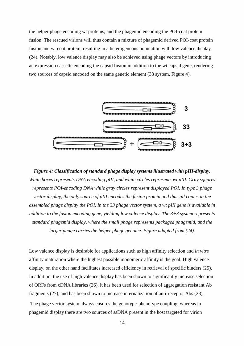
two sources of capsid encoded on the same genetic element (33 system, Figure 4).
Figure 4: Classification of standard phage display systems illustrated with pIII-display.
White boxes represents DNA encoding pIII, and white circles represents wt pIII. Gray squares
represents POI-encoding DNA while gray circles represent displayed POI. In type 3 phage
vector display, the only source of pIII encodes the fusion protein and thus all copies in the
assembled phage display the POI. In the 33 phage vector system, a wt pIII gene is available in
addition to the fusion encoding gene, yielding low valence display. The 3+3 system represents
standard phagemid display, where the small phage represents packaged phagemid, and the
larger phage carries the helper phage genome. Figure adapted from (24).
Low valence display is desirable for applications such as high affinity selection and in vitro
affinity maturation where the highest possible monomeric affinity is the goal. High valence
display, on the other hand facilitates increased efficiency in retrieval of specific binders (25).
In addition, the use of high valence display has been shown to significantly increase selection
of ORFs from cDNA libraries (26), it has been used for selection of aggregation resistant Ab
fragments (27), and has been shown to increase internalization of anti-receptor Abs (28).
The phage vector system always ensures the genotype-phenotype coupling, whereas in
phagemid display there are two sources of ssDNA present in the host targeted for virion

15
encapsulation; the phagemid, and the helper phage genome. During virion production in this
system, it is vital that the POI encoding phagemid, and not the helper phage genome, is
preferentially packaged in the phage progeny to ensure the genotype-phenotype link. This can
be monitored using the different antibiotic markers carried within the two vectors, and is
usually given as a ratio, where a ratio above one denotes a surplus of phagemid-carrying
particles. If the ratio drops below one, the majority of phage particles carry the helper phage
genome which may cause loss of clones during selection. Such situations may arise in e.g. the
initial selection round when highly diverse libraries are used in which only a limited
complexity (that is the number each unique clone is represented) input is feasible (29).
The most prominent advantage of using a phagemid instead of a phage genome vector is the
increased genomic stability when larger fusion are selected (30). Phage vectors have a
tendency to delete unnecessary DNA as a result of the selective growth advantage a smaller
phage has over a larger one. An additional major advantage of phagemid display is the high
transformation efficiency due to the small phagemid size (3-5kb) compared to phage vectors
(9-10 kb). Phagemids are constructed to facilitate quick exchange of the POI, e.g. by the use
of restriction cassettes (Figure 5). However, in standard phagemid-based systems, only about
10% of the phage population actually carries a coat protein fusion (31). This may lead to loss
of clones in the early rounds of selection as there may be no more than 100-1000 clones of
each individual specificity in the library. In high valence display however, as the only
available source of capsid is the phagemid encoded fusion, all produced phage will display the
peptide.
A more recent feature of pIII phagemid display is the development of modified helper phages
that enables high valence phagemid display on pIII by knocking down the wt helper phage
encoded pIII (32). These systems combine the features of a phage vector and the phagemid, in
that the high transformation efficiency and ease of use from the phagemids are retained, while
also keeping the selection capabilities of the phage vector system. By using a helper phage
incapable of providing wt pIII for phagemid rescue, one may in early rounds of panning first
select for specificity. In subsequent panning rounds one may then switch to classical helper
phages, rendering low valence display to select for affinity among the specific binders
isolated in the initial panning.

16
1.2.4 Capsid proteins for display
While polypeptides have been displayed on all five capsid proteins, only display on pIII or
pVIII have gained widespread use. pVIII display (used as an 8-system) enables presentation
of hundreds to thousands of peptides on a single phage depending on display system, but there
are size limitations that restrict its use to small peptide fusions (33,34). This constraint may be
alleviated to a certain extent by use of an 88 or 8+8 system (24).The minor coat protein pIII
tolerates large fusions and yields high display levels (i.e. amount of functional POI
displayed). However, pIII is responsible for host infection and fusions to this protein
unpredictably affects infectivity, especially in high valence formats (32). pIII fusions may
also affect phage propagation and cause repertoire bias (35,36), in that various constructs
behave differently, such as affecting host cell viability in a way that may give certain clones
unfavorable growth (37). Another effect from pIII fusions is large differences in the
phagemid/phage genome packaging ratio, where some clones are lost during selection
because of preferential packaging of helper phage. Furthermore, many pIII systems, and in
particular for scFv display, suffer from heterogeneous oligovalency effects (i.e. a
subpopulation carrying more than one POI) masking the true monomeric affinity. This effect
may cause the retrieval of low affinity binders due to avidity effects during selection on
polyvalent Ag (38).
Display on pVII and pIX were previously believed to be dependent on addition of an N-
terminal signal sequence to the POI for successful display. We recently reported that display
of folded domains on pVII and pIX is actually not dependent on a signal sequence (29). We
observed reduced host cell stress during growth, and surprisingly strong preferential
packaging of the phagemid independent of the construct used, which is likely to greatly
reduce the repertoire bias during library selection. Both pVII and pIX outperformed the pIII
counterpart in affinity selection even though display levels on these proteins were found to be
lower than on pIII. These observations make pVII and pIX highly attractive alternative
candidates to pIII for future use in selection of novel binder. In fact, the lower display levels
observed for pVII and pIX may not be a disadvantage in such selections as the low display
may be result in true monovalence, meaning that no virions carry more than one copy of the
POI, allowing for selection on intrinsic affinity only. Combined with the absence of pIII
immunity effects (36), and heterogeneous signal sequence cleavage (33,39,40) that may cause

17
random library repertoire bias, pVII and pIX indeed seems an attractive alternative to pIII
display.

18
2. Aim of the project
High valence phagemid display on pIII has become an important improvement of the phage
display technology as it combines the selection capabilities of the type 3 vector display
system with the ease of use of the 3+3 phagemid display system. However, there are several
issues that decrease the overall efficiency of this application in its present form, such as lower
end titers and severely reduced infectivity (32), in addition to the issues already present in the
pIII display system. We have recently reported that low valence phagemid display on pIX
have clear advantages over their pIII counterpart(29) in selection. However, high diversity in
selected repertoires is intimately coupled to high display levels during initial selection rounds,
meaning that a way to display peptides on all copies of pIX will broaden the area of
application of pIX phagemid display. A previous report indicated that display on pVII and
pIX were dependent on wt complementation, which implies that high valence display is
unachievable (41). However, we and others have recently shown that all copies of pVII in the
phage genome (corresponding to a type 7 phage vector system) may indeed tolerate a fusion
rendering high valence display (42,43). Due to the overlapping reading frames of pVII and
pIX , such N-terminal modifications in genomic pIX is however not straightforward, and may
give rise to polar effects (3). Thus, the aim of the project is to establish a novel helper phage
to facilitate high valence pIX display as a type 9+9 phagemid display system. The thesis
contains a thorough characterization of the new helper phage with regard to performance in
standard phagemid rescue, as well as in standard assays used for discovery of novel binders in
phage display technology.

19
3. Materials and methods
3.1 General Material
3.1.1 Bacteria
The E.coli XL1-Blue strain (recA1 endA1 gyrA96 thi-1 hsdR17 supE44 relA1 lac [F´ proAB
lacIqZ∆M15 Tn10 (Tetr)] was used for phage amplification and titration.
The E.coli TOP10 F’ strain (F'[lacIq Tn10(tet
R)] mcrA Δ(mrr-hsdRMS-mcrBC)
φ80lacZΔM15 ΔlacX74 deoR nupG recA1 araD139 Δ(ara-leu)7697 galU galK rpsL(StrR)
endA1 λ- (Invitrogen) was used for amplification of multivalent pIX phage
3.1.2 Plasmids
The pGALD9ΔL (GenBank accession no. HQ528249) phagemid was used for pIX display,
and the pSEX81 phagemid (GenBank accession no. Y14584) was used for pIII-display. Both
phagemids have been previously described in (29) and (44) respectively, and the POI
expression cassettes are illustrated in Figure 5.
Figure 5: Schematic drawing of pIII and pIX display phagemid. pSEX81 (A) is used for
display on pIII, and pGALD9ΔL (B) is used for display on pIX. There are two differences
between these phagemids, 1) the phage coat protein encoded, and 2) pSEX81 encodes a
signal sequence for periplasmic targeting (pelB) which is not included in pGALD9ΔL. The
POI is introduced using cassette exchange on NcoI/HindIII and MluI/NotI, creating a
continuous open reading frame (ORF). The two cassettes are connected by a synthetic linker,
containing the mAB Yol1/34 tubulin epitope (bold underlined). The POI is fused to the coat
protein through a 9 aa spacer, containing a trypsin protease site (KDIR bold underlined).
Abbreviations: lacPO, lac promoter; SD, Shine-Dalgarno sequence; pelB, signal sequence of
bacterial pectate lyase; t, T7 transcriptional terminator. Figure adapted from(29).

20
3.1.3 Helper phages
Two different commercially available helper phages were used in this work; M13K07 (45)
(Amersham Bioscience, Uppsala, Sweden) and Hyperphage (46) (Progen Biotechnik GmbH,
Heidelberg Germany).
Hyperphage has gene III deleted from its genome, hence the phagemid derived pIII fusion is
the sole source of pIII for phage assembly during phagemid rescue, which results in
multivalent display of the capsid fusion
M13K07 is referred to as wt as it has its complete genome from which the previous two are
derived. And thus provides lower valence display of fusion proteins.
In addition to these, the novel helper phage DeltaPhage was used in this work. Details
regarding the construction of this helper phage are included in the enclosed manuscript. The
construction was performed by Geir Åge Løset
3.2 Standard reagents
3.2.1 Antigens
The hapten NIP (4-hydroxy-3-iodo-5-nitrophenylacetate) conjugated to bovine serum albumin
(BSA) was prepared essentially as described (47) and was a kind gift from Dr. Terje
Michaelsen (Norwegian Institute of Public Health). The hapten 4-Ethoxymethylene-2-phenyl-
2-oxazolin-5-one (phOx) conjugated to BSA was prepared essentially as described (48). In
short, E-phOx (sigma) was dissolved in DMSO and incubated in a shaker over night (ON) at
room temperature (RT) followed by centrifugation at 3000g/5min/RT. The supernatant
volume was adjusted to 2.5 ml with 1x PBS pH 7.4, and filtered through a PD-10 desalting
column (Amersham Biosciences, Uppsala, Sweden) containing sephadex G-25. The sample
was then dialyzed against 4 L of 1x PBS pH 7.4 in a slide-a-lyzer dialysis cassette (Thermo
Scientific, Rockford, U.S.A.) with 10Kd cut-off, and stored at 4°C.

21
3.2.2 Antibodies
The anti-M13-HRP Ab was purchased from GE Healthcare (Uppsala, Sweden),
the rabbit anti-human Lλ chain Ab was from DakoCytomation (Glostrup, Denmark), the
donkey anti-rabbit HRP Ab was purchased from GE Healthcare (Uppsala, Sweden),
the mouse anti pIII Ab was purchased from MoBiTec (Göttingen, Germany) and
the Sheep Anti-Mouse HRP Ab was purchased from Amersham Bioscience (Uppsala,
Sweden) respectively.
The biotinylated mAbs GB113(49) and AB10 (Frigstad et. al. submitted) were kind gifts from
Professor Bjarne Bogen (Institute of Immunology, Oslo University Hospital)
3.3 Phage display protocols
3.3.1 Production of helper phage
E. coli XL1-Blue harboring either the DeltaPhage or the M13K07 genome was inoculated in 2
X YT containing 50µg/ml kanamycin, and incubated at 37°C ON with rigorous agitation. The
cells were pelleted by centrifugation, and the supernatant was sterile filtered into fresh 50 ml
tubes using 0.2 µm filters. The virions were purified and concentrated by PEG/NaCl
precipitation as described (50). Virion concentration was measured either by optical density at
A268 nm (51) or by infectious titration as described in 3.3.3.
3.3.2 Phagemid rescue
Phage propagation and purification was performed essentially as described in (44). Briefly,
phagemid-carrying E.coli XL1-Blue or TOP10F’ were inoculated in 2x YT-medium
supplemented with TAG (30µg/ml Tetracycline, 100µg/ml Ampicillin and 100mM Glucose)
and incubated at 37°C/250rpm/overnight (ON). The following day, the cultures were re-
inoculated to an OD of A600nm 0.025 in 50 ml 2x YT-TAG and incubated at 37°C/250rpm

22
until A600nm 0.1 was reached. At this point, super infection of the phagemid-harboring bacteria
with the appropriate helper phages according to phagemid and/or strain was then carried out
at a multiplicity of infection (MOI) 10. The cultures were then incubated at
37°C/80rpm/30mins, followed by incubation at 37°C/250 rpm/30 mins. The cells were then
pelleted by centrifugation, the supernatants discarded, and the pellets were resuspended in 50
ml 2x YT supplemented with 100µg/ml Ampicillin and 50µg/ml Kanamycin. The cultures
were incubated at 30°C/250rpm/ON. The following day, supernatants were harvested by
centrifugation and sterile filtration, followed by phage precipitation with 1/10 volume of 20%
polyethylene glycol (PEG) 8000 (Sigma Aldrich, Oslo, Norway) and 2.5M NaCl, and
incubated ON in ice-slurry at 4°C. The precipitated phage were pelleted by centrifugation at
4000-g/30 mins/4°C, and resuspended with 1ml of 1x PBS pH 7.4. Remaining cell debris was
removed by centrifugation at 16000-g/5 mins/ RT, and supernatants were transferred to new
tubes and stored as phage stocks.
3.3.3 Infection titration of rescued phages.
Phage stocks rescued with M13K07 and Deltaphage were titrated by standard spot titration
(52) while phages rescued with Hyperphage were titrated with a modified titration protocol
(53). In short, E.coli XL1-Blue was inoculated from glycerol stocks in 2xYT supplemented
with 30µg/ml Tetracycline and incubated at 37°C/250rpm/ON. The culture was the then re-
inoculated to an OD of A600nm=0.025 and grown to A600nm 0.4-0.8. A dilution series of phages
was prepared in 1x PBS, according to table 1. 10µl of each individual dilution was used to
infect 190µl of XL1-Blue culture and incubated at 37°C/45mins with agitation. 3µl of
infected culture were then spotted on Protran BA85/20 0.45µm nitrocellulose blotting
membrane with 5mm grid (Sleicher & Schell, Kent, UK) laid on top of LA-Amp and LA-Kan
plates (LB-agar plates supplemented with 100µg/ml Ampicillin or 50µg/ml Kanamycin
respectively) The plates were incubated at 37°C/ON and colony forming units (cfu)/ml were
calculated using the formula (cfu x dilution-1
x 20 x 1000)/3. Cfu/ml calculated from LA-Amp
plates equals number of phagemid-carrying phage in the solution, while cfu/ml calculated
from LA-Kan plates equals number of helper phage genome-carrying phage.

23
Dilution Gradient 1x PBS Transfer to tube
A 10-2 495 µl* 5 µl phage from stock
B 10-4 495 µl 5 µl from A
C 10-5 450 µl 50 µl from B
D 10-6
450 µl 50 µl from C
E 10-7 450 µl 50 µl from D
F 10-8
450µl 50µl from E
G 10-9
450µl 50µl from F
*for phages rescued with Hyperphage; 250µl
Trypsin/EDTA mix+245µl diluent
Table 1: Serial dilutions for infectious spot titration of phage preparations
3.4 Analysis protocols
3.4.1 Miniprep analysis of helper phage dsDNA
E.coli XL1-Blue cells were transduced with either M13K07 or Deltaphage at a MOI of 0.1,
plated on LA-Kan plates, and incubated at 37°C/ON. Single colonies were inoculated in
2xYT supplemented with 50µg/ml Kan, and grown to mid-log. The cultures were normalized
based on density (A600nm) and the phage dsDNA was isolated using the Wizard Plus miniprep
kit (Promega) according to manufacturers protocol. Equal amount of prep were visualized on
an agarose gel.

24
3.4.2 Western blotting
The samples (normalized with respect to infectious titers) were mixed with 4x XT sample
buffer. The samples were boiled for 5 min/95C, briefly centrifuged and loaded onto a 4-12%
Bis-Tris XT Precast gel (Bio-Rad). The gel ran at 140V (0.13A/20W)/100 min
Semi-dry blotting
The gels were put in Transfer Buffer (Tris/glycine/methanol) for 10 min. Meanwhile, an
appropriate sized Immobilon™-P membrane was cut and put in 100% methanol for 15-20 sec.
and immediately soaked in dH2O for 5 min before transferred to Transfer buffer for 5 min. An
extra thick filter paper (Bio-Rad) was wet in Transfer buffer and put on the Blot apparatus.
The membrane was then put on top of the filter paper, followed by the gel. An additional filter
paper was wet in Transfer buffer and put on top of the gel, and trapped air was removed using
a 50 ml tube. The blotting was allowed to occur for 30 min/25V. The blot was Blocked
ON/4C in PBSTM (PBST/4% w/v skimmed milk) with agitation.
Immunodetection of pIII
The blocked membrane was flushed once in PBST. The blot was then incubated for 1.5h/RT
with anti-pIII mAb diluted 1:5000 in PBSTM, followed by washing 3x 5 min with 50 ml
PBST. The blot was then Incubated 1.5h/RT with -mouse-HRP Ab (Amersham) diluted
1:10,000 in PBSTM. The blot was then washed 3x 5 min with PBST and 1x 10 min in PBS.
The reservoir was then completely emptied followed by the addition of ~ 10 ml SuperSignal
West Pico enhanced Chemiluminescent (ECL) solution (Thermo Scientific, Rockford,
U.S.A.), and 5 min/RT incubation. Excess liquid was drained from the membrane by use of a
paper towel. The membrane was laid on top of a transparency sheet on a film cassette, and a
second transparency sheet was overlain the membrane, and surplus ECL solution wiped off
using a paper towel.
The blot was developed on Hyperfilm, using <10 seconds exposure.

25
Immunodetection of scFv anti-phOx
The blot was incubated for 1.5h/RT, with the rabbit anti-human λ-light chain 1:5000 in
PBSTM. After washing 3x 5 mins with PBST, the blot was incubated 1.5h/RT with Donkey
α-rabbit IgG-HRP 1:10000 in PBST. The blot was washed 3x 5 min with PBST and 1x 10
min in PBS.
Development as above. Exposure time: 10 min.
3.4.3 ELISA (Phage dilutions)
MaxisorpTM
microtiter plates (NUNC, Wiesbaden, Germany) were coated with 10µg/ml
phOx-BSA or NIP-BSA at 4°C/ON. The plates were washed 3x with PBST, and blocked with
4%w/v skimmed milk powder in 1x PBS for 1h at RT. 100µl of a normalized 10-fold dilution
series were added to the plates and incubated 1.5h/RT. Bound phages were detected with anti-
M13-HRP (1:5000) in PBST for 1h/RT. The wells were developed with TMB soluble
substrate (Calbiochem, Darmstadt, Germany) by adding 100µl/well. The reaction was stopped
after 30min. by adding 100µl 1M HCl to the wells. The absorbance was measured at A450nm
3.4.4 ELISA (Antigen dilutions)
MaxisorpTM
microtiter plates were coated with a 10x dilution series of Ag starting at 10µg/ml.
after washing and blocking as described above, 100µl of 1x10¹¹
cfuamp
/ml of the individual
phage samples were added to the wells. Detection, development and measurement were done
as described above.
3.4.5 Flow staining of T cell Clone 4B2A1
2x 10⁵ cells/well were distributed into a V-shaped 96-well dish. The wells were then filled to
a total volume of 250 µl by adding 187µl/well with PBS/5% BSA. The plate was centrifuged
at 300-g/5 min/RT and the supernatant discarded.

26
Phage binding
The samples (50 µl/well) were added to the proper wells, whereas remaining wells (that
contained cells for mAb staining) received an equal volume of PBS/5% BSA only. The pellets
were then re-suspended by use of a multi-channel pipette and the dish incubated 1h/4˚C with
shaking
Ab binding
The wells were filled to a total volume of 250 µl by adding 200 µl/well with PBS/5% BSA.
The plate was centrifuged at 300-g/5 min/RT and the supernatant discarded.
All phage-containing wells then received a volume of 50 µl anti-fd (10 µg/ml: 2.5 ml PBS/5%
BSA + 6.6 µl Ab).
One well then received a volume of 50 µl mAb AB10 as a negative control (10 µg/ml: 99 µl
PBS/5% BSA + 1 µl Ab).
One well then received a volume of 50 µl mAb GB113 as positive control (10 µg/ml: 99 µl
PBS/5% BSA + 1 µl Ab), which corresponds to 67 nM
In addition, a positive control that is equimolar to phage input was added. The phage input
was 2.5x 10¹⁰
phages/well, which corresponds to 830pM.
The pellets were then re-suspended by use of a multi-channel pipette and the dish incubated
30 min/4˚C with shaking.
SA-PE binding
The wells were filled to a total volume of 250 µl by adding 200µl/well with PBS/5% BSA.
The plate was centrifuged at 300-g/5 min/RT and the supernatant discarded.
Volumes of 50 µl/well SA-PE (2 µg/ml; 2.9 ml PBS/5% BSA + 11 µl SA-PE) were added.
The pellets were then re-suspended by use of a multi-channel pipette and the dish incubated
15 min/4˚C with shaking. The plate was covered to minimized light damage to the
fluorophore.

27
Fixation
The wells were filled to a total volume of 250 µl by adding 200µl/well with PBS/5% BSA.
The plate was centrifuged at 300-g/5 min/RT and the supernatant discarded.
Volumes of 200 µl/well 2% PFA (freshly prepared by diluting the stock in PBS) were added,
the pellets re-suspended and the plate put in the dark at 4˚C until flow analysis
The samples were analyzed using a Becton Dickinson FACScalibur. The data analysis was
done using the CellQuest Pro (v5.2.1) software (BD Biosciences). This experiment was
performed by Geir Åge Løset and Terje Frigstad
3.4.6 Spiked Panning
A single round of panning was performed. 1µg/ml phOx-BSA and NIP-BSA was
immobilized on MaxisorpTM
microtiter plates. The plates were blocked with 4% skim milk
1xPBS for 1h/RT. Samples were prepared by mixing 4x10³ Ag specific phages with 4x10
10
phage of irrelevant specificity, and adjusting the volume to 200µl by addition of 1xPBS.
These samples were mixed 50:50 with PBSM and left for pre-blocking for 30 mins/RT.
Volumes of 100µl of the prepared samples were added to individual wells and incubated
2h/RT with agitation. The plates were washed 5x with PBST and 4x with dH2O and bound
phages were eluted by addition of 100µl Trypsin/EDTA mixture (BioWhittaker, Walkersville,
MD, USA) and incubation for 10min/RT. The Eluates were used to infect log-phase E.coli
XL1-Blue (or TOP10F’ for the Deltaphage samples). The infected cultures were incubated at
37°C/80rpm/30min followed by 37°/240rpm/30min. Volumes of 1ml from each culture were
transferred to 2ml tubes and spread on LA-Amp dishes for subsequent output calculations.
The remaining cultures were super infected, and phagemid rescue was done as described in
3.3.2 with the exception of PEG-precipitation.

28
3.4.7 Single colony screening of panning output.
Single clone Phagemid rescue
Single colonies were picked from the panning output plates and grown ON in 400µl YT-TAG
in a MegaBlock 96 well 2.2ml plate (Sarstedt, Nümbrecht, Germany) in a titramax at
37°C/900rpm. The colonies were re-inoculated by transferring 10µl of culture to fresh
identical deep-well plates and incubated 3h/37°C/900rpm. The cultures were super infected
with the appropriate helper phages and incubated 30min./37°C/no agitation followed by
30min/37°C/900rpm. The cultures were pelleted by centrifugation, the supernatant discarded
and the pellets were re-suspended in 2x YT supplemented with 100µg/ml Ampicillin and
50µg/ml Kanamycin, and incubated ON/37°C/900rpm. The bacteria were pelleted by
centrifugation and the supernatant used in a phage capture ELISA.
Single clone ELISA
Maxisorp plates were coated with 10µg/ml phOx-BSA or NIP-BSA at 4°C/ON. The plates
were blocked with 4% PBSM. Supernatants from the deep-well plates were added to
corresponding wells on plates with both Ags. Phage detection was done as in 3.3.3.
3.4.8 Establishment of protocol for PCR-screening of panning output.
To provide a template for the PCR, E.coli harboring scFv anti-phOx or –NIP phagemid was
inoculated in 2xYT supplemented with 30µg/ml tetracycline from glycerol stock and
incubated at ON/37°C/240rpm.
The primers were ordered from Eurofins MWG operon.
ColE1_frwd: 5’- TGGATAACCGTATTACCGC-3’ Tm 52C
phOx_VH_R: 5’- CGCACCCAGGTGATGC-3’ Tm 54C
NIP_VL_R: 5’- GAGTGTGACTGTTTCACCAG-3’ Tm 53C

29
Annealing temperatures were calculated using OligoCalc
(http://www.basic.northwestern.edu/biotools/oligocalc.html)
Amplification was done with Phusion HotStart DNA polymerase (Finnzymes)
The PCR reactions were set up according to table 2 and 1µl of culture was added to each
reaction (anti-phOx-template was added to tubes 1-4, and anti-NIP template added to 5-8).
Tube no 1 2 3 4 5 6 7 8
dH2O 32.5 µl 34.5 µl 34.5 µl 32.5 µl 32.5 µl 34.5 µl 34.5 µl 32.5 µl
dNTP 2 µl 2 µl 2 µl 2 µl 2 µl 2 µl 2 µl 2 µl
ColE1_frwd 2 µl 2 µl 2 µl 2 µl 2 µl 2 µl
phOx_VH_R primer 2 µl 2 µl 2 µl
NIP_VL_R primer 2 µl 2 µl 2 µl
Buffer 10µl 10µl 10µl 10µl 10µl 10µl 10µl 10µl
Phusion HS DNA pol 0.5 µl 0.5 µl 0.5 µl 0.5 µl 0.5 µl 0.5 µl 0.5 µl 0.5 µl
Total 50 µl 50 µl 50 µl 50 µl 50 µl 50 µl 50 µl 50 µl
Table 2: PCR reaction setup

30
To establish the optimal PCR-reaction, different variations of the PCR-cycle were used.
The initial PCR program is given in table 3
# Cycles Process Temperature Duration
1x
Lysis and
denaturation 98°C 1 min.
33x Denaturation 98°C 10 sec.
Annealing 54°C 15 sec.
Synthesis 72°C 15 sec.
1x Elongation 72°C 5 min.
Table 3: PCR program used for amplification of anti-phOx or –NIP DNA
Values for annealing temperature, synthesis duration and number of cycles were modified to
give the optimal PCR-yield.
The PCR-products were then visualized by agarose gel electrophoresis.

31
4. Manuscript for “Nucleic Acid Research”:
DeltaPhage – A novel helper phage for high valence pIX-display
Introduction
Phage display is a powerful technology for discovery of novel binding peptides and folded
domains. The technology has especially gained widespread use in the discovery of novel
antibody (Ab) specificities (15). By creating libraries of polypeptides fused to coat proteins on
a filamentous phage (Figure1), one may simultaneously screen over 1010
unique sequences for
binding to a specific antigen (Ag). Following selection, in vitro affinity maturation of the
selected binders is often carried out, from which Ag binding fragments (Fab), or single chain
variable fragments (scFv) with affinities in the low picomolar range may be retrieved. In the
case of Ab discovery, one may reformat the initially displayed Fab or scFv into complete
human monoclonal Abs (mAbs). What makes phage display a viable technology for selection
of novel binding peptides is the so called phenotype-genotype coupling in the phage particle.
During phage assembly in a host cell, the peptide coat protein fusion is incorporated in the
growing phage particle. The genetic information encoding the fusion protein, and therefore
the selected peptide, (in form of an ssDNA molecule), is packaged in the same particle. In
short, the protein of interest (POI) displayed on a capsid protein of a phage is encoded in the
ssDNA harbored in the phage particle. This allows rapid characterization of the selected
peptide using standard molecular biology techniques.

32
Figure 1: Schematic drawing of the filamentous phage. The filamentous phage M13 consists
of five structural proteins that coat a single stranded DNA (ssDNA) molecule. The major coat
protein pVIII is by far the most abundant with approximately 2700 copies covering the length
of the wild type (wt) phage. The phage particle is capped at one end by 3-5 copies of the
minor coat proteins pIII and pVI, and at the other end by pVII and pIX. All capsid proteins
are located in the inner membrane prior to virion assembly (3).
To display a polypeptide, the POI is normally placed in-frame between the N-terminal signal
sequence and coat proteins pIII or pVIII. POI-coat protein fusions may be encoded either in a
complete phage genome (phage vector display), or on a phagemid (phagemid display). As the
phagemid encodes the capsid protein only, a helper phage is used to provide the genetic
information required for phage production (Figure 2). In the case of phage vector display, all
components required for virion assembly and production is provided by the vector itself, and
hence there is only one genetic source of coat proteins. Thus, in phage vectors where an
endogenous coat protein is modified with a fusion, all copies of the coat protein will carry the
POI, yielding high valence display. In phagemid display, there are two sources of coat
proteins; the helper phage encoding wt proteins, and the phagemid encoding the POI-coat
protein fusion. The rescued virions will thus contain a mixture of phagemid derived POI-coat
protein fusion and wt coat protein, resulting in a heterogeneous population with low valence
display (24). Notably, low valence display may also be achieved using phage vectors by
introducing an expression cassette encoding the capsid fusion in addition to the wt capsid
gene, rendering two sources of capsid encoded on the same genetic element.

33
Figure 2: Schematic illustration of the phagemid rescue using a standard phagemid system.
The phagemid carries an antibiotic (e.g. ampicillin) resistance marker and hence can be
selectively propagated as a plasmid in E.coli. For virion assembly to occur, the host is super-
infected with a helper phage containing all elements necessary for new virion assembly. The
helper phage has a defect in the replicative system, and single strand conversion, while the
phagemid is a high copy-number plasmid, harboring a wt f1 ori which most often leads to a
preferential phagemid packaging into the virion progeny upon phagemid rescue with the
helper phage.
Low valence display is desirable for applications such as high affinity selection and in vitro
affinity maturation where the highest possible monomeric affinity is the goal. High valence
display, on the other hand facilitates increased efficiency in retrieval of specific binders (25).
In addition, the use of high valence display has been shown to significantly increase selection
of ORFs from cDNA libraries (26), it has been used for selection of aggregation resistant Ab
fragments (27), and has been shown to increase internalization of anti-receptor Abs (28).
The phage vector system always ensures the genotype-phenotype coupling, whereas in
phagemid display there are two sources of ssDNA present in the host targeted for virion
encapsulation; the phagemid, and the helper phage genome. During virion production in this
system, it is vital that the POI encoding phagemid, and not the helper phage genome, is
preferentially packaged in the phage progeny to ensure the genotype-phenotype link. This can
be monitored using the different antibiotic markers carried on the two vectors, and is usually
given as a ratio, where a ratio above one denotes a surplus of phagemid-carrying particles. If
the ratio drops below one, the majority of phage particles carry the helper phage genome

34
which may cause loss of clones during selection. Such situations may arise in e.g. the initial
selection round when highly diverse libraries are used in which only a limited complexity
(that is the number each unique clone is represented) input is feasible (29).
The most prominent advantage of using a phagemid instead of a phage genome vector is the
increased genomic stability when larger fusion are selected (30). An additional major
advantage of phagemid display is the high transformation efficiency due to the phagemid size
(3-5kb) compared to phage vectors (9-10 kb). Phagemids are constructed to facilitate quick
exchange of the POI, e.g. by the use of restriction cassettes (Figure 3). However, in standard
phagemid-based systems, only about 10% of the phage population actually carries a coat
protein fusion (31). This may lead to loss of clones in the early rounds of selection as there
may be no more than 100-1000 clones of each specificity in the library. In high valence
display however, as the only available source of pIII (or pVIII depending on the system used)
is the phagemid encoded fusion, all produced phage will display the peptide.
While polypeptides have been displayed on all five capsid proteins, only display on pIII or
pVIII have gained widespread use. pVIII display enables presentation of hundreds to
thousands of peptides on a single phage depending on display system, but there are size
limitations that restrict its use to small peptide fusions (33,34). The minor coat protein pIII
tolerates large fusions and yields high display levels (i.e. amount of functional POI
displayed), however, pIII is responsible for host infection, and fusions to this protein affects
infectivity, especially in high valence formats (53). pIII fusions also affect phage propagation
and cause repertoire bias (35,36), in that various constructs behave differently, such as
affecting host cell viability in a way that may give certain clones unfavorable growth (24).
Another effect from pIII fusions is large differences in the phagemid/phage genome
packaging ratio, where some clones are lost during selection because of preferential
packaging of helper phage. Furthermore, many pIII systems, and in particular for scFv
display, suffer from heterogeneous oligovalency effects (i.e. a subpopulation carrying more
than one POI) masking the true monomeric affinity. This effect may cause the retrieval of low
affinity binders due to avidity effects during selection on polyvalent Ag (38).
A more recent feature of pIII phagemid display is the development of modified helper phages
that enables high valence phagemid display on pIII by knocking down the wt helper phage
encoded pIII (32). This system combines the features of phage vector and phagemid, in that
the high transformation efficiency and ease of use from the phagemids are retained, while also
35
keeping the selection capabilities of the phage vector system. By using a helper phage
incapable of providing wt pIII during phagemid rescue, one may first select for specificity. In
subsequent panning rounds one may then switch to classical helper phages, rendering low
valence display to select for affinity among the specific binders isolated in the initial panning.
In the wt phage, only pIII and pVIII are synthesized with classical N-terminal signal
sequences for periplasmic translocation, while genes for pVI, pVII and pIX do not contain
any known signal sequence motifs. Thus, all three proteins are translocated by a hitherto
unknown mechanism (4). Display on pVII and pIX was previously believed to be dependent
on addition of an N-terminal signal sequence for successful heterologous POI for display.
Recently, we reported that display of folded domains on pVII and pIX is actually not
dependent on a signal sequence (29). We observed reduced host cell stress during growth,
and surprisingly strong preferential packaging of phagemid, independent of construct used,
which is likely to greatly reduce the repertoire bias during library selection. Both pVII and
pIX outperformed the pIII counterpart in affinity selection even though display levels on these
proteins were found to be lower than on pIII. These observations make pVII and pIX highly
attractive alternative candidates to pIII for future use in selection of novel binder. In fact, the
lower display levels observed for pVII and pIX may not be a disadvantage in such selections
as the low display may be attributed to true monovalence, meaning that no virions carry more
than one copy of the POI, allowing for selection on intrinsic affinity only. Combined with the
absence of pIII immunity effects (36), and heterogeneous signal sequence cleavage (33,39,40)
that may cause random library repertoire bias, pVII and pIX indeed seems an attractive
alternative to pIII display.
A previous report indicated that display on pVII and pIX is dependent on wt
complementation, such that high valence display was unachievable (41). However, we and
others have recently shown that all copies of pVII in the phage genome may indeed tolerate a
fusion rendering high valence display (42,43). Due to the overlapping reading frames of pVII
and pIX , similar modifications in genomic pIX is however not straightforward, and may give
rise to polar effects (3).
We demonstrate here a novel helper phage that indeed allows for high valence POI display on
pIX without any deleterious effects on virion titer or phenotype except for markedly increased
Ag reactivity due to functional affinity effects. Virions produced with this helper phage also
perform as well as high valence pIII display virions in selection in a direct side-by-side
comparison. Phagemid rescue with helper phages for high valence pIII-display usually gives

36
10-1000-fold lower end titers than rescue with “standard” helper phages such as M13K07
(32). We observe wt end titers with our novel helper phage, in conjunction with very high
phagemid to helper phage packaging ratios, no apparent clone dependent host bias and
absolutely no reduction in infectivity, as no modifications are done to the infection-mediating
pIII.
Materials and Methods
Standard reagents
Bovine serum albumin (BSA) was from Sigma-Aldrich (Oslo, Norway). Trypsin/EDTA was
purchased from BioWhittaker (Lonza Group Ltd., Visp, Switzerland).Restriction Enzymes
(RE) and T4 DNA ligase were purchased from New England Biolabs (Ipswich, MA, USA).
Pfu Turbo DNA and Phusion DNA polymerases were purchased from Stratagene (LaJolla,
CA, USA) and Sigma-Aldrich (Norway), respectively. DNA oligonucleotides were purchased
from Eurofins MWG operon (Ebersberg, Germany). The anti M13-HRP Ab was purchased
from GE Healthcare (Uppsala, Sweden). The biotinylated mAbs GB113 and AB10 were kind
gifts from Professor Bjarne Bogen (Institute of Immunology, Oslo University Hospital). The
haptens NIP (4-hydroxy-3-iodo-5-nitrophenylacetate) and 4-Ethoxymethylene-2-phenyl-2-
oxazolin-5-one (phOx) conjugated to BSA were prepared essentially as described (47,48).
Plasmids
The pSEX81 phagemid (GenBank accession no: Y14584), described in (44), was kindly
provided by Affitech Research AS (Oslo, Norway). The pGALD9ΔL phagemid (GenBank
accession no: HQ528249) has been previously described (29). The phagemids are illustrated
in Figure 3.

37
Figure 3: Schematic drawing of pIII and pIX display phagemid. PSEX81 (A) is used for
display on pIII and pGALD9ΔL (B) is used for display on pIX. There are two differences
between these phagemids, 1) the phage coat protein encoded, and 2) pSEX81 encodes a
signal sequence for periplasmic targeting (pelB) which is not included in pGALD9ΔL. The
POI is introduced using cassette exchange on NcoI/HindIII and MluI/NotI, creating a
continuous open reading frame (ORF). The two cassettes are connected by a synthetic linker,
containing the mAB Yol1/34 tubulin epitope (bold underlined). The POI is fused to the coat
protein through a 9 aa spacer, containing a trypsin protease site (KDIR bold underlined).
Abbreviations: lacPO, lac promoter; SD, Shine-Dalgarno sequence; pelB, signal sequence of
bacterial pectate lyase; t, T7 transcriptional terminator. Figure adapted from(29).
Helper phages and Bacterial strains
M13K07 Helper phage was purchased from GE Healthcare Bio-sciences AB (Uppsala,
Sweden), and is often referred to as a wt helper phage, as no modifications have been done to
the protein coding genes(45). The Hyperphage helper phage was purchased from Progen
Biotechnik (Heidelberg, Germany). This helper phage has gene III deleted from its genome,
hence the phagemid derived pIII fusion is the sole source of pIII for phage assembly during
phagemid rescue, which results in high valence display of the phage coat fusion(46).
The suppressor strain E.coli XL1-Blue was purchased from Stratagene. E.coli MC1061 was a
kind gift from Dr. G. P. Smith (Division of Biological Sciences, University of Missouri,
USA), and the non-suppressor strain E.coli TOP10F’ was obtained from life technologies.

38
Mutagenesis of M13K07 helper phage genome
The amber mutations in the pIX ORF in M13K07 was introduced by QuikChange in vitro
mutagenesis according to the manufacturer’s protocol (Stratagene, LaJolla, CA, USA), using
the synthetic oligonucleotides (1: 5’-
GCTGGGGGTCAAAGATGAGTTAGAGCTAGGTTTTAGTGTATTCTTTCGC-3’ and 2:
5’-GCGAAAGAATACACTAAAACCTAGCTCTAACTCATCTTTGACCCCCAGC-3’
mismatching bases underlined), and introduced to E.coli MC1061cells by electroporation.
Successful introduction of the mutation was verified by sequencing. To ensure a clean vector
background, the modified pIX fragment was moved into the M13K07 genome using the
BsrGI and SnaBI RE sites (fig3), using standard techniques. The DNA constructs were
subsequently introduced into E.coli XL1-Blue by heat shock. E.coli TOP10F’ cells were then
transduced with virions produced in the XL1-Blue cells.
Mutant helper phage production
E.coli XL1-Blue harboring the DeltaPhage genome was inoculated in 2 X YT containing
50µg/ml kanamycin, and incubated at 37°C ON with rigorous agitation. The cells were
pelleted by centrifugation, and the supernatant was sterile filtered into fresh 50 ml tubes using
0.2 µm filters. The virions were purified and concentrated by PEG/NaCl precipitation as
described (50). Virion concentration was measured by optical density at A268 nm (51).
Phagemid rescue
Phagemid rescue from E.coli XL1-Blue and TOP10F’ was done essentially as described (44).
Where applicable, virions were purified and concentrated by PEG/NaCl precipitation from
50ml supernatant and resuspended in 1ml PBS, pH 7.4. Virion assembly was monitored by
infectious spot titration as described (52) with the exception of Hyperphage-rescued samples
that required a modified titration protocol(53).

39
Phage Capture ELISA
The various Ags (NIP-BSA and phOx-BSA) were adsorbed to MaxiSorpTM
microtiter plate
wells (Nunc, Roskilde, Denmark) in concentrations of 5µg/ml in PBS, pH 7.4 ON at 4°C. The
wells were blocked with PBSTM (PBS supplemented with 0.05% v/v Tween 20 and 4% w/v
Skim milk) for 1h at room temperature (RT). Virion preparations were then added and
allowed to react for 1.5 hours at RT before captured virions were detected with anti M13-HRP
(1:5,000) for 1 h. at RT. A 3x washing step with PBST (PBS supplemented with 0.05% v/v
Tween 20) was applied between each incubation step. The wells were developed with TMB
soluble (Merck KGaA, Darmstadt, Germany), and the reaction terminated by addition of 1M
HCl, equilibrated, and the absorbance read at A450nm.
Flow cytometry
4B2A1 T-cells were incubated with either scFv GB113 (anti TCR-4B2A1 specificity) phage
preparations, or the biotinylated GB113 mAb as positive control, both in standard
concentration (10 µg/ml, which corresponds to 67 nM), and equimolar to the phage samples
(830 pM). mAb AB10 was used as isotype control.
The phages were then incubated with a biotinylated rabbit Anti-fd, before all samples were
incubated with Streptavidin-PE. 2% PFA were added to all samples and the plate put in the
dark at 4°C.
The samples were analyzed using a Becton Dickinson FACScalibur. The data analysis was
done using the CellQuest Pro (v5.2.1) software (BD Biosciences).
Spiked phOx-/NIP BSA selection
Fresh virion samples were prepared; PEG precipitated and titrated as described. Ag specific
virions were then spiked into an unspecific virion background at a 1:107 level, giving a known
diversity of 107, corresponding to a medium sized combinatorial library. For phOx-BSA
selection the scFv anti-phOx was spiked into an scFv anti-NIP background, and vice versa.
The initial input was 1010
cfuampR
resulting in a complexity level of 103 for all the libraries.

40
For selection, Ag was immobilized in MaxisorpTM
microtiter plate wells using 100 µl volumes
of 1 µg/ml per well. Prior to panning, wells were blocked with PBSTM for 2 h at RT, before
100 µl of the respective pre-blocked (in PBSTM) virion preparations were added and allowed
to react for 1.5 h at RT with agitation. The wells were washed 9X with PBST followed by 5X
with dH2O using a microtiter washer before Ag bound virions were eluted by adding 100
µl/well Trypsin/EDTA for 10 min with agitation. The eluates were then used to infect log-
phase E.coli XL1-Blue (M13K07 and Hyperphage rescued samples) or TOP10F’ (Deltaphage
rescued samples) cultures in 9 ml of YT-TAG (2 X YT supplemented with 30 µg/ml
tetracycline, 100 µg/ml ampicillin and 0.1 M glucose), incubated for 15 min at 37°C with low
agitation, before 1ml of YT-TAG supplemented with the appropriate helper phage at MOI 10
was added. The incubation was continued for 15 min at 37°C with low agitation followed by
30 min at 37°C with rigorous agitation, followed by centrifugation at 4000g/10 min/RT. The
supernatants were discarded and the pellets gently resuspended in 2 X YT containing
100µg/ml ampicillin and 50 µg/ml kanamycin, and all samples were incubated at 30°C with
rigorous agitation until A600nm >1 (approx. 36-40 h). The samples were then centrifuged at
4000-g/10 min/RT and the supernatants sterile filtered into fresh 15 ml tubes using 0.2 µm
filters.
Results and Discussion
Creation of a novel helper phage for high-valence pIX display: DeltaPhage
To achieve high valence display of phagemid encoded folded domains fused to pIX, the
helper phage encoded wt pIX must be knocked down. A selective knockdown is achievable
by introducing suppressible stop codons such as the Amber (TAG) in the ORF, disrupting
translation of the protein in the E.coli host. Amber suppressor host strains such as E.coli XL1-
Blue carry a tRNA mutation (supE) that recognizes the amber stop codon, and allows
translation read-through by inserting a glutamine (Q) residue hence producing a full length
protein, whereas in a non suppressor strain, such as TOP10F’, the amber codon leads to
translational stop.
41
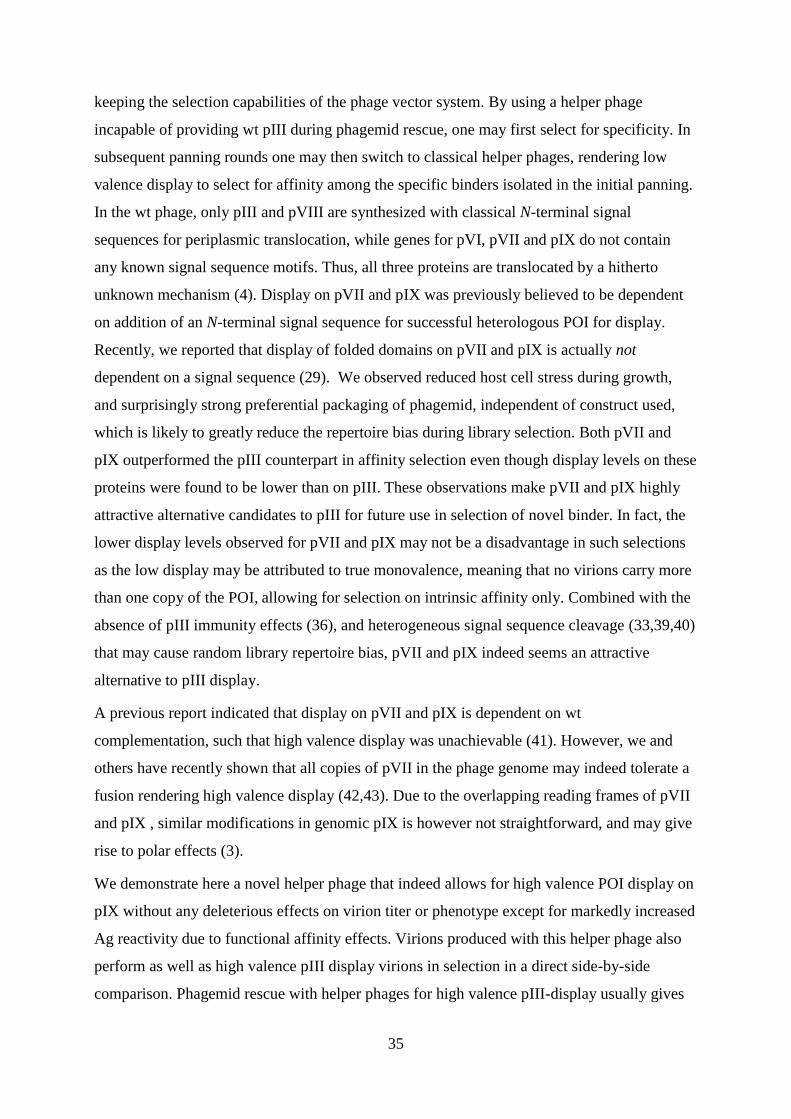
Figure 4. Illustration of the genomic Ff region with sequences important for the pIXAmber
mutant design. The wt (A) and modified with the pIXAmber
mutations (B). (C): schematic
illustration of phagemid rescue using the modified helper phage
To create the novel helper phage, two amber codons were introduced by inserting nine
nucleotides (5’-TAGAGCTAG-3’; Figure 4B, bold) close to the pIX start codon (ATG), but
after the pVII translational stop. The position of these mutations was selected not to interfere
with the pVII ORF as well as the pVIII ribosome binding site that is located within the 3’-part
of the pIX ORF. As a consequence of this insertion, two glutamines and a serine are added in
the solvent exposed N-terminal portion of the full length protein (3). This should have no
effect on pIX function, as minor N-terminal modifications are readily tolerated (41). Two
consecutive amber codons were chosen based on previous observations that a single amber
codon may be insufficient to achieve complete knockdown in the non-suppressor host (54).
As superior amber suppression has been observed if the amber codon is immediately followed
by a purine (55), the two amber codons were separated by 5’-AGC-3’ encoding a small
hydrophilic serine residue. Phagemid rescue with this modified helper phage in a non-
suppressor host should produce virions carrying a fusion on all copies of pIX (figure 4C).
This novel helper phage was named DeltaPhage.

42
Growth and production characteristics
To study how the amber mutations in DeltaPhage affected virion assembly in a suppressor
and a non-suppressor host strain, we cultured E.coli TOP10F’ (non-suppressor) and XL1-Blue
(suppressor) with DeltaPhage and the results are shown in Figure 5a. Wt helper phage
M13K07 was included for comparison.
Both host strains show roughly equal cell culture density after overnight culturing regardless
of helper phage. This showed that the pIXamber
mutations had no gross effect on host cell
viability beyond that observed for M13K07.
Virion yield was then assessed by infectious titration, in that serial dilutions of virion
containing supernatants were used to infect log-phase E.coli cells. The infected cultures were
spotted onto a nitrocellulose membrane on kanamycin-supplemented LA-plates. Colonies
were counted after incubation at 37°C/ON. We observed equal end titers for the two host
strains when infected with M13K07. Contrary, DeltaPhage gave a slightly reduced end titer
than wt helper phage in the suppressor strain, while virion production was completely
abolished in the non-suppressor strain TOP10F’ (Figure 5B).
Figure 5 Single colonies of either XL1-Blue or TOP10F’ were grown over night in selective
medium before the cell density was measured at A600nm (A). The virion content in the culture
supernatants were determined by infectious titration and the results given as kanamycin
resistant colony forming units (cfukanR
/ml) (B)
The absence of virion production from DeltaPhage in TOP10F’ showed that the intended
phenotype indeed was achieved.

43
The reduction in titer seen with DeltaPhage implies that the pIXAmber
mutations had a direct
influence on the virion production capacity. This effect has been observed, and often to a
larger extent, with other mutant helper phages as well (30). The phenotype might be caused
by lowered efficiency in translation of the pIX ORF as a result of incomplete suppression of
the amber codons. However, it should be of no practical consequence as high phage numbers
equal to those obtained for the M13K07 may be reached by an up-scale of volume and PEG
precipitation of the particles. Later experiments have also shown that optimal growth
conditions with increased agitation yields wt end titers also with DeltaPhage.
Performance in phagemid rescue
To characterize how DeltaPhage performs in phagemid rescue, the phagemid pGALD9ΔL
encoding either of two different scFvs fused to pIX, namely, anti-phOx and anti-NIP were
selected due to their differing properties in pIII display. Here, anti-phOx expresses well, and
has a strong genotype phenotype link, while anti-NIP expresses poorly and has a weak
genotype phenotype link. They also differ in monomeric affinity (~ 1x 10⁻⁹
M (56) and ~ 3x
10-7
M (57) respectively). The phagemids were rescued with DeltaPhage using the standard
phagemid rescue protocol followed by PEG precipitation and concentration. Both non-
suppressor and suppressor host strains were tested, and M13K07 was included for
comparison. Virion titers were determined by infectious titration, and given as cfuampR
/ml. We
also determined the phagemid to helper phage genome ratio of the rescued samples based on
the corresponding titers given as cfuampR
/cfukanR
.

44
Figure 6 End titer of phagemid virions after PEG precipitation was determined by infectious
titration and the results given as ampicillin resistant colony forming units (cfuampR
) per ml
(A). The phagemid to helper phage genome ratio was determined by infectious titration and
growth on both kanamycin and ampicillin. The ratio is given as cfuampR
/cfukanR
(B).
The results show that virions were produced in all cases in which similar and very high titers
close to the border of solubility were reached with both strains and helper phages (figure 6A).
The phage particles rescued with DeltaPhage exhibited a large increase in phagemid to helper
phage ratio, compared to the corresponding samples rescued with the wt M13K07, as
DeltaPhage is consistently >103 while M13K07 varies from 3 to 60 (Figure 6B). The effect
was highly pronounced and independent on host strain.
The high ratio value of the DeltaPhage-rescued samples may prove to be a significant
advantage during selections, as 99.9% of the virions carry the phagemid and thus the
genotype for the displayed fusion. In addition, the fact that wt-like titers are readily
achievable is a major advantage during screening of libraries with high diversity. Another
important observation is that there is no notable construct-dependent bias. This is previously
reported as an attribute of signal sequence independent pIX display (29).
The phagemid rescue properties of DeltaPhage were then compared with the current standard
helper phages, M13K07 as well as Hyperphage, and standard pIII display, using the phagemid
pSEX81 (Figure 3). Three different folded domains, namely scFv anti-phOx and –NIP along
with the single chain T-cell receptor (scTCR) 4B2A1 (58) were included in the comparison.
Phagemid rescue was done in E.coli XL1-Blue suppressor cells (to allow for pIX production
when using DeltaPhage) under identical conditions using the standard phagemid rescue

45
protocol. End titers and phagemid to helper phage genome packaging ratio were determined
as previously described.
Here, DeltaPhage supported the production of lower number of virions than M13K07.
Specifically, A 10 fold decrease in end titer was observed compared to M13K07, whereas
Hyperphage gave even lower end titers for two of the constructs (Figure 7a), which is
consistent with previous reports (46). Once again, a dramatic increase in phagemid packaging
was observed using DeltaPhage compared to M13K07. Hyperphage also showed higher ratio
than M13K07, albeit slightly lower than DeltaPhage (figure 7B).
Figure 7: End titer after phagemid rescue using three variants of the pSEX81 phagemid for
pIII-display and different helper phages(A), and phagemid to phage vector ratio(B).
The pIXAmber
mutation seems to affect phage production due to lowered production of pIX, as
the reduction in end titer is only seen when pIX is not encoded by the phagemid and thus
supplied by an exogenous source. Furthermore, there are notable clone variations for all three
helper phages in both titer and ratio, which confirms previous reports of clone dependent bias
in pIII display systems. Nonetheless, as compared to the standard M13K07 helper phage, the
overall high phagemid to helper phage ratios obtained with DeltaPhage also in the pIII display
system suggests that this helper phage indeed could be beneficial to use for low-valence
display and selection on pIII

46
Determination of display levels by ELISA
The expected phenotype of pIX encoding phagemids rescued with DeltaPhage in a non-
suppressor host is high valence display of the peptide fusion. This should directly translate to
an increase in Ag reactivity as a result of the avidity effect compared to rescue with a wt
helper phage such as M13K07. This may be studied in an Ag-specific phage capture ELISA.
Two different scFv specificities, anti-phOx and anti-NIP, rescued with M13K07 (suppressor
host) and DeltaPhage (non-suppressor host) were again tested, and serial dilutions of the
various phage preparations were tested for binding to a constant amount of Ag.
A significant increase in Ag reactivity from the DeltaPhage-rescued samples was observed
compared to those rescued with the wt M13K07 (Figure 8 A & B). Thus, the high-valence
phenotype was indeed achieved, as an increase in Ag reactivity is clearly shown. The effect of
high valence was more prominent when applied to the lower affinity anti-NIP specificity,
where reactivity was increased by almost two orders of magnitude when rescued with
DeltaPhage compared to the standard system (XL1-Blue and M13K07).
Figure 8 POI-capsid fusion display levels determined as Ag specific binding. Serial
dilutions of phagemid-rescued samples displaying either scFv anti-phOx (A) or –NIP (B)
were applied in a phage capture ELISA. Ag-bound virions were detected by anti-M13-HRP
and the data shown as function of number of phagemid-carrying virions/ml solution.

47
Performance in a whole-cell assay
Discovery of neutralizing Abs and other peptides that interact with transmembrane receptors
on cells is currently of very high interest, as these receptors comprise the largest group of drug
targets (59) . A major problem for selection using phage display is that these target proteins
often contain post-translational modifications, and thus cannot be expressed recombinantly in
their native form in bacteria. In addition, extracellular domains of integral membrane proteins
are dependent on the integration of the molecule in the bilayer, and will often not be
expressed in the correct conformation as a soluble recombinant protein (60). Primarily this is
solved by whole cell panning in which phage libraries are screened against living cells
expressing the membrane receptor of interest. To investigate the applicability of DeltaPhage
in such an assay, and to compare its performance with established pIII display, phage
preparations displaying the scFv version of the mAB GB113, an Ab specific for the T cell
receptor 4B2A1 (49) were tested for staining of 4B2A1 T-cells in a flow cytometer. Due to
the large size of the virion, there is a solubility threshold of about 1013
virions/ml, which
corresponds to 16.6 nM. However, due to the 10-fold lower end titer reached with
Hyperphage, we were forced to reduce the input concentration to 5x10¹¹
cfuampR
/ ml to allow
for direct comparison.
All scFv-displaying virions showed specific staining of the 4B2A1 T-cells (Figure 7A). The
high valence virions (pIX/DeltaPhage and pIII/Hyperphage) stained with similar intensities,
while the low-valence pIII (rescued with M13K07) exhibited approximately 10-fold stronger
staining than its pIX counterpart rescued with M13K07. Clearly, none of the virions stain as
bright as the GB113 mAb when using a standard mAb concentration for flow cytometry
(Figure 9A, cyan). We therefore included an additional mAb GB113 control employing an
equimolar amount to the phage inputs (830pM) for comparison. Notably, the two systems
obviously differ in valence (bivalent vs. pentavalent), as does the detection system.
Nonetheless, when comparing these samples (Figure 9A red), the phage system appears more
sensitive (about 0.5 log). The wt virion (VCSM13) and isotype control mAb were both
negative on 4B2A, and all samples were negative on the specificity related, yet different
7A10B2 T-cells (Figure 9B).

48
Figure 9 phage and mAB staining of 4B2A1 T-cells using flow cytometry. A normalized
phage input of 5x10¹¹
cfuampR
/ml which corresponds to a phage concentration of 830pM was
used (A). Staining of 7A10B2 T-cells as negative control (B).
There was a striking difference in target reactivity between low and high valence display on
pIX, corresponding to 1.5 log, while on pIII this difference was no more than 0.5 log. This
indicates that pIX exhibits enhanced discrimination between intrinsic and functional affinity
compared to the pIII system, which should be an asset during high affinity selection.
Performance in library selection
The primary application of phage display technology is to isolate specific binders from large
and diverse libraries. Importantly, increased number of selection cycles have been shown to
lower the retrieved diversity, hence a minimum amount of iterative Ag selection cycles, yet
sufficient to allow identification of Ag-specific clones, should be applied (61). To complete
the characterization of the novel helper phage by assessing its ability to retrieve binders
during selection, a mock library selection was done. 103 Ag specific phages were spiked into
1010
phages with a different specificity using the scFv anti- phOx and anti-NIP phages
(importantly, the specificities are not cross-reactive), creating a mock library with a diversity
of 107 (corresponding to a library of medium diversity). The libraries were then used in
selection for either phOx or NIP positive clones. Selection was done by adding the libraries to
wells coated with either phOx- or NIP-BSA. The wells were stringently washed, and bound

49
phages were eluted by cleavage of the fusion by incubation with trypsin. The eluates were
then used to infect E.coli cells. The infected cells were grown in selective medium, and the
supernatant was harvested. These supernatants were used for transduction to provide single
clones for screening. Screening was done by growing 40 randomly picked clones from each
selection in separate wells and testing the supernatant from these clones for binding to both
Ags.
Phagemid Helper phage Valence #Positives
pSEX81 scFv anti-phOx M13K07 Low 3/40
Hyperphage High 8/40
pSEX81 scFv anti-NIP M13K07 Low 0/40
Hyperphage High 9/40
pGALD9 scFv anti-phOx M13K07 Low 3/40
DeltaPhage High 8/40
pGALD9 scFv anti-NIP M13K07 Low 1/40
DeltaPhage High 7/40
Table 1 Single colony screening after one round of Ag selection. 40 single colonies from
each mock library were tested for Ag reactivity using ELISA A clone was regarded as positive
if it exhibited at least three-fold higher response than the background signal.
The screening procedure showed low efficiency of retrieval for low valence display after the
one round of selection, yielding from 3/40 positive clones for anti-phOx (both pIII and pIX),
to 0/40 (pIII anti-NIP). High valence display proved more efficient in retrieval of binders,
yielding 7-9/40 positive clones with no significant differences between the two systems.
After only one round of selection, it becomes apparent that high valence display on pIX
performs as well as the pIII counterpart, showing that DeltaPhage is indeed well suited for
selection of binders. In screening for new Abs, it is an advantage to utilize high valence
display during the first one or two rounds, as this effectively prevents loss of binders, due to
poor affinity or display, selecting primarily for specificity, before proceeding with low

50
valence display to select for affinity among the specific binders. Since display on pIX gives
increased discrimination between low and high valence, this system may superior during
affinity selection indeed.
Concluding remarks
Until now, the general consensus has been that display on pIX is dependent on wt
complementation, i.e. high valence display is unachievable (3,41). In this paper we clearly
demonstrate that not only is high valence display possible, but it has several clear advantages
over high valence pIII display, which represents the current standard. Wt end titers are easily
reachable, which gives one the opportunity to screen libraries of higher diversity. The
phagemid to phage vector ratios are uniformly very high which will ensure that no selected
clone is lost. In addition, high valence pIX-display does not affect infectivity in any way,
while high valence pIII-display severely and unpredictably affects infectivity (32). In
addition, pIX-display seems to overcome the clone dependent bias observed for pIII-display.
This prevents loss of binders due to unfavorable growth conditions. An observation with
strong implications for future affinity selection regimes, is the fact that the pIX system
apparently exhibits better discrimination between low and high valence display than is
observed using pIII. This may prove to be a significant advantage in primary selection, as well
as in affinity maturation, as it suggests that there are less avidity effects when using low
valence pIX-display than its pIII counterpart. Indeed, effective high affinity selection has been
shown to strongly benefit from very low display levels (20,35).

51
5. Supplementary results
5.1 Supplementary 1
Figure S1. Gel electrophoresis of plasmid miniprep from E.coli cultures containing either
M13K07 or DeltaPhage
To investigate if the differences in end titer between M13K07 and DeltaPhage occur due to
lowered copy number of phage genome, single clones of XL1-Blue harboring either phage
genome were grown under identical conditions. The cultures were normalized based on
culture density at A600nm, and a plasmid miniprep was performed. Equal amounts of prep
were run on a 1% agarose gel.
This analysis showed roughly equal amounts of phage dsDNA between the two helper
phages.
As the copy number of phage genome seemed to be equal, the lower end titer should not be
caused by lowered total protein production as a function of reduced gene dosage. Although
we cannot rule out putative distortions in dsDNA to ssDNA conversion, this observation
further substantiates incomplete suppression of the amber codons in the suppressor host,
possibly making pIX a limiting factor for phage assembly in this system. This should not be
of any practical issue, as phage production is not affected when pIX is supplied from a
phagemid, which is the intended use of DeltaPhage.

52
5.2 Supplementary 2
Figure S2. Western blot of normalized phage C: M13K07 wt control. K: phagemid rescued
with M13K07. D: phagemid rescued with DeltaPhage
To verify estimated titers in order to ensure correct inputs in subsequent experiments, equal
amounts of virions (108/lane) were separated by denaturing 14% SDS-PAGE, and blotted on
to a PDVF membrane, detected with an anti-pIII Ab.
The estimated titers were precise as seen from the equal signals obtained from all samples.
5.3 Supplementary 3
Figure S3. Western blot analysis of the scFv anti-phOx samples prepared in either E.coli
XL1-Blue or TOP10F’, and rescued with either M13K07 (K) or DeltaPhage (D).

53
In order to visually quantify display levels, a western blot analysis was done. Equal amounts
of virions (108/lane) were separated by denaturing 14% SDS-PAGE, blotted on to a PDVF
membrane, and the scFv fusions detected with an anti-human λ light chain Ab.
The samples rescued with M13K07 contained fusion levels outside the detection level.
Samples rescued from XL1-Blue with DeltaPhage show an increased amount of fusion, while
samples rescued with DeltaPhage from TOP10F’ show an even greater amount of fusion.
The samples rescued with M13K07 were undetectable, most likely because the fraction of
phage carrying the fusion in this system is only 1-10% due to the fact that pIX is produced in
large excess in the cytosol of the host (3). When phagemid rescue was performed with
DeltaPhage in XL1-Blue, an increase in fusion was detected. This might be an effect of
lowered [pIXwt
] in the host due to the effect proposed above. A lower concentration of wt pIX
in the host would directly translate into a higher fraction of the phagemid produced fusion
incorporated in the phage progeny. In TOP10F’ the detection is markedly stronger, which
points to high valence display of the POI due to the absence of wt pIX brought on by the
Amber mutations. However, this blot proved to not be reproducible and is therefore omitted in
the manuscript and discussion.
5.4 Supplementary 4
Figure S4. POI-capsid fusion display levels comparison of pIII vs. pIX display, determined
as Ag specific binding. Serial dilutions of phagemid-rescued samples displaying either scFv
anti-phOx (A) or –NIP (B) were applied in a phage capture ELISA. Ag-bound virions were
detected by anti-M13-HRP and the data shown as function of number of phagemid-carrying
virions/ml solution.

54
To assess functional display levels (i.e. amount of properly folded proteins displayed) on
DeltaPhage rescued virions compared with the current standard platforms, an ag-specific
phage capture ELISA was performed. Serial dilutions of the various phages were tested for
binding to a constant amount of Ag.
A significant increase in Ag reactivity was observed from samples rescued with DeltaPhage
when compared to M13K07 for both pIX phagemids. Phagemid rescue with Hyperphage
yields virions with superior reactivity for both constructs. pIII constructs rescued with
M13K07 show similar reactivity as DeltaPhage for anti-phOx (A), and higher reactivity for
anti-NIP (B).
The expected increase in functional display levels caused by the pIXamber
mutations in
DeltaPhage is apparent, giving an increase in Ag reactivity by up to two orders of magnitude
compared to M13K07 (B). This increase is larger than the one observed from low to high
valence in pIII-display. This difference may be attributed to the difference in functional
display levels in low valence display on pIII and pIX, as pIX is previously reported to have
lower display levels than pIII when using low valence platforms. This, meaning that the
switch from low valence to high valence display on pIX gives a larger relative increase in
functional display levels than on pIII. The higher reactivity observed for anti-NIP on pIII with
wt helper phage is most likely due to its poor ratio (5x10⁻²
). This poor ratio gives the
heterogeneous population of virions in this particular sample an advantage in this assay as the
majority of phages carry the helper phage genome, and these are not taken into account during
normalization. Furthermore the size of these virions are approximately double that of
phagemid-carrying virions, which may provide more binding sites for the detection Ab,
increasing the sensitivity of the assay.

55
5.5 Supplementary 5
Figure S5. Comparison of functional display levels for DeltaPhage (pIX) and Hyperphage
(pIII), along with M13K07 (pIX and pIII)
To study and compare the polyvalent binding properties of high valence display on pIX and
pIII, phagemids rescued with DeltaPhage and the current standard platform Hyperphage were
analyzed on a phage capture ELISA. Equal numbers of cfuampR
were tested for binding to
serial dilutions of phOx-BSA Ag. The ability of the virions to bind with polyvalence will
manifest itself as a shift toward the lower Ag concentrations.
The results showed roughly equal reactivity between pIX high valence and pIII low valence
display. Once again pIII rescued with Hyperphage gave the highest reactivity, while pIX
rescued with wt helper phage gave the lowest reactivity. Also worth noting is the larger
separation between the two high valence formats compared to the phage dilution ELISA
(figure S4).
The threshold for detection of the Hyperphage-rescued phagemid is 0.1pg/ml Ag while it is
100-fold higher for DeltaPhage. This can putatively be explained by the fact that pIII is a
fairly large protein with highly flexible glycine-rich linkers between its domains. This
facilitates polyvalent binding of Ag in a lot lower densities compared to pIX fusions. The five
copies of pIX are clustered together and are inflexible, which cause the scFvs to be localized
over a much smaller area, thus needing a higher density of Ag for high valence binding.
Surprisingly, pIII-phagemid rescued with wt phage has approximately the same detection
threshold. However, there is established knowledge that many pIII systems, and in particular

56
those based on the scFv format, suffers from heterogeneous oligovalency effects masking the
true monomeric affinity, which may cause the retrieval of artificial high affinity binders due
to avidity effects during selection on polyvalent Ag (38). In this particular assay the
subpopulation in question may act as oligovalent binders, giving artificially high reactivity. It
appears that pIX is less affected by this, which indicates that the pIX system is closer to an
actual monomeric display system. This observation is crucial, as such an effect may translate
into more effective affinity maturation, as there are no avidity effects masking the changes in
monomeric affinity brought on by mutation(30).
5.6 Supplementary 6
Figure S6. Western blot of pIII to verify normalization of scFv phages used in flow
cytometry. 1: pIII-M13K07 2: pIII-Hyperphage 3: pIX M13K07 4: pIX DeltaPhage C:
M13K07 wt control
In order to visualize correct normalization of phage input, a western blot was performed with
detection of pIII, and indeed the blot showed that the estimated titers are very precise.
As the normalization appears correct for all samples, it confers credibility to the results of the
flow cytometry experiment. The wt pIII-band seen for Hyperphage samples is most likely due
to proteolytic cleavage of the fusion in the periplasm.

57
Worth noting is that as the phages were detected by an anti-pIII Ab, the scFv fusions are
detectable for the samples that exhibit pIII-display. The increase in display levels from low to
high valence on pIII is clearly visualized on this western blot. However, due to the lack of an
anti-pIX Ab (there is no such Ab commercially available) we were not able to do the same for
pIX. Regardless, from all the observations done, especially in the flow cytometry experiment
(figure 9 in the manuscript) there is little doubt that a similar increase in display levels on pIX
is achieved when using DeltaPhage.
5.7 Supplementary 7
Figure S7: Flow cytometry staining. To investigate if more phages translated into stronger
staining, maximum input samples were compared to the normalized samples used in figure 9
To allow for a direct comparison of the various samples used in the flow cytometry
experiment, all samples were normalized. However, many of the samples had titers that
allowed for a higher input. To see if more phages translated into stronger staining, maximum
input samples were included.
The results showed that neither of the high valence display samples increased in staining
intensity when increasing the sample input with up to 3-fold indicating that target reactivity
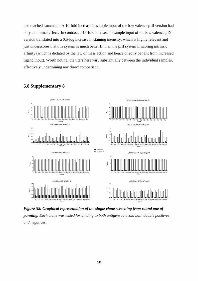
58
had reached saturation. A 10-fold increase in sample input of the low valence pIII version had
only a minimal effect. In contrast, a 16-fold increase in sample input of the low valence pIX
version translated into a 0.5-log increase in staining intensity, which is highly relevant and
just underscores that this system is much better fit than the pIII system in scoring intrinsic
affinity (which is dictated by the law of mass action and hence directly benefit from increased
ligand input). Worth noting, the titers here vary substantially between the individual samples,
effectively undermining any direct comparison.
5.8 Supplementary 8
Figure S8: Graphical representation of the single clone screening from round one of
panning. Each clone was tested for binding to both antigens to avoid both double positives
and negatives.

59
5.9 Supplementary 9
Figure S9. Gel electrophoresis of PCR products showing specific amplification of scFv
templates. Lanes 1-4: anti-phOx and lanes 5-8: anti-NIP.
As an alternative strategy for screening the output of the selection a PCR screening protocol
was devised. E.coli XL1-Blue harboring the pGALD9 scFv anti-phOx or –NIP phagemid was
inoculated in 2xYT supplemented with kanamycin and incubated ON. 1µl of culture was
added to the PCR reaction mix. Optimization of the PCR reaction was done by varying the
parameters for annealing temperature, synthesis duration and number of cycles.
The figure shows a highly specific PCR reaction, where all controls are negative, and clear
bands of the expected sizes were obtained. This was achieved by lowering the annealing
stringency (annealing temp. 50°C) and increasing the synthesis duration to 45s, as well as
increasing the number of cycles to 40.
This seems to be a quite effective way of screening the output in a spiked panning. Although,
as an output from a selection may yield >1000 clones, analysis using gel electrophoresis
would prove cumbersome. However, as the PCR reaction yielded such clean samples, and no
cross-amplification between the two templates, a quick analysis of the PCR reaction may be
done by utilizing a fluorescent detection agent for dsDNA.

60
6. Discussion
In phage display, pIII has up until now been the capsid protein of choice for display of fusion
proteins along with pVIII. The use of pVIII is limited to small peptides, as larger fusions to
this protein are not tolerated by the phage. For display of larger proteins, and folded domains,
pIII has become the industry standard, and it has had great success, despite the many
drawbacks it confers on the system, especially when high valence display is used, as pIII is
responsible for mediating infection (32). pIX was up until recently believed to be dependent
on a leader sequence for successful display of foreign proteins (41). This was disproved along
with the report that pIX seems to outperform pIII in affinity selection(29). Furthermore,
previous reports state that display on pIX is dependent on wt complementation, i.e. high
valence display is unachievable (3,41).
The aim of this study was to develop and characterize a novel mutant helper phage that
facilitates high valence pIX-display. This mutant was named DeltaPhage
6.1 Helper phage production and phagemid rescue
The first step of characterization was to produce the mutant helper phage. This was done by
culturing both suppressor and non-suppressor E.coli with DeltaPhage, while monitoring
bacterial growth and virion yield, and compared to the wt helper phage. The roughly equal
culture density observed showed that the amber mutations in pIX had no effect on host cell
viability beyond that observed for wt helper phage. The absence of virion production from
DeltaPhage in non-suppressor host showed that the intended phenotype was indeed achieved.
However, the reduction in titer seen with DeltaPhage implies that the amber mutations in pIX
have a direct influence on the virion production capacity, which has been observed, often to a
larger extent, with other mutant helper phages (30). This effect might be caused by lowered
efficiency in translation of the pIX ORF as a result of incomplete suppression of the amber
codons.
Following successful production of DeltaPhage, performance in phagemid rescue was
assessed. The phagemid encoding scFv anti-phOx and anti-NIP as fusion to pIX was rescued

61
with DeltaPhage from both suppressor and non-suppressor hosts. The high ratio value of the
DeltaPhage-rescued samples may prove to be a significant advantage during selections, as
99.9% of the virions carry the phagemid and thus the genotype for the displayed fusion. In
addition, the fact that wt-like titers are readily achievable is a major advantage during
screening of libraries with very high diversity. Another important observation is that there is
no notable construct-dependent bias. This is previously reported as an attribute of signal
sequence independent pIX display (29).
The phagemid rescue properties of DeltaPhage were then compared with the current standard
helper phages, M13K07 and Hyperphage, and standard pIII display. Three different folded
domains, namely scFv anti-phOx and –NIP along with the scTCR 4B2A1 (58) were included
in the comparison.
A slight drop in end titer is observed for DeltaPhage, which further supports the theory that
the amber mutations in pIX are incompletely suppressed, and thus negatively affecting phage
production, as the reduction in end titer is only seen when pIX is not encoded by the
phagemid and thus supplied by an exogenous source. The high phagemid to helper phage
ratios obtained with DeltaPhage indicate that this helper phage is better suited for low-valence
selection on pIII than the wt helper phage. Furthermore, there are notable clone variations for
all three helper phages in both titer and ratio, which confirms previous reports of clone
dependent bias in pIII display systems (35,36).
To further investigate the reduction in end titer for DeltaPhage in a suppressor host, a plasmid
miniprep and gel electrophoresis analysis was done to quantify the amount of double stranded
DNA (dsDNA) present in DeltaPhage producing cells. Wt helper phage was included for
comparison.
As the copy number of phage genome seems to be equal, the lower end titer should not be
caused by lowered total protein production as a function of reduced gene dosage. Although
we cannot rule out putative distortions in dsDNA to ssDNA conversion, this observation
further substantiates incomplete suppression of the amber codons in the suppressor host,
possibly making pIX a limiting factor for phage assembly in this system. This should not be
of any practical issue, as phage production is not affected when pIX is supplied from a
phagemid, which is the intended use of DeltaPhage.

62
6.2. Characterization of display levels
The expected phenotype of pIX encoding phagemids rescued with DeltaPhage in a non-
suppressor host is increased functional display levels (amount of correctly folded fusion
displayed) as a result of high valence display of the peptide fusion. This should directly
translate to an increase in Ag reactivity compared to rescue with a wt helper phage, as a result
of avidity effects. Importantly, correct virion titers were confirmed by western blotting (figure
S2). The high-valence phenotype is indeed achieved, as an increase in Ag reactivity is clearly
shown. The effect of high valence is more prominent when applied to the lower affinity anti-
NIP specificity, where reactivity is increased by almost two orders of magnitude when
rescued with DeltaPhage compared to the standard system.
When compared to the pIII-encoding phagemid rescued with M13K07 and Hyperphage, it is
notable that the pIII system indeed gives higher display levels as previously reported (29).
However the increase in display levels from low to high valence on pIII display is smaller
than the one observed for pIX. This implies that display on pIX discriminates better between
low and high valence display.
The higher reactivity observed for anti-NIP on pIII with wt helper phage is most likely due to
its poor ratio. This poor ratio gives the heterogeneous population of virions in this particular
sample an advantage in this assay as the majority of phages carry the helper phage genome
while still displaying the POI, and these are not taken into account during normalization.
Furthermore the size of these virions are approximately double that of phagemid-carrying
virions, which may provide more binding sites for the detection Ab, increasing the sensitivity
of the assay.
To study and compare the polyvalent binding properties of high valence display on pIX and
pIII, the anti-phOx phages were analyzed on a phage capture ELISA where equal numbers of
cfuampR
were tested for binding to serial dilutions of phOx-BSA Ag.
The threshold for detection of the Hyperphage-rescued virions was 100-fold lower than for
DeltaPhage- rescued virions. This can putatively be explained by the fact that pIII is a fairly
large protein with highly flexible glycine-rich linkers between its domains. This facilitates
polyvalent binding of Ag in a lot lower densities compared to pIX fusions. The five copies of
pIX are clustered together and are inflexible, which cause the scFvs to be localized over a
much smaller area, thus needing a higher density of Ag for high valence binding.

63
Surprisingly, the low valence pIII display virions had approximately the same detection
threshold as virions rescued with DeltaPhage. However, there is established knowledge that
many pIII systems, and in particular those based on the scFv format, suffers from
heterogeneous oligovalency effects masking the true monomeric affinity, which may cause
the retrieval of artificial high affinity binders due to avidity effects during selection on
polyvalent Ag (38). In this particular assay the subpopulation in question may act as
oligovalent binders, giving artificially high reactivity. It appears that pIX is less affected by
this, which indicates that the pIX system is closer to an actual monomeric display system.
This observation is crucial, as such an effect may translate into more effective affinity
maturation, as there are no avidity effects masking the changes in monomeric affinity brought
on by mutation(30).
6.3. Use of DeltaPhage rescued virions in a whole cell assay
Discovery of neutralizing Abs and other peptides that interact with transmembrane receptors
on cells is currently of very high interest, as these receptors comprise the largest group of
potential drug targets (59). A major problem for selection using phage display is that these
target proteins often contain post-translational modifications, and thus cannot be expressed
recombinantly in their native form in bacteria. In addition, extracellular domains of integral
membrane proteins are dependent on the integration of the molecule in the bilayer, and will
most often not be expressed in the correct conformation as a soluble recombinant protein (60).
Primarily this is solved by whole cell panning in which phage libraries are screened against
living cells expressing the membrane receptor of interest. To investigate the applicability of
DeltaPhage in such an assay, and to compare its performance with established pIII display,
phage preparations displaying the scFv version of an Ab specific for the T cell receptor
4B2A1 were tested for staining of 4B2A1 T-cells in a flow cytometer.
There is a striking difference between low and high valence display on pIX, while on pIII this
difference is markedly smaller. This indicates that pIX exhibits enhanced discrimination
between intrinsic and functional affinity compared to the pIII system, which should be an
asset during high affinity selection. The fact that high valence pIII and pIX display stains
similarly contrasts previous results where the high valence pIII system yields significantly

64
higher reactivity. It may be so that the functional affinity effects seen with pIX and pIII differ
between these two systems in an Ag dependent manner.
Indeed, the increased antigen reactivity observed could be directly attributed to increased
display level of the scFv fusion as shown for pIII by western blotting.
To allow for a direct comparison of the various samples used in the flow cytometry
experiment, all samples were normalized. However, many of the samples had titers that
allowed for a higher input. To see if more phages translated into stronger staining, maximum
input samples were included (Figure S7).
The results showed that neither of the high valence display samples increased in staining
intensity when increasing the sample input, indicating that target reactivity had reached
saturation. An increase in sample input of the low valence pIII version had only a minimal
effect. In contrast, a slightly larger increase in sample input of the low valence pIX version
translated into a distinct increase in staining intensity, which is highly relevant and just
underscores that this system is much better fit than the pIII system in scoring intrinsic affinity
(which is dictated by the law of mass action and hence directly benefit from increased ligand
input). Worth noting, the titers here vary substantially between the individual samples,
effectively undermining any direct comparison.
6.4. Affinity selection
The screening procedure showed low efficiency of retrieval for low valence display after the
one round of selection, yielding positive clones for anti-phOx (both pIII and pIX), none for
pIII anti-NIP, and one for pIX anti-NIP showing that low valence display on pIX is indeed
better suited for high affinity selection (29), as the retrieval of this clone can most likely be
attributed to the positive phagemid to helper phage ratio. High valence display proved more
efficient in retrieval of binders, several positive clones with no significant differences between
the two systems.
After round one of selection, it becomes apparent that high valence display on pIX performs
as well as the pIII counterpart, showing that DeltaPhage is indeed well suited for selection of
binders. In screening for new Abs, it is an advantage to utilize high valence display during the
first one or two rounds, as this effectively prevents loss of binders, selecting for specificity
only, before proceeding with low valence display to select for affinity among the specific

65
binders. Since display on pIX gives increased discrimination between low and high valence,
this system may superior during affinity selection indeed. Also worth noting is that the anti-
NIP selection (low affinity, poor expression) on low valence yielded one positive clone for
pIX while no positive clones were retrieved for pIII. This confirms our previous findings that
low valence pIX-display in the absence of a signal sequence seems to outperform standard
pIII in affinity selection (29).
6.5 Concluding remarks
Until now, the general consensus has been that display on pIX is dependent on wt
complementation, i.e. high valence display is unachievable (3,41).
We demonstrate here a novel helper phage that indeed allows for high valence display on pIX
(as a 9+9 system) without any deleterious effects on virion titer or phenotype except for
markedly increased Ag reactivity due to functional affinity effects. Virions produced with this
helper phage also perform as well as high valence pIII display virions during selection in a
direct side-by-side comparison. Phagemid rescue with helper phages for high valence pIII-
display usually gives 10-1000-fold lower end titers than rescue with “standard” helper phages
such as M13K07 (32). We observe wt end titers with our novel helper phage, in conjunction
with very high phagemid to helper phage packaging ratios, no apparent clone dependent host
bias and absolutely no reduction in infectivity, as no modifications are done to the infection-
mediating pIII.
An observation with strong implications for future affinity selection regimes, is the fact that
the pIX system apparently exhibits better discrimination between low and high valence
display than is observed using pIII. This may prove to be a significant advantage in primary
selection, as well as in affinity maturation, as it suggests that there are less avidity effects
when using low valence pIX-display than its pIII counterpart. Indeed, effective high affinity
selection has been shown to strongly benefit from very low display levels (20,35).
With all of these observations, one may argue that pIX could be the display protein of choice
for the future of this technology.

66
7. Future perspectives
In order to completely finalize the characterization of DeltaPhage, there are a several
experiments that should be conducted. First and foremost, an anti-pIX Ab should be made to
facilitate proper visualization of the increased display levels on pIX by western blotting, as is
easily done for pIII-display. As of now, no such Ab is commercially available, and all
attempts at immunodetection of the displayed scFvs have, due to unknown reasons and/or
lack of time for optimization, been inconclusive at best.
Secondly, a successful screening of e.g. a high diversity phage Ab library using DeltaPhage
during the first few rounds of panning to retrieve novel binders would provide valuable
information of how well this system performs on a complex selection.

67
8. References
1. Smith, G.P. (1985) Filamentous fusion phage: novel expression vectors that display cloned antigens on the virion surface. Science, 228, 1315-1317.
2. Paschke, M. (2006) Phage display systems and their applications. Appl Microbiol Biotechnol, 70, 2-11.
3. Endemann, H. and Model, P. (1995) Location of filamentous phage minor coat proteins in phage and in infected cells. J Mol Biol, 250, 496-506.
4. Simons, G.F., Konings, R.N. and Schoenmakers, J.G. (1981) Genes VI, VII, and IX of phage M13 code for minor capsid proteins of the virion. Proc Natl Acad Sci U S A, 78, 4194-4198.
5. Marvin, D.A., Welsh, L.C., Symmons, M.F., Scott, W.R. and Straus, S.K. (2006) Molecular structure of fd (f1, M13) filamentous bacteriophage refined with respect to X-ray fibre diffraction and solid-state NMR data supports specific models of phage assembly at the bacterial membrane. J Mol Biol, 355, 294-309.
6. Newman, J., Swinney, H.L. and Day, L.A. (1977) Hydrodynamic properties and structure of fd virus. J Mol Biol, 116, 593-603.
7. Russel, M. and Model, P. (1989) Genetic analysis of the filamentous bacteriophage packaging signal and of the proteins that interact with it. J Virol, 63, 3284-3295.
8. Rakonjac, J., Feng, J. and Model, P. (1999) Filamentous phage are released from the bacterial membrane by a two-step mechanism involving a short C-terminal fragment of pIII. J Mol Biol, 289, 1253-1265.
9. Rakonjac, J., Bennett, N.J., Spagnuolo, J., Gagic, D. and Russel, M. Filamentous Bacteriophage: Biology, Phage Display and Nanotechnology Applications. Curr Issues Mol Biol, 13, 51-76.
10. Boeke, J.D. and Model, P. (1982) A prokaryotic membrane anchor sequence: carboxyl terminus of bacteriophage f1 gene III protein retains it in the membrane. Proc Natl Acad Sci U S A, 79, 5200-5204.
11. Lubkowski, J., Hennecke, F., Pluckthun, A. and Wlodawer, A. (1999) Filamentous phage infection: crystal structure of g3p in complex with its coreceptor, the C-terminal domain of TolA. Structure, 7, 711-722.
12. Riechmann, L. and Holliger, P. (1997) The C-terminal domain of TolA is the coreceptor for filamentous phage infection of E. coli. Cell, 90, 351-360.
13. Higashitani, A., Higashitani, N. and Horiuchi, K. (1997) Minus-strand origin of filamentous phage versus transcriptional promoters in recognition of RNA polymerase. Proc Natl Acad Sci U S A, 94, 2909-2914.
14. Feng, J.N., Model, P. and Russel, M. (1999) A trans-envelope protein complex needed for filamentous phage assembly and export. Mol Microbiol, 34, 745-755.
15. Hoogenboom, H.R. (2005) Selecting and screening recombinant antibody libraries. Nat Biotechnol, 23, 1105-1116.
16. Schofield, D.J., Pope, A.R., Clementel, V., Buckell, J., Chapple, S., Clarke, K.F., Conquer, J.S., Crofts, A.M., Crowther, S.R., Dyson, M.R. et al. (2007) Application of phage display to high throughput antibody generation and characterization. Genome Biol, 8, R254.
17. Colwill, K., Persson, H., Jarvik, N.E., Wyrzucki, A., Wojcik, J., Koide, A., Kossiakoff, A.A., Koide, S., Sidhu, S., Dyson, M.R. et al. A roadmap to generate renewable protein binders to the human proteome. Nat Methods.
18. Chothia, C., Lesk, A.M., Tramontano, A., Levitt, M., Smith-Gill, S.J., Air, G., Sheriff, S., Padlan, E.A., Davies, D., Tulip, W.R. et al. (1989) Conformations of immunoglobulin hypervariable regions. Nature, 342, 877-883.
19. Hoogenboom, H.R., de Bruine, A.P., Hufton, S.E., Hoet, R.M., Arends, J.W. and Roovers, R.C. (1998) Antibody phage display technology and its applications. Immunotechnology, 4, 1-20.

68
20. Rothe, C., Urlinger, S., Lohning, C., Prassler, J., Stark, Y., Jager, U., Hubner, B., Bardroff, M., Pradel, I., Boss, M. et al. (2008) The human combinatorial antibody library HuCAL GOLD combines diversification of all six CDRs according to the natural immune system with a novel display method for efficient selection of high-affinity antibodies. J Mol Biol, 376, 1182-1200.
21. Lloyd, C., Lowe, D., Edwards, B., Welsh, F., Dilks, T., Hardman, C. and Vaughan, T. (2009) Modelling the human immune response: performance of a 1011 human antibody repertoire against a broad panel of therapeutically relevant antigens. Protein Eng Des Sel, 22, 159-168.
22. Hoet, R.M., Cohen, E.H., Kent, R.B., Rookey, K., Schoonbroodt, S., Hogan, S., Rem, L., Frans, N., Daukandt, M., Pieters, H. et al. (2005) Generation of high-affinity human antibodies by combining donor-derived and synthetic complementarity-determining-region diversity. Nat Biotechnol, 23, 344-348.
23. Edwards, B.M., Barash, S.C., Main, S.H., Choi, G.H., Minter, R., Ullrich, S., Williams, E., Du Fou, L., Wilton, J., Albert, V.R. et al. (2003) The remarkable flexibility of the human antibody repertoire; isolation of over one thousand different antibodies to a single protein, BLyS. J Mol Biol, 334, 103-118.
24. Bratkovic, T. (2009) Progress in phage display: evolution of the technique and its applications. Cell Mol Life Sci.
25. O'Connell, D., Becerril, B., Roy-Burman, A., Daws, M. and Marks, J.D. (2002) Phage versus phagemid libraries for generation of human monoclonal antibodies. J Mol Biol, 321, 49-56.
26. Hust, M., Meysing, M., Schirrmann, T., Selke, M., Meens, J., Gerlach, G.F. and Dubel, S. (2006) Enrichment of open reading frames presented on bacteriophage M13 using hyperphage. Biotechniques, 41, 335-342.
27. Jespers, L., Schon, O., Famm, K. and Winter, G. (2004) Aggregation-resistant domain antibodies selected on phage by heat denaturation. Nat Biotechnol, 22, 1161-1165.
28. Becerril, B., Poul, M.A. and Marks, J.D. (1999) Toward selection of internalizing antibodies from phage libraries. Biochem Biophys Res Commun, 255, 386-393.
29. Loset, G.A., Roos, N., Bogen, B. and Sandlie, I. (2011) Expanding the Versatility of Phage Display II: Improved Affinity Selection of Folded Domains on Protein VII and IX of the Filamentous Phage. PLoS One, 6, e17433.
30. Bradbury, A.R. and Marks, J.D. (2004) Antibodies from phage antibody libraries. J Immunol Methods, 290, 29-49.
31. Clackson, T. and Wells, J.A. (1994) In vitro selection from protein and peptide libraries. Trends Biotechnol, 12, 173-184.
32. Soltes, G., Hust, M., Ng, K.K., Bansal, A., Field, J., Stewart, D.I., Dubel, S., Cha, S. and Wiersma, E.J. (2007) On the influence of vector design on antibody phage display. J Biotechnol, 127, 626-637.
33. Malik, P., Terry, T.D., Gowda, L.R., Langara, A., Petukhov, S.A., Symmons, M.F., Welsh, L.C., Marvin, D.A. and Perham, R.N. (1996) Role of capsid structure and membrane protein processing in determining the size and copy number of peptides displayed on the major coat protein of filamentous bacteriophage. J Mol Biol, 260, 9-21.
34. Iannolo, G., Minenkova, O., Petruzzelli, R. and Cesareni, G. (1995) Modifying filamentous phage capsid: limits in the size of the major capsid protein. J Mol Biol, 248, 835-844.
35. Pavoni, E., Monteriu, G., Cianfriglia, M. and Minenkova, O. (2007) New display vector reduces biological bias for expression of antibodies in E. coli. Gene, 391, 120-129.
36. Shi, B., Wang, H., Guo, S., Xu, Y., Li, Z. and Gu, J. (2007) Protein III-based single-chain antibody phage display using bacterial cells bearing an additional genome of a gene-III-lacking helper phage. Biotechniques, 42, 760-765.
37. de Bruin, R., Spelt, K., Mol, J., Koes, R. and Quattrocchio, F. (1999) Selection of high-affinity phage antibodies from phage display libraries. Nat Biotechnol, 17, 397-399.
38. de Haard, H.J., van Neer, N., Reurs, A., Hufton, S.E., Roovers, R.C., Henderikx, P., de Bruine, A.P., Arends, J.W. and Hoogenboom, H.R. (1999) A large non-immunized human Fab

69
fragment phage library that permits rapid isolation and kinetic analysis of high affinity antibodies. J Biol Chem, 274, 18218-18230.
39. Malik, P., Terry, T.D., Bellintani, F. and Perham, R.N. (1998) Factors limiting display of foreign peptides on the major coat protein of filamentous bacteriophage capsids and a potential role for leader peptidase. FEBS Lett, 436, 263-266.
40. Shen, L.M., Lee, J.I., Cheng, S.Y., Jutte, H., Kuhn, A. and Dalbey, R.E. (1991) Use of site-directed mutagenesis to define the limits of sequence variation tolerated for processing of the M13 procoat protein by the Escherichia coli leader peptidase. Biochemistry, 30, 11775-11781.
41. Gao, C., Mao, S., Lo, C.H., Wirsching, P., Lerner, R.A. and Janda, K.D. (1999) Making artificial antibodies: a format for phage display of combinatorial heterodimeric arrays. Proc Natl Acad Sci U S A, 96, 6025-6030.
42. Kwasnikowski, P., Kristensen, P. and Markiewicz, W.T. (2005) Multivalent display system on filamentous bacteriophage pVII minor coat protein. J Immunol Methods, 307, 135-143.
43. Loset, G.A., Bogen, B. and Sandlie, I. Expanding the versatility of phage display I: efficient display of peptide-tags on protein VII of the filamentous phage. PLoS One, 6, e14702.
44. Welschof, M., Terness, P., Kipriyanov, S.M., Stanescu, D., Breitling, F., Dorsam, H., Dubel, S., Little, M. and Opelz, G. (1997) The antigen-binding domain of a human IgG-anti-F(ab')2 autoantibody. Proc Natl Acad Sci U S A, 94, 1902-1907.
45. Vieira, J. and Messing, J. (1987) Production of single-stranded plasmid DNA. Methods Enzymol, 153, 3-11.
46. Rondot, S., Koch, J., Breitling, F. and Dubel, S. (2001) A helper phage to improve single-chain antibody presentation in phage display. Nat Biotechnol, 19, 75-78.
47. Michaelsen, T.E., Aase, A., Westby, C. and Sandlie, I. (1990) Enhancement of complement activation and cytolysis of human IgG3 by deletion of hinge exons. Scand J Immunol, 32, 517-528.
48. Nakela, O., Kaartinen, M., Pelkonen, J.L. and Karjalainen, K. (1978) Inheritance of antibody specificity V. Anti-2-phenyloxazolone in the mouse. J Exp Med, 148, 1644-1660.
49. Bogen, B., Lauritzsen, G.F. and Weiss, S. (1990) A stimulatory monoclonal antibody detecting T cell receptor diversity among idiotype-specific, major histocompatibility complex-restricted T cell clones. Eur J Immunol, 20, 2359-2362.
50. Marks, J.D., Hoogenboom, H.R., Bonnert, T.P., McCafferty, J., Griffiths, A.D. and Winter, G. (1991) By-passing immunization. Human antibodies from V-gene libraries displayed on phage. J Mol Biol, 222, 581-597.
51. Sidhu, S.S., Lowman, H.B., Cunningham, B.C. and Wells, J.A. (2000) Phage display for selection of novel binding peptides. Methods Enzymol, 328, 333-363.
52. Koch, J., Breitling, F. and Dubel, S. (2000) Rapid titration of multiple samples of filamentous bacteriophage (M13) on nitrocellulose filters. Biotechniques, 29, 1196-1198, 2002.
53. Loset, G.A., Kristinsson, S.G. and Sandlie, I. (2008) Reliable titration of filamentous bacteriophages independent of pIII fusion moiety and genome size by using trypsin to restore wild-type pIII phenotype. Biotechniques, 44, 551-552, 554.
54. Baek, H., Suk, K.H., Kim, Y.H. and Cha, S. (2002) An improved helper phage system for efficient isolation of specific antibody molecules in phage display. Nucleic Acids Res, 30, e18.
55. Bossi, L. (1983) Context effects: translation of UAG codon by suppressor tRNA is affected by the sequence following UAG in the message. J Mol Biol, 164, 73-87.
56. Marks, J.D., Griffiths, A.D., Malmqvist, M., Clackson, T.P., Bye, J.M. and Winter, G. (1992) By-passing immunization: building high affinity human antibodies by chain shuffling. Biotechnology (N Y), 10, 779-783.
57. Løset, G.Å., Roux, K.H., Zhu, P., Michaelsen, T.E. and Sandlie, I. (2004) Differential segmental flexibility and reach dictate the antigen binding mode of chimeric IgD and IgM: implications for the function of the B cell receptor. J Immunol, 172, 2925-2934.

70
58. Gunnarsen, K.S., Lunde, E., Kristiansen, P.E., Bogen, B., Sandlie, I. and Loset, G.A. Periplasmic expression of soluble single chain T cell receptors is rescued by the chaperone FkpA. BMC Biotechnol, 10, 8.
59. Arinaminpathy, Y., Khurana, E., Engelman, D.M. and Gerstein, M.B. (2009) Computational analysis of membrane proteins: the largest class of drug targets. Drug Discov Today, 14, 1130-1135.
60. Molek, P., Strukelj, B. and Bratkovic, T. Peptide phage display as a tool for drug discovery: targeting membrane receptors. Molecules, 16, 857-887.
61. de Wildt, R.M., Mundy, C.R., Gorick, B.D. and Tomlinson, I.M. (2000) Antibody arrays for high-throughput screening of antibody-antigen interactions. Nat Biotechnol, 18, 989-994.
Related Documents


![Thesis for the Master’s degree in Molecular Biosciences ... … · In addition the body produce tears, mucus, and saliva containing lysozymes ([2] and [3] ). Commensal bacterias](https://static.cupdf.com/doc/110x72/605cd4600812773d5437ae53/thesis-for-the-masteras-degree-in-molecular-biosciences-in-addition-the.jpg)
