RESEARCH ARTICLE Open Access Therapy with un-engineered naïve rat umbilical cord matrix stem cells markedly inhibits growth of murine lung adenocarcinoma Dharmendra K Maurya, Chiyo Doi, Atsushi Kawabata, Marla M Pyle, Clay King, Zhihong Wu, Deryl Troyer, Masaaki Tamura * Abstract Background: Lung cancer remains the leading cause of cancer-related mortality despite continuous efforts to find effective treatments. Data from the American Cancer Society indicate that while the overall incidence of lung cancer is declining, it continues to rise in women. Stem cell-based therapy has been an emerging strategy to treat various diseases. The purpose of this paper is to determine the efficacy of an intrinsic anti-cancer effect of rat umbilical cord matrix stem cells (UCMSCs) on lung cancer. Methods: A mouse syngeneic lung carcinoma model was used to test the basic ability of UCMSCs to control the growth of lung cancer. Lung tumors were experimentally induced by tail vein administration of Lewis lung carcinoma (LLC) cells derived from the lung of C57BL/6 mouse. Rat UCMSCs were then administered intratracheally five days later or intravenously on days 5 and 7. The tumor burdens were determined by measuring lung weight three weeks after the treatment. Results: Co-culture of rat UCMSCs with LLC significantly attenuated the proliferation of LLC cells as monitored by MTT (3-(4,5-Dimethylthiazol-2-yl)-2,5-diphenyltetrazolium bromide), a tetrazole cell proliferation assay, thymidine uptake, and direct cell counts. In vitro colony assays with rat UCMSCs as feeder layers markedly reduced LLC colony size and number. Co-culture of rat UCMSCs with LLCs causes G0/G1 arrest of cancer cells. This is evident in the decrease of cyclin A and CDK2 expression. The in vivo studies showed that rat UCMSC treatment significantly decreased tumor weight and the total tumor mass. Histological study revealed that intratracheally or systemically administered rat UCMSCs homed to tumor areas and survived for at least 3 weeks without any evidence of differentiation or adverse effects. Conclusions: These results indicate that rat UCMSCs alone remarkably attenuate the growth of lung carcinoma cells in vitro and in a mouse syngeneic lung carcinoma graft model and could be used for targeted cytotherapy for lung cancer. Background Despite rapid advances in diagnostic and operative techni- ques, lung cancer remains one of the most difficult human malignancies to treat. The American Cancer Society esti- mates that 214,440 persons in the United States developed lung cancer in 2009, with 159,390 deaths [1]. Lung cancer- dependent deaths constituted 30% (men) and 26% (women) of the estimated total cancer-related deaths in 2009 [1]. Data from the American Cancer Society indicate that while the overall incidence of lung cancer is declining, it continues to rise in women [1]. The possible treatments for lung cancer include surgical resection, chemotherapy, radiotherapy, and/or combination therapy. Recently, mul- tiple new chemotherapeutic agents have been developed and some are in clinical trials [2,3]. Although some of these have produced promising results, their therapeutic spectrum is narrow. Genetically engineered mesenchymal stem cells derived from the umbilical cord matrix have great * Correspondence: [email protected] Department of Anatomy & Physiology, Kansas State University, College of Veterinary Medicine, Manhattan, KS 66506, USA Maurya et al. BMC Cancer 2010, 10:590 http://www.biomedcentral.com/1471-2407/10/590 © 2010 Maurya et al; licensee BioMed Central Ltd. This is an Open Access article distributed under the terms of the Creative Commons Attribution License (http://creativecommons.org/licenses/by/2.0), which permits unrestricted use, distribution, and reproduction in any medium, provided the original work is properly cited.

Welcome message from author
This document is posted to help you gain knowledge. Please leave a comment to let me know what you think about it! Share it to your friends and learn new things together.
Transcript
RESEARCH ARTICLE Open Access
Therapy with un-engineered naïve rat umbilicalcord matrix stem cells markedly inhibits growthof murine lung adenocarcinomaDharmendra K Maurya, Chiyo Doi, Atsushi Kawabata, Marla M Pyle, Clay King, Zhihong Wu, Deryl Troyer,Masaaki Tamura*
Abstract
Background: Lung cancer remains the leading cause of cancer-related mortality despite continuous efforts to findeffective treatments. Data from the American Cancer Society indicate that while the overall incidence of lungcancer is declining, it continues to rise in women. Stem cell-based therapy has been an emerging strategy to treatvarious diseases. The purpose of this paper is to determine the efficacy of an intrinsic anti-cancer effect of ratumbilical cord matrix stem cells (UCMSCs) on lung cancer.
Methods: A mouse syngeneic lung carcinoma model was used to test the basic ability of UCMSCs to control thegrowth of lung cancer. Lung tumors were experimentally induced by tail vein administration of Lewis lungcarcinoma (LLC) cells derived from the lung of C57BL/6 mouse. Rat UCMSCs were then administered intratracheallyfive days later or intravenously on days 5 and 7. The tumor burdens were determined by measuring lung weightthree weeks after the treatment.
Results: Co-culture of rat UCMSCs with LLC significantly attenuated the proliferation of LLC cells as monitored byMTT (3-(4,5-Dimethylthiazol-2-yl)-2,5-diphenyltetrazolium bromide), a tetrazole cell proliferation assay, thymidineuptake, and direct cell counts. In vitro colony assays with rat UCMSCs as feeder layers markedly reduced LLC colonysize and number. Co-culture of rat UCMSCs with LLCs causes G0/G1 arrest of cancer cells. This is evident in thedecrease of cyclin A and CDK2 expression. The in vivo studies showed that rat UCMSC treatment significantlydecreased tumor weight and the total tumor mass. Histological study revealed that intratracheally or systemicallyadministered rat UCMSCs homed to tumor areas and survived for at least 3 weeks without any evidence ofdifferentiation or adverse effects.
Conclusions: These results indicate that rat UCMSCs alone remarkably attenuate the growth of lung carcinomacells in vitro and in a mouse syngeneic lung carcinoma graft model and could be used for targeted cytotherapyfor lung cancer.
BackgroundDespite rapid advances in diagnostic and operative techni-ques, lung cancer remains one of the most difficult humanmalignancies to treat. The American Cancer Society esti-mates that 214,440 persons in the United States developedlung cancer in 2009, with 159,390 deaths [1]. Lung cancer-dependent deaths constituted 30% (men) and 26%(women) of the estimated total cancer-related deaths in
2009 [1]. Data from the American Cancer Society indicatethat while the overall incidence of lung cancer is declining,it continues to rise in women [1]. The possible treatmentsfor lung cancer include surgical resection, chemotherapy,radiotherapy, and/or combination therapy. Recently, mul-tiple new chemotherapeutic agents have been developedand some are in clinical trials [2,3]. Although some ofthese have produced promising results, their therapeuticspectrum is narrow.Genetically engineered mesenchymal stem cells
derived from the umbilical cord matrix have great* Correspondence: [email protected] of Anatomy & Physiology, Kansas State University, College ofVeterinary Medicine, Manhattan, KS 66506, USA
Maurya et al. BMC Cancer 2010, 10:590http://www.biomedcentral.com/1471-2407/10/590
© 2010 Maurya et al; licensee BioMed Central Ltd. This is an Open Access article distributed under the terms of the Creative CommonsAttribution License (http://creativecommons.org/licenses/by/2.0), which permits unrestricted use, distribution, and reproduction inany medium, provided the original work is properly cited.
potential for therapeutic application in various diseasesincluding cancer. However, a major advantage would berealized if tumor-trafficking stem cells that have not beengenetically modified exhibit an inherent anti-tumor effect,thus circumventing the necessity of the expression of exo-genous genes by the cells. It is well known that stem cellshave inherent tumoritropic migratory properties [4]. Sig-nals that mediate this effect appear to be similar or identi-cal to those that mediate recruitment of stromal ordefensive cells in tumors [5-7]. There are also a number ofreports showing that genetically engineered stem cells effi-ciently deliver therapeutic proteins to cancer and othersites of inflammation [4,6,8-12]. Stem cells we have iso-lated from the Wharton’s jelly of umbilical cord, termed‘umbilical cord matrix stem cells’ (UCMSCs), also exhibitinherent tumoritropic migratory properties [8].UCMSCs may be more useful for cancer therapy than
other adult stem cells, since they are easy to prepare inrelatively large quantities from umbilical cords afterdelivery and pose no ethical issues. They are potentiallyquite applicable to human patients without a completegenetic match, since they are unlikely to induce anacute immune response [13,14]. The versatility andavailability of umbilical cord stem cells makes them apotent resource for transplant therapies for various dis-eases, including cancer. When these cells are engineeredto secrete a cytokine, interferon beta (IFN-b), and areadministered intravenously, they can attenuate meta-static breast cancer in a SCID mouse model [8].Recently we found that rat UCMSCs completely abol-ished the growth of Mat B III cancer cells in vitro andin vivo [15]. Furthermore, un-engineered humanUCMSCs have been shown to attenuate human breastcancer xenografts in a SCID mouse model [16]. Accord-ingly, the primary objective of the present study was toexplore the therapeutic potential of rat UCMSCs againstlung cancer using an LLC tumor model in syngeneicimmunocompetent mice. This study surprisingly indi-cated that, even in trans-species transplantation, ratUCMSCs have exhibited a profound anti-tumor effecton lung carcinoma without any significant rejection oftransplanted rat UCMSCs.
MethodsCell cultureRat UCMSCs were prepared from E19 pregnant rats andisolated using the method described previously [15].Cells were maintained in defined medium, containing amixture of 56% low glucose Dulbecco’s Modified EagleMedium (DMEM, Invitrogen, CA); 37% MCDB 201(Sigma; St. Louis, MO); 2% fetal bovine serum (FBS,Atlanta Biologicals Inc, GA); 1x insulin-transferrin-sele-nium-X (ITS-X, Invitrogen); 1x ALBUMax1 (Invitrogen);1x penicillin/streptomycin (Pen/Strep, Invitrogen);
10 nM dexamethasone (Sigma); 100 μM ascorbic acid2-phosphate (Sigma); 10 ng/ml epidermal growth factor(EGF, R&D systems, Minneapolis, MN); and 10 ng/mlplatelet derived growth factor-BB (PDGF-BB, R&D sys-tems). Cells were maintained at 37°C in a humidifiedatmosphere containing 5% CO2.The LLC cell line was maintained in DMEM (Invitro-
gen) medium supplemented with 10% FBS and 1x Pen/Strep (Invitrogen). Cells were cultured at 37°C in ahumidified atmosphere containing 5% CO2.
Cell proliferation assayThe MTT assay was performed to study the effect of ratUCMSCs on LLC cell proliferation. In brief, differentratios of rat UCMSCs and LLCs (UCMSCs:LLCs = 1:10,1:6, and 1:3) in DMEM with 10% FBS were seeded in 96well plates; cells were allowed to grow for 72 hrs. MTTsolution (20 μl of 5 mg/ml) was added 4 hrs beforecompleting 72 hrs of incubation. Formazan crystalsformed were dissolved by adding 100 μl solublizationbuffer (10% SDS containing 0.01N HCl) and incubatedovernight at 37°C. The following day, color developedwas measured at 550 nm and background absorbancewas measured at 630 nm using the Molecular DevicesSpectramax 190 plate reader (Global Medical Instru-mentation, Inc. Ramsey, MN).
[3H] thymidine uptake assayTo evaluate cell proliferation from a different angle, a[3H] thymidine uptake assay was carried out. In brief,rat UCMSCs (1 × 10³ or 2 × 10³/well) were mixed with6 × 103 LLC cells/well, directly plated in 24-well cultureplates, and cultured in a CO2 incubator for 72 hrs.Radioactivity incorporated into the cells was analyzedusing our published protocol[15].
Transwell cell culture studyDirect cell counts via hemocytometer were performed tostudy the effect of rat UCMSCs on LLC cell growth inculture plates with cell culture inserts (BD Biosciences,San Jose, CA). In brief, LLC cells were seeded in normalgrowth medium at 1 × 10⁵ cells/well in 6-well plates.After allowing the cancer cells to adhere for 1 hr, 1.67 ×10⁴ and 3.33 × 10⁴ rat UCMSCs were seeded on the cellculture inserts (3.0 μm pore size). The insert pore size of3 μm is small enough to prevent cells migrating from theinserts to the culture dishes, since UCMSCs do not pene-trate this size pores in the insert. After 72 hrs co-culture,cells grown in the bottom culture dish were collected bytrypsinization and counted using a hemocytometer.
Colony formation studyVarious numbers of rat UCMSCs (8.33 × 10² and 1.67 ×10³ cells/well) were grown in a 12 well tissue culture
Maurya et al. BMC Cancer 2010, 10:590http://www.biomedcentral.com/1471-2407/10/590
Page 2 of 10
plate. One day after seeding, 0.75 ml 0.8% agar (Sea Pla-que agar, Cambrex Bio Science Rockland, Inc. Rockland,ME) in DMEM with 10% FBS was poured into the dish(bottom layer). After the bottom agar layer solidified,LLC cells (5 × 10³ cells) were suspended in 0.5 ml ofDMEM containing 10% FBS and 0.5% agar and platedon top of the bottom agar layer. The cells were incu-bated at 37°C with 5% CO2 for growth of colonies. Onday 16, colony growth was evaluated by an automatedphase contrast microscope equipped with Micro SuiteAnalysis Suite (Olympus CKX41, Center Valley, PA).Colonies with an area greater than 50000 μm2 werecounted using Micro Suite Analysis Suite software.
Cell cycle analysisTo analyze the effect of rat UCMSC co-culture on LLCcells, cell cycle analysis was carried out using propidiumiodide staining. In brief, rat UCMSCs and LLC cellswere co-cultured in 6 well Transwell culture dishes asdescribed in the Transwell cell culture study. At the endof incubation LLC cells in the bottom chamber werecollected and analyzed for cell cycle using our standardprotocol [16].
Western blot analysisTotal cellular protein was prepared using lysis buffer(1% TritonX-100, 0.1% SDS, 0.25M sucrose, 1 mMEDTA, 30 mM Tris-HCl (pH 8.0)) supplemented withprotease inhibitor cocktail (Boehringer Mannheim,Indianapolis, IN). Protein samples were separated by a10% SDS-PAGE gel, electroblotted onto nitrocellulosemembrane (GE Healthcare, Uppsala, Sweden) andblocked with 4% nonfat dry milk in 0.1% Tween20 inphosphate buffered saline (PBST) for 1 hr at room tem-perature. The membranes were washed and incubatedwith antibodies against cyclin A (1:100, Abcam,Cambridge, MA), cyclin E (1:100, Abcam), and CDK2(1:100, Santa Cruz Biotechnology, Santa Cruz, CA) with4% nonfat dry milk in PBST for 1 hr at room tempera-ture and then with a horseradish peroxidase-conjugatedanti-rabbit IgG secondary antibody (GE Healthcare).The protein expression signal was detected with PierceECL Western Blotting substrate (Pierce, Rockford, IL).GAPDH was used as the loading control of sample byreprobing with an anti-GAPDH antibody (1:4000, SantaCruz Biotechnology).
AnimalsWild-type female C57BL/6 mice were obtained fromthe Jackson Laboratory (Bar Harbor, ME). All micewere housed in an AAALAC-accredited clean facilityand held for 10 days to acclimatize. All animal proce-dures received prior approval from the Kansas StateUniversity Institutional Animal Care and Use
Committee (protocol # 2681) and were performed inadherence with all applicable international, federal,state, and local guidelines.
Systemic transplantation of LLC in lung and UCMSCtreatmentTo study the effect of rat UCMSCs on the growth oflung cancer grafts, a syngeneic LLC tumor model wasused. In brief, each mouse was injected via the tail veinwith 1.5 × 10⁶ LLC cells (for Experimental design I) or2 × 106 cells (for Experimental design II) suspended in200 μl of PBS. On day 5 all mice were randomized intotwo treatment groups: a PBS control group and a ratUCMSC treatment group. To evaluate the trafficking oftransplanted UCMSCs, cells were labeled with 10 μg/mlof SP-DiI fluorescent dye (Molecular Probes, CA) for 16hrs incubation in a 5% CO2 incubator. After removingexcess dye by washing the cells with PBS, cells wereincubated with dye-free medium for another 4 hrs.These cells were dispersed by trypsinization and mixedwith unlabeled UCMSCs so that 20% of the UCMSCswere labeled with SP-DiI. Although a preliminary cellculture study indicated that SP-DiI does not alter viabi-lity of UCMSCs, only 20% of transplanted cells werelabeled in order to minimize any potential adverseeffects of the dye. Two experimental designs were usedfor UCMSC injections.
Experimental design Ifive days after LLC cell injection, these mice wereinjected intratracheally using a 27 gauge needle witheither 35 μl PBS (n = 8) or 35 μl rat UCMSCs suspen-sion (3.5 × 10⁵ cells in PBS, n = 9). The cell suspensionin the syringe was released at approximately 5 mmabove the dividing point of the bronchi. Our preliminaryevaluations have shown that this injection methodevenly distributes injected cells into the right and leftlobes of the lung. Experimental design II: On day 5and day 7 after LLC injection, these mice were systemi-cally injected using a 28 gauge needle with either 200 μlPBS (n = 8) or 200 μl UCMSCs suspension (1 × 10⁶
cells in PBS, n = 8) through the tail vein (IV).All mice were kept in their cages and their physical
condition was monitored until sacrifice on day 21 afterLLC transplantation. Lungs of mice were collectedimmediately after sacrifice without inflation treatment,fixed in 10% formalin in saline, and used for histochem-ical analysis.
HistopathologyLung tissue fixed in 10% formalin in saline wasembedded in paraffin, serially sectioned at 4 μm, andstained with hematoxyline and eosin (H&E) for micro-scopic examination of morphology of tumors. Unstained
Maurya et al. BMC Cancer 2010, 10:590http://www.biomedcentral.com/1471-2407/10/590
Page 3 of 10
serial paraffin sections were counter stained with DAPIfor nuclei and observed under epifluorescence micro-scopy for tracking the rat UCMSCs.
Detection of apoptosis in tumorsTo determine apoptosis in the tumors, the DeadEndTMcolorimetric TUNEL system (Promega, Madison, WI)was used according to the manufacturer’s protocol witha slight modification. The sections were counterstainedwith methyl green. To determine the apoptotic index,10 nodules were selected randomly by light microscopyand the area of TUNEL positive cells in each nodulewas calculated using the NIH Image J analysis software.The index was assessed as the percentage of TUNEL-positive area in the tumor. The fold change was calcu-lated by dividing TUNEL-positive area in rat UCMSCtreated tumors by those in untreated tumors.
Statistical analysisAll values are expressed as means ± SE. For all in vitroand in vivo experiments, statistical significance wasassessed by Tukey-Kramer Pairwise Comparisons test.All experiments were conducted at least twice with mul-tiple sample determinations. Actual number of experi-ments repeated and sample numbers/experiments aredescribed in the figure legends. Since all experimentswere very closely reproduced, raw values were used forthe statistical analysis. Data from replicate experimentsas well as replicates within experiments were combinedwith no for experiment. A value of P < 0.05 was consid-ered significant.
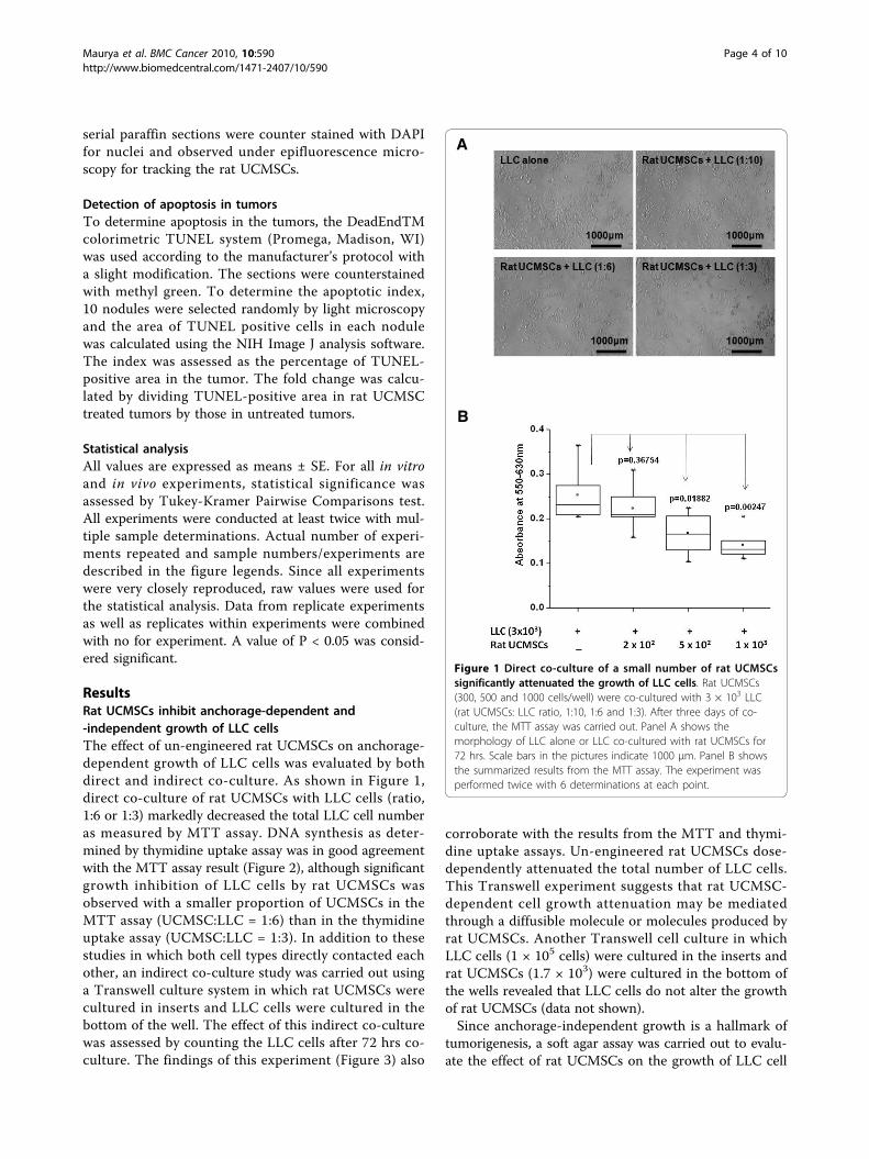
ResultsRat UCMSCs inhibit anchorage-dependent and-independent growth of LLC cellsThe effect of un-engineered rat UCMSCs on anchorage-dependent growth of LLC cells was evaluated by bothdirect and indirect co-culture. As shown in Figure 1,direct co-culture of rat UCMSCs with LLC cells (ratio,1:6 or 1:3) markedly decreased the total LLC cell numberas measured by MTT assay. DNA synthesis as deter-mined by thymidine uptake assay was in good agreementwith the MTT assay result (Figure 2), although significantgrowth inhibition of LLC cells by rat UCMSCs wasobserved with a smaller proportion of UCMSCs in theMTT assay (UCMSC:LLC = 1:6) than in the thymidineuptake assay (UCMSC:LLC = 1:3). In addition to thesestudies in which both cell types directly contacted eachother, an indirect co-culture study was carried out usinga Transwell culture system in which rat UCMSCs werecultured in inserts and LLC cells were cultured in thebottom of the well. The effect of this indirect co-culturewas assessed by counting the LLC cells after 72 hrs co-culture. The findings of this experiment (Figure 3) also
corroborate with the results from the MTT and thymi-dine uptake assays. Un-engineered rat UCMSCs dose-dependently attenuated the total number of LLC cells.This Transwell experiment suggests that rat UCMSC-dependent cell growth attenuation may be mediatedthrough a diffusible molecule or molecules produced byrat UCMSCs. Another Transwell cell culture in whichLLC cells (1 × 10⁵ cells) were cultured in the inserts andrat UCMSCs (1.7 × 10³) were cultured in the bottom ofthe wells revealed that LLC cells do not alter the growthof rat UCMSCs (data not shown).Since anchorage-independent growth is a hallmark of
tumorigenesis, a soft agar assay was carried out to evalu-ate the effect of rat UCMSCs on the growth of LLC cell
Figure 1 Direct co-culture of a small number of rat UCMSCssignificantly attenuated the growth of LLC cells. Rat UCMSCs(300, 500 and 1000 cells/well) were co-cultured with 3 × 10³ LLC(rat UCMSCs: LLC ratio, 1:10, 1:6 and 1:3). After three days of co-culture, the MTT assay was carried out. Panel A shows themorphology of LLC alone or LLC co-cultured with rat UCMSCs for72 hrs. Scale bars in the pictures indicate 1000 μm. Panel B showsthe summarized results from the MTT assay. The experiment wasperformed twice with 6 determinations at each point.
Maurya et al. BMC Cancer 2010, 10:590http://www.biomedcentral.com/1471-2407/10/590
Page 4 of 10
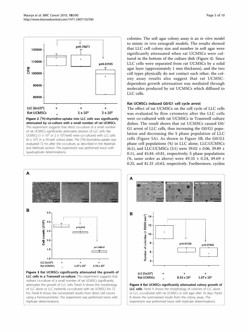
colonies. The soft agar colony assay is an in vitro modelto mimic in vivo xenograft models. The results showedthat LLC cell colony size and number in soft agar weresignificantly attenuated when rat UCMSCs were cul-tured in the bottom of the culture dish (Figure 4). SinceLLC cells were separated from rat UCMSCs by a solidagar layer (approximately 1 mm thickness), and the twocell types physically do not contact each other, the col-ony assay results also suggest that rat UCMSC-dependent growth attenuation was mediated throughmolecules produced by rat UCMSCs which diffused toLLC cells.
Rat UCMSCs induced G0/G1 cell cycle arrestThe effect of rat UCMSCs on the cell cycle of LLC cellswas evaluated by flow cytometry after the LLC cellswere co-cultured with rat UCMSCs in Transwell culturedishes. The result shows that rat UCMSCs caused G0/G1 arrest of LLC cells, thus increasing the G0/G1 popu-lation and decreasing the S phase population of LLCcells (Figure 5A). As shown in Figure 5B, the G0/G1phase cell populations (%) in LLC alone, LLC:UCMSCs(6:1), and LLC:UCMSCs (3:1) were 39.02 ± 0.06, 39.89 ±0.11, and 45.84 ±0.81, respectively; S phase populations(%, same order as above) were 49.35 ± 0.24, 49.69 ±0.25, and 41.33 ±0.63, respectively. Furthermore, cyclins
Figure 2 [3H]-thymidine-uptake into LLC cells was significantlyattenuated by co-culture with a small number of rat UCMSCs.This experiment suggests that direct co-culture of a small numberof rat UCMSCs significantly attenuates division of LLC cells. RatUCMSCs (1 × 10³ or 2 × 10³/well) were co-cultured with LLC cells(6 × 10³) in a 24-well culture plate. The [3H] thymidine-uptake wasevaluated 72 hrs after the co-culture, as described in the Materialsand Methods section. The experiment was performed twice withquadruplicate determinations.
Figure 3 Rat UCMSCs significantly attenuated the growth ofLLC cells in a Transwell co-culture. This experiment suggests thatindirect co-culture of a small number of rat UCMSCs significantlyattenuates the growth of LLC cells. Panel A shows the morphologyof LLC alone or LLC indirectly co-cultured with rat UCMSCs for 72hrs. Panel B shows the summarized results from direct cell countsusing a hemocytometer. The experiment was performed twice withtriplicate determinations.
Figure 4 Rat UCMSCs significantly attenuated colony growth ofLLC cells. Panel A shows the morphology of colonies of LLC aloneor LLC co-cultured with rat UCMSCs in soft agar after 16 days. PanelB shows the summarized results from the colony assay. Theexperiment was performed twice with triplicate determinations.
Maurya et al. BMC Cancer 2010, 10:590http://www.biomedcentral.com/1471-2407/10/590
Page 5 of 10
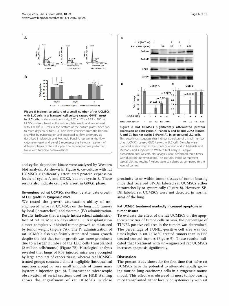
and cyclin-dependent kinase were analyzed by Westernblot analysis. As shown in Figure 6, co-culture with ratUCMSCs significantly attenuated protein expressionlevels of cyclin A and CDK2, but not cyclin E. Theseresults also indicate cell cycle arrest in G0/G1 phase.
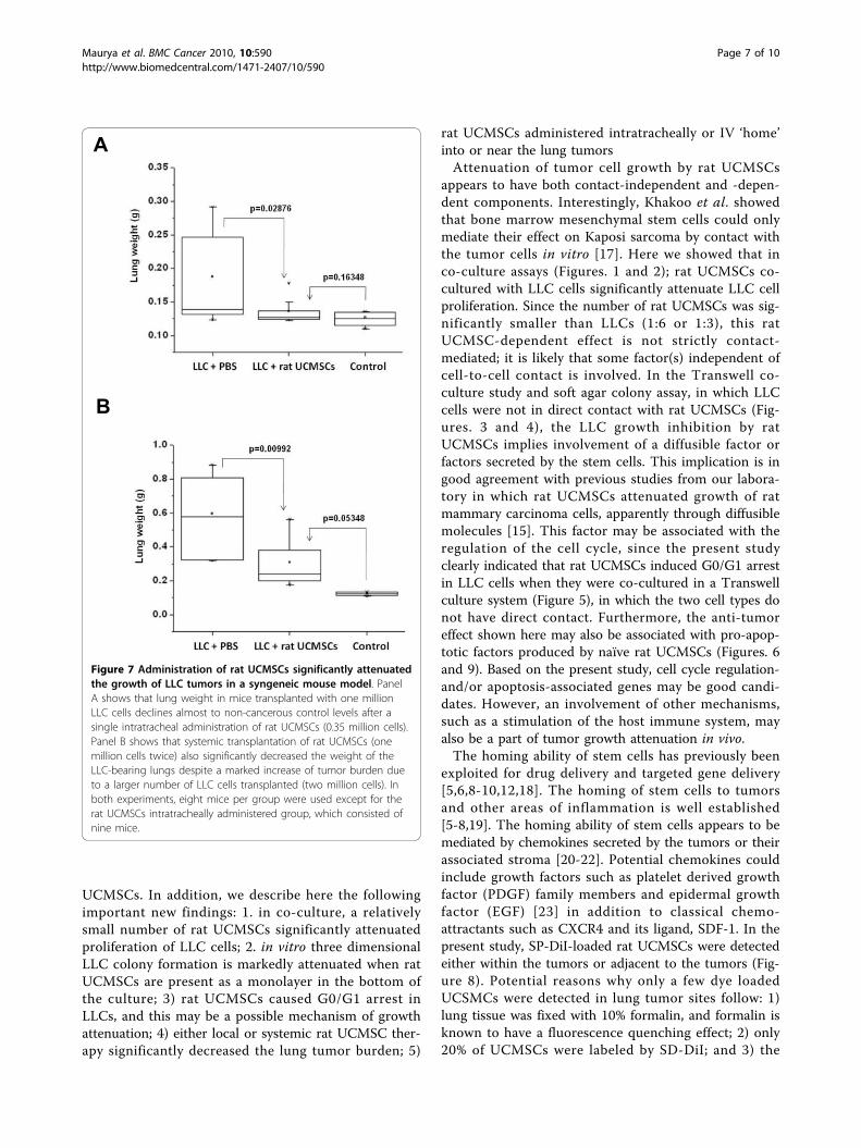
Un-engineered rat UCMSCs significantly attenuate growthof LLC grafts in syngeneic miceWe tested the growth attenuation ability of un-engineered naïve rat UCMSCs on the lung LLC tumorsby local (intratracheal) and systemic (IV) administration.Results indicate that a single intratracheal administra-tion of rat UCMSCs 5 days after LLC transplantationalmost completely inhibited tumor growth as measuredby tumor weight (Figure 7A). The IV administration ofrat UCMSCs also significantly attenuated tumor growthdespite the fact that tumor growth was more prominentdue to a larger number of the LLC cells transplanted(2 million cells/mouse) (Figure 7B). Histological analysisrevealed that lungs of PBS injected mice were occupiedby large amounts of cancer tissue, whereas rat UCMSC-treated groups contained almost negligible (intratrachealinjection group) or very small amounts of tumor mass(systemic injection group). Fluorescence microscopicobservation of serial sections used for H&E stainingshows the engraftment of rat UCMSCs in close
proximity to or within tumor tissues of tumor bearingmice that received SP-DiI labeled rat UCMSCs eitherintratracheally or systemically (Figure 8). However, SP-DiI labeled rat UCMSCs were not detected in normalareas of the lung.
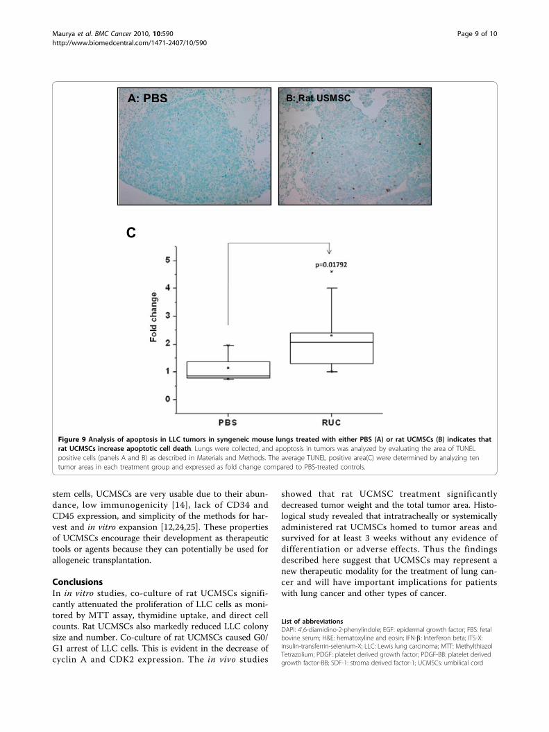
Rat UCMSC treatment markedly increased apoptosis intumor tissuesTo evaluate the effect of the rat UCMSCs on the apop-totic activities of tumor cells in vivo, the percentage ofTUNEL-positive cell area in the tumors was determined.The percentage of TUNEL-positive cell area was twotimes higher in rat UCMSC treated tumors than in PBStreated control tumors (Figure 9). These results indi-cated that treatment with un-engineered rat UCMSCsincreases apoptosis significantly.
DiscussionThe present study shows for the first time that naïve ratUCMSCs have the potential to attenuate rapidly grow-ing murine lung carcinoma cells in a syngeneic mousemodel. This effect was observed in most tumor-bearingmice transplanted either locally or systemically with rat
Figure 5 Indirect co-culture of a small number of rat UCMSCswith LLC cells in a Transwell cell culture caused G0/G1 arrestin LLC cells. In the co-culture study, 1.67 × 10⁴ or 3.33 × 10⁴ ratUCMSCs were placed in the culture plate inserts and co-culturedwith 1 × 10⁵ LLC cells in the bottom of the culture plates. After twoto three days co-culture, LLC cells were collected from the bottomchamber by trypsinization and subjected to flow cytometry asdescribed in Materials and Methods. Panel A represents the flowcytometry result and panel B represents the histogram pattern ofdifferent phases of the cell cycle. The experiment was performedtwice with triplicate determinations.
Figure 6 Rat UCMSCs significantly attenuated proteinexpression of both cyclin A (Panels A and B) and CDK2 (PanelsA and C), but not cyclin E (Panel A), in co-cultured LLC cells.This experiment suggests that indirect co-culture of a small numberof rat UCMSCs caused G0/G1 arrest in LLC cells. Samples wereprepared as described in the Figure 5 legend and in Materials andMethods, and subjected to Western blot analysis. Samplepreparation and Western blot analysis were performed three timeswith duplicate determinations. The pictures (Panel A) representtypical blotting results. P values were calculated as compared to thelevel of control.
Maurya et al. BMC Cancer 2010, 10:590http://www.biomedcentral.com/1471-2407/10/590
Page 6 of 10
UCMSCs. In addition, we describe here the followingimportant new findings: 1. in co-culture, a relativelysmall number of rat UCMSCs significantly attenuatedproliferation of LLC cells; 2. in vitro three dimensionalLLC colony formation is markedly attenuated when ratUCMSCs are present as a monolayer in the bottom ofthe culture; 3) rat UCMSCs caused G0/G1 arrest inLLCs, and this may be a possible mechanism of growthattenuation; 4) either local or systemic rat UCMSC ther-apy significantly decreased the lung tumor burden; 5)
rat UCMSCs administered intratracheally or IV ‘home’into or near the lung tumorsAttenuation of tumor cell growth by rat UCMSCs
appears to have both contact-independent and -depen-dent components. Interestingly, Khakoo et al. showedthat bone marrow mesenchymal stem cells could onlymediate their effect on Kaposi sarcoma by contact withthe tumor cells in vitro [17]. Here we showed that inco-culture assays (Figures. 1 and 2); rat UCMSCs co-cultured with LLC cells significantly attenuate LLC cellproliferation. Since the number of rat UCMSCs was sig-nificantly smaller than LLCs (1:6 or 1:3), this ratUCMSC-dependent effect is not strictly contact-mediated; it is likely that some factor(s) independent ofcell-to-cell contact is involved. In the Transwell co-culture study and soft agar colony assay, in which LLCcells were not in direct contact with rat UCMSCs (Fig-ures. 3 and 4), the LLC growth inhibition by ratUCMSCs implies involvement of a diffusible factor orfactors secreted by the stem cells. This implication is ingood agreement with previous studies from our labora-tory in which rat UCMSCs attenuated growth of ratmammary carcinoma cells, apparently through diffusiblemolecules [15]. This factor may be associated with theregulation of the cell cycle, since the present studyclearly indicated that rat UCMSCs induced G0/G1 arrestin LLC cells when they were co-cultured in a Transwellculture system (Figure 5), in which the two cell types donot have direct contact. Furthermore, the anti-tumoreffect shown here may also be associated with pro-apop-totic factors produced by naïve rat UCMSCs (Figures. 6and 9). Based on the present study, cell cycle regulation-and/or apoptosis-associated genes may be good candi-dates. However, an involvement of other mechanisms,such as a stimulation of the host immune system, mayalso be a part of tumor growth attenuation in vivo.The homing ability of stem cells has previously been
exploited for drug delivery and targeted gene delivery[5,6,8-10,12,18]. The homing of stem cells to tumorsand other areas of inflammation is well established[5-8,19]. The homing ability of stem cells appears to bemediated by chemokines secreted by the tumors or theirassociated stroma [20-22]. Potential chemokines couldinclude growth factors such as platelet derived growthfactor (PDGF) family members and epidermal growthfactor (EGF) [23] in addition to classical chemo-attractants such as CXCR4 and its ligand, SDF-1. In thepresent study, SP-DiI-loaded rat UCMSCs were detectedeither within the tumors or adjacent to the tumors (Fig-ure 8). Potential reasons why only a few dye loadedUCSMCs were detected in lung tumor sites follow: 1)lung tissue was fixed with 10% formalin, and formalin isknown to have a fluorescence quenching effect; 2) only20% of UCMSCs were labeled by SD-DiI; and 3) the
Figure 7 Administration of rat UCMSCs significantly attenuatedthe growth of LLC tumors in a syngeneic mouse model. PanelA shows that lung weight in mice transplanted with one millionLLC cells declines almost to non-cancerous control levels after asingle intratracheal administration of rat UCMSCs (0.35 million cells).Panel B shows that systemic transplantation of rat UCMSCs (onemillion cells twice) also significantly decreased the weight of theLLC-bearing lungs despite a marked increase of tumor burden dueto a larger number of LLC cells transplanted (two million cells). Inboth experiments, eight mice per group were used except for therat UCMSCs intratracheally administered group, which consisted ofnine mice.
Maurya et al. BMC Cancer 2010, 10:590http://www.biomedcentral.com/1471-2407/10/590
Page 7 of 10
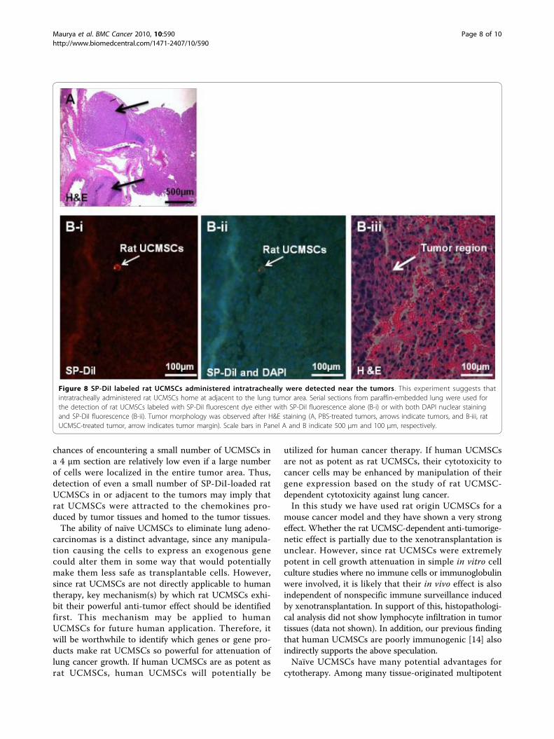
chances of encountering a small number of UCMSCs ina 4 μm section are relatively low even if a large numberof cells were localized in the entire tumor area. Thus,detection of even a small number of SP-DiI-loaded ratUCMSCs in or adjacent to the tumors may imply thatrat UCMSCs were attracted to the chemokines pro-duced by tumor tissues and homed to the tumor tissues.The ability of naïve UCMSCs to eliminate lung adeno-
carcinomas is a distinct advantage, since any manipula-tion causing the cells to express an exogenous genecould alter them in some way that would potentiallymake them less safe as transplantable cells. However,since rat UCMSCs are not directly applicable to humantherapy, key mechanism(s) by which rat UCMSCs exhi-bit their powerful anti-tumor effect should be identifiedfirst. This mechanism may be applied to humanUCMSCs for future human application. Therefore, itwill be worthwhile to identify which genes or gene pro-ducts make rat UCMSCs so powerful for attenuation oflung cancer growth. If human UCMSCs are as potent asrat UCMSCs, human UCMSCs will potentially be
utilized for human cancer therapy. If human UCMSCsare not as potent as rat UCMSCs, their cytotoxicity tocancer cells may be enhanced by manipulation of theirgene expression based on the study of rat UCMSC-dependent cytotoxicity against lung cancer.In this study we have used rat origin UCMSCs for a
mouse cancer model and they have shown a very strongeffect. Whether the rat UCMSC-dependent anti-tumorige-netic effect is partially due to the xenotransplantation isunclear. However, since rat UCMSCs were extremelypotent in cell growth attenuation in simple in vitro cellculture studies where no immune cells or immunoglobulinwere involved, it is likely that their in vivo effect is alsoindependent of nonspecific immune surveillance inducedby xenotransplantation. In support of this, histopathologi-cal analysis did not show lymphocyte infiltration in tumortissues (data not shown). In addition, our previous findingthat human UCMSCs are poorly immunogenic [14] alsoindirectly supports the above speculation.Naïve UCMSCs have many potential advantages for
cytotherapy. Among many tissue-originated multipotent
Figure 8 SP-DiI labeled rat UCMSCs administered intratracheally were detected near the tumors. This experiment suggests thatintratracheally administered rat UCMSCs home at adjacent to the lung tumor area. Serial sections from paraffin-embedded lung were used forthe detection of rat UCMSCs labeled with SP-DiI fluorescent dye either with SP-DiI fluorescence alone (B-i) or with both DAPI nuclear stainingand SP-DiI fluorescence (B-ii). Tumor morphology was observed after H&E staining (A, PBS-treated tumors, arrows indicate tumors, and B-iii, ratUCMSC-treated tumor, arrow indicates tumor margin). Scale bars in Panel A and B indicate 500 μm and 100 μm, respectively.
Maurya et al. BMC Cancer 2010, 10:590http://www.biomedcentral.com/1471-2407/10/590
Page 8 of 10
stem cells, UCMSCs are very usable due to their abun-dance, low immunogenicity [14], lack of CD34 andCD45 expression, and simplicity of the methods for har-vest and in vitro expansion [12,24,25]. These propertiesof UCMSCs encourage their development as therapeutictools or agents because they can potentially be used forallogeneic transplantation.
ConclusionsIn in vitro studies, co-culture of rat UCMSCs signifi-cantly attenuated the proliferation of LLC cells as moni-tored by MTT assay, thymidine uptake, and direct cellcounts. Rat UCMSCs also markedly reduced LLC colonysize and number. Co-culture of rat UCMSCs caused G0/G1 arrest of LLC cells. This is evident in the decrease ofcyclin A and CDK2 expression. The in vivo studies
showed that rat UCMSC treatment significantlydecreased tumor weight and the total tumor area. Histo-logical study revealed that intratracheally or systemicallyadministered rat UCMSCs homed to tumor areas andsurvived for at least 3 weeks without any evidence ofdifferentiation or adverse effects. Thus the findingsdescribed here suggest that UCMSCs may represent anew therapeutic modality for the treatment of lung can-cer and will have important implications for patientswith lung cancer and other types of cancer.
List of abbreviationsDAPI: 4’,6-diamidino-2-phenylindole; EGF: epidermal growth factor; FBS: fetalbovine serum; H&E: hematoxyline and eosin; IFN-b: Interferon beta; ITS-X:insulin-transferrin-selenium-X; LLC: Lewis lung carcinoma; MTT: MethylthiazolTetrazolium; PDGF: platelet derived growth factor; PDGF-BB: platelet derivedgrowth factor-BB; SDF-1: stroma derived factor-1; UCMSCs: umbilical cord
Figure 9 Analysis of apoptosis in LLC tumors in syngeneic mouse lungs treated with either PBS (A) or rat UCMSCs (B) indicates thatrat UCMSCs increase apoptotic cell death. Lungs were collected, and apoptosis in tumors was analyzed by evaluating the area of TUNELpositive cells (panels A and B) as described in Materials and Methods. The average TUNEL positive area(C) were determined by analyzing tentumor areas in each treatment group and expressed as fold change compared to PBS-treated controls.
Maurya et al. BMC Cancer 2010, 10:590http://www.biomedcentral.com/1471-2407/10/590
Page 9 of 10
matrix stem cells; CDK2: Cyclin-dependent Kinase 2; GAPDH: Glyceraldehyde3-phosphate dehydrogenase;
AcknowledgementsThis work was supported by Kansas State University (KSU) Terry C. JohnsonCenter for Basic Cancer Research, Kansas Bioscience Authority CollaborativeCancer Research Initiative grant, KSU Targeted Excellence research grant,Kansas State Legislative Appropriation, KSU College of Veterinary MedicineDean’s fund, NIH grants P20 RR017686, P20 RR01556 and R21 CA135599 andJoan’s Legacy Foundation.
Authors’ contributionsDKM, CD, AK, CK, ZW, and MT were responsible for the study design,experimental work, data evaluation and analysis, and drafting themanuscript. MMP and DT were consulted extensively in the experimentaldesign and interpretation of results, as well as in the preparation of themanuscript. MT was the research supervisor and participated in the studydesign, assessment of the results, and drafting the manuscript. All authorsread and approved the manuscript.
Competing interestsThe authors declare that they have no competing interests.
Received: 25 September 2009 Accepted: 28 October 2010Published: 28 October 2010
References1. Jemal A, Siegel R, Ward E, Hao Y, Xu J, Thun MJ: Cancer Statistics, 2009. CA
Cancer J Clin 2009.2. Johnson DH, Schiller JH: Novel therapies for the treatment of non-small
cell lung cancer. Cancer Chemother Biol Response Modif 2002, 20:763-786.3. Lynch TJ, Adjei AA, Bunn PA Jr, DuBois RN, Gandara DR, Giaccone G,
Govindan R, Herbst RS, Johnson BE, Khuri FR, et al: Novel agents in thetreatment of lung cancer: conference summary statement. Clin CancerRes 2004, 10(12 Pt 2):4199s-4204s.
4. Corsten MF, Shah K: Therapeutic stem-cells for cancer treatment: hopesand hurdles in tactical warfare. Lancet Oncol 2008, 9(4):376-384.
5. Aboody KS, Brown A, Rainov NG, Bower KA, Liu S, Yang W, Small JE,Herrlinger U, Ourednik V, Black PM, et al: Neural stem cells displayextensive tropism for pathology in adult brain: evidence fromintracranial gliomas. Proc Natl Acad Sci USA 2000, 97(23):12846-12851.
6. Nakamizo A, Marini F, Amano T, Khan A, Studeny M, Gumin J, Chen J,Hentschel S, Vecil G, Dembinski J, et al: Human bone marrow-derivedmesenchymal stem cells in the treatment of gliomas. Cancer Res 2005,65(8):3307-3318.
7. Rachakatla RS, Marini F, Weiss ML, Tamura M, Troyer D: Development ofhuman umbilical cord matrix stem cell-based gene therapy forexperimental lung tumors. Cancer Gene Ther 2007, 14(10):828-835.
8. Studeny M, Marini FC, Champlin RE, Zompetta C, Fidler IJ, Andreeff M: Bonemarrow-derived mesenchymal stem cells as vehicles for interferon-betadelivery into tumors. Cancer Res 2002, 62(13):3603-3608.
9. Studeny M, Marini FC, Dembinski JL, Zompetta C, Cabreira-Hansen M,Bekele BN, Champlin RE, Andreeff M: Mesenchymal stem cells: potentialprecursors for tumor stroma and targeted-delivery vehicles foranticancer agents. J Natl Cancer Inst 2004, 96(21):1593-1603.
10. Aboody KS, Najbauer J, Schmidt NO, Yang W, Wu JK, Zhuge Y, Przylecki W,Carroll R, Black PM, Perides G: Targeting of melanoma brain metastasesusing engineered neural stem/progenitor cells. Neuro Oncol 2006,8(2):119-126.
11. Kumar S, Chanda D, Ponnazhagan S: Therapeutic potential of geneticallymodified mesenchymal stem cells. Gene Ther 2008, 15(10):711-715.
12. Mitchell KE, Weiss ML, Mitchell BM, Martin P, Davis D, Morales L, Helwig B,Beerenstrauch M, Abou-Easa K, Hildreth T, et al: Matrix cells fromWharton’s jelly form neurons and glia. Stem Cells 2003, 21(1):50-60.
13. Cho PS, Messina DJ, Hirsh EL, Chi N, Goldman SN, Lo DP, Harris IR,Popma SH, Sachs DH, Huang CA: Immunogenicity of umbilical cord tissuederived cells. Blood 2008, 111(1):430-438.
14. Weiss ML, Anderson C, Medicetty S, Seshareddy KB, Weiss RJ, VanderWerff I,Troyer D, McIntosh KR: Immune properties of human umbilical cordWharton’s jelly-derived cells. Stem Cells 2008, 26(11):2865-2874.
15. Ganta C, Chiyo D, Ayuzawa R, Rachakatla R, Pyle M, Andrews G, Weiss M,Tamura M, Troyer D: Rat umbilical cord stem cells completely abolish ratmammary carcinomas with no evidence of metastasis or recurrence 100days post-tumor cell inoculation. Cancer Res 2009, 69(5):1815-1820.
16. Ayuzawa A, Doi C, Rachakatla RS, Pyle MM, Maurya DK, Troyer D, Tamura M:Naïve human umbilical cord matrix derived stem cells significantlyattenuate growth of human breast cancer cells in vitro and in vivo.Cancer letters 2009, 280(1):31-37.
17. Khakoo AY, Pati S, Anderson SA, Reid W, Elshal MF, Rovira II, Nguyen AT,Malide D, Combs CA, Hall G, et al: Human mesenchymal stem cells exertpotent antitumorigenic effects in a model of Kaposi’s sarcoma. J ExpMed 2006, 203(5):1235-1247.
18. Aboody KS, Najbauer J, Danks MK: Stem and progenitor cell-mediatedtumor selective gene therapy. Gene Ther 2008, 15(10):739-752.
19. Hall B, Andreeff M, Marini F: The participation of mesenchymal stem cellsin tumor stroma formation and their application as targeted-genedelivery vehicles. Handb Exp Pharmacol 2007, , 180: 263-283.
20. Muller A, Homey B, Soto H, Ge N, Catron D, Buchanan ME, McClanahan T,Murphy E, Yuan W, Wagner SN, et al: Involvement of chemokine receptorsin breast cancer metastasis. Nature 2001, 410(6824):50-56.
21. Lee BC, Lee TH, Avraham S, Avraham HK: Involvement of the chemokinereceptor CXCR4 and its ligand stromal cell-derived factor 1alpha inbreast cancer cell migration through human brain microvascularendothelial cells. Mol Cancer Res 2004, 2(6):327-338.
22. Karnoub AE, Dash AB, Vo AP, Sullivan A, Brooks MW, Bell GW,Richardson AL, Polyak K, Tubo R, Weinberg RA: Mesenchymal stem cellswithin tumour stroma promote breast cancer metastasis. Nature 2007,449(7162):557-563.
23. Arbab AS, Janic B, Knight RA, Anderson SA, Pawelczyk E, Rad AM, Read EJ,Pandit SD, Frank JA: Detection of migration of locally implanted AC133+stem cells by cellular magnetic resonance imaging with histologicalfindings. Faseb J 2008, 22(9):3234-3246.
24. Weiss ML, Medicetty S, Bledsoe AR, Rachakatla RS, Choi M, Merchav S,Luo Y, Rao MS, Velagaleti G, Troyer D: Human umbilical cord matrix stemcells: preliminary characterization and effect of transplantation in arodent model of Parkinson’s disease. Stem Cells 2006, 24(3):781-792.
25. Weiss ML, Troyer DL: Stem cells in the umbilical cord. Stem Cell Rev 2006,2(2):155-162.
Pre-publication historyThe pre-publication history for this paper can be accessed here:http://www.biomedcentral.com/1471-2407/10/590/prepub
doi:10.1186/1471-2407-10-590Cite this article as: Maurya et al.: Therapy with un-engineered naïve ratumbilical cord matrix stem cells markedly inhibits growth of murinelung adenocarcinoma. BMC Cancer 2010 10:590.
Submit your next manuscript to BioMed Centraland take full advantage of:
• Convenient online submission
• Thorough peer review
• No space constraints or color figure charges
• Immediate publication on acceptance
• Inclusion in PubMed, CAS, Scopus and Google Scholar
• Research which is freely available for redistribution
Submit your manuscript at www.biomedcentral.com/submit
Maurya et al. BMC Cancer 2010, 10:590http://www.biomedcentral.com/1471-2407/10/590
Page 10 of 10
Related Documents