RESEARCH ARTICLE Open Access Therapeutic potentials of Crataegus azarolus var. eu- azarolus Maire leaves and its isolated compounds Eman Abu-Gharbieh 1,2* and Naglaa Gamil Shehab 3 Abstract Background: Hyperglycemia is a complicated condition accompanied with high incidence of infection and dyslipidemia. This study aimed to explore the phyto-constituents of Crataegus azarolus var. eu- azarolus Maire leaves, and to evaluate the therapeutic potentials particularly antimicrobial, antihyperglycemic and antihyperlipidemic of the extract and the isolated compound (3β-O-acetyl ursolic acid). Methods: Total phenolics and flavonoidal contents were measured by RP-HPLC analysis. Free radicals scavenging activity of different extraction solvents was tested in-vitro on DPPH free radicals. The antimicrobial activity of the ethanolic extract and its fractions as well as the isolated compounds were evaluated in-vitro on variable microorganisms. Animal models were used to evaluate the antihyperglycemic and antihyperlipidemic activities of the ethanolic extract along with the isolated compound (3β-O acetyl ursolic acid). Results: RP- HPLC analysis of the phenolics revealed high content of rutin, salicylic and ellagic acids. Six compounds belonging to triterpenes and phenolics were isolated from chloroform and n-butanol fractions namely: ursolic acid, 3β-O-acetyl ursolic acid, ellagic acid, quercetin 3-O-β methyl ether, rutin and apigenin7-O-rutinoside. Ethanolic extract showed the highest DPPH radical scavenger activity compared to other solvents. Ethanolic extract, hexane fraction, ursolic acid, 3β-O acetyl ursolic acid and quercetin 3-O-methyl ether showed variable antimicrobial activity against E. coli, P. aeruginosa, S. aureus, and C. albicans. Administration of the ethanolic extract or 3β-O acetyl ursolic acid orally to the mice reduced blood glucose significantly in a time- and dose-dependent manner. Ethanolic extract significantly reduced LDL-C, VLDL-C, TC and TG and increased HDL-C in rats. Ethanolic extract and 3β-O acetyl ursolic acid reduced in-vitro activity of pancreatic lipase. Conclusion: This study reveals that Crataegus azarolus var. eu- azarolus Maire has the efficiency to control hyperglycemia with its associated complications. This study is the first to evaluate antihyperglycemic and antihyperlipidemic potentials of 3β-O acetyl ursolic acid. Keywords: Crataegus azarolus var. eu- azarolus Maire, 3β-O acetyl ursolic acid, Antimicrobial, Antihyperglycemic, Antihyperlipidemic * Correspondence: [email protected] 1 Department of Basic Medical Sciences, College of Medicine, University of Sharjah, Sharjah, United Arab Emirates 2 Department of Pharmacology and Toxicology, Dubai Pharmacy College, Dubai, United Arab Emirates Full list of author information is available at the end of the article © The Author(s). 2017 Open Access This article is distributed under the terms of the Creative Commons Attribution 4.0 International License (http://creativecommons.org/licenses/by/4.0/), which permits unrestricted use, distribution, and reproduction in any medium, provided you give appropriate credit to the original author(s) and the source, provide a link to the Creative Commons license, and indicate if changes were made. The Creative Commons Public Domain Dedication waiver (http://creativecommons.org/publicdomain/zero/1.0/) applies to the data made available in this article, unless otherwise stated. Abu-Gharbieh and Shehab BMC Complementary and Alternative Medicine (2017) 17:218 DOI 10.1186/s12906-017-1729-9

Welcome message from author
This document is posted to help you gain knowledge. Please leave a comment to let me know what you think about it! Share it to your friends and learn new things together.
Transcript
RESEARCH ARTICLE Open Access
Therapeutic potentials of Crataegusazarolus var. eu- azarolus Maire leavesand its isolated compoundsEman Abu-Gharbieh1,2* and Naglaa Gamil Shehab3
Abstract
Background: Hyperglycemia is a complicated condition accompanied with high incidence of infection anddyslipidemia. This study aimed to explore the phyto-constituents of Crataegus azarolus var. eu- azarolus Maireleaves, and to evaluate the therapeutic potentials particularly antimicrobial, antihyperglycemic andantihyperlipidemic of the extract and the isolated compound (3β-O-acetyl ursolic acid).Methods: Total phenolics and flavonoidal contents were measured by RP-HPLC analysis. Free radicals scavengingactivity of different extraction solvents was tested in-vitro on DPPH free radicals. The antimicrobial activity of theethanolic extract and its fractions as well as the isolated compounds were evaluated in-vitro on variablemicroorganisms. Animal models were used to evaluate the antihyperglycemic and antihyperlipidemic activities ofthe ethanolic extract along with the isolated compound (3β-O acetyl ursolic acid).
Results: RP- HPLC analysis of the phenolics revealed high content of rutin, salicylic and ellagic acids. Sixcompounds belonging to triterpenes and phenolics were isolated from chloroform and n-butanol fractions namely:ursolic acid, 3β-O-acetyl ursolic acid, ellagic acid, quercetin 3-O-β methyl ether, rutin and apigenin7-O-rutinoside.Ethanolic extract showed the highest DPPH radical scavenger activity compared to other solvents. Ethanolic extract,hexane fraction, ursolic acid, 3β-O acetyl ursolic acid and quercetin 3-O-methyl ether showed variable antimicrobialactivity against E. coli, P. aeruginosa, S. aureus, and C. albicans. Administration of the ethanolic extract or 3β-Oacetyl ursolic acid orally to the mice reduced blood glucose significantly in a time- and dose-dependent manner.Ethanolic extract significantly reduced LDL-C, VLDL-C, TC and TG and increased HDL-C in rats. Ethanolic extract and3β-O acetyl ursolic acid reduced in-vitro activity of pancreatic lipase.
Conclusion: This study reveals that Crataegus azarolus var. eu- azarolus Maire has the efficiency to controlhyperglycemia with its associated complications. This study is the first to evaluate antihyperglycemic andantihyperlipidemic potentials of 3β-O acetyl ursolic acid.
Keywords: Crataegus azarolus var. eu- azarolus Maire, 3β-O acetyl ursolic acid, Antimicrobial, Antihyperglycemic,Antihyperlipidemic
* Correspondence: [email protected] of Basic Medical Sciences, College of Medicine, University ofSharjah, Sharjah, United Arab Emirates2Department of Pharmacology and Toxicology, Dubai Pharmacy College,Dubai, United Arab EmiratesFull list of author information is available at the end of the article
© The Author(s). 2017 Open Access This article is distributed under the terms of the Creative Commons Attribution 4.0International License (http://creativecommons.org/licenses/by/4.0/), which permits unrestricted use, distribution, andreproduction in any medium, provided you give appropriate credit to the original author(s) and the source, provide a link tothe Creative Commons license, and indicate if changes were made. The Creative Commons Public Domain Dedication waiver(http://creativecommons.org/publicdomain/zero/1.0/) applies to the data made available in this article, unless otherwise stated.
Abu-Gharbieh and Shehab BMC Complementary andAlternative Medicine (2017) 17:218 DOI 10.1186/s12906-017-1729-9
BackgroundDyslipidemia and increased susceptibility to infections aretwo typical complications of diabetes mellitus. High glu-cose levels are highly associated with immune system im-pairment, particularly on neutrophils [1]. Hyperglycemiareduces the phagocytic activity and ability of neutrophilsto form extracellular traps to kill bacteria [1]. On the otherhand, hyperglycemia due to both insulin deficiency and in-sulin resistance significantly affect the lipid metabolicpathways [2]. Diabetic patients usually experience variouscardiovascular complications of which dyslipidemia repre-sents a main risk factor [2].There has been a noticeable increase in the use of
both traditional home remedies and herbal medicineinstead of relying on conventional treatments [3]. Thishas caused traditional medicine to become of worldwideimportance, with medicinal and economic effects [4].Crataegus azarolus is indigenous to the Mediterranean
Basin. Crataegus azarolus var. eu- azarolus Maire is alow, dense, spiny tree with a beautiful inflorescence upto 6 m tall and with orange fruits [5]. Phytochemical in-vestigation of the plant was performed mainly on theflowers. Antioxidant activity and phenolic compositionof the flowers extract were studied [6]. No available liter-atures concerning the pharmacological activity and thephytochemical constituents of the leaves were found.In folk medicine, genus Crataegus (commonly called
hawthorn in English and Zaarour in Arabic) is used forcuring several ailments viz. central nervous, reproductive,cardiovascular and immune systems [7]. It also showedanti-inflammatory, cytotoxic, antioxidant, gastroprotec-tive, antimicrobial, cardioprotective, antidiabetic and anti-HIV activities [8–14].Phytochemical investigations on genus Crataegus were
mainly performed on the leaves, flowers and berries. Theisolated compounds were: oligomeric procyanidins,bioflavonoid, polysaccharides, catecholamines, vitaminC, saponins, tannins, cardiotonic amines, purine deriva-tives and ursolic acid [8–10, 15–18].This study aimed to explore the phytochemical com-
position of Crataegus azarolus var. eu- azarolus Maireleaves’ extract, assess its acute toxicity and investigatethe free radical scavenging and therapeutic potentialsparticularly antimicrobial, antihyperglycemic and antihy-perlipidemic activities.
MethodsGeneralShimadzu 1700 spectrophotometer was used for UVabsorption spectra. Melting points were determined onElectrothermal 9100 equipment. Mass spectra were mea-sured on a Jeol Mass Spectrometer SSQ 7000, DigitalDEC 300. NMR spectra were measured in DMSO orCD3OD or CDCl3;
1H–NMR spectra were obtained at
400 MHz and 13 C-NMR spectra at 100 MHz on a JEOLGX-400 spectrometer with the chemical shifts (δ ppm)expressed relative to TMS as internal standard. Pre-coated silica gel 60 F254 (Merck, Darmstadt, Germany)was used for the TLC analysis. Vacuum liquid chroma-tography (VLC) was performed on silica gel 60 GF(Merck, Darmstadt, Germany). Sephadex LH-20 (Sigma-Aldrich, St. Louis, Missouri, United States) and silica gel100 C18-Reversed Phase (Fluka, Switzerland) were alsoused. Analysis of phenolics was performed on HewlettPackard HPLC (HP 1050HPLCDADw/Data System)equipped with a Hypersil-ODS (4.6 X 250 mm, 5 μm)column and a UV detector.
Plant materialLeaves of Crataegus azarolus var. eu- azarolus were col-lected during the fruiting stage in July 2012 from plantscultivated in Nablus, Palestine. The plant was identifiedby Professor Hassnaa Ahmed Hosny, Department ofBotany, Faculty of Science, Cairo University, Egypt. Avoucher specimen has been kept at the Herbarium ofthe Department of Pharmacognosy, Faculty of Pharmacy,Cairo University.
Chemicals, drugs and biochemical kitsAll solvents were of analytical grade and obtained fromFisher Scientific. Sodium carboxymethylcellulose (CMC),2,2-diphenyl-1-picrylhydrazyl (DPPH), gentamicin, flu-conazole, alloxan, glibenclamide, cholesterol, lovastatin,orlistat, procaine pancreatic lipase type II, p-nitrophenylbutyrate (PNPB) and HMG-CoA reductase assay kitwere purchased from Sigma-Aldrich (St. Louis, Missouri,United States). Folin-Ciocalteu reagent was obtainedfrom Merck (Darmstadt, Germany). Lipid profile assess-ment kits including total cholesterol (TC), low densitylipoprotein cholesterol (LDL-C), very low density lipo-protein cholesterol (VLDL-C), high density lipoproteincholesterol (HDL-C) and triglycerides (TG) were pur-chased from Abcam-Cambridge, UK. Glucose estimationkit (kit Glu 1108, Test Strips, One Touch, Lifescan) wasused to evaluate the blood glucose level.
Plant leaves extractionSelection of best extraction solvent for phenolic contentsAcetone, ethyl acetate, Methanol and 70% ethanol wereused individually for the extraction of the powdered leavesmaterial (each 100 g). Spectrophotometric methods wereused to determine the phenolic and the flavonoid con-tents. The experiments were carried out in triplicate.
Spectrophotometric determination of total phenolic andflavonoid contentsFolin-Ciocalteu reagent was used to measure the phen-olic content according to Oktay et al. [19]. Different
Abu-Gharbieh and Shehab BMC Complementary and Alternative Medicine (2017) 17:218 Page 2 of 13
concentrations from gallic acid (10–50 µg/mL) were pre-pared for the standard calibration curve. The absorbancewas determined at 750 nm. Results were calculated asmg gallic acid/g dry plant weight equivalent.Aluminum chloride was used to assess the total
flavonoid contents of the different extracts according tothe procedure described by Dewanto et al. [20]. Serialdilutions of quercetin were used for preparation of thestandard calibration curve. The absorbance was mea-sured at 510 nm. All the experiments were carried outin triplicate.
RP- HPLC analysisPhenolic composition of C. azarolus var. eu- azarolusleaves was investigated in aliquots (1 g, each) of themethanolic extract of the plant via RP-HPLC [21, 22]based on the method previously described by theauthors [23]. For the phenolic acids composition, theUV detector was set at 280 nm while for flavonoidscomposition the UV detector was adjusted at 330 nm.Quantification was based on peak area calculation andwas done in triplicate.
Large scale extraction and fractionationAir-dried powdered leaves of C. azarolus var. eu- azarolus(2.5 Kg) were extracted at room temperature by macer-ation in ethanol (70%, 10 L × 3). The ethanolic extract wasevaporated under reduced pressure at temperature 55 °Cto provide 280 g residue. 200 g residue were successivelyfractionated with different solvents according to the polar-ity viz. n-hexane, chloroform and n-butanol saturated withwater; while the remaining residual amount was saved forbiological evaluation. Partitioning the ethanolic extractwith different solvents yielded 20, 15 and 18 g of driedextractive respectively.
Isolation of the constituents of the chloroform extractAn accurately weighed amount of the chloroform ex-tract (13.0 g) was applied on silica gel 60 GF VLCcolumn (25 X 5 cm). Gradient elution was performedusing hexane-chloroform, chloroform-ethyl acetatemixtures and ethyl acetate. Fractions (100 mL each)were gathered and monitored by TLC using differentmobile phase (System A, chloroform-methanol 9.5:0.5 andSystem B, ethyl acetate: formic acid: acetic acid: water10.0:1.0:1.0:0.5). Spots were located by visualization underUV365 nm before and after exposure to ammonia vaporand by spraying with p-anisaldehyde at 110 °C. Similarfractions were pooled to yield 6 collective fractions(F1-F6). According to the weight of the fraction andthe number of spots, fractions F3 and F5 were se-lected for further isolation.F3: (2.5 g; 6 spots, Rf values 0.83, 0.70, 0.60, 0.50, 0.40
and 0.34, system A) upon rechromatography on a silica
gel 100 C18-RP column under reduced pressure (20 X1.5 cm), using methanol–water 9:1 as eluent, resulted inisolation of compounds 1 and 2.F5: (1.2 g; 2 spots, Rf values 0.77 and 0.47, System B)
was exposed to three columns the first and second wereon sephadex LH-20 (35 X 3 cm; elution, methanol–water 9:1 then 8:2) the third one was a silica gel 100C18-RP under reduced pressure (20 X 1.5 cm; elution,methanol–water 1:1) to afford compound 3.
Isolation of the constituents of the n-butanol extractThe n-butanol residue (15 g) was fractionated by silicagel 60 GF VLC column (30 × 3.5 cm). Mixtures fromdifferent solvents were used (chloroform-ethyl acetate,ethyl acetate and ethyl acetate-methanol). Fractions(200 mL each) were gathered and seen by TLC (Sys-tem B and System C, chloroform: methanol: water8.5:1.5: 0.1). Spots were located before and afterexposure to ammonia vapor by visualization underUV365 nm and by spraying with p-anisaldehyde at110 °C. Similar fractions were pooled to yield 10collective fractions (P1-P10). According to the weightof the fractions and the number of spots, fractions P7and P8 were selected for further isolation.P7: (1.5 g; 2 spots, Rf values 0.72 and 0.50, System C)
upon rechromatography on a silica gel 100 C18-RPcolumn under reduced pressure(20 X 1.5 cm) usingmethanol: water 1:1 as eluent, resulted in isolation ofcompound 4.P8: (0.8 g; 2 spots, Rf values 0.38 and 0.22, System
B) upon rechromatography on Sephadex LH-20 col-umn (35 X 3 cm) using methanol: water 8:2 as eluentthen on silica gel 100 C18-RP column (20 X 1.5 cm)using methanol: water 1:1 resulted in isolation of com-pounds 5 and 6.
Antioxidant activityDPPH radical scavenging assayEthanol (70%), methanol, acetone, and ethyl acetate wereused individually to extract the leaves. The free radical-scavenging activity was assessed by DPPH radical [24].Different concentrations of leaves extracts concentra-tions (3.9, 7.8, 15.6, 31.3, 62.5, 125, 250 μg/ml) and as-corbic acid (AA), as standard compound, were analyzedin triplicate. The percentage inhibition of DPPH radicalwas calculated as follow:% inhibition = [A0 - (A1- A2)]/A0 ×100%.A0: control absorbance, A1: absorbance of the sample,
A2: absorbance of the sample in ethanol without DPPH.
Biological studyMicroorganismsThree bacterial strains and one fungus, were kindlyprovided by Rashid hospital (Dubai-UAE) and were used
Abu-Gharbieh and Shehab BMC Complementary and Alternative Medicine (2017) 17:218 Page 3 of 13
for the antimicrobial screening. This included one repre-sentative of the Gram-positive group (Staphylococcusaureus RMTCC 3161), two representatives of the Gram-negative group (Escherichia coli RMTCC 2682 andPseudomonas aeruginosa RMTCC 1687) and one fungus(Candida albicans RMTCC 5122). Microorganisms weregrown on appropriate media: nutrient agar for S. aureusand P. aeruginosa, MacConkey agar for E. coli andSabouraud dextrose agar for C. albicans.
Antimicrobial activityThe ethanolic extract, and its fractions, n-hexane,chloroform and n-butanol as well as the isolated com-pounds of C. azarolus at doses of 375 μg/mL for eachextract, and 140 μg/mL for the isolated compounds weresubjected to in-vitro qualitative screening, for evaluationof their antimicrobial potentialities. The agar diffusiontechnique was used [25]. Solubilization of the samples wasassisted by sterile DMSO. The effects were compared withgentamicin (30 μg/mL) and antifungal, fluconazole(30 μg/mL). Diameters of zones of inhibition (in mm)were taken as a measure for the growth inhibitory ac-tivity against the selected strains.
Experimental animalsMale albino mice (30 ± 5 g) were used for acute toxicityand antihyperglycemic studies. Antihyperlipidemicexperiments were performed on Sprague Dawley ratsweighing 210 ± 5 g. All animals were kept under stand-ard conditions, fed with regular diet and water suppliedad libitum. Mice were accommodated for 1 week priorto the experiments. All animal investigations wereaccepted from the Ethical Research Committee of theDubai Pharmacy College, Dubai UAE and done accord-ing to the ethical standards of laboratory animals [26].
Acute oral toxicity studyLD50 was determined by probit test [27]. Mice weredivided into five groups (10 each) and they receivedvarious oral doses of the ethanolic extract (250, 500,1000, 2500 and 5000 mg/kg). Later, they were observedover 72 h for any signs of morbidity or abnormal behav-ior and their death was recorded [27].
Evaluation of antihyperglycemic activityInduction of diabetes in miceOne night before the induction of hyperglycemia, theanimals were kept fasted but given water ad libitum. Thenext morning, animals were injected 150 mg/kg alloxanmonohydrate solution in acetate buffer (0.15 M, pH 4.5)intraperitoneally. The animals were observed over aweek and their blood glucose values were measured.Mice with blood glucose levels between 180 and
360 mg/dL were assigned diabetic and were used laterfor further studies [28].
Oral glucose tolerance testAnimals were divided into four groups (n = 6 each). Themice were fasted for 18 h and provided water ad libitum.Each animal serves as its own control, Group I receivedonly glucose at dose of 2 g/kg. Groups II and III received250 and 500 mg/kg of the ethanolic leaves extractrespectively, while group IV received the isolatedcompound in a dose of 50 mg/kg. All the tested sampleswere given orally 90 min before the glucose dose (2 g/kg,p.o.). Levels of blood glucose were calculated before andsubsequently at 30, 60, 120 and 240 min after the adminis-tration of glucose dose. Blood glucose was measured byglucose estimation kit.
Experimental procedureThe diabetic animals were divided into five groups(n = 6 each). Group I kept as control, group II received5 mg/kg glibenclamide as positive control, groups III andIV received the extract at doses of 250 and 500 mg/kg re-spectively, and group V received the isolated compound(2) at dose of 50 mg/kg. Levels of the blood glucose weremeasured pre and post (120 and 240 min) the treatment.
Evaluation of antihyperlipidemic activityInduction of hyperlipidemia in ratsThe rats were fed daily by means of gavage tube withcholesterol at 25 mg/kg suspended in coconut oil givenat 10 mL/kg daily for 30 days [29].
Experimental procedureThe animals were grouped into five treatment categories(n = 6, each), as following: Group I, kept as controlgroup received daily 1% w/v sodium CMC suspension.Animals in groups II-V were hyperlipidemic and receiveddaily cholesterol (25 mg/kg/day) in oil at 10:00 am. GroupII represented the hyperlipidemic group, group III servedas positive control and received lovastatin (10 mg/kg/day)at 3:00 pm. Similarly groups IV and V were given the etha-nolic leaves extract at doses of 250 and 500 respectively at3:00 pm. For a period of 30 days, the original and the finalbody weights and food intake of rats were monitored.After this period, the fasted rats were sacrificed. Under
ether anesthesia, samples of the blood were gathered bycardiac puncture. Lipid profile test was done includingTC, HL-C, LDL-C, VLDL-C and TG.Additionally, cardiac risk indicators were calculated by
the “Atherogenic Index” TC: HDL-C ratio and LDL-C:HDL-C ratio.
Abu-Gharbieh and Shehab BMC Complementary and Alternative Medicine (2017) 17:218 Page 4 of 13

In-vitro evaluation of the effect of the ethanolic extract andits isolated compound (2) on pancreatic lipase andHMGCoA reductase activitiesIn-vitro lipase inhibitory effect of the ethanolic leavesextract and the isolated compound (2) was assessedaccording to the method previously described [30]. Thefinal concentrations of the tested samples of the plantextract and isolated compound were ranged from 50 to500 μg/mL and 20–200 μg/mL respectively.The following formula was used to calculate the per-
centage inhibitory activity (I):
I ¼ 100�½ðB−bÞ=ðA−aÞ � 100�
Where A: activity without inhibitor; a: negative controlin absence of inhibitor; B: activity in presence of inhibi-tor; and b: negative control in presence of inhibitor.Orlistat and DMSO were used as positive and negative
control respectively and their activities were also tested.For the in-vitro evaluation of HMG-CoA inhibitory
activity, similar concentrations range of the plant extractand isolated compound were used. Pravastatin was usedas standard drug with concentrations ranged from 0.1–2.5 μg/mL according to the method previously described[31]. HMG-CoA reductase inhibitory activity was calcu-lated by using the following formula:Inhibitory activity (I %) = (Δ Absorbance control- Δ
Absorbance test/Δ Absorbance control) × 100.
Statistical analysisThe results were expressed as mean + S.E.M (StandardError Mean). Data was analyzed by GraphPad Softwareversion 6.00 (San Diego, CA). One-Way ANOVA followedby Bonferroni’s multiple comparison tests against the con-trol was performed. For repeated measures in glucose tol-erance test, two-way ANOVA assessed the interactive andindependent effects of treatment and time. P values < 0.05were considered significant. IC50 values for the DPPHradical scavenging, pancreatic lipase Inhibition and HMG-CoA inhibition assays were determined from the dose–re-sponse curves using a linear regression analysis. For in-vitro evaluation of pancreatic lipase and HMG-CoA inhib-ition activities, inhibition of less than 40% was consideredirrelevant and was selected as a cutoff point.
ResultsSpectrophotometric determination of total phenolic andflavonoid contentsDifferent solvents were used for leaves extraction for theflavonoid and the phenolic contents to select the safestand the most effective extracting solvent as shown inTable 1. Spectrophotometric analysis revealed that etha-nol was the best solvent to extract both flavonoids andphenolic acids.
RP- HPLC analysisRP-HPLC analyses of the methanolic leaves extract of C.azarolus var. eu- azarolus Maire revealed that 11 compo-nents were identified at λ=280 nm (corresponding to37.73% of the total composition, Table 2) among which 8were phenolic acids (30.77%) with prevalence of salicylicacid (11.91%) and ellagic acid (9.78%) and one flavonoid(catechin) besides the diphenol, catechol; meanwhile,at λ=330 nm, 7 components were known (Table 3);six of which were flavonoidal compounds with themajor rutin (6.50%). (RP-HPLC chromatograms areavailable as Additional file 1).
Isolation of the constituents of the chloroform andn-butanol extractsIsolated compoundsCompound 1 (ursolic acid): Molecular formula, C30H48 O3; white powder; 20 mg; soluble in chloroform;Rf: 0.70 (System A); gave positive test for sterols and/ortriterpenes; m.p., 286 °C; EI-MS m/z (% rel. Intensity)456; 1HNMR (400 MHz, CDCl3): δH 0.79, 0.98, 0.97, 1.0and 1.2 (15 H, 5 s, all CH3), 0.93, (3H, d, J = 6.4 Hz, H-30), 0.94 (3H, d, J = 6 Hz. H-29), 1.62 (2H, m, H-21),1.38 (2H, m, H-16), 2.10 (d, 1H, J = 15 Hz, H-18), 3.32(1H, dd, J = 10.8, 4.4 Hz, H-3), 5.30 (1 H, t; J = 3.6 Hz,H-12).
Table 1 Flavonoid and phenolic acids contents of C.azarolusvar. eu- azarolus leaves extracts
Solvent Extractionyield (%)a
Total flavonoid content(mg quercetin/g)
Total phenoliccontent (mg GAE/g)
Ethanol 20.0 ± 1.2 1.5 ± 0.2 1.5 ± 0.6
Methanol 12.3 ± 1.7 1.1 ± 0.3 1.3 ± 0.7
Ethyl acetate 9.7 ± 0.8 0.9 ± 0.0 0.6 ± 0.0
Acetone 2.4 ± 0.3 0.7 ± 0.1 0.1 ± 0.0aExpressed as 100 x (g dry extract/g dry leaves)
Table 2 RP-HPLC analysis of phenolics components of C.azarolus var. eu- azarolus leaves extract at λ = 280 nm
Retention time Identified component Relative area %
6.81 Pyrogallol 0.17
6.92 Gallic acid 0.24
8.235 Protocatechuic acid 3.95
8.444 Catechin 4.82
8.593 Chlorogenic acid 2.97
8.950 Catechol 1.90
10.040 Caffeic acid 0.75
11.620 Ferulic acid 0.75
12.466 Salicylic acid 11.91
12.943 Ellagic acid 9.78
14.980 Cinnamic acid 0.47
Total identified constituents 37.73
Abu-Gharbieh and Shehab BMC Complementary and Alternative Medicine (2017) 17:218 Page 5 of 13
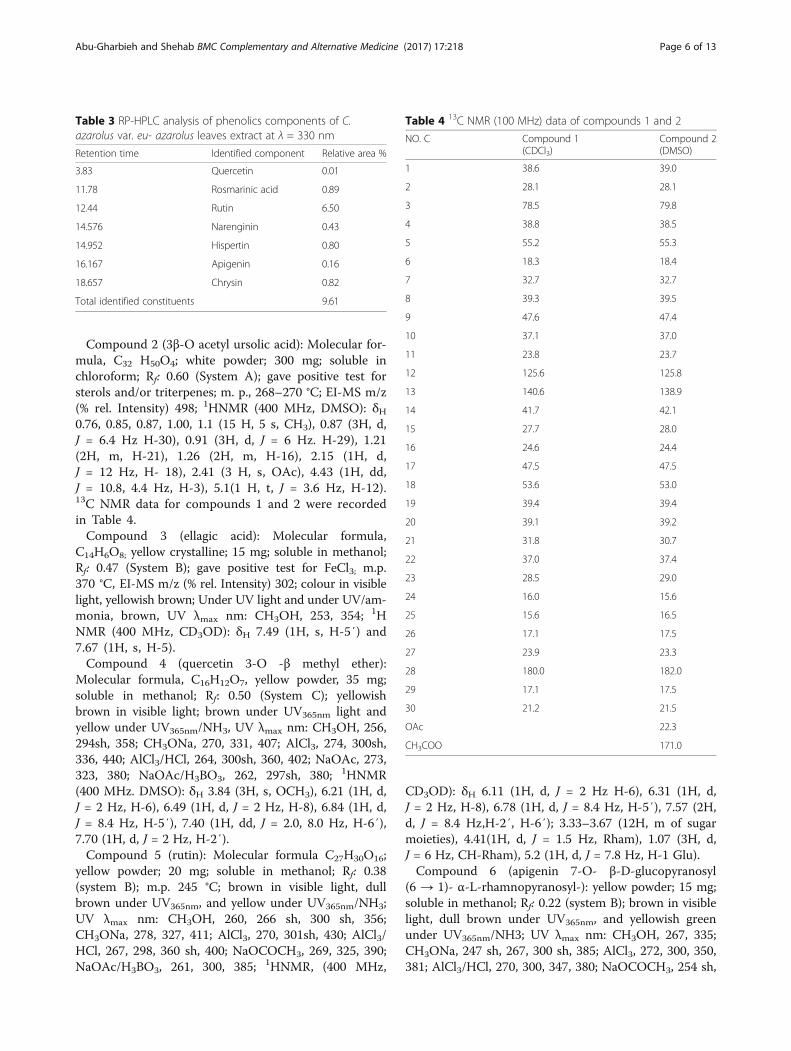
Compound 2 (3β-O acetyl ursolic acid): Molecular for-mula, C32 H50O4; white powder; 300 mg; soluble inchloroform; Rf: 0.60 (System A); gave positive test forsterols and/or triterpenes; m. p., 268–270 °C; EI-MS m/z(% rel. Intensity) 498; 1HNMR (400 MHz, DMSO): δH0.76, 0.85, 0.87, 1.00, 1.1 (15 H, 5 s, CH3), 0.87 (3H, d,J = 6.4 Hz H-30), 0.91 (3H, d, J = 6 Hz. H-29), 1.21(2H, m, H-21), 1.26 (2H, m, H-16), 2.15 (1H, d,J = 12 Hz, H- 18), 2.41 (3 H, s, OAc), 4.43 (1H, dd,J = 10.8, 4.4 Hz, H-3), 5.1(1 H, t, J = 3.6 Hz, H-12).13C NMR data for compounds 1 and 2 were recordedin Table 4.Compound 3 (ellagic acid): Molecular formula,
C14H6O8; yellow crystalline; 15 mg; soluble in methanol;Rf: 0.47 (System B); gave positive test for FeCl3; m.p.370 °C, EI-MS m/z (% rel. Intensity) 302; colour in visiblelight, yellowish brown; Under UV light and under UV/am-monia, brown, UV λmax nm: CH3OH, 253, 354; 1HNMR (400 MHz, CD3OD): δH 7.49 (1H, s, H-5′) and7.67 (1H, s, H-5).Compound 4 (quercetin 3-O -β methyl ether):
Molecular formula, C16H12O7, yellow powder, 35 mg;soluble in methanol; Rf: 0.50 (System C); yellowishbrown in visible light; brown under UV365nm light andyellow under UV365nm/NH3, UV λmax nm: CH3OH, 256,294sh, 358; CH3ONa, 270, 331, 407; AlCl3, 274, 300sh,336, 440; AlCl3/HCl, 264, 300sh, 360, 402; NaOAc, 273,323, 380; NaOAc/H3BO3, 262, 297sh, 380; 1HNMR(400 MHz. DMSO): δH 3.84 (3H, s, OCH3), 6.21 (1H, d,J = 2 Hz, H-6), 6.49 (1H, d, J = 2 Hz, H-8), 6.84 (1H, d,J = 8.4 Hz, H-5′), 7.40 (1H, dd, J = 2.0, 8.0 Hz, H-6′),7.70 (1H, d, J = 2 Hz, H-2′).Compound 5 (rutin): Molecular formula C27H30O16;
yellow powder; 20 mg; soluble in methanol; Rf: 0.38(system B); m.p. 245 °C; brown in visible light, dullbrown under UV365nm, and yellow under UV365nm/NH3;UV λmax nm: CH3OH, 260, 266 sh, 300 sh, 356;CH3ONa, 278, 327, 411; AlCl3, 270, 301sh, 430; AlCl3/HCl, 267, 298, 360 sh, 400; NaOCOCH3, 269, 325, 390;NaOAc/H3BO3, 261, 300, 385; 1HNMR, (400 MHz,
CD3OD): δH 6.11 (1H, d, J = 2 Hz H-6), 6.31 (1H, d,J = 2 Hz, H-8), 6.78 (1H, d, J = 8.4 Hz, H-5′), 7.57 (2H,d, J = 8.4 Hz,H-2′, H-6′); 3.33–3.67 (12H, m of sugarmoieties), 4.41(1H, d, J = 1.5 Hz, Rham), 1.07 (3H, d,J = 6 Hz, CH-Rham), 5.2 (1H, d, J = 7.8 Hz, H-1 Glu).Compound 6 (apigenin 7-O- β-D-glucopyranosyl
(6 → 1)- α-L-rhamnopyranosyl-): yellow powder; 15 mg;soluble in methanol; Rf: 0.22 (system B); brown in visiblelight, dull brown under UV365nm, and yellowish greenunder UV365nm/NH3; UV λmax nm: CH3OH, 267, 335;CH3ONa, 247 sh, 267, 300 sh, 385; AlCl3, 272, 300, 350,381; AlCl3/HCl, 270, 300, 347, 380; NaOCOCH3, 254 sh,
Table 3 RP-HPLC analysis of phenolics components of C.azarolus var. eu- azarolus leaves extract at λ = 330 nm
Retention time Identified component Relative area %
3.83 Quercetin 0.01
11.78 Rosmarinic acid 0.89
12.44 Rutin 6.50
14.576 Narenginin 0.43
14.952 Hispertin 0.80
16.167 Apigenin 0.16
18.657 Chrysin 0.82
Total identified constituents 9.61
Table 4 13C NMR (100 MHz) data of compounds 1 and 2
NO. C Compound 1(CDCl3)
Compound 2(DMSO)
1 38.6 39.0
2 28.1 28.1
3 78.5 79.8
4 38.8 38.5
5 55.2 55.3
6 18.3 18.4
7 32.7 32.7
8 39.3 39.5
9 47.6 47.4
10 37.1 37.0
11 23.8 23.7
12 125.6 125.8
13 140.6 138.9
14 41.7 42.1
15 27.7 28.0
16 24.6 24.4
17 47.5 47.5
18 53.6 53.0
19 39.4 39.4
20 39.1 39.2
21 31.8 30.7
22 37.0 37.4
23 28.5 29.0
24 16.0 15.6
25 15.6 16.5
26 17.1 17.5
27 23.9 23.3
28 180.0 182.0
29 17.1 17.5
30 21.2 21.5
OAc 22.3
CH3COO 171.0
Abu-Gharbieh and Shehab BMC Complementary and Alternative Medicine (2017) 17:218 Page 6 of 13
265; 355, 385; NaOAc/H3BO3, 272, 373; 1H–NMR,(400 MHz, CD3OD): δH 7.91 (2H, d, J = 9.1 Hz, H-2′,H-6′), 7.03 (2H, d, J = 8.8 Hz, H-3′, H-5′), 6.86 (1H, d,J = 2.2 Hz, H-8), 6.51 (1H, d, J = 2.2 Hz, H-6), 6.32 (1H,s, H-3), 3.14–3.53 (12 H, m, sugar moieties), 4.40 (1H, d,J = 2 Hz, −Rham), 1.07 (3H, d, J = 6.0 Hz, CH3-Rham),5.1 (1H, d, J = 7.3 Hz, H-1 Glu). 13C NMR data for com-pounds 4–6 were recorded in Table 5.Compounds 1 and 2 gave positive Salkoweski reactions
confirming their triterpenoidal nature [32]. 1H- and13C–NMR spectra showed that compounds 1 and 2 werepentacyclic triterpene. Compound 1 was identified asursolic acid while compound 2 was identified as 3β-Oacetyl ursolic acid from their physical properties andtheir spectral data (1H–NMR and 13C–NMR) [33, 34].The structure of compound 3 was identified as ellagic
acid from its physical properties and different spectro-scopic spectra (UV and1H–NMR) [35].
1H and 13C–NMR spectra of compound 4 demon-strated a methoxy group at δH 3.84 and at δC 58.2respectively attached at position 3; compound 4 wasidentified as quercetin 3-O -β methyl ether [36].The structure of compound 5 was identified as rutin
from its physical properties and different spectral data(UV, 1H–NMR and 13C–NMR) [37].The UV λmax (335 nm) of compound 6 suggested that
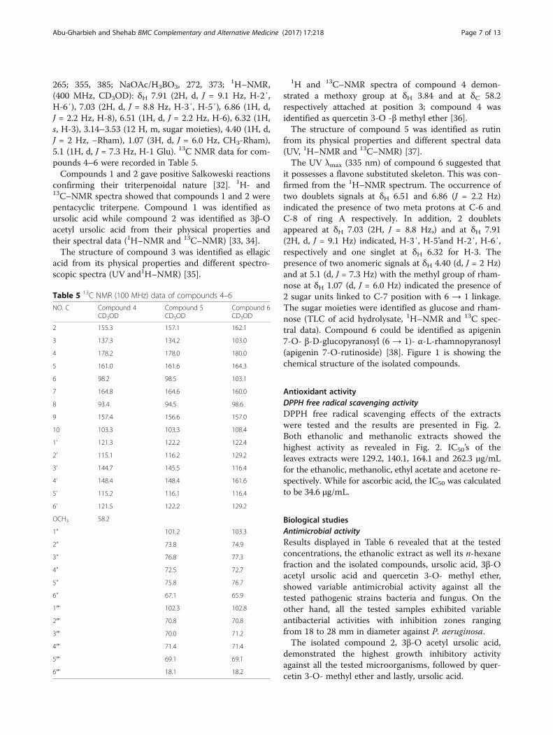
it possesses a flavone substituted skeleton. This was con-firmed from the 1H–NMR spectrum. The occurrence oftwo doublets signals at δH 6.51 and 6.86 (J = 2.2 Hz)indicated the presence of two meta protons at C-6 andC-8 of ring A respectively. In addition, 2 doubletsappeared at δH 7.03 (2H, J = 8.8 Hz,) and at δH 7.91(2H, d, J = 9.1 Hz) indicated, H-3′, H-5’and H-2′, H-6′,respectively and one singlet at δH 6.32 for H-3. Thepresence of two anomeric signals at δH 4.40 (d, J = 2 Hz)and at 5.1 (d, J = 7.3 Hz) with the methyl group of rham-nose at δH 1.07 (d, J = 6.0 Hz) indicated the presence of2 sugar units linked to C-7 position with 6 → 1 linkage.The sugar moieties were identified as glucose and rham-nose (TLC of acid hydrolysate, 1H–NMR and 13C spec-tral data). Compound 6 could be identified as apigenin7-O- β-D-glucopyranosyl (6 → 1)- α-L-rhamnopyranosyl(apigenin 7-O-rutinoside) [38]. Figure 1 is showing thechemical structure of the isolated compounds.
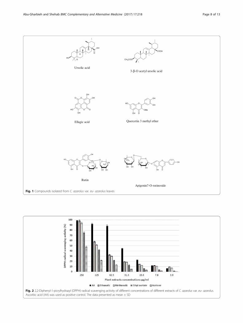
Antioxidant activityDPPH free radical scavenging activityDPPH free radical scavenging effects of the extractswere tested and the results are presented in Fig. 2.Both ethanolic and methanolic extracts showed thehighest activity as revealed in Fig. 2. IC50’s of theleaves extracts were 129.2, 140.1, 164.1 and 262.3 μg/mLfor the ethanolic, methanolic, ethyl acetate and acetone re-spectively. While for ascorbic acid, the IC50 was calculatedto be 34.6 μg/mL.
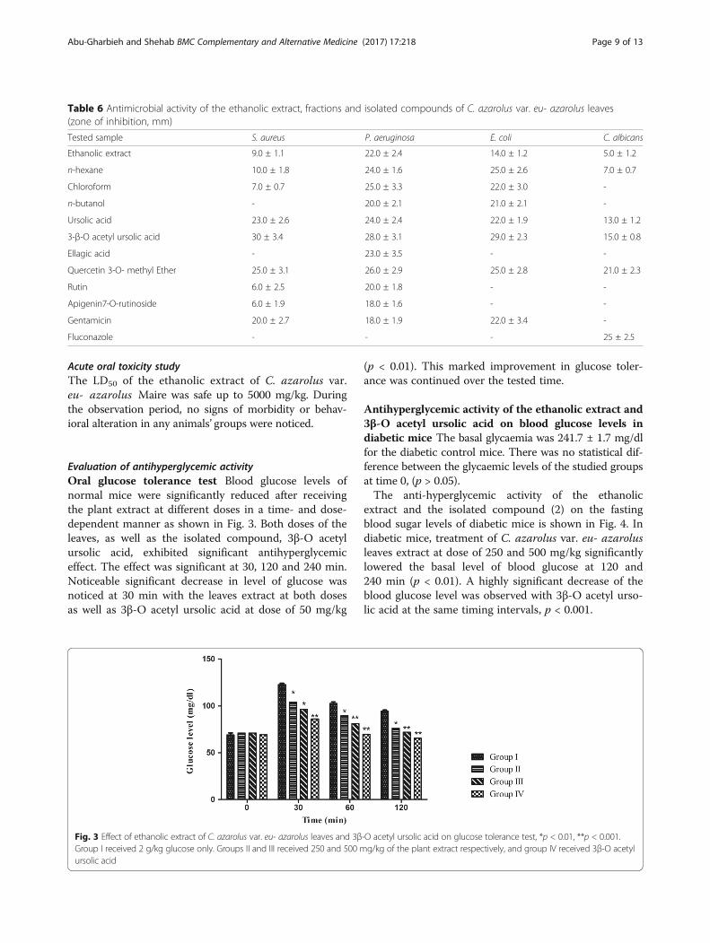
Biological studiesAntimicrobial activityResults displayed in Table 6 revealed that at the testedconcentrations, the ethanolic extract as well its n-hexanefraction and the isolated compounds, ursolic acid, 3β-Oacetyl ursolic acid and quercetin 3-O- methyl ether,showed variable antimicrobial activity against all thetested pathogenic strains bacteria and fungus. On theother hand, all the tested samples exhibited variableantibacterial activities with inhibition zones rangingfrom 18 to 28 mm in diameter against P. aeruginosa.The isolated compound 2, 3β-O acetyl ursolic acid,
demonstrated the highest growth inhibitory activityagainst all the tested microorganisms, followed by quer-cetin 3-O- methyl ether and lastly, ursolic acid.
Table 5 13C NMR (100 MHz) data of compounds 4–6
NO. C Compound 4CD3OD
Compound 5CD3OD
Compound 6CD3OD
2 155.3 157.1 162.1
3 137.3 134.2 103.0
4 178.2 178.0 180.0
5 161.0 161.6 164.3
6 98.2 98.5 103.1
7 164.8 164.6 160.0
8 93.4 94.5 98.6
9 157.4 156.6 157.0
10 103.3 103.3 108.4
1’ 121.3 122.2 122.4
2’ 115.1 116.2 129.2
3’ 144.7 145.5 116.4
4’ 148.4 148.4 161.6
5’ 115.2 116.1 116.4
6’ 121.5 122.2 129.2
OCH3 58.2
1” 101.2 103.3
2” 73.8 74.9
3” 76.8 77.3
4” 72.5 72.7
5” 75.8 76.7
6” 67.1 65.9
1″‘ 102.3 102.8
2″‘ 70.8 70.8
3″‘ 70.0 71.2
4″‘ 71.4 71.4
5″‘ 69.1 69.1
6″‘ 18.1 18.2
Abu-Gharbieh and Shehab BMC Complementary and Alternative Medicine (2017) 17:218 Page 7 of 13
HOH
O
OHH
H
Ursolic acid3-β-O acetyl ursolic acid
O
OOH
OH
OH
OHO
O
Ellagic acid
O
OH
OHOH
OH
OMe
O
Quercetin 3 methyl ether
CH3COO
COOH
Rutin
Apigenin7-O-rutinoside
O
OH
OHOH
OH
O
O
OOHO
OH
OH
O
OHOH
OHCH3
OOH O
OH
OH
O
OH
CH3
OH OH
O
OOH
OH O
Fig. 1 Compounds isolated from C. azarolus var. eu- azarolus leaves
Fig. 2 2,2-Diphenyl-1-picrylhydrazyl (DPPH) radical scavenging activity of different concentrations of different extracts of C. azarolus var. eu- azarolus.Ascorbic acid (AA) was used as positive control. The data presented as mean ± SD
Abu-Gharbieh and Shehab BMC Complementary and Alternative Medicine (2017) 17:218 Page 8 of 13
Acute oral toxicity studyThe LD50 of the ethanolic extract of C. azarolus var.eu- azarolus Maire was safe up to 5000 mg/kg. Duringthe observation period, no signs of morbidity or behav-ioral alteration in any animals’ groups were noticed.
Evaluation of antihyperglycemic activityOral glucose tolerance test Blood glucose levels ofnormal mice were significantly reduced after receivingthe plant extract at different doses in a time- and dose-dependent manner as shown in Fig. 3. Both doses of theleaves, as well as the isolated compound, 3β-O acetylursolic acid, exhibited significant antihyperglycemiceffect. The effect was significant at 30, 120 and 240 min.Noticeable significant decrease in level of glucose wasnoticed at 30 min with the leaves extract at both dosesas well as 3β-O acetyl ursolic acid at dose of 50 mg/kg
(p < 0.01). This marked improvement in glucose toler-ance was continued over the tested time.
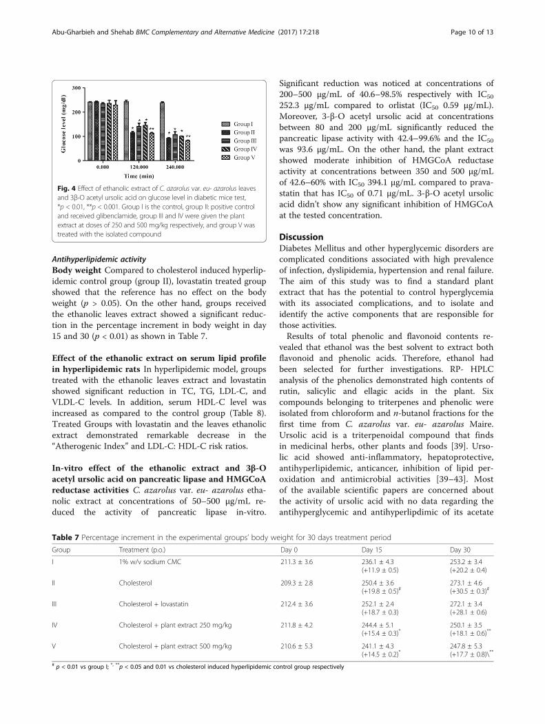
Antihyperglycemic activity of the ethanolic extract and3β-O acetyl ursolic acid on blood glucose levels indiabetic mice The basal glycaemia was 241.7 ± 1.7 mg/dlfor the diabetic control mice. There was no statistical dif-ference between the glycaemic levels of the studied groupsat time 0, (p > 0.05).The anti-hyperglycemic activity of the ethanolic
extract and the isolated compound (2) on the fastingblood sugar levels of diabetic mice is shown in Fig. 4. Indiabetic mice, treatment of C. azarolus var. eu- azarolusleaves extract at dose of 250 and 500 mg/kg significantlylowered the basal level of blood glucose at 120 and240 min (p < 0.01). A highly significant decrease of theblood glucose level was observed with 3β-O acetyl urso-lic acid at the same timing intervals, p < 0.001.
Table 6 Antimicrobial activity of the ethanolic extract, fractions and isolated compounds of C. azarolus var. eu- azarolus leaves(zone of inhibition, mm)
Tested sample S. aureus P. aeruginosa E. coli C. albicans
Ethanolic extract 9.0 ± 1.1 22.0 ± 2.4 14.0 ± 1.2 5.0 ± 1.2
n-hexane 10.0 ± 1.8 24.0 ± 1.6 25.0 ± 2.6 7.0 ± 0.7
Chloroform 7.0 ± 0.7 25.0 ± 3.3 22.0 ± 3.0 -
n-butanol - 20.0 ± 2.1 21.0 ± 2.1 -
Ursolic acid 23.0 ± 2.6 24.0 ± 2.4 22.0 ± 1.9 13.0 ± 1.2
3-β-O acetyl ursolic acid 30 ± 3.4 28.0 ± 3.1 29.0 ± 2.3 15.0 ± 0.8
Ellagic acid - 23.0 ± 3.5 - -
Quercetin 3-O- methyl Ether 25.0 ± 3.1 26.0 ± 2.9 25.0 ± 2.8 21.0 ± 2.3
Rutin 6.0 ± 2.5 20.0 ± 1.8 - -
Apigenin7-O-rutinoside 6.0 ± 1.9 18.0 ± 1.6 - -
Gentamicin 20.0 ± 2.7 18.0 ± 1.9 22.0 ± 3.4 -
Fluconazole - - - 25 ± 2.5
Fig. 3 Effect of ethanolic extract of C. azarolus var. eu- azarolus leaves and 3β-O acetyl ursolic acid on glucose tolerance test, *p < 0.01, **p < 0.001.Group I received 2 g/kg glucose only. Groups II and III received 250 and 500 mg/kg of the plant extract respectively, and group IV received 3β-O acetylursolic acid
Abu-Gharbieh and Shehab BMC Complementary and Alternative Medicine (2017) 17:218 Page 9 of 13
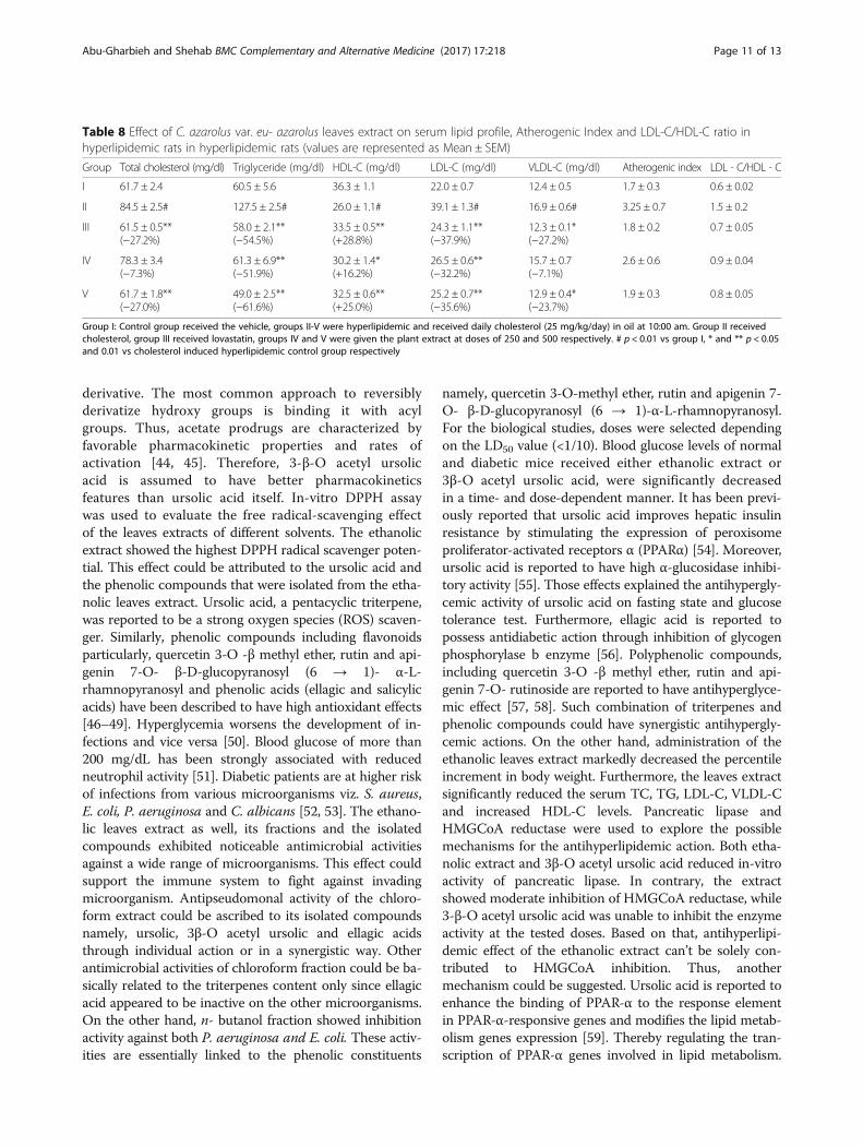
Antihyperlipidemic activityBody weight Compared to cholesterol induced hyperlip-idemic control group (group II), lovastatin treated groupshowed that the reference has no effect on the bodyweight (p > 0.05). On the other hand, groups receivedthe ethanolic leaves extract showed a significant reduc-tion in the percentage increment in body weight in day15 and 30 (p < 0.01) as shown in Table 7.
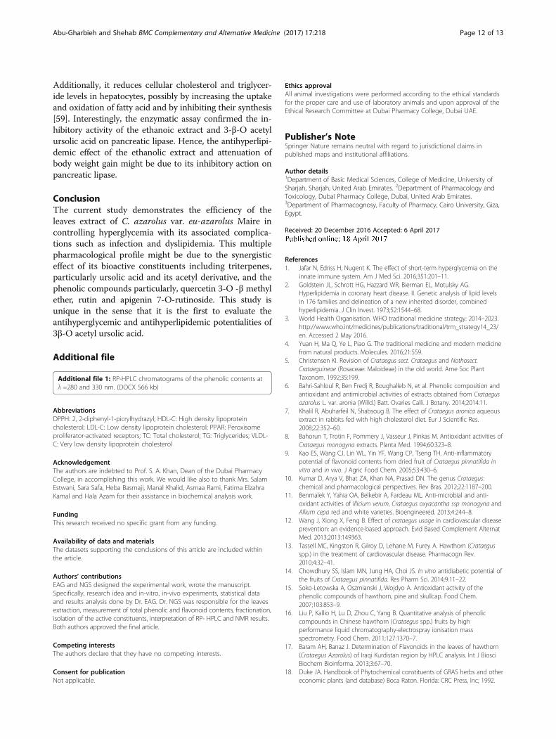
Effect of the ethanolic extract on serum lipid profilein hyperlipidemic rats In hyperlipidemic model, groupstreated with the ethanolic leaves extract and lovastatinshowed significant reduction in TC, TG, LDL-C, andVLDL-C levels. In addition, serum HDL-C level wasincreased as compared to the control group (Table 8).Treated Groups with lovastatin and the leaves ethanolicextract demonstrated remarkable decrease in the“Atherogenic Index” and LDL-C: HDL-C risk ratios.
In-vitro effect of the ethanolic extract and 3β-Oacetyl ursolic acid on pancreatic lipase and HMGCoAreductase activities C. azarolus var. eu- azarolus etha-nolic extract at concentrations of 50–500 μg/mL re-duced the activity of pancreatic lipase in-vitro.
Significant reduction was noticed at concentrations of200–500 μg/mL of 40.6–98.5% respectively with IC50
252.3 μg/mL compared to orlistat (IC50 0.59 μg/mL).Moreover, 3-β-O acetyl ursolic acid at concentrationsbetween 80 and 200 μg/mL significantly reduced thepancreatic lipase activity with 42.4–99.6% and the IC50
was 93.6 μg/mL. On the other hand, the plant extractshowed moderate inhibition of HMGCoA reductaseactivity at concentrations between 350 and 500 μg/mLof 42.6–60% with IC50 394.1 μg/mL compared to prava-statin that has IC50 of 0.71 μg/mL. 3-β-O acetyl ursolicacid didn’t show any significant inhibition of HMGCoAat the tested concentration.
DiscussionDiabetes Mellitus and other hyperglycemic disorders arecomplicated conditions associated with high prevalenceof infection, dyslipidemia, hypertension and renal failure.The aim of this study was to find a standard plantextract that has the potential to control hyperglycemiawith its associated complications, and to isolate andidentify the active components that are responsible forthose activities.Results of total phenolic and flavonoid contents re-
vealed that ethanol was the best solvent to extract bothflavonoid and phenolic acids. Therefore, ethanol hadbeen selected for further investigations. RP- HPLCanalysis of the phenolics demonstrated high contents ofrutin, salicylic and ellagic acids in the plant. Sixcompounds belonging to triterpenes and phenolic wereisolated from chloroform and n-butanol fractions for thefirst time from C. azarolus var. eu- azarolus Maire.Ursolic acid is a triterpenoidal compound that findsin medicinal herbs, other plants and foods [39]. Urso-lic acid showed anti-inflammatory, hepatoprotective,antihyperlipidemic, anticancer, inhibition of lipid per-oxidation and antimicrobial activities [39–43]. Mostof the available scientific papers are concerned aboutthe activity of ursolic acid with no data regarding theantihyperglycemic and antihyperlipdimic of its acetate
Fig. 4 Effect of ethanolic extract of C. azarolus var. eu- azarolus leavesand 3β-O acetyl ursolic acid on glucose level in diabetic mice test,*p < 0.01, **p < 0.001. Group I is the control, group II: positive controland received glibenclamide, group III and IV were given the plantextract at doses of 250 and 500 mg/kg respectively, and group V wastreated with the isolated compound
Table 7 Percentage increment in the experimental groups’ body weight for 30 days treatment period
Group Treatment (p.o.) Day 0 Day 15 Day 30
I 1% w/v sodium CMC 211.3 ± 3.6 236.1 ± 4.3(+11.9 ± 0.5)
253.2 ± 3.4(+20.2 ± 0.4)
II Cholesterol 209.3 ± 2.8 250.4 ± 3.6(+19.8 ± 0.5)#
273.1 ± 4.6(+30.5 ± 0.3)#
III Cholesterol + lovastatin 212.4 ± 3.6 252.1 ± 2.4(+18.7 ± 0.3)
272.1 ± 3.4(+28.1 ± 0.6)
IV Cholesterol + plant extract 250 mg/kg 211.8 ± 4.2 244.4 ± 5.1(+15.4 ± 0.3)*
250.1 ± 3.5(+18.1 ± 0.6)**
V Cholesterol + plant extract 500 mg/kg 210.6 ± 5.3 241.1 ± 4.3(+14.5 ± 0.2)*
247.8 ± 5.3(+17.7 ± 0.8)\**
# p < 0.01 vs group I; *, **p < 0.05 and 0.01 vs cholesterol induced hyperlipidemic control group respectively
Abu-Gharbieh and Shehab BMC Complementary and Alternative Medicine (2017) 17:218 Page 10 of 13
derivative. The most common approach to reversiblyderivatize hydroxy groups is binding it with acylgroups. Thus, acetate prodrugs are characterized byfavorable pharmacokinetic properties and rates ofactivation [44, 45]. Therefore, 3-β-O acetyl ursolicacid is assumed to have better pharmacokineticsfeatures than ursolic acid itself. In-vitro DPPH assaywas used to evaluate the free radical-scavenging effectof the leaves extracts of different solvents. The ethanolicextract showed the highest DPPH radical scavenger poten-tial. This effect could be attributed to the ursolic acid andthe phenolic compounds that were isolated from the etha-nolic leaves extract. Ursolic acid, a pentacyclic triterpene,was reported to be a strong oxygen species (ROS) scaven-ger. Similarly, phenolic compounds including flavonoidsparticularly, quercetin 3-O -β methyl ether, rutin and api-genin 7-O- β-D-glucopyranosyl (6 → 1)- α-L-rhamnopyranosyl and phenolic acids (ellagic and salicylicacids) have been described to have high antioxidant effects[46–49]. Hyperglycemia worsens the development of in-fections and vice versa [50]. Blood glucose of more than200 mg/dL has been strongly associated with reducedneutrophil activity [51]. Diabetic patients are at higher riskof infections from various microorganisms viz. S. aureus,E. coli, P. aeruginosa and C. albicans [52, 53]. The ethano-lic leaves extract as well, its fractions and the isolatedcompounds exhibited noticeable antimicrobial activitiesagainst a wide range of microorganisms. This effect couldsupport the immune system to fight against invadingmicroorganism. Antipseudomonal activity of the chloro-form extract could be ascribed to its isolated compoundsnamely, ursolic, 3β-O acetyl ursolic and ellagic acidsthrough individual action or in a synergistic way. Otherantimicrobial activities of chloroform fraction could be ba-sically related to the triterpenes content only since ellagicacid appeared to be inactive on the other microorganisms.On the other hand, n- butanol fraction showed inhibitionactivity against both P. aeruginosa and E. coli. These activ-ities are essentially linked to the phenolic constituents
namely, quercetin 3-O-methyl ether, rutin and apigenin 7-O- β-D-glucopyranosyl (6 → 1)-α-L-rhamnopyranosyl.For the biological studies, doses were selected dependingon the LD50 value (<1/10). Blood glucose levels of normaland diabetic mice received either ethanolic extract or3β-O acetyl ursolic acid, were significantly decreasedin a time- and dose-dependent manner. It has been previ-ously reported that ursolic acid improves hepatic insulinresistance by stimulating the expression of peroxisomeproliferator-activated receptors α (PPARα) [54]. Moreover,ursolic acid is reported to have high α-glucosidase inhibi-tory activity [55]. Those effects explained the antihypergly-cemic activity of ursolic acid on fasting state and glucosetolerance test. Furthermore, ellagic acid is reported topossess antidiabetic action through inhibition of glycogenphosphorylase b enzyme [56]. Polyphenolic compounds,including quercetin 3-O -β methyl ether, rutin and api-genin 7-O- rutinoside are reported to have antihyperglyce-mic effect [57, 58]. Such combination of triterpenes andphenolic compounds could have synergistic antihypergly-cemic actions. On the other hand, administration of theethanolic leaves extract markedly decreased the percentileincrement in body weight. Furthermore, the leaves extractsignificantly reduced the serum TC, TG, LDL-C, VLDL-Cand increased HDL-C levels. Pancreatic lipase andHMGCoA reductase were used to explore the possiblemechanisms for the antihyperlipidemic action. Both etha-nolic extract and 3β-O acetyl ursolic acid reduced in-vitroactivity of pancreatic lipase. In contrary, the extractshowed moderate inhibition of HMGCoA reductase, while3-β-O acetyl ursolic acid was unable to inhibit the enzymeactivity at the tested doses. Based on that, antihyperlipi-demic effect of the ethanolic extract can’t be solely con-tributed to HMGCoA inhibition. Thus, anothermechanism could be suggested. Ursolic acid is reported toenhance the binding of PPAR-α to the response elementin PPAR-α-responsive genes and modifies the lipid metab-olism genes expression [59]. Thereby regulating the tran-scription of PPAR-α genes involved in lipid metabolism.
Table 8 Effect of C. azarolus var. eu- azarolus leaves extract on serum lipid profile, Atherogenic Index and LDL-C/HDL-C ratio inhyperlipidemic rats in hyperlipidemic rats (values are represented as Mean ± SEM)
Group Total cholesterol (mg/dl) Triglyceride (mg/dl) HDL-C (mg/dl) LDL-C (mg/dl) VLDL-C (mg/dl) Atherogenic index LDL - C/HDL - C
I 61.7 ± 2.4 60.5 ± 5.6 36.3 ± 1.1 22.0 ± 0.7 12.4 ± 0.5 1.7 ± 0.3 0.6 ± 0.02
II 84.5 ± 2.5# 127.5 ± 2.5# 26.0 ± 1.1# 39.1 ± 1.3# 16.9 ± 0.6# 3.25 ± 0.7 1.5 ± 0.2
III 61.5 ± 0.5**(−27.2%)
58.0 ± 2.1**(−54.5%)
33.5 ± 0.5**(+28.8%)
24.3 ± 1.1**(−37.9%)
12.3 ± 0.1*(−27.2%)
1.8 ± 0.2 0.7 ± 0.05
IV 78.3 ± 3.4(−7.3%)
61.3 ± 6.9**(−51.9%)
30.2 ± 1.4*(+16.2%)
26.5 ± 0.6**(−32.2%)
15.7 ± 0.7(−7.1%)
2.6 ± 0.6 0.9 ± 0.04
V 61.7 ± 1.8**(−27.0%)
49.0 ± 2.5**(−61.6%)
32.5 ± 0.6**(+25.0%)
25.2 ± 0.7**(−35.6%)
12.9 ± 0.4*(−23.7%)
1.9 ± 0.3 0.8 ± 0.05
Group I: Control group received the vehicle, groups II-V were hyperlipidemic and received daily cholesterol (25 mg/kg/day) in oil at 10:00 am. Group II receivedcholesterol, group III received lovastatin, groups IV and V were given the plant extract at doses of 250 and 500 respectively. # p < 0.01 vs group I, * and ** p < 0.05and 0.01 vs cholesterol induced hyperlipidemic control group respectively
Abu-Gharbieh and Shehab BMC Complementary and Alternative Medicine (2017) 17:218 Page 11 of 13
Additionally, it reduces cellular cholesterol and triglycer-ide levels in hepatocytes, possibly by increasing the uptakeand oxidation of fatty acid and by inhibiting their synthesis[59]. Interestingly, the enzymatic assay confirmed the in-hibitory activity of the ethanoic extract and 3-β-O acetylursolic acid on pancreatic lipase. Hence, the antihyperlipi-demic effect of the ethanolic extract and attenuation ofbody weight gain might be due to its inhibitory action onpancreatic lipase.
ConclusionThe current study demonstrates the efficiency of theleaves extract of C. azarolus var. eu-azarolus Maire incontrolling hyperglycemia with its associated complica-tions such as infection and dyslipidemia. This multiplepharmacological profile might be due to the synergisticeffect of its bioactive constituents including triterpenes,particularly ursolic acid and its acetyl derivative, and thephenolic compounds particularly, quercetin 3-O -β methylether, rutin and apigenin 7-O-rutinoside. This study isunique in the sense that it is the first to evaluate theantihyperglycemic and antihyperlipidemic potentialities of3β-O acetyl ursolic acid.
Additional file
Additional file 1: RP-HPLC chromatograms of the phenolic contents atλ =280 and 330 nm. (DOCX 566 kb)
AbbreviationsDPPH: 2, 2-diphenyl-1-picrylhydrazyl; HDL-C: High density lipoproteincholesterol; LDL-C: Low density lipoprotein cholesterol; PPAR: Peroxisomeproliferator-activated receptors; TC: Total cholesterol; TG: Triglycerides; VLDL-C: Very low density lipoprotein cholesterol
AcknowledgementThe authors are indebted to Prof. S. A. Khan, Dean of the Dubai PharmacyCollege, in accomplishing this work. We would like also to thank Mrs. SalamEstwani, Sara Safa, Heba Basmaji, Manal Khalid, Asmaa Rami, Fatima ElzahraKamal and Hala Azam for their assistance in biochemical analysis work.
FundingThis research received no specific grant from any funding.
Availability of data and materialsThe datasets supporting the conclusions of this article are included withinthe article.
Authors’ contributionsEAG and NGS designed the experimental work, wrote the manuscript.Specifically, research idea and in-vitro, in-vivo experiments, statistical dataand results analysis done by Dr. EAG. Dr. NGS was responsible for the leavesextraction, measurement of total phenolic and flavonoid contents, fractionation,isolation of the active constituents, interpretation of RP- HPLC and NMR results.Both authors approved the final article.
Competing interestsThe authors declare that they have no competing interests.
Consent for publicationNot applicable.
Ethics approvalAll animal investigations were performed according to the ethical standardsfor the proper care and use of laboratory animals and upon approval of theEthical Research Committee at Dubai Pharmacy College, Dubai UAE.
Publisher’s NoteSpringer Nature remains neutral with regard to jurisdictional claims inpublished maps and institutional affiliations.
Author details1Department of Basic Medical Sciences, College of Medicine, University ofSharjah, Sharjah, United Arab Emirates. 2Department of Pharmacology andToxicology, Dubai Pharmacy College, Dubai, United Arab Emirates.3Department of Pharmacognosy, Faculty of Pharmacy, Cairo University, Giza,Egypt.
Received: 20 December 2016 Accepted: 6 April 2017
References1. Jafar N, Edriss H, Nugent K. The effect of short-term hyperglycemia on the
innate immune system. Am J Med Sci. 2016;351:201–11.2. Goldstein JL, Schrott HG, Hazzard WR, Bierman EL, Motulsky AG.
Hyperlipidemia in coronary heart disease. II. Genetic analysis of lipid levelsin 176 families and delineation of a new inherited disorder, combinedhyperlipidemia. J Clin Invest. 1973;52:1544–68.
3. World Health Organisation. WHO traditional medicine strategy: 2014–2023.http://www.who.int/medicines/publications/traditional/trm_strategy14_23/en. Accessed 2 May 2016.
4. Yuan H, Ma Q, Ye L, Piao G. The traditional medicine and modern medicinefrom natural products. Molecules. 2016;21:559.
5. Christensen KI. Revision of Crataegus sect. Crataegus and Nothosect.Crataeguineae (Rosaceae: Maloideae) in the old world. Ame Soc PlantTaxonom. 1992;35:199.
6. Bahri-Sahloul R, Ben Fredj R, Boughalleb N, et al. Phenolic composition andantioxidant and antimicrobial activities of extracts obtained from Crataegusazarolus L. var. aronia (Willd.) Batt. Ovaries Calli. J Botany. 2014;2014:11.
7. Khalil R, Abuharfeil N, Shabsoug B. The effect of Crataegus aronica aqueousextract in rabbits fed with high cholesterol diet. Eur J Scientific Res.2008;22:352–60.
8. Bahorun T, Trotin F, Pommery J, Vasseur J, Pinkas M. Antioxidant activities ofCrataegus monogyna extracts. Planta Med. 1994;60:323–8.
9. Kao ES, Wang CJ, Lin WL, Yin YF, Wang CP, Tseng TH. Anti-inflammatorypotential of flavonoid contents from dried fruit of Crataegus pinnatifida invitro and in vivo. J Agric Food Chem. 2005;53:430–6.
10. Kumar D, Arya V, Bhat ZA, Khan NA, Prasad DN. The genus Crataegus:chemical and pharmacological perspectives. Rev Bras. 2012;22:1187–200.
11. Benmalek Y, Yahia OA, Belkebir A, Fardeau ML. Anti-microbial and anti-oxidant activities of Illicium verum, Crataegus oxyacantha ssp monogyna andAllium cepa red and white varieties. Bioengineered. 2013;4:244–8.
12. Wang J, Xiong X, Feng B. Effect of crataegus usage in cardiovascular diseaseprevention: an evidence-based approach. Evid Based Complement AlternatMed. 2013;2013:149363.
13. Tassell MC, Kingston R, Gilroy D, Lehane M, Furey A. Hawthorn (Crataegusspp.) in the treatment of cardiovascular disease. Pharmacogn Rev.2010;4:32–41.
14. Chowdhury SS, Islam MN, Jung HA, Choi JS. In vitro antidiabetic potential ofthe fruits of Crataegus pinnatifida. Res Pharm Sci. 2014;9:11–22.
15. Soko-Letowska A, Oszmianski J, Wojdyo A. Antioxidant activity of thephenolic compounds of hawthorn, pine and skullcap. Food Chem.2007;103:853–9.
16. Liu P, Kallio H, Lu D, Zhou C, Yang B. Quantitative analysis of phenoliccompounds in Chinese hawthorn (Crataegus spp.) fruits by highperformance liquid chromatography-electrospray ionisation massspectrometry. Food Chem. 2011;127:1370–7.
17. Baram AH, Banaz J. Determination of Flavonoids in the leaves of hawthorn(Crataegus Azarolus) of Iraqi Kurdistan region by HPLC analysis. Int J BiosciBiochem Bioinforma. 2013;3:67–70.
18. Duke JA. Handbook of Phytochemical constituents of GRAS herbs and othereconomic plants (and database) Boca Raton. Florida: CRC Press, Inc; 1992.
Abu-Gharbieh and Shehab BMC Complementary and Alternative Medicine (2017) 17:218 Page 12 of 13
19. Oktay M, Gulcin I, Kufrevioglu OI. Determination of in vitro antioxidantactivity of fennel (Foeniculum vulgare) seed extracts. LWT Food Sci Technol.2003;36:263–71.
20. Dewanto V, Wu X, Adom KK, Liu RH. Thermal processing enhances thenutritional value of tomatoes by increasing total antioxidant activity.J Agric Food Chem. 2002;50:3010–4.
21. Goupy P, Hugues M, Boivin P, Amiot MJ. Antioxidant composition andactivity of barley (Hordeum vulgare) and malt extracts and of isolatedphenolic compounds. J Sci Food Agric. 1999;79:1625–34.
22. Mattila P, Astola J, Kumpulainen J. Determination of Flavonoids in plantmaterial by HPLC with diode-Array and electro-Array detections. J AgricFood Chem. 2000;48:5834–41.
23. Shehab NG, Abu-Gharbieh E, Bayoumi FA. Impact of phenolic compositionon hepatoprotective and antioxidant effects of four desert medicinal plants.BMC Complement Altern Med. 2015;15:401.
24. Cheng Z, Moore J, Yu L. High-throughput relative DPPH radical scavengingcapacity assay. J Agric Food Chem. 2006;54:7429–36.
25. Lorian V. Antibiotics in laboratory medicine. 5th ed. Philadelphia: LippincottWilliams and Wilkins; 2005.
26. National Research Council (US) Committee for the Update of the Guide forthe Care and Use of Laboratory Animals. Guide for the Care and Use ofLaboratory Animals. 2011.
27. Lorke D. A new approach to practical acute toxicity testing. Arch Toxicol.1983;54:275–87.
28. Syiem D, Syngai G, Khup PZ, Khongwir BS, Kharbuli B, Kayang H.Hypoglycemic effects of Potentilla fulgens L in normal and alloxan-induceddiabetic mice. J Ethnopharmacol. 2002;83:55–61.
29. Arichi H, Kimura Y, Okuda H, Baba K, Kozawa M, Arichi S. Effects of stilbenecomponents of the roots of Polygonum cuspidatum Sieb. Et Zucc. On lipidmetabolism. Chem Pharm Bull (Tokyo). 1982;30:1766–70.
30. Kim YS, Lee YM, Kim H, et al. Anti-obesity effect of Morus bombycis rootextract: anti-lipase activity and lipolytic effect. J Ethnopharmacol.2010;130:621–4.
31. Xie W, Wang W, Su H, Xing D, Cai G, Du L. Hypolipidemic mechanisms ofAnanas comosus L. leaves in mice: different from fibrates but similar tostatins. J Pharmacol Sci. 2007;103:267–74.
32. Finar IL. Organic chemistry, The Fundamental Principles. 4th ed, Vol 1. NewYork: Wiley; 1963.
33. Do Nascimento GP, Lemos LT, Bizerra MA, et al. Antibacterial andantioxidant activities of Ursolic acid and derivatives. Molecules.2014;19:1317–27.
34. Martins D, Carrion LL, Ramos DF, et al. Triterpenes and the antimycobacterialactivity of Duroia macrophylla Huber (Rubiaceae). Biomed Res Int.2013;2013:605831.
35. Yan XH, Guo YW. Two new ellagic acid glycosides from leaves ofDiplopanax stachyanthus. J Asian Nat Prod Res. 2004;6:271–6.
36. Krenn L, Miron A, Pemp E, Petr U, Kopp B. Flavonoids from Achillea nobilis L.Z Naturforsch C. 2003;58:11–6.
37. Biruk S, Kaleab A, Raghavendra Y. Radical scavenging activities of the leafextracts and a flavonoid glycoside isolated from Cineraria abyssinica Sch.Bip. Exa. Rich. J App Pharmac Sci. 2012;2:44–9.
38. Moon BH, Lee SC, et al. Complete assignments of the 1H and 13C NMRdata of Flavone derivatives. Bull Kor Chem Soc. 2005;26:603–8.
39. Liu J. Pharmacology of oleanolic acid and ursolic acid. J Ethnopharmacol.1995;49:57–68.
40. Ovesna Z, Kozics K, Slamenova D. Protective effects of ursolic acid andoleanolic acid in leukemic cells. Mutat Res. 2006;600:131–7.
41. Ramachandran S, Prasad NR. Effect of ursolic acid, a triterpenoid antioxidant,on ultraviolet-B radiation-induced cytotoxicity, lipid peroxidation and DNAdamage in human lymphocytes. Chem Biol Interact. 2008;176:99–107.
42. Huang CY, Lin CY, Tsai CW, Yin MC. Inhibition of cell proliferation, invasionand migration by ursolic acid in human lung cancer cell lines. Toxicol inVitro. 2011;25:1274–80.
43. Somova LO, Nadar A, Rammanan P, Shode FO. Cardiovascular,antihyperlipidemic and antioxidant effects of oleanolic and ursolic acids inexperimental hypertension. Phytomedicine. 2003;10:115–21.
44. Jana S, Mandlekar S, Marathe P. Prodrug design to improvepharmacokinetic and drug delivery properties: challenges to the discoveryscientists. Curr Med Chem. 2010;17:3874–908.
45. Prodrug TB. Objectives and design A2 - Taylor, John B. In: Triggle DJ, editor.Comprehensive medicinal chemistry II. Oxford: Elsevier; 2007. p. 1009–41.
46. Li ZG. Synergistic effect of antioxidant system and osmolyte in hydrogensulfide and salicylic acid crosstalk-induced heat tolerance in maize(Zea mays L.) seedlings. Plant Signal Behav. 2015;10:e105–1278.
47. Usta C, Ozdemir S, Schiariti M, Schiariti MF, Puddu PE. The pharmacologicaluse of ellagic acid-rich pomegranate fruit. Int J Food Sci Nutr. 2013;64:907–13.
48. Lee YH, Kim HJ, Yoo H, et al. Synthesis of (2-amino) ethyl derivatives ofquercetin 3-O-methyl ether and their antioxidant and neuroprotectiveeffects. Bioorg Med Chem. 2015.
49. Metodiewa D, Kochman AF, Karolczak S. Evidence for antiradical andantioxidant properties of four biologically active N,N-diethylaminoethylethers of flavanone oximes: a comparison with natural polyphenolicflavonoid (rutin) action. Biochem Mol Biol Int. 1997:1067–75.
50. Koh GC, Peacock SJ, van der PT, Wiersinga WJ. The impact of diabetes onthe pathogenesis of sepsis. Eur J Clin Microbiol Infect Dis. 2012;31:379–88.
51. Pettersson US, Christoffersson G, Massena S, et al. Increased recruitment butimpaired function of leukocytes during inflammation in mouse models oftype 1 and type 2 diabetes. PLoS One. 2011;6:e22480.
52. Casqueiro J, Casqueiro J, Alves C. Infections in patients with diabetesmellitus: a review of pathogenesis. Indian J Endocrinol Metab.2012;16(Suppl 1):S27–36.
53. Knapp S. Diabetes and infection: is there a link? A mini-review. Gerontology.2013;59:99–104.
54. Wang L, Wang GL, Liu JH, Li D, Zhu DZ, Wu LN. Effects of ursolic acid inameliorating insulin resistance in liver of KKAy mice via peroxisomeproliferator-activated receptors alpha and gamma. Zhong Xi Yi Jie He XueBao. 2012;10:793–9.
55. He K, Song S, Zou Z, et al. The Hypoglycemic and synergistic effect ofLoganin, Morroniside, and Ursolic acid isolated from the fruits of Cornusofficinalis. Phytother Res. 2016:283–91.
56. Kyriakis E, Stravodimos GA, Kantsadi AL, Chatzileontiadou DS, Skamnaki VT,Leonidas DD. Natural flavonoids as antidiabetic agents. The binding of gallicand ellagic acids to glycogen phosphorylase b. FEBS Lett. 2015:1787–94.
57. Habtemariam S, Lentini G. The therapeutic potential of rutin for diabetes: anupdate. Mini Rev Med Chem. 2015;15:524–8.
58. Jadhav R, Puchchakayala G. Hypoglycemic and antidiabetic activity offlavonoids: boswellic acid, ellagic acid, quercetin, rutin on streptozotocin-nicotinamide induced type 2 diabetic rats. Int J Pharm Pharm Sci.2012;4:251–6.
59. Jia Y, Bhuiyan MJ, Jun HJ, et al. Ursolic acid is a PPAR-alpha agonist thatregulates hepatic lipid metabolism. Bioorg Med Chem Lett. 2011;21:5876–80.
• We accept pre-submission inquiries
• Our selector tool helps you to find the most relevant journal
• We provide round the clock customer support
• Convenient online submission
• Thorough peer review
• Inclusion in PubMed and all major indexing services
• Maximum visibility for your research
Submit your manuscript atwww.biomedcentral.com/submit
Submit your next manuscript to BioMed Central and we will help you at every step:
Abu-Gharbieh and Shehab BMC Complementary and Alternative Medicine (2017) 17:218 Page 13 of 13
Related Documents


