Therapeutic Effect of Autologous Activated Platelet-rich Plasma Therapy on Mid-dermal to Full-thickness Burns: A Case Series Karina Karina, MD, PhD 1,2,3,4 Krista Ekaputri, MD 1 Johannes Albert Biben, MD 1 Pritha Hadi, MD 5,6 Hubert Andrew 2,7 Patricia Marcellina Sadikin, MD 1 1 Klinik Hayandra, Yayasan Hayandra Peduli, Jakarta Pusat, Indonesia 2 HayandraLab, Yayasan Hayandra Peduli, Jakarta Pusat, Indonesia 3 Faculty of Medicine, Universitas Pembangunan Nasional Veteran Jakarta, Indonesia 4 Pusat Kajian Stem Cell, Universitas Pembangunan Nasional Veteran Jakarta, Indonesia 5 Faculty of Medicine and Health Sciences, Universitas Jambi, Jambi, Indonesia 6 Raden Mattaher Regional Hospital, Jambi, Indonesia 7 Faculty of Medicine, Universitas Indonesia, Jakarta Pusat, Indonesia Arch Plast Surg 2022;49:405–412. Address for correspondence Karina Karina, MD, PhD, Klinik Hayandra, Yayasan Hayandra Peduli, Jl. Kramat VI No. 11, Jakarta Pusat, Indonesia (e-mail: [email protected]). Burn injuries are painful and could lead to morbidity through complications such as pain, infection, dyspigmentation, scar- ring, psychological conditions, and others. 1 Activated autolo- gous platelet-rich plasma (aaPRP) is an interesting alternative to tackle these problems associated with burn injuries. aaPRP contains abundant growth factors, which are able to compen- sate for the depleted growth factors in wounds and provide an ideal microenvironment for wound repair. 2 We present seven cases of mid-dermal and full-thickness burns that were treated with intralesional and intravenous (IV) aaPRP to demonstrate its therapeutic effects through wound re-epithe- lialization, anti-inflammation, and pain relief. Case This study reports a case series of seven patients with varying degrees of burns. The aaPRP was prepared with a method that our group developed. Exactly 24 mL of blood was Keywords ► platelet-rich plasma ► burns ► skin transplantation Abstract Although modern medicine has made great strides in the management of burn injuries, associated complications such as pain, infection, dyspigmentation, and scarring have yet to be fully dealt with. Although skin grafting and meshing are routinely performed on burn patients, this method poses a risk for adverse effects. Activated autologous platelet-rich plasma (aaPRP), which is increasingly used in the field of plastic surgery, contains growth factors beneficial for wound regeneration. Seven cases of burns with varying severity and conditions that were treated with intralesional subcutaneous injection and intravenous aaPRP are presented and discussed herein. This case series indicates that subcutaneous and intravenous aaPRP is a safe procedure with the potential to be an alternative when skin grafting cannot be done or as an adjunct treatment to skin grafting. DOI https://doi.org/ 10.1055/s-0042-1744423. ISSN 2234-6163. © 2022. The Korean Society of Plastic and Reconstructive Surgeons. All rights reserved. This is an open access article published by Thieme under the terms of the Creative Commons Attribution-NonDerivative-NonCommercial-License, permitting copying and reproduction so long as the original work is given appropriate credit. Contents may not be used for commercial purposes, or adapted, remixed, transformed or built upon. (https://creativecommons.org/ licenses/by-nc-nd/4.0/) Thieme Medical Publishers, Inc., 333 Seventh Avenue, 18th Floor, New York, NY 10001, USA THIEME Pediatric/Craniomaxillofacial/Head & Neck: Case Report 405 Article published online: 2022-05-27

Welcome message from author
This document is posted to help you gain knowledge. Please leave a comment to let me know what you think about it! Share it to your friends and learn new things together.
Transcript
Therapeutic Effect of Autologous ActivatedPlatelet-rich Plasma Therapy on Mid-dermal toFull-thickness Burns: A Case SeriesKarina Karina, MD, PhD1,2,3,4 Krista Ekaputri, MD1 Johannes Albert Biben, MD1 Pritha Hadi, MD5,6
Hubert Andrew2,7 Patricia Marcellina Sadikin, MD1
1Klinik Hayandra, Yayasan Hayandra Peduli, Jakarta Pusat, Indonesia2HayandraLab, Yayasan Hayandra Peduli, Jakarta Pusat, Indonesia3Faculty of Medicine, Universitas Pembangunan Nasional VeteranJakarta, Indonesia
4Pusat Kajian Stem Cell, Universitas Pembangunan Nasional VeteranJakarta, Indonesia
5Faculty of Medicine and Health Sciences, Universitas Jambi, Jambi,Indonesia
6Raden Mattaher Regional Hospital, Jambi, Indonesia7Faculty of Medicine, Universitas Indonesia, Jakarta Pusat, Indonesia
Arch Plast Surg 2022;49:405–412.
Address for correspondence Karina Karina, MD, PhD, Klinik Hayandra,Yayasan Hayandra Peduli, Jl. Kramat VI No. 11, Jakarta Pusat,Indonesia (e-mail: [email protected]).
Burn injuries are painful and could lead to morbidity throughcomplications such as pain, infection, dyspigmentation, scar-ring, psychological conditions, and others.1 Activated autolo-gous platelet-rich plasma (aaPRP) is an interesting alternativeto tackle these problems associated with burn injuries. aaPRPcontains abundant growth factors, which are able to compen-sate for the depleted growth factors inwounds and provide anideal microenvironment for wound repair.2 We present sevencases of mid-dermal and full-thickness burns that were
treated with intralesional and intravenous (IV) aaPRP todemonstrate its therapeutic effects throughwound re-epithe-lialization, anti-inflammation, and pain relief.
Case
This study reports a case series of seven patients withvarying degrees of burns. The aaPRP was prepared with amethod that our group developed. Exactly 24mL of bloodwas
Keywords
► platelet-rich plasma► burns► skin transplantation
Abstract Althoughmodernmedicine hasmade great strides in themanagement of burn injuries,associated complications such as pain, infection, dyspigmentation, and scarring haveyet to be fully dealt with. Although skin grafting and meshing are routinely performedon burn patients, this method poses a risk for adverse effects. Activated autologousplatelet-rich plasma (aaPRP), which is increasingly used in the field of plastic surgery,contains growth factors beneficial for wound regeneration. Seven cases of burns withvarying severity and conditions that were treated with intralesional subcutaneousinjection and intravenous aaPRP are presented and discussed herein. This case seriesindicates that subcutaneous and intravenous aaPRP is a safe procedure with thepotential to be an alternative when skin grafting cannot be done or as an adjuncttreatment to skin grafting.
DOI https://doi.org/10.1055/s-0042-1744423.ISSN 2234-6163.
© 2022. The Korean Society of Plastic and Reconstructive Surgeons.All rights reserved.This is an open access article published by Thieme under the terms of the
Creative Commons Attribution-NonDerivative-NonCommercial-License,
permitting copying and reproduction so long as the original work is given
appropriate credit. Contents may not be used for commercial purposes, or
adapted, remixed, transformed or built upon. (https://creativecommons.org/
licenses/by-nc-nd/4.0/)
Thieme Medical Publishers, Inc., 333 Seventh Avenue, 18th Floor,New York, NY 10001, USA
THIEME
Pediatric/Craniomaxillofacial/Head & Neck: Case Report 405
Article published online: 2022-05-27
distributed into eight tubes containing sodium citrate andcentrifuged at 1,000 rpm for 10minutes. The plasma portionwas drawn and recentrifuged at 3,000 rpm for 10minutes. Theplatelet-poor portion was then discarded, leaving a concen-trate of 2.5mL in each tube (with �8.85 times more plateletsthan whole blood). Precisely 0.15mL of calcium activator (H-Remedy, HayandraLab, Indonesia) was added to each tube toactivate the PRP, and the subsequent clots were removed.Finally, 10mL of normal saline was added into each tube,andphotoactivation (AdiLight-1,AdiStemLtd.,HongKong)wasperformed afterward. This processing method rendered theaaPRP devoid of leukocytes and platelets, and it was alsosuitable for subsequent IV administration.
For subcutaneous (SC) administration, we injected multi-ple doses of 0.1mL of aaPRP. The injections were performedat the edge of the wound, with each injection 1 to 2 cm apartfrom the other.We also injected 0.1mL of aaPRP at the centerof the woundwith a similar technique. The remaining aaPRPwas then suspended in 100mL of saline and deliveredintravenously. In patients with a large wound area, thesame dose of SC aaPRP was administered, but the distancebetween each injection was adjusted.
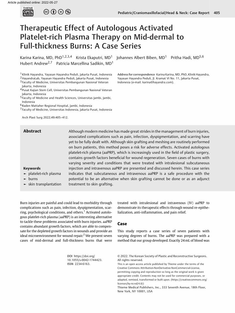
Case 1A 43-year-oldmanpresentedwith bilateral deep-dermal andfull-thickness burns on his legs (18% of the total body surfacearea [TBSA]). Surgical debridement was performed 5 daysafter theflame burn injury. aaPRP therapywas then given viaSC injection and IV infusion. The wound was dressed withparaffin gauze and dry gauze. The second dose of aaPRP wasgiven on the 14th day postinjury. On the 18th day, the patient
was discharged, and 100% epithelization was seen on the23rd day (►Fig. 1).
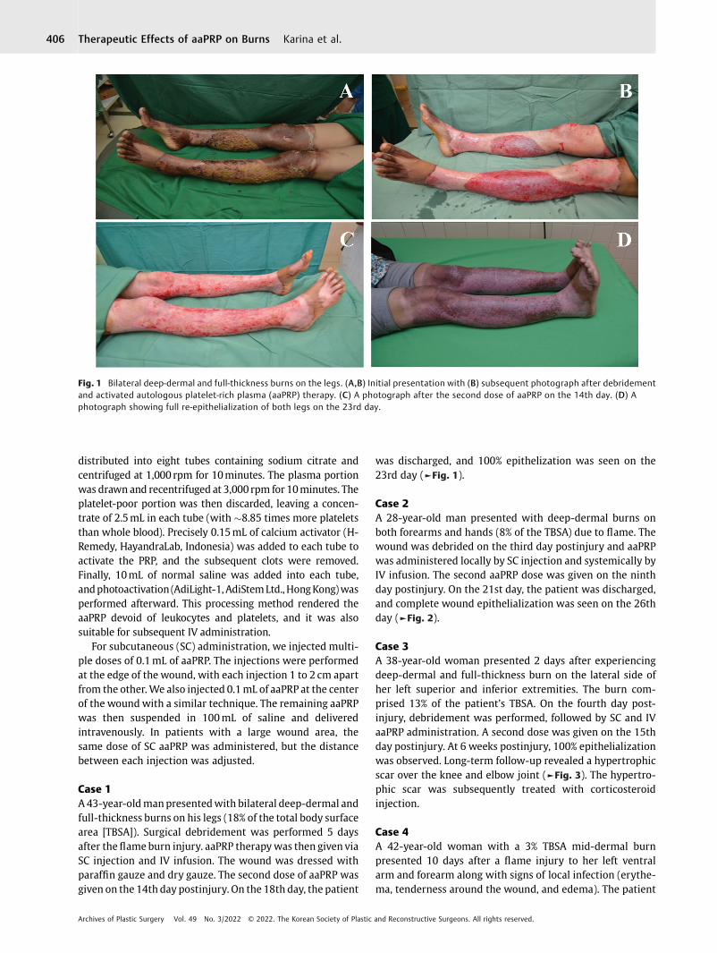
Case 2A 28-year-old man presented with deep-dermal burns onboth forearms and hands (8% of the TBSA) due to flame. Thewound was debrided on the third day postinjury and aaPRPwas administered locally by SC injection and systemically byIV infusion. The second aaPRP dose was given on the ninthday postinjury. On the 21st day, the patient was discharged,and complete wound epithelialization was seen on the 26thday (►Fig. 2).
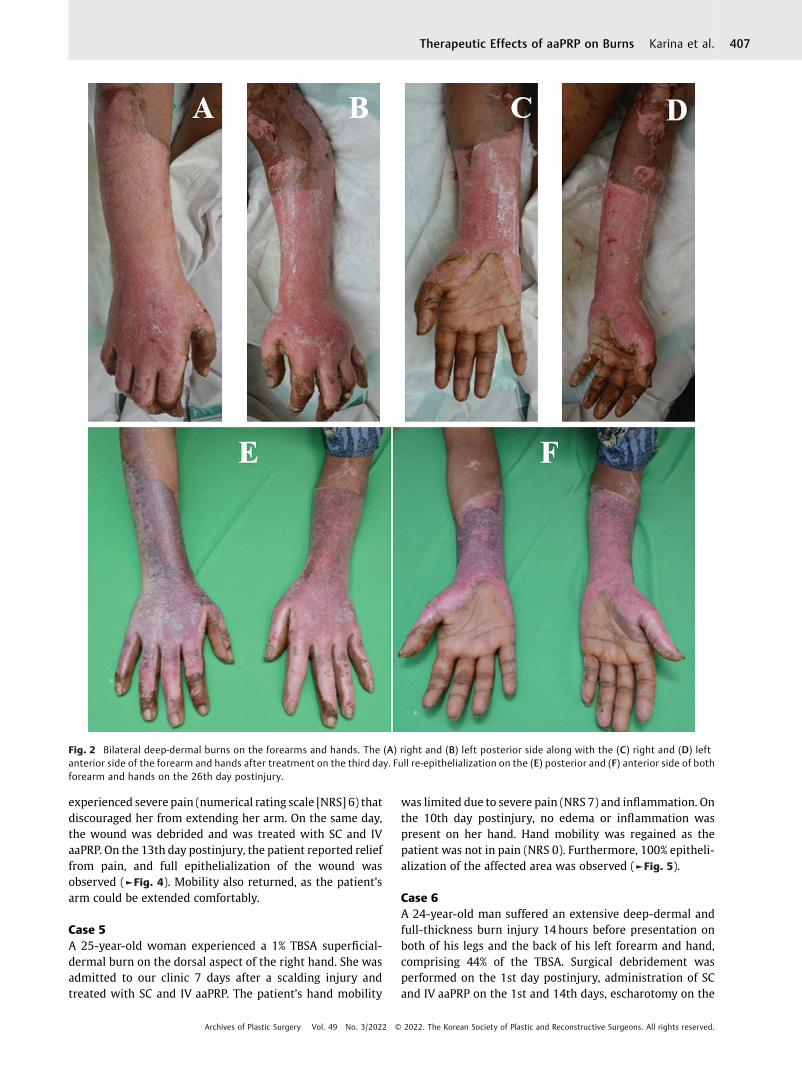
Case 3A 38-year-old woman presented 2 days after experiencingdeep-dermal and full-thickness burn on the lateral side ofher left superior and inferior extremities. The burn com-prised 13% of the patient’s TBSA. On the fourth day post-injury, debridement was performed, followed by SC and IVaaPRP administration. A second dose was given on the 15thday postinjury. At 6 weeks postinjury, 100% epithelializationwas observed. Long-term follow-up revealed a hypertrophicscar over the knee and elbow joint (►Fig. 3). The hypertro-phic scar was subsequently treated with corticosteroidinjection.
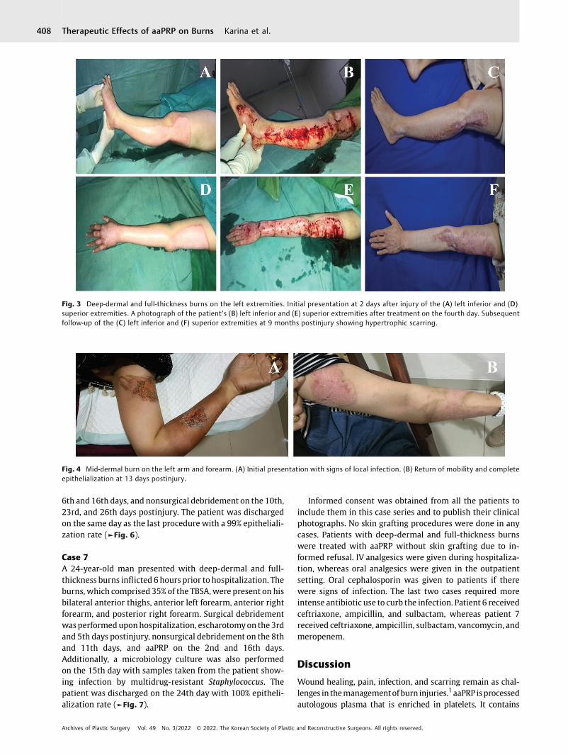
Case 4A 42-year-old woman with a 3% TBSA mid-dermal burnpresented 10 days after a flame injury to her left ventralarm and forearm along with signs of local infection (erythe-ma, tenderness around the wound, and edema). The patient
Fig. 1 Bilateral deep-dermal and full-thickness burns on the legs. (A,B) Initial presentation with (B) subsequent photograph after debridementand activated autologous platelet-rich plasma (aaPRP) therapy. (C) A photograph after the second dose of aaPRP on the 14th day. (D) Aphotograph showing full re-epithelialization of both legs on the 23rd day.
Archives of Plastic Surgery Vol. 49 No. 3/2022 © 2022. The Korean Society of Plastic and Reconstructive Surgeons. All rights reserved.
Therapeutic Effects of aaPRP on Burns Karina et al.406
experienced severe pain (numerical rating scale [NRS] 6) thatdiscouraged her from extending her arm. On the same day,the wound was debrided and was treated with SC and IVaaPRP. On the 13th day postinjury, the patient reported relieffrom pain, and full epithelialization of the wound wasobserved (►Fig. 4). Mobility also returned, as the patient’sarm could be extended comfortably.
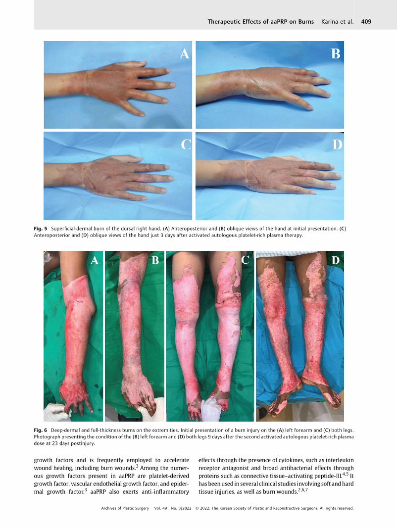
Case 5A 25-year-old woman experienced a 1% TBSA superficial-dermal burn on the dorsal aspect of the right hand. She wasadmitted to our clinic 7 days after a scalding injury andtreated with SC and IV aaPRP. The patient’s hand mobility
was limited due to severe pain (NRS 7) and inflammation. Onthe 10th day postinjury, no edema or inflammation waspresent on her hand. Hand mobility was regained as thepatient was not in pain (NRS 0). Furthermore, 100% epitheli-alization of the affected area was observed (►Fig. 5).
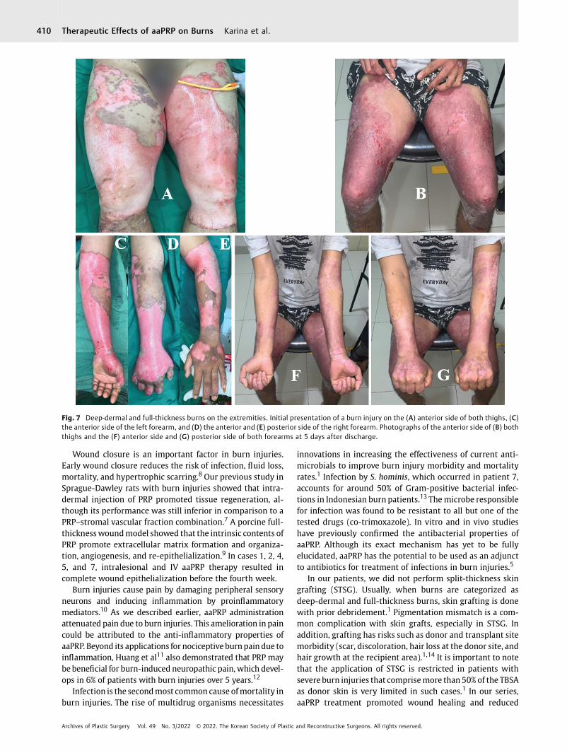
Case 6A 24-year-old man suffered an extensive deep-dermal andfull-thickness burn injury 14 hours before presentation onboth of his legs and the back of his left forearm and hand,comprising 44% of the TBSA. Surgical debridement wasperformed on the 1st day postinjury, administration of SCand IV aaPRP on the 1st and 14th days, escharotomy on the
Fig. 2 Bilateral deep-dermal burns on the forearms and hands. The (A) right and (B) left posterior side along with the (C) right and (D) leftanterior side of the forearm and hands after treatment on the third day. Full re-epithelialization on the (E) posterior and (F) anterior side of bothforearm and hands on the 26th day postinjury.
Archives of Plastic Surgery Vol. 49 No. 3/2022 © 2022. The Korean Society of Plastic and Reconstructive Surgeons. All rights reserved.
Therapeutic Effects of aaPRP on Burns Karina et al. 407
6th and 16th days, and nonsurgical debridement on the 10th,23rd, and 26th days postinjury. The patient was dischargedon the same day as the last procedure with a 99% epitheliali-zation rate (►Fig. 6).
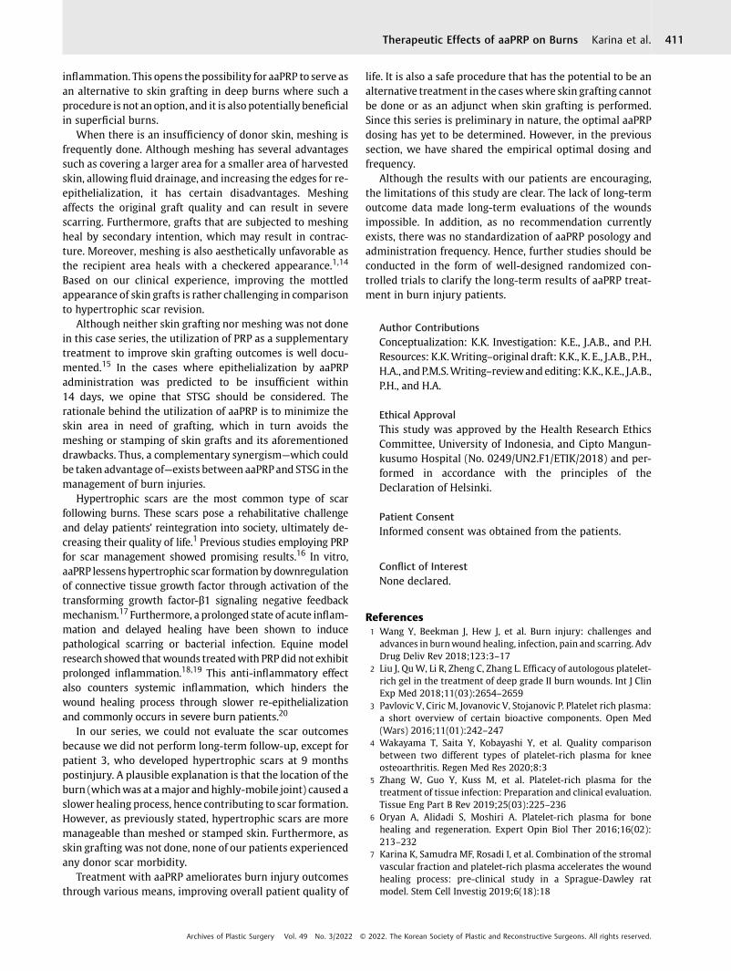
Case 7A 24-year-old man presented with deep-dermal and full-thickness burns inflicted 6hours prior to hospitalization. Theburns, which comprised 35% of the TBSA, were present on hisbilateral anterior thighs, anterior left forearm, anterior rightforearm, and posterior right forearm. Surgical debridementwas performeduponhospitalization, escharotomyon the 3rdand 5th days postinjury, nonsurgical debridement on the 8thand 11th days, and aaPRP on the 2nd and 16th days.Additionally, a microbiology culture was also performedon the 15th day with samples taken from the patient show-ing infection by multidrug-resistant Staphylococcus. Thepatient was discharged on the 24th day with 100% epitheli-alization rate (►Fig. 7).
Informed consent was obtained from all the patients toinclude them in this case series and to publish their clinicalphotographs. No skin grafting procedures were done in anycases. Patients with deep-dermal and full-thickness burnswere treated with aaPRP without skin grafting due to in-formed refusal. IV analgesics were given during hospitaliza-tion, whereas oral analgesics were given in the outpatientsetting. Oral cephalosporin was given to patients if therewere signs of infection. The last two cases required moreintense antibiotic use to curb the infection. Patient 6 receivedceftriaxone, ampicillin, and sulbactam, whereas patient 7received ceftriaxone, ampicillin, sulbactam, vancomycin, andmeropenem.
Discussion
Wound healing, pain, infection, and scarring remain as chal-lenges in themanagementofburn injuries.1aaPRP isprocessedautologous plasma that is enriched in platelets. It contains
Fig. 3 Deep-dermal and full-thickness burns on the left extremities. Initial presentation at 2 days after injury of the (A) left inferior and (D)superior extremities. A photograph of the patient’s (B) left inferior and (E) superior extremities after treatment on the fourth day. Subsequentfollow-up of the (C) left inferior and (F) superior extremities at 9 months postinjury showing hypertrophic scarring.
Fig. 4 Mid-dermal burn on the left arm and forearm. (A) Initial presentation with signs of local infection. (B) Return of mobility and completeepithelialization at 13 days postinjury.
Archives of Plastic Surgery Vol. 49 No. 3/2022 © 2022. The Korean Society of Plastic and Reconstructive Surgeons. All rights reserved.
Therapeutic Effects of aaPRP on Burns Karina et al.408
growth factors and is frequently employed to acceleratewound healing, including burn wounds.3 Among the numer-ous growth factors present in aaPRP are platelet-derivedgrowth factor, vascular endothelial growth factor, and epider-mal growth factor.3 aaPRP also exerts anti-inflammatory
effects through the presence of cytokines, such as interleukinreceptor antagonist and broad antibacterial effects throughproteins such as connective tissue–activating peptide-III.4,5 Ithas beenused in several clinical studies involving soft andhardtissue injuries, as well as burn wounds.2,6,7
Fig. 5 Superficial-dermal burn of the dorsal right hand. (A) Anteroposterior and (B) oblique views of the hand at initial presentation. (C)Anteroposterior and (D) oblique views of the hand just 3 days after activated autologous platelet-rich plasma therapy.
Fig. 6 Deep-dermal and full-thickness burns on the extremities. Initial presentation of a burn injury on the (A) left forearm and (C) both legs.Photograph presenting the condition of the (B) left forearm and (D) both legs 9 days after the second activated autologous platelet-rich plasmadose at 23 days postinjury.
Archives of Plastic Surgery Vol. 49 No. 3/2022 © 2022. The Korean Society of Plastic and Reconstructive Surgeons. All rights reserved.
Therapeutic Effects of aaPRP on Burns Karina et al. 409
Wound closure is an important factor in burn injuries.Early wound closure reduces the risk of infection, fluid loss,mortality, and hypertrophic scarring.8 Our previous study inSprague-Dawley rats with burn injuries showed that intra-dermal injection of PRP promoted tissue regeneration, al-though its performance was still inferior in comparison to aPRP–stromal vascular fraction combination.7 A porcine full-thicknesswoundmodel showed that the intrinsic contents ofPRP promote extracellular matrix formation and organiza-tion, angiogenesis, and re-epithelialization.9 In cases 1, 2, 4,5, and 7, intralesional and IV aaPRP therapy resulted incomplete wound epithelialization before the fourth week.
Burn injuries cause pain by damaging peripheral sensoryneurons and inducing inflammation by proinflammatorymediators.10 As we described earlier, aaPRP administrationattenuated pain due to burn injuries. This amelioration in paincould be attributed to the anti-inflammatory properties ofaaPRP. Beyond its applications for nociceptive burn pain due toinflammation, Huang et al11 also demonstrated that PRP maybebeneficial for burn-induced neuropathic pain, which devel-ops in 6% of patients with burn injuries over 5 years.12
Infection is the secondmost common cause ofmortality inburn injuries. The rise of multidrug organisms necessitates
innovations in increasing the effectiveness of current anti-microbials to improve burn injury morbidity and mortalityrates.1 Infection by S. hominis, which occurred in patient 7,accounts for around 50% of Gram-positive bacterial infec-tions in Indonesian burn patients.13 Themicrobe responsiblefor infection was found to be resistant to all but one of thetested drugs (co-trimoxazole). In vitro and in vivo studieshave previously confirmed the antibacterial properties ofaaPRP. Although its exact mechanism has yet to be fullyelucidated, aaPRP has the potential to be used as an adjunctto antibiotics for treatment of infections in burn injuries.5
In our patients, we did not perform split-thickness skingrafting (STSG). Usually, when burns are categorized asdeep-dermal and full-thickness burns, skin grafting is donewith prior debridement.1 Pigmentation mismatch is a com-mon complication with skin grafts, especially in STSG. Inaddition, grafting has risks such as donor and transplant sitemorbidity (scar, discoloration, hair loss at the donor site, andhair growth at the recipient area).1,14 It is important to notethat the application of STSG is restricted in patients withsevere burn injuries that comprisemore than 50% of the TBSAas donor skin is very limited in such cases.1 In our series,aaPRP treatment promoted wound healing and reduced
Fig. 7 Deep-dermal and full-thickness burns on the extremities. Initial presentation of a burn injury on the (A) anterior side of both thighs, (C)the anterior side of the left forearm, and (D) the anterior and (E) posterior side of the right forearm. Photographs of the anterior side of (B) boththighs and the (F) anterior side and (G) posterior side of both forearms at 5 days after discharge.
Archives of Plastic Surgery Vol. 49 No. 3/2022 © 2022. The Korean Society of Plastic and Reconstructive Surgeons. All rights reserved.
Therapeutic Effects of aaPRP on Burns Karina et al.410
inflammation. This opens the possibility for aaPRP to serve asan alternative to skin grafting in deep burns where such aprocedure is not an option, and it is also potentially beneficialin superficial burns.
When there is an insufficiency of donor skin, meshing isfrequently done. Although meshing has several advantagessuch as covering a larger area for a smaller area of harvestedskin, allowing fluid drainage, and increasing the edges for re-epithelialization, it has certain disadvantages. Meshingaffects the original graft quality and can result in severescarring. Furthermore, grafts that are subjected to meshingheal by secondary intention, which may result in contrac-ture. Moreover, meshing is also aesthetically unfavorable asthe recipient area heals with a checkered appearance.1,14
Based on our clinical experience, improving the mottledappearance of skin grafts is rather challenging in comparisonto hypertrophic scar revision.
Although neither skin grafting nor meshing was not donein this case series, the utilization of PRP as a supplementarytreatment to improve skin grafting outcomes is well docu-mented.15 In the cases where epithelialization by aaPRPadministration was predicted to be insufficient within14 days, we opine that STSG should be considered. Therationale behind the utilization of aaPRP is to minimize theskin area in need of grafting, which in turn avoids themeshing or stamping of skin grafts and its aforementioneddrawbacks. Thus, a complementary synergism—which couldbe taken advantage of—exists between aaPRP and STSG in themanagement of burn injuries.
Hypertrophic scars are the most common type of scarfollowing burns. These scars pose a rehabilitative challengeand delay patients’ reintegration into society, ultimately de-creasing their quality of life.1 Previous studies employing PRPfor scar management showed promising results.16 In vitro,aaPRP lessens hypertrophic scar formation by downregulationof connective tissue growth factor through activation of thetransforming growth factor-β1 signaling negative feedbackmechanism.17 Furthermore, a prolonged state of acute inflam-mation and delayed healing have been shown to inducepathological scarring or bacterial infection. Equine modelresearch showed that wounds treatedwith PRP did not exhibitprolonged inflammation.18,19 This anti-inflammatory effectalso counters systemic inflammation, which hinders thewound healing process through slower re-epithelializationand commonly occurs in severe burn patients.20
In our series, we could not evaluate the scar outcomesbecause we did not perform long-term follow-up, except forpatient 3, who developed hypertrophic scars at 9 monthspostinjury. A plausible explanation is that the location of theburn (whichwas at amajor and highly-mobile joint) caused aslower healing process, hence contributing to scar formation.However, as previously stated, hypertrophic scars are moremanageable than meshed or stamped skin. Furthermore, asskin grafting was not done, none of our patients experiencedany donor scar morbidity.
Treatment with aaPRP ameliorates burn injury outcomesthrough various means, improving overall patient quality of
life. It is also a safe procedure that has the potential to be analternative treatment in the caseswhere skin grafting cannotbe done or as an adjunct when skin grafting is performed.Since this series is preliminary in nature, the optimal aaPRPdosing has yet to be determined. However, in the previoussection, we have shared the empirical optimal dosing andfrequency.
Although the results with our patients are encouraging,the limitations of this study are clear. The lack of long-termoutcome data made long-term evaluations of the woundsimpossible. In addition, as no recommendation currentlyexists, there was no standardization of aaPRP posology andadministration frequency. Hence, further studies should beconducted in the form of well-designed randomized con-trolled trials to clarify the long-term results of aaPRP treat-ment in burn injury patients.
Author ContributionsConceptualization: K.K. Investigation: K.E., J.A.B., and P.H.Resources: K.K.Writing–original draft: K.K., K. E., J.A.B., P.H.,H.A., andP.M.S.Writing–reviewandediting:K.K., K.E., J.A.B.,P.H., and H.A.
Ethical ApprovalThis study was approved by the Health Research EthicsCommittee, University of Indonesia, and Cipto Mangun-kusumo Hospital (No. 0249/UN2.F1/ETIK/2018) and per-formed in accordance with the principles of theDeclaration of Helsinki.
Patient ConsentInformed consent was obtained from the patients.
Conflict of InterestNone declared.
References1 Wang Y, Beekman J, Hew J, et al. Burn injury: challenges and
advances in burnwound healing, infection, pain and scarring. AdvDrug Deliv Rev 2018;123:3–17
2 Liu J, QuW, Li R, Zheng C, Zhang L. Efficacy of autologous platelet-rich gel in the treatment of deep grade II burn wounds. Int J ClinExp Med 2018;11(03):2654–2659
3 Pavlovic V, Ciric M, Jovanovic V, Stojanovic P. Platelet rich plasma:a short overview of certain bioactive components. Open Med(Wars) 2016;11(01):242–247
4 Wakayama T, Saita Y, Kobayashi Y, et al. Quality comparisonbetween two different types of platelet-rich plasma for kneeosteoarthritis. Regen Med Res 2020;8:3
5 Zhang W, Guo Y, Kuss M, et al. Platelet-rich plasma for thetreatment of tissue infection: Preparation and clinical evaluation.Tissue Eng Part B Rev 2019;25(03):225–236
6 Oryan A, Alidadi S, Moshiri A. Platelet-rich plasma for bonehealing and regeneration. Expert Opin Biol Ther 2016;16(02):213–232
7 Karina K, Samudra MF, Rosadi I, et al. Combination of the stromalvascular fraction and platelet-rich plasma accelerates the woundhealing process: pre-clinical study in a Sprague-Dawley ratmodel. Stem Cell Investig 2019;6(18):18
Archives of Plastic Surgery Vol. 49 No. 3/2022 © 2022. The Korean Society of Plastic and Reconstructive Surgeons. All rights reserved.
Therapeutic Effects of aaPRP on Burns Karina et al. 411
8 Singer AJ, Boyce ST. Burn wound healing and tissue engineering. JBurn Care Res 2017;38(03):e605–e613
9 Vermeulen P, Dickens S, Degezelle K, Van den Berge S, HendrickxB, Vranckx JJ. A plasma-based biomatrix mixed with endothelialprogenitor cells and keratinocytes promotes matrix formation,angiogenesis, and reepithelialization in full-thickness wounds.Tissue Eng Part A 2009;15(07):1533–1542
10 Morgan M, Deuis JR, Frøsig-Jørgensen M, et al. Burn pain: asystematic and critical review of epidemiology, pathophysiology,and treatment. Pain Med 2018;19(04):708–734
11 Huang SH, Wu SH, Lee SS, et al. Platelet-rich plasma injection inburn scar areas alleviates neuropathic scar pain. Int J Med Sci2018;15(03):238–247
12 Klifto KM, Dellon AL, Hultman CS. Prevalence and associatedpredictors for patients developing chronic neuropathic painfollowing burns. Burn Trauma 2020;8:tkaa011
13 Soedjana H, Nadia J, Sundoro A, et al. The profile of severe burninjury patients with sepsis in hasan sadikin bandung generalhospital. Ann Burns Fire Disasters 2020;33(04):312–316
14 Chen J, Jain SA. Principles of skin grafts. In: Weinzweig J, ed.Plastic Surgery Secrets Plus. 2nd ed. Baltimore,MD:Mosby; 2010:677–683
15 Aggarwal A, Chittoria RK, Dutta S, et al. Autologous platelet richplasma: an adjunct to early tangential excision and grafting inburns. Plast Aesthetic Res 2018;5:47
16 Alser OH, Goutos I. The evidence behind the use of platelet-richplasma (PRP) in scar management: a literature review. Scars BurnHeal 2018;4:2059513118808773
17 Nam SM, Kim YB. The effects of platelet-rich plasma on hypertro-phic scars fibroblasts. Int Wound J 2018;15(04):547–554
18 Finnerty CC, Jeschke MG, Branski LK, Barret JP, Dziewulski P,Herndon DN. Hypertrophic scarring: the greatest unmet chal-lenge after burn injury. Lancet 2016;388(10052):1427–1436
19 Carter CA, Jolly DG,Worden CE Sr, Hendren DG, Kane CJM. Platelet-rich plasma gel promotes differentiation and regeneration duringequine wound healing. Exp Mol Pathol 2003;74(03):244–255
20 Rowan MP, Cancio LC, Elster EA, et al. Burn wound healing andtreatment: review and advancements. Crit Care 2015;19:243
Archives of Plastic Surgery Vol. 49 No. 3/2022 © 2022. The Korean Society of Plastic and Reconstructive Surgeons. All rights reserved.
Therapeutic Effects of aaPRP on Burns Karina et al.412
Related Documents


