Biomolecules 2021, 11, 1775. https://doi.org/10.3390/biom11121775 www.mdpi.com/journal/biomolecules Review Therapeutic Approaches in Lysosomal Storage Diseases Carlos Fernández-Pereira 1 , Beatriz San Millán-Tejado 1 , María Gallardo-Gómez 1 , Tania Pérez-Márquez 1 , Marta Alves-Villar 1 , Cristina Melcón-Crespo 1,2 , Julián Fernández-Martín 1,3 and Saida Ortolano 1, * 1 Rare Disease and Pediatric Medicine Group, Galicia Sur Health Research Institute (IIS Galicia Sur), SERGAS-UVIGO, 36312 Vigo, Spain; [email protected] (C.F.-P.); [email protected] (B.S.M.-T.); [email protected] (M.G.-G.); [email protected] (T.P.-M.); [email protected] (M.A.-V.); [email protected] (C.M.-C.); [email protected] (J.F.-M.) 2 Department of Pediatrics, Hospital Álvaro Cunqueiro, SERGAS, 36213 Vigo, Spain 3 Department of Internal Medicine, Hospital Álvaro Cunqueiro, SERGAS, 36213 Vigo, Spain * Correspondence: [email protected]; Tel.: +34-986217466 Abstract: Lysosomal Storage Diseases are multisystemic disorders determined by genetic variants, which affect the proteins involved in lysosomal function and cellular metabolism. Different thera- peutic approaches, which are based on the physiologic mechanisms that regulate lysosomal func- tion, have been proposed for these diseases. Currently, enzyme replacement therapy, gene therapy, or small molecules have been approved or are under clinical development to treat lysosomal storage disorders. The present article reviews the main therapeutic strategies that have been proposed so far, highlighting possible limitations and future perspectives. Keywords: lysosomal storage diseases; enzyme replacement therapy; gene therapy; small molecules; autophagy 1. Introduction 1.1. General Considerations concerning Rare Diseases The last 25 years have been characterized by an upgrowing interest among the scien- tific community towards rare diseases and the development of specific treatments for these pathologies. This initiative finds its impulse in the increased sense of social and eth- ical responsibility to provide therapeutic solutions to the affected groups, as well as in the rising efforts of the pharma industry to dedicate resources to this field of knowledge. The discovery of orphan drugs is a matter of interest for the industry, since most rare diseases exhibit well defined pathophysiological mechanisms, as they are congenital pa- thologies that affect a single gene. In addition, the process of approving orphan drugs comes with a specific designation and a relatively less complex process to facilitate possi- ble solutions for unmet medical needs. Additionally, an orphan drug can potentially have secondary indications directed to the treatment of frequent pathologies, with more complex etiology, which share molecu- lar mechanisms with the rare disease for which the drug was originally developed. For all these reasons, in the last years, the development of treatments orientated to rare diseases has largely increased, also favoring innovation of the biological tools and advances in the knowledge of disease natural history. These considerations are particularly applicable to the field of Lysosomal Storage Diseases (LSDs) that show favorable characteristics for different therapeutic approaches. Citation: Fernández-Pereira, C.; San Millán, B.; Gallardo-Gómez, M.; Pérez-Márquez, T.; Alves-Villar, M.; Melcón-Crespo, C.; Fernández-Martín, J.; Ortolano, S. Therapeutic Approaches in Lysosomal Storage Diseases. Biomolecules 2021, 11, 1775. https://doi.org/10.3390/ biom11121775 Academic Editor: Enrico Moro Received: 31 October 2021 Accepted: 24 November 2021 Published: 26 November 2021 Publisher’s Note: MDPI stays neu- tral with regard to jurisdictional claims in published maps and institu- tional affiliations. Copyright: © 2021 by the authors. Li- censee MDPI, Basel, Switzerland. This article is an open access article distributed under the terms and con- ditions of the Creative Commons At- tribution (CC BY) license (http://crea- tivecommons.org/licenses/by/4.0/).

Welcome message from author
This document is posted to help you gain knowledge. Please leave a comment to let me know what you think about it! Share it to your friends and learn new things together.
Transcript

Biomolecules 2021, 11, 1775. https://doi.org/10.3390/biom11121775 www.mdpi.com/journal/biomolecules
Review
Therapeutic Approaches in Lysosomal Storage Diseases Carlos Fernández-Pereira 1, Beatriz San Millán-Tejado 1, María Gallardo-Gómez 1, Tania Pérez-Márquez 1, Marta Alves-Villar 1, Cristina Melcón-Crespo 1,2, Julián Fernández-Martín 1,3 and Saida Ortolano 1,*
1 Rare Disease and Pediatric Medicine Group, Galicia Sur Health Research Institute (IIS Galicia Sur), SERGAS-UVIGO, 36312 Vigo, Spain; [email protected] (C.F.-P.); [email protected] (B.S.M.-T.); [email protected] (M.G.-G.); [email protected] (T.P.-M.); [email protected] (M.A.-V.); [email protected] (C.M.-C.); [email protected] (J.F.-M.)
2 Department of Pediatrics, Hospital Álvaro Cunqueiro, SERGAS, 36213 Vigo, Spain 3 Department of Internal Medicine, Hospital Álvaro Cunqueiro, SERGAS, 36213 Vigo, Spain * Correspondence: [email protected]; Tel.: +34-986217466
Abstract: Lysosomal Storage Diseases are multisystemic disorders determined by genetic variants, which affect the proteins involved in lysosomal function and cellular metabolism. Different thera-peutic approaches, which are based on the physiologic mechanisms that regulate lysosomal func-tion, have been proposed for these diseases. Currently, enzyme replacement therapy, gene therapy, or small molecules have been approved or are under clinical development to treat lysosomal storage disorders. The present article reviews the main therapeutic strategies that have been proposed so far, highlighting possible limitations and future perspectives.
Keywords: lysosomal storage diseases; enzyme replacement therapy; gene therapy; small molecules; autophagy
1. Introduction 1.1. General Considerations concerning Rare Diseases
The last 25 years have been characterized by an upgrowing interest among the scien-tific community towards rare diseases and the development of specific treatments for these pathologies. This initiative finds its impulse in the increased sense of social and eth-ical responsibility to provide therapeutic solutions to the affected groups, as well as in the rising efforts of the pharma industry to dedicate resources to this field of knowledge.
The discovery of orphan drugs is a matter of interest for the industry, since most rare diseases exhibit well defined pathophysiological mechanisms, as they are congenital pa-thologies that affect a single gene. In addition, the process of approving orphan drugs comes with a specific designation and a relatively less complex process to facilitate possi-ble solutions for unmet medical needs.
Additionally, an orphan drug can potentially have secondary indications directed to the treatment of frequent pathologies, with more complex etiology, which share molecu-lar mechanisms with the rare disease for which the drug was originally developed.
For all these reasons, in the last years, the development of treatments orientated to rare diseases has largely increased, also favoring innovation of the biological tools and advances in the knowledge of disease natural history.
These considerations are particularly applicable to the field of Lysosomal Storage Diseases (LSDs) that show favorable characteristics for different therapeutic approaches.
Citation: Fernández-Pereira, C.;
San Millán, B.; Gallardo-Gómez, M.;
Pérez-Márquez, T.; Alves-Villar, M.;
Melcón-Crespo, C.;
Fernández-Martín, J.; Ortolano, S.
Therapeutic Approaches in
Lysosomal Storage Diseases.
Biomolecules 2021, 11, 1775.
https://doi.org/10.3390/
biom11121775
Academic Editor: Enrico Moro
Received: 31 October 2021
Accepted: 24 November 2021
Published: 26 November 2021
Publisher’s Note: MDPI stays neu-
tral with regard to jurisdictional
claims in published maps and institu-
tional affiliations.
Copyright: © 2021 by the authors. Li-
censee MDPI, Basel, Switzerland.
This article is an open access article
distributed under the terms and con-
ditions of the Creative Commons At-
tribution (CC BY) license (http://crea-
tivecommons.org/licenses/by/4.0/).

Biomolecules 2021, 11, 1775 2 of 20
1.2. Molecular Basis of LSDs and Possible Therapeutical Strategies LSDs are caused by mutations in genes which encode for acid hydrolases, integral
membrane proteins, activators and transporter proteins, or other proteins involved in ly-sosomal function. Deficiencies of these molecules unleash a metabolic imbalance, which results in substrate accumulation in multiple organs or tissues [1].
The therapeutic options currently available or under development for LSDs are based on the physiology of the lysosome, which is considered as a master regulator of cell me-tabolism.
Lysosomes are organelles principally dedicated to cell catabolic function through the action of more than 60 hydrolytic enzymes contained inside their lumen. However, now-adays we know that the lysosome is also important in the regulation of anabolic processes. In fact, it acts as a nutrient sensor and it regulates signal transduction, cellular growth, and vesicle trafficking [2].
These processes are widely related to the activity of the mammalian target of rapamy-cin complex 1 (mTORC1) of proteins, the principal regulator of cell growth, which is an-chored to lysosomal membrane and senses variations in cytoplasmic amino acid concen-tration. At low amino acid concentrations, mTORC1 triggers autophagy, a process by which the cells sequester their own content to degrade it and recycle its components [3,4].
Moreover, the expression of genes that encode for lysosomal proteins depends on the action of the transcriptional factor EB (TFEB), which controls lysosomal biogenesis and exocytosis, lipid catabolism and energy metabolism, allowing the adaptation of the lyso-some to different pathophysiological conditions [5]. The mTORC1 regulates TFEB by phosphorylation and retains it in the cytoplasm. Dephosphorylation of TFEB occurs in fasting conditions, following mTORC1 inactivation (Figure 1). Therefore, this mechanism is a proof of lysosome to nucleus communication and supports the concept of the lyso-some as the main actor in the regulation of cellular metabolism [6].
Figure 1. Autophagy activation route and lysosome-nuclei communication. The mTORC1 complex, attached to the lysosomal membrane, controls the activation of TFEB, which is translocated to the nucleus to activate the genes that regulate the autophagic process.
Lysosomal enzymes are produced in the cytoplasm and migrate to the lysosome through well-established endocytic routes. In most cases, enzyme functionalization occurs in the Golgi apparatus by the addition of mannose-6-phosphate (MP-6) groups, which enables their entrance in the lysosome through the MP-6 receptor (M6PR) [7]. Nonethe-less, some enzymes use different mechanisms; for instance, β-glucocerebrosidase, which
Biomolecules 2021, 11, 1775 3 of 20
reaches the lysosome through the lysosomal integral membrane protein-2 (LIMP-2) recep-tor [8].
Vesicles transport of substrates to the lysosome occurs throughout a variety of endo-cytic mechanisms (phagocytosis, macropinocytosis, and endocytosis, independent or me-diated by clathrin or caveolin) determined by vesicle content [9]. By this means, substrate accumulation generated in LSDs determines the blockage of the autophagic process, as described in different disorders.
In Pompe Disease (PD), the autophagic compartment has been reported to be ex-panded in muscular tissues [10], and in Multiple Sulfatase Deficiency (MSD), all sulfatases are affected by a post-translational defective activation of the sulfatase modifier factor 1 that impairs autophagolysosome formation [11]. Finally, in Niemann Pick disease C (NPC), sphingosine storage leads to calcium increased concentration that blocks autoph-agy and increases the accumulation of cholesterol, sphingomyelin, and glycosphin-golipids [12].
Therefore, the recently discovered connection between many LSDs, such as Gaucher disease (GD) and NPC, and other conditions, like ageing or neurodegenerative disorders (e.g., Parkinson and Alzheimer Disease), is easier to explain considering that these dis-eases are also caused by imbalance in the lysosome-autophagy axis [13].
On the other hand, substrate accumulation may also trigger an anomalous activation of other routes, such as the activation of a Toll-like receptor 4 (TLR-4) by Globotrialosil ceramide (Gb3) in Fabry disease (FD), which determines the response of the innate im-mune system [14] and endothelial disfunction [15].
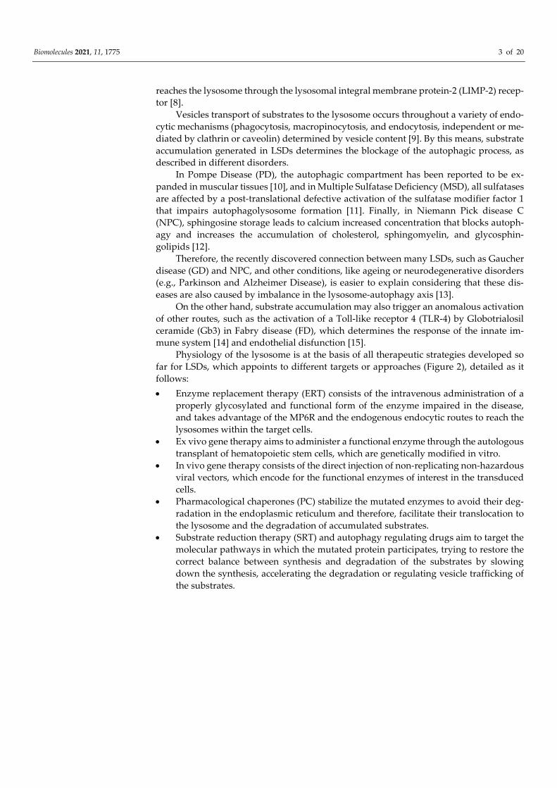
Physiology of the lysosome is at the basis of all therapeutic strategies developed so far for LSDs, which appoints to different targets or approaches (Figure 2), detailed as it follows: • Enzyme replacement therapy (ERT) consists of the intravenous administration of a
properly glycosylated and functional form of the enzyme impaired in the disease, and takes advantage of the MP6R and the endogenous endocytic routes to reach the lysosomes within the target cells.
• Ex vivo gene therapy aims to administer a functional enzyme through the autologous transplant of hematopoietic stem cells, which are genetically modified in vitro.
• In vivo gene therapy consists of the direct injection of non-replicating non-hazardous viral vectors, which encode for the functional enzymes of interest in the transduced cells.
• Pharmacological chaperones (PC) stabilize the mutated enzymes to avoid their deg-radation in the endoplasmic reticulum and therefore, facilitate their translocation to the lysosome and the degradation of accumulated substrates.
• Substrate reduction therapy (SRT) and autophagy regulating drugs aim to target the molecular pathways in which the mutated protein participates, trying to restore the correct balance between synthesis and degradation of the substrates by slowing down the synthesis, accelerating the degradation or regulating vesicle trafficking of the substrates.
Biomolecules 2021, 11, 1775 4 of 20
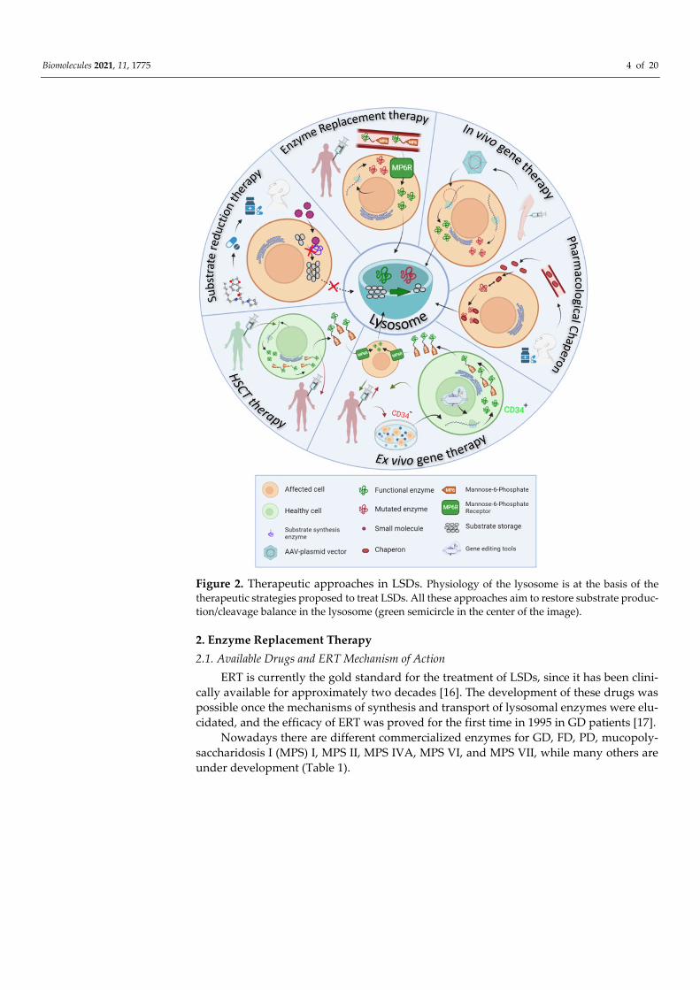
Figure 2. Therapeutic approaches in LSDs. Physiology of the lysosome is at the basis of the therapeutic strategies proposed to treat LSDs. All these approaches aim to restore substrate produc-tion/cleavage balance in the lysosome (green semicircle in the center of the image).
2. Enzyme Replacement Therapy 2.1. Available Drugs and ERT Mechanism of Action
ERT is currently the gold standard for the treatment of LSDs, since it has been clini-cally available for approximately two decades [16]. The development of these drugs was possible once the mechanisms of synthesis and transport of lysosomal enzymes were elu-cidated, and the efficacy of ERT was proved for the first time in 1995 in GD patients [17].
Nowadays there are different commercialized enzymes for GD, FD, PD, mucopoly-saccharidosis I (MPS) I, MPS II, MPS IVA, MPS VI, and MPS VII, while many others are under development (Table 1).

Biomolecules 2021, 11, 1775 5 of 20
Table 1. Available ERT based medicaments for LSDs. In the reference column there are indicated either reference articles or clinical trial numbers referring to www.clinicaltrials.gov (accessed on 15 November 2021)web page.
LSDs Affected Enzyme Available ERT Development Status Reference
GD Glucocerebrosidase Imglucerase Approved
[18] Velaglucerase Approved Taliglucerase alpha Approved
FD α-Galactosidase A
Agalsidase alpa Approved [19] Agalsidase beta Approved NCT03018730
PRX-102 Phase III NCT02795676 NCT03180840
PD α-Glycosidase
Alglucosidase alpha Approved [20] Avalglucosidase alfa Approved [21]
VAL-1221 Phase I-II NCT02898753 ATB200 Phase III NCT03729362
MPS I α-L-iduronidase Laronidase Approved [22]
MPS II Iduronate-2-sulfatase Idursulfasa Approved [23]
AGT-182 Phase I NCT02262338 JR-141 Approved [24]
MPS III A B
Heparan N-sulphatase rhHNS Phase I-II NCT01299727
N-acetyl-glucosaminidase BMN250 Phase I-II NCT02754076
MPS VI N-acetylgalactosamine-4-
sulfatase Galsufase Approved [24]
MLD Arilsulphatase A HGT1110 Phase I-II NCT01887938 TAK611 Phase II NCT015128 [25]
NPA and NPB Acid
Sphingomyelinase rh-ASM Phase I NCT00410566
As previously mentioned, ERT is based on the intravenous administration of a func-tional recombinant human enzyme, which can compensate the consequences of the con-genital defect. ERT exploits the mechanism of lysosomal enzyme biogenesis and recycling. The recombinant protein, enriched with M6P groups, enters the cell using the MP6R re-ceptor and reaches the lysosome. A wider distribution of the enzyme is possible thanks to the phenomenon of the cross-correction, which was described for the first time by Barton and Neufeld [26]. They observed that cells derived from LSD patients were able to metab-olize the accumulated substrates, when co-cultivated with cells from healthy donors, which is possible because a fraction of lysosomal enzymes is secreted towards the extra-cellular space and enters the surrounding cells through the MP6R.
Recombinant human enzymes are produced in stable cell lines as Chinese Ovary Hamster (CHO) cells or human fibroblasts, which provide different glycosylation patterns [27]. Recently, vegetable cells (e.g., tobacco derived cells) [28] or yeasts [29] have also been used to produce the recombinant enzyme with lower production costs.
ERT has shown its efficacy in delaying the progression of the disease and improving the life quality of the patients. As an example, type 1 GD patients respond fairly well to ERT that can easily reach the macrophage to cleave glucosylceramide storage. Macro-phages are responsible for the major clinical signs in this condition; nonetheless, ERT is not effective in neurological manifestations of type III GD and differentially distributed to tissues that are more difficult to target, such as the bone [18]. In FD, ERT reduces Gb3 levels in plasma and tissues, improves gastrointestinal symptoms and neuropathic pain and delays disease progression, by partially stabilizing heart and renal function [30].
Biomolecules 2021, 11, 1775 6 of 20
ERT effectively reduces urinary glycosaminoglycans (GAGs), and liver and spleen volume in MPS patients (MPS I, II, VI and IVA), while cartilaginous organs such as tra-chea, bronchi, bones, eyes, and cognitive impairment, are poorly impacted by ERT [31].
2.2. Limitations of Enzymatic Replacement Therapy Despite its undoubtable benefits, ERT entails different limitations that new genera-
tion enzymes aspire to improve. One of the major drawbacks of ERT is the low half-life of the recombinant injected
enzyme, which obligates to frequent administrations (usually biweekly infusion) during the whole life of the patient. Hence, enzyme concentrations in plasma fluctuate due to the fast degradation of recombinant proteins, which causes an On-Off effect in the therapeutic pattern.
In addition, the drug bioavailability is variable, since organs such as the liver and spleen, which express high MP6 receptor levels, tend to sequester the majority of the ad-ministered dose [32], while other organs are more hardly targeted. The enzymes have lim-ited expression in bones, cartilage, or eyes, and are excluded from the central nervous system (CNS), as they are not able to cross the blood brain barrier (BBB).
To overcome this problem, intrathecal and intraventricular routes of administration are being tested to treat LSDs with neurological involvement (e.g. MPS I and VI, meta-chromatic leukodystrophy, MLD, etc.) by ERT and special equipment for the continuing administration of the enzymes to the CNS have been also developed [33,34]. Nonetheless, this is a manageable solution for patients with LSDs and CNS impairment.
Finally, is it possible that the patients treated with ERT can develop IgG antibodies against the recombinant enzyme. These antibodies can either be of neutralizing type, which directly binds to the enzyme and suppresses its catalytical function, or of non-neu-tralizing type, promoting enzyme elimination from immune cells throughout the Fc re-ceptors [35]. Both mechanisms have been observed in patients under ERT treatment [36].
Complete neutralization of ERT effects was observed in PD patients, who do not ex-press acid α-glycosidase. These patients are known as cross-reactive immunologic mate-rial (CRIM) subjects and may present a strong immune response to ERT [37], which can be attenuated by immunomodulation [38]. Allergic reactions with IgE antibody produc-tion has also been reported in a few cases [39].
2.3. New Generation Enzyme Replacement Therapy New generation ERT attempts to produce more stable recombinant enzymes, using
liposomes or nanoparticles envelops, which can enhance the half-life of the recombinant protein in the blood. Nanoparticles such as polystyrene or polyelectrolyte capsules, lipo-somes or extracellular vesicles are being tested [40].
Among the new generation enzymes, pegunigalsidase (PRX-102) is in advanced stage of development for the treatment of FD. PRX-102 is a recombinant α-galactosidase A covered by covalently bounded polyethylene glycol (PEG) moieties produced in to-bacco derived cells. The PEG coating slows down protein degradation and facilitates the binding between enzyme monomers [41]. PRX-102 has been recently tested in an open label Phase I/II clinical trial (NCT01678898), involving 18 patients with FD. The drug was administered by intravenous infusion every 2 weeks during 12 months at three doses (0.2 mg/kg, 1 mg/kg, and 2 mg/kg) and enzyme levels were constantly maintained in the blood, while Gb3 and Lyso-Gb3 decreased [42]. In the extension study (NCT01769001), the drug was well tolerated with only moderate side effects. Following 24 months, gastro-intestinal symptoms and Lyso Gb3 levels significantly decreased. Renal and cardiac func-tions remained stable after treatment, based on eGFR and LVMI data, respectively. Cur-rently, PRX-102 efficacy is being evaluated in three phase III clinical trials in patients who were previously treated with agalsidase alfa (NTC03018730) or algasidase beta (NTC02795676 or NCT03180840).
Biomolecules 2021, 11, 1775 7 of 20
The recombinant proteins used in ERT can also be functionalized with groups that allow receptor mediated internalization of the enzyme in the target cells, independently of its glycosylation pattern. These approaches to reach difficult targets, such as the CNS, recalls the Trojan Horse strategy and, therefore, the functional groups that are used are often denominated as trojans. These functional groups (i.e., recognized signal peptides by the insulin or transferrin receptor) allow to the recombinant enzymes to trespass the BBB. Examples of this strategy with good preclinic results have been described for MPS IIIA, a neurodegenerative disease that affects the Sulfoglucosamine Sulfamidase (SGSH) en-zyme. It has been demonstrated that the chimeric forms of SGSH bound to antibody frag-ments against, respectively, transferrin or insulin receptor, are expressed at therapeutic levels in the brain of knock out mice or primates (rhesus monkey) and it can significantly reduce GAG accumulation [43].
3. Hematopoietic Stem Cell Transplantation Hematopoietic stem cell transplantation (HSCT) was the main therapeutic option for
LSDs before ERT became available, and it is still a useful strategy in certain disease with CNS involvement.
Indeed, an important advantage of HSCT is that donor-derived cells that produce functional enzymes are able to migrate to the brain, thus delaying neurocognitive degen-eration [44]. Improvement in CNS manifestations and life quality is more efficiently achieved when the transplant is performed at an early stage of the disease. HSCT is indi-cated for the treatment of MPS I, in Krabbe disease, and in the attenuated forms of meta-chromatic leukodystrophy. In severe MPSI, HSCT increases life expectancy and improves clinical symptoms in children; therefore, it is the preferred treatment for patients diag-nosed before the age of 2.5 years [45]. In Krabbe disease and MLD, the disease phenotype and stage of disease progression are of fundamental importance in determining successful outcomes [46,47].
However, the most important limitations of HSCT are the high morbidity and mor-tality rates of the process, related to rejection and infections, which, together with the var-iables levels of cell engraftment, eventually limits the applicability of this strategy to a few cases.
4. Gene Therapy in LSDs Rare diseases, and LSDs in particular, have been an object of study for the develop-
ment of gene therapy strategy due to their favorable characteristics. LSDs are mostly caused by mutations in genes encoding hydrolytic enzymes, which, due to their inher-ently catalytic nature, do not need to be expressed at high levels to determine therapeutic effects. It has been described that storage accumulation does not occur if residual activity is about 10% of physiologic values [48]. In addition, the cross-correction phenomenon helps to avoid substrate accumulation in different target organs, although not all cells are transduced by gene therapy vectors.
Currently, the only approved gene therapy for LSDs is OTL-200 for the treatment of MLD, however there are several candidates that are being tested in clinical trials and which will possibly be available at short-term.
4.1. Ex Vivo Genetic Therapy Ex vivo gene therapy is based on a similar approach to allogenic transplantation of
stem cells of hematopoietic origin (CD34+); however, in this case, the re-implanted cells are extracted from the affected patient and the genetic defect is corrected in vitro. Genome editing in extracted cells is possible through the action of endonucleases (TALEN system, Zinc-finger nucleases, and the CRISPR-Cas9) that specifically cut the genomic DNA in the mutation locus [49,50]. These systems allow a directed homologous recombination and, therefore, reduce the risk of mistarget in unrelated loci.

Biomolecules 2021, 11, 1775 8 of 20
Viral vectors (e.g., AAV, Lentivirus, Adenovirus, etc.) are usually used to drive the editing system into target cells, however, liposomes or other type of nanoparticles can also vehicle these DNA sequences.
Compared to the allogenic transplant of hematopoietic cells, ex vivo gene therapy has the advantage of allowing autologous cell reimplantation and, therefore, can avoid immune rejection. Moreover, it maintains the favorable regenerative power of stem cells in damaged tissues.
Given its recent development, gene editing tools are continuously improving to over-come the limitations that still concern the application of these techniques. In fact, there is still a necessity to improve the percentage of corrected cells that are efficiently delivered. Moreover, reimplantation protocols need to be preceded by aggressive administration of immunosuppressive drugs to facilitate engraftment.
In addition, mistargeting is still a possibility as the guide RNAs may include short sequences that could be repeated along the whole genome. This risk obligates the need to sequence the genome of in vitro treated cells and select for the correctly modified clones.
Among the examples of successful ex vivo gene therapy drugs, approved or in the clinical trial phase (Table 2), there are Lentiviral vectors for the treatment of MLD [51] or FD [52].
Table 2. Ex vivo gene therapy vectors under development for the treatment of LSDs.
LSD Drug Name and Trial ID Action Mechanism Trial Phase
GD AVR-RD-02 (NCT04145037) CD34+ cells from patients treated with a Lentiviral vector to cor-
rect mutations in GBA I/II
FD AVR-RD-01 (NCT03454893) CD34+ cells from patients treated with a Lentiviral vector to cor-
rect mutations in GLA I/II
MLD
OTL-200 [53] (NCT04283227 III)
(NCT03392987) (NCT01560182)
CD34+ cells from patients treated with a Lentiviral vector to cor-rect mutations in ARSA
I/II
MPSI IDUA LV
(NCT03488394) Lentivirus-based vector to correct defects in IDUA gene in
CD34+ cells I/II
MPS II L2SN-transduced lympho-
cytes
Mononuclear cells from blood are extracted from patients and transduced with a retroviral vector which express iduronate-2-sulphatase. Cells are stimulated in order to enrich the lympho-cyte T population which are re-implanted in the same patient
I/II
In MLD, the phase III clinical trial (NCT04283227) assessed the pharmacodynamic effects and the long-term safety and efficacy of OTL-200 in late juvenile patients. The main end points were focused on measuring human arylsulfatase A (ARSA) activity in cerebro-spinal fluid (CSF) and neuronal metabolite N-acetyl-aspartate (NAA) to creatinine (Cr) ratio, in white matter regions of the brain [53,54].
AVR-RD-01 is a Lentivirus based ex vivo gene therapy for FD. The first data obtained from the phase I/II clinical trial (NCT02800070) showed no serious adverse effects in the five enrolled male patients. All subjects presented increased α-GalA activity levels follow-ing intravenous infusion. After reaching a peak, activity levels decreased but did not re-turn to null values (1 nmol/h/mL) [55]. In the extended study (NCT03454893), AVR-RD-01 showed similar results in producing functional enzyme and reducing Gb3 levels, as well as achieving a controlled eGFR state.
A similar strategy was developed for ex vivo gene therapy of GD and is now under evaluation (NCT04145037, phase I/II). The trial compares the results from naïve patients

Biomolecules 2021, 11, 1775 9 of 20
versus treated patients, evaluating outcomes like glucocerebrosidase activity levels and concentration of Lyso-GL1 [56].
In MPSI, phase I/II clinical trial (NCT03488394), aims to transduce the human α-L-iduronidase gene in cells from eight participants (28 days–11 years). The main endpoints of the trial are the effective hematological engraftment achievement, the overall survival and the normalization of urinary GAGs levels [57].
On the other hand, in MPSII, the phase I/II clinical trial (NCT03566043) aims to assess a dose regimen for efficient delivery of functional iduronate-2-sulfatase gene to the CNS of 12 enrolled patients [58].
4.2. In Vivo Genetic Therapy In vivo gene therapy is based on direct injection of a viral vector encoding the gene
of interest. Therefore, vectors that integrate into the genome are not usually employed for this technique, due to the difficult control of the transgene insertion locus in the genome.
Adeno-Associated Virus (AAV) are the most used vectors for in vivo gene therapy. AAVs are viruses from the Dependoparvovirus genus of the Parvoviridae family that need co-infection with an adenovirus or herpes virus for their replication [59]. Moreover, in AAV-based vectors, the viral genome is almost entirely replaced by the transgene and the only viral regions that are conserved are the ones that allow viral particles to enter the cells (Inverted Terminal Repeats), while capsid and replication related genes are removed. Viral particles assembly is only possible in vitro through the co-transfection of the vector of interest with other plasmids that express the Rep and Cap genes, as well as the Helper gene from the adenovirus [60].
In vivo gene therapy injected vector remains in the cell in the form of episome (cir-cular DNA fragment) and takes advantage of the cell machinery to produce the protein encoded by the transgene.
Therefore, this therapy is similar to ERT, but the continuous production of recombi-nant protein ensures higher half-life and bioavailability of the enzyme of interest. In fact, with a single injection of the viral vector, sustained expression of the recombinant protein can be observed in many tissues.
Different serotypes of AAV with specific tissue tropisms have been described, in-cluding serotypes that are able to transduce the BBB [61].
Although it is still unknown if the injection of AAV vectors can cover life-long ex-pression of the transgene, preclinical studies have demonstrated that these vectors drive long-term expression of the transgene in animal models [62].
The AAV can also be employed to deliver gene editing tools, which drive safe inte-gration of the transgenes in human genome. This is the principle applied in the liver di-rected therapy that is being developed for different LSDs (i.e., PD and FD) [63–66]. In this strategy, AAVs transport gene editing tools and the transgene to a safe locus (i.e., albumin locus) in the liver, where the proteins of interest are selectively produced through the ac-tion of hepato-specific promoters. The recombinant enzyme is redistributed to other or-gans, thanks to the cross-correction effect (Figure 3).

Biomolecules 2021, 11, 1775 10 of 20
Figure 3. Schematic view of liver targeting in vivo gene therapy.
AAV vectors are currently being tested in clinical trials for the treatment of LSDs (Table 3).
Table 3. In vivo gene therapy vectors under development for the treatment of LSDs.
LSD Drug Name and Trial ID Action Mechanism Trial Phase
FD
FLT190 (NCT04040049) AAV vector (AAV8) which drive the GLA functional gene in the
liver I/II
4D-310 (NCT04519749) AAV based vector, which express GLA gene under CAG pro-
moter action I/II
ST920 (NCT04046224) vector based on AAV (AAV2/6) which drive the GLA functional
gene in the liver I/II
PD
SPK-3006 (NCT04093349) AAV based vector to express GAA in liver I/II Raav9-DES-Hgaa (NCT02240407)
Intramuscular AAV9, to express GAA I/II
AT845 (NCT04174105) Intravenous AAV8 to express GAA I/II
MLD TYF-ARSA (NCT03725670) Self-inactivating lentiviral vector injected intracerebrally. The vec-
tor transports a correct version of ARSA gene. I/II
MPSI RGX-111 (NCT03580083)
Intracisternal, intracerebroventricular or lumbar puncture of AAV9 which express IDUA
I/II
SB913 (NCT03041324) (NCT04628871)
Permanent expression of iduronidase in hepatocytes, obtained by in vivo genetic editing in albumin locus driven by AAV
I/II
MPSII
RGX-121 (NCT0457190) AAV9-based vector which directs iduronate-2-sulfatase expres-
sion I/II
SBFIX Permanent expression of iduronate-2-sulphatase in hepatocytes, obtained by in vivo genetic editing from albumin locus directed
by AAV-based vectors I/II
MPSIII (A, B)
LYS-SAF302 AAVrh10 intracerebral injection which express SGSH gene I/II
SAF-301 (NCT02053064) AAV10 intracerebral injection that express SGSH and SUMF1
genes I/II
ABO101 (NCT04655911) AAV9 which express NAGLU gene by intracerebral injection I/II

Biomolecules 2021, 11, 1775 11 of 20
MPS VI AAV2/8.TBG.hARSB AAV8 to express ARSB gene in liver I/II
Krabbe Disease
AAVrh10 AAV vector expressing galactosylceramidase is combined with
HSCT and delivered intravenously I/II
AAV Hu68 Intracisternal AAV vector expressing galactosylceramidase I/II
In PD, a phase I/II clinical trial (NCT04093349) seeks to evaluate a SPK-3006 single dose in 20 participants in order to test safety and possible immune response against AAV capsid, as well as effective acid-α-glucosidase production [67].
FLT190 is an AAV8 based vector driving α-Galactosidase A expression to the liver, which is currently being tested (NCT04040049, phase I/II) for the treatment of FD. The trial aims to evaluate safety and efficacy in 12 enrolled patients [68,69].
A similar approach is used by ST-920, which is an AAV2/6 based vector driving GLA expression in the liver. This vector showed efficacy in a FD mouse model [66] and is cur-rently being evaluated in a phase I/II clinical trial (NCT04046224).
In MPS I, a first-in-human phase I/II clinical trial (NCT03580083) is intended to de-liver a functional copy of the α-L-iduronidase in the CNS, through an in vivo injected AAV9 vector (RGX-111). RGX-111 is intramuscularly injected to assess the dose in five enrolled patients. The main outcomes focus on neurodevelopmental parameters [70].
A similar vector has been developed for the treatment of MPSII and it is being tested in a phase I/II clinical trial (NCT04571970, NCT03566043, NCT04597385) to assess safety and long-term follow-up. Outcomes include GAG levels and iduronate-2-sulfatase activ-ity [70].
AAV vector SAF-301 has been developed for the treatment of MPS III A and its long-term safety and efficacy are under evaluation in a phase I/II clinical trial (NCT02053064) after intracerebral injection of the vector in 4 participants [70].
Finally, vectors based respectively on AAV9, AVVrh10, and Hu68 serotypes, have been developed to treat Krabbe Disease. Positive preclinical outcomes were reported in preclinical studies on dog models, upon direct injection of the drug in the CNS [71,72]. Two of these gene therapy tools are going to be tested in clinical trials that started to re-cruit patients in September 2021 and aim to deliver functional galactosylceramidase in the CNS (NCT04771416, NCT04693598).
Nonetheless, AAV- based in vivo gene therapy still presents some limitations to over-come. One of the major drawbacks of AAV is the viral particle size, which only allows the inclusion of DNA fragments of the maximum size of 5Kb, restricting this application to small genes. However, by combining different constructs and editing tools, AAV vectors can also be eventually suitable to correct genetic defects in larger genes.
Perhaps the major limitation when using in vivo gene therapy is the pre-existing im-munity towards AAV (45). These viruses were isolated for the first time from human tis-sues, and the majority of the population produce antibodies against their capsids, which may limit the efficacy of this strategy, especially when it comes to AAV1 and AAV2, which are the most frequently detected serotypes. Therefore, in an attempt to by-pass pre-exist-ing immunity, the most used serotypes, up to date, are the least abundant in nature (e.g., AAV8, AAV9, and AVV10) or the recently developed AAVs with chimeric capsids. How-ever, antibody production against the virus can also be controlled by employing immu-nosuppressive drugs, before vector administration.
Synthetic capsids also provide a promising solution to control the immune response in case re-injection is required when episomal DNA is lost, as a consequence of repeated cell cycles.
5. Small Molecules of Oral Administration The so-called small molecules are synthetic compounds of low molecular weight that
can be orally administered and do not entail immune system activation. Small molecules can diffuse through cell membranes and reach different tissues, including the CNS.
Biomolecules 2021, 11, 1775 12 of 20
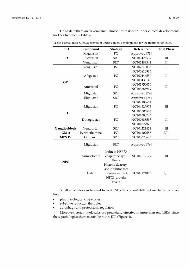
Up to date there are several small molecules in use, or under clinical development, for LSD treatment (Table 4.
Table 4. Small molecules, approved of under clinical development, for the treatment of LSDs.
LSD Compound Strategy Reference Trial Phase
FD Migalastat PC Approved [73]
Lucerastat SRT NCT03425539 III Venglustat SRT NCT02489344 II
GD
Venglustat PC NCT02843035 II
Afegostat PC NCT00813865 NCT00446550 NCT00433147
II
Ambroxol PC NCT03950050 NCT04388969
II
Miglustat SRT Approved [74]
Eliglustat SRT Approved [75]
PD
Miglustat PC NCT02185651 NCT04327973 NCT04808505
III
Duvoglustat PC NCT01380743 NCT00688597 NCT04327973
II
Gangliosidosis GM-2
Venglustat SRT NCT04221451 III Pyrimethamina PC NCT01102686 I/II
MPS IV Odiparcil SRT NCT03370653 II
NPC
Miglustat SRT Approved [76]
Arimoclomol Induces HSP70 chaperone syn-
thesis NCT02612129 III
Ostat
Histone deacety-lase inhibitor that increase mutant NPC1 protein
levels
NCT02124083 I/II
Small molecules can be used to treat LSDs throughout different mechanisms of ac-tion: • pharmacological chaperones • substrate reduction therapies • autophagy and proteostasis regulators
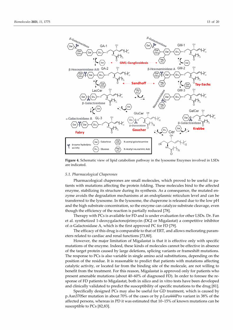
Moreover, certain molecules are potentially effective in more than one LSDs, since these pathologies share metabolic routes [77] (Figure 4).
Biomolecules 2021, 11, 1775 13 of 20
Figure 4. Schematic view of lipid catabolism pathway in the lysosome Enzymes involved in LSDs are indicated.
5.1. Pharmacological Chaperones Pharmacological chaperones are small molecules, which proved to be useful in pa-
tients with mutations affecting the protein folding. These molecules bind to the affected enzyme, stabilizing its structure during its synthesis. As a consequence, the mutated en-zyme avoids the degradation mechanisms at an endoplasmic reticulum level and can be transferred to the lysosome. In the lysosome, the chaperone is released due to the low pH and the high substrate concentration, so the enzyme can catalyze substrate cleavage, even though the efficiency of the reaction is partially reduced [78].
Therapy with PCs is available for FD and is under evaluation for other LSDs. Dr. Fan et al. synthetized 1-deoxygalactonojirimycin (DGJ or Migalastat) a competitive inhibitor of α-Galactosidase A, which is the first approved PC for FD [79].
The efficacy of this drug is comparable to that of ERT, and allows meliorating param-eters related to cardiac and renal functions [73,80].
However, the major limitation of Migalastat is that it is effective only with specific mutations of the enzyme. Indeed, these kinds of molecules cannot be effective in absence of the target protein caused by large deletions, splicing variants or frameshift mutations. The response to PCs is also variable in single amino acid substitutions, depending on the position of the residue. It is reasonable to predict that patients with mutations affecting catalytic activity, or located far from the binding site of the molecule, are not willing to benefit from the treatment. For this reason, Migalastat is approved only for patients who present amenable mutations (about 40–60% of diagnosed FD). In order to foresee the re-sponse of FD patients to Migalastat, both in silico and in vitro tests have been developed and clinically validated to predict the susceptibility of specific mutations to the drug [81].
Specifically designed PCs may also be useful for GD treatment, which is caused by p.Asn370Ser mutation in about 70% of the cases or by p.Leu444Pro variant in 38% of the affected persons, whereas in PD it was estimated that 10–15% of known mutations can be susceptible to PCs [82,83].

Biomolecules 2021, 11, 1775 14 of 20
Nevertheless, not all enzymes involved in LSDs respond sufficiently well to the ac-tion of PCs. In fact, 1-deoxynojirimycin (DNJ) was successfully tested in mice models, where it proved to enhance α-Glycosidase stability (enzyme causing PD), but these effects were considerably lower in primates and in leukocytes from healthy human subjects.
One way of enhancing the efficacy of the PCs is by applying a discontinuous admin-istration regime. This is because the half-life of the enzyme is a matter of days whereas the PC is a matter of hours. When the drug is in the bloodstream, the traffic of the enzyme to the lysosome is maximized, while, after its concentration decays, substrate metabolization is enhanced, favoring chaperone release at the lysosome. In FD mice models the discon-tinuous administration (alternate days) of Migalastat resulted in a major storage metabo-lization compared with the daily drug administration [83].
Furthermore, it is possible that new generation PCs, which interact with allosteric sites of the protein, may better stabilize the protein and increase the spectrum of suscep-tible mutations. These molecules would not need to be released by the protein and would further maintain the correctly folded structure of the protein. Possible therapeutic targets with allosteric function have been identified in α-Galactosidase, in acid α-Glycosidase and β-Glycosidase [84–86].
Currently, the co-administration of PCs with ERT is under investigation in order to evaluate the possible synergic effect of both treatments. In fact, PCs can also bind to re-combinant enzymes, stabilizing them and favoring their transit towards the lysosome. Data that confirm this hypothesis have been obtained in vitro and in vivo for Migalastat [87] and for other experimental PCs for LSDs treatment [88].
5.2. Substrate Reduction Therapy SRT consists of the use of small molecules to inhibit the enzyme that synthetizes the
substrate (or one of its precursors) accumulated in a specific LSDs, to restore synthe-sis/degradation balance.
Miglustat and Eliglustat are competitive inhibitors of the glucosylceramide synthe-tase developed as SRT for GD treat GD [77].
Miglustat is an oral drug, conditionally approved for GD patients who cannot toler-ate ERT, because it seems to act less efficiently than the recombinant enzymes. Despite of its capacity to cross BBB, Miglustat can only be used for GD Type I patients, due to its potentially neurotoxic effects, as it is an agonist of the glucose sensor SGLT3, which is expressed at the neuromuscular junction [89]. Moreover, Miglustat may also cause less severe side effects like diarrhea, abdominal pain, and weight loss, due to the impaired intestinal absorption of disaccharide determined by the inhibition of glycosidase isomers at an enteric level [90]. In spite of these limitations, Miglustat proved to be effective to improve cognitive and motor function in NPC patients, since it decreases glucosylcer-amide synthesis and it is used for the treatment of this disease [76].
On the other hand, Eliglustat proved to be effective and comparable to ERT in non-treated GD I patients [75]. Eliglustat does not inhibit intestinal glycosidases neither is it neurotoxic. Nonetheless, this drug is a substrate of the Glycoprotein-P transporter and can compete with other substrates of this receptor (e.g., drugs such as digoxin, phenytoin, and colchicine) which can affect its pharmacokinetics [91]. In addition, this active principle is metabolized by the P450 cytochrome and can interact with other drugs eliminated through this route, so pharmacokinetics of Eliglustat can also be affected by the genetic heterogenicity of CYP2D6 enzyme at the P450 cytochrome. For this reason, the dose of this treatment has to be adjusted according to the kind of CYP2D6 enzyme expressed by the subject, which determines a patient’s status of extensive, intermediate, or poor metab-olizer.
SRT is currently under development also for the treatment of FD. Lucearstat is an iminosugar which inhibits the production of glucosylceramide synthase and, therefore, the synthesis of Gb3. In phase I clinical trials (NCT02930655), Lucerastat was able to re-duce Glucosphyngolipids accumulation (GL-1,Gb3 and lactosilceramide) [92]. Currently,

Biomolecules 2021, 11, 1775 15 of 20
the molecule is under evaluation in phase III clinical trial MODIFY (NCT03425539), an aleatory double-blind placebo controlled study to assess safety and efficacy [92,93].
Venglustat is a new SRT drug, which inhibits glucosylceramide synthase (GCS) and so it reduces glucosylceramide synthesis [94]. This drug has been tested for different ther-apeutic indications and appears to be useful for the treatment of FD. A phase II clinical trial (NCT02489344) assessed long-term safety of Venglustat (GZ/SAR402671) in eight adult male patients with FD, finding reduced levels of Gb3, Lyso-Gb3 and monosialodi-hexosylganglioside (GM3) in treated patients.
5.3. Autophagy and Proteostasis Regulatory Molecules Proteostasis is a complex network that regulates the synthesis, folding, trafficking,
aggregation, and the degradation of proteins in the cell. Proteostasis regulators (Celastrol and MG-132), especially if associated with a PCs, can be an alternative for LSDs treatment as they facilitate the enzyme transit towards the lysosome. These compounds were as-sayed in vitro for GM2 Gangliosidosis (Tay-Sachs disease) and GD treatment [95].
In addition, targeting autophagy related pathways may help to contrast storage ac-cumulation in LSDs, by reactivation of substrate degradation. The advantage of this strat-egy is that the same treatment can ideally be effective for different LSDs, independently of the genetic defect.
As an example, it has been shown that the stimulation of Ca+2-dependent exocytosis mediated by TFEB increases the metabolization of accumulated substrates in MSD and in PD [96]. Medina et al., described how TFEB overexpression in a PD mouse model reduces the glycogen storage and lysosome size, improving the autophagolysosome processing and reducing the excessive presence of autophagic vesicles. A similar strategy was proved in fibroblasts of patients with GD [97].
Genistein is a natural isoflavonoid that has been successfully tested to stimulate the autophagy through TFEB activation. As a consequence, GAGs levels were find to be re-duced and so it was effective as a MPSs treatment [98].
Finally, HSp70 protein increases the lysosome stability by modulating the sphin-golipid membrane composition. The acid sphingomyelinase deficiency is partially re-stored by the Hsp70 administration in cells from Niemann-Pick A patients [99].
6. Conclusions The wide range of possible new treatments and therapeutic strategies for LSDs, de-
scribed in the present article, gives an idea of the great efforts made by the scientific com-munity to provide patients with options against unmet medical needs, which will hope-fully be soon available in the clinic.
Nevertheless, there is still margin for improvement in respect to future research in this field. Relevant challenges include the optimization of the available strategies to im-prove not only lifespan, but also patient’s life quality; and the reduction in the costs asso-ciated with these chronic patient’s treatment and follow up. Moreover, therapeutic proto-cols also need to be improved in future to introduce combinatory therapies and personal-ized medicine, which will face the specific clinical issues experienced by these patients, who are affected with complex multisystemic and heterogeneous disorders.
Author Contributions: C.F.-P. wrote the manuscript and conducted bibliographic search and illus-tration preparation. M.G.-G., T.P.-M., and M.A.-V. conducted bibliographic search and illustration preparation. B.S.M.-T., J.F.-M., and C.M.-C. wrote and revised the manuscript. S.O. was responsible for conceptualization, coordination, manuscript writing, and revision. All authors have read and agreed to the published version of the manuscript.
Funding: This research received no external funding.
Institutional Review Board Statement: Not applicable
Informed Consent Statement: Not applicable

Biomolecules 2021, 11, 1775 16 of 20
Data Availability Statement: Not applicable
Acknowledgments: The authors kindly acknowledge Tania Vázquez-Santos for English language revision. Graphic art was prepared using Biorender and Draw Chem software.
Conflicts of Interest: S.O., J.F.M. received research founding, honorary, or travel expenses from Takeda, Sanofi-Genzyme and Amicus Therapeutics. B.S.M.-T. and C.M.-C. received travel expenses by Takeda, Sanofi Genzyme and Amicus Therapeutics.
References 1. Parkinson-Lawrence, E.J.; Shandala, T.; Prodoehl, M.; Plew, R.; Borlace, G.N.; Brooks, D.A. Lysosomal storage Disease: Reveal-
ing lysosomal function and physiology. Physiology 2010, 25, 102–115. 2. Parenti, G.; Andria, G.; Ballabio, A. Lysosomal storage diseases: From pathophysiology to therapy. Annu. Rev. Med. 2015, 66,
471–486. 3. Zoncu, R.; Bar-Peled, L.; Efeyan, A.; Wang, S.; Sancak, Y.; Sabatini, D.M. mTORC1 senses lysosomal amino acids through an
inside-out mechanism that requires the vacuolar H+-ATPase. Science 2011, 334, 678–683. 4. Sancak, Y.; Bar-Peled, L.; Zoncu, R.; Markhard, A.L.; Nada, S.; Sabatini, D.M. Ragulator-rag complex targets mTORC1 to the
lysosomal surface and is necessary for its activation by amino acids. Cell 2010, 141, 290–303. 5. Sardiello, M.; Palmieri, M.; Di Ronza, A.; Medina, D.L.; Valenza, M.; Gennarino, V.A.; Di Malta, C.; Donaudy, F.; Embrione, V.;
Polishchuk, R.S.; et al. A Gene Network Regulating Lysosomal Biogenesis and Function. Science 2009, 325, 473–477. 6. Settembre, C.; Zoncu, R.; Medina, D.L.; Vetrini, F.; Erdin, S.; Erdin, S.; Huynh, T.; Ferron, M.; Karsenty, G.; Vellard, M.C.; et al.
A lysosome-to-nucleus signalling mechanism senses and regulates the lysosome via mTOR and TFEB. EMBO J. 2012, 31, 1095–1108.
7. Braulke, T.; Bonifacino, J.S. Sorting of lysosomal proteins. Biochim. Biophys. Acta-Mol. Cell Res. 2009, 1793, 605–614. 8. Reczek, D.; Schwake, M.; Schröder, J.; Hughes, H.; Blanz, J.; Jin, X.; Brondyk, W.; Van Patten, S.; Edmunds, T.; Saftig, P. LIMP-
2 Is a Receptor for Lysosomal Mannose-6-Phosphate-Independent Targeting of β-Glucocerebrosidase. Cell 2007, 131, 770–783. 9. Lieberman, A.P.; Puertollano, R.; Raben, N.; Slaugenhaupt, S.; Walkley, S.U.; Ballabio, A. Autophagy in lysosomal storage dis-
orders. Autophagy 2012, 8, 719–730. 10. Shea, L.; Raben, N. Autophagy in skeletal muscle: Implications for Pompe disease. Int. J. Clin. Pharmacol. Ther. 2009, 47, S42. 11. Settembre, C.; Fraldi, A.; Jahreiss, L.; Spampanato, C.; Venturi, C.; Medina, D.L.; De Pablo, R.; Tacchetti, C.; Rubinsztein, D.C.;
Ballabio, A. A block of autophagy in lysosomal storage disorders. Hum. Mol. Genet. 2007, 17, 119–129. 12. Lloyd-Evans, E.; Morgan, A.J.; He, X.; A Smith, D.; Elliot-Smith, E.; Sillence, D.J.; Churchill, G.C.; Schuchman, E.H.; Galione, A.;
Platt, F.M. Niemann-Pick disease type C1 is a sphingosine storage disease that causes deregulation of lysosomal calcium. Nat. Med. 2008, 14, 1247–1255.
13. Settembre, C.; Fraldi, A.; Medina, D.L.; Ballabio, A. Signals from the lysosome: A control centre for cellular clearance and energy metabolism. Nat. Rev. Mol. Cell Biol. 2013, 14, 283–296.
14. Mauhin, W.; Lidove, O.; Masat, E.; Mingozzi, F.; Mariampillai, K.; Ziza, J.-M.; Benveniste, O. Innate and adaptive immune response in fabry disease. JIMD Rep. 2015, 22, 1–10.
15. Satoh, K. Globotriaosylceramide Induces Endothelial Dysfunction in Fabry Disease. Arter. Thromb. Vasc. Biol. 2014, 34, 2–4. 16. Lachmann, R.H. Enzyme replacement therapy for lysosomal storage diseases. Curr. Opin. Pediatr. 2011, 23, 588–593. 17. Barton, N.W.; Brady, R.O.; Dambrosia, J.M.; Di Bisceglie, A.M.; Doppelt, S.H.; Hill, S.C.; Mankin, H.J.; Murray, G.J.; Parker, R.I.;
Argoff, C.E.; et al. Replacement Therapy for Inherited Enzyme Deficiency—Macrophage-Targeted Glucocerebrosidase for Gau-cher’s Disease. N. Engl. J. Med. 1991, 324, 1464–1470.
18. Dinur, T.; Grittner, U.; Revel-Vilk, S.; Becker-Cohen, M.; Istaiti, M.; Cozma, C.; Rolfs, A.; Zimran, A. Impact of Long-Term En-zyme Replacement Therapy on Glucosylsphingosine (Lyso-Gb1) Values in Patients with Type 1 Gaucher Disease: Statistical Models for Comparing Three Enzymatic Formulations. Int. J. Mol. Sci. 2021, 22, 7699.
19. Arends, M.; Biegstraaten, M.; Wanner, C.; Sirrs, S.; Mehta, A.; Elliott, P.; Oder, D.; Watkinson, O.T.; Bichet, D.G.; Khan, A.; et al. Agalsidase alfa versus agalsidase beta for the treatment of Fabry disease: an international cohort study. J. Med Genet. 2018, 55, 351–358.
20. Schoser, B.; Stewart, A.; Kanters, S.; Hamed, A.; Jansen, J.; Chan, K.; Karamouzian, M.; Toscano, A. Survival and long-term outcomes in late-onset Pompe disease following alglucosidase alfa treatment: a systematic review and meta-analysis. J. Neurol. 2017, 264, 621–630.
21. Pena, L.D.M.; Barohn, R.J.; Byrne, B.J.; Desnuelle, C.; Goker-Alpan, O.; Ladha, S.; Laforêt, P.; Mengel, K.E.; Pestronk, A.; Pouget, J.; et al. Safety, tolerability, pharmacokinetics, pharmacodynamics, and exploratory efficacy of the novel enzyme replacement therapy avalglucosidase alfa (neoGAA) in treatment-naïve and alglucosidase alfa-treated patients with late-onset Pompe dis-ease: A phase 1, open-label, multicenter, multinational, ascending dose study. Neuromuscul. Disord. 2019, 29, 167–186.
22. Jameson, E.; Jones, S.; Remmington, T. Enzyme replacement therapy with laronidase (Aldurazyme®) for treating mucopolysac-charidosis type I. Cochrane Database Syst. Rev. 2019, 6,1–24.
23. Whiteman, D.A.H.; Kimura, A. Development of idursulfase therapy for mucopolysaccharidosis type II(Hunter syndrome): The past, the present and the future. Drug Des. Dev. Ther. 2017, 11, 2467–2480.
Biomolecules 2021, 11, 1775 17 of 20
24. Okuyama, T.; Eto, Y.; Sakai, N.; Minami, K.; Yamamoto, T.; Sonoda, H.; Yamaoka, M.; Tachibana, K.; Hirato, T.; Sato, Y. Idu-ronate-2-Sulfatase with Anti-human Transferrin Receptor Antibody for Neuropathic Mucopolysaccharidosis II: A Phase 1/2 Trial. Mol. Ther. 2019, 27, 456–464.
25. í Dali, C.; Sevin, C.; Krägeloh-Mann, I.; Giugliani, R.; Sakai, N.; Wu, J.; Wasilewski, M. Safety of intrathecal delivery of recom-binant human arylsulfatase A in children with metachromatic leukodystrophy: Results from a phase 1/2 clinical trial. Mol. Genet. Metab. 2020, 131, 235–244.
26. Barton, R.W.; Neufeld, E.F. The Hurler corrective factor. Purification and some properties. J. Biol. Chem. 1971, 246, 7773–7779. 27. Tian, W.; Ye, Z.; Wang, S.; Schulz, M.A.; Van Coillie, J.; Sun, L.; Chen, Y.-H.; Narimatsu, Y.; Hansen, L.; Kristensen, C.; et al. The
glycosylation design space for recombinant lysosomal replacement enzymes produced in CHO cells. Nat. Commun. 2019, 10, 1–13.
28. He, X.; Galpin, J.D.; Tropak, M.B.; Mahuran, D.; Haselhorst, T.; Von Itzstein, M.; Kolarich, D.; Packer, N.; Miao, Y.; Jiang, L.; et al. Production of active human glucocerebrosidase in seeds of Arabidopsis thaliana complex-glycan-deficient (cgl) plants. Gly-cobiology 2011, 22, 492–503.
29. Chiba, Y.; Akeboshi, H. Glycan Engineering and Production of 'Humanized' Glycoprotein in Yeast Cells. Biol. Pharm. Bull. 2009, 32, 786–795.
30. Oder, D.; Nordbeck, P.; Wanner, C. Long Term Treatment with Enzyme Replacement Therapy in Patients with Fabry Disease. Nephron 2016, 134, 30–36.
31. Concolino, D.; Deodato, F.; Parini, R. Enzyme replacement therapy: efficacy and limitations. Ital. J. Pediatr. 2018, 44, 117–126. 32. Sly, W.S.; Vogler, C.; Grubb, J.H.; Levy, B.; Galvin, N.; Tan, Y.; Nishioka, T.; Tomatsu, S. Enzyme therapy in mannose receptor-
null mucopolysaccharidosis VII mice defines roles for the mannose 6-phosphate and mannose receptors. Proc. Natl. Acad. Sci. USA 2006, 103, 15172–15177.
33. Stroobants, S.; Gerlach, D.; Matthes, F.; Hartmann, D.; Fogh, J.; Gieselmann, V.; D'Hooge, R.; Matzner, U. Intracerebroventricular enzyme infusion corrects central nervous system pathology and dysfunction in a mouse model of metachromatic leukodystro-phy. Hum. Mol. Genet. 2011, 20, 2760–2769.
34. Chen, J.C.; Luu, A.R.; Wise, N.; De Angelis, R.; Agrawal, V.; Mangini, L.; Vincelette, J.; Handyside, B.; Sterling, H.; Lo, M.J.; et al. Intracerebroventricular enzyme replacement therapy with β-galactosidase reverses brain pathologies due to GM1 gangli-osidosis in mice. J. Biol. Chem. 2020, 295, 13532–13555.
35. Solomon, M.; Muro, S. Lysosomal enzyme replacement therapies: Historical development, clinical outcomes, and future per-spectives. Adv. Drug Deliv. Rev. 2017, 118, 109–134. Available online: http://europepmc.org/articles/pmc5828774?pdf=render (ac-cessed on 15 November 2021)
36. Rombach, S.M.; Aerts, J.; Poorthuis, B.J.H.M.; Groener, J.E.M.; Donker-Koopman, W.; Hendriks, E.; Mirzaian, M.; Kuiper, S.; Wijburg, F.A.; Hollak, C.E.M.; et al. Long-Term Effect of Antibodies against Infused Alpha-Galactosidase A in Fabry Disease on Plasma and Urinary (lyso)Gb3 Reduction and Treatment Outcome. PLOS ONE 2012, 7, e47805.
37. Banugaria, S.G.; Prater, S.N.; Ng, Y.-K.; Kobori, J.A.; Finkel, R.S.; Ladda, R.L.; Chen, Y.-T.; Rosenberg, A.S.; Kishnani, P.S. The impact of antibodies on clinical outcomes in diseases treated with therapeutic protein: Lessons learned from infantile Pompe disease. Genet. Med. 2011, 13, 729–736.
38. Banugaria, S.G.; Prater, S.N.; Patel, T.T.; DeArmey, S.M.; Milleson, C.; Sheets, K.B.; Bali, D.S.; Rehder, C.W.; Raiman, J.A.J.; Wang, R.A.; et al. Algorithm for the Early Diagnosis and Treatment of Patients with Cross Reactive Immunologic Material-Negative Classic Infantile Pompe Disease: A Step towards Improving the Efficacy of ERT. PLOS ONE 2013, 8, e67052.
39. Lenders, M.; Stappers, F.; Brand, E. In Vitro and In Vivo Amenability to Migalastat in Fabry Disease. Mol. Ther. - Methods Clin. Dev. 2020, 19, 24–34.
40. Abasolo, I.; Seras-Franzoso, J.; Moltó-Abad, M.; Díaz-Riascos, V.; Corchero, J.L.; Pintos-Morell, G.; Schwartz, S., Jr. Nanotech-nology-based approaches for treating lysosomal storage disorders, a focus on Fabry disease. Wiley Interdiscip. Rev. Nanomed. Nanobiotechnology 2020, 13, e1684.
41. Kizhner, T.; Azulay, Y.; Hainrichson, M.; Tekoah, Y.; Arvatz, G.; Shulman, A.; Ruderfer, I.; Aviezer, D.; Shaaltiel, Y. Character-ization of a chemically modified plant cell culture expressed human α-Galactosidase-A enzyme for treatment of Fabry disease. Mol. Genet. Metab. 2015, 114, 259–267.
42. Schiffmann, R.; Goker-Alpan, O.; Holida, M.; Giraldo, P.; Barisoni, L.; Colvin, R.B.; Jennette, C.J.; Maegawa, G.; Boyadjiev, S.A.; Gonzalez, D.; et al. Pegunigalsidase alfa, a novel PEGylated enzyme replacement therapy for Fabry disease, provides sustained plasma concentrations and favorable pharmacodynamics: A 1-year Phase 1/2 clinical trial. J. Inherit. Metab. Dis. 2019, 42, 534–544.
43. Boado, R.J.; Hui, E.K.-W.; Lu, J.Z.; Zhou, Q.-H.; Pardridge, W.M. Reversal of Lysosomal Storage in Brain of Adult MPS-I Mice with Intravenous Trojan Horse-Iduronidase Fusion Protein. Mol. Pharm. 2011, 8, 1342–1350.
44. Parenti, G.; Pignata, C.; Vajro, P.; Salerno, M. New strategies for the treatment of lysosomal storage diseases (Review). Int. J. Mol. Med. 2012, 31, 11–20.
45. Valayannopoulos, V.; Wijburg, F.A. Therapy for the mucopolysaccharidoses. Rheumatology 2011, 50 (Suppl. 5), v49–v59. 46. Yoon, I.C.; Bascou, N.A.; Poe, M.D.; Szabolcs, P.; Escolar, M.L. Long-Term Neurodevelopmental Outcomes of Hematopoietic
Stem Cell Transplantation for Late-Infantile Krabbe Disease. Blood 2020, 137, 1719–1730. 47. Orchard, P.J.; Tolar, J. Transplant Outcomes in Leukodystrophies. Semin. Hematol. 2010, 47, 70–78.
Biomolecules 2021, 11, 1775 18 of 20
48. Ries, M.; Clarke, J.T.; Whybra, C.; Timmons, M.; Robinson, C.; Schlaggar, B.L.; Pastores, G.; Lien, Y.H.; Kampmann, C.; Brady, R.O.; et al. Enzyme-Replacement Therapy With Agalsidase Alfa in Children With Fabry Disease. Pediatrics 2006, 118, 924–932.
49. Knott, G.J.; Doudna, J.A. CRISPR-Cas guides the future of genetic engineering. Science 2018, 361, 866–869. 50. Gupta, S.K.; Shukla, P. Gene editing for cell engineering: trends and applications. Crit. Rev. Biotechnol. 2016, 37, 672–684. 51. Biffi, A.; Montini, E.; Lorioli, L.; Cesani, M.; Fumagalli, F.; Plati, T.; Baldoli, C.; Martino, S.; Calabria, A.; Canale, S.; et al. Lenti-
viral Hematopoietic Stem Cell Gene Therapy Benefits Metachromatic Leukodystrophy. Science 2013, 341, 1233158. 52. Medin, J.A.; Khan, A.; Huang, J.; Barber, D.; Rupar, C.A.; Auray-Blais, C.; Fraser, G.; Fowler, D.H.; Keating, A.; West, M.L.; et
al. FACTs Fabry gene therapy clinical trial: Two-year data. Mol. Genet. Metab. 2019, 126, S99. 53. Fumagalli, F.; Calbi, V.; Sessa, M.; Zambon, A.; Baldoli, C.; Rancoita, P.M.; Acquati, S.; De Mattia, F.; Tucci, F.; Gallo, V.; et al.
Lentiviral hematopoietic stem and progenitor cell gene therapy (HSPC-GT) for metachromatic leukodystrophy (MLD): Clinical outcomes from 33 patients. Mol. Genet. Metab. 2020, 129, S59.
54. NCT04283227. Available online: https://clinicaltrials.gov/ct2/show/study/NCT04283227?cond=OTL-200&draw=2&rank=1 (ac-cessed on 15 November 2021).
55. Khan, A.; Barber, D.L.; Huang, J.; Rupar, C.A.; Rip, J.W.; Auray-Blais, C.; Boutin, M.; O’Hoski, P.; Gargulak, K.; McKillop, W.M.; et al. Lentivirus-mediated gene therapy for Fabry disease. Nat. Commun. 2021, 12, 1–9.
56. NCT04145037. Available online: https://clinicaltrials.gov/ct2/show/NCT04145037 (accessed on 15 November 2021). 57. NCT03488394. Available online: https://clinicaltrials.gov/ct2/show/NCT03488394?term=NCT03488394&draw=2&rank=1 (ac-
cessed on November 15th 2021) 58. NCT03566043. Available online: https://clinicaltrials.gov/ct2/show/NCT03566043?cond=RGX-121&draw=2&rank=2 (accessed
on 15 November 2021) 59. Wang, D.; Tai, P.W.L.; Gao, G. Adeno-associated virus vector as a platform for gene therapy delivery. Nat. Rev. Drug Discov.
2019, 18, 358–378. 60. Ortolano, S.; Spuch, C.; Navarro, C. Present and future of adeno associated virus based gene therapy approaches.. Recent Patents
Endocrine, Metab. Immune Drug Discov. 2012, 6, 47–66. 61. Manfredsson, F.P.; Rising, A.C.; Mandel, R.J. AAV9: a potential blood-brain barrier buster. Mol. Ther. 2009, 17, 403–405. 62. Biferi, M.G.; Cohen-Tannoudji, M.; García-Silva, A.; Souto-Rodríguez, O.; Viéitez-González, I.; San-Millán-Tejado, B.; Fernán-
dez-Carrera, A.; Pérez-Márquez, T.; Teijeira-Bautista, S.; Barrera, S.; et al. Systemic Treatment of Fabry Disease Using a Novel AAV9 Vector Expressing α-Galactosidase A. Mol. Ther. - Methods Clin. Dev. 2020, 20, 1–17.
63. Aravalli, R.N.; Belcher, J.D.; Steer, C.J. Liver-targeted gene therapy: Approaches and challenges. Liver Transplant. 2015, 21, 718–737.
64. Sharma, R.; Anguela, X.M.; Doyon, Y.; Wechsler, T.; DeKelver, R.C.; Sproul, S.; Paschon, D.E.; Miller, J.C.; Davidson, R.J.; Shivak, D.; et al. In vivo genome editing of the albumin locus as a platform for protein replacement therapy. Blood 2015, 126, 1777–1784.
65. Puzzo, F.; Colella, P.; Biferi, M.G.; Bali, D.; Paulk, N.K.; Vidal, P.; Collaud, F.; Simon-Sola, M.; Charles, S.; Hardet, R.; et al. Rescue of Pompe disease in mice by AAV-mediated liver delivery of secretable acid α-glucosidase. Sci. Transl. Med. 2017, 9 (418), 1-27.
66. Yasuda, M.; Huston, M.W.; Pagant, S.; Gan, L.; Martin, S.S.; Sproul, S.; Richards, D.; Ballaron, S.; Hettini, K.; Ledeboer, A.; et al. AAV2/6 Gene Therapy in a Murine Model of Fabry Disease Results in Supraphysiological Enzyme Activity and Effective Sub-strate Reduction. Mol. Ther.-Methods Clin. Dev. 2020, 18, 607–619. https://doi.org/10.1016/j.omtm.2020.07.002.
67. NCT04093349. Available online: https://clinicaltrials.gov/ct2/show/NCT04093349?term=SPK-3006&draw=2&rank=1 (accessed on 15 November 2021).
68. Hughes, D.A.; Patel, N.; Kinch, R.; Dronfield, L.; Short, G.; Sheridan, R.; Kia, A.; Jeyakumar, J.; Corbau, R.; Nathwani, A. First-in-human study of a liver-directed AAV gene therapy (FLT190) in Fabry disease. Mol. Genet. Metab. 2020, 129, S77–S78.
69. NCT04040049. Available online: https://clinicaltrials.gov/ct2/show/NCT04040049?cond=FLT190&draw=2 (accessed on 15 No-vember 2021).
70. NTC03580083. Available online: https://clinicaltrials.gov/ct2/show/NCT03580083?cond=RGX-111&draw=2 (accessed on 15 No-vember 2021).
71. Bradbury, A.M.; Rafi, M.A.; Bagel, J.H.; Brisson, B.K.; Marshall, M.S.; Salvador, J.P.; Jiang, X.; Swain, G.P.; Prociuk, M.L.; Odon-nell, P.A.; et al. AAVrh10 Gene Therapy Ameliorates Central and Peripheral Nervous System Disease in Canine Globoid Cell Leukodystrophy (Krabbe Disease). Hum. Gene Ther. 2018, 29, 785–801.
72. Bradbury, A.M.; Bagel, J.H.; Nguyen, D.; Lykken, E.A.; Salvador, J.P.; Jiang, X.; Swain, G.P.; Assenmacher, C.A.; Hendricks, I.J.; Miyadera, K.; et al. Krabbe disease successfully treated via monotherapy of intrathecal gene therapy. J. Clin. Investig. 2020, 130, 4906–4920.
73. Germain, D.P.; Nicholls, K.; Giugliani, R.; Bichet, D.G.; Hughes, D.A.; Barisoni, L.M.; Colvin, R.B.; Jennette, J.C.; Skuban, N.; Castelli, J.P.; et al. Efficacy of the pharmacologic chaperone migalastat in a subset of male patients with the classic phenotype of Fabry disease and migalastat-amenable variants: data from the phase 3 randomized, multicenter, double-blind clinical trial and extension study. Genet. Med. 2019, 21, 1987–1997.
74. Pastores, G.M. Miglustat: Substrate reduction therapy for lysosomal storage disorders associated with primary central nervous system involvement. Recent Pat. CNS Drug Discov. 2006, 1, 77–82.
75. Cox, T.M.; Drelichman, G.I.; Cravo, R.; Balwani, M.; Burrow, T.A.; Martins, A.M.; Lukina, E.; Rosenbloom, B.E.; Ross, L.H.; Angell, J.; et al. ENCORE, a randomized, controlled, open-label non-inferiority study comparing eliglustat to imiglucerase in

Biomolecules 2021, 11, 1775 19 of 20
Gaucher disease type 1 patients stabilized on enzyme replacement therapy: 24-month results. Mol. Genet. Metab. 2015, 114, S33–S34.
76. Patterson, M.C.; Vecchio, D.; Jacklin, E.; Abel, L.; Chadha-Boreham, H.; Luzy, C.; Giorgino, R.; Wraith, J.E. Long-Term Miglustat Therapy in Children With Niemann-Pick Disease Type C. J. Child Neurol. 2010, 25, 300–305.
77. Ortolano, S. Small molecules: Substrate inhibitors, chaperones, stop-codon read through, and beyond. J. Inborn. Errors Metab. Screen. 2016, 4, 1–11.
78. Parenti, G.; Andria, G.; Valenzano, K.J. Pharmacological chaperone therapy: Preclinical development, clinical translation, and prospects for the treatment of lysosomal storage disorders. Mol. Ther. 2015, 23, 1138–1148. Available online: http://euro-pepmc.org/search?query=(DOI:10.1038/mt.2015.62)%0Ahttp://search.ebscohost.com/login.aspx?di-rect=true&scope=site&site=ehost-live&db=mdc&AN=25881001%0Ahttps://gateway.proquest.com/openurl?ctx_ver=Z39.88-2004&res_id=xri:pqm&req_dat=xri:pqil:pq_cln (accessed on 15 November 2021).
79. Fan, J.Q.; Ishii, S. Active-site-specific chaperone therapy for Fabry disease: Yin and Yang of enzyme inhibitors. FEBS J. 2007, 274, 4962–4971.
80. Hughes, D.A.; Nicholls, K.; Sunder-Plassmann, G.; Jovanovic, A.; Feldt-Rasmussen, U.; Schiffmann, R.; Giugliani, R.; Jain, V.; Viereck, C.; Castelli, J.P.; et al. Safety of switching to Migalastat from enzyme replacement therapy in Fabry disease: Experience from the Phase 3 ATTRACT study. Am. J. Med. Genet. Part A 2019, 179, 1069–1073.
81. Schiffmann, R.; Bichet, D.G.; Benjamin, E.; Wu, X.; Giugliani, R. The migalastat GLP-HEK assay is the gold standard for deter-mining amenability in patients with Fabry disease. Mol. Genet. Metab. Rep. 2019, 20, 100494.
82. Flanagan, J.J.; Rossi, B.; Tang, K.; Wu, X.; Mascioli, K.; Donaudy, F.; Tuzzi, M.R.; Fontana, F.; Cubellis, M.V.; Porto, C.; et al. The pharmacological chaperone 1-deoxynojirimycin increases the activity and lysosomal trafficking of multiple mutant forms of acid alpha-glucosidase. Hum. Mutat. 2009, 30, 1683–1692.
83. Khanna, R.; Powe, A.C.; Lun, Y.; Soska, R.; Feng, J.; Dhulipala, R.; Frascella, M.; Garcia, A.; Pellegrino, L.J.; Xu, S.; et al. The Pharmacological Chaperone AT2220 Increases the Specific Activity and Lysosomal Delivery of Mutant Acid Alpha-Gluco-sidase, and Promotes Glycogen Reduction in a Transgenic Mouse Model of Pompe Disease. PLOS ONE 2014, 9, e102092.
84. Guce, A.I.; Clark, N.E.; Salgado, E.N.; Ivanen, D.R.; Kulminskaya, A.A.; Brumer, H.; Garman, S.C. Catalytic Mechanism of Hu-man α-Galactosidase. J. Biol. Chem. 2010, 285, 3625–3632.
85. Zheng, W.; Padia, J.; Urban, D.J.; Jadhav, A.; Goker-Alpan, O.; Simeonov, A.; Goldin, E.; Auld, D.; LaMarca, M.E.; Inglese, J.; et al. Three classes of glucocerebrosidase inhibitors identified by quantitative high-throughput screening are chaperone leads for Gaucher disease. Proc. Natl. Acad. Sci. 2007, 104, 13192–13197.
86. Marugan, J.J.; Zheng, W.; Ferrer, M.; Motabar, O.; Southall, N.; Goldin, E.; Westbroek, W. and Sidransky, E. Discovery, SAR, and Biological Evaluation of a Non-Inhibitory Chaperone for Acid Alpha Glucosidase. Probe Reports from the NIH Molecular Libraries Program. 2010. Available online: http://www.ncbi.nlm.nih.gov/pubmed/23905202 (accessed on 15 November 2021).
87. Warnock, D.G.; Bichet, D.-G.; Holida, M.; Goker-Alpan, O.; Nicholls, K.; Thomas, M.; Eyskens, F.; Shankar, S.; Adera, M.; Si-taraman, S.; et al. Oral Migalastat HCl Leads to Greater Systemic Exposure and Tissue Levels of Active α-Galactosidase A in Fabry Patients when Co-Administered with Infused Agalsidase. PLOS ONE 2015, 10, e0134341.
88. Shen, J.-S.; Edwards, N.J.; Bin Hong, Y.; Murray, G.J. Isofagomine increases lysosomal delivery of exogenous glucocerebro-sidase. Biochem. Biophys. Res. Commun. 2008, 369, 1071–1075.
89. Voss, A.A.; Díez-Sampedro, A.; Hirayama, B.A.; Loo, D.D.F.; Wright, E.M. Imino Sugars Are Potent Agonists of the Human Glucose Sensor SGLT3. Mol. Pharmacol. 2006, 71, 628–634.
90. Amiri, M.; Naim, H.Y. Miglustat-induced intestinal carbohydrate malabsorption is due to the inhibition of α-glucosidases, but not β-galactosidases. J. Inherit. Metab. Dis. 2012, 35, 949–954.
91. Shayman, J.A. Eliglustat tartrate. Glucosylceramide synthase inhibitor treatment of type 1 gaucher disease. Drugs Future 2010, 35, 613–620.
92. Guérard, N.; Oder, D.; Nordbeck, P.; Zwingelstein, C.; Morand, O.; Welford, R.W.; Dingemanse, J.; Wanner, C. Lucerastat, an Iminosugar for Substrate Reduction Therapy: Tolerability, Pharmacodynamics, and Pharmacokinetics in Patients With Fabry Disease on Enzyme Replacement. Clin. Pharmacol. Ther. 2017, 103, 703–711.
93. Guérard, N.; Morand, O.; Dingemanse, J. Lucerastat, an iminosugar with potential as substrate reduction therapy for glycolipid storage disorders: safety, tolerability, and pharmacokinetics in healthy subjects. Orphanet J. Rare Dis. 2017, 12, 1–10.
94. Peterschmitt, M.J.; Crawford, N.P.S.; Gaemers, S.J.M.; Ji, A.J.; Sharma, J.; Pham, T.T. Pharmacokinetics, Pharmacodynamics, Safety, and Tolerability of Oral Venglustat in Healthy Volunteers. Clin. Pharmacol. Drug Dev. 2020, 10, 86–98.
95. Mu, T.; Ong, D.S.T.; Wang, Y.-J.; Balch, W.E.; Yates, J.R.; Segatori, L.; Kelly, J.W. Chemical and Biological Approaches Synergize to Ameliorate Protein-Folding Diseases. Cell 2008, 134, 769–781.
96. Medina, D.L.; Fraldi, A.; Bouche, V.; Annunziata, F.; Mansueto, G.; Spampanato, C.; Puri, C.; Pignata, A.; Martina, J.; Sardiello, M.; et al. Transcriptional Activation of Lysosomal Exocytosis Promotes Cellular Clearance. Dev. Cell 2011, 21, 421–430.
Biomolecules 2021, 11, 1775 20 of 20
97. Song, W.; Wang, F.; Savini, M.; Ake, A.; Di Ronza, A.; Sardiello, M.; Segatori, L. TFEB regulates lysosomal proteostasis. Hum. Mol. Genet. 2013, 22, 1994–2009.
98. Moskot, M.; Montefusco, S.; Jakóbkiewicz-Banecka, J.; Mozolewski, P.; Wegrzyn, A.; di Bernardo, D.; Wegrzyn, G.; Medina, D.L.; Ballabio, A.; Gabig-Cimińska, M. The Phytoestrogen Genistein Modulates Lysosomal Metabolism and Transcription Fac-tor EB (TFEB) Activation. J. Biol. Chem. 2014, 289, 17054–17069.
99. Petersen, N.H.; Kirkegaard, T. HSP70 and lysosomal storage disorders: novel therapeutic opportunities. Biochem. Soc. Trans. 2010, 38, 1479–1483.
Related Documents