-- -- -- -- T T T T FUNDA FUNDA FUNDA FUNDA G G G G Course No Co Assi Co N.A.U., B COLLEGE OF AGRICU COLLEGE OF AGRICU COLLEGE OF AGRICU COLLEGE OF AGRICU NAVSARI AGRICULT NAVSARI AGRICULT NAVSARI AGRICULT NAVSARI AGRICULT UNIVERSITY UNIVERSITY UNIVERSITY UNIVERSITY, BHAR BHAR BHAR BHAR (GUJARAT) (GUJARAT) (GUJARAT) (GUJARAT) THEORY NOTE THEORY NOTE THEORY NOTE THEORY NOTE -- -- -- -- AMENTALS AMENTALS AMENTALS AMENTALS GENETICS GENETICS GENETICS GENETICS o.:- GPB 2.2, Credit- 3(2 + ompiled & Edited By Dr. Sunil S. Patil istant Professor (GPB) ollege of Agriculture, Bharuch, Gujarat (India ULTURE, ULTURE, ULTURE, ULTURE, TURAL TURAL TURAL TURAL RUCH RUCH RUCH RUCH S S S S OF OF OF OF + 1) a).

Welcome message from author
This document is posted to help you gain knowledge. Please leave a comment to let me know what you think about it! Share it to your friends and learn new things together.
Transcript
-------- THEORY NOTE THEORY NOTE THEORY NOTE THEORY NOTE
FUNDAMENTALS FUNDAMENTALS FUNDAMENTALS FUNDAMENTALS
GENETICSGENETICSGENETICSGENETICS
Course No.:
Compiled & Edited By
Assistant Professor (GPB)
College of Agriculture,
N.A.U., Bharuch
COLLEGE OF AGRICULTURE,COLLEGE OF AGRICULTURE,COLLEGE OF AGRICULTURE,COLLEGE OF AGRICULTURE,
NAVSARI AGRICULTURAL NAVSARI AGRICULTURAL NAVSARI AGRICULTURAL NAVSARI AGRICULTURAL UNIVERSITYUNIVERSITYUNIVERSITYUNIVERSITY,,,, BHARUCH BHARUCH BHARUCH BHARUCH
(GUJARAT)(GUJARAT)(GUJARAT)(GUJARAT)
THEORY NOTE THEORY NOTE THEORY NOTE THEORY NOTE --------
FUNDAMENTALS FUNDAMENTALS FUNDAMENTALS FUNDAMENTALS
GENETICSGENETICSGENETICSGENETICS
Course No.:- GPB 2.2, Credit- 3(2 + 1)
Compiled & Edited By
Dr. Sunil S. Patil
Assistant Professor (GPB)
College of Agriculture,
Bharuch, Gujarat (India).
COLLEGE OF AGRICULTURE,COLLEGE OF AGRICULTURE,COLLEGE OF AGRICULTURE,COLLEGE OF AGRICULTURE,
NAVSARI AGRICULTURAL NAVSARI AGRICULTURAL NAVSARI AGRICULTURAL NAVSARI AGRICULTURAL BHARUCH BHARUCH BHARUCH BHARUCH
FUNDAMENTALS FUNDAMENTALS FUNDAMENTALS FUNDAMENTALS OF OF OF OF
3(2 + 1)
Gujarat (India).
1
1. INTRODUCTION TO GENETICS
Genetics is a biological science which deals with the principles of heredity and variation.
Heredity refers to the transmission of characters from parents to their offspring. Thus, genetics
is a science which unravels the inheritance of various characters from one generation to another.
The foundation of this new branch of biology was laid by Mendel in 1866 when he
discovered the basic principles of heredity. However, Mendel's findings came into light only in
1900 when similar results were obtained independently by three scientists, viz., de Vres, Carl
Correns and Tschermak. Thus, genetics was born in 1900.
The term genetics was first used by Bateson in 1905, i.e., five year, after its birth. The
word genetics has been derived from the Greek word gene, which means to become or to grow.
Since characters are governed by genes, genetics is the study of structure, composition and
function of genes.
BRANCHES OF GENETICS
There are several branches of genetics. These branches have been identified on the basis
of experimental material used for the study and are briefly described below:
Plant Genetics:- It deals with inheritance of characters in various plant species. Microbial
Genetics:- It deals with inheritance of traits in microorganisms like bacteria,viruses and fungi.
Animal Genetics:- It deals with inheritance of traits in animals.
Molecular Genetics:- It deals with the structure, composition, function and replication of
chromosomes and genes, representing genetic material, viz., DNA and RNA.
Biochemical Genetics:- It deals with the role of genes in controlling biochemical pathways in an
organism.
Population Genetics:- It deals with frequencies of genes and genotypes in a population as well
as with various agencies which tend to alter gene frequencies in a population leading to
evolutionary changes.
Radiation Genetics. It deals with effects of various types of radiations on chromosomes and
genes.
Eugenics. It deals with the application of genetic principles for the betterment of human race.
Mendelian Genetics. It deals with the inheritance of qualitative characters or
oligogeniccharacters which display discontinuous variation.
2
Quantitative Genetics. It deals with the inheritance of polygenic or quantitative characterswhich
display continuous variation. This branch of genetics is also known as biometricalgenetics or
statistical genetics or mathematical genetics.
Cytogenetics. It deals with combined study of cytology and genetics. Euphenics. It deals with
the control of hereditary diseases especially inborn errors of metabolism.
PRE-MENDELIAN CONCEPTS ABOUT HEREDITY
Various views were prevailing about heredity before rediscovery of Mendel's laws of
inheritance. Some of the important theories or concepts about the heredity which were proposed
by various scientists prior to the discovery of Mendel are: (i) preformation, (it) epigenesis, (iii)
inheritance of acquired characters, (iv) pangenesis, and, (v) germplasm theory. These are briefly
presented below:
(i) Preformation Theory
This theory was proposed by two Dutch biologists, Swammerdam and Bonnet (1720-
1793). This theory states that a miniature human called humunculus was already present in the
egg and sperm. In other words, a miniature human was preformed in the gametes. The
development of zygote resulted only in the growth of miniature human who was already present
in the egg and sperm. However, this theory was soon given up because this could not be proved
scientifically.
(ii) Theory of Epigenesis
This theory was advocated by Wolff (1738—1794), a German biologist. This theory
states that egg or sperm cells do not contain miniature human. In other words, egg or sperm cells
are undifferentiated. The differentiation into various organs or parts takes place only after
fertilization from the zygote resulting into development of adult tissues and organs. This concept
is known as epigenesis which is universally accepted.
(iii) Theory of Acquired Characters
This concept was proposed by Lamarck (1744—1829), a French biologist. This theory
states that a new character once acquired by an individual shall pass on to its progeny. It means if
3
a man develops a strong muscle by exercise all his children will have strong muscle. On the other
hand, if a person becomes weak all his children will be weak. This theory was disproved by
Weismann. He cut the tail of mice for 22 successive generations and always got the baby mice
with tail. Thus, this theory was soon given up.
(iv) Theory of Pangenes
This theory was proposed by Charles Darwin (1809—1882), this theory states that very
small, exact but invisible copies of each body organ (gemmules) are transported by the blood
stream to the sex organs. These invisible copies of each body organ are called the gemmules.
These gemmules are assembled in the gametes. After fertilization these gemules move out to
different parts of the body resulting in the development of respective organ. A defective
gemmule will lead to the development of defective organ in an individual. This theory was given
up because it did not have scientific basis.
(v) Germplasm Theory
This theory was advocated by August Weismann (1889), a German biologist. This theory
states that body tissues are of two types, viz., germplasm and somatoplasm. The germplasm
refers to the reproductive tissues or cells which produce gametes. The somatoplasm includes all
other body tissues which are not related to sexual reproduction. Thus, transmission of characters
from one generation to other takes place only through germplasm. Any change in the germplasm
will lead to change in the next generation. This theory is accepted in a broad sense.
CONTRIBUTION OF SOME GENETICISTS
Several scientists have contributed for the advancement of genetics. A brief contribution
of some earlier geneticists is presented below:
Mendel Gregor (Johann)
He was an Austrian botanist who laid the foundation of the science of genetics. He
worked with garden gea (Pisum sativum) and formulated two important laws of inheritance, viz.,
(i) law of segregation and, (li) law of independent assortment. For this pioneer work he is rightly
called as the father of genetics. He presented his results in two papers at the meetings of Natural
4
History Society on February 8 and March 8 in 1865 which were published in the proceedings of
the society in 1866. However, his results were neglected for 34 years. Mendel died in 1884 and
his work came into being after 16 yrs. of his death in 1900 when same results were
independently discovered by de Vries, Correns and Tschermak.
Correns, Carl Erich
He was a German botanist and geneticist who in 1900, independently but simultaneously
with the biologists Tschermak (Austria) and Hugo de Vries rediscovered Mendel’s historic paper
outlining the principles of heredity. He conducted research with garden peas and came to the
same conclusions which were drawn by Mendel in 1865. Later on, he worked with variegated
plants such as four O' clock (Mirabilis jalapa) and established the first conclusive example of
extrachromosomal or cytoplasmic Inheritance.
Hugo, de Vries
He was a Dutch biologist and geneticist. He rediscovered independently but
simultaneously with Correns and Tschermak in 1900 Mendel's Law's of inheritance. Later on,
working with Oenothera lamarckiana he coined the term mutation for sudden heritable changes
in the
Characters.
Tschermak, V.S.E.
He was an Austrian botanist and geneticist. He was one of the codiscoverers of Mendel's
classic papers on the garden pea. Working with garden pea, Tschermak saw a cross reference to
Mendel's work and found that his results were in agreement with the findings of Mendel. In the
same year 1900, when Tschermak reported his findings, Hugo de Vries and Correns also
reported their discoveries of Mendel's papers. Later on, he applied Mendel's Laws of heredity in
barley, wheat-rye hybrids and oats hybrids for development of new plants.
Bateson, William and Punnett, R.C.
He was a British biologist who coined the term Genetics in 1905. Bateson translated
Mendel's paper from German into English and became Mendel's champion in England. He
worked with pea and discovered the phenomena of linkage which is now known to be the result
5
of closely locater1 genes on the same chromosome. He also demonstrated that in pea certain
characters are governed by two or more genes.
Punnett was an English geneticist who with Bateson discovered genetic linkage in 1905.
Working with poultry and sweet peas, Punnett and Bateson discovered some fundamental
concepts of genetics like linkage, sex determination, sex linkage and first case of autosomal
linkage. In 1901, Bateson and Punnett founded the journal of genetics.
Johannsen, W.L.
He was a Danish botanist and geneticist. He developed the concept of pure line. He
worked with princess beans and coined the terms phenotype and genotype in 1903. He supported
the mutation theory of Hugo de Vries, which refers to sudden heritable changes in a gene. The
terms phenotype and genotype are widely used in genetics. He also recognized the importance of
environment in the expression of characters.
Morgan, T.H.
He was an American zoologist and geneticist famous for his experimental research with
the fruitfly, Drosophila melanogaster. He established the chromosome theory of heredity in
1910. He showed that genes are linked in a series on chromosomes and are responsible for
observable genetic traits. For this work he received Nobel Prize for Physiology or Medicine in
1933. Thus, he was awarded Nobel Prize for his discovery of hereditary transmission
mechanisms in Drosophila. He also observed sex linkage in Drosophila.
Bridges, C.B.
He was a U.S. geneticist who helped establish the chromosomal basis of heredity and sex.
He constructed detailed gene mape of the giant chromosomes found in the salivary gland cells of
fruitfly larva. He also discovered genie balance theory of sex determination and gene duplication
in Drosophila. He had opportunity to work with Morgan.
Muller, H.J.
He was a U.S. geneticist best known for his demonstration in 1927, that X-rays speed up
the natural process of mutation. For experimental induction of mutation he was awarded Nobel
Prize for physiology or medicine in 1946. He went to USSR and worked with N.I. Vavilov
6
inLeningrad for about 4 years. He also helped to organize 7th International Congress of Genetics
in Great Britain.
Beadle, G.W.
He is a U.S. geneticist. He worked in the field of biochemical genetics and discovered
that genes affect heredity by determining enzymatic structure. He worked on fruitfly with
Morgan and on Neurospora with Edward Tatum. They exposed Neurvspora with X-rays and
studied the altered nutritional requirements of the mutants thus produced. These experiments
enabled them to conclude that each gene determined the structure of a specific enzyme which in
turn allowed a single chemical reaction to proceed. This one gene one enzyme hypothesis
concept won Beadle and Tatum (with Joshua Lederbcrg) the Nobel Prize for physiology or
Medicine in, 1958.
Tatum, E.L.
He was a U.S. biochemist, whose research on bacteria, yeast and Neurospora created a
new field of genetic studies known as molecular genetics. He worked in collaboration with
Beadle and Lederberg and developed the concept of one gene one enzyme hypothesis in 1941 for
which they were awarded Nobel Prize for Medicine or Physiology in 1958. They demonstrated
that :
1. All biochemical processes in all organisms are ultimately governed by genes.
2. Each reaction in some way is controlled by a single gene and,
3. Mutation of a single gene results only in an alteration in the ability of a cell to carry out a
single chemical reaction. Later on, with Lederberg he discovered genetic recombination or sex in
E. coli bacteria. This bacteria has become important source of genetic investigations for
biochemical process after the discovery of one gene one enzyme hypothesis.
Sutton, W.S.
Sutton was a U.S. geneticist who working with Grasshopper gave a hypothesis in 1903
that chromosomes carry the units of inheritance and they are the physical basis of the Mendelian
laws of heredity. Thus, his work formed the basis for the chromosomal theory of heredity.
7
Avery, O.T. ; Mac Leod, CM. and Mc Carty, M.
Avery was a Canada born U.S. bacteriologist. His research on pneumococcus bacteria
laid the foundation of immunochemistry. He discovered that pneumonia causing bacteria
produce a capsular envelope consisting of polysaccharide. He also discovered the phenomenon
of nansformation. Avery with his coworkers (Mac Leod and Mc Carty) reported in 1944 that the
substance which caused the transformation was deoxyribonucleic acid (DNA). Thus, they were
the pioneer workers to demonstrate that DNA was the genetic material.
Mc Carty is a U.S. biologist, who with Avery and Mac Leod provided the first
experimental evidence in 1944 that the genetic material of living cells is composed of DNA.
When the DNA extracted from capsulated bacteria (virulent) was mixed with living cells of
second type of bacteria lacking capsules, the transformation occurred. The results of this
experiment indicated that the substance responsible for the change was DNA.
Watson, J.D. and Crick, F.H.C.
Watson is a U.S. geneticist and biophysicist. He is famous for his discovery of the
molecular structure of deoxyribonucleic acid (DNA), the genetic material, in 1953. This
investigation brought him (with Francis Crick and Maurice Wilkins) the Nobel Prize for
Physiology or Medicine in 1962. They proposed double helical model for DNA. This model also
showed how the DNA molecule could duplicate itself. Thus, it became known how genes and
eventually chromosomes duplicate themselves. Watson published three books, viz., (1)
Molecular Biology of Gene in 1965, (2) The Double Helix in 1968, and (3) The DNA story in
1981. Crick is a British biophysicist, who worked with Watson and Wilkins and discovered the
DNA double helical model.
M.H. Wilkins is a New Zealand born British biophysicist whose X-ray diffraction
studies of DNA proved crucial for the discovery of the molecular structure of DNA by James
Watson and Sir Francis Crick. For this work the three Scientists were awarded Nobel Prize as
mentioned above.
Barbara McClintock
Miss. Barbara McClintock (1950), a U.S. geneticist working with maize observed that
some genes are capable of changing their position on a chromosome and from one chromosome
8
to another. Such genes are known as transpozons or transposable elements or jumping genes.
Since this was an unusual finding, people did not appreciate it for a long time. This concept was
recognized in early seventies and McClintock was awarded Nobel Prize for this work in 1983.
Benzer, S.
He is a U.S. molecular biologist, who developed a method in 1955 for determining the
detailed structure of viral genes and coined the term cistron to denote functional subunit of
genes. He also explained non-sense mutations in terms of molecular sequence of DNA. Benzer
(1955) gave sub-divisions of genes viz., Cistron, Recon and Muton. These are the units of
function, recombination and mutation with in a gene. He worked with r-II locus of T4
bacteriophage.
Jacob, F. and Monod, J.L.
Jacob is a French biologist and Monod was French biochemist. They explained in 1961
the way in which genes regulate cell metabolism by directing the biosynthesis of enzymes. Jacob
discovered that genes of bacteria are arranged in linear fashion in a ring and the ring can be
broken at almost any point. They developed the concept of gene regulation known as operon
concept in 1962. They discovered regulatory genes which control the activities of structural
genes. For this work, they were awarded Nobel Prize for physiology or medicine in 1965.
Nirenberg, M.W. and Khorana, H.G.
Nirenberg is a U.S. biochemist, who played a major role in deciphering the genetic co He
demonstrated that with the exceptions of non-sense codons, each possible triplet (c-.lle a codon)
of four different kinds of nitrogen containing bases found in DNA and in sone ther viruses in
RNA ultimately causes the incorporation of specific amino-acid into a cell protein.
Har Gobind Khorana is an Indian born U.S. biochemist. He discovered how the genetic
components of the cell nucleus control the synthesis of protein. He was awarded Nobel Prize for
physiology or medicine with Nirenberg and Holley in 1968. Later on, he prepared the first
artificial copy of a yeast gene in 1970.
9
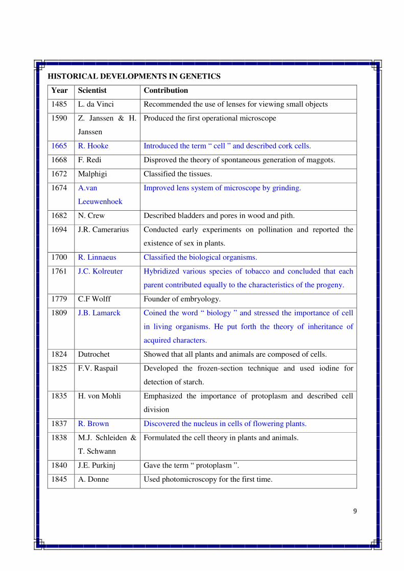
HISTORICAL DEVELOPMENTS IN GENETICS
Year Scientist Contribution
1485 L. da Vinci Recommended the use of lenses for viewing small objects
1590 Z. Janssen & H.
Janssen
Produced the first operational microscope
1665 R. Hooke Introduced the term “ cell ” and described cork cells.
1668 F. Redi Disproved the theory of spontaneous generation of maggots.
1672 Malphigi Classified the tissues.
1674 A.van
Leeuwenhoek
Improved lens system of microscope by grinding.
1682 N. Crew Described bladders and pores in wood and pith.
1694 J.R. Camerarius Conducted early experiments on pollination and reported the
existence of sex in plants.
1700 R. Linnaeus Classified the biological organisms.
1761 J.C. Kolreuter Hybridized various species of tobacco and concluded that each
parent contributed equally to the characteristics of the progeny.
1779 C.F Wolff Founder of embryology.
1809 J.B. Lamarck Coined the word “ biology ” and stressed the importance of cell
in living organisms. He put forth the theory of inheritance of
acquired characters.
1824 Dutrochet Showed that all plants and animals are composed of cells.
1825 F.V. Raspail Developed the frozen-section technique and used iodine for
detection of starch.
1835 H. von Mohli Emphasized the importance of protoplasm and described cell
division
1837 R. Brown Discovered the nucleus in cells of flowering plants.
1838 M.J. Schleiden &
T. Schwann
Formulated the cell theory in plants and animals.
1840 J.E. Purkinj Gave the term “ protoplasm ”.
1845 A. Donne Used photomicroscopy for the first time.
10
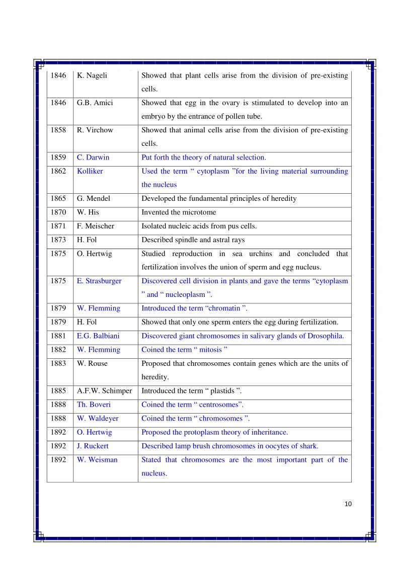
1846 K. Nageli Showed that plant cells arise from the division of pre-existing
cells.
1846 G.B. Amici Showed that egg in the ovary is stimulated to develop into an
embryo by the entrance of pollen tube.
1858 R. Virchow Showed that animal cells arise from the division of pre-existing
cells.
1859 C. Darwin Put forth the theory of natural selection.
1862 Kolliker Used the term “ cytoplasm ”for the living material surrounding
the nucleus
1865 G. Mendel Developed the fundamental principles of heredity
1870 W. His Invented the microtome
1871 F. Meischer Isolated nucleic acids from pus cells.
1873 H. Fol Described spindle and astral rays
1875 O. Hertwig Studied reproduction in sea urchins and concluded that
fertilization involves the union of sperm and egg nucleus.
1875 E. Strasburger Discovered cell division in plants and gave the terms “cytoplasm
” and “ nucleoplasm ”.
1879 W. Flemming Introduced the term “chromatin ”.
1879 H. Fol Showed that only one sperm enters the egg during fertilization.
1881 E.G. Balbiani Discovered giant chromosomes in salivary glands of Drosophila.
1882 W. Flemming Coined the term “ mitosis ”
1883 W. Rouse Proposed that chromosomes contain genes which are the units of
heredity.
1885 A.F.W. Schimper Introduced the term “ plastids ”.
1888 Th. Boveri Coined the term “ centrosomes”.
1888 W. Waldeyer Coined the term “ chromosomes ”.
1892 O. Hertwig Proposed the protoplasm theory of inheritance.
1892 J. Ruckert Described lamp brush chromosomes in oocytes of shark.
1892 W. Weisman Stated that chromosomes are the most important part of the
nucleus.
11
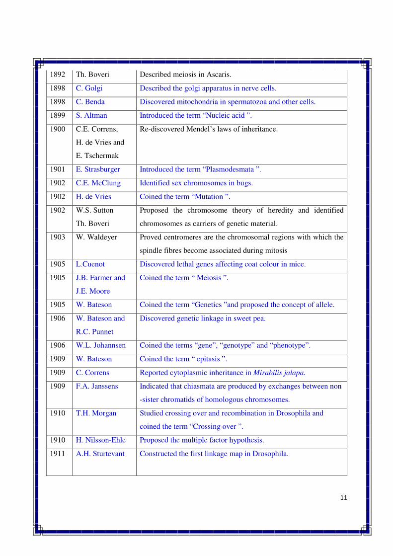
1892 Th. Boveri Described meiosis in Ascaris.
1898 C. Golgi Described the golgi apparatus in nerve cells.
1898 C. Benda Discovered mitochondria in spermatozoa and other cells.
1899 S. Altman Introduced the term “Nucleic acid ”.
1900 C.E. Correns,
H. de Vries and
E. Tschermak
Re-discovered Mendel’s laws of inheritance.
1901 E. Strasburger Introduced the term “Plasmodesmata ”.
1902 C.E. McClung Identified sex chromosomes in bugs.
1902 H. de Vries Coined the term “Mutation ”.
1902 W.S. Sutton
Th. Boveri
Proposed the chromosome theory of heredity and identified
chromosomes as carriers of genetic material.
1903 W. Waldeyer Proved centromeres are the chromosomal regions with which the
spindle fibres become associated during mitosis
1905 L.Cuenot Discovered lethal genes affecting coat colour in mice.
1905 J.B. Farmer and
J.E. Moore
Coined the term “ Meiosis ”.
1905 W. Bateson Coined the term “Genetics ”and proposed the concept of allele.
1906 W. Bateson and
R.C. Punnet
Discovered genetic linkage in sweet pea.
1906 W.L. Johannsen Coined the terms “gene”, “genotype” and “phenotype”.
1909 W. Bateson Coined the term “ epitasis ”.
1909 C. Correns Reported cytoplasmic inheritance in Mirabilis jalapa.
1909 F.A. Janssens Indicated that chiasmata are produced by exchanges between non
-sister chromatids of homologous chromosomes.
1910 T.H. Morgan Studied crossing over and recombination in Drosophila and
coined the term “Crossing over ”.
1910 H. Nilsson-Ehle Proposed the multiple factor hypothesis.
1911 A.H. Sturtevant Constructed the first linkage map in Drosophila.
12
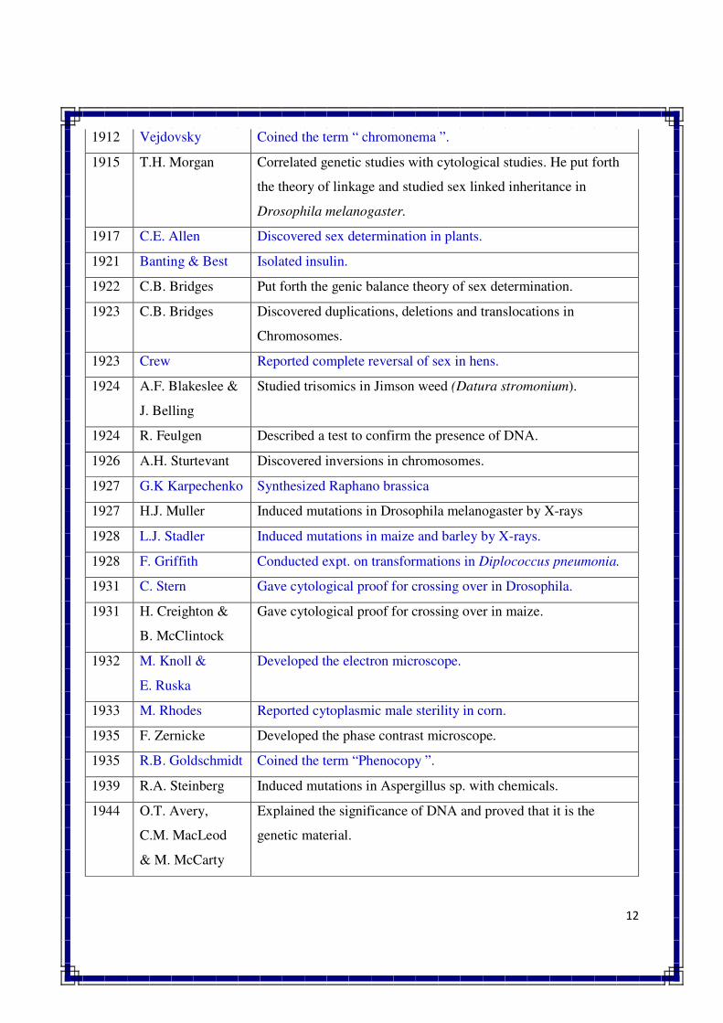
1912 Vejdovsky Coined the term “ chromonema ”.
1915 T.H. Morgan Correlated genetic studies with cytological studies. He put forth
the theory of linkage and studied sex linked inheritance in
Drosophila melanogaster.
1917 C.E. Allen Discovered sex determination in plants.
1921 Banting & Best Isolated insulin.
1922 C.B. Bridges Put forth the genic balance theory of sex determination.
1923 C.B. Bridges Discovered duplications, deletions and translocations in
Chromosomes.
1923 Crew Reported complete reversal of sex in hens.
1924 A.F. Blakeslee &
J. Belling
Studied trisomics in Jimson weed (Datura stromonium).
1924 R. Feulgen Described a test to confirm the presence of DNA.
1926 A.H. Sturtevant Discovered inversions in chromosomes.
1927 G.K Karpechenko Synthesized Raphano brassica
1927 H.J. Muller Induced mutations in Drosophila melanogaster by X-rays
1928 L.J. Stadler Induced mutations in maize and barley by X-rays.
1928 F. Griffith Conducted expt. on transformations in Diplococcus pneumonia.
1931 C. Stern Gave cytological proof for crossing over in Drosophila.
1931 H. Creighton &
B. McClintock
Gave cytological proof for crossing over in maize.
1932 M. Knoll &
E. Ruska
Developed the electron microscope.
1933 M. Rhodes Reported cytoplasmic male sterility in corn.
1935 F. Zernicke Developed the phase contrast microscope.
1935 R.B. Goldschmidt Coined the term “Phenocopy ”.
1939 R.A. Steinberg Induced mutations in Aspergillus sp. with chemicals.
1944 O.T. Avery,
C.M. MacLeod
& M. McCarty
Explained the significance of DNA and proved that it is the
genetic material.
13
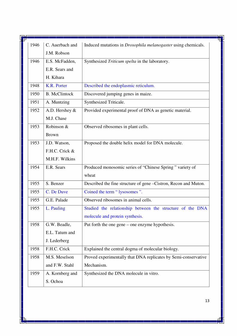
1946 C. Auerbach and
J.M. Robson
Induced mutations in Drosophila melanogaster using chemicals.
1946 E.S. McFadden,
E.R. Sears and
H. Kihara
Synthesized Triticum spelta in the laboratory.
1948 K.R. Porter Described the endoplasmic reticulum.
1950 B. McClintock Discovered jumping genes in maize.
1951 A. Muntzing Synthesized Triticale.
1952 A.D. Hershey &
M.J. Chase
Provided experimental proof of DNA as genetic material.
1953 Robinson &
Brown
Observed ribosomes in plant cells.
1953 J.D. Watson,
F.H.C. Crick &
M.H.F. Wilkins
Proposed the double helix model for DNA molecule.
1954 E.R. Sears Produced monosomic series of “Chinese Spring ” variety of
wheat
1955 S. Benzer Described the fine structure of gene –Cistron, Recon and Muton.
1955 C. De Duve Coined the term “ lysosomes ”.
1955 G.E. Palade Observed ribosomes in animal cells.
1955 L. Pauling Studied the relationship between the structure of the DNA
molecule and protein synthesis.
1958 G.W. Beadle,
E.L. Tatum and
J. Lederberg
Put forth the one gene – one enzyme hypothesis.
1958 F.H.C. Crick Explained the central dogma of molecular biology.
1958 M.S. Meselson
and F.W. Stahl
Proved experimentally that DNA replicates by Semi-conservative
Mechanism.
1959 A. Kornberg and
S. Ochoa
Synthesized the DNA molecule in vitro.
14
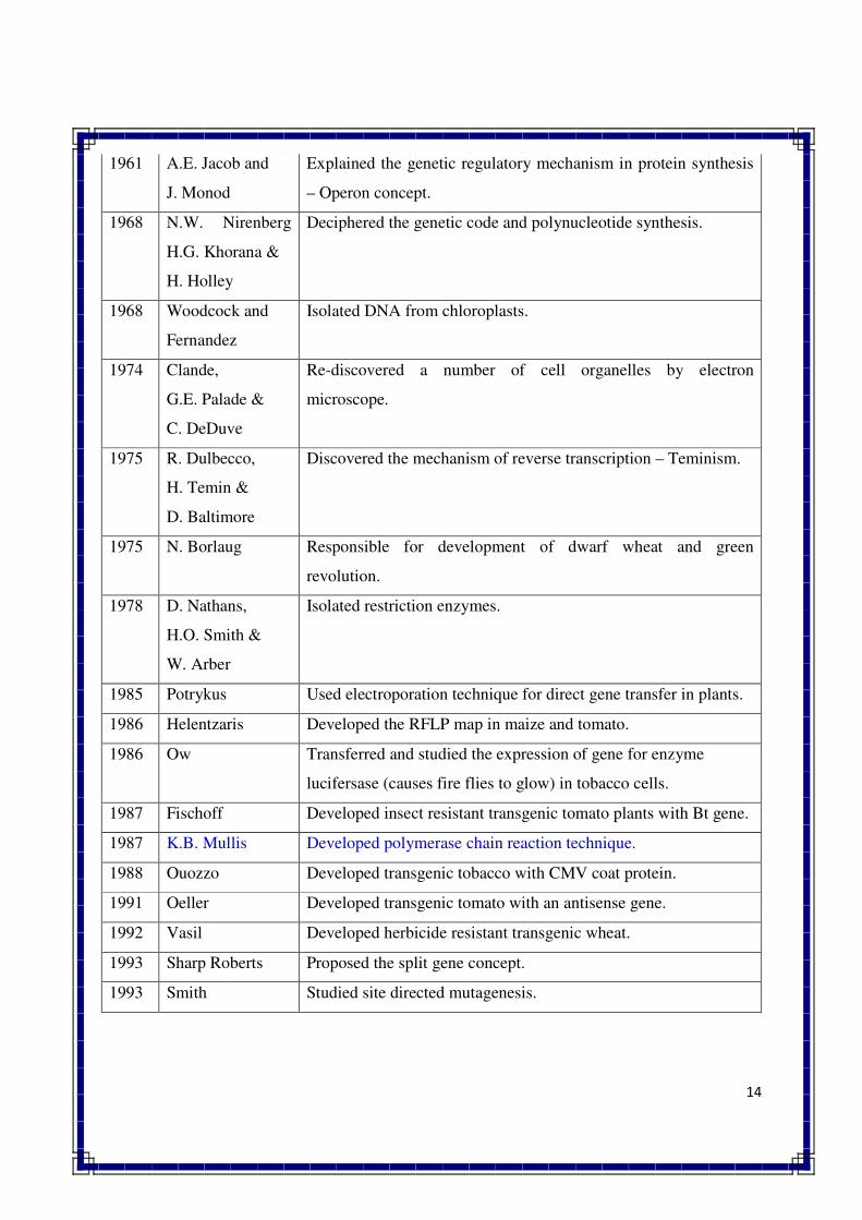
1961 A.E. Jacob and
J. Monod
Explained the genetic regulatory mechanism in protein synthesis
– Operon concept.
1968 N.W. Nirenberg
H.G. Khorana &
H. Holley
Deciphered the genetic code and polynucleotide synthesis.
1968 Woodcock and
Fernandez
Isolated DNA from chloroplasts.
1974 Clande,
G.E. Palade &
C. DeDuve
Re-discovered a number of cell organelles by electron
microscope.
1975 R. Dulbecco,
H. Temin &
D. Baltimore
Discovered the mechanism of reverse transcription – Teminism.
1975 N. Borlaug Responsible for development of dwarf wheat and green
revolution.
1978 D. Nathans,
H.O. Smith &
W. Arber
Isolated restriction enzymes.
1985 Potrykus Used electroporation technique for direct gene transfer in plants.
1986 Helentzaris Developed the RFLP map in maize and tomato.
1986 Ow Transferred and studied the expression of gene for enzyme
lucifersase (causes fire flies to glow) in tobacco cells.
1987 Fischoff Developed insect resistant transgenic tomato plants with Bt gene.
1987 K.B. Mullis Developed polymerase chain reaction technique.
1988 Ouozzo Developed transgenic tobacco with CMV coat protein.
1991 Oeller Developed transgenic tomato with an antisense gene.
1992 Vasil Developed herbicide resistant transgenic wheat.
1993 Sharp Roberts Proposed the split gene concept.
1993 Smith Studied site directed mutagenesis.
15
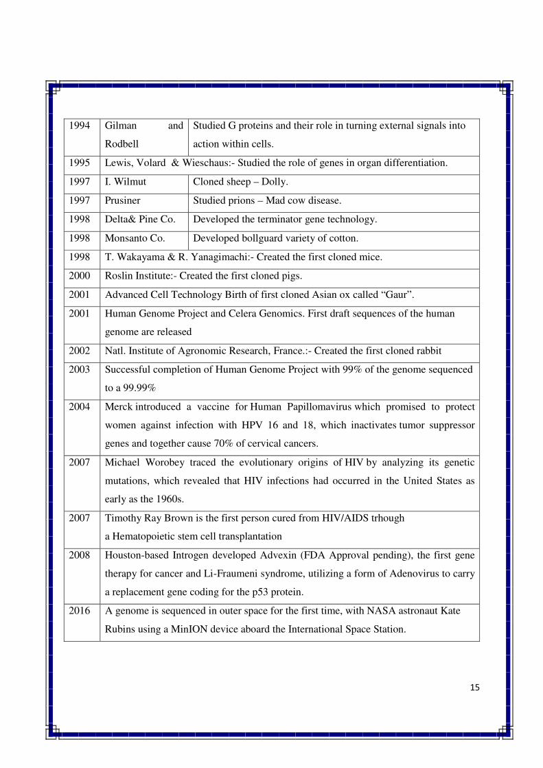
1994 Gilman and
Rodbell
Studied G proteins and their role in turning external signals into
action within cells.
1995 Lewis, Volard & Wieschaus:- Studied the role of genes in organ differentiation.
1997 I. Wilmut Cloned sheep – Dolly.
1997 Prusiner Studied prions – Mad cow disease.
1998 Delta& Pine Co. Developed the terminator gene technology.
1998 Monsanto Co. Developed bollguard variety of cotton.
1998 T. Wakayama & R. Yanagimachi:- Created the first cloned mice.
2000 Roslin Institute:- Created the first cloned pigs.
2001 Advanced Cell Technology Birth of first cloned Asian ox called “Gaur”.
2001 Human Genome Project and Celera Genomics. First draft sequences of the human
genome are released
2002 Natl. Institute of Agronomic Research, France.:- Created the first cloned rabbit
2003 Successful completion of Human Genome Project with 99% of the genome sequenced
to a 99.99%
2004 Merck introduced a vaccine for Human Papillomavirus which promised to protect
women against infection with HPV 16 and 18, which inactivates tumor suppressor
genes and together cause 70% of cervical cancers.
2007 Michael Worobey traced the evolutionary origins of HIV by analyzing its genetic
mutations, which revealed that HIV infections had occurred in the United States as
early as the 1960s.
2007 Timothy Ray Brown is the first person cured from HIV/AIDS trhough
a Hematopoietic stem cell transplantation
2008 Houston-based Introgen developed Advexin (FDA Approval pending), the first gene
therapy for cancer and Li-Fraumeni syndrome, utilizing a form of Adenovirus to carry
a replacement gene coding for the p53 protein.
2016 A genome is sequenced in outer space for the first time, with NASA astronaut Kate
Rubins using a MinION device aboard the International Space Station.
16
GLOSSARY
PLANT GENETICS: A branch of genetics which deals with inheritance and variation of
characters in plant species.
ANIMAL GENETICS: A branch of genetics which deals with inheritance and variation of
traits in animals.
MICROBIAL GENETICS: A branch of genetics which deals with inheritance of characters in
microorganisms like bacteria, viruses and fungi.
MOLECULAR GENETICS: A branch of genetics which deals with structure, composition,
function and replication of chromosomes and genes.
POPULATION GENETICS: A branch of genetics which deals with frequencies of genes and
genotypes in a population, and also with various forces which tend to alter gene frequencies in a
population leading to evolutionary changes.
RADIATION GENETICS: A branch of genetics which deals with effects of various types of
radiations on chromosomes and genes.
EUGENICS: A branch of genetics which deals with the application of the principles of heredity
for the betterment of human race.
MENDELIAN GENETICS: Genetics which deals with the inheritance of oligogenic
characters.
QUANTITATIVE GENETICS: A branch of genetics which deals with the inheritance of
quantitative or polygenic characters.
CYTOGENETICS: Combined study of cytology and genetics.
EUPHENICS: Genetics which deals with the control of hereditary diseases especially inborn
errors of metabolism.
PROKARYOTE:Unicellular organisms whose cells lack nucleus like bacteria, bacteriophages
and blue green algae.
EUKARYOTE: Organisms whose cells contain well defined nucleus.
BACTERIA: Unicellular free living organisms without well defined nucleus.
BACTERIOPHAGE: Special types of virus which grow only inside the bacteria and kill them.
HEREDITY: Transmission of characters from parents to their offspring.
VARIATION: Differences for various characters among the individuals of the same species.
GEMMULES: Invisible copies of each body organ as per theory of pangenes.
17
QUESTIONS
Q.1 Define genetics. Describe in brief its role in crop improvement.
Q.2 Give a brief account of pre-Mendelian concepts about heredity.
Q.3 Describe in brief the role of various disciplines in the advancement of genetics.
Q.4 Write short notes on the following
1. Genetics 5. Drosophila
2. Theory of epigenesis 6. Bacteriophages
3. Theory of pangenes 7. Neurospora
4. Germplasm theory 8. Garden pea
Q.5 List the names of Nobel Prize winners in genetics. Describe in brief the contribution of
any five Nobel Prize winners.
Q.6 Describe in brief the role of following disciplines in the advancement of genetic
1. Cytology 3. Biophysics
2. Biochemistry 4. Statistics
Q.7 Give in brief the contribution of the following scientists in the field of genetics
1. Gregor Mendel 9. T.H. Morgan 16. S. Benzer
2. C.E. Correns 10. C.B. Bridges 17. F. Jacob
3. Hugo de Vries 11. E.L. Tatum 18. M.W. Nirenberg
4. V.S.E. Tschermak 12. Barbara Mc Clintock 19. H.J. Muller
5. William Bateson 13. O.T.Avery 20. G.W. Beadle
6. J.D. Watson 14. F.H.C. Crick
7. J.L. Monod 15. Har Gobind Khorana
Reference Book:-
Elements of Genetics by Phundan Singh (Kalyani Publisher)
2. CELL & CELL DIVISION
Cell structure and organelles
Cell – It is the structural and functional unit of all living organism
Plant cell : A structural and physiological unit of plant, which have protoplasm. Which
consist following organelles. Plant cell consist following types of parts
� The basic constituents of plant and animal cells are the same, viz., nucleic acid,
proteins, carbohydrates, lipids and various inorganic substances.
� They organized in the same fundamental manner.
� The shape of plant cell is rectangular and that of animal cell is round with irregular
appearance.
� Cell organelles: various membrane bound structures that are found within a cell such
as nucleus, plastids, mitochondria, endoplasmic reticulum, etc.,
CELL
Non-living inclusions (Cell wall) Living materials (Protoplast)
Nucleus Cytoplasm
Nuclear membrane Plasma-membrane
Nucleoplasm Endoplasmic reticulum
Chromatin Ribosomes
Nucleolus Golgi body
Vacuoles
Mitochondria
Plastids
Lysosomes
Centrosomes
Microtubules
Cell wall
• It is the outermost part of the cell and is always non-living (Rigid and Strong) but it is
produced and nourished by living protoplasm.
• The cell wall is found only in plants and absent in animals.
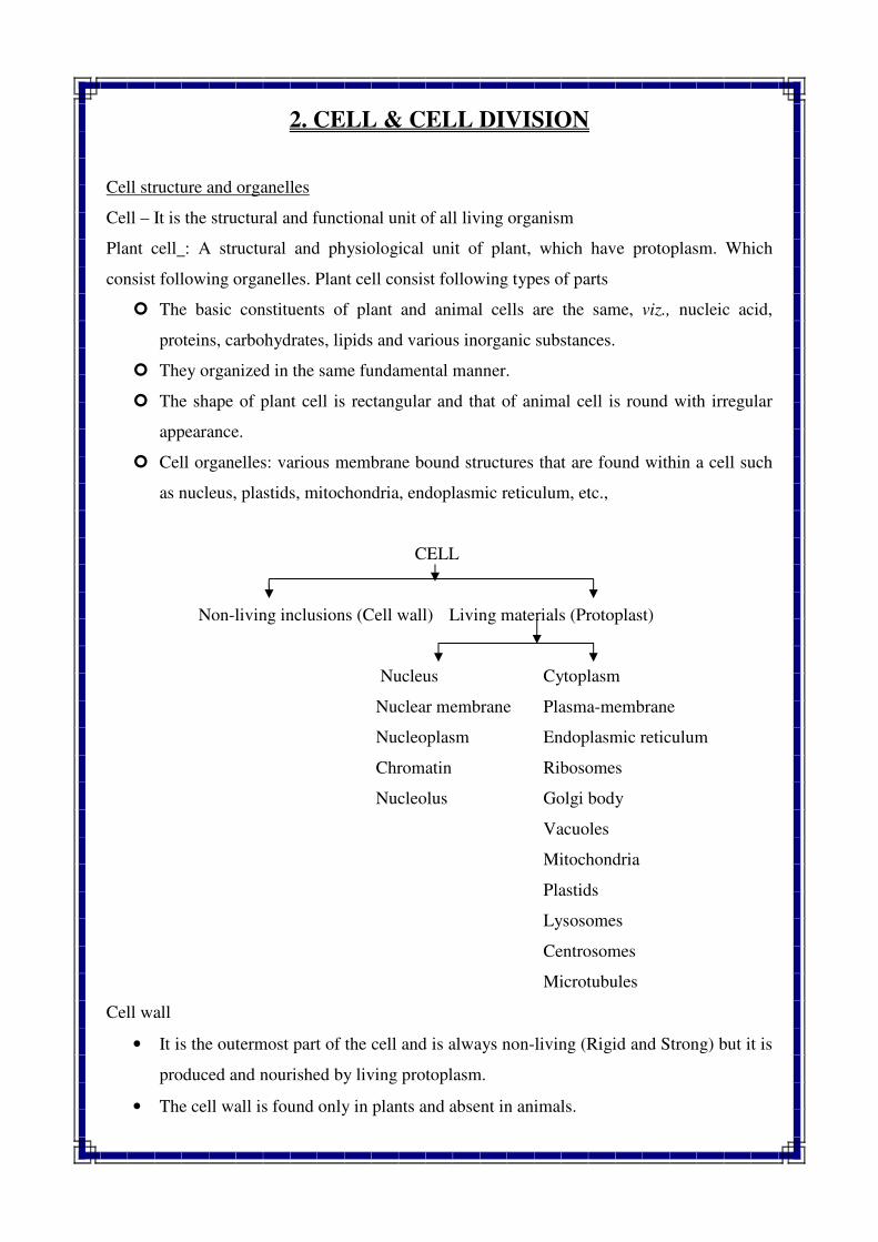
• Multilayered viz., middle lamella, primary cell wall (dispersed arrangement) and
secondary cell wall (parallel arrangement of cellulose fibrils).
Functions
1. To protect inner parts of
the cell
2. To give a definite shape
to the cell
3. To provide mechanical
support
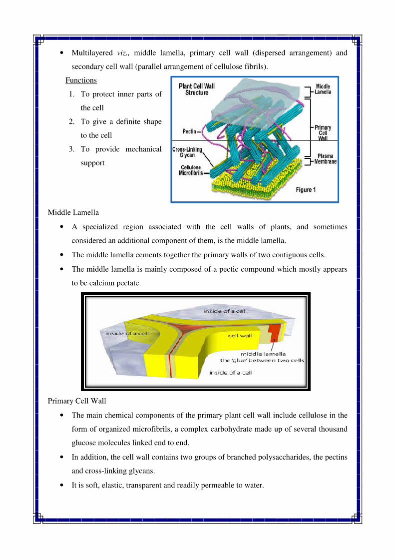
Middle Lamella
• A specialized region associated with the cell walls of plants, and sometimes
considered an additional component of them, is the middle lam
• The middle lamella cements together the primary walls of two contiguous cells.
• The middle lamella is mainly composed of a pectic compound which mostly appears
to be calcium pectate.
Primary Cell Wall
• The main chemical components of the primary plant cell wall include cellulose in the
form of organized microfibrils, a complex carbohydrate made up of several thousand
glucose molecules linked end to end.
• In addition, the cell wall contains two groups of
and cross-linking glycans.
• It is soft, elastic, transparent and readily permeable to water.
middle lamella, primary cell wall (dispersed arrangement) and
secondary cell wall (parallel arrangement of cellulose fibrils).
protect inner parts of
To give a definite shape
To provide mechanical
A specialized region associated with the cell walls of plants, and sometimes
considered an additional component of them, is the middle lamella.
The middle lamella cements together the primary walls of two contiguous cells.
The middle lamella is mainly composed of a pectic compound which mostly appears
to be calcium pectate.
The main chemical components of the primary plant cell wall include cellulose in the
form of organized microfibrils, a complex carbohydrate made up of several thousand
glucose molecules linked end to end.
In addition, the cell wall contains two groups of branched polysaccharides, the pectins
linking glycans.
It is soft, elastic, transparent and readily permeable to water.
middle lamella, primary cell wall (dispersed arrangement) and
A specialized region associated with the cell walls of plants, and sometimes
ella.
The middle lamella cements together the primary walls of two contiguous cells.
The middle lamella is mainly composed of a pectic compound which mostly appears
The main chemical components of the primary plant cell wall include cellulose in the
form of organized microfibrils, a complex carbohydrate made up of several thousand
branched polysaccharides, the pectins
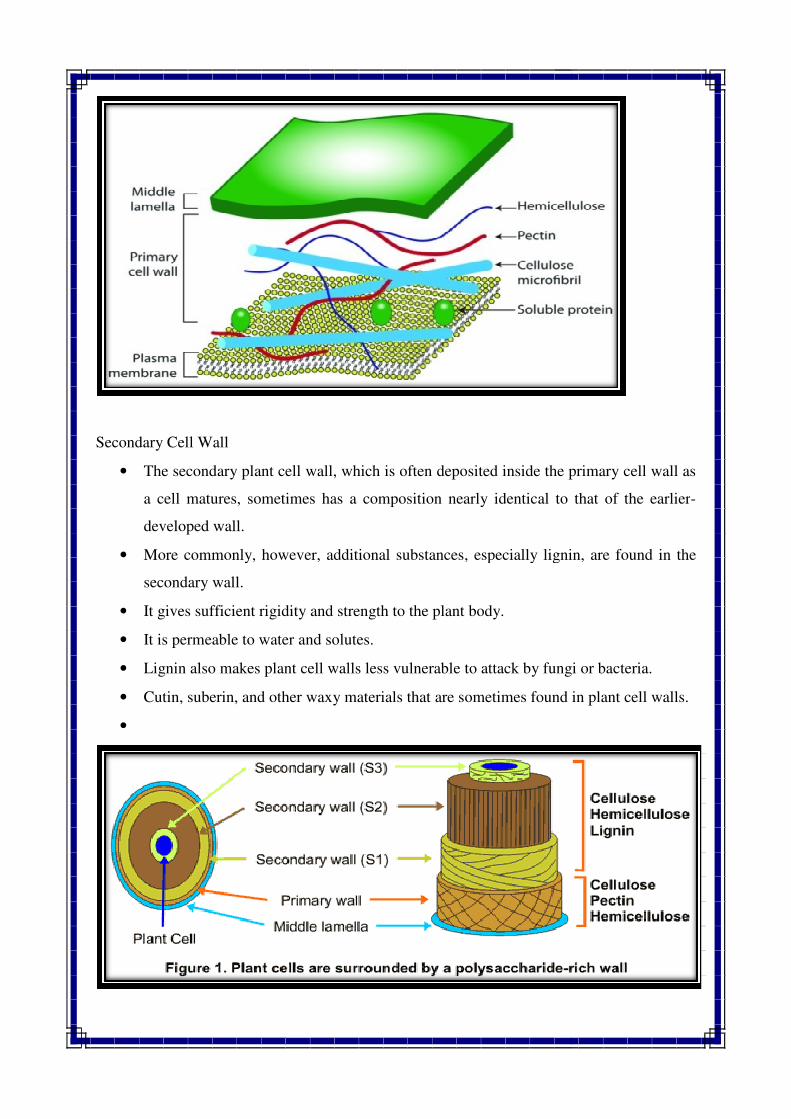
Secondary Cell Wall
• The secondary plant cell wall, which is often deposited inside the primary cell wall as
a cell matures, sometimes has a composition nearly identical to that of the earlier
developed wall.
• More commonly, however, additional substances, especially lignin, a
secondary wall.
• It gives sufficient rigidity and strength to the plant body.
• It is permeable to water and solutes.
• Lignin also makes plant cell walls less vulnerable to attack by fungi or bacteria.
• Cutin, suberin, and other waxy material
•
The secondary plant cell wall, which is often deposited inside the primary cell wall as
a cell matures, sometimes has a composition nearly identical to that of the earlier
More commonly, however, additional substances, especially lignin, a
It gives sufficient rigidity and strength to the plant body.
It is permeable to water and solutes.
Lignin also makes plant cell walls less vulnerable to attack by fungi or bacteria.
Cutin, suberin, and other waxy materials that are sometimes found in plant cell walls.
The secondary plant cell wall, which is often deposited inside the primary cell wall as
a cell matures, sometimes has a composition nearly identical to that of the earlier-
More commonly, however, additional substances, especially lignin, are found in the
Lignin also makes plant cell walls less vulnerable to attack by fungi or bacteria.
s that are sometimes found in plant cell walls.
Plasma lemma or plasma membrane
� The cytoplasm which is surrounded by thin and flexible membrane is called plasma
membrane
� It present both in plant as well as animal cell
� Which composed of lipids and proteins
Functions
1. It regulates the passage in and out of the cell
2. It act as selective permeable membrane
3. It checks the entry of toxic elements from outside into cytoplasm
4. It permits passage of molecules like minerals into the cell and restrict their outward
movement
Protoplasm
� Its substance which provide life to the plants/animals
� It is granular, semi fluid – translucent
� Protoplasm differentiated into Cytoplasm and Nucleus
Cytoplasm
� Variety of structure remain suspended such as living and non living
� Non living : Non membrane bounded – lipid, starch granules
� Living : membrane bounded
Cytoplasmic inclusions (Non living) :
� They are not metabolic active parts but they are the storage site of end products
� Suspended in cytoplasmic matrix
� It includes oil drops, yolk granules, pigments, starch granules etc.
Cytoplasmic organelles
� They are membrane bounded, living structures-mitochondria, ER, chloroplast etc.
� They perform important activities like biosynthesis, metabolic and respiratory
� They are also engaged in transportation and storage of food materials and
reproduction
Microtubules
� Complex structure made up of 13 individual protofilaments arranged to form hallow
cylinder
� Responsible for transportation of small molecules
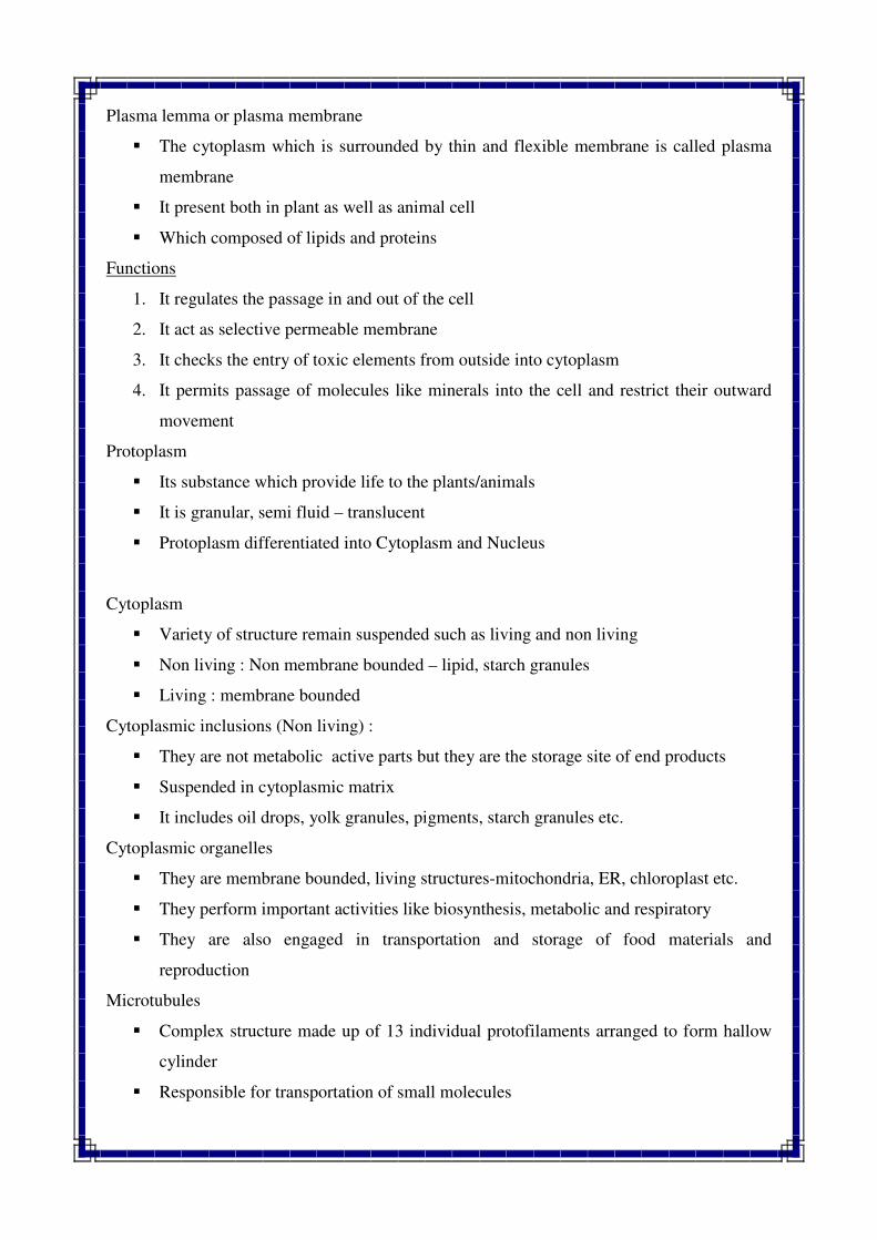
Endoplasmic Reticulum (E. R.)
� The term E. R. was first used by Porter in 1948 to describe a fine reticulum in the
endoplasmic cells
� It is thread or tube like floating in cytoplasm on which ribosomes are attached
� Two types of E. R.
� Smooth E. R. – In this case both ou
have attached ribosomes. They do not involved in protein synthesis
� Rough E. R. – Outer and inner membrane found attached with ribosomes. They also
actively involved in protein synthesis. Both smooth and rou
interchangeable as per the needs of the cell
Functions: (E.R.)
1. It is associated with the synthesis of proteins (rough E. R.), lipids and phospholipids
(Both E. R.)
2. Provide channels for the transportation of synthesized material to various pa
3. Provide controlled passage for the export of m
4. Several enzymes are embedded in the membrane eg. Glucose
etc.
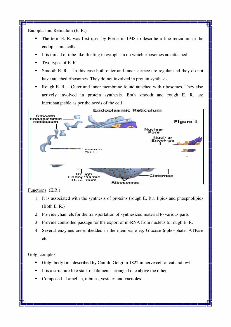
Golgi complex
� Golgi body first described by Camilo Golgi in 1822 in nerve cell of cat and
� It is a structure like stalk of filaments arranged one above the other
� Composed –Lamellae, tubules, vesicles and vacuoles
Reticulum (E. R.)
The term E. R. was first used by Porter in 1948 to describe a fine reticulum in the
It is thread or tube like floating in cytoplasm on which ribosomes are attached
In this case both outer and inner surface are regular and they do not
have attached ribosomes. They do not involved in protein synthesis
Outer and inner membrane found attached with ribosomes. They also
actively involved in protein synthesis. Both smooth and rou
interchangeable as per the needs of the cell
It is associated with the synthesis of proteins (rough E. R.), lipids and phospholipids
Provide channels for the transportation of synthesized material to various pa
Provide controlled passage for the export of m-RNA from nucleus to rough E. R.
Several enzymes are embedded in the membrane eg. Glucose-6-phosphate, ATPase
Golgi body first described by Camilo Golgi in 1822 in nerve cell of cat and
It is a structure like stalk of filaments arranged one above the other
Lamellae, tubules, vesicles and vacuoles
The term E. R. was first used by Porter in 1948 to describe a fine reticulum in the
It is thread or tube like floating in cytoplasm on which ribosomes are attached
ter and inner surface are regular and they do not
have attached ribosomes. They do not involved in protein synthesis
Outer and inner membrane found attached with ribosomes. They also
actively involved in protein synthesis. Both smooth and rough E. R. are
It is associated with the synthesis of proteins (rough E. R.), lipids and phospholipids
Provide channels for the transportation of synthesized material to various parts
RNA from nucleus to rough E. R.
phosphate, ATPase
Golgi body first described by Camilo Golgi in 1822 in nerve cell of cat and owl
Functions:
1. Packaging food materials such as proteins, lipids and phospholipids for transport to
other cells
2. It secrete many granules and lysosomes
Lysosomes :
� The term lysosome was first used by Dave in 1955
� In plant cell they are bounded storage granules and containing hydrolylic digestive
enzymes
Functions:
1. It is responsible for digestion of intracellular substances and foreign particles.
2. When cell dies lysosomes releases its enzymes, which digest the dead cell resulting in
cleaning of debris.
Cytoplasmic vacuoles:
� Its small to large sized liquid filled structure
� Sometimes more numbers of vacuoles fuse to gather and formed large size structure
� Each vacuoles is surrounded by vacuolar membrane called as tonoplast. Tonoplast
having a selective semi permeable membrane composed of lipoprotein
� The fluid inside the vacuoles is called as cell sap, which made up organic substances
like sugars, organic acids, inorganic salts, proteins and pigments
Functions:
1. Storage and transmission of the materials and maintenance of the internal pressure
Ribosomes:
� Ribosomes are the small cellular particles composed of RNA + Protein
� Ribosomes are the site of protein synthesis
� They contain nearly 40
� In young actively dividing cell they are usually free in the cytoplasm but in the mature
cells, they are attached with ER.
� The size or weight of the ribo
rate or coefficient)
� Mainly three kinds –
(1) Mitochondrion
Functions:
1. To carry out protein synthesis with the help of m
Mitochondria:
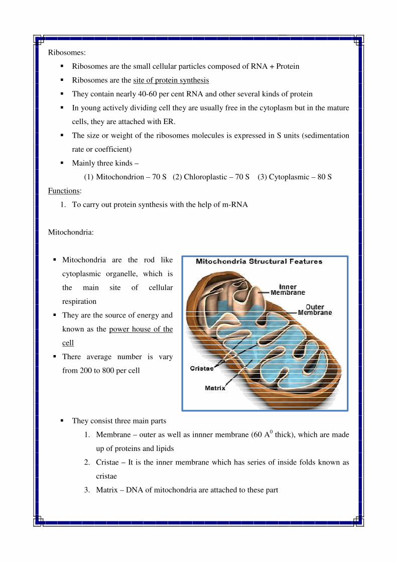
� Mitochondria are the rod like
cytoplasmic organelle, which is
the main site of cellular
respiration
� They are the source of energy and
known as the power house of the
cell
� There average number is vary
from 200 to 800 per cell
� They consist three main pa
1. Membrane – outer as well as innner membrane (60 A
up of proteins and lipids
2. Cristae – It is the inner membrane which has series of inside folds known as
cristae
3. Matrix – DNA of mitochondria are attached to these part
Ribosomes are the small cellular particles composed of RNA + Protein
site of protein synthesis
They contain nearly 40-60 per cent RNA and other several kinds of protein
In young actively dividing cell they are usually free in the cytoplasm but in the mature
cells, they are attached with ER.
The size or weight of the ribosomes molecules is expressed in S units (sedimentation
Mitochondrion – 70 S (2) Chloroplastic – 70 S (3) Cytoplasmic
To carry out protein synthesis with the help of m-RNA
Mitochondria are the rod like
cytoplasmic organelle, which is
the main site of cellular
They are the source of energy and
power house of the
There average number is vary
They consist three main parts
outer as well as innner membrane (60 A0 thick), which are made
up of proteins and lipids
It is the inner membrane which has series of inside folds known as
DNA of mitochondria are attached to these part
Ribosomes are the small cellular particles composed of RNA + Protein
60 per cent RNA and other several kinds of protein
In young actively dividing cell they are usually free in the cytoplasm but in the mature
somes molecules is expressed in S units (sedimentation
70 S (3) Cytoplasmic – 80 S
thick), which are made
It is the inner membrane which has series of inside folds known as
Functions:
1. It involved in respiration, oxidation and metabolism of energy (Power house of the
cell)
2. They contain circular DNA and ribosomes, so they are capable of synthesis of certain
proteins
3. They contain DNA, so also contribute to heredity by the way of
inheritance
Plastid:
� They are the self replicating cytoplasmic organelles founds in plant cell
� They are absent in bacteria, cerain fungi and animals
� Mainly three types : Leucoplast, Chromoplast and Chloroplast
� Leucoplast – They are colourless
fats
� Chromoplast – Coloured but other than green viz., Plucoxanthin and Phycocynin.
There functions are still not clear. They contain pigments of different colour
Yellow, orange and red. The coluring ma
Carotene.
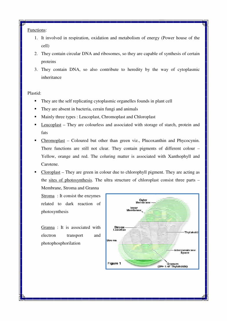
� Cloroplast – They are green in colour due to chlorophyll pigment. They are acting as
the sites of photosynthesis
Membrane, Stroma and Granna
Stroma : It consist the enzymes
related to dark reaction of
photosynthesis
Granna : It is associated with
electron transport and
photophosphorilation
It involved in respiration, oxidation and metabolism of energy (Power house of the
They contain circular DNA and ribosomes, so they are capable of synthesis of certain
They contain DNA, so also contribute to heredity by the way of
They are the self replicating cytoplasmic organelles founds in plant cell
They are absent in bacteria, cerain fungi and animals
Mainly three types : Leucoplast, Chromoplast and Chloroplast
They are colourless and associated with storage of starch, protein and
Coloured but other than green viz., Plucoxanthin and Phycocynin.
There functions are still not clear. They contain pigments of different colour
Yellow, orange and red. The coluring matter is associated with Xanthophyll and
They are green in colour due to chlorophyll pigment. They are acting as
sites of photosynthesis. The ultra structure of chloroplast consist three parts
Membrane, Stroma and Granna
: It consist the enzymes
related to dark reaction of
: It is associated with
electron transport and
It involved in respiration, oxidation and metabolism of energy (Power house of the
They contain circular DNA and ribosomes, so they are capable of synthesis of certain
They contain DNA, so also contribute to heredity by the way of cytoplasmic
They are the self replicating cytoplasmic organelles founds in plant cell
and associated with storage of starch, protein and
Coloured but other than green viz., Plucoxanthin and Phycocynin.
There functions are still not clear. They contain pigments of different colour –
tter is associated with Xanthophyll and
They are green in colour due to chlorophyll pigment. They are acting as
. The ultra structure of chloroplast consist three parts –
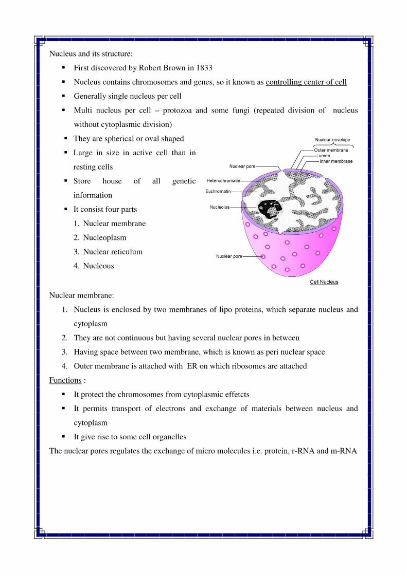
Nucleus and its structure:
� First discovered by Robert Brown in 1833
� Nucleus contains chromosomes and genes, so it known as controlling center of cell
� Generally single nucleus per cell
� Multi nucleus per cell – protozoa and some fungi (repeated division of nucleus
without cytoplasmic division)
� They are spherical or oval shaped
� Large in size in active cell than in
resting cells
� Store house of all genetic
information
� It consist four parts
1. Nuclear membrane
2. Nucleoplasm
3. Nuclear reticulum
4. Nucleous
Nuclear membrane:
1. Nucleus is enclosed by two membranes of lipo proteins, which separate nucleus and
cytoplasm
2. They are not continuous but having several nuclear pores in between
3. Having space between two membrane, which is known as peri nuclear space
4. Outer membrane is attached with ER on which ribosomes are attached
Functions :
� It protect the chromosomes from cytoplasmic effetcts
� It permits transport of electrons and exchange of materials between nucleus and
cytoplasm
� It give rise to some cell organelles
The nuclear pores regulates the exchange of micro molecules i.e. protein, r-RNA and m-RNA
Nucleoplasm
1. Its watery substances in higher nucleolus
2. It is also known as nucleoplasm or nuclear sap or Karyolymph
3. It is shapeless and contain dissolved phosphorous ribose sugar proteins, nucleotides
and had nucleic acid
Nucleolus
� A spherical body found in the nucleus is called nucleolus
� It is found in the higher organisms and is attached with specific region of a particular
chromosome.
� It disappear during prophase of mitosis and meiosis and reappear during telophase.
� Chemically it is composed of ribosomal proteins and RNA
Functions :
1. Formation of ribosome and synthesis of proteins
2. It provide energy for all nuclear activities
Chromatin or Nuclear reticulum
� Its thread like, coiled and much elongated structures
� During cell division process (mitosis and meiosis) chromatin structure becomes thick
ribbon like structure which are known as chromosomes
Functions :
1. Chromatin is the basic unit of chromosomes contain genes and thus play important
role in the inheritance of the character from the parents to offspring
Structural differences between Eukaryotic and Prokaryotic cells
PROKARY PROTIC CELL EUKARYOTIC CELL
1. Prokaryotes are primitive organisms (Pro =
primitive; Karyon = nucleus)
1. Eukaryotes are higher organisms
(Eu = good or true; Karyon = nucleus)
2. They are generally uni-cellular 2. They are generally multi-cellular
3. The average diameter of prokaryotic cell
ranges from 5 - 10µm
3. The average diameter of eukaryotic cell
ranges from 10–100 µm
4. Posses only one envelope system 4. Posses two envelope system
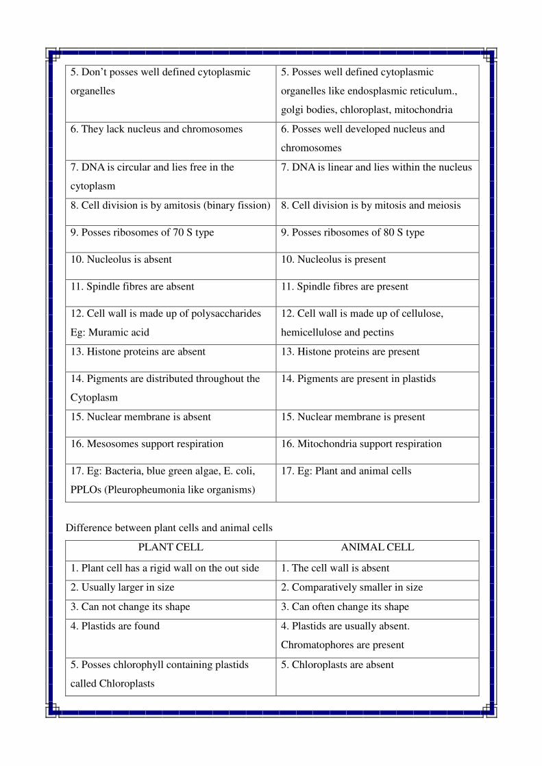
5. Don’t posses well defined cytoplasmic
organelles
5. Posses well defined cytoplasmic
organelles like endosplasmic reticulum.,
golgi bodies, chloroplast, mitochondria
6. They lack nucleus and chromosomes 6. Posses well developed nucleus and
chromosomes
7. DNA is circular and lies free in the
cytoplasm
7. DNA is linear and lies within the nucleus
8. Cell division is by amitosis (binary fission) 8. Cell division is by mitosis and meiosis
9. Posses ribosomes of 70 S type 9. Posses ribosomes of 80 S type
10. Nucleolus is absent 10. Nucleolus is present
11. Spindle fibres are absent 11. Spindle fibres are present
12. Cell wall is made up of polysaccharides
Eg: Muramic acid
12. Cell wall is made up of cellulose,
hemicellulose and pectins
13. Histone proteins are absent 13. Histone proteins are present
14. Pigments are distributed throughout the
Cytoplasm
14. Pigments are present in plastids
15. Nuclear membrane is absent 15. Nuclear membrane is present
16. Mesosomes support respiration 16. Mitochondria support respiration
17. Eg: Bacteria, blue green algae, E. coli,
PPLOs (Pleuropheumonia like organisms)
17. Eg: Plant and animal cells
Difference between plant cells and animal cells
PLANT CELL ANIMAL CELL
1. Plant cell has a rigid wall on the out side 1. The cell wall is absent
2. Usually larger in size 2. Comparatively smaller in size
3. Can not change its shape 3. Can often change its shape
4. Plastids are found 4. Plastids are usually absent.
Chromatophores are present
5. Posses chlorophyll containing plastids
called Chloroplasts
5. Chloroplasts are absent
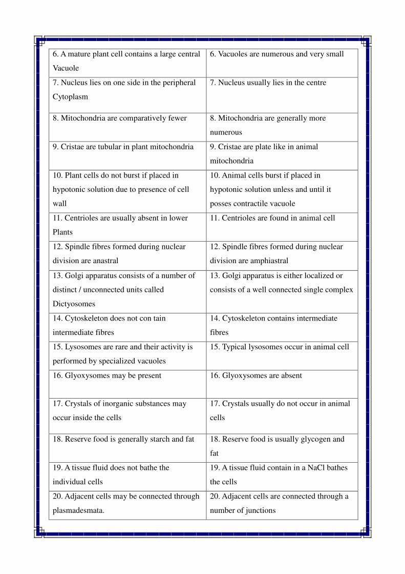
6. A mature plant cell contains a large central
Vacuole
6. Vacuoles are numerous and very small
7. Nucleus lies on one side in the peripheral
Cytoplasm
7. Nucleus usually lies in the centre
8. Mitochondria are comparatively fewer 8. Mitochondria are generally more
numerous
9. Cristae are tubular in plant mitochondria 9. Cristae are plate like in animal
mitochondria
10. Plant cells do not burst if placed in
hypotonic solution due to presence of cell
wall
10. Animal cells burst if placed in
hypotonic solution unless and until it
posses contractile vacuole
11. Centrioles are usually absent in lower
Plants
11. Centrioles are found in animal cell
12. Spindle fibres formed during nuclear
division are anastral
12. Spindle fibres formed during nuclear
division are amphiastral
13. Golgi apparatus consists of a number of
distinct / unconnected units called
Dictyosomes
13. Golgi apparatus is either localized or
consists of a well connected single complex
14. Cytoskeleton does not con tain
intermediate fibres
14. Cytoskeleton contains intermediate
fibres
15. Lysosomes are rare and their activity is
performed by specialized vacuoles
15. Typical lysosomes occur in animal cell
16. Glyoxysomes may be present 16. Glyoxysomes are absent
17. Crystals of inorganic substances may
occur inside the cells
17. Crystals usually do not occur in animal
cells
18. Reserve food is generally starch and fat 18. Reserve food is usually glycogen and
fat
19. A tissue fluid does not bathe the
individual cells
19. A tissue fluid contain in a NaCl bathes
the cells
20. Adjacent cells may be connected through
plasmadesmata.
20. Adjacent cells are connected through a
number of junctions
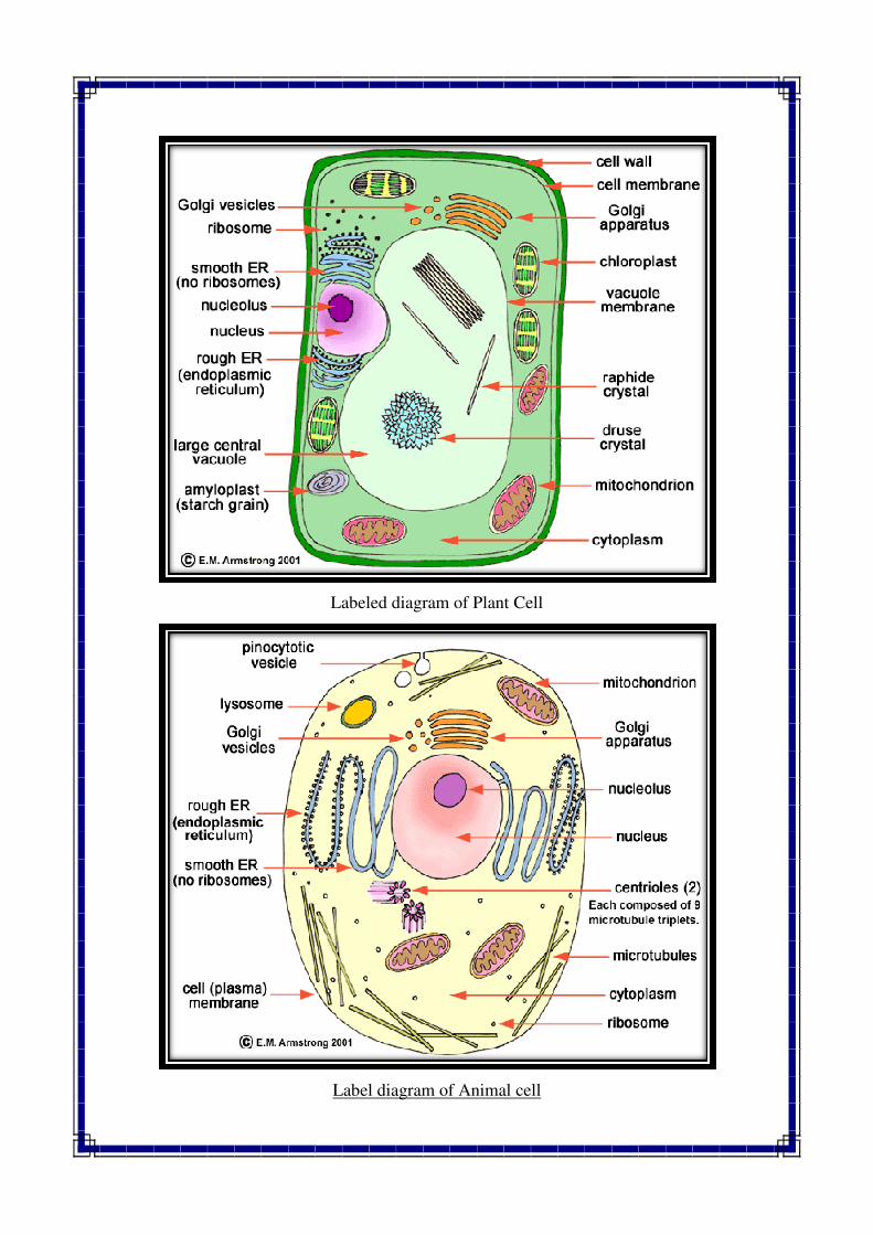
Labeled diagram of Plant Cell
Label diagram of Animal cell
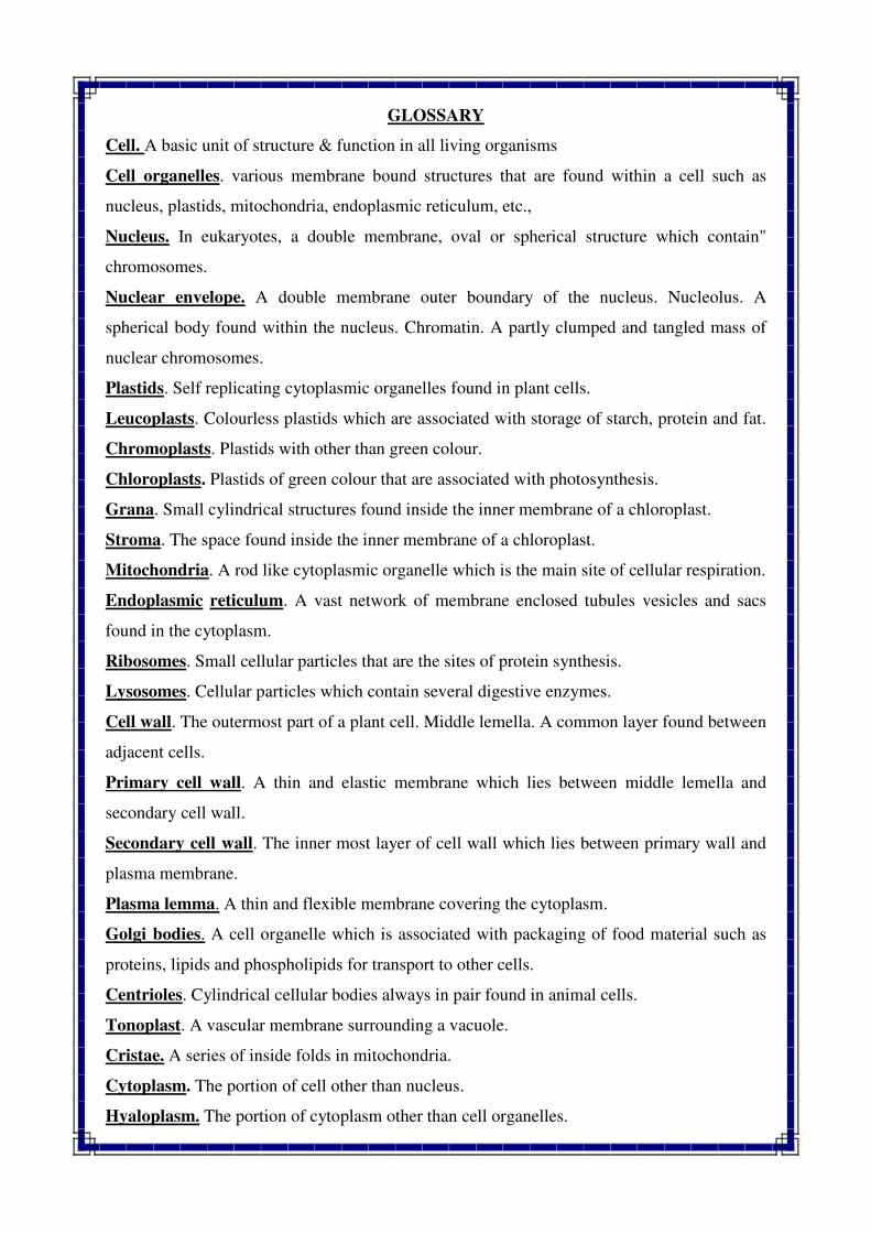
GLOSSARY
Cell. A basic unit of structure & function in all living organisms
Cell organelles. various membrane bound structures that are found within a cell such as
nucleus, plastids, mitochondria, endoplasmic reticulum, etc.,
Nucleus. In eukaryotes, a double membrane, oval or spherical structure which contain"
chromosomes.
Nuclear envelope. A double membrane outer boundary of the nucleus. Nucleolus. A
spherical body found within the nucleus. Chromatin. A partly clumped and tangled mass of
nuclear chromosomes.
Plastids. Self replicating cytoplasmic organelles found in plant cells.
Leucoplasts. Colourless plastids which are associated with storage of starch, protein and fat.
Chromoplasts. Plastids with other than green colour.
Chloroplasts. Plastids of green colour that are associated with photosynthesis.
Grana. Small cylindrical structures found inside the inner membrane of a chloroplast.
Stroma. The space found inside the inner membrane of a chloroplast.
Mitochondria. A rod like cytoplasmic organelle which is the main site of cellular respiration.
Endoplasmic reticulum. A vast network of membrane enclosed tubules vesicles and sacs
found in the cytoplasm.
Ribosomes. Small cellular particles that are the sites of protein synthesis.
Lysosomes. Cellular particles which contain several digestive enzymes.
Cell wall. The outermost part of a plant cell. Middle lemella. A common layer found between
adjacent cells.
Primary cell wall. A thin and elastic membrane which lies between middle lemella and
secondary cell wall.
Secondary cell wall. The inner most layer of cell wall which lies between primary wall and
plasma membrane.
Plasma lemma. A thin and flexible membrane covering the cytoplasm.
Golgi bodies. A cell organelle which is associated with packaging of food material such as
proteins, lipids and phospholipids for transport to other cells.
Centrioles. Cylindrical cellular bodies always in pair found in animal cells.
Tonoplast. A vascular membrane surrounding a vacuole.
Cristae. A series of inside folds in mitochondria.
Cytoplasm. The portion of cell other than nucleus.
Hyaloplasm. The portion of cytoplasm other than cell organelles.
QUESTIONS
Q.1 Define nucleus. Describe in brief ultrastructure and functions of nucleus.
Q.2 Give a brief account of the ultrastructure, origin and functions of chloroplasts.
Q.3 Describe briefly ultrastructure, origin and functions of mitochondria.
Q.4 What are cell organelles? Give a list of various organelles found in a plant cell.
Describe anyone of them in detail.
Q.5 What is cell wall? Describe briefly the structure and functions of cell wall with
suitable diagrams.
Q.6 Write short notes on the following
1. Endoplasmic reticulum 5. Ribosomes
2. Lysosomes 6. Golgi complex
3. Centrioles 7. Microtubules
4. Peroxisomes 8. Spherosomes
Q.7 Give differences between the following:
1. Plant cell and animal cell. 3. Primary and secondary cell wall.
2. Nucleus and nucleolus 4. Plastids and mitochondria.
//*//*//*//
Cell is the basic unit of structure and function of all living systems. The process of
formation of new cells from pre
process, the cell going under the division process is referred as mother ce
which are formed due to the process of cell division is termed as daughter cells. There are
mainly two types of cell division
The process of cell division is divided into two parts : (1) Karyokinesis and (2
1. Karyokinesis : It is the process of nucleus division.
2. Cytokinesis : It is the process of division of cytoplasm. Cytokinesis follows to
karyokinesis.
Cell cycle with different sub
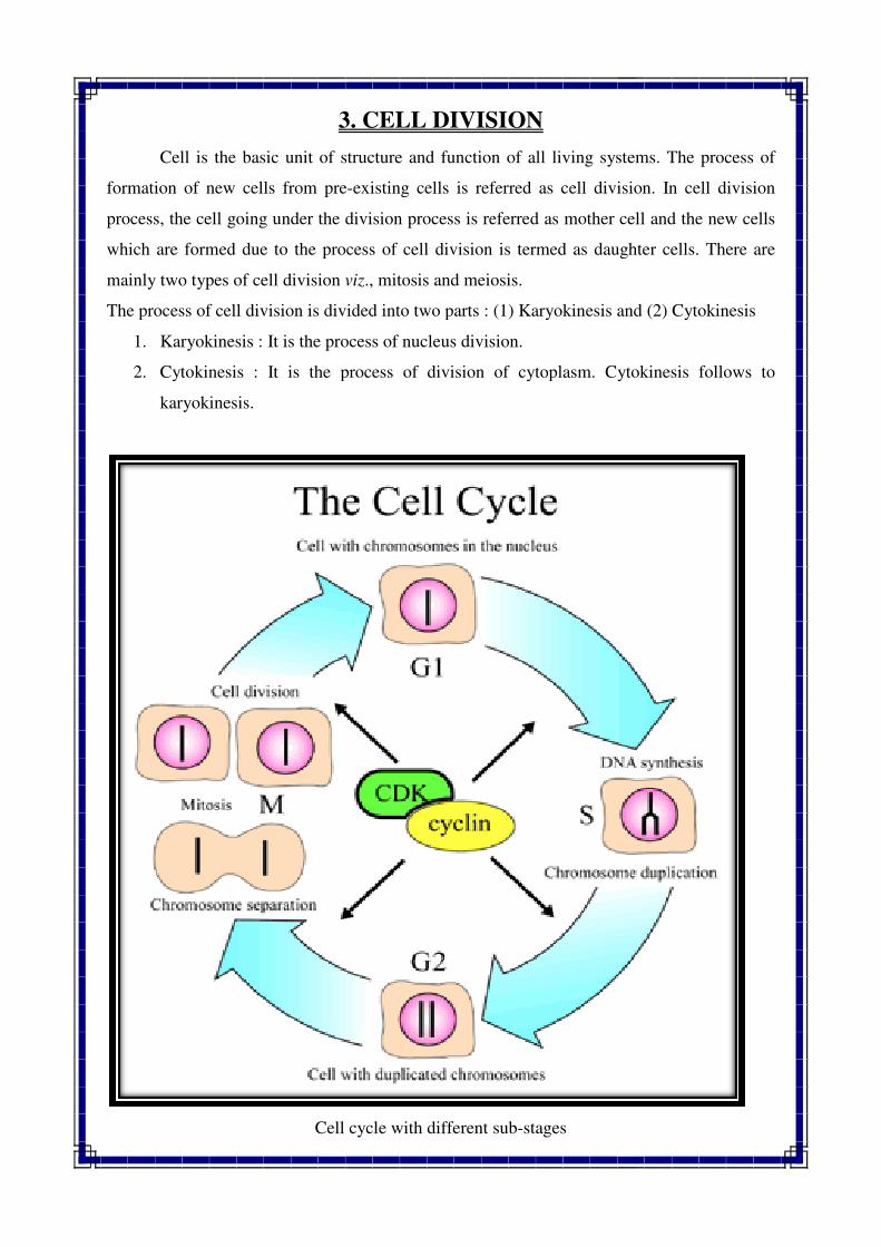
3. CELL DIVISION
Cell is the basic unit of structure and function of all living systems. The process of
formation of new cells from pre-existing cells is referred as cell division. In cell division
process, the cell going under the division process is referred as mother cell and the new cells
which are formed due to the process of cell division is termed as daughter cells. There are
mainly two types of cell division viz., mitosis and meiosis.
The process of cell division is divided into two parts : (1) Karyokinesis and (2
Karyokinesis : It is the process of nucleus division.
Cytokinesis : It is the process of division of cytoplasm. Cytokinesis follows to
Cell cycle with different sub-stages
Cell is the basic unit of structure and function of all living systems. The process of
existing cells is referred as cell division. In cell division
ll and the new cells
which are formed due to the process of cell division is termed as daughter cells. There are
The process of cell division is divided into two parts : (1) Karyokinesis and (2) Cytokinesis
Cytokinesis : It is the process of division of cytoplasm. Cytokinesis follows to
CELL CYCLE
It is the period in which one cycle of cell division is completed is called cell cycle. It consists
of two phases viz., Interphase and Mitotic phase.
INTERPHASE :
It is the phase of the DNA synthesis in which the chromosomal material is in special stage,
which is known as metabolic stage or interphase. It occupies the time between the end of
telophase of previous mitotic division and the beginning of the next prophase. It occupies the
largest period in a cell cycle. It is often not regarded as a stage of cell division. The interphase
is divided into three sub stages i.e. G1, S and G2 (Fig-3.1).
1. G1 : Synthesis of RNA and protein (Pre DNA replication phase). It occupies 25-50 %
of interphase duration
2. S : Synthesis of DNA (DNA replication phase). It occupies 35-40 % of interphase
time
3. G2 : Synthesis of mRNA and some fraction of RNA (Post DNA replication phase). It
occupies 15-25 % of interphase duration
During interphase chromosome do not goes any observable cytological changes but
chromosomes are in form of chromatin fibers. While, during mitotic phase (M phase)
chromosomes are duplicated and as a result chromosome number remain constant and
definite in each species. The M phase consists of four stages viz., prophase, metaphase,
anaphase and telophase. (Fig 3.2).
MITOSIS
The term mitosis was coined by Flemming in 1892. Mitosis refers to the cell division
process in which two identical daughter cells are produced from a mother cell. The
chromosome numbers of newly developed daughter cells are also remaining same as mother
cell in mitotic division. Mitotic division is take place in somatic cells, so it is also referred as
a somatic cell division.
SIGNIFICANCE OF MITOSIS :
1. The chief function of the mitosis is growth of organisms and regeneration of damaged
tissues.
2. To keep the chromosome number constant.
3. It multiplies the cell number and causes vegetative growth and development.
4. Regeneration of damaged tissues and organs.
5. Replacement of old tissues and organs.
6. Production of new tissues and organs like roots, shoots, branches etc.
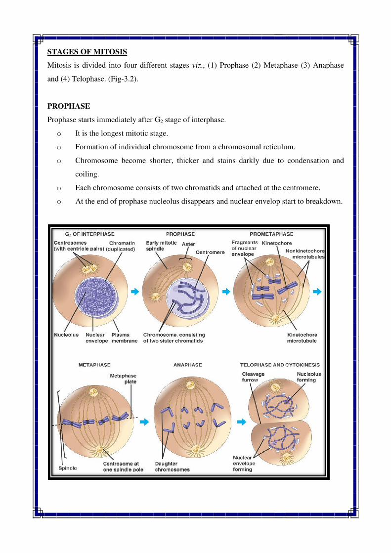
STAGES OF MITOSIS
Mitosis is divided into four different stages
and (4) Telophase. (Fig-3.2).
PROPHASE
Prophase starts immediately after G
o It is the longest mitotic stage.
o Formation of individual chromosome from a chromo
o Chromosome become shorter, thicker and stains darkly due to condensation and
coiling.
o Each chromosome consists of two chromatids and attached at the centromere.
o At the end of prophase nucleolus disappears and nuclear envelop start to break
ed into four different stages viz., (1) Prophase (2) Metaphase (3) Anaphase
Prophase starts immediately after G2 stage of interphase.
It is the longest mitotic stage.
Formation of individual chromosome from a chromosomal reticulum.
Chromosome become shorter, thicker and stains darkly due to condensation and
Each chromosome consists of two chromatids and attached at the centromere.
At the end of prophase nucleolus disappears and nuclear envelop start to break
., (1) Prophase (2) Metaphase (3) Anaphase
somal reticulum.
Chromosome become shorter, thicker and stains darkly due to condensation and
Each chromosome consists of two chromatids and attached at the centromere.
At the end of prophase nucleolus disappears and nuclear envelop start to breakdown.
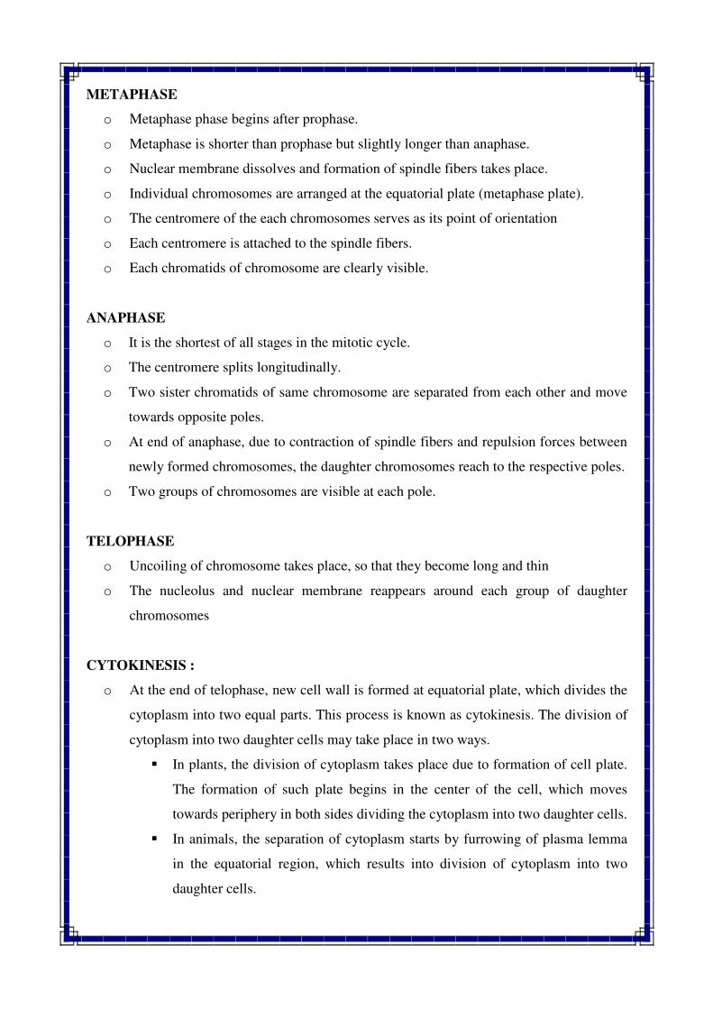
METAPHASE
o Metaphase phase begins after prophase.
o Metaphase is shorter than prophase but slightly longer than anaphase.
o Nuclear membrane dissolves and formation of spindle fibers takes place.
o Individual chromosomes are arranged at the equatorial plate (metaphase plate).
o The centromere of the each chromosomes serves as its point of orientation
o Each centromere is attached to the spindle fibers.
o Each chromatids of chromosome are clearly visible.
ANAPHASE
o It is the shortest of all stages in the mitotic cycle.
o The centromere splits longitudinally.
o Two sister chromatids of same chromosome are separated from each other and move
towards opposite poles.
o At end of anaphase, due to contraction of spindle fibers and repulsion forces between
newly formed chromosomes, the daughter chromosomes reach to the respective poles.
o Two groups of chromosomes are visible at each pole.
TELOPHASE
o Uncoiling of chromosome takes place, so that they become long and thin
o The nucleolus and nuclear membrane reappears around each group of daughter
chromosomes
CYTOKINESIS :
o At the end of telophase, new cell wall is formed at equatorial plate, which divides the
cytoplasm into two equal parts. This process is known as cytokinesis. The division of
cytoplasm into two daughter cells may take place in two ways.
� In plants, the division of cytoplasm takes place due to formation of cell plate.
The formation of such plate begins in the center of the cell, which moves
towards periphery in both sides dividing the cytoplasm into two daughter cells.
� In animals, the separation of cytoplasm starts by furrowing of plasma lemma
in the equatorial region, which results into division of cytoplasm into two
daughter cells.
MEIOSIS
The term meiosis was coined by Moore and Farmer (1905). It is a cell division
process in which, from a single mother cell four haploid daughter cells are produced. The
process of meiosis is divided into two types of division. The first division (meiosis-I) is
known as reductional division and the second division (meiosis-II) is known as equational
division.
IMPORTANT FEATURES OF THE MEIOSIS:
o Meiosis results in the formation of four daughter cells from a single mother cell in
each cycle of cell division.
o Newly developed daughter cells are identical to mother cell in shape and size but it
differ in chromosome number.
o Meiosis occurs in reproductive organs like anthers and ovaries.
o The complete process of meiosis consists of two types of division. The first division
results in the reduction of chromosome number to half (Reductional division) and the
second division is like mitotic division (Equational division).
o Meiosis results in segregation of chromosomes and genes and their independent
assortment. Crossing over and recombination also occurs during meiosis.
SIGNIFICANCE/IMPORTANCE OF MEIOSIS :
o Meiosis maintains a definite and constant number of chromosomes from one
generation to the next generation produced by sexual reproduction.
o It facilitates the segregation and independent assortment of chromosomes and genes.
o The recombination of genes takes place during meiosis, which act as the basis of
genetic variation.
STAGES OF MEIOSIS
FIRST MEIOTIC DIVISION (REDUCTIONAL DIVISION)
In first meiotic division, the chromosome number of newly developed cells is half in
compared to the mother cell, therefore it is referred as reductional division. It consist four
different phases viz., Prophase-I, Metaphase-I, Anaphase-I and Telophase-I.
1) PROPHASE –I : This phase consist very long duration and it is sub divided into five
stages viz., Leptotene, Zygotene, Pachytene, Diplotene and Diakinesis.
a) LEPTOTENE :
o Chromosomes look like long, thin thread under light microscope, They are inter
woven like a loose ball of wool.
o Chromosomes are scattered throughout the nucleus in a random manner.
o RNA and protein synthesis also take place.
b) ZYGOTENE :
o Each chromosome divides into two chromatids. Chromatids become clear due to
continuous coiling.
o Chromosomes become shorter and thicker.
o This stage is also characterized by pairing of homologous chromosomes
(Synapsis).
o The pairing take place in zipper like fashion and may start at centromere, at
chromosome ends or any other position.
c) PACHYTENE :
o Chromosomes look like bivalent and each bivalent has now two chromatids. Thus
each chromosome has four chromatids generally known as tetrads.
o In this stage, the chromosome number look likes haploid number (but actually it is
diploid).
o Nucleolus is present and attached to a chromosome.
o Formation of chiasmata and crossing over take place i.e exchange of segments
between non homologous chromatids of homologous chromosomes take place.
d) DIPLOTENE :
o In this stage further thickening and shortening of chromosomes take place.
o Homologous chromosomes start separating from each other. The separation starts
from centromere and proceeds towards terminal end (Chromosome
terminalization)
o Homologous chromosomes are held together only at certain point, such points are
referred as chiasma or chiasmata.
o Nucleolus decrease in size.
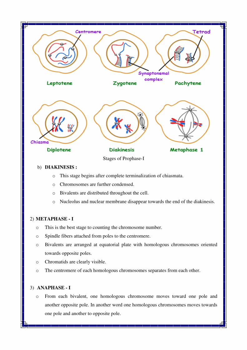
Stages of Prophase-I
b) DIAKINESIS :
o This stage begins after complete terminalization of chiasmata.
o Chromosomes are further condensed.
o Bivalents are distributed throughout the cell.
o Nucleolus and nuclear membrane disappear towards the end of the diakinesis.
2) METAPHASE - I
o This is the best stage to counting the chromosome number.
o Spindle fibers attached from poles to the centromere.
o Bivalents are arranged at equatorial plate with homologous chromosomes oriented
towards opposite poles.
o Chromatids are clearly visible.
o The centromere of each homologous chromosomes separates from each other.
3) ANAPHASE - I
o From each bivalent, one homologous chromosome moves toward one pole and
another opposite pole. In another word one homologous chromosomes moves towards
one pole and another to opposite pole.
o Sister chromatids of each chromosome remain attached at the centromere.
o Homologous chromosomes reach the opposite pole at the end of this phase.
4) TELOPHASE - I
o Chromosomes uncoiled and relax and regrouping of chromosome occurs.
o Nucleolus and nuclear membrane reappear.
o Two haploid daughter nuclei are formed.
CYTOKINESIS :
o At the end of telophase-I, cytoplasm is divided into two halves and each two halves
are staying to gether, this structure is called Dyad.
SECOND MEIOTIC DIVISION (EQUATIONAL DIVISION)
The first meiotic division (meiosis-I) results in reduction of chromosomes number
from diploid to haploid. The second nuclear division (meiosis-II) is required to reduce the
number of chromatids per chromosomes. Meiosis –II differs from mitosis in the following
three aspects :
1) The interphase prior to meiosis II is very short. It does not have S phase because each
chromosome already contains two chromatids.
2) The two sister chromatids in each chromosomes are not sister chromatid throughout.
In other words, some chromatids have alternate segments of non sister chromatids due
to recombination.
3) The meiosis-II deals with haploid chromosome number, whereas normal mitosis deals
with diploid chromosome number.
Rest of the features of meiosis
prophase-II, metaphase-II, anaphse
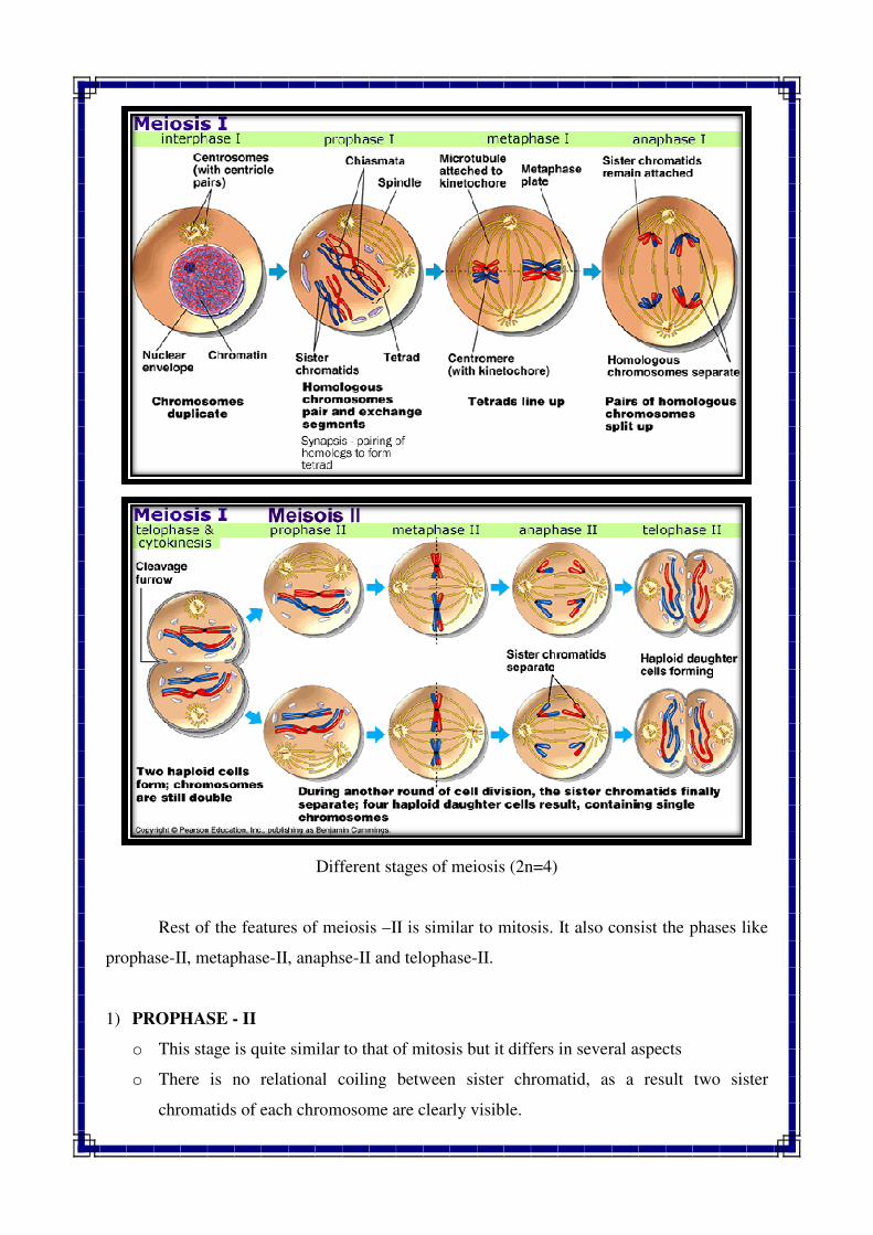
1) PROPHASE - II
o This stage is quite similar to that of mitosis but it differs in several aspects
o There is no relational coiling between sister chromatid, as a result two sister
chromatids of each chromosome are clearly visible.
Different stages of meiosis (2n=4)
Rest of the features of meiosis –II is similar to mitosis. It also consist the phases like
II, anaphse-II and telophase-II.
This stage is quite similar to that of mitosis but it differs in several aspects
o relational coiling between sister chromatid, as a result two sister
chromatids of each chromosome are clearly visible.
II is similar to mitosis. It also consist the phases like
This stage is quite similar to that of mitosis but it differs in several aspects
o relational coiling between sister chromatid, as a result two sister
o The chromosomes are much more condensed and appeared shorter and thicker.
o At the end of prophase, nucleolus and nuclear membrane are disappearing.
2) METAPHASE - II
o In this stage, chromosomes become arranged on the equatorial plate.
o Nucleolus and nuclear envelope are absent.
o Spindle apparatus is present and centromere of each chromosome is arranged at the
equatorial plate.
o Two sister chromatids of each chromosome are distinctly separated from each other.
o Chromosomes become more condensed, thicker and shorter.
o The stage is quite short in duration.
3) ANAPHASE - II
o In this stage, centromere of the each chromosome divides longitudinally.
o Two sister chromatids of each chromosome begins to separate and move away to
opposite poles.
4) TELOPHASE - II
o In this stage, uncoiling of chromosomes take place.
o Reappearance of nucleolus and reformation of nuclear envelop around each group of
chromosomes.
CYTOKINESIS :
o By the end of telophase- II, the cytoplasm of each of the two cells divides into two
parts, so total four haploid daughter cells are produced after completion of two
meiotic divisions. These four haploid daughter cells are all to gether referred as
Tetrad.
o Then this four haploid cells differentiate into gamete and this process is known as
gametogenesis
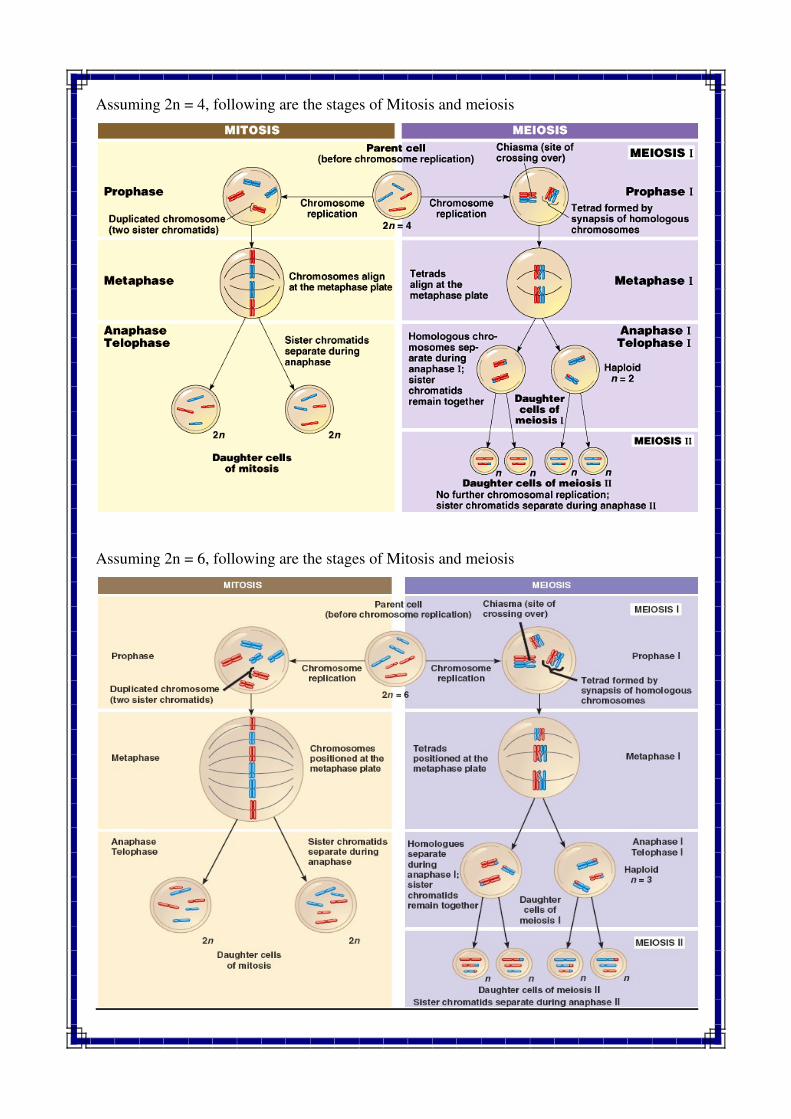
Assuming 2n = 4, following are the stages of Mitosis and meiosis
Assuming 2n = 6, following are the stages of Mitosis and meiosis
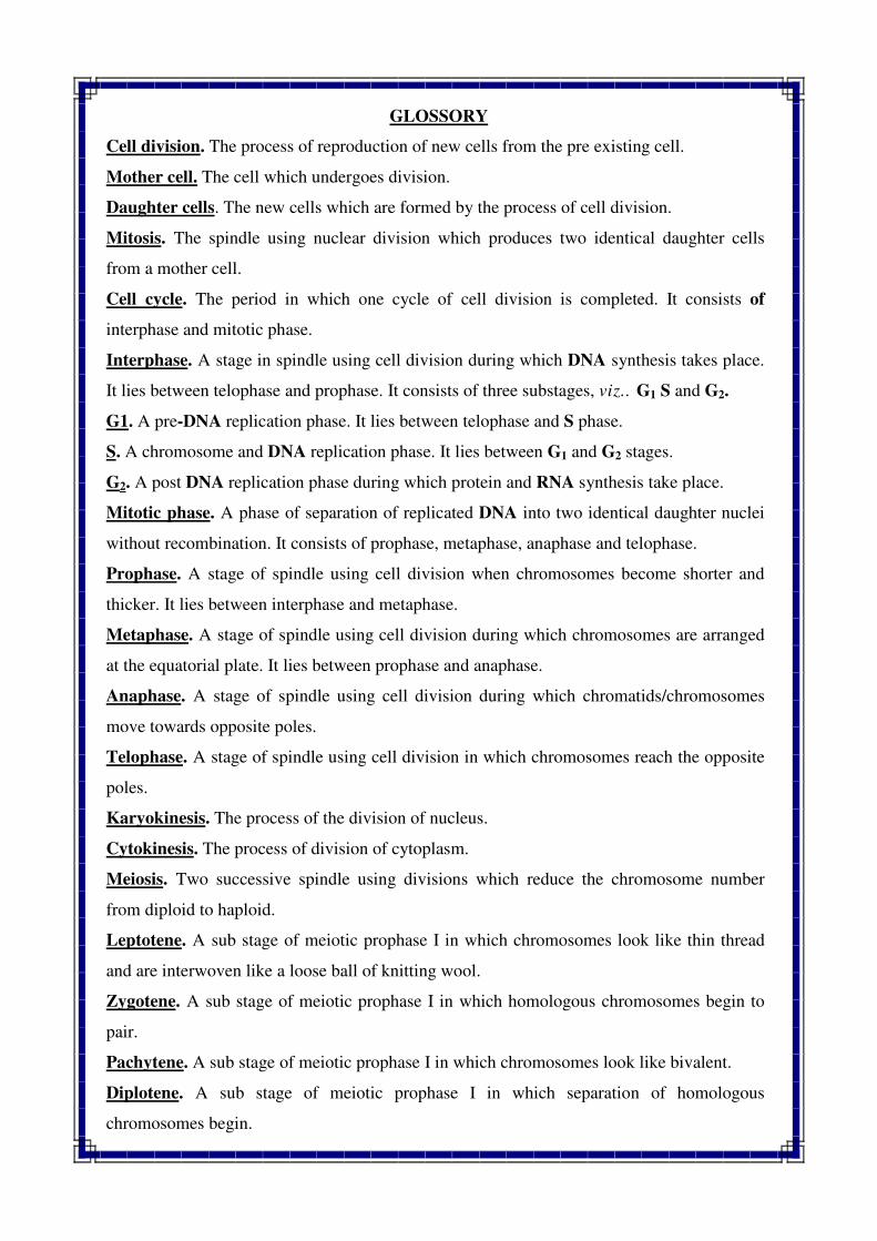
GLOSSORY
Cell division. The process of reproduction of new cells from the pre existing cell.
Mother cell. The cell which undergoes division.
Daughter cells. The new cells which are formed by the process of cell division.
Mitosis. The spindle using nuclear division which produces two identical daughter cells
from a mother cell.
Cell cycle. The period in which one cycle of cell division is completed. It consists of
interphase and mitotic phase.
Interphase. A stage in spindle using cell division during which DNA synthesis takes place.
It lies between telophase and prophase. It consists of three substages, viz.. G1 S and G2.
G1. A pre-DNA replication phase. It lies between telophase and S phase.
S. A chromosome and DNA replication phase. It lies between G1 and G2 stages.
G2. A post DNA replication phase during which protein and RNA synthesis take place.
Mitotic phase. A phase of separation of replicated DNA into two identical daughter nuclei
without recombination. It consists of prophase, metaphase, anaphase and telophase.
Prophase. A stage of spindle using cell division when chromosomes become shorter and
thicker. It lies between interphase and metaphase.
Metaphase. A stage of spindle using cell division during which chromosomes are arranged
at the equatorial plate. It lies between prophase and anaphase.
Anaphase. A stage of spindle using cell division during which chromatids/chromosomes
move towards opposite poles.
Telophase. A stage of spindle using cell division in which chromosomes reach the opposite
poles.
Karyokinesis. The process of the division of nucleus.
Cytokinesis. The process of division of cytoplasm.
Meiosis. Two successive spindle using divisions which reduce the chromosome number
from diploid to haploid.
Leptotene. A sub stage of meiotic prophase I in which chromosomes look like thin thread
and are interwoven like a loose ball of knitting wool.
Zygotene. A sub stage of meiotic prophase I in which homologous chromosomes begin to
pair.
Pachytene. A sub stage of meiotic prophase I in which chromosomes look like bivalent.
Diplotene. A sub stage of meiotic prophase I in which separation of homologous
chromosomes begin.
Diakinesis. A sub-stage of meiotic prophase. I in which bivalents are distributed throughout
the cell.
Syneptonemal complex. A protein frame work which is found between paired
chromosomes.
QUESTIONS
Q.1 Define mitosis. Describe in brief various features of mitotic cell division and give its
significance.
Q.2 What is meiosis? Discuss important features of meiosis and its significance.
Q.3 Assuming 2n = 6, draw various stages of meiosis I and meiosis II.
Q.4 Mitosis and meiosis are genetically controlled. Give evidences in support of this
statement.
Q.5 Describe in brief various similarities and dissimilarities between mitosis and meiosis.
Q.6 Write short notes on the following
1. Cell cycle 5. Karyokinesis
2. Significance of mitosis 6. Spindle fibres
3. Significance of meiosis 7. Cytokinesis
4. Synaptonemal complex
//*//*//*//
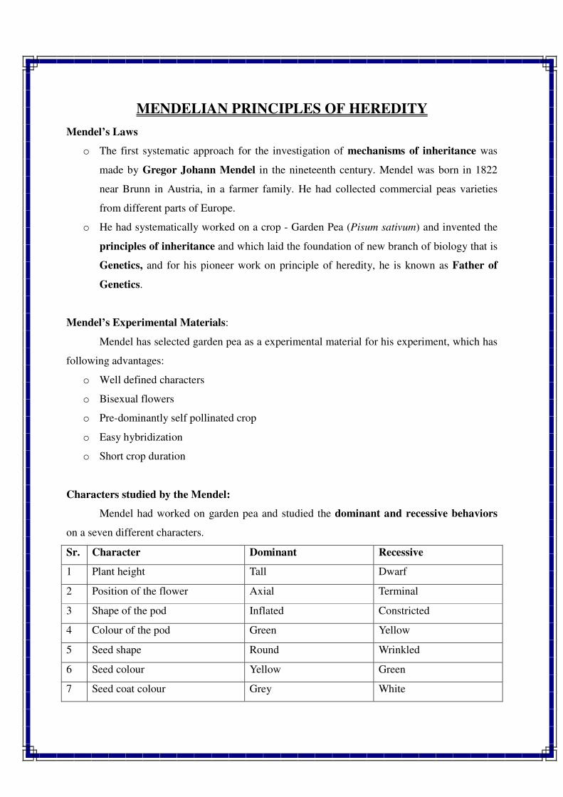
MENDELIAN PRINCIPLES OF HEREDITY
Mendel’s Laws
o The first systematic approach for the investigation of mechanisms of inheritance was
made by Gregor Johann Mendel in the nineteenth century. Mendel was born in 1822
near Brunn in Austria, in a farmer family. He had collected commercial peas varieties
from different parts of Europe.
o He had systematically worked on a crop - Garden Pea (Pisum sativum) and invented the
principles of inheritance and which laid the foundation of new branch of biology that is
Genetics, and for his pioneer work on principle of heredity, he is known as Father of
Genetics.
Mendel’s Experimental Materials:
Mendel has selected garden pea as a experimental material for his experiment, which has
following advantages:
o Well defined characters
o Bisexual flowers
o Pre-dominantly self pollinated crop
o Easy hybridization
o Short crop duration
Characters studied by the Mendel:
Mendel had worked on garden pea and studied the dominant and recessive behaviors
on a seven different characters.
Sr. Character Dominant Recessive
1 Plant height Tall Dwarf
2 Position of the flower Axial Terminal
3 Shape of the pod Inflated Constricted
4 Colour of the pod Green Yellow
5 Seed shape Round Wrinkled
6 Seed colour Yellow Green
7 Seed coat colour Grey White
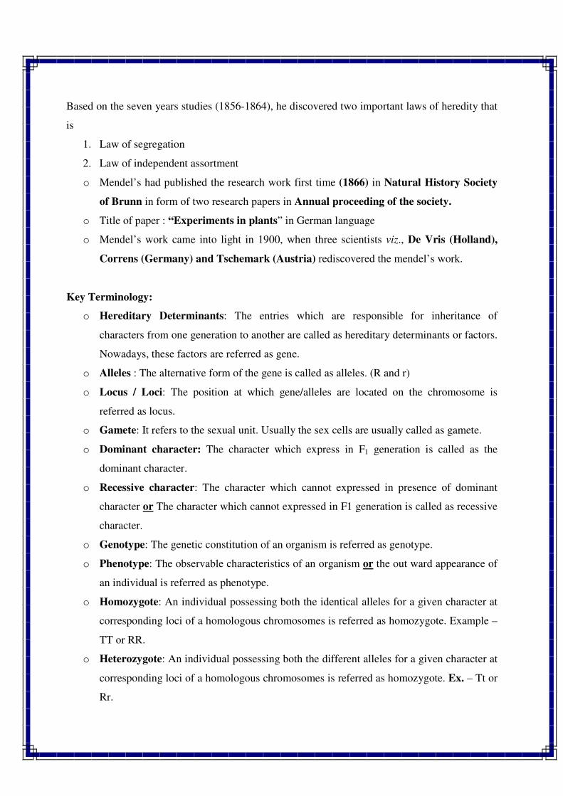
Based on the seven years studies (1856-1864), he discovered two important laws of heredity that
is
1. Law of segregation
2. Law of independent assortment
o Mendel’s had published the research work first time (1866) in Natural History Society
of Brunn in form of two research papers in Annual proceeding of the society.
o Title of paper : “Experiments in plants” in German language
o Mendel’s work came into light in 1900, when three scientists viz., De Vris (Holland),
Correns (Germany) and Tschemark (Austria) rediscovered the mendel’s work.
Key Terminology:
o Hereditary Determinants: The entries which are responsible for inheritance of
characters from one generation to another are called as hereditary determinants or factors.
Nowadays, these factors are referred as gene.
o Alleles : The alternative form of the gene is called as alleles. (R and r)
o Locus / Loci: The position at which gene/alleles are located on the chromosome is
referred as locus.
o Gamete: It refers to the sexual unit. Usually the sex cells are usually called as gamete.
o Dominant character: The character which express in F1 generation is called as the
dominant character.
o Recessive character: The character which cannot expressed in presence of dominant
character or The character which cannot expressed in F1 generation is called as recessive
character.
o Genotype: The genetic constitution of an organism is referred as genotype.
o Phenotype: The observable characteristics of an organism or the out ward appearance of
an individual is referred as phenotype.
o Homozygote: An individual possessing both the identical alleles for a given character at
corresponding loci of a homologous chromosomes is referred as homozygote. Example –
TT or RR.
o Heterozygote: An individual possessing both the different alleles for a given character at
corresponding loci of a homologous chromosomes is referred as homozygote. Ex. – Tt or
Rr.
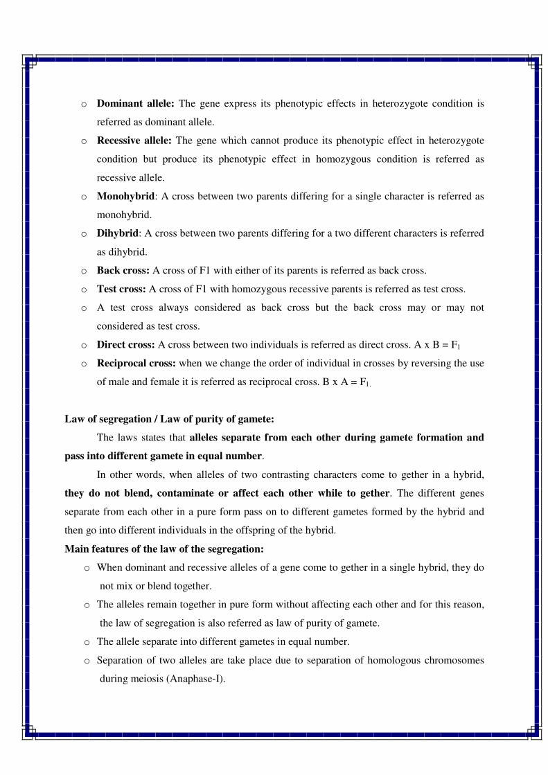
o Dominant allele: The gene express its phenotypic effects in heterozygote condition is
referred as dominant allele.
o Recessive allele: The gene which cannot produce its phenotypic effect in heterozygote
condition but produce its phenotypic effect in homozygous condition is referred as
recessive allele.
o Monohybrid: A cross between two parents differing for a single character is referred as
monohybrid.
o Dihybrid: A cross between two parents differing for a two different characters is referred
as dihybrid.
o Back cross: A cross of F1 with either of its parents is referred as back cross.
o Test cross: A cross of F1 with homozygous recessive parents is referred as test cross.
o A test cross always considered as back cross but the back cross may or may not
considered as test cross.
o Direct cross: A cross between two individuals is referred as direct cross. A x B = F1
o Reciprocal cross: when we change the order of individual in crosses by reversing the use
of male and female it is referred as reciprocal cross. B x A = F1.
Law of segregation / Law of purity of gamete:
The laws states that alleles separate from each other during gamete formation and
pass into different gamete in equal number.
In other words, when alleles of two contrasting characters come to gether in a hybrid,
they do not blend, contaminate or affect each other while to gether. The different genes
separate from each other in a pure form pass on to different gametes formed by the hybrid and
then go into different individuals in the offspring of the hybrid.
Main features of the law of the segregation:
o When dominant and recessive alleles of a gene come to gether in a single hybrid, they do
not mix or blend together.
o The alleles remain together in pure form without affecting each other and for this reason,
the law of segregation is also referred as law of purity of gamete.
o The allele separate into different gametes in equal number.
o Separation of two alleles are take place due to separation of homologous chromosomes
during meiosis (Anaphase-I).
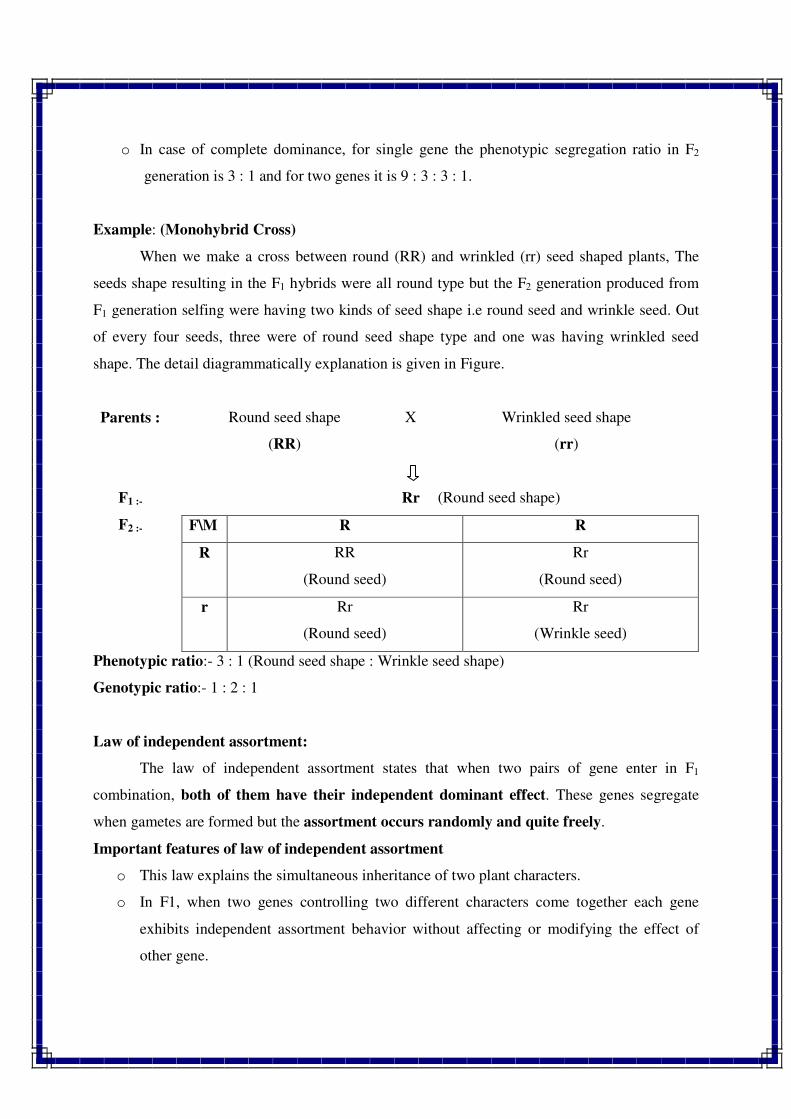
o In case of complete dominance, for single gene the phenotypic segregation ratio in F2
generation is 3 : 1 and for two genes it is 9 : 3 : 3 : 1.
Example: (Monohybrid Cross)
When we make a cross between round (RR) and wrinkled (rr) seed shaped plants, The
seeds shape resulting in the F1 hybrids were all round type but the F2 generation produced from
F1 generation selfing were having two kinds of seed shape i.e round seed and wrinkle seed. Out
of every four seeds, three were of round seed shape type and one was having wrinkled seed
shape. The detail diagrammatically explanation is given in Figure.
Parents : Round seed shape
(RR)
X Wrinkled seed shape
(rr)
F1 :- Rr (Round seed shape)
F2 :- F\M R R
R RR
(Round seed)
Rr
(Round seed)
r Rr
(Round seed)
Rr
(Wrinkle seed)
Phenotypic ratio:- 3 : 1 (Round seed shape : Wrinkle seed shape)
Genotypic ratio:- 1 : 2 : 1
Law of independent assortment:
The law of independent assortment states that when two pairs of gene enter in F1
combination, both of them have their independent dominant effect. These genes segregate
when gametes are formed but the assortment occurs randomly and quite freely.
Important features of law of independent assortment
o This law explains the simultaneous inheritance of two plant characters.
o In F1, when two genes controlling two different characters come together each gene
exhibits independent assortment behavior without affecting or modifying the effect of
other gene.
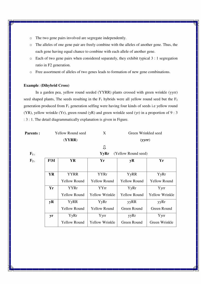
o The two gene pairs involved are segregate independently.
o The alleles of one gene pair are freely combine with the alleles of another gene. Thus, the
each gene having equal chance to combine with each allele of another gene.
o Each of two gene pairs when considered separately, they exhibit typical 3 : 1 segregation
ratio in F2 generation.
o Free assortment of alleles of two genes leads to formation of new gene combinations.
Example: (Dihybrid Cross)
In a garden pea, yellow round seeded (YYRR) plants crossed with green wrinkle (yyrr)
seed shaped plants, The seeds resulting in the F1 hybrids were all yellow round seed but the F2
generation produced from F1 generation selfing were having four kinds of seeds i.e yellow round
(YR), yellow wrinkle (Yr), green round (yR) and green wrinkle seed (yr) in a proportion of 9 : 3
: 3 : 1. The detail diagrammatically explanation is given in Figure.
Parents : Yellow Round seed
(YYRR)
X Green Wrinkled seed
(yyrr)
F1 : YyRr (Yellow Round seed)
F2 : F\M YR Yr yR Yr
YR YYRR
Yellow Round
YYRr
Yellow Round
YyRR
Yellow Round
YyRr
Yellow Round
Yr YYRr
Yellow Round
YYrr
Yellow Wrinkle
YyRr
Yellow Round
Yyrr
Yellow Wrinkle
yR YyRR
Yellow Round
YyRr
Yellow Round
yyRR
Green Round
yyRr
Green Round
yr YyRr
Yellow Round
Yyrr
Yellow Wrinkle
yyRr
Green Round
Yyrr
Green Wrinkle
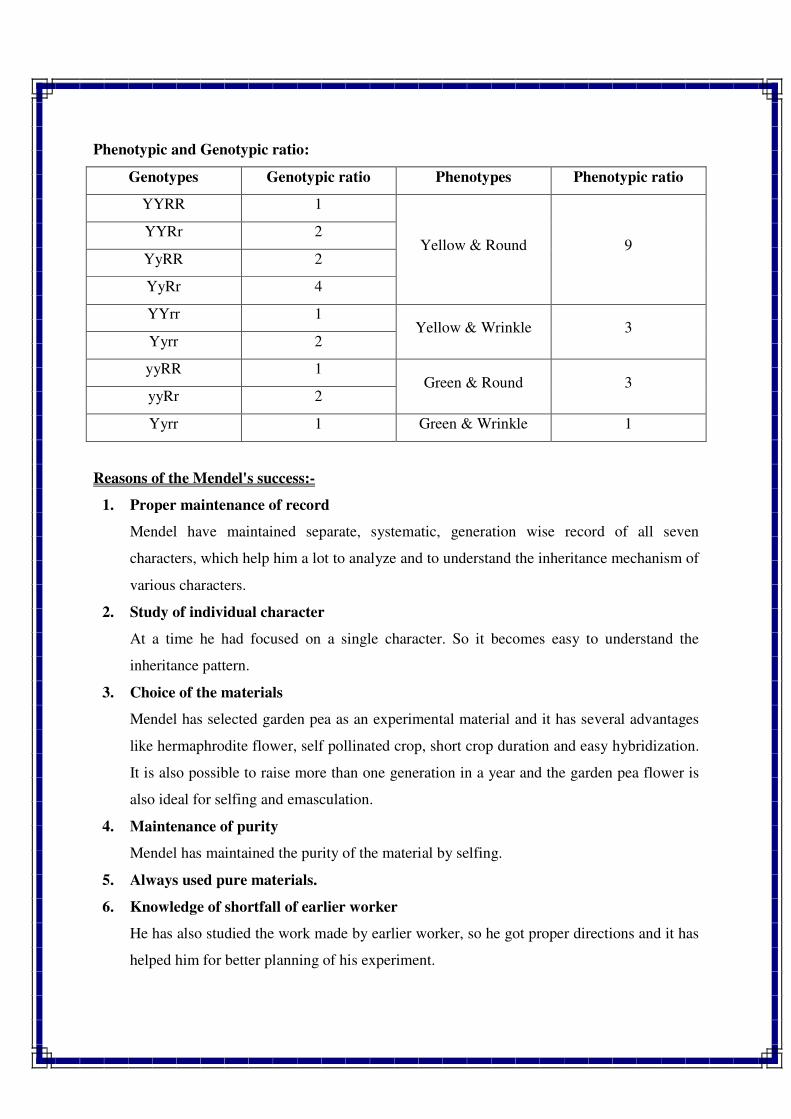
Phenotypic and Genotypic ratio:
Genotypes Genotypic ratio Phenotypes Phenotypic ratio
YYRR 1
Yellow & Round 9 YYRr 2
YyRR 2
YyRr 4
YYrr 1 Yellow & Wrinkle 3
Yyrr 2
yyRR 1 Green & Round 3
yyRr 2
Yyrr 1 Green & Wrinkle 1
Reasons of the Mendel's success:-
1. Proper maintenance of record
Mendel have maintained separate, systematic, generation wise record of all seven
characters, which help him a lot to analyze and to understand the inheritance mechanism of
various characters.
2. Study of individual character
At a time he had focused on a single character. So it becomes easy to understand the
inheritance pattern.
3. Choice of the materials
Mendel has selected garden pea as an experimental material and it has several advantages
like hermaphrodite flower, self pollinated crop, short crop duration and easy hybridization.
It is also possible to raise more than one generation in a year and the garden pea flower is
also ideal for selfing and emasculation.
4. Maintenance of purity
Mendel has maintained the purity of the material by selfing.
5. Always used pure materials.
6. Knowledge of shortfall of earlier worker
He has also studied the work made by earlier worker, so he got proper directions and it has
helped him for better planning of his experiment.
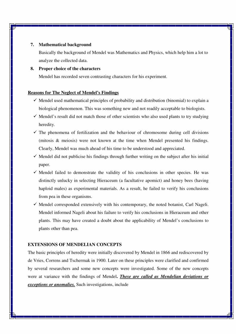
7. Mathematical background
Basically the background of Mendel was Mathematics and Physics, which help him a lot to
analyze the collected data.
8. Proper choice of the characters
Mendel has recorded seven contrasting characters for his experiment.
Reasons for The Neglect of Mendel’s Findings
� Mendel used mathematical principles of probability and distribution (binomial) to explain a
biological phenomenon. This was something new and not readily acceptable to biologists.
� Mendel’s result did not match those of other scientists who also used plants to try studying
heredity.
� The phenomena of fertilization and the behaviour of chromosome during cell divisions
(mitosis & meiosis) were not known at the time when Mendel presented his findings.
Clearly, Mendel was much ahead of his time to be understood and appreciated.
� Mendel did not publicise his findings through further writing on the subject after his initial
paper.
� Mendel failed to demonstrate the validity of his conclusions in other species. He was
distinctly unlucky in selecting Hieraceum (a facultative apomict) and honey bees (having
haploid males) as experimental materials. As a result, he failed to verify his conclusions
from pea in these organisms.
� Mendel corresponded extensively with his contemporary, the noted botanist, Carl Nageli.
Mendel informed Nageli about his failure to verify his conclusions in Hieraceum and other
plants. This may have created a doubt about the applicability of Mendel’s conclusions to
plants other than pea.
EXTENSIONS OF MENDELIAN CONCEPTS
The basic principles of heredity were initially discovered by Mendel in 1866 and rediscovered by
de Vries, Correns and Tschermak in 1900. Later on these principles were clarified and confirmed
by several researchers and some new concepts were investigated. Some of the new concepts
were at variance with the findings of Mendel. These are called as Mendelian deviations or
exceptions or anomalies. Such investigations, include
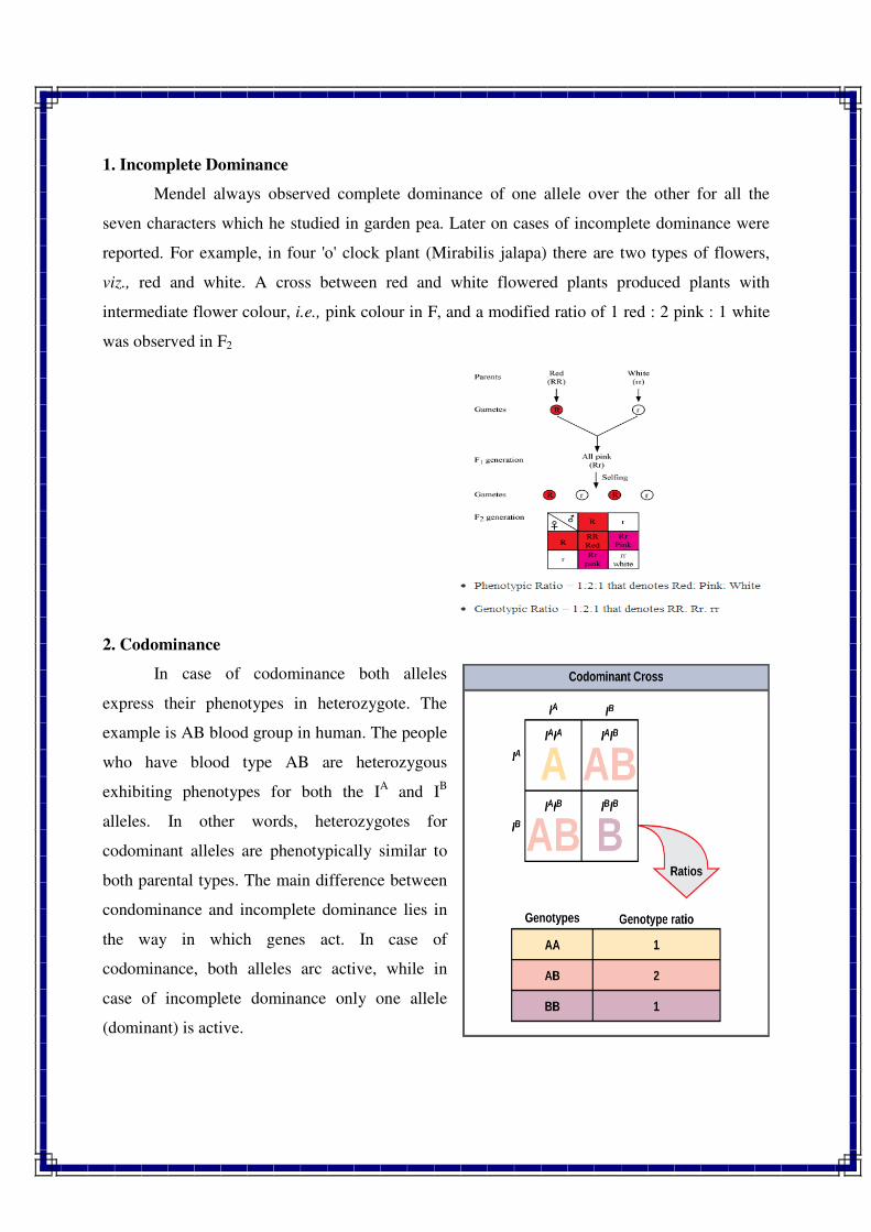
1. Incomplete Dominance
Mendel always observed complete dominance of one allele over the other for all the
seven characters which he studied in garden pea. Later on cases of incomplete dominance were
reported. For example, in four 'o' clock plant (Mirabilis jalapa) there are two types of flowers,
viz., red and white. A cross between red and white flowered plants produced plants with
intermediate flower colour, i.e., pink colour in F, and a modified ratio of 1 red : 2 pink : 1 white
was observed in F2
2. Codominance
In case of codominance both alleles
express their phenotypes in heterozygote. The
example is AB blood group in human. The people
who have blood type AB are heterozygous
exhibiting phenotypes for both the IA and I
B
alleles. In other words, heterozygotes for
codominant alleles are phenotypically similar to
both parental types. The main difference between
condominance and incomplete dominance lies in
the way in which genes act. In case of
codominance, both alleles arc active, while in
case of incomplete dominance only one allele
(dominant) is active.
3. Multiple Alleles
Mendel always observed two allelic forms of a gene. Now cases are known where a gene
has more than two allelic forms, although only two can exist in a diploid cell at a time. Existence
of more than two alleles for a gene is called multiple alleles. Examples of multiple alleles are
ABO blood group alleles in human, coat color in rabbit and self incompatibility alleles in
tobacco.
4. Linkage
Mendel always observed independent assortment of genes. Later on cases of Linkage were
reported by Bateson and Punnett in 1906 in pea, Hutchinson in maize and Morgan (1910) in
Drosophila. In a dihybrid test cross, they observed higher frequencies of parental types than
recombinants instead of 1 : 1 : 1 : 1 ratio. This led to modification of the concept of independent
assortment.
5. Lethal Genes
Gene which causes the death of its carrier when in homozygous condition is called lethal
gene. Mendel's findings were based on equal survival of all genotypes. In the presence of lethal
genes, the normal segregation ratio of 3 : 1 is modified into 2 : 1 ratio. Lethal genes have been
reported in both animals as well as plants. In mice, allele for yellow coat color is dominant over
grey. When a cross is made between yellow and grey, a ratio of 1:1 for yellow and grey mice
was observed. This indicated that yellow mice are always heterozygous, because yellow
homozygotes are never born because of homozygous lethality. Such genes were not observed by
Mendel. He always got 3 : 1 ratio in F2 for single gene characters.
6. Gene Interactions
When the expression of an allele of one gene pair depends on the presence of a specific allele
of another pair, it is known as gene interaction. Mendel observed 9 : 3 : 3 : 1 ratio in F2 from a
dihybrid cross. Later on many deviations of this phenotypic ratio were observed in dihybrid
crosses. The modified ratios included 9 : 7, 9 : 3 : 4, 12 : 3 : 1, 13 : 3 : 15 : 1 and 9 : 6 : 1 in
different crop plants.
7. Pleiotropic Gene Effects
Mendel observed that one gene controls the expression of only one character. Later on cases
were observed in which one gene was found to govern the expression of two or more characters.
Example is white eye allele in Drosophila. This allele affects eye colour, shape of spermathica,
fecundity and testicular membrane.
8. Polygenes
Mendel always observed that each character is governed by a single gene. Later on Nilsson
Ehle observed that some characters are controlled by several genes and each of such gene has
additive effect in the expression of character. This concept led to the foundation of polygenic
inheritance. East (1916) demonstrated that polygenic characters were perfectly in agreement with
Mendelian segregation and later on Fisher and Wright provided a mathematical basis for the
genetic inteipretation of polygenic characters.
9. Environmental Effects
Genes can interact not only with other genes but also with the environment to produce the -
final phenotype. Thus phenotype is the result of the interaction between genotype and
environment. It leads directly to the concept of penetrance and expressivity. The importance of
environment was first realised by Johannsen. He coined the terms genotype and phenotype.
10. Maternal Effects
Mendel did not observe any difference between direct and reciprocal crosses. Later
investigations revealed the presence of significant difference in the reciprocal crosses, which led
to the concept of cytoplasmic inheritance.
//*//*//*//
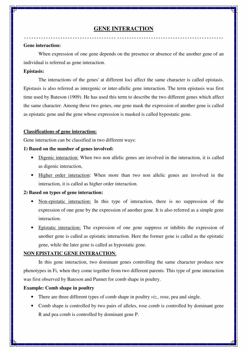
GENE INTERACTION
- - - - - - - - - - - - - - - - - - - - - - - - - - - - - - - - - - - - - - - - - - - - - - - - - - - - - - - - - - - - - - - - - - - - - - - - - - - - - - - - - -
Gene interaction:
When expression of one gene depends on the presence or absence of the another gene of an
individual is referred as gene interaction.
Epistasis:
The interactions of the genes' at different loci affect the same character is called epistasis.
Epistasis is also referred as intergenic or inter-allelic gene interaction. The term epistasis was first
time used by Bateson (1909). He has used this term to describe the two different genes which affect
the same character. Among these two genes, one gene mask the expression of another gene is called
as epistatic gene and the gene whose expression is masked is called hypostatic gene.
Classifications of gene interaction:
Gene interaction can be classified in two different ways:
1) Based on the number of genes involved:
• Digenic interaction: When two non allelic genes are involved in the interaction, it is called
as digenic interaction,
• Higher order interaction: When more than two non allelic genes are involved in the
interaction, it is called as higher order interaction.
2) Based on types of gene interaction:
• Non-epistatic interaction: In this type of interaction, there is no suppression of the
expression of one gene by the expression of another gene. It is also referred as a simple gene
interaction.
• Epistatic interaction: The expression of one gene suppress or inhibits the expression of
another gene is called as epistatic interaction. Here the former gene is called as the epistatic
gene, while the later gene is called as hypostatic gene.
NON EPISTATIC GENE INTERACTION:
In this gene interaction, two dominant genes controlling the same character produce new
phenotypes in Fi, when they come together from two different parents. This type of gene interaction
was first observed by Bateson and Punnet for comb shape in poultry.
Example: Comb shape in poultry
• There are three different types of comb shape in poultry viz., rose, pea and single.
• Comb shape is controlled by two pairs of alleles, rose comb is controlled by dominant gene
R and pea comb is controlled by dominant gene P.
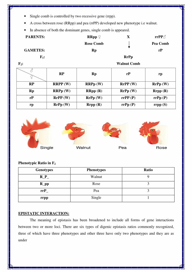
• Single comb is controlled by two recessive gene (rrpp).
• A cross between rose (RRpp) and pea (rrPP) developed new phenotype i.e walnut.
• In absence of both the dominant genes, single comb is appeared.
PARENTS: RRpp ♀ X rrPP♂
Rose Comb Pea Comb
GAMETES: Rp rP
F1:
F2:
RrPp
Walnut Comb
♂
♀ RP Rp rP rp
RP RRPP (W) RRPp (W) RrPP (W) RrPp (W)
Rp RRPp (W) RRpp (R) RrPp (W) Rrpp (R)
rP RrPP (W) RrPp (W) rrPP (P) rrPp (P)
rp RrPp (W) Rrpp (R) rrPp (P) rrpp (S)
Phenotypic Ratio in F2
Genotypes Phenotypes Ratio
R_P_ Walnut 9
R_pp Rose 3
rrP_ Pea 3
rrpp Single 1
EPISTATIC INTERACTION:
The meaning of epistasis has been broadened to include all forms of gene interactions
between two or more loci. There are six types of digenic epistasis ratios commonly recognized,
three of which have three phenotypes and other three have only two phenotypes and they are as
under
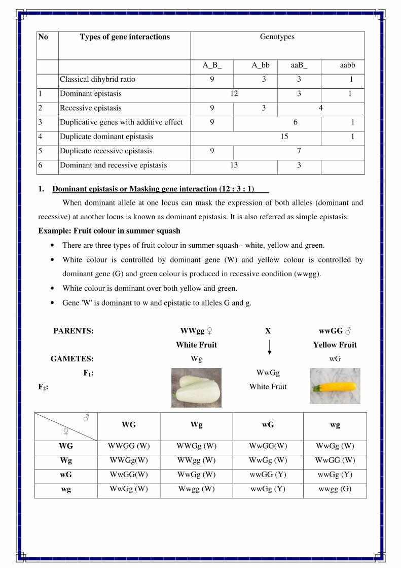
1. Dominant epistasis or Masking gene interaction (12 : 3 : 1)
When dominant allele at one locus can mask the expression of both alleles (dominant and
recessive) at another locus is known as dominant epistasis. It is also referred as simple epistasis.
Example: Fruit colour in summer squash
• There are three types of fruit colour in summer squash - white, yellow and green.
• White colour is controlled by dominant gene (W) and yellow colour is controlled by
dominant gene (G) and green colour is produced in recessive condition (wwgg).
• White colour is dominant over both yellow and green.
• Gene 'W' is dominant to w and epistatic to alleles G and g.
PARENTS: WWgg ♀ X wwGG ♂
White Fruit Yellow Fruit
GAMETES: Wg wG
F1:
F2:
WwGg
White Fruit
♂
♀ WG Wg wG wg
WG WWGG (W) WWGg (W) WwGG(W) WwGg (W)
Wg WWGg(W) WWgg (W) WwGg (W) WwGG (W)
wG WwGG(W) WwGg (W) wwGG (Y) wwGg (Y)
wg WwGg (W) Wwgg (W) wwGg (Y) wwgg (G)
No Types of gene interactions
Genotypes
A_B_ A_bb aaB_ aabb
Classical dihybrid ratio 9 3 3 1
1 Dominant epistasis 12 3 1
2 Recessive epistasis 9 3 4
3 Duplicative genes with additive effect 9 6 1
4 Duplicate dominant epistasis 15 1
5 Duplicate recessive epistasis 9 7
6 Dominant and recessive epistasis 13 3
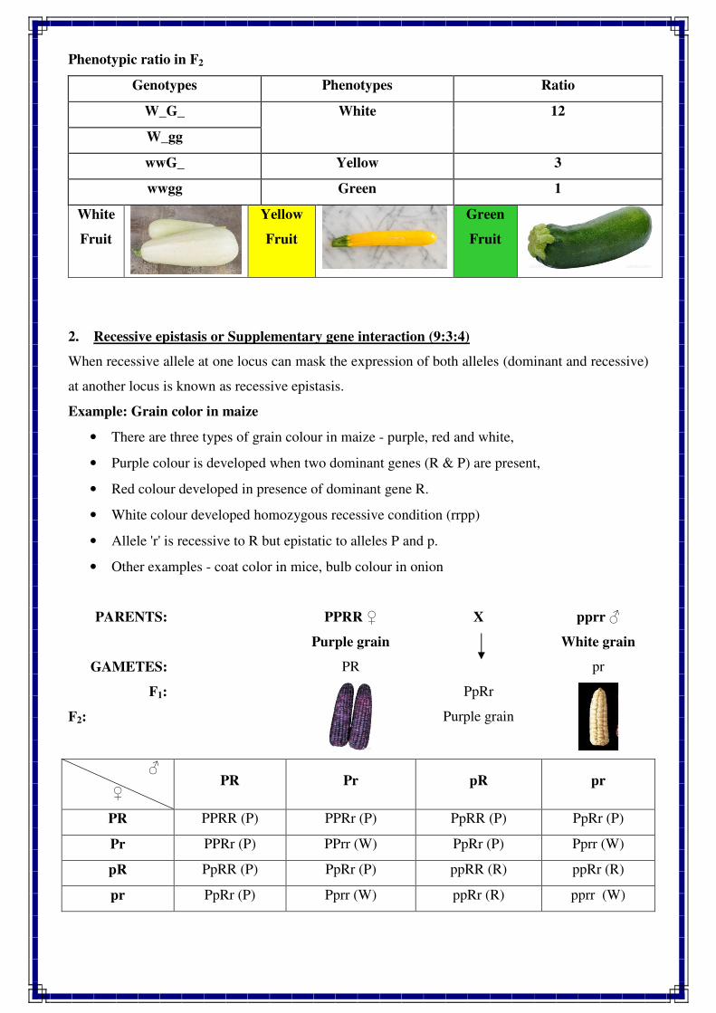
Phenotypic ratio in F2
Genotypes Phenotypes Ratio
W_G_ White 12
W_gg
wwG_ Yellow 3
wwgg Green 1
White
Fruit
Yellow
Fruit
Green
Fruit
2. Recessive epistasis or Supplementary gene interaction (9:3:4)
When recessive allele at one locus can mask the expression of both alleles (dominant and recessive)
at another locus is known as recessive epistasis.
Example: Grain color in maize
• There are three types of grain colour in maize - purple, red and white,
• Purple colour is developed when two dominant genes (R & P) are present,
• Red colour developed in presence of dominant gene R.
• White colour developed homozygous recessive condition (rrpp)
• Allele 'r' is recessive to R but epistatic to alleles P and p.
• Other examples - coat color in mice, bulb colour in onion
PARENTS: PPRR ♀ X pprr ♂
Purple grain White grain
GAMETES: PR pr
F1:
F2:
PpRr
Purple grain
♂
♀ PR Pr pR pr
PR PPRR (P) PPRr (P) PpRR (P) PpRr (P)
Pr PPRr (P) PPrr (W) PpRr (P) Pprr (W)
pR PpRR (P) PpRr (P) ppRR (R) ppRr (R)
pr PpRr (P) Pprr (W) ppRr (R) pprr (W)
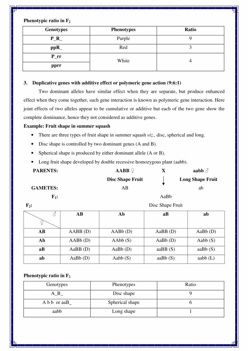
Phenotypic ratio in F2
Genotypes Phenotypes Ratio
P_R_ Purple 9
ppR_ Red 3
P_rr White 4
pprr
3. Duplicative genes with additive effect or polymeric gene action (9:6:1)
Two dominant alleles have similar effect when they are separate, but produce enhanced
effect when they come together, such gene interaction is known as polymeric gene interaction. Here
joint effects of two alleles appear to be cumulative or additive but each of the two gene show the
complete dominance, hence they not considered as additive genes.
Example: Fruit shape in summer squash
• There are three types of fruit shape in summer squash viz., disc, spherical and long.
• Disc shape is controlled by two dominant genes (A and B).
• Spherical shape is produced by either dominant allele (A or B).
• Long fruit shape developed by double recessive homozygous plant (aabb).
PARENTS: AABB ♀ X aabb ♂
Disc Shape Fruit Long Shape Fruit
GAMETES: AB ab
F1:
F2:
AaBb
Disc Shape Fruit
♂
♀
AB Ab aB ab
AB AABB (D) AABb (D) AaBB (D) AaBb (D)
Ab AABb (D) AAbb (S) AaBb (D) Aabb (S)
aB AaBB (D) AaBb (D) aaBB (S) aaBb (S)
ab AaBb (D) Aabb (S) aaBb (S) aabb (L)
Phenotypic ratio in F2
Genotypes Phenotypes Ratio
A_B_ Disc shape 9
A b b or aaB_ Spherical shape 6
aabb Long shape 1
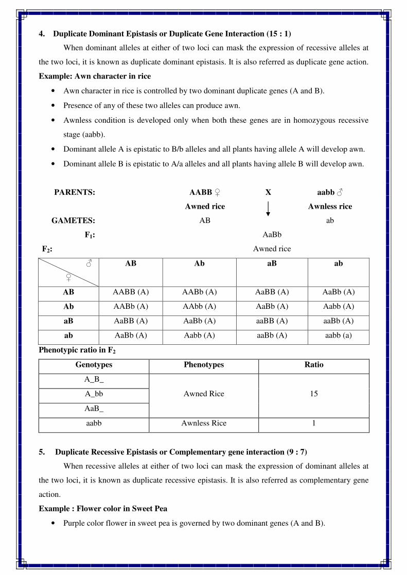
4. Duplicate Dominant Epistasis or Duplicate Gene Interaction (15 : 1)
When dominant alleles at either of two loci can mask the expression of recessive alleles at
the two loci, it is known as duplicate dominant epistasis. It is also referred as duplicate gene action.
Example: Awn character in rice
• Awn character in rice is controlled by two dominant duplicate genes (A and B).
• Presence of any of these two alleles can produce awn.
• Awnless condition is developed only when both these genes are in homozygous recessive
stage (aabb).
• Dominant allele A is epistatic to B/b alleles and all plants having allele A will develop awn.
• Dominant allele B is epistatic to A/a alleles and all plants having allele B will develop awn.
PARENTS: AABB ♀ X aabb ♂
Awned rice Awnless rice
GAMETES: AB ab
F1:
F2:
AaBb
Awned rice
♂
♀
AB Ab aB ab
AB AABB (A) AABb (A) AaBB (A) AaBb (A)
Ab AABb (A) AAbb (A) AaBb (A) Aabb (A)
aB AaBB (A) AaBb (A) aaBB (A) aaBb (A)
ab AaBb (A) Aabb (A) aaBb (A) aabb (a)
Phenotypic ratio in F2
Genotypes Phenotypes Ratio
A_B_
Awned Rice 15 A_bb
AaB_
aabb Awnless Rice 1
5. Duplicate Recessive Epistasis or Complementary gene interaction (9 : 7)
When recessive alleles at either of two loci can mask the expression of dominant alleles at
the two loci, it is known as duplicate recessive epistasis. It is also referred as complementary gene
action.
Example : Flower color in Sweet Pea
• Purple color flower in sweet pea is governed by two dominant genes (A and B).
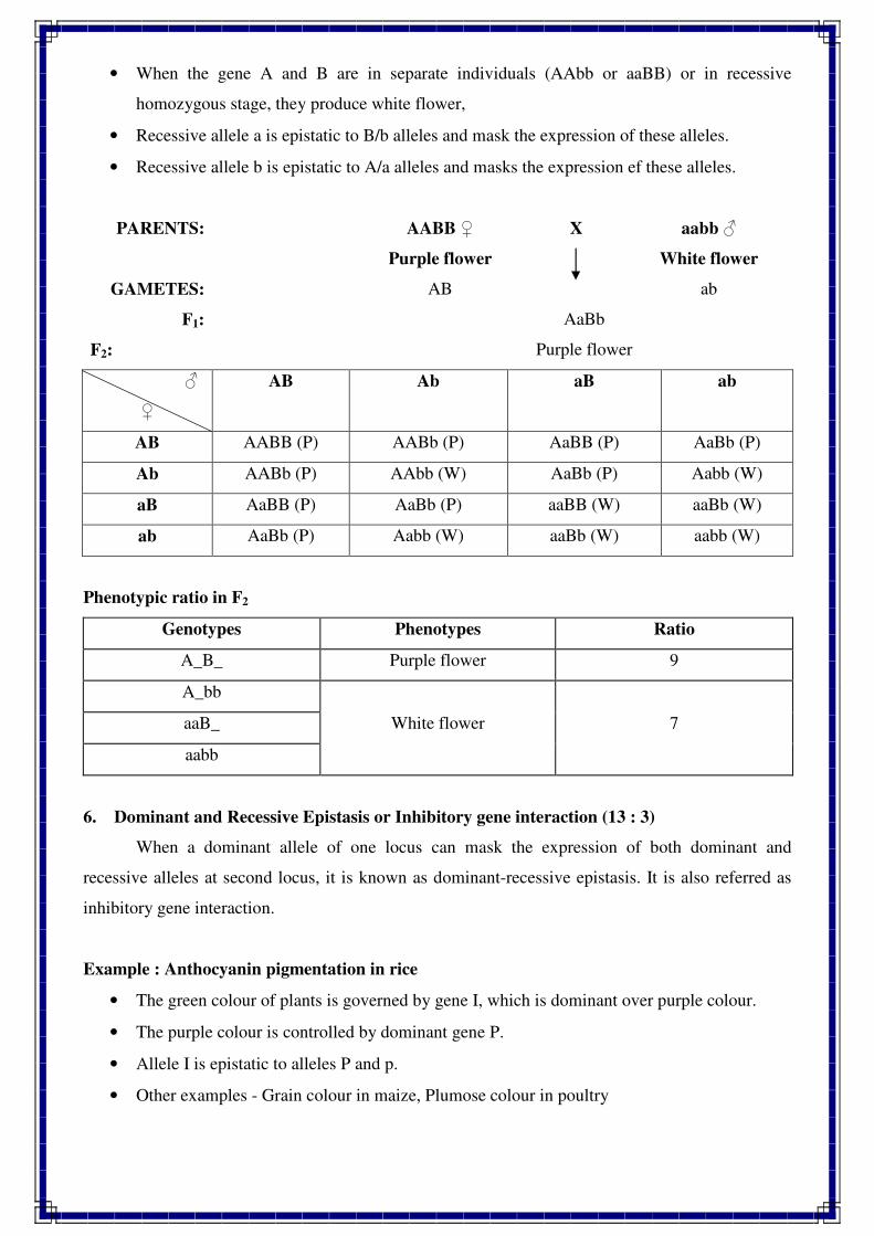
• When the gene A and B are in separate individuals (AAbb or aaBB) or in recessive
homozygous stage, they produce white flower,
• Recessive allele a is epistatic to B/b alleles and mask the expression of these alleles.
• Recessive allele b is epistatic to A/a alleles and masks the expression ef these alleles.
PARENTS: AABB ♀ X aabb ♂
Purple flower White flower
GAMETES: AB ab
F1:
F2:
AaBb
Purple flower
♂
♀
AB Ab aB ab
AB AABB (P) AABb (P) AaBB (P) AaBb (P)
Ab AABb (P) AAbb (W) AaBb (P) Aabb (W)
aB AaBB (P) AaBb (P) aaBB (W) aaBb (W)
ab AaBb (P) Aabb (W) aaBb (W) aabb (W)
Phenotypic ratio in F2
Genotypes Phenotypes Ratio
A_B_ Purple flower 9
A_bb
White flower 7 aaB_
aabb
6. Dominant and Recessive Epistasis or Inhibitory gene interaction (13 : 3)
When a dominant allele of one locus can mask the expression of both dominant and
recessive alleles at second locus, it is known as dominant-recessive epistasis. It is also referred as
inhibitory gene interaction.
Example : Anthocyanin pigmentation in rice
• The green colour of plants is governed by gene I, which is dominant over purple colour.
• The purple colour is controlled by dominant gene P.
• Allele I is epistatic to alleles P and p.
• Other examples - Grain colour in maize, Plumose colour in poultry
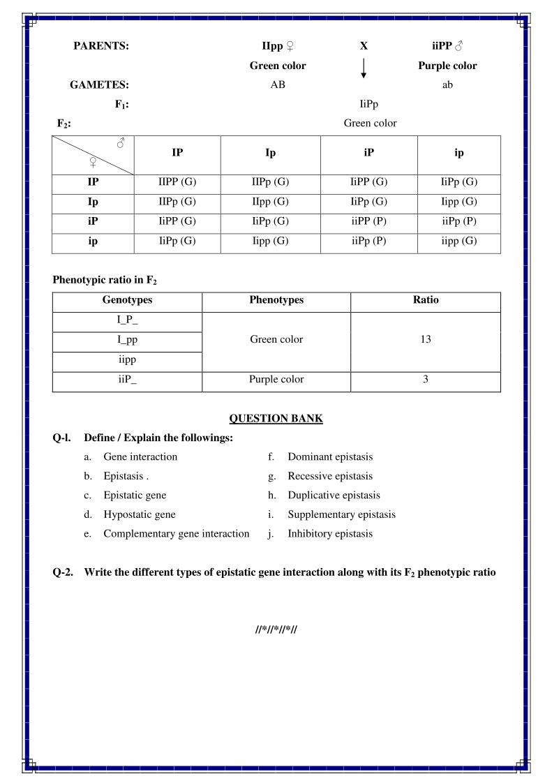
PARENTS: IIpp ♀ X iiPP ♂
Green color Purple color
GAMETES: AB ab
F1:
F2:
IiPp
Green color
♂
♀ IP Ip iP ip
IP IIPP (G) IIPp (G) IiPP (G) IiPp (G)
Ip IIPp (G) IIpp (G) IiPp (G) Iipp (G)
iP IiPP (G) IiPp (G) iiPP (P) iiPp (P)
ip IiPp (G) Iipp (G) iiPp (P) iipp (G)
Phenotypic ratio in F2
Genotypes Phenotypes Ratio
I_P_
Green color 13 I_pp
iipp
iiP_ Purple color 3
QUESTION BANK
Q-l. Define / Explain the followings:
a. Gene interaction f. Dominant epistasis
b. Epistasis . g. Recessive epistasis
c. Epistatic gene h. Duplicative epistasis
d. Hypostatic gene i. Supplementary epistasis
e. Complementary gene interaction j. Inhibitory epistasis
Q-2. Write the different types of epistatic gene interaction along with its F2 phenotypic ratio
//*//*//*//
1
STUDY OF CHROMOSOME STRUCTURE
CHROMOSOME
The darkly stained rod shaped bodies observed under light microscope in a cell during
metaphase stage of mitosis is referred as chromosome.
Strasburger discovered the chromosomes (thread like structure) in 1875.
The term chromosome was coined by Waldeyer in 1888
Features of Eukaryotic chromosomes:
� Chromosomes are not visible during interphase. They are visible during other stages of
cell division but more clearly visible during mitotic metaphase
� The genes are located on the chromosome in linear fashion, which are responsible for
transmission of characters from generation to generation
� In eukaryotes the chromosomes are enclosed by nuclear membrane
� They are vary in shape, size and number in different species of plants and animals
� Chromosomes having property of self replication, segregation and mutation
� Chromosomes are composed of DNA, RNA and histone proteins. DNA is the major
genetic material of chromosome.
CHROMOSOME SHAPE
Chromosome shape is usually observed during anaphase. The shape of chromosomes is
determined by the position of centromere, a part of chromosome on which spindle fibres are
attached during metaphase. Chromosomes have generally three different shapes, viz.. rod
shape, J shape and V shape. These shapes are observed when the centromere occupies
terminal, sub-terminal and median (middle) position on the chromosomes, respectively.
CHROMOSOME SIZE
Chromosome size is measured with the help of micrometer at mitotic metaphase. It is
measured in two ways, viz., in length and in diameter. Plants usually have longer
chromosomes than animals. Moreover, species or individuals which have fewer chromosome
number have larger chromosomes. The maximum length of chromosome is observed during
interphase and minimum during anaphase. Thus chromosome size varies from species to
species (Table 4.1). Giant chromosomes have length upto 300 p
2
TABLE 4.1. Chromosome size in some plants and animals
Name of Plant/animal Chromosome size in p.
Maize 8-12
Lilium. Allium and Tradescantia 10-12
Trillium 30-32
Human 4
Drosophila 3
CHROMOSOME NUMBER
There are three types of chromosome number, viz., haploid, diploid and basic number
as given below :
Haploid
It represents half of the somatic chromosome number of a species and is denoted by n.
Since haploid chromosome number is usually found in the gametes, it is also known as
gametic number.
Diploid
It refers to somatic chromosome number of a species and is represented by 2n.
Since diploid chromosome number is found in zygotic or somatic cells it is also referred to as
zygotic or somatic number.
Basic Number
The gametic chromosome number of a true diploid species is called basic number. It
is the minimum haploid chromosome number of any species which is denoted by x. For
example, in wheat, the basic number is 7, whereas the haploid number is 7, 14 and 21 for
diploid, tetraploid and hexaploid species, respectively.
Thus haploid chromosome number differs from basic number. Both are same in case
of true diploid species but differ in case of polyploid species. Thus, basic number can be a
haploid number but all haploid numbers cannot be basic number. Chromosome number
differs from species to species (Table 4.2). In plant kingdom, chromosome number usually is
higher in dicots than in monocots.
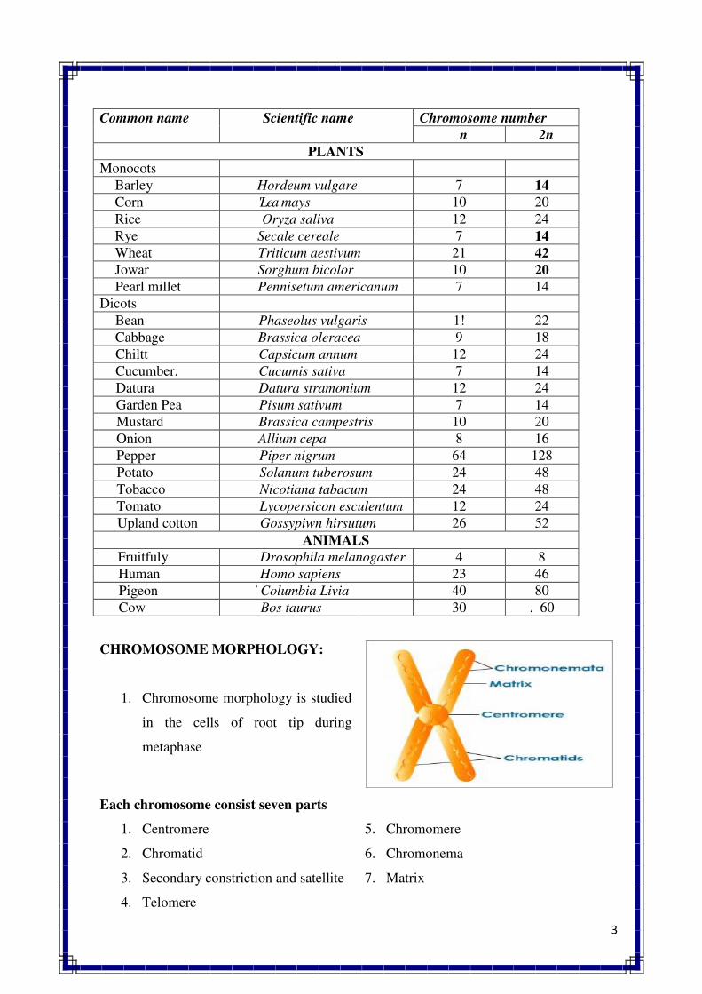
3
Common name Scientific name Chromosome number
n 2n
PLANTS
Monocots
Barley Hordeum vulgare 7 14
Corn 'Lea mays 10 20
Rice Oryza saliva 12 24
Rye Secale cereale 7 14
Wheat Triticum aestivum 21 42
Jowar Sorghum bicolor 10 20
Pearl millet Pennisetum americanum 7 14
Dicots
Bean Phaseolus vulgaris 1! 22
Cabbage Brassica oleracea 9 18
Chiltt Capsicum annum 12 24
Cucumber. Cucumis sativa 7 14
Datura Datura stramonium 12 24
Garden Pea Pisum sativum 7 14
Mustard Brassica campestris 10 20
Onion Allium cepa 8 16
Pepper Piper nigrum 64 128
Potato Solanum tuberosum 24 48
Tobacco Nicotiana tabacum 24 48
Tomato Lycopersicon esculentum 12 24
Upland cotton Gossypiwn hirsutum 26 52
ANIMALS
Fruitfuly Drosophila melanogaster 4 8
Human Homo sapiens 23 46
Pigeon ' Columbia Livia 40 80
Cow Bos taurus 30 . 60
CHROMOSOME MORPHOLOGY:
1. Chromosome morphology is studied
in the cells of root tip during
metaphase
Each chromosome consist seven parts
1. Centromere
2. Chromatid
3. Secondary constriction and satellite
4. Telomere
5. Chromomere
6. Chromonema
7. Matrix
4
Centromere :
The region of the chromosome with which spindle fibers are attached during metaphase is
known as centromere or primary constriction or kinetochore
Functions
� Responsible for orientation of chromosome
� For movement of chromosome during anaphase
� For the formation of chromatid
� For the chromosome shape
Depending upon the position of centromere and its number on the chromosome, it cab be
grouped into five classes
Position of
centromere
Chromosome type Number of
centromere
Chromosome type
Median Metacentric Nil Acentric
Sub-median Sub metacentric One Monocentric
Sub-terminal Acrocentric Two Dicentric
Terminal Telocentric Three Tricentric
Diffuses Holokinetic/holocentric Many Polycentric
Chromatid :
� One of the two distinct longitudinal subunits of chromosome is called chromatid
� These two subunits get separated during anaphase. Basically they are two types :
sister chromatid and non sister chromatid
� Sister chromatid : It is derived from the same chromosome
� Non sister chromatid : They are not from the same chromosome but they originate
from homologous chromosome
� Two chromatids of a chromosomes are held to gather by centromere
Secondary constriction :
� The constriction or narrow region other than that of centromere is called secondary
constriction
� Generally found on the short arm of the chromosome and away from the centromere,
but some time it is also found on long arm
� A chromosome segment separated from the main body of the chromosome by one
secondary constriction is called satellite
� The chromosome with secondary constriction is referred as satellite chromosome or
sat-chromosome
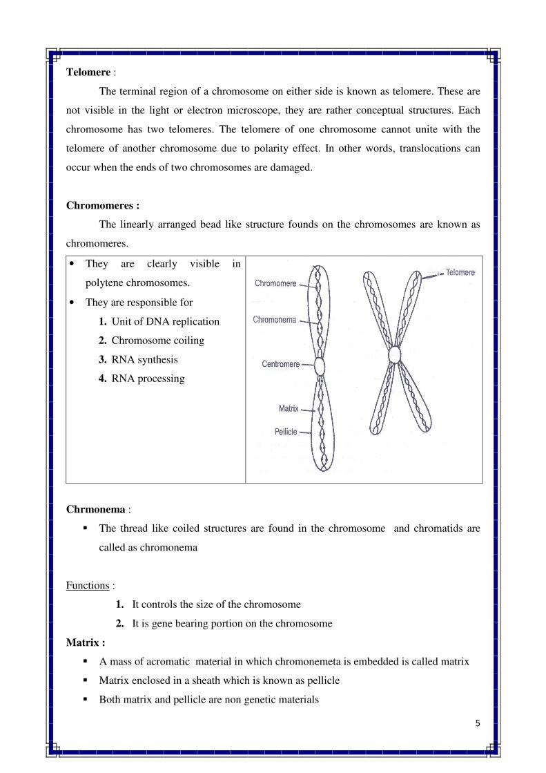
5
Telomere :
The terminal region of a chromosome on either side is known as telomere. These are
not visible in the light or electron microscope, they are rather conceptual structures. Each
chromosome has two telomeres. The telomere of one chromosome cannot unite with the
telomere of another chromosome due to polarity effect. In other words, translocations can
occur when the ends of two chromosomes are damaged.
Chromomeres :
The linearly arranged bead like structure founds on the chromosomes are known as
chromomeres.
• They are clearly visible in
polytene chromosomes.
• They are responsible for
1. Unit of DNA replication
2. Chromosome coiling
3. RNA synthesis
4. RNA processing
Chrmonema :
� The thread like coiled structures are found in the chromosome and chromatids are
called as chromonema
Functions :
1. It controls the size of the chromosome
2. It is gene bearing portion on the chromosome
Matrix :
� A mass of acromatic material in which chromonemeta is embedded is called matrix
� Matrix enclosed in a sheath which is known as pellicle
� Both matrix and pellicle are non genetic materials
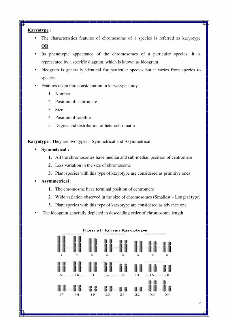
6
Karyotype :
� The characteristics features of chromosome of a species is referred as karyotype
OR
� Its phenotypic appearance of the chromosomes of a particular species. It is
represented by a specific diagram, which is known as ideogram
� Ideogram is generally identical for particular species but it varies from species to
species
� Features taken into consideration in karyotype study
1. Number
2. Position of centromere
3. Size
4. Position of satellite
5. Degree and distribution of heterochromatin
Karyotype : They are two types – Symmetrical and Asymmetrical
� Symmetrical :
1. All the chromosomes have median and sub-median position of centromere
2. Less variation in the size of chromosome
3. Plant species with this type of karyotype are considered as primitive ones
� Asymmetrical :
1. The chromosme have terminal position of centromere
2. Wide variation observed in the size of chromosomes (Smallest – Longest type)
3. Plant species with this type of karyotype are considered as advance one
� The ideogram generally depicted in descending order of chromosome length
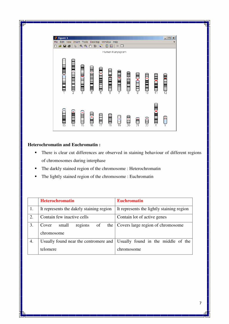
7
Heterochromatin and Euchromatin :
� There is clear cut differences are observed in staining behaviour of different regions
of chromosomes during interphase
� The darkly stained region of the chromosome : Heterochromatin
� The lightly stained region of the chromosome : Euchromatin
Heterochromatin Euchromatin
1. It represents the dakrly staining region It represents the lightly staining region
2. Contain few inactive cells Contain lot of active genes
3. Cover small regions of the
chromosome
Covers large region of chromosome
4. Usually found near the centromere and
telomere
Usually found in the middle of the
chromosome
8
CLASSIFICATION OF CHROMOSOMES
Chromosomes can be classified in different ways. The various criteria which are
usually used for the classification of chromosomes include, (1) position of centromere, (2)
number of centromere, (3) shape at anaphase, (4) structure and appearance, (5) role in
heredity essentiality, (6) role in sex determination, and (7) structure and function. A brief
classification on the bases of these criteria is presented below
1.Based on the Position of centromere
Metacentric
Chromosome
A chromosome in which centromere is located in the middle portion,
such chromosomes assume V shape at anaphase.
Sub-metacentic A chromosome in which centromere is located slightly away from the
centre point or has sub-median position. Such chromosomes assume J
shape at anaphase.
Acrocentric
Chromosome
A chromosome in which centromere is located very near to one end or
has subterminal position. Also called as sub-terminal chromosome.
Such chromosome assumes J shape or rod shape during anaphase.
Holokinetic
Chromosome
A chromosome with diffused centromere. Centromere does not occupy
a specific pqsition, but is diffuses throughout the body of chromosome.
Whole body of such chromosome exhibits centromeric activity. Also
called holocentric chromosome.
2. Based on the Number of centromere
Acentric
Chromosome
A chromosome without centromere. Such chromosome remains as
laggard during cell division and is eventually lost.
Monocentric
Chromosome
A chromosome with one centromere. It represents normal type of
chromosomes.
Dicentric
Chromosome
A chromosome having two centromeres. Such chromosome makes
dicentric bridge at anaphase and are produced due to inversion and
translocations.
3. Based on the Shape at anaphase
V shaped
Chromosome
A chromosome which assumes V shape at anaphase. It includes
metacentric chromosome.
J shaped A chromosome which assumes J shape at anaphase. It includes sub-
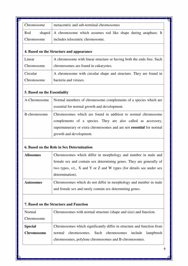
9
Chromosome metacentric and sub-terminal chromosomes
Rod shaped
Chromosome
A chromosome which assumes rod like shape during anaphase. It
includes telocentric chromosome.
4. Based on the Structure and appearance
Linear
Chromosome
A chromosome with linear structure or having both the ends free. Such
chromosomes are found in cukaryotes.
Circular
Chromosome
A chromosome with circular shape and structure. They are found in
bacteria and viruses.
5. Based on the Essentiality
A-Chromosome Normal members of chromosome complements of a species which are
essential for normal growth and development.
B-chromosome Chromosomes which are found in addition to normal chromosome
complements of a species. They are also called as accessory,
supernumerary or extra chromosomes and are not essential for normal
growth and development.
6. Based on the Role in Sex Determination
Allosomes
Chromosomes which differ in morphology and number in male and
female sex and contain sex determining genes. They are generally of
two types, viz., X and Y or Z and W types (for details see under sex
determination).
Autosomes
Chromosomes which do not differ in morphology and number in male
and female sex and rarely contain sex determining genes.
7. Based on the Structure and Function
Normal
Chromosome
Chromosomes with normal structure (shape and size) and function.
Special
Chromosome
Chromosomes which significantly differ in structure and function from
normal chromosomes. Such chromosomes include lampbrush
chromosomes, polylene chromosomes and B-chromosomes.
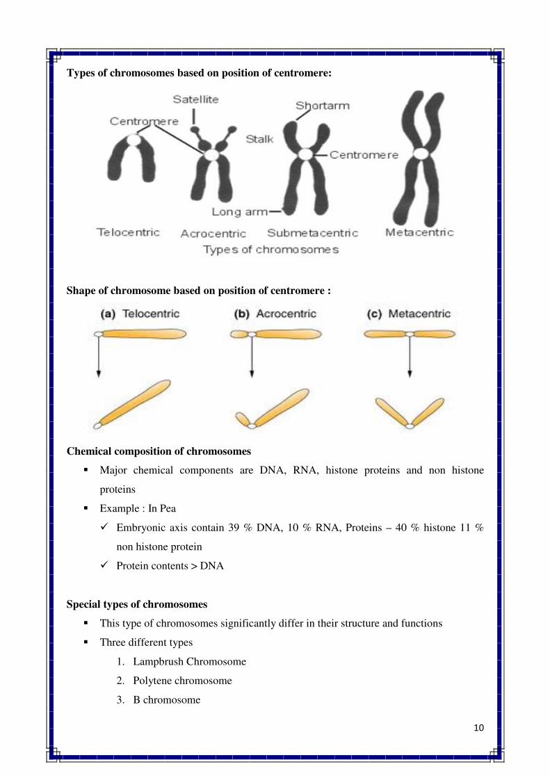
10
Types of chromosomes based on position of centromere:
Shape of chromosome based on position of centromere :
Chemical composition of chromosomes
� Major chemical components are DNA, RNA, histone proteins and non histone
proteins
� Example : In Pea
� Embryonic axis contain 39 % DNA, 10 % RNA, Proteins – 40 % histone 11 %
non histone protein
� Protein contents > DNA
Special types of chromosomes
� This type of chromosomes significantly differ in their structure and functions
� Three different types
1. Lampbrush Chromosome
2. Polytene chromosome
3. B chromosome
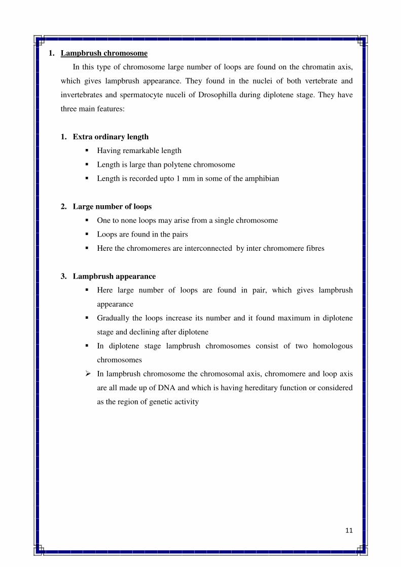
11
1. Lampbrush chromosome
In this type of chromosome large number of loops are found on the chromatin axis,
which gives lampbrush appearance. They found in the nuclei of both vertebrate and
invertebrates and spermatocyte nuceli of Drosophilla during diplotene stage. They have
three main features:
1. Extra ordinary length
� Having remarkable length
� Length is large than polytene chromosome
� Length is recorded upto 1 mm in some of the amphibian
2. Large number of loops
� One to none loops may arise from a single chromosome
� Loops are found in the pairs
� Here the chromomeres are interconnected by inter chromomere fibres
3. Lampbrush appearance
� Here large number of loops are found in pair, which gives lampbrush
appearance
� Gradually the loops increase its number and it found maximum in diplotene
stage and declining after diplotene
� In diplotene stage lampbrush chromosomes consist of two homologous
chromosomes
� In lampbrush chromosome the chromosomal axis, chromomere and loop axis
are all made up of DNA and which is having hereditary function or considered
as the region of genetic activity
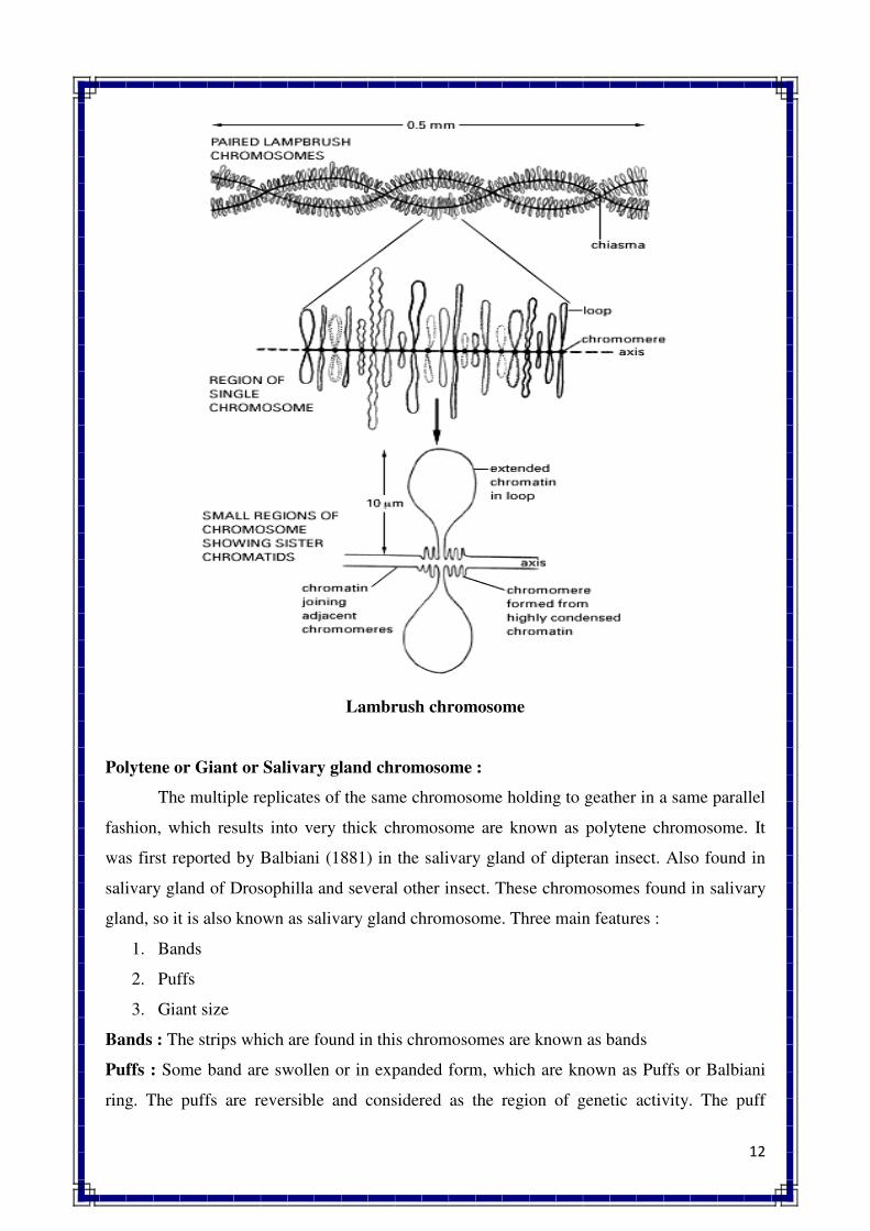
12
Lambrush chromosome
Polytene or Giant or Salivary gland chromosome :
The multiple replicates of the same chromosome holding to geather in a same parallel
fashion, which results into very thick chromosome are known as polytene chromosome. It
was first reported by Balbiani (1881) in the salivary gland of dipteran insect. Also found in
salivary gland of Drosophilla and several other insect. These chromosomes found in salivary
gland, so it is also known as salivary gland chromosome. Three main features :
1. Bands
2. Puffs
3. Giant size
Bands : The strips which are found in this chromosomes are known as bands
Puffs : Some band are swollen or in expanded form, which are known as Puffs or Balbiani
ring. The puffs are reversible and considered as the region of genetic activity. The puff
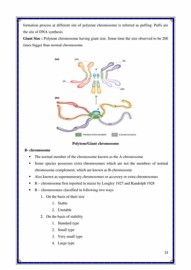
13
formation process at different site of polytene chromosome is referred as puffing. Puffs are
the site of DNA synthesis
Giant Size : Polytene chromosome having giant size. Some time the size observed to be 200
times bigger than normal chromosome
Polytene/Giant chromosome
B- chromosome
� The normal member of the chromosome known as the A-chromosome
� Some species possesses extra chromosomes which are not the members of normal
chromosome complement, which are known as B-chromosome
� Also known as supernumerary chromosomes or accesory or extra chromosomes
� B – chromosome first reported in maize by Longley 1927 and Randolph 1928
� B – chromosomes classified in following two ways
1. On the basis of their size
1. Stable
2. Unstable
2. On the basis of stability
1. Standard type
2. Small type
3. Very small type
4. Large type
14
GLOSSORY
CHROMOSOME. Darkly stained rod shaped bodies visible under light microscope in a cell during
mitotic metaphase.
HAPLOID NUMBER. The gametic chromosome number of a species (n)
DIPLOID NUMBER. The somatic chromosome number of a speciest (2n)
BASIC NUMBER. The gametic chromosome number of a true diploid species (x)
CENTROMERE. The region of chromosome with which spindle fibres are attached during
metaphase. Also called primary constriction or kinetochore.
CHROMATID. One of the two distinct longitudinal sub-units of a chromosome.
SECONDARY CONSTRICTION. The narrow or constricted region in a chromosome other than
centromere. Telomere. The terminal region of chromosome on either side.
CHROMOMERE. The linearly arranged bead like structures found on the chromosomes.
CHROMONEMA. Thread like coiled structures found in the chromosomes and chromatids (Pleural
chromonemeta).
MATRIX. A mass of acromatic material in which chromonemeta are embedded.
KARYOTYPE. The characteristic features of chromosomes of a species.
IDIOGRAM. The diagram which is used to represent karyotype.
EUCHROMATIN. Lightly staining region of chromosomes during interphase. Usually found in the
middle of chromosome, genetically active and takes part in transcription.
HETEROCHROMATIN. Darkly staining region of chromosomes during interphase, usually
inactive in transcription, found near centromere and telomere.
A-CHROMOSOME. Normal members of chromosome complements of a species which are
essential for normal growth and development.
B-CHROMOSOME. Chromosomes which are found in addition to normal chromosome
complements of a species and are not essential for normal growth and development. Also known as
accessory supernumerary or extra chromosomes.
NORMAL CHROMOSOMES. Chromosomes with normal structure (shape and size) and function.
15
SPECIAL CHROMOSOMES. Chromosomes which significantly differ in structure and function
from normal chromosomes such as lampbrush chromosomes, polytene chromosomes and B-
chromosornes.
METACENTRIC CHROMOSOME. A chromosome in which centromere is located in the middle
portion. Such
chromosomes assume V shape at anaphase. Sub-Metacentric chromosome. A chromosome in which
centromere has sub-median position. Acrocentric chromosome. A chromosome in which centromere
is located very near to one end or has sub-terminal position.
TELOCENTRIC CHROMOSOME. A chromosome in which centromere is located at one end.
HOLOKINETIC CHROMOSOME. A chromosome with diffused centromere.
ACENTRIC CHROMOSOME. A chromosome without centromere.
MONOCENTRIC CHROMOSOME. A chromosome with one centromere.
DICENTRIC CHROMOSOME. A chromosome having two centromeres.
V-SHAPED CHROMOSOME. A chromosome which assumes V shape at anaphase.
J-SHAPED CHROMOSOME. A chromosome which assumes J share at anaphase.
ROD SHAPED CHROMOSOME. A chromosome which assumes rod shape at anaphase.
LINEAR CHROMOSOME. A chromosome having both the ends free; found in etikaryotes.
CIRCULAR CHROMOSOME. A chromosome with circular shape and structures; found in bacteria
and viruses.
LAMPBRUSH CHROMOSOME. A chromosome having lampbrush appearance.
POLYTENE Chromosome. The multiple replicate of the same chromosome holding together in a
parallel fashion resulting in very thick structure; also called giant or salivary gland chromosome.
BANDS. The strips which are found on polytene chromosome.
PUFFS. The swollen regions found on polytene chromosome; also called Balbiani rings.
ISO-CHROMOSOME. A chromosome with two identical arms.
RING CHROMOSOME. A physically circular chromosome, usually found in prokaryotes such as
bacteria and viruses.
CHROMOSOME MODELS. The pattern of organization of chromatin fibres in a chromosome.
16
CHROMOSOME BANDING. The differentially stained regions of chromosomes, as a result of
treatment with various dyes, visible under light or fluorescence microscope.
QUESTIONS
Q.1 Define chromosome. Describe briefly the variation found in shape, size and number
of chromosomes in crop plants.
Q.2 Give a brief account of the internal morphology of chromosomes in eukaryotes.
Q.3 What are special chromosomes ? Describe anyone of them in detail with suitable
diagrams.
Q.4 What are polytene chromosomes ? Describe briefly their origin and significance.
Q.5 Defince B chromosomes. Give their classification and describe their meiotic
behaviour and genetic effects.
Q.6 Describe briefly the role of chromosomes in heredity.
Q.7 What are chromosome models ? Describe two types of chromosome models with
suitable diagrams.
Q.8 What is chromosome banding ? Explain various types of chromosome banding and
their significance in genetics.
Q.9 Write short notes on the following
1. Basic number 5. Chromonema
2. Chromatid 6. Lampbrush chromosome
3. Centromere 7. Giant chromosome
4. Karyotype 8. Isochromosome
Q.10 Differentiate between the following :
1. Heterochromatin and euchromatin
2. Chromosome and chromatid
3. Primary constriction and secondary constriction
//*//*//*//
MULTIPLE ALLELE
Multiple allele
Existence of more than one allele at a locus is referred as multiple allele and the
phenomenon is referred as multiple allelism OR
The condition in which a particular gene occurs in three or more allelic form in
population of organisms
Main features/characteristics of multiple allele
o Multiple allele always belong to the same locus and one allele is present at a locus at a
time in a chromosome
o Multiple allele always control the same character of an individual. The expression of a
character will differ depending upon the allele present
o There is no crossing over in multiple allele series
o In series of multiple allele wild type is always dominant.
o The cross between two mutant type will always produce mutant phenotype
(intermediates). Such cross will never produce wild phenotype. In other words multiple
allele do not show complementation
Examples of multiple allele :
o Fur colour in rabbit
o Wing type in Drosophila
o Eye colour in Drosophila
o Self incompatibility allele in plants
o ABO blood group in man
o Waxy gene of maize
Self Incompatibility Alleles in Plants
The most common example of multiple alleles in plants is the series of self incompatibility
alleles. Such alleles were reported in Nicotiana and later on they were found in several other
plant species like Brassica, radish, tomato, potato, etc. In these species, self incompatibility is
governed by a single gene S which has multiple alleles, viz., S1, S
2, S
3, S
4 and so on. Now cases
of digenic and trigenic self incompatibility have also been reported. In evening primrose 37 and
in red clover 41 alleles of self incompatibility have been reported. Crosses between individuals
having self incompatibility alleles will lead to three types of situations as given below :
Fully Sterile
When both male female have similar alleles, viz., S'S2 x S'S
2 the cross will be incompatible
and there will be no seed setting.
Partially Fertile
Such crosses are obtained when male and female plants differ for one allele, viz., S'S2 x S'S3.
This cross will produces, S'S3 and S
2S
3 progeny. In other words, half of the progeny will be
fertile.
Fully Fertile
The fully fertile crosses are obtained when male and female plants differ in respect of both
alleles, viz., S'S2 x S3S4. This cross will produce four fertile genotypes, viz., S'S3, S'S4, S2S3 and
S2S
4. Thus, plants which have self incompatibility alleles are always heterozygous for this gene.
ABO blood group in man
o ABO blood group in man was discovered by Landsteiner (1900)
o Antibody : It is a type of protein which is commonly referred to as immunoglobin and
found in the serum or plasma. The presence of antibody can be demonstrated by its
specific reaction with antigen.
o Antigen : It refers to a substance or agent which introduced into the system of a
vertebrate animal (like cow, goat, rabbit, man etc.) for the production of specific
antibody, which binds specifically to this substance. Antigens are located in the RBC (red
blood corpuscles)
o In human RBC there is two types of antigens viz., A and B
o Depending upon the presence and absence of antigen A & B, the blood group in human is
of four types viz., A, B, AB & O.
o As far as antigen is concern, a person with the blood group A has antigen A on the
surface of RBCs, similarly for the blood group B will have antigen B, for the blood
group AB will have the antigens A & B and for the blood group O have no antigen on the
surface of their RBCs
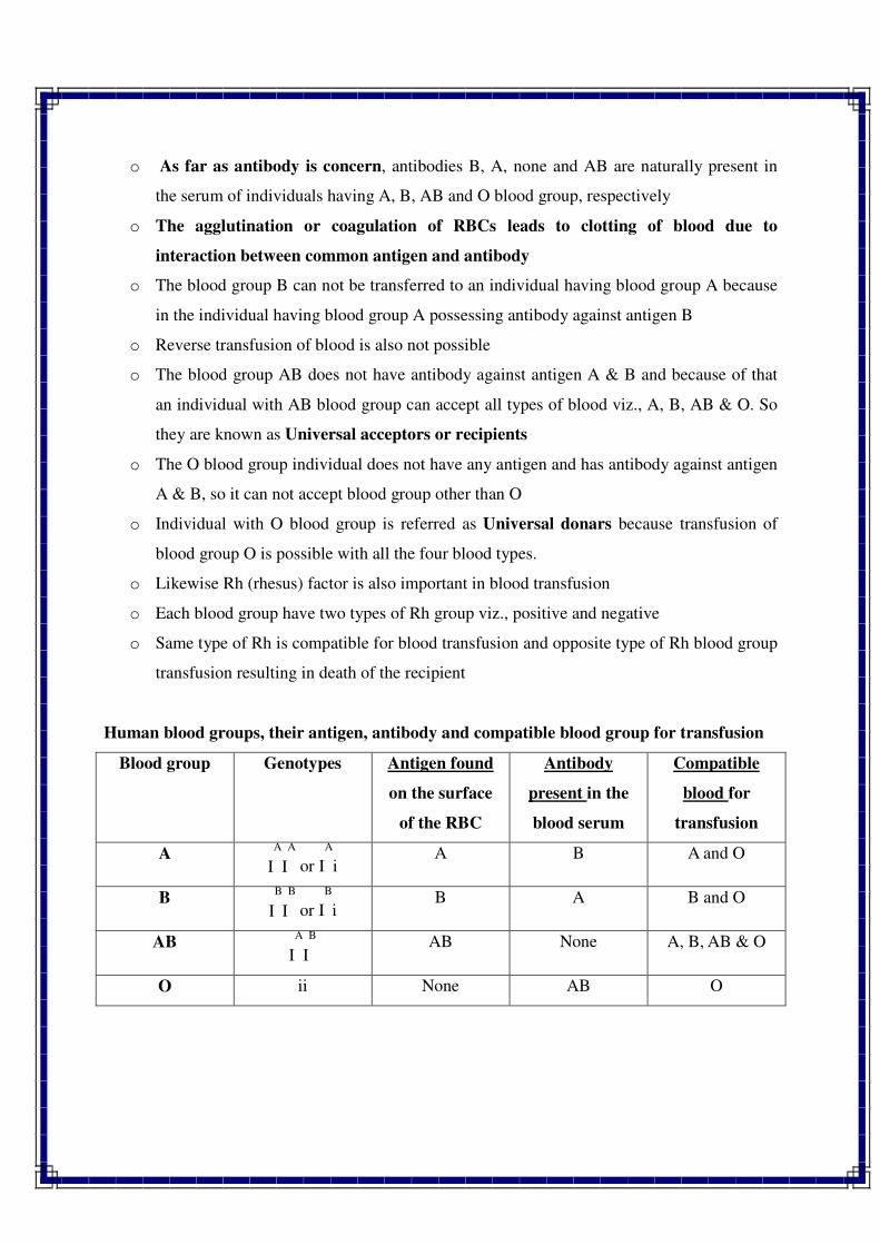
o As far as antibody is concern, antibodies B, A, none and AB are naturally present in
the serum of individuals having A, B, AB and O blood group, respectively
o The agglutination or coagulation of RBCs leads to clotting of blood due to
interaction between common antigen and antibody
o The blood group B can not be transferred to an individual having blood group A because
in the individual having blood group A possessing antibody against antigen B
o Reverse transfusion of blood is also not possible
o The blood group AB does not have antibody against antigen A & B and because of that
an individual with AB blood group can accept all types of blood viz., A, B, AB & O. So
they are known as Universal acceptors or recipients
o The O blood group individual does not have any antigen and has antibody against antigen
A & B, so it can not accept blood group other than O
o Individual with O blood group is referred as Universal donars because transfusion of
blood group O is possible with all the four blood types.
o Likewise Rh (rhesus) factor is also important in blood transfusion
o Each blood group have two types of Rh group viz., positive and negative
o Same type of Rh is compatible for blood transfusion and opposite type of Rh blood group
transfusion resulting in death of the recipient
Human blood groups, their antigen, antibody and compatible blood group for transfusion
Blood group Genotypes Antigen found
on the surface
of the RBC
Antibody
present in the
blood serum
Compatible
blood for
transfusion
A I
A
I
A
or I
A
i A B A and O
B I
B
I
B
or I
B
i B A B and O
AB I
A
I
B
AB None A, B, AB & O
O ii None AB O
PSEUDOALLELES
Pseudoalleles refer to closely linked and functionally related genes. A cluster of pseudoalleles is
known as pseudoallele series or a complex locus or a complex region. Main characteristics of
pseudoalleles are given below :
1. Pseudoalleles govern different expressions of the same character. In other words, they
are functionally related.
2. Pseudoalleles are considered to occupy a complex locus which is divided into sub loci.
Thus, they occupy different positions, but on the same complex locus.
3. They exhibit low frequency of genetic recombination by crossing over. In other words,
crossingover occurs between pseudoalleles, but at a very low frequency.
4. They exhibit cis-trans position effect. In trans heterozygotes such mutants produce
mutant phenotype, but in cis-heterozygotes they produce a wild phenotype.
TEST FOR ALLELISM
There are two types of tests that are used for allelism, viz., recombination test and
complementation test. These are briefly discussed below :
Recombination Test
Earlier it was believed that recombination can occur between two genes but not within a
gene. Thus, if a cross between two mutants say tr^mi and m2m2 produces wild type in test cross
or in F2 then iri| and m2 are considered as non-allelic because production of wild type is not
possible without recombination. If no wild type appears in test cross or F2 then m, and m2 are
considered as allelic forms. Now intragenic recombination has been reported in many organisms.
Hence this concept is no more valid.
Complementation Test
Alleles may be arranged in two ways, viz., cis position and trans position (Fig. 13.1).
When two wild alleles are located in one chromosome and their mutant alleles in homologous
chromosomes (++/ab), it is known as cis-arrangement. Thus, in cis position alleles are linked in
coupling phase. On the other hand, when one wild and one mutant type alleles are located in
each homologous chromosome (+a/+b), it is known as trans position or repulsion phase of
alleles. Complementation refers to appearance of wild phenotype when two mutants are crossed.
Complementation test is used to determine whether two mutant alleles belong to same gene or
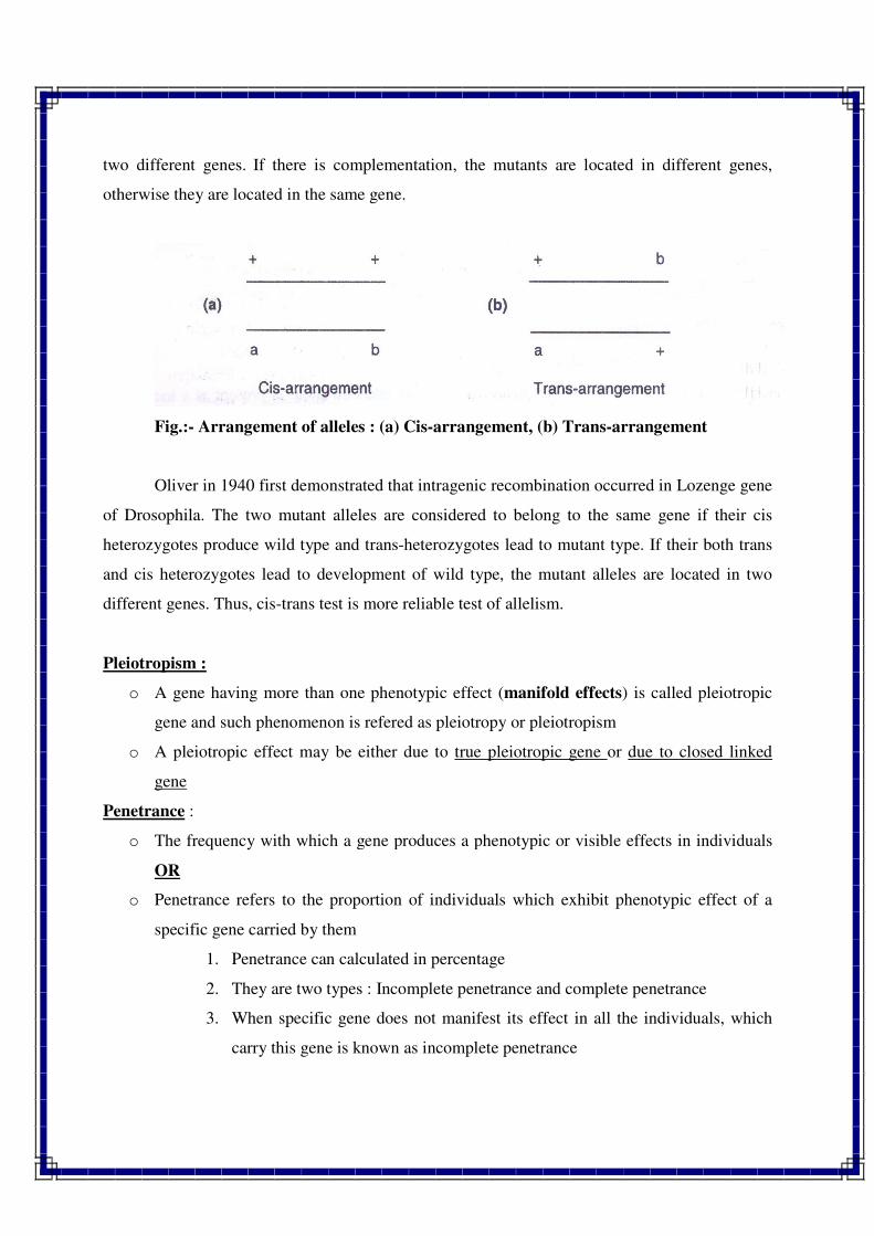
two different genes. If there is complementation, the mutants are located in different genes,
otherwise they are located in the same gene.
Fig.:- Arrangement of alleles : (a) Cis-arrangement, (b) Trans-arrangement
Oliver in 1940 first demonstrated that intragenic recombination occurred in Lozenge gene
of Drosophila. The two mutant alleles are considered to belong to the same gene if their cis
heterozygotes produce wild type and trans-heterozygotes lead to mutant type. If their both trans
and cis heterozygotes lead to development of wild type, the mutant alleles are located in two
different genes. Thus, cis-trans test is more reliable test of allelism.
Pleiotropism :
o A gene having more than one phenotypic effect (manifold effects) is called pleiotropic
gene and such phenomenon is refered as pleiotropy or pleiotropism
o A pleiotropic effect may be either due to true pleiotropic gene or due to closed linked
gene
Penetrance :
o The frequency with which a gene produces a phenotypic or visible effects in individuals
OR
o Penetrance refers to the proportion of individuals which exhibit phenotypic effect of a
specific gene carried by them
1. Penetrance can calculated in percentage
2. They are two types : Incomplete penetrance and complete penetrance
3. When specific gene does not manifest its effect in all the individuals, which
carry this gene is known as incomplete penetrance
4. When all the individuals which carry a particular gene exhibit its phenotypic
effect is known as complete penetrance
Expressivity :
o The degree of phenotypic expression of a penetrant gene is called as expressivity
Expressivity are of two types :
1. Uniform
2. Variable
o When the phenotypic expression of a gene is identical or similar in all the individuals
which carry such gene is known as uniform expressivity
o Most of the qualitative characters exhibit uniform expressivity
o Most of the qualitative characters exhibit uniform expressivity
o When the phenotypic expression differs in different carriers of a gene it is known as
variable expressivity
GLOSSORY
Allele. Alternative form of a gene. Alleles may be of two types, viz,, either dominant and
recessive or wild and mutant.
Multiple allele. Existence of more than two alleles at a locus.
cis-position. Presence of two wild alleles in one homologous chromosome and their mutant
alleles in another homologous chromosome. (++/ab); also called coupling phase of alleles.
Trans-position. Existence of one wild and one mutant allele in each homologous chromosome
(+ a/+b); also called repulsion phase of alleles.
Complementation. Appearance of wild phenotype when two mutants are crossed together.
Antibody. A type of protein which is commonly known as immunoglobin.
Antigen. A substance or agent which, when introduced into a system of a vertebrate animal like
cow, goat, rabbit, man, etc. induces the production of specific antibody which binds specifically
to this substance.
Pseudo alleles. Closely linked and functionally related genes. A cluster of pseudo alleles in
known as a complex locus or complex region. Examples are lozenge and star asteroid eyes in
Drosophila.
Isoallele. An allele which is similar in its phenotypic expression to that of other independently
occurring allele.
Mutant isoallele. An isoallele which acts within the phenotypic range of a mutant character.
Normal isoallele. An isoallele which acts within the phenotypic range of a wild character.
Gene. A basic unit of inheritance, sequence of DNA nucleotides which codes for a functional
product of RNA or a polypeptide. The term gene was coined by Johannsen in 1909. A gene
consists of recon, muton and cistron.
Recons. The regions (units) within a gene between which recombination can occur, but the
recombinationcannot occur within a recon.
Muton. The smallest element within a gene, which can give rise to mutant phenotype or
mutation.
Cistron. The largest element within a gene which is the unit of function.
Split genes. The genes with intervening or interrupted sequences; usually found in eukaryotes.
Exons. Coding sequences of DNA in split genes.
Introns. Non coding sequences in split genes which are removed during processing of mRNA.
Jumping genes. The genes which keep on changing their position in a chromosome and also
between the chromosomes in a genome. Also called transposons or transposable elements. The
first case of jumping gene was reported by McClinlock in 1950 in maize.
Overlapping Genes. Genes which code for more than one protein. In such gene, the complete
nucleotide sequence codes for one protein and part of such nucleotide sequence for another
protein. Such genes have been reported in tumor producing viruses such as SV40 and G4.
Pseudo Genes. A non-functional sequence of DNA or a defective copy of a normal gene in
eukaryotes.Such genes have been reported in humans, mouse and Drosophila.
********
Linkage & Crossing over
Linkage : The tendency of two or more genes to stay together during inheritance is known as
linkage.
Linkage groups :
A group of genes, which are present in one chromosome OR All genes which are located in one
chromosome constitute one linkage group.
� Maximum number of linkage group = Haploid number of an organism. Example : 4
linkage group in Drosophilla (2n=8) and 23 in man (2n=46)
� Linked genes do not shown independent segregation was first reported by Bateson and
Punnet in 1905 in pea.
� Effect of linkage is clearly noticeable in test cross generation.
� The frequency of parental character combinations are more than expected, while
new character combinations are relatively lower.
Main features of linkage :
1. Linkage involves two or more genes which are located in the same chromosome in a liner
fashion.
2. Linkage may involve either dominant genes or recessive genes or some dominant and
some recessive genes.
3. Linkage usually involves those genes which are located closely.
4. Presence of linkage leads to higher frequency of parental types than recombinants in a
test cross progeny. When two genes are linked the segregation ratio of a test cross
progeny deviates significantly from the 1 : 1 : 1 :1 ratio.
5. Linkage may involve either two or more desirable traits or all undesirable traits or some
desirable and some undesirable traits.
6. Linkage is observed for both oligogenic traits as well as polygenic traits. However, it is
more common for the former than latter.
7. Besides pleiotropy, linkage is an important cause of genetic correlation between various
plant characters.
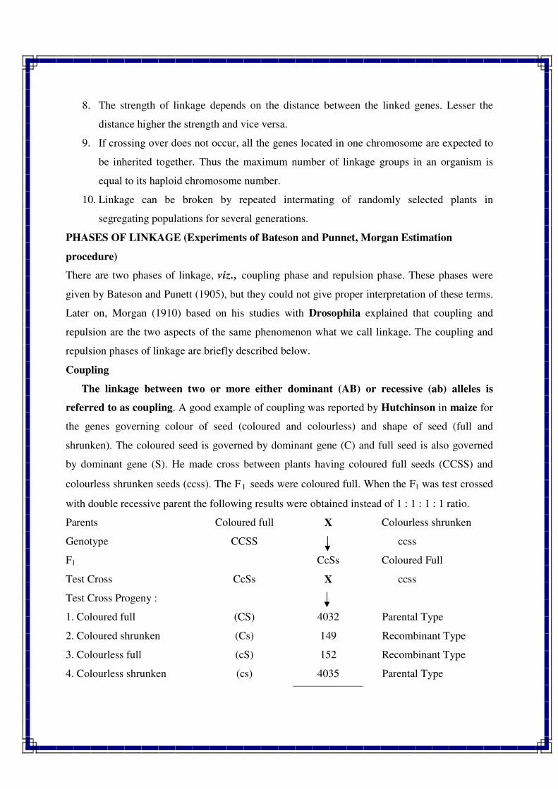
8. The strength of linkage depends on the distance between the linked genes. Lesser the
distance higher the strength and vice versa.
9. If crossing over does not occur, all the genes located in one chromosome are expected to
be inherited together. Thus the maximum number of linkage groups in an organism is
equal to its haploid chromosome number.
10. Linkage can be broken by repeated intermating of randomly selected plants in
segregating populations for several generations.
PHASES OF LINKAGE (Experiments of Bateson and Punnet, Morgan Estimation
procedure)
There are two phases of linkage, viz., coupling phase and repulsion phase. These phases were
given by Bateson and Punett (1905), but they could not give proper interpretation of these terms.
Later on, Morgan (1910) based on his studies with Drosophila explained that coupling and
repulsion are the two aspects of the same phenomenon what we call linkage. The coupling and
repulsion phases of linkage are briefly described below.
Coupling
The linkage between two or more either dominant (AB) or recessive (ab) alleles is
referred to as coupling. A good example of coupling was reported by Hutchinson in maize for
the genes governing colour of seed (coloured and colourless) and shape of seed (full and
shrunken). The coloured seed is governed by dominant gene (C) and full seed is also governed
by dominant gene (S). He made cross between plants having coloured full seeds (CCSS) and
colourless shrunken seeds (ccss). The F1 seeds were coloured full. When the F1 was test crossed
with double recessive parent the following results were obtained instead of 1 : 1 : 1 : 1 ratio.
Parents Coloured full X Colourless shrunken
Genotype CCSS ccss
F1 CcSs Coloured Full
Test Cross CcSs X ccss
Test Cross Progeny :
1. Coloured full (CS) 4032 Parental Type
2. Coloured shrunken (Cs) 149 Recombinant Type
3. Colourless full (cS) 152 Recombinant Type
4. Colourless shrunken (cs) 4035 Parental Type
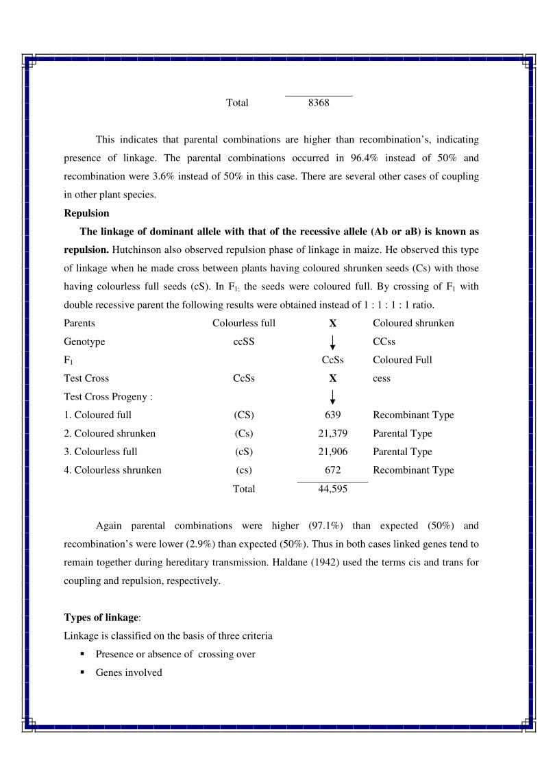
Total 8368
This indicates that parental combinations are higher than recombination’s, indicating
presence of linkage. The parental combinations occurred in 96.4% instead of 50% and
recombination were 3.6% instead of 50% in this case. There are several other cases of coupling
in other plant species.
Repulsion
The linkage of dominant allele with that of the recessive allele (Ab or aB) is known as
repulsion. Hutchinson also observed repulsion phase of linkage in maize. He observed this type
of linkage when he made cross between plants having coloured shrunken seeds (Cs) with those
having colourless full seeds (cS). In F1; the seeds were coloured full. By crossing of F1 with
double recessive parent the following results were obtained instead of 1 : 1 : 1 : 1 ratio.
Parents Colourless full X Coloured shrunken
Genotype ccSS CCss
F1 CcSs Coloured Full
Test Cross CcSs X cess
Test Cross Progeny :
1. Coloured full (CS) 639 Recombinant Type
2. Coloured shrunken (Cs) 21,379 Parental Type
3. Colourless full (cS) 21,906 Parental Type
4. Colourless shrunken (cs) 672 Recombinant Type
Total 44,595
Again parental combinations were higher (97.1%) than expected (50%) and
recombination’s were lower (2.9%) than expected (50%). Thus in both cases linked genes tend to
remain together during hereditary transmission. Haldane (1942) used the terms cis and trans for
coupling and repulsion, respectively.
Types of linkage:
Linkage is classified on the basis of three criteria
� Presence or absence of crossing over
� Genes involved
� The chromosome involved
Based on crossing over :
1. Complete linkage :
1. The linkage in which crossing over does not occur is known as complete linkage
or absolute linkage.
2. In this situation only parental types are obtained from the test cross progeny
2. Incomplete linkage :
1. If some frequency of crossing over occur between linked genes, it is known as
incomplete linkage
2. In this situation, recombinants are observed in the test cross progeny along with
parental combination
Based on genes involved
1. Coupling linkage : It refers to linkage either between dominant genes or between
recessive genes
2. Repulsion linkage : It refers to linkage of some dominant genes with some recessive
genes
Based on the chromosome involved
1. Autosomal linkage : it refers to linkage of such genes, which are located in other than
sex chromosomes
2. X- chromosomal linkage : It refers to the linkage of genes which are located in sex
chromosomes
Significance or practical utility of linkage :
1. Effect of selection : Linkage between two or more loci controlling different desirable
character is advantageous for a plant breeder, because desirable alleles comes more
frequently in segregating population than would be expected with independent
assortment. Linkage is undesirable when desirable gene is linked with undesirable gene.
2. Effect on genetic variance : The estimates of genetic variance for quantitative characters
are greatly influenced by presence of linkage.
3. Effect of genetic correlation : The linked characters showed high values of genetic
correlation and coheritability
Crossing over :
Crossing over refers to the exchange of chromosomal segment between non sister chromatids of
homologous chromosomes during meiotic prophase.
� The term crossing over was first used by Morgan and Cattell in 1912
Main features of crossing over :
1. Crossing over take place during meiotic prophase, i.e., during pachytene.
2. Crossing over occurs between non-sister chromatids. Thus one chromatid from each of
the two homologous chromosomes is involved in crossing over.
3. It is universally accepted that crossing over takes place at four strand stage.
4. Each crossing over involves only two of four chromatids of two homologous
chromosomes. However, double or multiple crossing over may involve all four, three or
two of the four chromatids, which is very rare.
5. Crossing over leads to recombination or new combinations between linked genes.
Crossing over generally yields two recombinant types or crossover types and two parental
types or non-crossover types.
6. Crossing over generally leads to exchanges of equal segments of genes and
recombination is always reciprocal. However, unequal crossing over has also been
reported.
7. The value of crossover or recombinants may vary from 0-50%.
8. The frequency of recombinants can be worked out from the test cross progeny. It is
expressed as the percentage ratio of recombinants to the total population (recombinants +
parental types ) Thus,
Crossingover frequency (%) = No. of recombinants X 100
Total progeny
Linkage Crossing over
1. It keeps the genes together It leads to separation of linked genes.
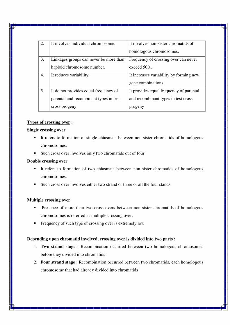
2. It involves individual chromosome. It involves non-sister chromatids of
homologous chromosomes.
3. Linkages groups can never be more than
haploid chromosome number.
Frequency of crossing over can never
exceed 50%.
4. It reduces variability. It increases variability by forming new
gene combinations.
5. It do not provides equal frequency of
parental and recombinant types in test
cross progeny
It provides equal frequency of parental
and recombinant types in test cross
progeny
Types of crossing over :
Single crossing over
� It refers to formation of single chiasmata between non sister chromatids of homologous
chromosomes.
� Such cross over involves only two chromatids out of four
Double crossing over
� It refers to formation of two chiasmata between non sister chromatids of homologous
chromosomes.
� Such cross over involves either two strand or three or all the four stands
Multiple crossing over
� Presence of more than two cross overs between non sister chromatids of homologous
chromosomes is referred as multiple crossing over.
� Frequency of such type of crossing over is extremely low
Depending upon chromatid involved, crossing over is divided into two parts :
1. Two strand stage : Recombination occurred between two homologous chromosomes
before they divided into chromatids
2. Four strand stage : Recombination occurred between two chromatids, each homologous
chromosome that had already divided into chromatids
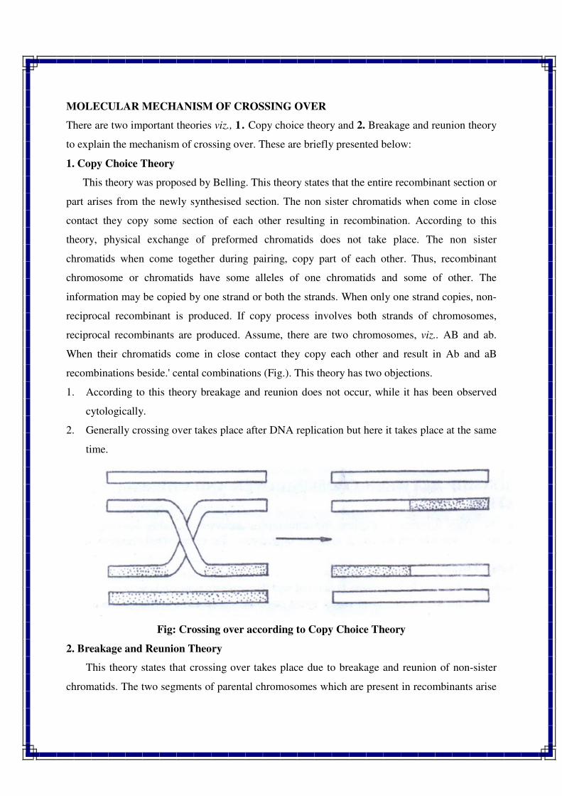
MOLECULAR MECHANISM OF CROSSING OVER
There are two important theories viz., 1. Copy choice theory and 2. Breakage and reunion theory
to explain the mechanism of crossing over. These are briefly presented below:
1. Copy Choice Theory
This theory was proposed by Belling. This theory states that the entire recombinant section or
part arises from the newly synthesised section. The non sister chromatids when come in close
contact they copy some section of each other resulting in recombination. According to this
theory, physical exchange of preformed chromatids does not take place. The non sister
chromatids when come together during pairing, copy part of each other. Thus, recombinant
chromosome or chromatids have some alleles of one chromatids and some of other. The
information may be copied by one strand or both the strands. When only one strand copies, non-
reciprocal recombinant is produced. If copy process involves both strands of chromosomes,
reciprocal recombinants are produced. Assume, there are two chromosomes, viz.. AB and ab.
When their chromatids come in close contact they copy each other and result in Ab and aB
recombinations beside.' cental combinations (Fig.). This theory has two objections.
1. According to this theory breakage and reunion does not occur, while it has been observed
cytologically.
2. Generally crossing over takes place after DNA replication but here it takes place at the same
time.
Fig: Crossing over according to Copy Choice Theory
2. Breakage and Reunion Theory
This theory states that crossing over takes place due to breakage and reunion of non-sister
chromatids. The two segments of parental chromosomes which are present in recombinants arise
from physical breaks in the parental chromosomes with subsequent exchange of broken segments
(Fig. 9.2). The breakage results due to mechanical strains that result from the separation of paired
homologous chromosomes and chromatids in each chromosome during pachytene stage. The
broken ends of non-sister chromatids unite to produce chiasmata resulting in crossing over.
Fig: Crossing over according to Breakage & Reunion Theory
Factors affecting the crossing over
1. Distance between gene
o Greater the distance between genes higher is chance of crossing over and vice
versa.
2. Age of female
o Generally the crossing over decreases with advancement of the age in the female
of Drosophila.
3. Temperature
o The rate of the crossing over in Drosophila increases above and below the
temperature 22 0C.
4. Sex
o The rate of crossing over also differs according to sex. There is lack of crossing
over in Drosophila male and female silk moth.
5. Nutrition
o Presence of metallic ions like calcium and magnesium in the food caused the
reductions in the recombination in the Drosophila. However, removal of such
chemicals from the diet increased the rate of crossing over.
6. Chemicals
o Treatment with mutagenic chemicals like alkylating agents was found to increase
the rate of crossing over in female of Drosophila.
7. Irradiation
o Irradiation with X-rays and gamma rays was found to enhanced the rate of
crossing over in female of Drosophila.
8. Structural changes
o Structural changes mainly inversion and translocations reduce the frequency of
crossing over in the chromosomes where such changes are involved.
9. Centromere effect
o Generally the genes are located near to centromere show reduced frequency of
crossing over.
10. Cytoplasmic genes
o In some species, cytoplasmic genes also lead to reduction in crossing over.
Chromosome mapping
The line diagram which shows various genes present on a chromosome and
recombination frequency between them such map is called as chromosome map or
linkage map or genetic map. The mapping of the genes on the chromosome is done with
the help of two point test cross and three point test cross
Two point test cross –
A cross between dihybrid (F1 differing in two genes) with its homozygous recessive parent is
referred as two point test cross.
� It provides information about recombination frequency between two genes. In a two point
test cross, four different phenotypic classes are obtained. Out of four classes, two classes
are known as parental types (which have maximum number of individuals or maximum
frequencies). The remaining two types are known as recombinant types (which have
lowest number of individuals or minimum frequencies).
Three point test cross –
A cross between trihybrid (F1 differing in three genes) with its homozygous recessive parent is
referred as three point test cross.
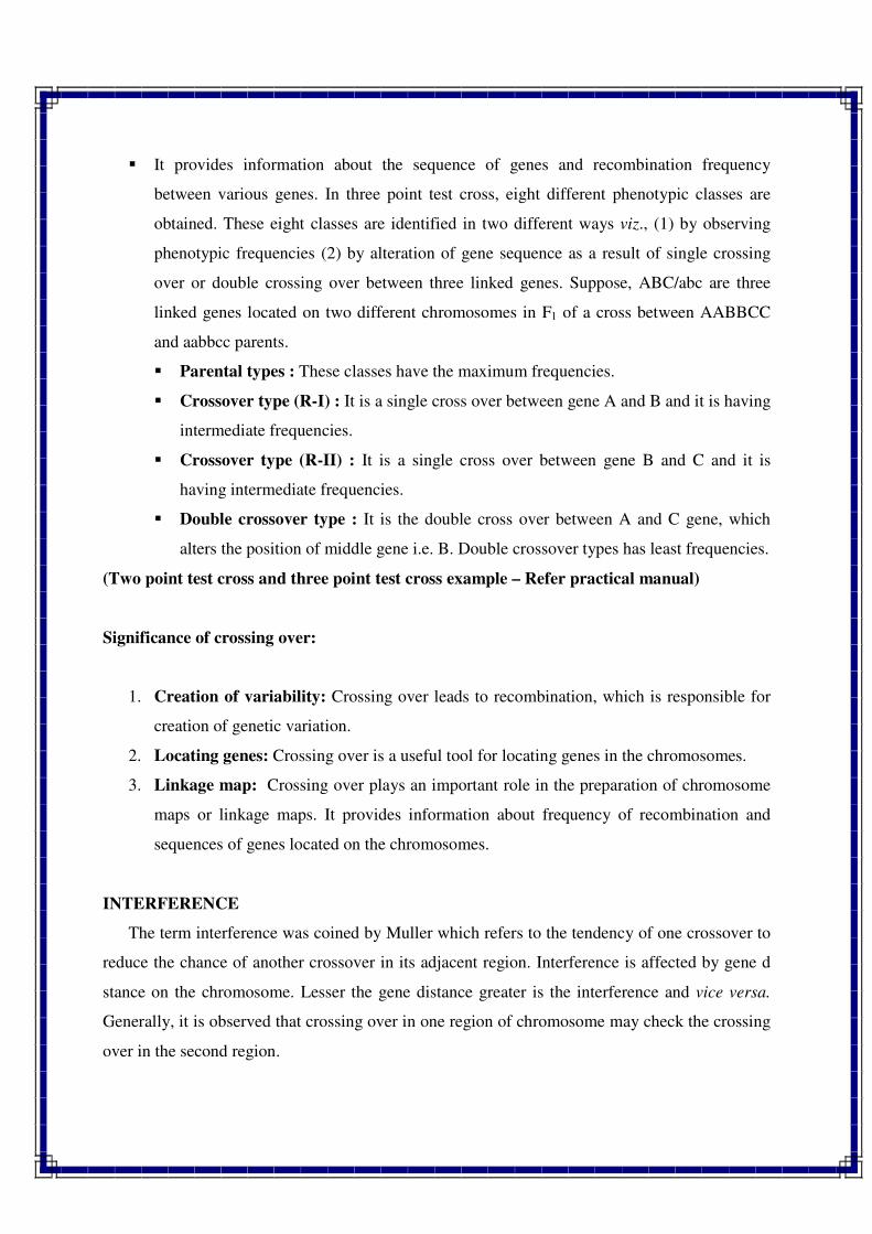
� It provides information about the sequence of genes and recombination frequency
between various genes. In three point test cross, eight different phenotypic classes are
obtained. These eight classes are identified in two different ways viz., (1) by observing
phenotypic frequencies (2) by alteration of gene sequence as a result of single crossing
over or double crossing over between three linked genes. Suppose, ABC/abc are three
linked genes located on two different chromosomes in F1 of a cross between AABBCC
and aabbcc parents.
� Parental types : These classes have the maximum frequencies.
� Crossover type (R-I) : It is a single cross over between gene A and B and it is having
intermediate frequencies.
� Crossover type (R-II) : It is a single cross over between gene B and C and it is
having intermediate frequencies.
� Double crossover type : It is the double cross over between A and C gene, which
alters the position of middle gene i.e. B. Double crossover types has least frequencies.
(Two point test cross and three point test cross example – Refer practical manual)
Significance of crossing over:
1. Creation of variability: Crossing over leads to recombination, which is responsible for
creation of genetic variation.
2. Locating genes: Crossing over is a useful tool for locating genes in the chromosomes.
3. Linkage map: Crossing over plays an important role in the preparation of chromosome
maps or linkage maps. It provides information about frequency of recombination and
sequences of genes located on the chromosomes.
INTERFERENCE
The term interference was coined by Muller which refers to the tendency of one crossover to
reduce the chance of another crossover in its adjacent region. Interference is affected by gene d
stance on the chromosome. Lesser the gene distance greater is the interference and vice versa.
Generally, it is observed that crossing over in one region of chromosome may check the crossing
over in the second region.
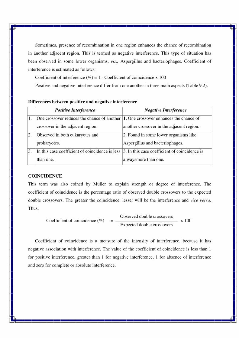
Sometimes, presence of recombination in one region enhances the chance of recombination
in another adjacent region. This is termed as negative interference. This type of situation has
been observed in some lower organisms, viz., Aspergillus and bacteriophages. Coefficient of
interference is estimated as follows:
Coefficient of interference (%) = 1 - Coefficient of coincidence x 100
Positive and negative interference differ from one another in three main aspects (Table 9.2).
Differences between positive and negative interference
Positive Interference Negative Interference
1. One crossover reduces the chance of another
crossover in the adjacent region.
1. One crossover enhances the chance of
another crossover in the adjacent region.
2. Observed in both eukaryotes and
prokaryotes.
2. Found in some lower organisms like
Aspergillus and bacteriophages.
3. In this case coefficient of coincidence is less
than one.
3. In this case coefficient of coincidence is
alwaysmore than one.
COINCIDENCE
This term was also coined by Muller to explain strength or degree of interference. The
coefficient of coincidence is the percentage ratio of observed double crossovers to the expected
double crossovers. The greater the coincidence, lesser will be the interference and vice versa.
Thus,
Coefficient of coincidence (%) = Observed double crossovers
x 100 Expected double crossovers
Coefficient of coincidence is a measure of the intensity of interference, because it has
negative association with interference. The value of the coefficient of coincidence is less than 1
for positive interference, greater than 1 for negative interference, 1 for absence of interference
and zero for complete or absolute interference.
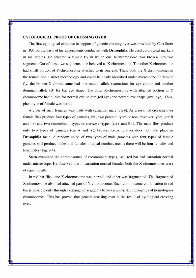
CYTOLOGICAL PROOF OF CROSSING OVER
The first cytological evidence in support of genetic crossing over was provided by Curt Stern
in 1931 on the basis of his experiments conducted with Drosophila. He used cytological markers
in his studies. He selected a female fly in which one X-chromosome was broken into two
segments. Out of these two segments, one behaved as X-chromosome. The other X-chromosome
had small portion of Y-chromosome attached to its one end. Thus, both the X-chromosomes in
the female had distinct morphology and could be easily identified under microscope. In female
fly, the broken X-chromosome had one mutant allele (carnation) for eye colour and another
dominant allele (B) for bar eye shape. The other X-chromosome with attached portion of Y
chromosome had alleles for normal eye colour (red eye) and normal eye shape (oval eye). Thus,
phenotype of female was barred.
A cross of such females was made with carnation male (car+). As a result of crossing over
female flies produce four types of gametes, viz., two parental types or non crossover types (car B
and ++) and two recombinant types or crossover types (car+ and B+). The male flies produce
only two types of gametes (car + and Y), because crossing over does not take place in
Drosophila male. A random union of two types of male gametes with four types of female
gametes will produce males and females in equal number, means there will be four females and
four males (Fig. 9.4).
Stern examined the chromosomes of recombinant types, viz., red bar and carnation normal
under microscope. He observed that in carnation normal females both the X-chromosomes were
of equal length.
In red bar flies, one X-chromosome was normal and other was fragmented. The fragmented
X-chromosome also had attached part of Y-chromosome. Such chromosome combination in red
bar is possible only through exchange of segments between non-sister chromatids of homologous
chromosomes. This has proved that genetic crossing over is the result of cytological crossing
over.
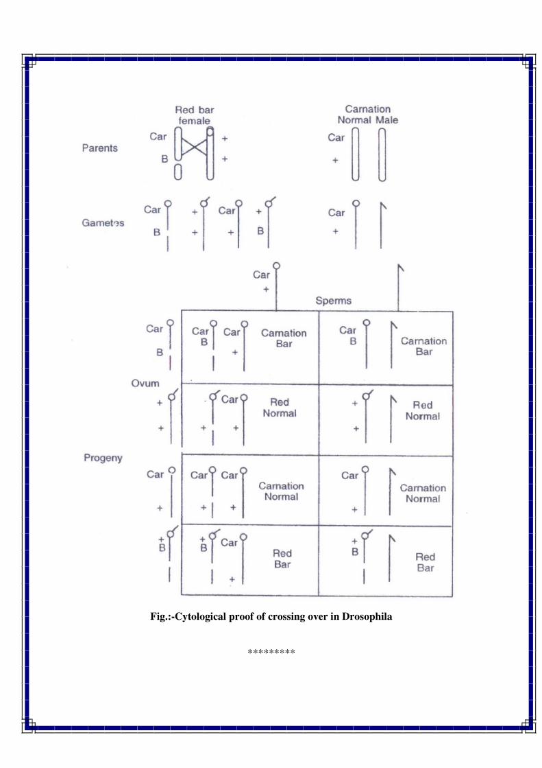
Fig.:-Cytological proof of crossing over in Drosophila
*********
Sex Determination and Sex Linkage, Sex Limited and Sex
Influenced Traits
INTRODUCTION
Sex refers to contrasting features of male and female individuals of the same species.
Thus sex is usually of two types, viz., male and female.
Sex determination is the process of sex differentiation, which utilizes various genetical
concepts to decide whether a particular individual will develop into male or female sex.
Some important points related to sex determination are given below:
1. Applicability of Sex Determination
Majority of animals are unisexual, whereas most of the plants are bisexual or hermaphrodite.
The sex determination is applicable to unisexual animals and monoecious and dioecious plant
species. In case of dioecious plants, male and female flowers are produced on different plants
such as in cucumber, papaya, asparagus, etc. On the other hand, in monoecious plants male and
female flowers are produced on the same plant such as in maize, castor and coconut.
2. Identification of Sex
Sex is identified with the help of sex characters. In plants, sex is identified with the help of
male and female floral parts. Thus in plants, morphological distinction of sex is possible only
after flowering. In mammals, sex characters are of two types, viz., primary sex characters and
secondary sex characters. Primary sex characters include gonads or sex organs which are
distinct since birth and take part in reproduction. Secondary sex characters express differently
in males and females in adulthood and do not take part in reproduction. In human, such
characters include voice, facial hair, mammary glands, muscles, etc.
3. Control of Sex
Sex expression is governed by chromosomes and genes. In some cases environment also
plays an important role in the expression of sex or determination of sex. In unisexual animals,
chromosomes are of two types, viz., autosomes and allosomes. Those chromosomes which do
not differ in morphology and number in male and female sex are referred to as autosomes. On
the other hand, those chromosomes which differ in morphology and number in male and female
sex and contain genes determining sex are known as allosomes or sex chromosomes. Sex
chromosomes were first discovered by Mc Lung in 1902 in grasshopers and later on by Wilson
and Stevens in 1905 in Protenor. Each unisexual diploid individual has two sex chromosomes
and rest autosomes. Thus autosomes and allosomes differ from one another in several aspects as
follows
Autosomes Allosomes or Sex chromosomes
1. Refer to other than sex chromosomes. 1. These are sex chromosomes.
2. Morphology is similar in male and
female
2. Morphology is different in male and female
3. The number is same in both the sexes. 3. The number is sometimes different in male
and female sex.
4. Generally control traits other than sex. 4. Usually determine sex of an individual.
5. Number of autosomes differs from
species to species.
5. Each diploid organism usually has two
allosomes.
6. Do not exhibit sex linkage. 6. Exhibit sex linkage.
MECHANISMS OF SEX DETERMINATION
There are three important mechanisms of sex determination, viz., 1. Chromosomal sex
determination, 2. Monogenic sex determination, and 3. Environmental sex determination (Table
10.2). These are briefly described below :
1. CHROMOSOMAL SEX DETERMINATION
Chromosomal sex determination is again of three types, viz., (a) Sex determination by
allosomes, (b) the diploid-haploid system of sex determination, and (c) genic balance system. All
these systems are briefly discussed below:
(a) ALLOSOMAL SEX DETERMINATION
Allosomes or sex chromosomes are generally of X and Y types, but in some birds they
are of Z and W types. Sex with similar type of sex chromosomes (XX) is known as homogametic
sex and with dissimilar type of sex chromosomes (XY) as heterogametic sex. There are four
different systems of allosomal sex determination, viz., 1. XX-XY female-male system, 2. XX-
XO female-male system, 3. XO-XX female-male system, and 4. ZW-ZZ female-male system.
These are briefly discussed below.
1. XX-XY [Female-Male] System
This system is found in Drosophila, man and some other mammals. In this system
female has two X chromosomes, is homogametic and produces only one type of gamete, i.e., X.
The male has one X and one Y chromosome, is heterogametic and produces two types of
gametes, viz, X and Y. Union of X ovum with X sperm leads to development of female (XX)
sex. If X ovum units with Y sperm, it produces male (XY) sex.
Table:-Mechanisms of sex determination found in animals and plants
Mechanism of sex
determination
Found in
Animals Plants
1. Chromosomal system
a) Allosomal system (Female—Male)
i) XX-XY System Drosophila, man and some
other mammals
Melandrium, Cannabis,
Coccinia, Huniulus lupulus,
Salix, Bryonea etc.
ii) XX-XO System Grasshoppers, many insects of
hemiptera and orthoptera.
Dioscorea sinuata,
Vallisneria spiralis
iii) XO-XX System Fumea —
iv) ZW-ZZ System
(XY-XX System)
Birds, butterflies, moths Fragaria
b) Diploid-Haploid System Honey bee —
c) Genie Balance System Drosophila —
2. Monogenic System Drosophila, man, goats, pigs,
dogs
Asparagus, maize, spinach,
papaya, etc.
3. Environmental Effect Bonellia and Dinophilus Equisetum, cucumber,
Muskmelon, and Cannabis.
Sex determination in Melandrium album
In this plant pistillate plants are XX and staminate plants are XY. In this plant one dose of
Y is able to produce male sex even in the presence of four X chromosomes. In other words,
presence of one Y chromosome can produce male flowers in diploid, triploid and tetraploid
species (Table)
Detailed investigation of the sex chromosomes of Melandrium revealed that Y
chromosome has four regions, viz., (1) male suppression region, (2) male promoter region, (3)
male fertility region, and (4) pairing region. Region four is common in X and Y
chromosomes. Hence pairing occurs in this region (Fig. 10.1). If first region is lost a bisexual
plant is produced. When second region is lost, female plant is produced, and when third region is
lost, male sterile plants with abortive anthers develop.
Fig. Sex chromosomes of Melandrium album. Region IV is homologous in X and Y
chromosomes, hence pairing occurs in this region.
This system differs from genic balance system in Drosophila. In this system, the male sex is
determined by the genes presented on Y chromosome, whereas in genic balance system male sex
is determined by the genes located on autosomes.
Table:- Sex determination in Melandrium in relation to autosomes and sex chromosomes
2. XX-XO [Female-Male] System
This type of sex determination is common in grasshoppers and many orthoptera and
hemiptera insects. In this system, female has double X chromosomes (XX) and male has single
X chromosome (XO). Here female is homogametic and produces all the eggs with X
chromosome. The male is heterogametic, which produces sperms half of which have X
chromosome and other have none. Union of egg with sperm having X chromosome will give rise
to female sex and with sperm having none results in development of male sex. In plants, this
system operates in Dioscorea sinuata and Vallisneria spiralis. Here the pistillate plants are XX
and staminate plants are XO.
3. XO-XX [Female-Male] System
This system has been reported in very few species of insects like Fumea. In this system,
female has only one X chromosome and hence is heterogametic. As a result of meiosis. 50%
eggs of such female carry an X chromosome and remaining 50% have none. On the other hand,
male has two X chromosomes and produces all the sperms with one X chromosome. Thus, male
sex is homogametic. Union of X sperm with ovum having X chromosome gives rise to male sex
and union of X sperm with ovum having none leads to development of female sex, Thus, system
is reverse of the system described above. This system has not been reported in plant so far.
4 ZW-ZZ [Female-Male] System
This system is found in birds, butterflies and moths. In this system, the female is
heterogametic and produces two types of gametes, namely Z and W types. On the other hand,
male is homogametic and, as a result of meiosis, produces all the sperms of same type carrying
one Z chromosome. Union of Z sperm with ovum having Z chromosome gives rise to male and
union of Z sperm with ovum carrying W chromosome leads to the development of female sex. In
plants, this system operates in Fragaria.
(b) DIPLOID-HAPLOID [FEMALE-MALE] SYSTEM
This system operates in honey bees, ants and termites. In honey bees, the females have
diploid (2n = 32) chromosomes and drones or males have haploid (n = 16) chromosomes. The
females are of two types, viz., queen and workers. Those females which feed on royal jelly
develop into queen. The queen is fertile and workers are sterile females. The queen produces
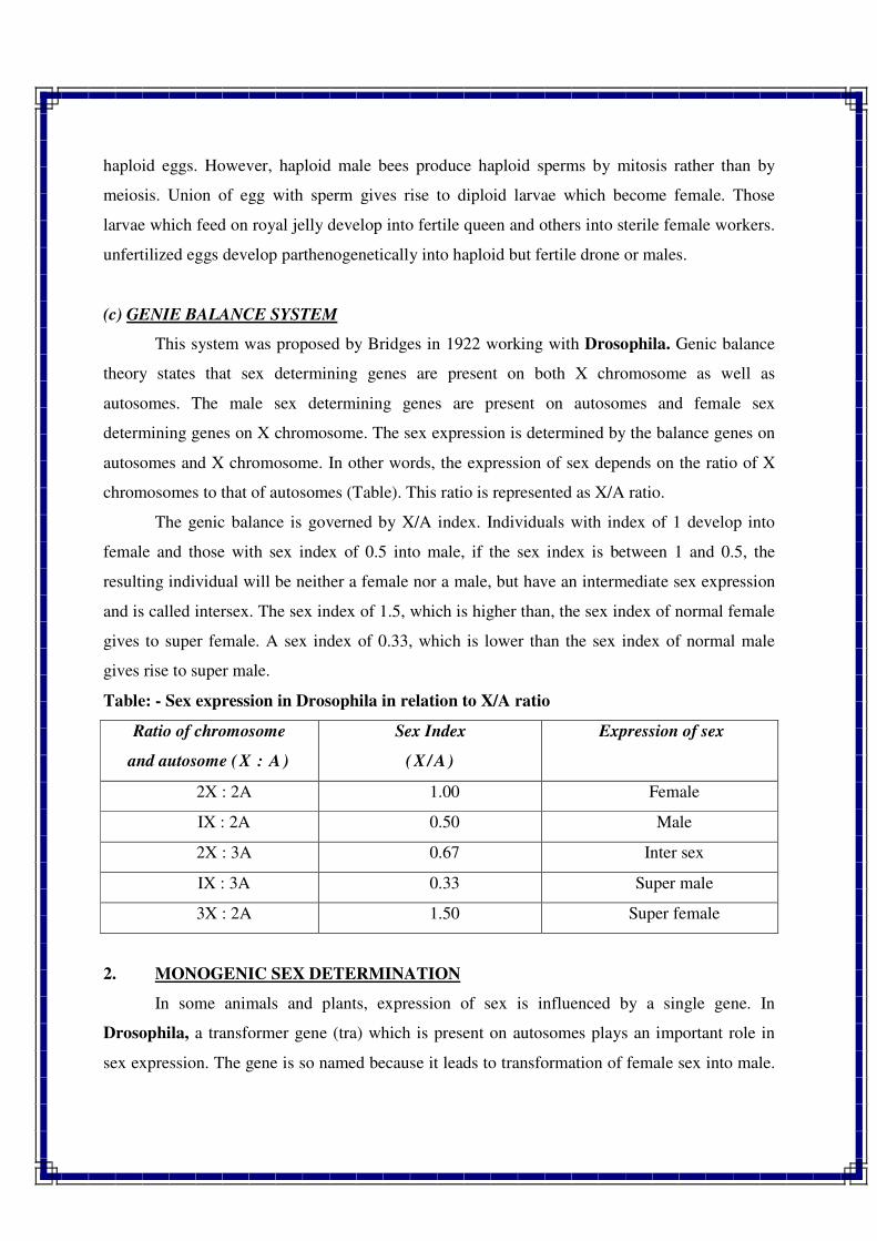
haploid eggs. However, haploid male bees produce haploid sperms by mitosis rather than by
meiosis. Union of egg with sperm gives rise to diploid larvae which become female. Those
larvae which feed on royal jelly develop into fertile queen and others into sterile female workers.
unfertilized eggs develop parthenogenetically into haploid but fertile drone or males.
(c) GENIE BALANCE SYSTEM
This system was proposed by Bridges in 1922 working with Drosophila. Genic balance
theory states that sex determining genes are present on both X chromosome as well as
autosomes. The male sex determining genes are present on autosomes and female sex
determining genes on X chromosome. The sex expression is determined by the balance genes on
autosomes and X chromosome. In other words, the expression of sex depends on the ratio of X
chromosomes to that of autosomes (Table). This ratio is represented as X/A ratio.
The genic balance is governed by X/A index. Individuals with index of 1 develop into
female and those with sex index of 0.5 into male, if the sex index is between 1 and 0.5, the
resulting individual will be neither a female nor a male, but have an intermediate sex expression
and is called intersex. The sex index of 1.5, which is higher than, the sex index of normal female
gives to super female. A sex index of 0.33, which is lower than the sex index of normal male
gives rise to super male.
Table: - Sex expression in Drosophila in relation to X/A ratio
Ratio of chromosome
and autosome (X : A )
Sex Index
( X / A)
Expression of sex
2X : 2A 1.00 Female
IX : 2A 0.50 Male
2X : 3A 0.67 Inter sex
IX : 3A 0.33 Super male
3X : 2A 1.50 Super female
2. MONOGENIC SEX DETERMINATION
In some animals and plants, expression of sex is influenced by a single gene. In
Drosophila, a transformer gene (tra) which is present on autosomes plays an important role in
sex expression. The gene is so named because it leads to transformation of female sex into male.
Transformer gene is recessive and hence does not have any effect in heterozygous condition
(Tra/tra) on either sex, i.e., male or female. In homozygous condition (tra/tra), this gene
transforms the normal diploid females into sterile males.
A similar gene is found in human. The gene is probably present on autosome and
transforms the normal male (XY) into female. In this case, the change is reverse as compared to
Drosophila case. Such males have feminine characters such as breast and vagina. They also have
internal degenerated testes and are sterile. This condition is known as testicular feminization.
Similar effects of single recessive gene on expression of sex have been found in some other
animals such as goats, pigs, dogs, etc.
Monogenic control of sex has also been reported in some plants like asparagus, maize,
papaya, spinach, etc. In papaya, the sex is postulated to be governed by three alleles, viz.. m, M1
and M2 of a single gene. Homozygous recessive (m m) produces female plants, heterozygous,
viz., M1m and M2m produce male and hermaphrodite plants, respectively. However, combination
of both dominant alleles (M1 and M2) produces inviable plants both in homozygous condition
(M1M1 and M2M2) as well as heterozygous condition (M1M2), Crosses between female (mm) and
male (M1m) produce females and males in 1 : 1 ratio. Similarly, crosses between female (mm)
and hermaphrodite (M2m) will produce females and hermaphrodite in 1 : 1 ratio. Seeds obtained
from hermaphrodite plants will produce hermaphrodite and female progeny in 2 : 1 ratio.
Maize being a monoecious plant bears both female (silk) and male (tassel) inflorescence
on the same plant. A recessive gene ba (barren cob) in homozygous condition (ba/ba) makes the
cobs barren or non-functional. Similarly, a recessive gene (ts) in homozygous condition (ts/ts)
converts the male flowers of tassel into female flowers. Thus, homozygous state of gene ba
(ba/ba} converts the monoecious plant into male. Similarly, gene (ts) in homozygous condition
(ts/ts) converts the monoecious plant into female. Plants with both dominant genes (BaBa/TsTs)
are normal monoecious, with baba/TsTs normal male, with BaBa/tsts normal female and with
baba/tsts rudimentary females.
3. ENVIRONMENTAL SEX DETERMINATION
Sex determination in some organisms such as sea worm (Bonellia and Dinophilus) and
horse tail plant (Equisetum) is governed by environmental conditions and also includes some
hormonal effects.
In Bonellia, those larvae which remain free in the sea water and settle on the sea bottom
are differentiated into females. On the other hand, those larvae which settle on the proboscis of
female develop into tiny males. They do not have digestive organs, hence move from the
proboscis to the uterus of female where they remain as parasites.
In Dinophilus, the determination of sex appears to depend solely on the size of egg
produced by the females. Larger eggs after fertilization always develop into females and smaller
eggs into male animals.
In horse tail plant (Equisetum), development of sex depends on growing conditions.
Plants grown under good conditions develop as females and those grown under stress conditions
develop into males. In some plants, sex differentiation depends on day length, temperature and
hormones. For example, in cucumber (Cucumis sativa) and muskmelon, treatment with ethylene
enhances production of female flowers.
SEX MOSAIC IN DROSOPHILA
Combination of male and female features in the body of an individual is known as sex
mosaic. In Drosophila, flies with a very low frequency (1 in 2000) have male tissues in one part
of the body and female tissues in other. Individuals with such sex mosaic are known as
gynandromorphs or gynanders.
Three patterns of sex mosaic may be found in Drosophila, viz., bilateral, antero/ posterior
and in patches. In case of bilateral sex mosaic, one side of the fly is male and the other side is
female. This is the most common type of sex mosaic found in Drosophila. In case of
antero/posterior pattern of sex mosaic, the front half of the fly is one sex and the rear half of the
other sex. Sometimes, only a quarter of the body is male and rest is female or just a small patch
of tissues may be male and rest of the body is female. The last two patterns are of rare
occurrence.
Gynandromorphs develop from a normal female having one pair of X-chromosomes. In
the first mitotic division of zygote, one X-chromosome is lost in the cytoplasm. As a result, one
of the two cells has only one X-chromosome. Thus, one cell is differentiated into male tissue.
Further, division of these two types of cells results in the development of bilateral type of
gynander. If such mistake occurs at the second mitosis of embryogenesis, only one cell out of
four will produce male tissue. In this case only one fourth of the body will be male. If such
mistake in mitosis takes place at later mitotic divisions, only a small portion of male tissues will
be produced. Gynandromorphs are detected with the help of phenotypic characters such as
banding pattern of abdomen, presence or absence of sex combs on the fore legs and structure of
external genitalia.
SEX REVERSAL
Sex reversal refers to transformation of one sex to another. A good example of partial sex
reversal is found in chickens. In rare cases, old hens which have been good egg layers for many
years start exhibiting such features which are indicative of male sex. They develop male comb
and male plumage, begin to crow and try to mate with other hens. Crew reported such extreme
case of sex reversal in hen. A normal fertile hen, after several years of egg laying changed into
male, produced viable sperms and became father of two chicks. In birds, there is only one
functional ovary (left sided). In the centre of this ovary a small mass of tissues similar to male
testes is present. The ovary usually produces female hormone which results in full expression of
female features. In some cases, the ovary is destroyed due to tubercular infection. In such cases,
the testes like tissues enlarge and produce male hormone in ample quantity resulting in reversal
of sex. Removal of ovary by way of surgery, and leaving the testes like portion may also result in
sex reversal in hens.
HUMAN SEX ANOMALIES
There are two types of human sex abnormalities, viz., 1. Klinefelter's syndrome, and 2.
Turner's syndrome. These are briefly described below :
1. Klinefelter's Syndrome
Klinefelter's syndrome refers to the characteristics of an abnormal human male who has an XXY
chromosome constitution. Such individuals have 47 chromosomes (44 autosomes plus XXY),
whereas normal human has 46 chromosomes (44 autosomes + XX or XY). Frequency of such
persons is 1 in every 5000 births.
Phenotype
Such persons have typical male sex organs, but testes are about half of the normal size,
sperms are usually not produced and most of such persons are mentally retarded. Their arms are
longer than average. Some degree of breast development also occurs. Hair are feminine in nature
and voice is higher pitched than in normal males. Such persons are always sterile and are sex
chromatin positive.
Origin
The XXY individuals may arise when an XX egg is fertilized by a Y sperm or an X egg is
fertilized by XY sperm. The majority of Klinefelter's syndromes are born to mothers after the
age of 32. It results mainly due to non-disjunction of the X chromosomes in the ageing oocytes
than XY non-disjunction during spermatogenesis. In very rare cases, Klinefelter's syndromes
have more than two X chromosomes or more than one Y chromosome. Generally, greater the
number of X chromosomes greater the degree of mental retardation. Presence of one Y
chromosome leads to general phenotype of male even in the presence of several X chromosomes.
2. Turner's Syndrome
It refers to the characteristics of an abnormal human female who has an XO chromosome
constitution. Such individuals have 45 chromosomes (44 autosomes plus one X chromosome),
their frequency is 1 in every 3000 births.
Phenotype
Such individuals are phenotypically females, but have poorly developed and sterile
ovaries. They are dwarf (below 5 feet), often show some mental retardation, webbing of the
neck, low set ears, broad chest and under developed breasts. They are sex chromatin negative
which confirms presence of single X chromosome.
Origin
The XO individuals may arise due to non-disjunction of X and Y chromosomes in the
male. In the absence of Y chromosome, the general sex phenotype is the female. Thus, union of
A+X egg with A+O sperm produces AA+XO individuals.
BARR BODY OR SEX CHROMATIN
Barr body refers to relatively coiled and inactive interphase chromatin of the X
chromosomes of the normal mammalian females but not the males. The Barr body was first
discovered by a geneticist, Murray Barr in nerve cells of cat.
Barr bodies were found in the nucleus of female cats only. They take dark stain like
DNA. Later on Barr bodies were found in females of many mammals including man. Barr
bodies are also known as sex chromatin.
In humans, Barr bodies are studied in the epithelial cells, which can be easily obtained
from the lining of the mouth, vagina or urethra. In women the Barr bodies are found near the
nuclear membrane and look like a round disc. The number of Barr bodies in a female cell is
always one less than the number of X chromosomes. Thus, a normal woman has two X
chromosomes and one Barr body. A woman with Turner's syndrome has one X chromosome but
no Barr body. Normal males have no Barr body, but males with Klinefelter's syndrome have two
X chromosomes and one Barr body. Thus Barr bodies are useful tools for detection of sex
abnormalities and also in the determination of sex in humans. Individuals which have Barr
bodies are known as sex chromatin positive and those who do not have Barr bodies are called as
sex chromatin negative.
LYONS HYPOTHESIS
This hypothesis was proposed by Lyon in 1969, which states that in a normal female only
one X chromosome is active which is invisible at interphase. The other X chromosome remains
inactive, takes dark stain and becomes visible in interphase nucleus.
In other words, an individual may have any number of X chromosomes, but only one
remains active and others become condensed or inactive. The number of inactive chromosomes
is represented by the number of sex chromatin bodies.
Though the male and female individuals in Drosophila and man differ from one another
for dose of X chromosome, the male has one X chromosome and female has two X
chromosomes. However, they do not differ in their gene products. This reveals that either one X
chromosome should be inactivated in female or the efficiency of single X chromosome of male
should be doubled. This phenomenon is known as dosage compensation which was proposed by
Muller in 1932. Discovery of Barr bodies and Lyon's hypothesis have confirmed the
concept of dosage compensation.
SEX LINKAGE
Characters for which genes are located on sex or X chromosomes are known as sex
linked traits, such genes are called sex linked genes and linkage of such genes is referred to as
sex linkage. Inheritance of such genes or characters is known as sex linked inheritance. Main
features of sex linked genes are briefly discussed below:
1. Location
Sex linked genes are located on X chromosomes only. Hence, inheritance of sex linked
genes follows the inheritance pattern of X chromosomes. The Y chromosome determines only
sex, is usually inert, and does not contain genes for other characters.
2. Number
In a diploid organism, each homogametic sex (XX or ZZ) has two copies of linked
alleles. These alleles may be either in homozygous condition (AA or aa) or in heterozygous state
(Aa). The heterogametic sex (XY or ZW) has only one copy of the sex linked allele (either A or
a), which may be either dominant or recessive. Thus heterozygous sex is hemizygous for a sex
linked gene.
3. Expression
A recessive gene, in a homogametic sex can express only when it is in homozygous state.
However, in heterogametic sex, a recessive allele can express even in a single dose (hemizygous
condition) because the Y chromosome is genetically inactive. As a result, expression of recessive
sex linked character is higher in heterogametic sex than in homogametic sex.
4. Transmission
Sex linked genes are transmitted from female to the male and not from the male to the
male, because males receive their X chromosome from the female only. Similarly, sex linked
genes are inherited from male to the female, because female receives one of its X chromosome
from the male. Thus sex linked genes are first inherited from male to female in the first
generation and then back to male in the second generation. This type of inheritance in which a
sex linked gene is inherited from grandfather to grandson through daughter is called cris-cross
inheritance.
5. Pattern of Segregation
Inheritance of sex linked characters does not follow normal segregation pattern. It
exhibits several deviations from the normal segregation pattern. The deviation of sex linked
inheritance from the normal one is due to location of such genes in the X chromosome and
absence of such alleles in the Y chromosome.
SEX LINKED CHARACTERS
Sex linked characters are known in man (colour blindness, hemophilia, baldness, etc.),
Drosophila (white eye, vermilion eye) and several other organisms. Some examples of sex
linked genes are given below:
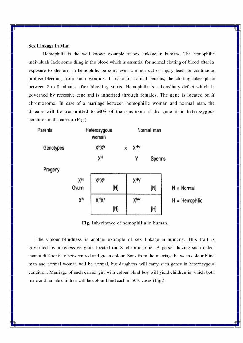
Sex Linkage in Man
Hemophilia is the well known example of sex linkage in humans. The hemophilic
individuals lack some thing in the blood which is essential for normal clotting of blood after its
exposure to the air, in hemophilic persons even a minor cut or injury leads to continuous
profuse bleeding from such wounds. In case of normal persons, the clotting takes place
between 2 to 8 minutes after bleeding starts. Hemophilia is a hereditary defect which is
governed by recessive gene and is inherited through females. The gene is located on X
chromosome. In case of a marriage between hemophilic woman and normal man, the
disease will be transmitted to 50% of the sons even if the gene is in heterozygous
condition in the carrier (Fig.)
Fig. Inheritance of hemophilia in human.
The Colour blindness is another example of sex linkage in humans. This trait is
governed by a recessive gene located on X chromosome. A person having such defect
cannot differentiate between red and green colour. Sons from the marriage between colour blind
man and normal woman will be normal, but daughters will carry such genes in heterozygous
condition. Marriage of such carrier girl with colour blind boy will yield children in which both
male and female children will be colour blind each in 50% cases (Fig.).
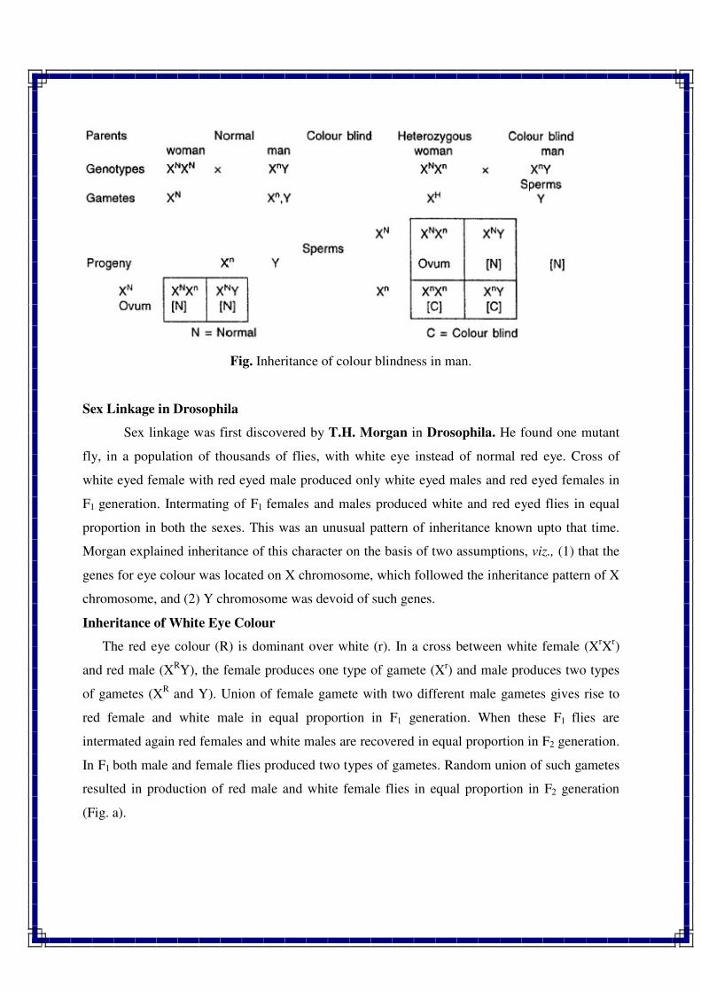
Fig. Inheritance of colour blindness in man.
Sex Linkage in Drosophila
Sex linkage was first discovered by T.H. Morgan in Drosophila. He found one mutant
fly, in a population of thousands of flies, with white eye instead of normal red eye. Cross of
white eyed female with red eyed male produced only white eyed males and red eyed females in
F1 generation. Intermating of F1 females and males produced white and red eyed flies in equal
proportion in both the sexes. This was an unusual pattern of inheritance known upto that time.
Morgan explained inheritance of this character on the basis of two assumptions, viz., (1) that the
genes for eye colour was located on X chromosome, which followed the inheritance pattern of X
chromosome, and (2) Y chromosome was devoid of such genes.
Inheritance of White Eye Colour
The red eye colour (R) is dominant over white (r). In a cross between white female (XrX
r)
and red male (XRY), the female produces one type of gamete (Xr) and male produces two types
of gametes (XR and Y). Union of female gamete with two different male gametes gives rise to
red female and white male in equal proportion in F1 generation. When these F1 flies are
intermated again red females and white males are recovered in equal proportion in F2 generation.
In F1 both male and female flies produced two types of gametes. Random union of such gametes
resulted in production of red male and white female flies in equal proportion in F2 generation
(Fig. a).
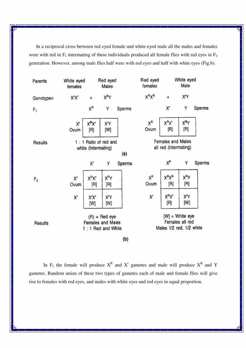
In a reciprocal cross between red eyed female and white eyed male all the males and females
were with red in F1 intermating of these individuals produced all female flies with red eyes in F2
generation. However, among male flies half were with red eyes and half with white eyes (Fig.b).
In F1 the female will produce XR and X
r gametes and male will produce X
R and Y
gametes. Random union of these two types of gametes each of male and female flies will give
rise to females with red eyes, and males with white eyes and red eyes in equal proportion.
SEX LIMITED TRAITS
Characters which are expressed in one sex only are referred to as sex limited characters.
These characters have three main characteristics as given below :
1. They express in one sex only and not in the other sex.
2. Sex limited genes may be located either in sex chromosome or autosomes.
3. Sex limited genes control the expression of primary and secondary sex characters.
Table:-. Differences between sex linked and sex limited characters
Sex Linked Characters Sex Limited Characters
1. They are located on sex or X
chromosomes.
1. They are located on sex chromosomes or
autosomes.
2. They can express in both the sexes. 2. They express in one sex only.
3. Include characters not related to sex. 3. Include primary and secondary sex
characters.
4. Examples are white eye in Drosophila,
hemophilia and colour blindness in man.
4. Examples are breast development in
woman and beard in man.
SEX INFLUENCED GENES
The dominance expression of some genes depends on the sex of individual. Such genes are
known as sex influenced genes and characters governed by such genes are referred to as sex
influenced characters. These genes have three main features as given below :
1. Such genes are located in the autosomes.
2. They express more frequently in one sex than other.
3. Expression of such characters appears to be governed by sex hormones.
The best known example of sex influenced character in pattern of baldness in humans. The
gene responsible for baldness behaves as dominant in males and as recessive in females.
Similarly, horns in sheep behave as dominant in males and recessive in females. The baldness
character expresses in woman only when it is in dominant homozygous state, while in man it
expresses even in heterozygous condition.
GLOSSARY
1. Sex. Contrasting features of male and female individuals of the same species.
2. Sex determination. The process of sex differentiation which utilizes various genetical
concepts to decide whether a particular genotype will develop into male or female sex.
3. Monoecious. Plant species which bear male and female flowers on the same plant such as
maize, caster and coconut.
4. Dioecious. Plant species in which male and female flowers are produced on different plants
such as cucumber, papaya, asparagus, etc.
5. Primary sex characters. Characters which are distinct since birth and take part in
reproduction such as gonads or sex organs in mammals.
6. Secondary sex characters. Characters which express in adulthood differently i n males and
females and do not take part in reproduction. In humans, such characters include voice, facial
hair, mammary glands, muscles, etc.
7. Autosomes. Those chromosomes which do not differ in number and morphology in male and
female sex.
8. Allosomes. Those chromosomes which differ in number and morphology in male and female
sex; also known as sex chromosomes.
9. Heterogametic. Sex with dissimilar type of sex chromosomes such as XY, XO and ZW.
10. Homogametic sex. Sex with similar type of sex chromosomes such as XX or ZZ.
11. Sex Mosaic. Combination of male and female features in the body of an individual.
12. Gynandromorphs. Individuals with sex mosaic; also called gynanders.
13. Sex reversal. Transformation of one sex into another.
14. Klinefelter's syndrome. The characteristics of an abnormal human male who has XXY
chromosome constitution.
15. Turner's syndrome. The characteristics of an abnormal human female who has XO
chromosome constitution.
16. Sex chromatin. Relatively coiled and inactive interphase chromatin of the X chromosomes
of the normal mammalian females but not the males; also called Barr Body.
17. Lyon's Hypothesis. This hypothesis states that an individual may have any number of X-
chromosomes, but only one remains active and others become inactive or condensed.
18. Sex linkage. The linkage of genes which are located on sex or X chromosomes. Inheritance
of such genes is known as sex linked inheritance.
19. Sex linked traits. Characters for which genes are located on sex- or X-chromosomes.
20. Sex limited traits. Characters which express in one sex only.
21. Sex influenced genes. Genes whose expression depends on the sex of an individual such as
baldness in humans.
22. Primary non-disjunction. Failure of homologous chromosomes to separate at meiotic
anaphase in a normal Drosophila female.
23. Secondary non-disjunction. Failure of chromosomes to separate in XXY individuals
resulting in production of four types of egg cells, viz., XX, XY, X and Y.
24. Hemizygous. A recessive allele in a single dose.
25. Cris-cross inheritance. Inheritance of sex linked genes from grand father to grand son
through daughter.
//*//*//*//
1
POLYGENIC INHERITANCE
QUANTITATIVE TRAITS / QUALITATIVE CHARACTERS:
o Quantitative traits : The characters, which are controlled by polygenes are referred as
quantitative character or polygenic characters. e.g. grain yield, leaf size, corolla length,
maturity etc.
o Qualitative traits : The characters, which are controlled by one or two genes are referred
as qualitative characters or oligogenic characters. e.g. fruit colour, seed colour etc.
o Oligogene : One or few genes governs the same character.
o Polygene : Several genes governs the same character.
Characteristics of Quantitative Inheritance (Polygenic Trait)
o Each quantitative character is controlled by several independent genes and each gene
having small, similar and cumulative effect.
o Each quantitative character exhibits continuous variation rather than discontinuous
variation. The variability observed for quantitative trait cannot be classified into distinct
phenotypic classes.
o Effect of individual gene is not easily detectable in case of quantitative characters and
therefore, such traits are also known as minor gene characters.
o The statistical analysis of quantitative character is based on means, variances and co-
variances. Thus, quantitative characters are studied in quantitative genetics.
o Quantitative characters are highly sensitive to environmental variation.
o Classification of quantitative characters into different clear cut phenotypic classes is not
possible because of continuous variation from one extreme to the other extreme.
o Generally, the expression of quantitative characters is governed by additive gene action
but now the cases are known where quantitative characters are governed by dominance
and epistatic gene action.
o Each quantitative character involves metric measurements like size, weight, height etc.
o Transgressive segregants are only possible from the crosses between two parents for
quantitative characters.
2
o The transmission of quantitative characters is generally low because of high influence of
environmental variation.
o If a substitution of an allele occurs in a gene locus then such allelic substitution have
different effects.
o Quantitative traits are controlled by polygenes and polygenes have pleiotropic effects i.e.
one gene may modify or suppress more than one phenotypic trait.
Characteristics of Qualitative Inheritance (Oligogenic Trait)
o Each qualitative character is controlled by few independent genes and each gene has its
independent effect.
o Oligogenic characters exhibits discontinuous variation.
o Effect of individual gene is easily detectable in case of qualititative characters.
o The statistical analysis of the discontinuous variation can be analyzed with the help of
frequencies and ratios.
o Qualitative characters are less sensitive to environmental variation.
o Qualitative characters are classified into distinct phenotypic classes.
o Generally, the expression of quantitative characters is governed by non-additive gene
action (dominance and epistatic gene action).
o Transgressive segregants are not possible in case of qualitative or oligogenic inheritance.
o Qualitative characters exhibit high transmission because there is limited difference
between the genotype and phenotype of such characters.
PARTITIONING OF POLYGENIC VARIABILITY (Components of Variation)
The polygenic variation or variability present in a genetic population is measured in
terms of variances. The polygenic variation is of three types, viz., (1) phenotypic, (2) genotypic,
(3) and environmental. These are briefly described below:
1. Phenotypic Variability
It is the total variability which is observable. It includes both genotypic and environmental
variation and hence changes under different environmental conditions. Such variation is
measured in terms of phenotypic variance.
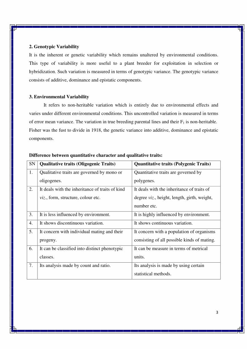
3
2. Genotypic Variability
It is the inherent or genetic variability which remains unaltered by environmental conditions.
This type of variability is more useful to a plant breeder for exploitation in selection or
hybridization. Such variation is measured in terms of genotypic variance. The genotypic variance
consists of additive, dominance and epistatic components.
3. Environmental Variability
It refers to non-heritable variation which is entirely due to environmental effects and
varies under different environmental conditions. This uncontrolled variation is measured in terms
of error mean variance. The variation in true breeding parental lines and their F1 is non-heritable.
Fisher was the fust to divide in 1918, the genetic variance into additive, dominance and epistatic
components.
Difference between quantitative character and qualitative traits:
SN Qualitative traits (Oligogenic Traits) Quantitative traits (Polygenic Traits)
1. Qualitative traits are governed by mono or
oligogenes.
Quantitative traits are governed by
polygenes.
2. It deals with the inheritance of traits of kind
viz., form, structure, colour etc.
It deals with the inheritance of traits of
degree viz., height, length, girth, weight,
number etc.
3. It is less influenced by environment. It is highly influenced by environment.
4. It shows discontinuous variation. It shows continuous variation.
5. It concern with individual mating and their
progeny.
It concern with a population of organisms
consisting of all possible kinds of mating.
6. It can be classified into distinct phenotypic
classes.
It can be measure in terms of metrical
units.
7. Its analysis made by count and ratio. Its analysis is made by using certain
statistical methods.
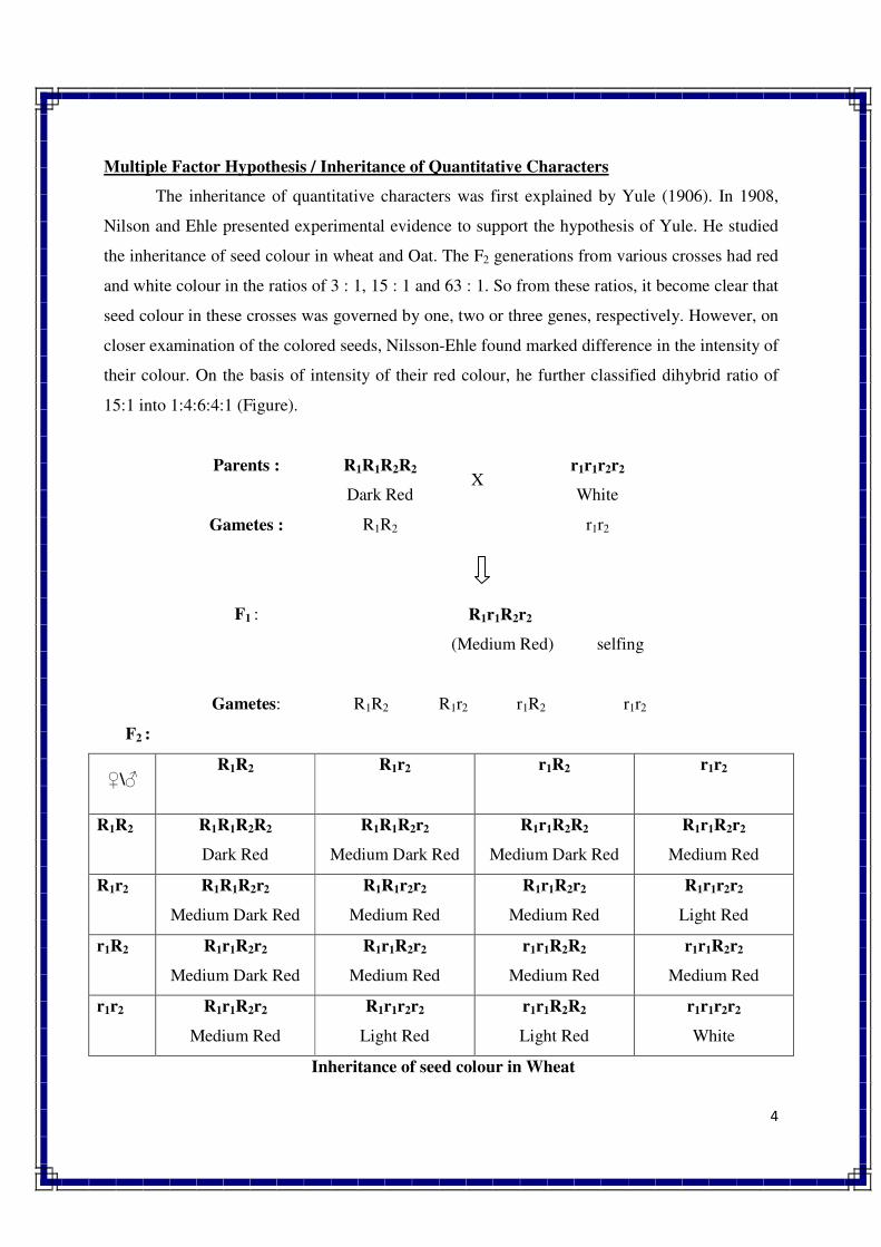
4
Multiple Factor Hypothesis / Inheritance of Quantitative Characters
The inheritance of quantitative characters was first explained by Yule (1906). In 1908,
Nilson and Ehle presented experimental evidence to support the hypothesis of Yule. He studied
the inheritance of seed colour in wheat and Oat. The F2 generations from various crosses had red
and white colour in the ratios of 3 : 1, 15 : 1 and 63 : 1. So from these ratios, it become clear that
seed colour in these crosses was governed by one, two or three genes, respectively. However, on
closer examination of the colored seeds, Nilsson-Ehle found marked difference in the intensity of
their colour. On the basis of intensity of their red colour, he further classified dihybrid ratio of
15:1 into 1:4:6:4:1 (Figure).
Parents : R1R1R2R2
Dark Red X
r1r1r2r2
White
Gametes : R1R2 r1r2
F1 : R1r1R2r2
(Medium Red) selfing
Gametes: R1R2 R1r2 r1R2 r1r2
F2 :
♀\♂ R1R2 R1r2 r1R2 r1r2
R1R2 R1R1R2R2
Dark Red
R1R1R2r2
Medium Dark Red
R1r1R2R2
Medium Dark Red
R1r1R2r2
Medium Red
R1r2 R1R1R2r2
Medium Dark Red
R1R1r2r2
Medium Red
R1r1R2r2
Medium Red
R1r1r2r2
Light Red
r1R2 R1r1R2r2
Medium Dark Red
R1r1R2r2
Medium Red
r1r1R2R2
Medium Red
r1r1R2r2
Medium Red
r1r2 R1r1R2r2
Medium Red
R1r1r2r2
Light Red
r1r1R2R2
Light Red
r1r1r2r2
White
Inheritance of seed colour in Wheat
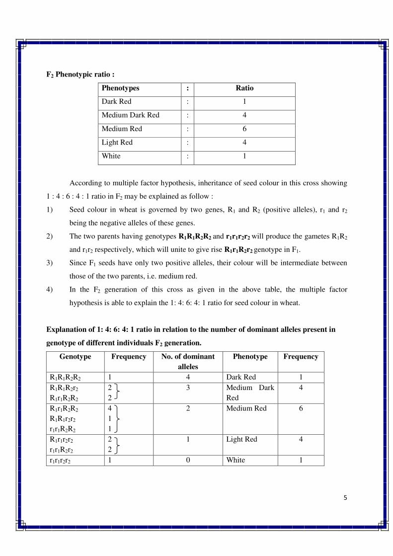
5
F2 Phenotypic ratio :
Phenotypes : Ratio
Dark Red : 1
Medium Dark Red : 4
Medium Red : 6
Light Red : 4
White : 1
According to multiple factor hypothesis, inheritance of seed colour in this cross showing
1 : 4 : 6 : 4 : 1 ratio in F2 may be explained as follow :
1) Seed colour in wheat is governed by two genes, R1 and R2 (positive alleles), r1 and r2
being the negative alleles of these genes.
2) The two parents having genotypes R1R1R2R2 and r1r1r2r2 will produce the gametes R1R2
and r1r2 respectively, which will unite to give rise R1r1R2r2 genotype in F1.
3) Since F1 seeds have only two positive alleles, their colour will be intermediate between
those of the two parents, i.e. medium red.
4) In the F2 generation of this cross as given in the above table, the multiple factor
hypothesis is able to explain the 1: 4: 6: 4: 1 ratio for seed colour in wheat.
Explanation of 1: 4: 6: 4: 1 ratio in relation to the number of dominant alleles present in
genotype of different individuals F2 generation.
Genotype Frequency No. of dominant
alleles
Phenotype Frequency
R1R1R2R2 1 4 Dark Red 1
R1R1R2r2
R1r1R2R2
2
2
3 Medium Dark
Red
4
R1r1R2R2
R1R1r2r2
r1r1R2R2
4
1
1
2 Medium Red 6
R1r1r2r2
r1r1R2r2
2
2
1 Light Red 4
r1r1r2r2 1 0 White 1
6
MULTIPLE FACTOR HYPOTHESIS: Continuous variation of the characters is controlled by
polygene with no dominance and here each of the genes having small, similar and cumulative
effect. This is the essence of the multiple factor hypothesis.
//*//*//*//
CYTOPLASMIC INHERITANCE
Cytoplasmic inheritance:
o The transmission of characters from parents to offspring is governed by cytoplasmic
genes, it is known as cytoplasmic inheritance.
o Cytoplasmic inheritance is also known as extra-nuclear inheritance or extra
chromosomal inheritance or non-mendelian inheritance or organellar inheritance.
o The first case of cytoplasmic inheritance was reported by correns in four o’clock
(Mirabillis jalaba) for leaf colour.
Characteristics / Features of cytoplasmic inheritance:
1. Reciprocal differences
Characters govern by cytoplasmic inheritance showed the marked differences in
reciprocal crosses in F1. Whereas, such kinds of reciprocal differences are not observed in
case of nuclear inheritance.
2. Maternal Effects
In case of cytoplasmic inheritance, distinct maternal effect is observed and it is
mainly due to more contribution of cytoplasm to the zygote by female parent than male
parent.
3. Mappability
Nuclear genes are easily mapped on chromosomes but it is very difficult to map
cytoplasmic genes.
4. Non-Mendelian Segregation
In case of nuclear inheritance, typical mendelian inheritance pattern is found but
in case of cytoplasmic inheritance such type of inheritance pattern is not observed.
5. Somatic Segregation
Characters governed by cytoplasmic genes usually exhibits segregation in somatic
tissues such as leaf variegation but such type of segregation is very rare for nuclear genes.
6. Infection-like Transmission
Cytoplasmic traits in some organisms exhibit infections like transmission. They
are associated with parasites or viruses present in the cytoplasm but such type of cases do
not come under true cytoplasmic inheritance.
7. Governed by Plasma Genes
The true cases on cytoplasmic inheritance are governed by chloroplast or
mitochondrial DNA. In other words, plasma genes are made up of cp-DNA or mt-DNA.
Difference between nuclear inheritance vs cytoplasmic inheritance :
Sr. Mendelian (Nuclear) inheritance Non mendelian (Cytoplasmic) inheritance
1. Governed by nuclear genes Governed by plasma genes
2. Exhibits distinct segregation pattern Does not exhibit distinct segregation
3. Reciprocal differences are not observed. Reciprocal differences are observed.
4. Does not show maternal effects. Exhibits maternal effects.
5. Genes can be easily mapped on
chromosomes.
Mapping of plasma genes is very difficult.
6. Nuclear genes are associated with
chromosomes.
Plasma genes are associated with either
chloroplast DNA or mitochondrial DNA.
Classes of Cytoplasmic Inheritance
There are three different classes of cytoplasmic inheritance or non-Mendelian inheritance viz.,
1) Materal effects: e.g. coiling pattern of shell in snail.
2) Inheritance due to infective particles: e.g. Kappa particles in Paramecium.
3) Cytoplasmic inheritance:
a. Plastid inheritance: e.g. Leaf color in Four o'clock
b. Mitochondrial inheritance: e.g. cytoplasmic male sterility.
1) MATERNAL EFFECTS
Coiling pattern of shell in Snail :
o The effect of maternal genotype on the coiling behaviour in water snail was studied by
Sturtevant.
o There are two types of coiling pattern of shell in snail viz., right handed (dextral) and
left handed (sinistral).
o The coiling behaviour is controlled by a single gene.
o The dextral coiling behaviour is governed by dominant allele D and sinistral by recessive
allele d.
o When a cross is made between dextral female and sinistral male, it produces dextral
snails in F1 as well in F2.
o However, in F3 segregation ratio of 3 dextral and 1 sinistral is observed.
o Similarly, reciprocal cross is made, i.e., sinistral as female and dextral as male, all the
snails are sinistral in F1 and dextral in F2. Again in F3 ratio of 3 dextral and 1 sinistral is
observed.
o This indicates that the inheritance of coiling direction in water snail depends on the
genotype of female parent and not on its own genotype.
Parents : Dextral Female Sinistral Male Sinistral Female Dextral Male
DD X dd dd X DD
F1 Dd
Dextral
(Intermating)
Dd
Sinistral
(Intermating)
F2 1 DD 2 Dd 1 dd
All Dextral
1 DD 2 Dd 1 dd
All Dextral
F3 DD 1 DD 2 Dd 1 dd dd
dextral dextral sinistral
DD 1 DD 2 Dd 1 dd dd
dextral dextral sinistral
Maternal effect on coiling direction of water snail
2) INHERITANCE DUE TO INFECTIVE PARTICLES
In some cases, cytoplasmic inheritance is associated with infective particles like parasite,
symbiont or viruses which are present in the cytoplasm of an organismSome examples of this
type are given below:
(i) Kappa Particles in Paramecium
There are two types of strains in Paramecium. One has kappa particles in its cytoplasm and
other does not have such particles. The presence of kappa particles in the cytoplasm leads to
production of a toxin known as paramecin. This toxin can kill the strain of Paramecium which
lacks kappa particle. Thus, the strain with kappa particle is known as killer strain and that
without kappa particle is called as sensitive strain.
Multiplication of kappa particles in the cytoplasm takes place by Fission. However, their
multiplication is governed by a dominant nuclear gene (K). They can multiply in the
homozygous dominant (KK) or heterozygous (Kk) individuals. Kappa particles cannot multiply
in recessive (kk) individuals. Even if kappa particles are introduced into kk strains, they will
gradually disappear due to their inability to multiply and the strain will become sensitive.
Though the multiplication of kappa particles is dependent on nuclear genes, but their
action is independent of nuclear gene. The inheritance of kappa particles can be studied by
conjugation (transfer of genetic material between bacterial cells by direct cell-to-cell contact or by a bridge-like
connection between two cells) between killer and sensitive strains. The conjugation may be of two
types, viz., I. short duration conjugation and 2. long duration conjugation. The consequences of
such conjugations are given below :
( a ) Short Duration Conjugation. Short duration conjugation leads to exchange of nuclear
genes between the killer and sensitive strains. Exchange of cytoplasm does not take place
in such conjugation. Thus, the ex-conjugants (resultant strains) will be heterozygous (Kk)
for killer gene. However, the strain with killer cytoplasm produces killer (KK) and
sensitive (kk) strains by further division, whereas the sensitive stain produces only
sensitive strains (kk) by further division. This clearly indicates that the killer character is
not governed by nuclear gene.
( b ) Long Duration Conjugation. Such conjugation between killer and sensitive strains leads
to exchange of both nuclear genes as well as cytoplasm. Here both the ex-conjugants are
heterozygous (Kk) but killer. Autogamy of both the ex-conjugants produces killer and
sensitive strains in 1 : 1 ratio. This has demonstrated that kappa particles have cytoplasmic
inheritance .
3) CYTOPLASMIC INHERITANCE:
a) Plastid inheritance: e.g. Iojap in Maize, Leaf color in Four o'clock
In maize, three types of leaves are found, viz, green, iojap (green and white stripes) and
white. Crosses between green female and iojap male produced all green individuals in F1 and a
single gene segregation ratio, i.e., 3 green and 1 iojap in F2. However, the reciprocal cross (iojap
female x green male) produced individuals with all the three phenotypes, viz., green, white and
striped in F1.
The iojap phenotype is governed by plastids. The green leaves have normal plastids; white
leaves have mutant plastids and striped leaves have mixture of normal and mutant plastids. In a
cross between iojap female and green male, there are three types of plastids, viz., green, white or
both in the egg cell. Depending upon the presence of these three types of plastids in the egg cell,
a cross between iojap and green will produce three types of individuals, viz., green, white and
striped in F1, because male parent does not contribute cytoplasm and thereby plastids to the
zygote.
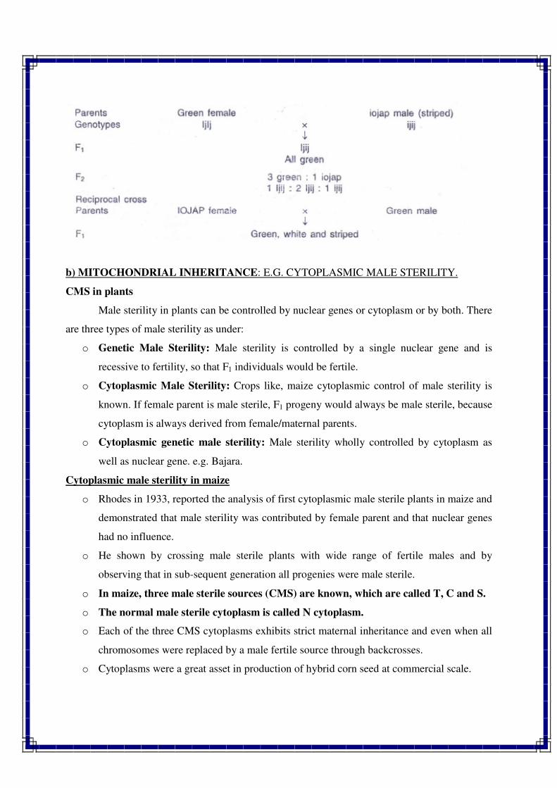
b) MITOCHONDRIAL INHERITANCE: E.G. CYTOPLASMIC MALE STERILITY.
CMS in plants
Male sterility in plants can be controlled by nuclear genes or cytoplasm or by both. There
are three types of male sterility as under:
o Genetic Male Sterility: Male sterility is controlled by a single nuclear gene and is
recessive to fertility, so that F1 individuals would be fertile.
o Cytoplasmic Male Sterility: Crops like, maize cytoplasmic control of male sterility is
known. If female parent is male sterile, F1 progeny would always be male sterile, because
cytoplasm is always derived from female/maternal parents.
o Cytoplasmic genetic male sterility: Male sterility wholly controlled by cytoplasm as
well as nuclear gene. e.g. Bajara.
Cytoplasmic male sterility in maize
o Rhodes in 1933, reported the analysis of first cytoplasmic male sterile plants in maize and
demonstrated that male sterility was contributed by female parent and that nuclear genes
had no influence.
o He shown by crossing male sterile plants with wide range of fertile males and by
observing that in sub-sequent generation all progenies were male sterile.
o In maize, three male sterile sources (CMS) are known, which are called T, C and S.
o The normal male sterile cytoplasm is called N cytoplasm.
o Each of the three CMS cytoplasms exhibits strict maternal inheritance and even when all
chromosomes were replaced by a male fertile source through backcrosses.
o Cytoplasms were a great asset in production of hybrid corn seed at commercial scale.
o T (Texas) cytoplasm was associated with susceptibility to two diseases, viz., southern
corn leaf blight and yellow leaf blight, so it is not used for commercially.
//*//*//*//
17-18. MUTATION
Mutation in a broad sense includes all those heritable changes which alter the phenotype
of an individual. Thus mutation can be defined as a sudden heritable & permanent change in the
character of an organism which is not due to either segregation or recombination.
• The term mutation was first used by Hugo de Vries to describe the sudden phenotypic
changes which were heritable, while working with Oenothera lamarckiana.
• But the earliest record of mutations dates back to 1791 when Seth Wright noticed a male
lamb with unusually short legs in his flock of sheep. This lamb served as a source of short
leg trait for the development of Ancon breed of sheep.
• However the systematic studies on mutations were started in 1910 by T.H. Morgan who
used Drosophila melanogaster for his studies.
• In 1927, H.J. Muller demonstrated for the first time the artificial induction of mutations
by using x-rays in Drosophila.
• Similarly in 1928, L.J. Stadler demonstrated an increase in the rate of mutations due to x-
rays in barley and maize.
• Induction of mutations by chemicals in fungus Aspergillus was demonstrated by R.A.
Steinberg in 1939.
• C. Auerbach and J.N. Robson in 1946 used chemicals to induce mutations in Drosophila.
• The first plant breeding programme using mutations (mutation breeding) was initiated in
1929 in Sweden, Germany and Russia.
• In India it was initiated in early 1930s.
Terminology
Muton: The smallest unit of gene capable of undergoing mutation and it is represented by a
nucleotide.
Mutator gene: A gene which causes another gene or genes to undergo spontaneous mutation.
Mutable genes: Genes which show very high rates of mutation as compared to other genes.
Mutant: An organism or cell showing a mutant phenotype due to mutant allele of a gene.
Mutagen: A physical or chemical agent which induces mutation.
Hot spots: Highly mutable sites with in a gene.
Gene mutations or point mutations: The changes which alter the chemical structure of a gene
at molecular level.
Classification of mutations: Mutations can be classified in several ways.
1. Based on direction of mutations :
a) Forward mutation : Any change from wild type allele to mutant allele
b) Backward mutation or reverse mutation: A change from mutant allele to wild type
2. Based on source / cause of mutations :
a) Spontaneous mutation: Mutation that occur naturally
b) Induced mutation: Mutation that originates in response to mutagenic treatment
3. Based on tissue of origin :
a) Somatic mutation: A mutation in somatic tissue
b) Germinal mutation: A mutation in germline cells or in reproductive tissues
4. Based on effect on survival :
a) Lethal mutation: Mutation which kills the individual that carries it. (survival 0%)
b) Sub-lethal mutation: When mortality is more than 50% of individuals that carry mutation
c) Sub-vital mutation: When mortality is less than 50% of individual that carry mutation.
d) Vital mutation: When all the mutant individuals survive (survival-100%)
5. Based on trait or character effected:
a) Morphological mutation: A mutation that alters the morphological features of an individual
b) Biochemical mutation: A mutation that alters the biochemical functions of an individual.
6. Based on visibility or quantum of morphological effect produce:
a) Macro-mutations: Produce a distinct morphological change in phenotype (which can be
detected easily without any confusion due to environmental effects) Generally found in
qualitative characters. Eg : colour of flowers, height of plant etc.
b) Micro-mutations: Mutations with invisible phenotypic changes, (which can be easily
confused with effects produced due to environment). Generally observed in quantitative
characters.
7. Based on the site of mutation or on cytological basis:
a) Chromosomal mutations: Mutations associated with detectable changes in either
chromosome number or structure.
b) Gene or point mutations: Mutations produced by alterations in base sequences of concerned
genes.
c) Cytoplasmic mutations: Mutations associated with the changes in chloroplast DNA
(cpDNA) and mitochondrial DNA (mtDNA).
CHARACTERISTIC FEATURES OF MUTATIONS:
1. Mutations are mostly recessive and very rarely dominant.
2. Most mutations have harmful effects and very few (less than 0.1 %) are beneficial.
3. Mutations may be due to a change in a gene, a group of genes or in entire chromosome.
4. If gene mutations are not lethal, the mutant individuals may survive. However, chromosomal
mutations are generally lethal and such mutants do not survive.
5. If mutation occurs at both loci simultaneously, the mutants can be identified in M1 generation.
However, if it is restricted to one locus only, (dominant to recessive) the effect can be seen only
in M2 generation.
6. Macro-mutations are visible and can be easily identified, while micro-mutations can not be
seen with naked eye and need special statistical tests (or statistical analysis).
7. Many of the mutants show sterility.
8. Most mutants are of negative selection value.
9. Mutation for altogether new character generally does not occur.
10. Mutations are random i.e. they can occur in any tissue or cell of an organism. However some
genes show higher mutation rate than others.
11. Mutations can be sectorial. The branches arising from mutated sector show mutant
characters.
12. Mutations are recurrent i.e. the same mutation may occur again and again.
13. Induced mutations commonly show pleiotropy often due mutation in closely linked genes.
I. Spontaneous mutations:
Spontaneous mutations occur naturally without any apparent cause. There are two
possible sources of origin of these mutations.
1. Due to error during DNA replication.
2. Due to mutagenic effect of natural environment Eg : UV rays from sunlight
The rate of spontaneous mutations is very low. 1 in 10 lakhs i.e. 10-6
. But different genes may
show considerably different mutation rates. In crop plants some varieties were developed
through spontaneous mutations. They are
Crop Variety
1. Rice= GEB-24, Dee-Geo-Woo-Gen
2. Wheat= Norin
3. Groundnut= TMV-10
4. Sorghum= Co-4 (coimbatore 4)
II. Induced mutations:
Mutations can be induced artificially through treatment with either physical or chemical
mutagens. The exploitation of induced mutations for crop improvement is called mutation
breeding. The rate of induced mutations is very high. The induced mutations did not differ from
spontaneous mutations in expression.
Examples of popular induced mutants in crop plants are:
Crop Mutant variety Original variety Mutagen
1. Rice Jagannath T-141 X-rays
Mahsuri mutant Mahsuri g-rays
2. Wheat Sharbati sonara Sonara 64 UV rays
NP-836 NP-799 x rays
3. French Beans Pusa Parvati Wax podded x-rays
Pusa Lal Meeruti Meeruti x-rays
4. Tomato S-12 Sioux g-rays
5. Castor Aruna HC-6 Thermal neutrons
6. Cotton MCU 7 1143 EE x-rays
MCU 10 MCU 4 g-rays
Artificial induction of mutations: Mutations can be induced artificially using
1. Physical mutagens or radiations
2. Chemical agents
1. Physical mutagens:
Include various types of radiations, viz., x-rays, g-rays, a-rays, ß-rays, fast neutrons,
thermal or slow neutrons, UV rays etc. The physical mutagens are classified into
a) Ionizing radiations:
They work through the release of ions. They have deep penetrating capacity. Eg : x-rays,
g-rays, a -particles etc.
b) Non-ionizing radiations :
They function through excitation and have a very low penetrating capacity. They are used
for studies on bacteria and viruses. Eg : UV rays.
Sources of physical mutagens:
Gamma garden X-ray machine
Gamma green house Isotopes
Vertical gamma irradiation facility Small portable irradiators, accelerators and cyclotrons
Horizontal gamma irradiation facility Nuclear reactors
2. Chemical mutagens: These can be divided into four groups.
a) Alkylating agents:
This is the most powerful group of mutagens. These are the chemicals which are mainly
used to induce mutations in cultivated plants. They induce mutations especially transitions and
transversions by adding an alkyl group (either ethyl or methyl) at various positions in DNA.
Alkylation produces mutation by changing hydrogen bonding in various ways. Eg: Dimethyl
sulphonate (DMS), Ethyl methane sulphonate (EMS), Nitrosomethyl Urea (NMU), Nitrosoethyl
Urea (NEU), Methyl methane sulphonate (MMS).
b) Base analogues:
These are chemicals which are very similar to DNA bases, such chemicals are sometimes
incorporated in DNA in place of normal bases during replication. Thus they can cause mutation
by wrong base pairing. An incorrect base pairing results in transitions or transversions after DNA
replication. Eg: 5– bromouracil, 3-bromodeoxy uridine, 2-amino purine.
c) Antibiotics:
A number of antibiotics like mitomycin and streptomycin have been found to possess
chromosome breaking properties. Their usefulness for practical purposes is very limited.
d) Acridine dyes:
Acridine dyes Eg: proflavin, acriflavin, acridine orange, etc. are very effective mutagens.
These are positively charged and they insert themselves between two base pairs of DNA. This is
known as intercalation. Replication of intercalated DNA molecules results in addition or deletion
of one or few base pairs which produces frame shift mutations.
e) Miscellaneous:
Hydoxyl amine produce chromosomal aberrations. Nitrous acid (deaminating agent) has
strong mutagenic activity in a variety of viruses and micro organisms. But not useful in higher
plants.
Materials used for treating with mutagens:
Seeds, pollen, vegetative buds, whole plants, bulbils, tubers, suckers etc.
DETECTION OF MUTATION
Detection of mutations depends on their types. Morphological mutations are detected
either by change in the phenotype of an individual or by change in the segregation ratio in a cross
between normal (with marker) and irradiated individuals. The molecular mutations are detected
by a change in the nucleotide, and a biochemical mutation can be detected by alteration in a
biochemical reaction. The methods of detection of morphological mutants have been developed
mainly with Drosophila. Four methods, viz.. (1) ClB method, (2) Muller's 5 method (3) attached
X-chromosome method, and (4) curly lobe plum method are in common use for detection if
mutations in Drosophila. A brief description of each method is presented below
ClB Method
This method was developed by Muller for detection of induced sex linked recessive lethal
mutations in Drosophila male. In this technique, C represents a paracentric inversion in large part
of X-chromosome which suppresses crossing over in the inverted portion. The l is a recessive
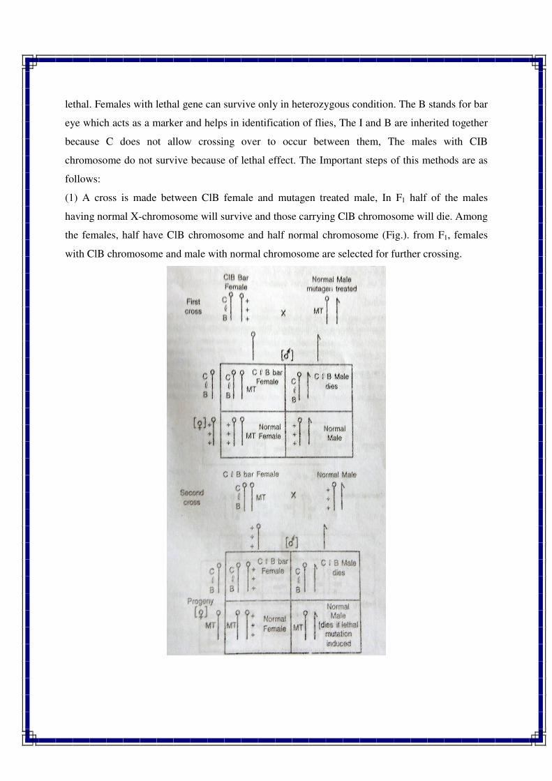
lethal. Females with lethal gene can survive only in heterozygous condition. The B stands for bar
eye which acts as a marker and helps in identification of flies, The I and B are inherited together
because C does not allow crossing over to occur between them, The males with CIB
chromosome do not survive because of lethal effect. The Important steps of this methods are as
follows:
(1) A cross is made between ClB female and mutagen treated male, In F1 half of the males
having normal X-chromosome will survive and those carrying ClB chromosome will die. Among
the females, half have ClB chromosome and half normal chromosome (Fig.). from F1, females
with ClB chromosome and male with normal chromosome are selected for further crossing.
(2) Now a cross is made between ClB female and normal male. This time the ClB female has one
ClB chromosome and one mutagen treated chromosome received from the male in earlier cross.
This will produce two types of females, viz., half with ClB chromosome and half with mutagen
treated chromosome (with normal phenotype). Both the progeny will survive. In case of males,
half with ClB will die and other half have mutagen treated chromosome. If a lethal mutation was
induced in mutagen treated X-chromosome, the remaining half males will also die, resulting in
absence of male progeny in the above cross. Absence of male progeny in F2 confirms the
induction of sex linked recessive lethal mutation in the mutagen treated Drosophila male.
//*//*//*//
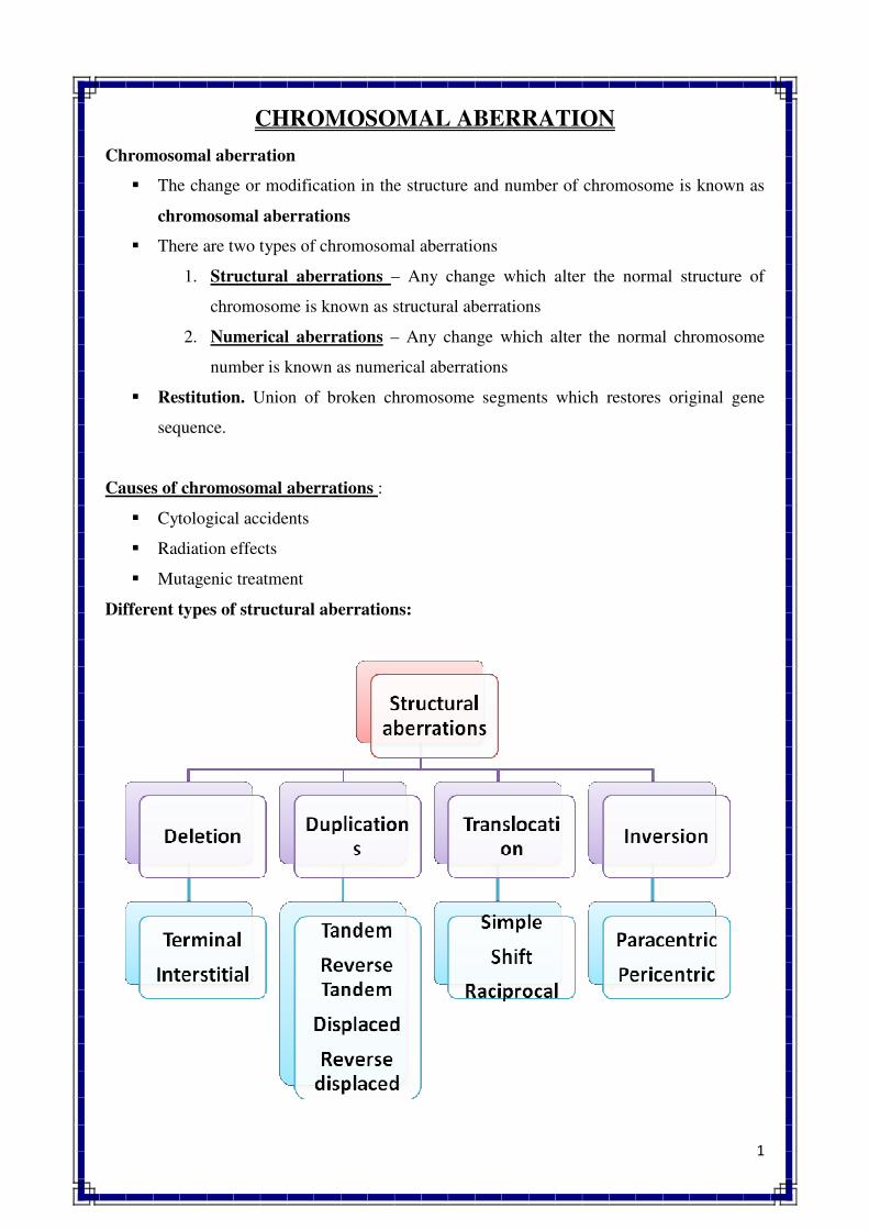
1
CHROMOSOMAL ABERRATION
Chromosomal aberration
� The change or modification in the structure and number of chromosome is known as
chromosomal aberrations
� There are two types of chromosomal aberrations
1. Structural aberrations – Any change which alter the normal structure of
chromosome is known as structural aberrations
2. Numerical aberrations – Any change which alter the normal chromosome
number is known as numerical aberrations
� Restitution. Union of broken chromosome segments which restores original gene
sequence.
Causes of chromosomal aberrations :
� Cytological accidents
� Radiation effects
� Mutagenic treatment
Different types of structural aberrations:
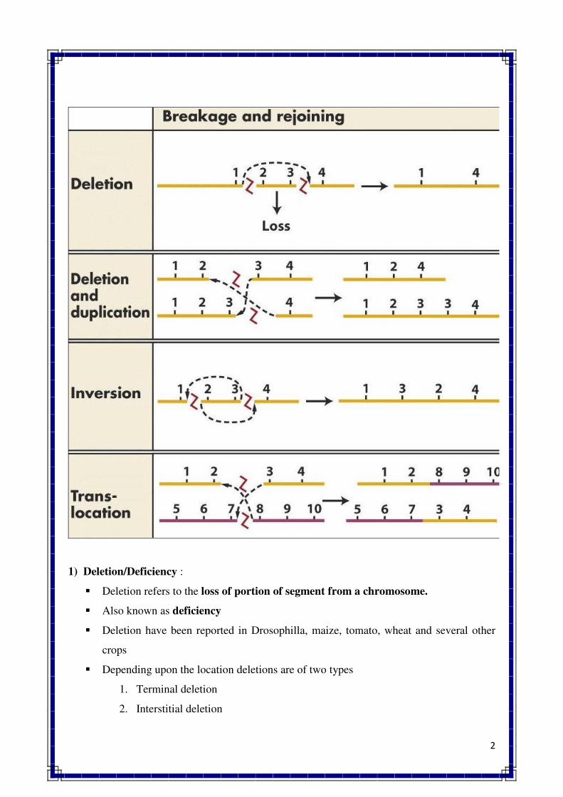
2
1) Deletion/Deficiency :
� Deletion refers to the loss of portion of segment from a chromosome.
� Also known as deficiency
� Deletion have been reported in Drosophilla, maize, tomato, wheat and several other
crops
� Depending upon the location deletions are of two types
1. Terminal deletion
2. Interstitial deletion
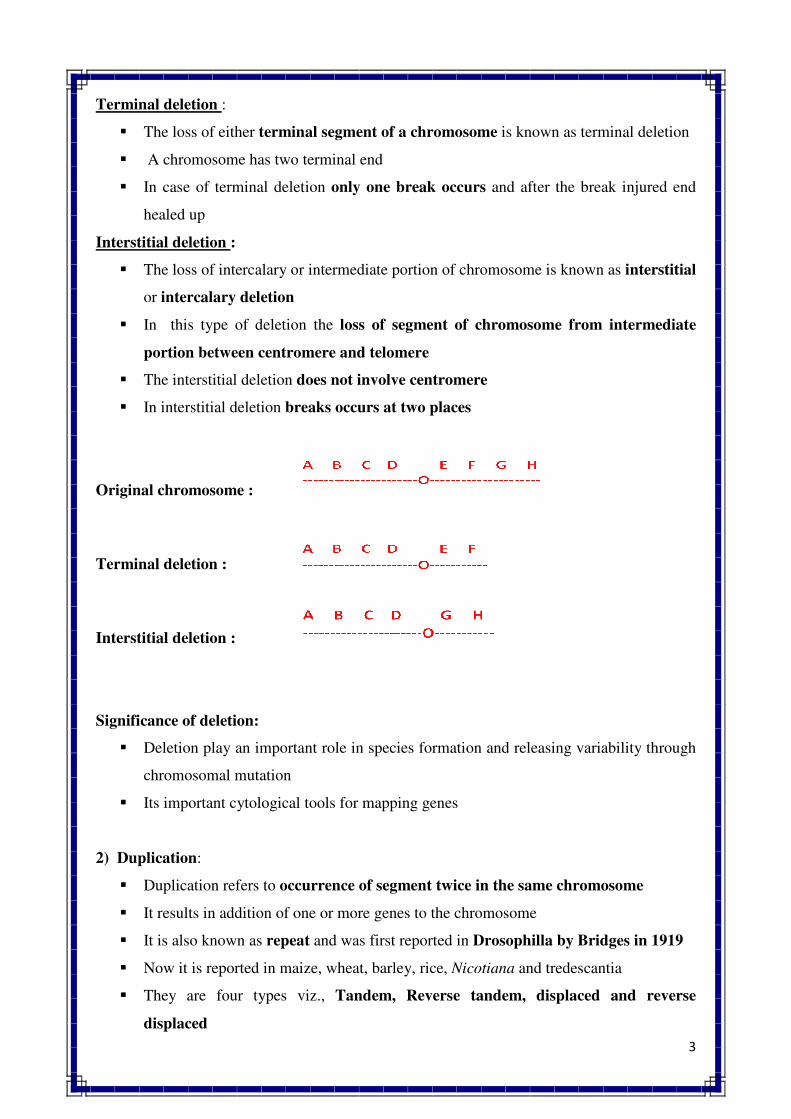
3
Terminal deletion :
� The loss of either terminal segment of a chromosome is known as terminal deletion
� A chromosome has two terminal end
� In case of terminal deletion only one break occurs and after the break injured end
healed up
Interstitial deletion :
� The loss of intercalary or intermediate portion of chromosome is known as interstitial
or intercalary deletion
� In this type of deletion the loss of segment of chromosome from intermediate
portion between centromere and telomere
� The interstitial deletion does not involve centromere
� In interstitial deletion breaks occurs at two places
Original chromosome :
Terminal deletion :
Interstitial deletion :
Significance of deletion:
� Deletion play an important role in species formation and releasing variability through
chromosomal mutation
� Its important cytological tools for mapping genes
2) Duplication:
� Duplication refers to occurrence of segment twice in the same chromosome
� It results in addition of one or more genes to the chromosome
� It is also known as repeat and was first reported in Drosophilla by Bridges in 1919
� Now it is reported in maize, wheat, barley, rice, Nicotiana and tredescantia
� They are four types viz., Tandem, Reverse tandem, displaced and reverse
displaced
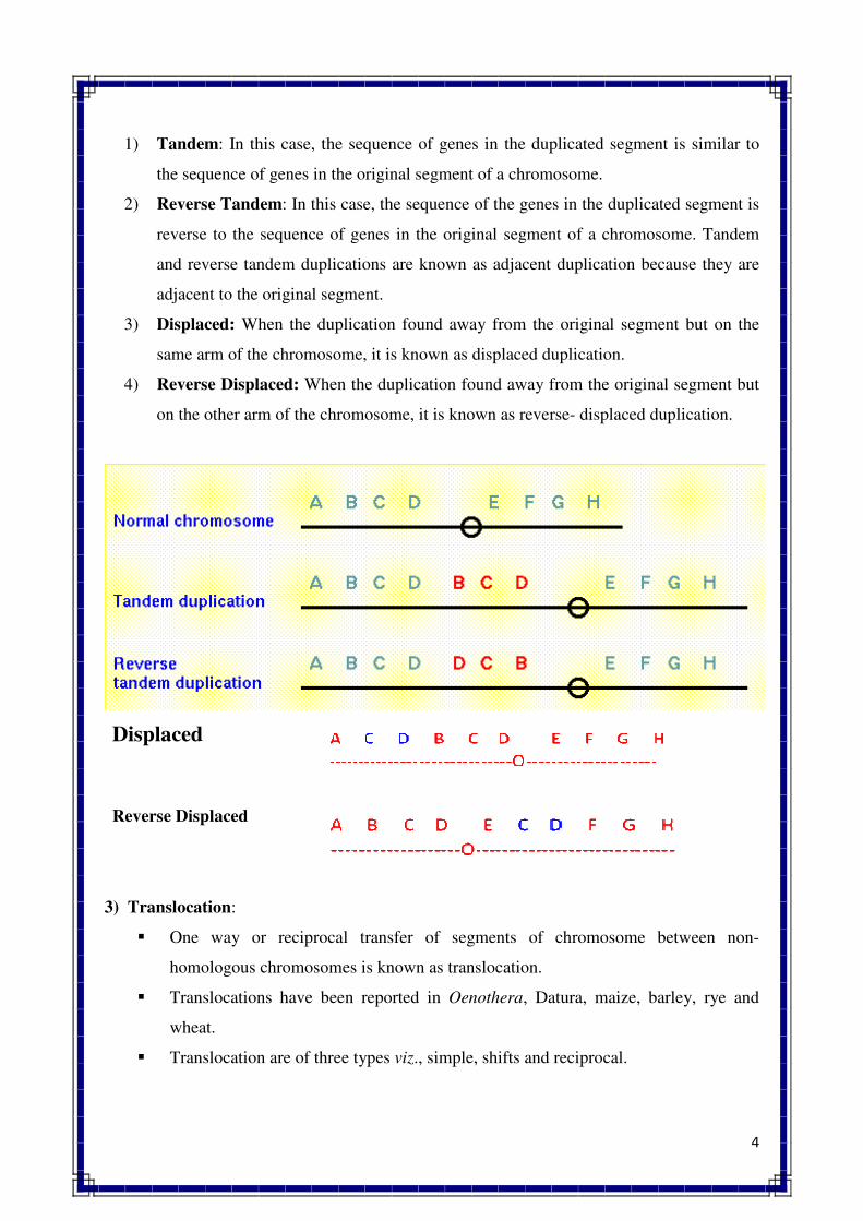
4
1) Tandem: In this case, the sequence of genes in the duplicated segment is similar to
the sequence of genes in the original segment of a chromosome.
2) Reverse Tandem: In this case, the sequence of the genes in the duplicated segment is
reverse to the sequence of genes in the original segment of a chromosome. Tandem
and reverse tandem duplications are known as adjacent duplication because they are
adjacent to the original segment.
3) Displaced: When the duplication found away from the original segment but on the
same arm of the chromosome, it is known as displaced duplication.
4) Reverse Displaced: When the duplication found away from the original segment but
on the other arm of the chromosome, it is known as reverse- displaced duplication.
Displaced
Reverse Displaced
3) Translocation:
� One way or reciprocal transfer of segments of chromosome between non-
homologous chromosomes is known as translocation.
� Translocations have been reported in Oenothera, Datura, maize, barley, rye and
wheat.
� Translocation are of three types viz., simple, shifts and reciprocal.

5
1) Simple translocation: When a segment from one chromosome is transferred and
attached to the end of a non-homologous chromosome is known as simple
translocation.
2) Shifts: Transfer of an intercalary segment from one chromosome to the intercalary
position in a non-homologus chromosome is referred as shift.
3) Reciprocal translocation: When there is mutual exchange of segments between
non-homologous chromosomes is known as reciprocal translocation.
4) Inversion :
� Inversion refers to structural change in a chromosome in which segment is oriented
in a reverse order.
� It was first time discovered by Strutevant (1926) in Drosophila. It has been also
reported in maize, Nicotiana and several other plants.
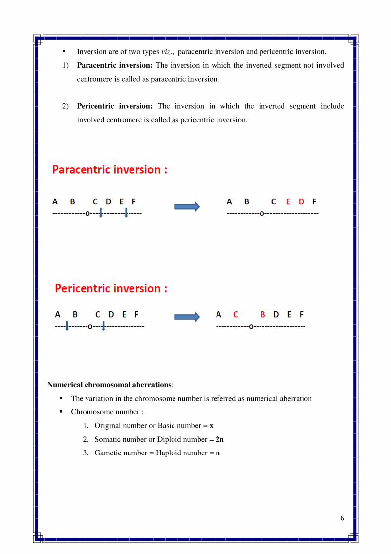
6
� Inversion are of two types viz., paracentric inversion and pericentric inversion.
1) Paracentric inversion: The inversion in which the inverted segment not involved
centromere is called as paracentric inversion.
2) Pericentric inversion: The inversion in which the inverted segment include
involved centromere is called as pericentric inversion.
Numerical chromosomal aberrations:
� The variation in the chromosome number is referred as numerical aberration
� Chromosome number :
1. Original number or Basic number = x
2. Somatic number or Diploid number = 2n
3. Gametic number = Haploid number = n
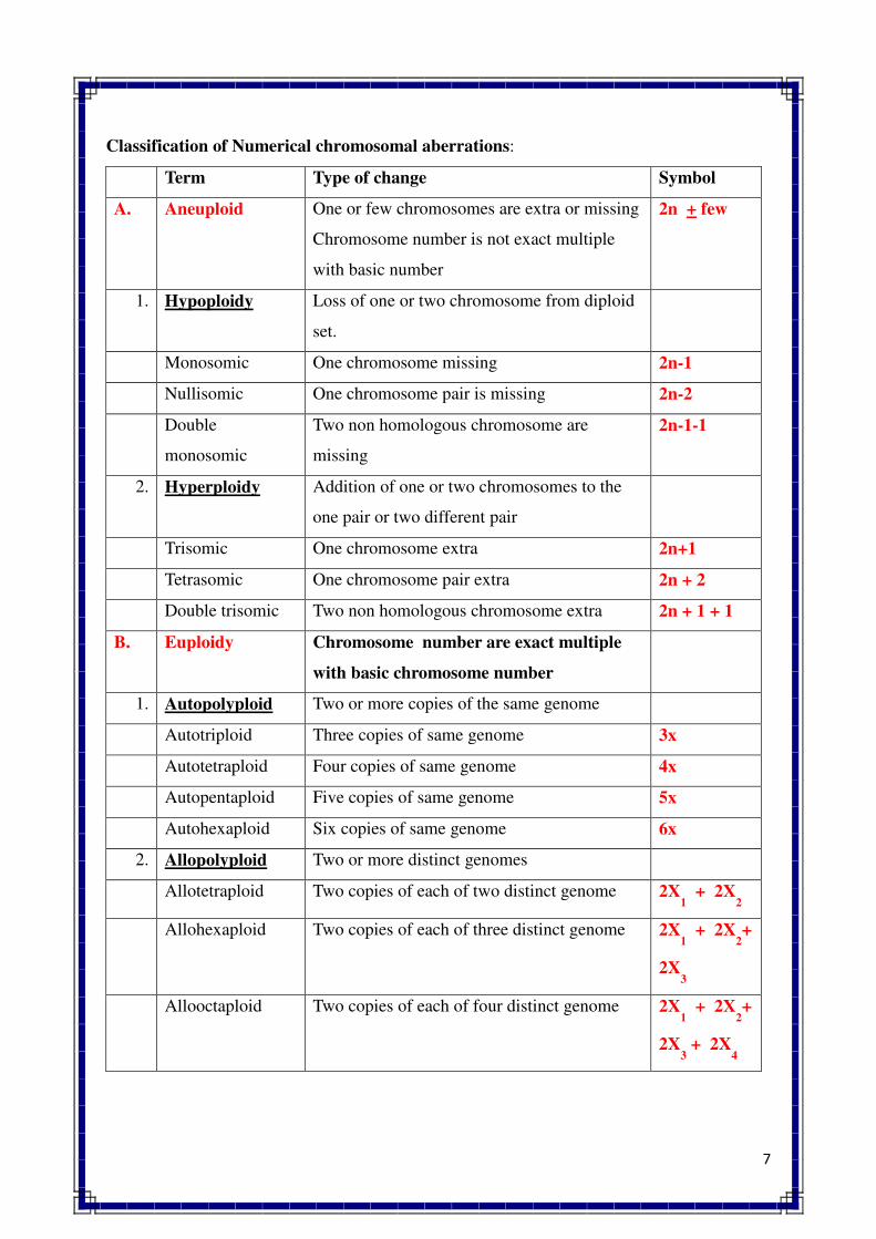
7
Classification of Numerical chromosomal aberrations:
Term Type of change Symbol
A. Aneuploid One or few chromosomes are extra or missing
Chromosome number is not exact multiple
with basic number
2n + few
1. Hypoploidy Loss of one or two chromosome from diploid
set.
Monosomic One chromosome missing 2n-1
Nullisomic One chromosome pair is missing 2n-2
Double
monosomic
Two non homologous chromosome are
missing
2n-1-1
2. Hyperploidy Addition of one or two chromosomes to the
one pair or two different pair
Trisomic One chromosome extra 2n+1
Tetrasomic One chromosome pair extra 2n + 2
Double trisomic Two non homologous chromosome extra 2n + 1 + 1
B. Euploidy Chromosome number are exact multiple
with basic chromosome number
1. Autopolyploid Two or more copies of the same genome
Autotriploid Three copies of same genome 3x
Autotetraploid Four copies of same genome 4x
Autopentaploid Five copies of same genome 5x
Autohexaploid Six copies of same genome 6x
2. Allopolyploid Two or more distinct genomes
Allotetraploid Two copies of each of two distinct genome 2X1 + 2X
2
Allohexaploid Two copies of each of three distinct genome 2X1 + 2X
2+
2X3
Allooctaploid Two copies of each of four distinct genome 2X1 + 2X
2+
2X3 + 2X
4
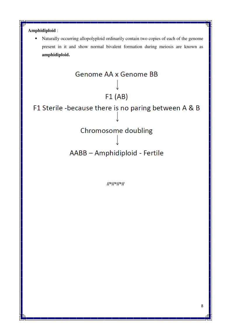
8
Amphidiploid :
� Naturally occurring allopolyploid ordinarily contain two copies of each of the genome
present in it and show normal bivalent formation during meiosis are known as
amphidiploid.
//*//*//*//
1
POLYPLOIDY
Polyploidy :
An organism or individual having more than two basic sets of chromosomes is called
polyploid and such condition is known as polyploidy. About one third species of flowering
plants are polyploids. The polyploidy has been reported upto 70% in case of wild species of
grass family. In animals, polyploidy is very rare occurrence because of its lethal effects.
However, it is found only in those species of animal which develop parthnogenetically.
Classification of Polyploidy : Polyploidy is of two main types.
1. Autopolyploid
2. Allopolyploid
Autopolyploidy :
o Polyploids which originate by multiplication of the chromosome number of a single
species are called as autoployploids or autoploids and such condition is referred to as
autopolyploidy.
o Autoploids are also known as simple polyploids or single species polyploids.
o Autoploids includes autotriploids (3x), autotetraploids (4x), autopentaploids (5x),
autohexaploids (6x) etc.
o Examples : Seedless watermelon, sugarbeet, banana, jowar, tomato, wheat etc.
Autotriploids :
o They can occur naturally or can be produced artificially by crossing autotetraploid and
diploid species.
o They are generally sterile due to defective gamete formation (due to meiotic
abnormalities).
o Example: Banana, sugarcane, apple, sugarbeet and watermelon (seedless or
rudimentary seeds), etc.
o All other autotriploids with odd chromosomes sets, viz., autopentaploids (5x),
autoseptaploids (7x) etc also behave like autotriploids.
Autotetraploids :
o They may occur spontaneously in nature or can be induced artificially by doubling the
chromosomes of a diploid species with colchicine treatment.
2
o They are usually very stable and fertile due to availability of pairing partners during
meiosis and it lead to diploid gamete (2n) formation.
o Autotetraploids are generally larger in size and more vigorous than the diploid
species.
o Examples : Aurotetraploid rye, grapes, alfalfa, groundnut, potato, coffee etc.
o All other autoploids with even chromosomes sets, viz., autohexaploids (6x),
autooctaploids (8x) etc also behave like autotetraploids.
Allopolyploidy :
o Polyploids which originate by combining complete chromosome sets from two or
more species are called as alloployploids or alloploids and such condition is referred
to as allopolyploidy.
o Alloploids are also known as hybrid polyploids or bispecies or multispecies
polyploids.
o Alloploids includes allotetraploids (2x1 + 2x2), Allohexapolids (2x1 + 2x2 + 2x3),
allooctaploids (2x1 + 2x2 + 2x3 + 2x4) etc.
o An alloplyploid which arises by combining the genomes of two diploid specis
followed by chromosome doubling with colchicines is termed as amphidiploids.
o They can be produced by interspecific cross and fertility is restored by chromosome
doubling with colchicine treatment.
o About 50% of crop plants are allopolyploids. Therefore, allopolyploidy has played
greater role in crop evolution than autopolyploidy.
o Allopolyploids are of two types (a) Natural allopolyploid (b) Artificial allopolyploid.
(a) Natural Allopolyploids :
o Alloploids which naturally originate through interspecific crossing followed by
chromosome doubling in nature are called natural allopolyploids.
Examples:
Wheat
The bread wheat (Triticum aestivum) is an allopolyploid. It is believed that A
genome of wheat has come from Triticum monococcum (2n = 14), D genome from
Triticum tauschi (2n = 14) and B genome from unknown source probably from an
extinct species (2n = 14). Thus hexaploid wheat has two copies of the genomes from
three species. First allotetraploid Triticum turgidum developed from a cross between
3
Triticum monococcum and unknown species of B genome. Then cross between T.
turgidum and T. tauschi resulted in the development of hexaploid wheat T. aestivum.
Tobacco
There are two cultivated species of tobacco, viz., Nicotiana tabacum and N.
rustica. N. tabacum is an amphidiploid between N. sylvestris (2n = 24) and N.
tomentosa (2n = 24). N. roptica is believed to be amphidiploid between N. paniculata
and N. undulata. Each of these two species has 2n = 24.
Cotton
The tetraploid American cotton (Gossypium hirsutum) is believed to be an
amphidiploid between G. africanum, an old world species, and G. raimondii, a new
world species. Both these species arc diploid with 2n = 26. The chromosomes of old
world species are larger than new world species.
Oat
The cultivated oat (Avena sativa, n = 21) is an allohexaploid which is considered
to have originated from a cross between A. barbata (tetraploid, n = 14) and A. strigosa
(a diploid, n = 7).
Brassica
In brassica, there are three basic species, viz., Brassica nigra (BB, n =8) B.
oleracea (CC, n = 9) and B. campestris (AA, n = 10). The cross between B. nigra and
B. oleracea gave rise to B. carinata. Cross between B. campestris and B. oleracea led
to the development of B. napus, and cross between B. campestris and B. nigra
resulted in the development of B. juncea. All the resulting species are amphidiploids.
o
o (b) Artificial Allopolyploids :
o Alloploids which have been synthesized through artificial interspecific crossing
followed by chromosome doubling in nature are called artificial allopolyploids.
o They are synthesized with two main objectives:
1. To study the origin of naturally available allopolyploids
2. To explore the possibilities of creating new species.
Example :
1. Raphnobrassica : A cross between radish (2n=18) and cabbage (2n=18) was first
developed by Karpencheko (1927). He wanted to develop a fertile hybrid with roots
of radish and leaves of cabbage. But he got a fertile allotetraploid (4n=36) by
spontaneous chromosome doubling which had roots of cabbage and leaves of radish.
4
2. Triticale : A cross between tetraploid wheat (2n=28) or hexaploid wheat (2n=42) and
rye (2n=14) was first developed by Rimpu (1889) produced allohexaploid triticle or
allooctaploid triticle, respectively. Triticle is commonly grown in Canada, Mexico etc.
Methods to induce polyploids :
A) Induction of polyploidy using colchicine :
o Polyploidy is mainly induced by treatment with a chemical known as colchicines. The
colchicines induced polyploidy is known as Colchiploidy.
o It is the most effective and the widely used treatment for chromosome doubling.
o It is widely used in large numbers of monocot and dicot crop plants.
o Colchicine [C22H25O6N] is poisonous alkaloid which is isolated from seeds (0.2 – 0.8
%) and bulbs (0.1 – 0.5 %) of autumn crocus (Colchicum autumnale), which belongs
to the family Liliaceae.
o It is soluble in alcohol, chloroform or cold water but is relatively less soluble in hot
water.
o Colchicine does not affect Colchicum from which it is extracted, as this plant has an
anticolchicine substance.
o Colchicine is applied in very low concentration, because high concentration is highly
toxic to the cells.
Mode of action of Colchicine :
o Colchicine induces polyploidy by blocking the formation of spindle fibres.
o The chromosomes do not line up on the equatorial plate and thus inhibits the
movement of sister chromatids to opposite poles. The nuclear membrane is formed
around them and cell enters into interphase. As a result, the chromosome number of
cell is doubled in the nucleus without division of cell.
o For effective induction of polyploidy, usually concentration of 0.01 to 0.5 % is used
in different plant species. In plant species, it is applied to growing tips, meristematic
cells, seeds and axillary buds in freshly prepared aqueous solution or mixed with
lanolin. The duration of treatment varies from 24 to 96 hours depending upon the
species of plants.
o Colchicines affects only dividing cells, so it should be applied to a shoot-tip meristem
only when its cells are actively dividing. At any given time, only small proportion of
the cells would be in division, so repeated treatments should be given at brief intervals
to double the chromosome number in large number of cells of apex.
5
Methods of colchicine application :
a) Seed Treatment : The seeds are generally soaked in 0.001 to 1 % (0.2 % most
common) freshly prepared aqueous solution of colchicines for one to ten days.
b) Seedling Treatment : Germinating seedling or in vitro derived seedling may be
inverted so that only young shoots are exposed to colchicines solution for 3 to 24
hours and roots are protected.
c) Growing shoot apices :
o It is most commonly used treatment with 0.1 to 1.0 % colchicines, which is
applied by brush or with a dropper. The treatment is repeated once or twice
daily for 5-7 days.
o A small cotton wool piece may also be placed at the shoot-tip meristem, which
daily soaked with colchicine solution.
o Colchicine 0.5 to 1.0 % solution mixed in lanoline paste may be smeared on the
shoot apex and this treatment is repeated 2 to 3 times per week.
d) Woody plants : 1 % colchicine is used for application on shoot buds. For better
wetting and penetration, a small quantity of wetting agent is added in the colchicine
solution.
e) Special treatments: Special methods for the treatments in cereals, grasses, pulses,
oilseeds crop etc have been developed to contact the colchicines solution with shoot
apical meristem.
B) Induction of autopolyploidy using physical agents :
o Heat or cold treatments, centrifugation, X-rays or gamma rays irradiation may also
produce polyploids but with very low frequencies and are not used commercially.
C) Induction of autopolyploidy using other chemicals :
o Several other chemicals like acenaphthene, 8-hydroxyquinoline, nitrous oxide etc. are
used to induce autopolyploid but these chemicals are much less effective than
colchicines and are not commonly used.
APPLICATIONS IN CROP IMPROVEMENT
Polyploidy plays an important role in crop improvement. Both, autopolyloidy and
allopolyploidy are useful in several ways. However, allopolyploidy has wider applications
than autopolyloidy. Applications of allopolyploidy and autopolyloidy in crop improvement
are briefly presented below :
6
Autoploidy
Both triploids and tetraploids have been used in crop improvement. However, their
applications have been limited to few species only.
• Autotriploids have been developed in sugar beets and water melon only. The triploid
sugar beets have larger roots and higher sugar content than diploids.
• The triploid water melons are seedless or have rudimentary and soft seeds like
cucumber. The triploid seed is produced by using tetraploid as female and diploid as
male.
• Tetraploid varieties of rye are grown in Sweden and Germany. They have larger seeds
and higher protein than diploids.
• Tetraploid grapes have been developed in California USA, which have larger fruits
and fewer seeds per fruit than diploids.
Alloploidy
Alloploidy is useful in four principal ways, viz-, (1) in tracing the origin of natural
allopolyploids, (2) in creating new species, (3) in interspecific gene transfer, and (4) as a
bridging species. These are briefly described below :
1. Tracing the Origin of Crop Species. Alloploidy plays an important role in tracing the
origin of natural allopolyploids. Study of chromosome pairing in a cross between
allopolyploid and a diploid species helps in tracing the origin of polyploid species
2. Creation of New Species. Alloploidy sometimes leads to the creation of new crop
species. Triticale is the best example, which is alloployploid between wheat and rye. It
combines desirable character of both the species i.e., grain quality of wheat and
hardiness of rye.
3. Interspecific Gene Transfer. When the desirable character is not found within the
species, it is transferred from the related species. Interspecific gene transfer is done in
two ways, viz., by alien addition and alien substitution.
4. 4. Bridging Cross. Sometimes direct cross between two species is not posssible due
to sterility in Fl. In such case, first an amphidiploid is made between such species and
then amphidiploid is crossed with the recipient species. Such types of bridging crosses
have been made for transfer of genes from wild species particularly in crops like
tobacco and cotton.
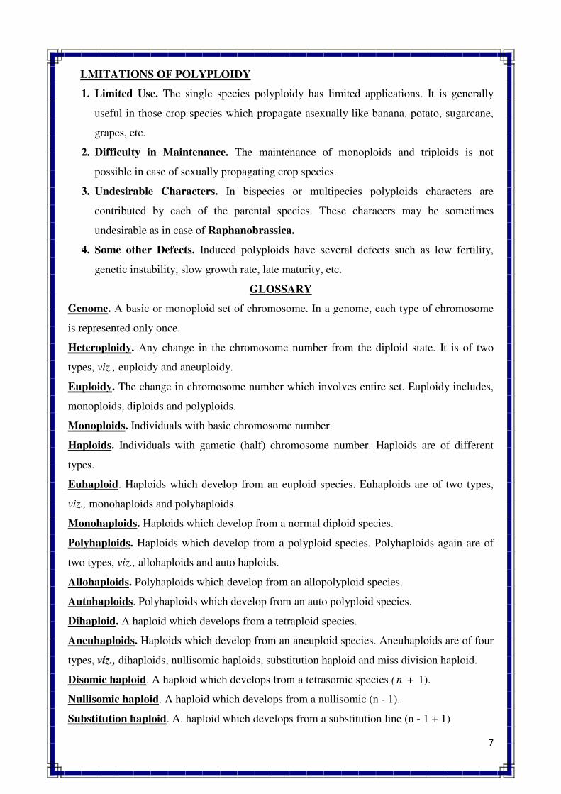
7
LMITATIONS OF POLYPLOIDY
1. Limited Use. The single species polyploidy has limited applications. It is generally
useful in those crop species which propagate asexually like banana, potato, sugarcane,
grapes, etc.
2. Difficulty in Maintenance. The maintenance of monoploids and triploids is not
possible in case of sexually propagating crop species.
3. Undesirable Characters. In bispecies or multipecies polyploids characters are
contributed by each of the parental species. These characers may be sometimes
undesirable as in case of Raphanobrassica.
4. Some other Defects. Induced polyploids have several defects such as low fertility,
genetic instability, slow growth rate, late maturity, etc.
GLOSSARY
Genome. A basic or monoploid set of chromosome. In a genome, each type of chromosome
is represented only once.
Heteroploidy. Any change in the chromosome number from the diploid state. It is of two
types, viz., euploidy and aneuploidy.
Euploidy. The change in chromosome number which involves entire set. Euploidy includes,
monoploids, diploids and polyploids.
Monoploids. Individuals with basic chromosome number.
Haploids. Individuals with gametic (half) chromosome number. Haploids are of different
types.
Euhaploid. Haploids which develop from an euploid species. Euhaploids are of two types,
viz., monohaploids and polyhaploids.
Monohaploids. Haploids which develop from a normal diploid species.
Polyhaploids. Haploids which develop from a polyploid species. Polyhaploids again are of
two types, viz., allohaploids and auto haploids.
Allohaploids. Polyhaploids which develop from an allopolyploid species.
Autohaploids. Polyhaploids which develop from an auto polyploid species.
Dihaploid. A haploid which develops from a tetraploid species.
Aneuhaploids. Haploids which develop from an aneuploid species. Aneuhaploids are of four
types, viz., dihaploids, nullisomic haploids, substitution haploid and miss division haploid.
Disomic haploid. A haploid which develops from a tetrasomic species (n + 1).
Nullisomic haploid. A haploid which develops from a nullisomic (n - 1).
Substitution haploid. A. haploid which develops from a substitution line (n - 1 + 1)
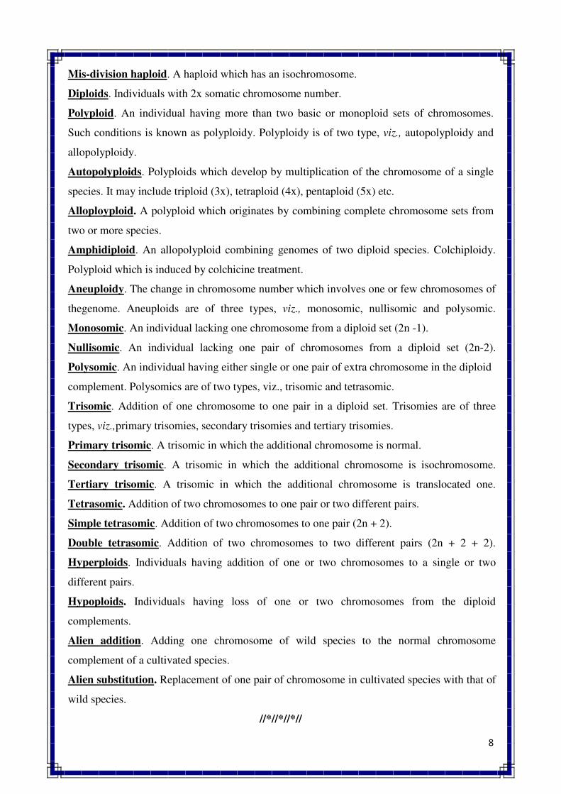
8
Mis-division haploid. A haploid which has an isochromosome.
Diploids. Individuals with 2x somatic chromosome number.
Polyploid. An individual having more than two basic or monoploid sets of chromosomes.
Such conditions is known as polyploidy. Polyploidy is of two type, viz., autopolyploidy and
allopolyploidy.
Autopolyploids. Polyploids which develop by multiplication of the chromosome of a single
species. It may include triploid (3x), tetraploid (4x), pentaploid (5x) etc.
Alloployploid. A polyploid which originates by combining complete chromosome sets from
two or more species.
Amphidiploid. An allopolyploid combining genomes of two diploid species. Colchiploidy.
Polyploid which is induced by colchicine treatment.
Aneuploidy. The change in chromosome number which involves one or few chromosomes of
thegenome. Aneuploids are of three types, viz., monosomic, nullisomic and polysomic.
Monosomic. An individual lacking one chromosome from a diploid set (2n -1).
Nullisomic. An individual lacking one pair of chromosomes from a diploid set (2n-2).
Polysomic. An individual having either single or one pair of extra chromosome in the diploid
complement. Polysomics are of two types, viz., trisomic and tetrasomic.
Trisomic. Addition of one chromosome to one pair in a diploid set. Trisomies are of three
types, viz.,primary trisomies, secondary trisomies and tertiary trisomies.
Primary trisomic. A trisomic in which the additional chromosome is normal.
Secondary trisomic. A trisomic in which the additional chromosome is isochromosome.
Tertiary trisomic. A trisomic in which the additional chromosome is translocated one.
Tetrasomic. Addition of two chromosomes to one pair or two different pairs.
Simple tetrasomic. Addition of two chromosomes to one pair (2n + 2).
Double tetrasomic. Addition of two chromosomes to two different pairs (2n + 2 + 2).
Hyperploids. Individuals having addition of one or two chromosomes to a single or two
different pairs.
Hypoploids. Individuals having loss of one or two chromosomes from the diploid
complements.
Alien addition. Adding one chromosome of wild species to the normal chromosome
complement of a cultivated species.
Alien substitution. Replacement of one pair of chromosome in cultivated species with that of
wild species.
//*//*//*//
EVOLUTION OF CROP PLANTS
A divergent process which increases genetic diversity and leads to change in allelic
frequencies in a population is known as evolution. OR
A process which leads to significant deviation in the characteristic features of existing
individuals as compared to their pre-existing individuals is termed evolution.
The evolution is of two types, viz,
1. Natural evolution: Natural selection operates
2. Man made evolution: Human selection operates
NATURAL EVOLUTION:
EVOLUTION OF WHEAT (Triticum aestivum) Hexaploid wheat and Tetraploid wheat
(Durum wheat):
Wheat is a cereal crop of global importance. It belongs to the genus Triticum of the
family Poaceae (old Gramineae). There are three types of species in the genus Triticum, viz.,
diploid, tetraploid and hexaploid. The somatic chromosome number of these species is 14, 28
and 42, respectively. Bread wheat (Triticum aestivum) is the predominantly cultivated
species, which belongs to the hexaploid group. Other cultivated species are T. monococcum
in diploid group and T. turgidum in tetraploid group.
The tetraploid species developed as an amphidiplod between two diploid species, and
hexaploid species originated from a cross between tetraploid and diploid species. It is
believed that tetraploid species Triticum turgidum evolved as an amphidiploid between
Triticum monococcum (AA) and an unknown species (now probably extinct) with BB
genome. The hexaploid bread wheat originated as an amphidiploid between Triticum
turgidum (AABB) and T. tauschii (DD). The overall process can be represented as follows
Thus, A genome of bread wheat has derived from T. monococcum, B genome from
an unknown species which is now probably extinct, and D genome from T. tauschii. The F1
was sterile at both the stages, which became fertile through chromosome doubling in nature.
Thus, interspecific hybridization and polyploidy have played key role in the evolution of
bread wheat.
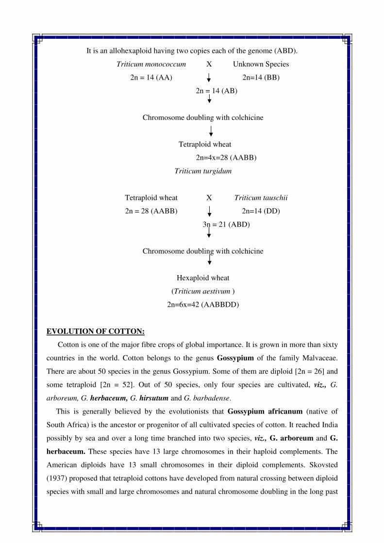
It is an allohexaploid having two copies each of the genome (ABD).
EVOLUTION OF COTTON:
Cotton is one of the major fibre crops of global importance. It is grown in more than sixty
countries in the world. Cotton belongs to the genus Gossypium of the family Malvaceae.
There are about 50 species in the genus Gossypium. Some of them are diploid [2n = 26] and
some tetraploid [2n = 52]. Out of 50 species, only four species are cultivated, viz., G.
arboreum, G. herbaceum, G. hirsutum and G. barbadense.
This is generally believed by the evolutionists that Gossypium africanum (native of
South Africa) is the ancestor or progenitor of all cultivated species of cotton. It reached India
possibly by sea and over a long time branched into two species, viz., G. arboreum and G.
herbaceum. These species have 13 large chromosomes in their haploid complements. The
American diploids have 13 small chromosomes in their diploid complements. Skovsted
(1937) proposed that tetraploid cottons have developed from natural crossing between diploid
species with small and large chromosomes and natural chromosome doubling in the long past
Triticum monococcum X Unknown Species
2n = 14 (AA) 2n=14 (BB)
2n = 14 (AB)
Chromosome doubling with colchicine
Tetraploid wheat
2n=4x=28 (AABB)
Triticum turgidum
Tetraploid wheat X Triticum tauschii
2n = 28 (AABB) 2n=14 (DD)
3n = 21 (ABD)
Chromosome doubling with colchicine
Hexaploid wheat
(Triticum aestivum )
2n=6x=42 (AABBDD)
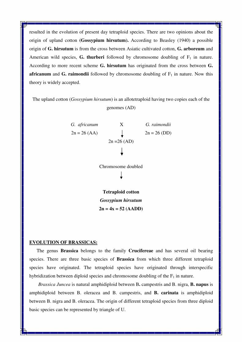
resulted in the evolution of present day tetraploid species. There are two opinions about the
origin of upland cotton (Gossypium hirsutum). According to Beasley (1940) a possible
origin of G. hirsutum is from the cross between Asiatic cultivated cotton, G. arboreum and
American wild species, G. thurberi followed by chromosome doubling of F1 in nature.
According to more recent scheme G. hirsutum has originated from the cross between G.
africanum and G. raimondii followed by chromosome doubling of F1 in nature. Now this
theory is widely accepted.
The upland cotton (Gossypium hirsutum) is an allotetraploid having two copies each of the
genomes (AD)
EVOLUTION OF BRASSICAS:
The genus Brassica belongs to the family Crucifereae and has several oil bearing
species. There are three basic species of Brassica from which three different tetraploid
species have originated. The tetraploid species have originated through interspecific
hybridization between diploid species and chromosome doubling of the F1 in nature.
Brassica Juncea is natural amphidiploid between B. campestris and B. nigra, B. napus is
amphidiploid between B. oleracea and B. campestris, and B. carinata is amphidiploid
between B. nigra and B. oleracea. The origin of different tetraploid species from three diploid
basic species can be represented by triangle of U.
G. africanum X G. raimondii
2n = 26 (AA) 2n = 26 (DD)
2n =26 (AD)
Chromosome doubled
Tetraploid cotton
Gossypium hirsutum
2n = 4x = 52 (AADD)
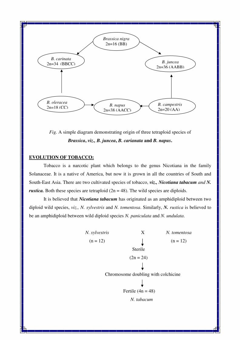
Fig. A simple diagram demonstrating origin of three tetraploid species of
Brassica, viz., B. juncea, B. carianata and B. napus.
EVOLUTION OF TOBACCO:
Tobacco is a narcotic plant which belongs to the genus Nicotiana in the family
Solanaceae. It is a native of America, but now it is grown in all the countries of South and
South-East Asia. There are two cultivated species of tobacco, viz., Nicotiana tabacum and N.
rustica. Both these species are tetraploid (2n = 48). The wild species are diploids.
It is believed that Nicotiana tabacum has originated as an amphidiploid between two
diploid wild species, viz., N. sylvestris and N. tomentosa. Similarly, N. rustica is believed to
be an amphidiploid between wild diploid species N. paniculata and N. undulata.
N. sylvestris X N. tomentosa
(n = 12) (n = 12)
Sterile
(2n = 24)
Chromosome doubling with colchicine
Fertile (4n = 48)
N. tabacum
Brassica nigra
2n=16 (BB)
B. carinata
2n=34 (BBCC) B. juncea
2n=36 (AABB)
B. napus
2n=38 (AACC)
B. oleracea
2n=18 (CC)B. campestris
2n=20 (AA)
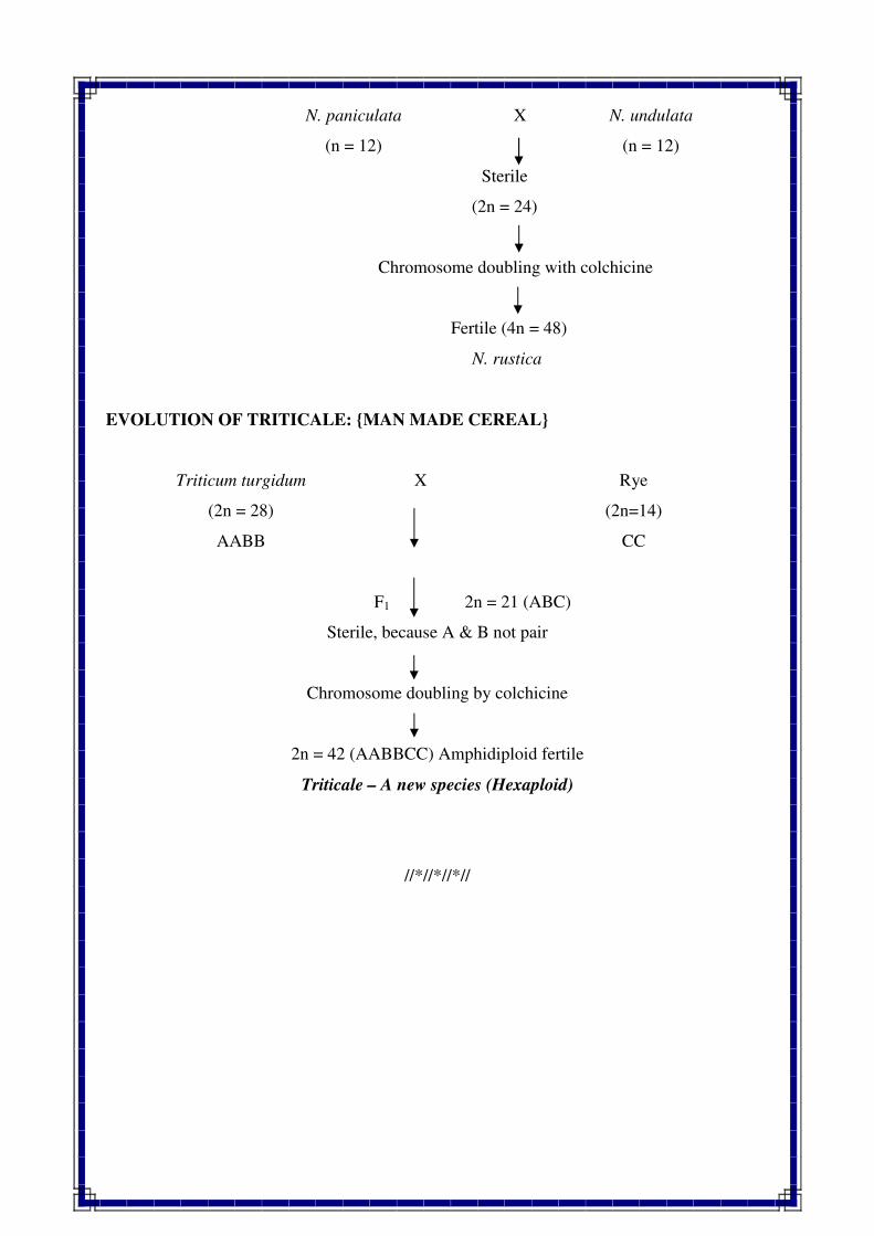
N. paniculata X N. undulata
(n = 12) (n = 12)
Sterile
(2n = 24)
Chromosome doubling with colchicine
Fertile (4n = 48)
N. rustica
EVOLUTION OF TRITICALE: {MAN MADE CEREAL}
Triticum turgidum X Rye
(2n = 28)
AABB
(2n=14)
CC
F1 2n = 21 (ABC)
Sterile, because A & B not pair
Chromosome doubling by colchicine
2n = 42 (AABBCC) Amphidiploid fertile
Triticale – A new species (Hexaploid)
//*//*//*//
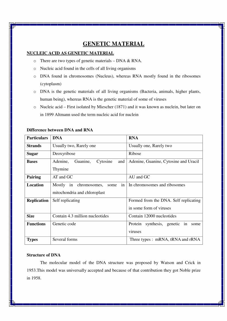
GENETIC MATERIAL
NUCLEIC ACID AS GENETIC MATERIAL
o There are two types of genetic materials – DNA & RNA.
o Nucleic acid found in the cells of all living organisms
o DNA found in chromosomes (Nucleus), whereas RNA mostly found in the ribosomes
(cytoplasm)
o DNA is the genetic materials of all living organisms (Bacteria, animals, higher plants,
human being), whereas RNA is the genetic material of some of viruses
o Nucleic acid – First isolated by Miescher (1871) and it was known as nuclein, but later on
in 1899 Altmann used the term nucleic acid for nuclein
Difference between DNA and RNA
Particulars DNA RNA
Strands Usually two, Rarely one Usually one, Rarely two
Sugar Deoxyribose Ribose
Bases Adenine, Guanine, Cytosine and
Thymine
Adenine, Guanine, Cytosine and Uracil
Pairing AT and GC AU and GC
Location Mostly in chromosomes, some in
mitochondria and chloroplast
In chromosomes and ribosomes
Replication Self replicating Formed from the DNA. Self replicating
in some form of viruses
Size Contain 4.3 million nucleotides Contain 12000 nucleotides
Functions Genetic code Protein synthesis, genetic in some
viruses
Types Several forms Three types : mRNA, tRNA and rRNA
Structure of DNA
The molecular model of the DNA structure was proposed by Watson and Crick in
1953.This model was universally accepted and because of that contribution they got Noble prize
in 1958.
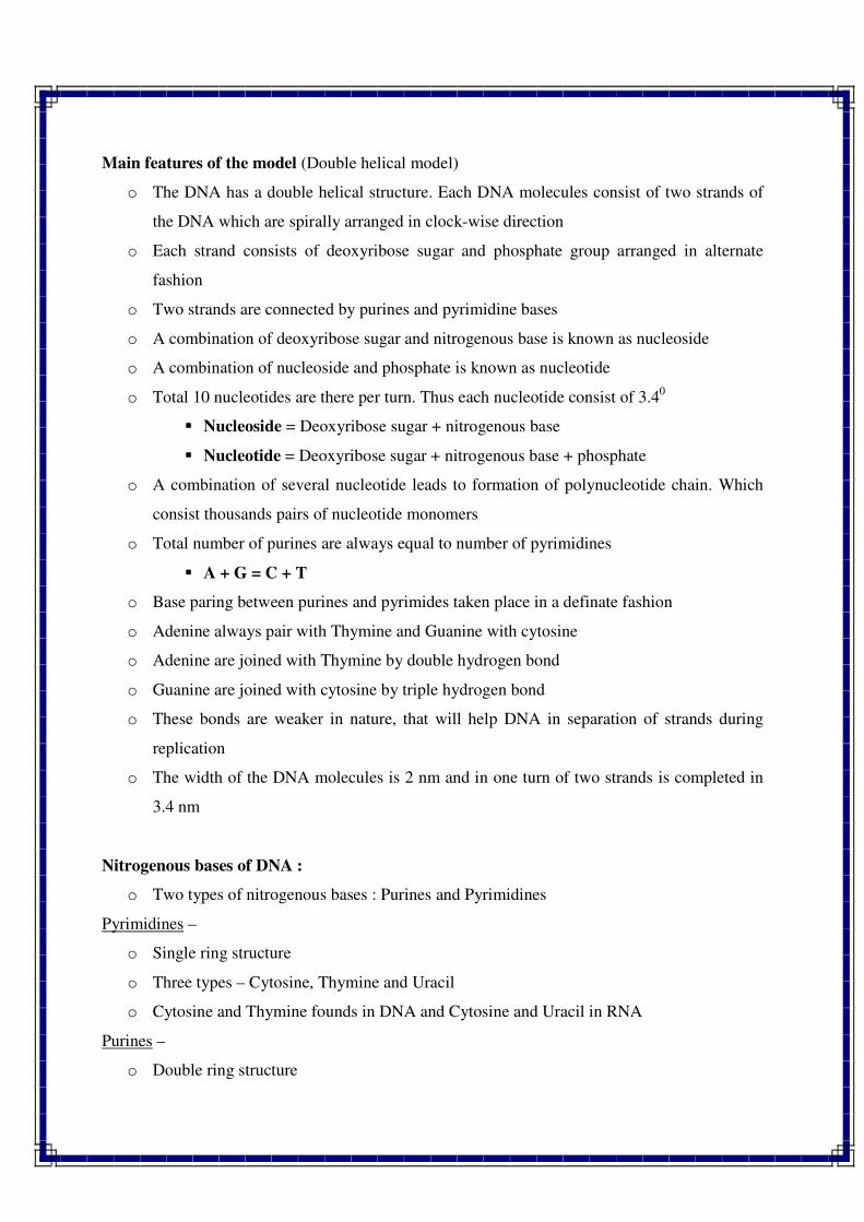
Main features of the model (Double helical model)
o The DNA has a double helical structure. Each DNA molecules consist of two strands of
the DNA which are spirally arranged in clock-wise direction
o Each strand consists of deoxyribose sugar and phosphate group arranged in alternate
fashion
o Two strands are connected by purines and pyrimidine bases
o A combination of deoxyribose sugar and nitrogenous base is known as nucleoside
o A combination of nucleoside and phosphate is known as nucleotide
o Total 10 nucleotides are there per turn. Thus each nucleotide consist of 3.40
� Nucleoside = Deoxyribose sugar + nitrogenous base
� Nucleotide = Deoxyribose sugar + nitrogenous base + phosphate
o A combination of several nucleotide leads to formation of polynucleotide chain. Which
consist thousands pairs of nucleotide monomers
o Total number of purines are always equal to number of pyrimidines
� A + G = C + T
o Base paring between purines and pyrimides taken place in a definate fashion
o Adenine always pair with Thymine and Guanine with cytosine
o Adenine are joined with Thymine by double hydrogen bond
o Guanine are joined with cytosine by triple hydrogen bond
o These bonds are weaker in nature, that will help DNA in separation of strands during
replication
o The width of the DNA molecules is 2 nm and in one turn of two strands is completed in
3.4 nm
Nitrogenous bases of DNA :
o Two types of nitrogenous bases : Purines and Pyrimidines
Pyrimidines –
o Single ring structure
o Three types – Cytosine, Thymine and Uracil
o Cytosine and Thymine founds in DNA and Cytosine and Uracil in RNA
Purines –
o Double ring structure
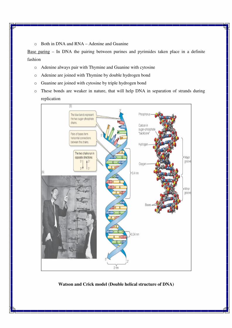
o Both in DNA and RNA – Adenine and Guanine
Base paring – In DNA the pairing between purines and pyrimides taken place in a definite
fashion
o Adenine always pair with Thymine and Guanine with cytosine
o Adenine are joined with Thymine by double hydrogen bond
o Guanine are joined with cytosine by triple hydrogen bond
o These bonds are weaker in nature, that will help DNA in separation of strands during
replication
Watson and Crick model (Double helical structure of DNA)
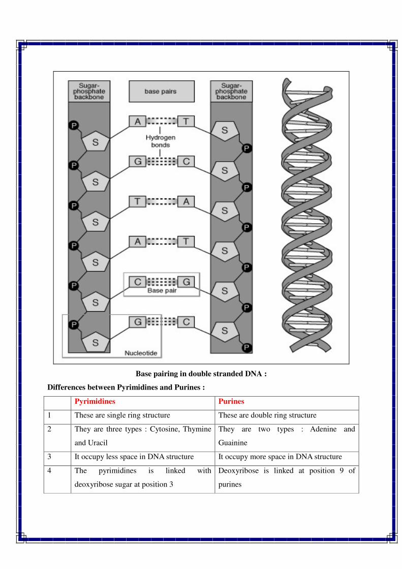
Base pairing in double stranded DNA :
Differences between Pyrimidines and Purines :
Pyrimidines Purines
1 These are single ring structure These are double ring structure
2 They are three types : Cytosine, Thymine
and Uracil
They are two types : Adenine and
Guainine
3 It occupy less space in DNA structure It occupy more space in DNA structure
4 The pyrimidines is linked with
deoxyribose sugar at position 3
Deoxyribose is linked at position 9 of
purines
Functions of the DNA :
o DNA (Deoxyribose nucleic acid) is the act as the genetic material of majority of living
organisms like in prokaryotes, eukaryotes and some of the viruses.
o DNA is the genetic code, which ensures that daughter cells inheritate the same
characteristics as the parent cells
o DNA is the code from which all protein is synthesized
Types of the DNA
The DNA can be classified on the basis of
1. Number of base pair per turn
2. Coiling pattern
3. Location
4. Structure
5. Nucleotide sequences
6. Number of strands
1. Number of base pair per turn :
Depending upon the nucleotide base per turn of the helix, pitch of the helix, tilt of the
base pair and humidity of the sample, the DNA can be observed in four different forms namely
A-DNA, B-DNA, C-DNA & D-DNA.
2. Coiling pattern :
On the basis of coiling pattern of the helix, DNA is of two types i.e Right handed and left
handed
o Right handed DNA : The coiling of the helix is in the right direction.
o Left handed DNA : The coiling of the DNA is in the left direction. The Z-DNA has left
handed double helical structure.
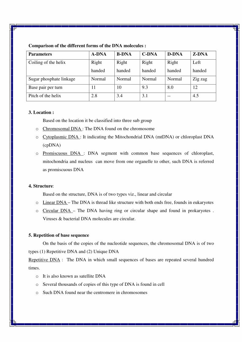
Comparison of the different forms of the DNA molecules :
Parameters A-DNA B-DNA C-DNA D-DNA Z-DNA
Coiling of the helix Right
handed
Right
handed
Right
handed
Right
handed
Left
handed
Sugar phosphate linkage Normal Normal Normal Normal Zig zag
Base pair per turn 11 10 9.3 8.0 12
Pitch of the helix 2.8 3.4 3.1 -- 4.5
3. Location :
Based on the location it be classified into three sub group
o Chromosomal DNA : The DNA found on the chromosome
o Cytoplasmic DNA : It indicating the Mitochondrial DNA (mtDNA) or chloroplast DNA
(cpDNA)
o Promiscuous DNA : DNA segment with common base sequences of chloroplast,
mitochondria and nucleus can move from one organelle to other, such DNA is referred
as promiscuous DNA
4. Structure:
Based on the structure, DNA is of two types viz., linear and circular
o Linear DNA – The DNA is thread like structure with both ends free, founds in eukaryotes
o Circular DNA – The DNA having ring or circular shape and found in prokaryotes .
Viruses & bacterial DNA molecules are circular.
5. Repetition of base sequence
On the basis of the copies of the nucleotide sequences, the chromosomal DNA is of two
types (1) Repetitive DNA and (2) Unique DNA
Repetitive DNA : The DNA in which small sequences of bases are repeated several hundred
times.
o It is also known as satellite DNA
o Several thousands of copies of this type of DNA is found in cell
o Such DNA found near the centromere in chromosomes
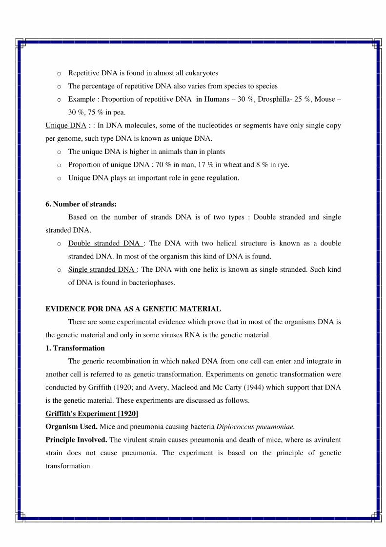
o Repetitive DNA is found in almost all eukaryotes
o The percentage of repetitive DNA also varies from species to species
o Example : Proportion of repetitive DNA in Humans – 30 %, Drosphilla- 25 %, Mouse –
30 %, 75 % in pea.
Unique DNA : : In DNA molecules, some of the nucleotides or segments have only single copy
per genome, such type DNA is known as unique DNA.
o The unique DNA is higher in animals than in plants
o Proportion of unique DNA : 70 % in man, 17 % in wheat and 8 % in rye.
o Unique DNA plays an important role in gene regulation.
6. Number of strands:
Based on the number of strands DNA is of two types : Double stranded and single
stranded DNA.
o Double stranded DNA : The DNA with two helical structure is known as a double
stranded DNA. In most of the organism this kind of DNA is found.
o Single stranded DNA : The DNA with one helix is known as single stranded. Such kind
of DNA is found in bacteriophases.
EVIDENCE FOR DNA AS A GENETIC MATERIAL
There are some experimental evidence which prove that in most of the organisms DNA is
the genetic material and only in some viruses RNA is the genetic material.
1. Transformation
The generic recombination in which naked DNA from one cell can enter and integrate in
another cell is referred to as genetic transformation. Experiments on genetic transformation were
conducted by Griffith (1920; and Avery, Macleod and Mc Carty (1944) which support that DNA
is the genetic material. These experiments are discussed as follows.
Griffith's Experiment [1920]
Organism Used. Mice and pneumonia causing bacteria Diplococcus pneumoniae.
Principle Involved. The virulent strain causes pneumonia and death of mice, where as avirulent
strain does not cause pneumonia. The experiment is based on the principle of genetic
transformation.
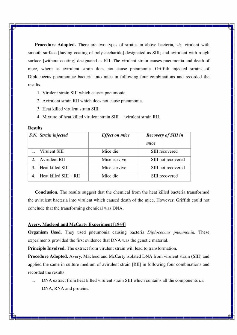
Procedure Adopted. There are two types of strains in above bacteria, viz. virulent with
smooth surface [having coating of polysaccharide] designated as SIII; and avirulent with rough
surface [without coating] designated as RII. The virulent strain causes pneumonia and death of
mice, where as avirulent strain does not cause pneumonia. Griffith injected strains of
Diplococcus pneumoniae bacteria into mice in following four combinations and recorded the
results.
1. Virulent strain SIII which causes pneumonia.
2. Avirulent strain RII which does not cause pneumonia.
3. Heat killed virulent strain SIII.
4. Mixture of heat killed virulent strain SIII + avirulent strain RII.
Results
S.N. Strain injected Effect on mice Recovery of SHI in
mice
1. Virulent SIII Mice die SIII recovered
2. Avirulent RII Mice survive SIII not recovered
3. Heat killed SIII Mice survive SIII not recovered
4. Heat killed SIII + RII Mice die SIII recovered
Conclusion. The results suggest that the chemical from the heat killed bacteria transformed
the avirulent bacteria into virulent which caused death of the mice. However, Griffith could not
conclude that the transforming chemical was DNA.
Avery, Macleod and McCarty Experiment [1944]
Organism Used. They used pneumonia causing bacteria Diplococcus pneumonia. These
experiments provided the first evidence that DNA was the genetic material.
Principle Involved. The extract from virulent strain will lead to transformation.
Procedure Adopted. Avery, Macleod and McCarty isolated DNA from virulent strain (SIII) and
applied the same in culture medium of avirulent strain [RII] in following four combinations and
recorded the results.
I. DNA extract from heat killed virulent strain SIII which contains all the components i.e.
DNA, RNA and proteins.
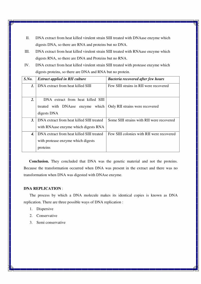
II. DNA extract from heat killed virulent strain SIII treated with DNAase enzyme which
digests DNA, so there are RNA and proteins but no DNA.
III. DNA extract from heat killed virulent strain SIII treated with RNAase enzyme which
digests RNA, so there are DNA and Proteins but no RNA.
IV. DNA extract from heat killed virulent strain SIII treated with protease enzyme which
digests proteins, so there are DNA and RNA but no protein.
S.No. Extract applied in RII culture Bacteria recovered after few hours
1. DNA extract from heat killed SIII Few SIII strains in RII were recovered
2. DNA extract from heat killed SIII
treated with DNAase enzyme which
digests DNA
Only RII strains were recovered
3. DNA extract from heat killed SIII treated
with RNAase enzyme which digests RNA
Some SIII strains with RII were recovered
4. DNA extract from heat killed SIII treated
with protease enzyme which digests
proteins
Few SIII colonies with RII were recovered
Conclusion. They concluded that DNA was the genetic material and not the proteins.
Because the transformation occurred when DNA was present in the extract and there was no
transformation when DNA was digested with DNAse enzyme.
DNA REPLICATION :
The process by which a DNA molecule makes its identical copies is known as DNA
replication. There are three possible ways of DNA replication :
1. Dispersive
2. Conservative
3. Semi conservative
Dispersive Replication :
o In this method, the two strands of DNA break at several points resulting in several pieces
of DNA.
o Each piece replicates and reunited randomly.
o Finally two copies of the DNA form from a single copy.
o The new DNA molecules are hybrid, which have old and new DNA in patches.
o This method is not accepted because replication could not prove experimentally.
Conservative replication :
o In conservative replication, two DNA molecules are formed.
o One molecule has both parental strands and the other contain both newly synthesized
strands.
o This method is also not accepted because of no experimental proof in support of this
model.
Semi conservative replication :
o Semi conservative mode of DNA replication was proposed by Watson and Crick.
o In this type of replication, both the strands of parental DNA separates from one another
and from each of the old strand new strand synthesize.
o Thus each of two resulting DNA molecules has one parental and one new strand.
o This method of DNA replication was universally accepted, because so many evidence are
there in support of this replication.
Steps of semi conservative method of DNA replication
Semi conservative method of DNA replication involved following steps :
1. Initiation of DNA replication :
o DNA replication is start at a specific point of the chromosome and this unique site is
referred as origin.
o This origin site is differ from organism to organism.
o Some time it start with an incision, which is made by enzyme which is known as
endonuclease.
2. Unwinding of strands :
o The two strands of the DNA double helix unwind.
o The opening of DNA strands taken place with the help of unwinding protein.
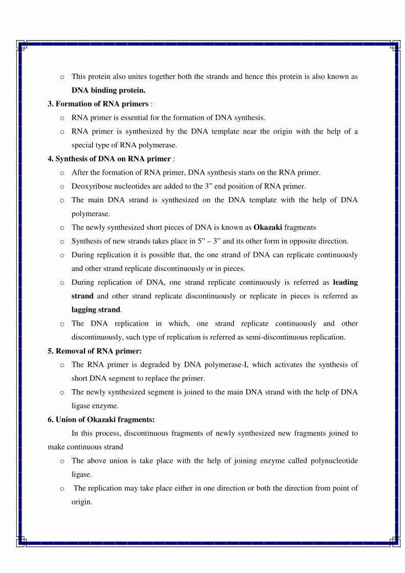
o This protein also unites together both the strands and hence this protein is also known as
DNA binding protein.
3. Formation of RNA primers :
o RNA primer is essential for the formation of DNA synthesis.
o RNA primer is synthesized by the DNA template near the origin with the help of a
special type of RNA polymerase.
4. Synthesis of DNA on RNA primer :
o After the formation of RNA primer, DNA synthesis starts on the RNA primer.
o Deoxyribose nucleotides are added to the 3” end position of RNA primer.
o The main DNA strand is synthesized on the DNA template with the help of DNA
polymerase.
o The newly synthesized short pieces of DNA is known as Okazaki fragments
o Synthesis of new strands takes place in 5” – 3” and its other form in opposite direction.
o During replication it is possible that, the one strand of DNA can replicate continuously
and other strand replicate discontinuously or in pieces.
o During replication of DNA, one strand replicate continuously is referred as leading
strand and other strand replicate discontinuously or replicate in pieces is referred as
lagging strand.
o The DNA replication in which, one strand replicate continuously and other
discontinuously, such type of replication is referred as semi-discontinuous replication.
5. Removal of RNA primer:
o The RNA primer is degraded by DNA polymerase-I, which activates the synthesis of
short DNA segment to replace the primer.
o The newly synthesized segment is joined to the main DNA strand with the help of DNA
ligase enzyme.
6. Union of Okazaki fragments:
In this process, discontinuous fragments of newly synthesized new fragments joined to
make continuous strand
o The above union is take place with the help of joining enzyme called polynucleotide
ligase.
o The replication may take place either in one direction or both the direction from point of
origin.
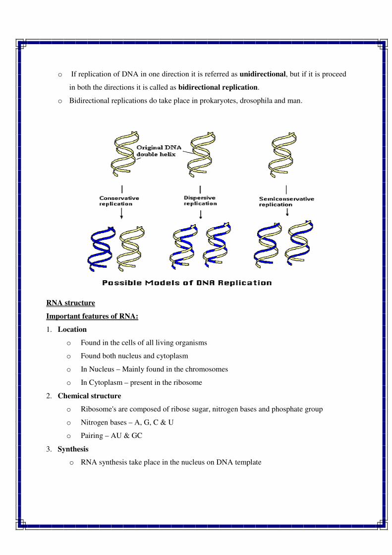
o If replication of DNA in one direction it is referred as unidirectional, but if it is proceed
in both the directions it is called as bidirectional replication.
o Bidirectional replications do take place in prokaryotes, drosophila and man.
RNA structure
Important features of RNA:
1. Location
o Found in the cells of all living organisms
o Found both nucleus and cytoplasm
o In Nucleus – Mainly found in the chromosomes
o In Cytoplasm – present in the ribosome
2. Chemical structure
o Ribosome's are composed of ribose sugar, nitrogen bases and phosphate group
o Nitrogen bases – A, G, C & U
o Pairing – AU & GC
3. Synthesis
o RNA synthesis take place in the nucleus on DNA template
o After synthesis it moves from nucleus to cytoplasm (RNA does not having self
duplication property)
o Exception of self duplicationsis found in TMV and plantango viruses
4. Strand number
o RNA is usually single stranded
o Double stranded RNA – reo virus in animals & Wound tumor virus in plants
o Single stranded RNA folded upon itself either entirely or in some regions
o Most of the bases in the folded regions are complementary and are joined by
hydrogen bond
o In unfolded regions bases do not have complements and due to it the purines and
pyrimidine ratio are not equal
5. Types
o Mainly of three types – mRNA, rRNA and tRNA or sRNA (Soluble RNA)
o rRNA and tRNA consist 98 % of total RNA
o All three forms of RNA synthesis on DNA templates
6. Size
o RNA molecules are much in smaller size than DNA
o RNA consist 12000 nucleotides
o DNA consist 4.3 million nucleotides
7. Functions
o The common function of RNA is to transfer genetic message from nucleus to
cytoplasm.
o Synthesis of protein in the ribosome.
o In some form of viruses it also act as genetic materials and regulate the gene action
Types of RNA
On the basis of functions RNA is of two types:
1. Genetic RNA – One type
2. Non-Genetic RNA – Three types (m-RNA, r-RNA and t-RNA)
Genetic RNA:
o The RNA which acts as the genetic materials or like DNA is called genetic RNA.
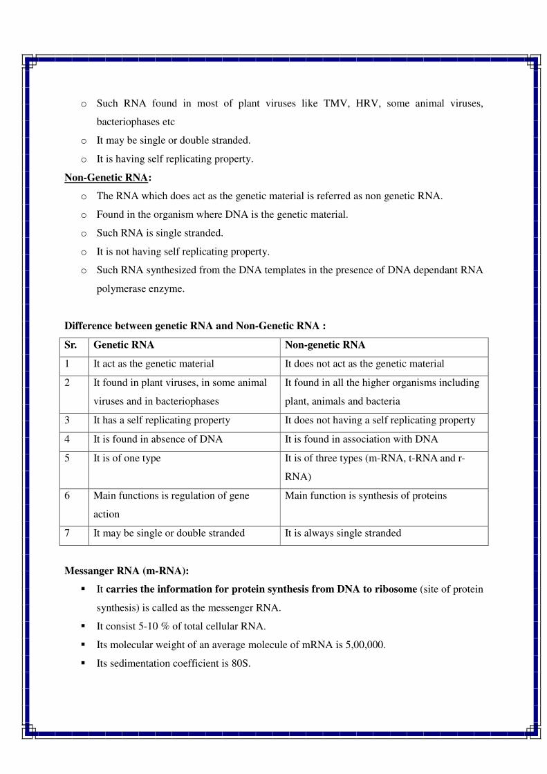
o Such RNA found in most of plant viruses like TMV, HRV, some animal viruses,
bacteriophases etc
o It may be single or double stranded.
o It is having self replicating property.
Non-Genetic RNA:
o The RNA which does act as the genetic material is referred as non genetic RNA.
o Found in the organism where DNA is the genetic material.
o Such RNA is single stranded.
o It is not having self replicating property.
o Such RNA synthesized from the DNA templates in the presence of DNA dependant RNA
polymerase enzyme.
Difference between genetic RNA and Non-Genetic RNA :
Sr. Genetic RNA Non-genetic RNA
1 It act as the genetic material It does not act as the genetic material
2 It found in plant viruses, in some animal
viruses and in bacteriophases
It found in all the higher organisms including
plant, animals and bacteria
3 It has a self replicating property It does not having a self replicating property
4 It is found in absence of DNA It is found in association with DNA
5 It is of one type It is of three types (m-RNA, t-RNA and r-
RNA)
6 Main functions is regulation of gene
action
Main function is synthesis of proteins
7 It may be single or double stranded It is always single stranded
Messanger RNA (m-RNA):
� It carries the information for protein synthesis from DNA to ribosome (site of protein
synthesis) is called as the messenger RNA.
� It consist 5-10 % of total cellular RNA.
� Its molecular weight of an average molecule of mRNA is 5,00,000.
� Its sedimentation coefficient is 80S.
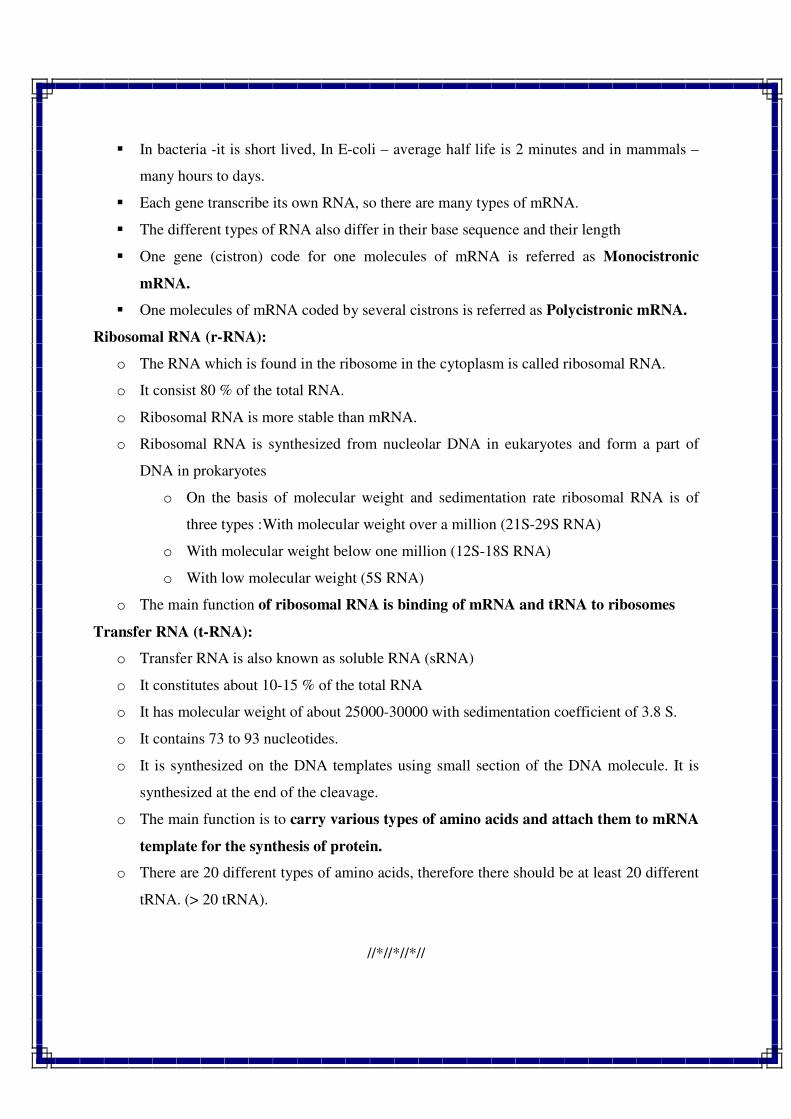
� In bacteria -it is short lived, In E-coli – average half life is 2 minutes and in mammals –
many hours to days.
� Each gene transcribe its own RNA, so there are many types of mRNA.
� The different types of RNA also differ in their base sequence and their length
� One gene (cistron) code for one molecules of mRNA is referred as Monocistronic
mRNA.
� One molecules of mRNA coded by several cistrons is referred as Polycistronic mRNA.
Ribosomal RNA (r-RNA):
o The RNA which is found in the ribosome in the cytoplasm is called ribosomal RNA.
o It consist 80 % of the total RNA.
o Ribosomal RNA is more stable than mRNA.
o Ribosomal RNA is synthesized from nucleolar DNA in eukaryotes and form a part of
DNA in prokaryotes
o On the basis of molecular weight and sedimentation rate ribosomal RNA is of
three types :With molecular weight over a million (21S-29S RNA)
o With molecular weight below one million (12S-18S RNA)
o With low molecular weight (5S RNA)
o The main function of ribosomal RNA is binding of mRNA and tRNA to ribosomes
Transfer RNA (t-RNA):
o Transfer RNA is also known as soluble RNA (sRNA)
o It constitutes about 10-15 % of the total RNA
o It has molecular weight of about 25000-30000 with sedimentation coefficient of 3.8 S.
o It contains 73 to 93 nucleotides.
o It is synthesized on the DNA templates using small section of the DNA molecule. It is
synthesized at the end of the cleavage.
o The main function is to carry various types of amino acids and attach them to mRNA
template for the synthesis of protein.
o There are 20 different types of amino acids, therefore there should be at least 20 different
tRNA. (> 20 tRNA).
//*//*//*//
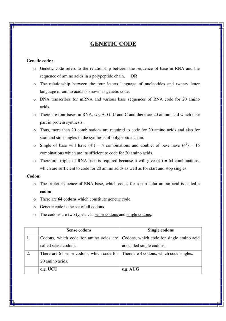
GENETIC CODE
Genetic code :
o Genetic code refers to the relationship between the sequence of base in RNA and the
sequence of amino acids in a polypeptide chain. OR
o The relationship between the four letters language of nucleotides and twenty letter
language of amino acids is known as genetic code.
o DNA transcribes for mRNA and various base sequences of RNA code for 20 amino
acids.
o There are four bases in RNA, viz, A, G, U and C and there are 20 amino acid which take
part in protein synthesis.
o Thus, more than 20 combinations are required to code for 20 amino acids and also for
start and stop singles in the synthesis of polypeptide chain.
o Single of base will have (41) = 4 combinations and doublet of base have (4
2) = 16
combinations which are insufficient to code for 20 amino acids.
o Therefore, triplet of RNA base is required because it will give (43) = 64 combinations,
which are sufficient to code for 20 amino acids as well as for start and stop singles
Codon:
o The triplet sequence of RNA base, which codes for a particular amino acid is called a
codon
o There are 64 codons which constitute genetic code.
o Genetic code is the set of all codons
o The codons are two types, viz, sense codons and single codons.
Sense codons Single codons
1. Codons, which code for amino acids are
called sense codons.
Codons, which code for single amino acid
are called single codons.
2. There are 61 sense codons, which code for
20 amino acids.
There are 4 codons, which code singles.
e.g. UCU e.g. AUG
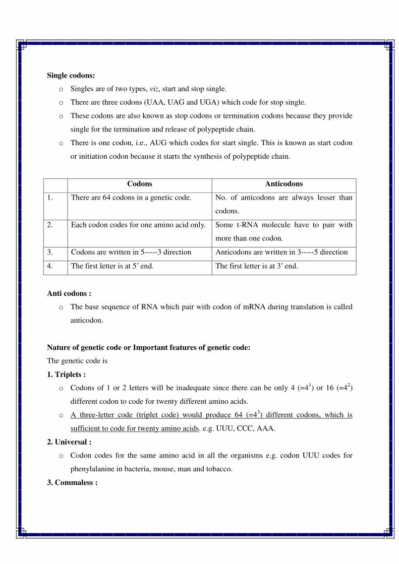
Single codons:
o Singles are of two types, viz, start and stop single.
o There are three codons (UAA, UAG and UGA) which code for stop single.
o These codons are also known as stop codons or termination codons because they provide
single for the termination and release of polypeptide chain.
o There is one codon, i.e., AUG which codes for start single. This is known as start codon
or initiation codon because it starts the synthesis of polypeptide chain.
Codons Anticodons
1. There are 64 codons in a genetic code. No. of anticodons are always lesser than
codons.
2. Each codon codes for one amino acid only. Some t-RNA molecule have to pair with
more than one codon.
3. Codons are written in 5-----3 direction Anticodons are written in 3-----5 direction
4. The first letter is at 5’ end. The first letter is at 3’ end.
Anti codons :
o The base sequence of RNA which pair with codon of mRNA during translation is called
anticodon.
Nature of genetic code or Important features of genetic code:
The genetic code is
1. Triplets :
o Codons of 1 or 2 letters will be inadequate since there can be only 4 (=41) or 16 (=4
2)
different codon to code for twenty different amino acids.
o A three-letter code (triplet code) would produce 64 (=43) different codons, which is
sufficient to code for twenty amino acids. e.g. UUU, CCC, AAA.
2. Universal :
o Codon codes for the same amino acid in all the organisms e.g. codon UUU codes for
phenylalanine in bacteria, mouse, man and tobacco.
3. Commaless :
o All the bases in a segment of polynucleotide are parts of codons and no base serves as a
punctuation mark.
o When there are no intermediary nucleotides between words, UUUCCC two amino acids
in triplet.
4. Non-overlapping :
o It means no single base can again take part in the formation of next codon.
o When only as many amino acids are coded as there are code under in end to end sequence
(e.g. triplet code UUUCCC = phynylalanine + praline).
5. Non-ambiguous :
o When one codon can code for more than one amino acid e.g. GGA = glycine, glutamic
acid.
6. Degenerate :
o When there is more than one codon for a particular amino acid e.g. UUU UAC =
phenylalanine Synonymous code.
7. Polarity :
o The code has a definite direction for reading of message, which is referred to as polarity.
o Reading of code in opposite direction will specify for another amino acids due to
alteration in the base sequences in the codon.
Central dogma :
o In 1958, crick proposed that the information present in DNA (in the form of base
sequence) is transferred to DNA (via replication) and RNA and then from RNA it is
transferred to protein (in the form of amino acid sequence); but this information does no
flow in the reverse direction, that is from protein to RNA to DNA.
o DNA molecules provide the information for their own replication. This relationship
between DNA; RNA and protein molecules is known as central dogma.
//*//*//*//
OUTLINE OF PROTEIN SYNTHESIS
Transcription:
o The process of synthesis of messanger RNA (mRNA) from a DNA template is known as
transcription.
o The mRNA is synthesized only from one strand of DNA in the presence of RNA
polymerase enzyme.
o The RNA polymerase which catalyses the synthesis of RNA from a DNA template is
called transcriptase.
This process can be represented as under:
Transcriptase
DNA mRNA
Transcription
o The process of transcription requires DNA template activated precursors (ATP, GTP,Utp.
And CTp) BIVALENT METAL IONS (Mg++
or Mn) and RNA polymerase.
The process of transcription consists of three main steps:
1. Initiation of transcription.
2. Elongation of RNA.
3. Termination of RNA chain.
Reverse Transcription:
o It was first reported by Temin and Baltimore in 1970 for which they got nobel prize in
1975.
o It is also known as Teminism.
o Reverse transcription means synthesis of DNA from RNA in the presence of DNA
polymerase.
o DNA polymerase is also known as reverse transcriptase.
o It is reported in certain tumor producing viruses.
Reverse Transcriptase Translation
RNA DNA Protein
Reverse Transcription
Translation:
o The process of protein synthesis from information in mRNA is known as translation.
Reverse Translation
RNA Protein
o The translation process requires mRNA, rRNA, ribosones, 20 amino acids kinds of the
process of translation consists of five major steps:
1. Activation of amino acids
2. Transfer of amino acids to tRNA
3. Chain initiation
4. Chain elongation
5. Chain termination
Fine structure of Gene
Classical Vs Modern concept of Gene:
Classical concept of gene Modern concept of gene
1 According to this concept, the term
factors for genes and reported that
factors were responsible for
transmission of characters.
According to this concept, a gene is a sequence of
nucleotides in DNA which controls a single
polypeptide chain.
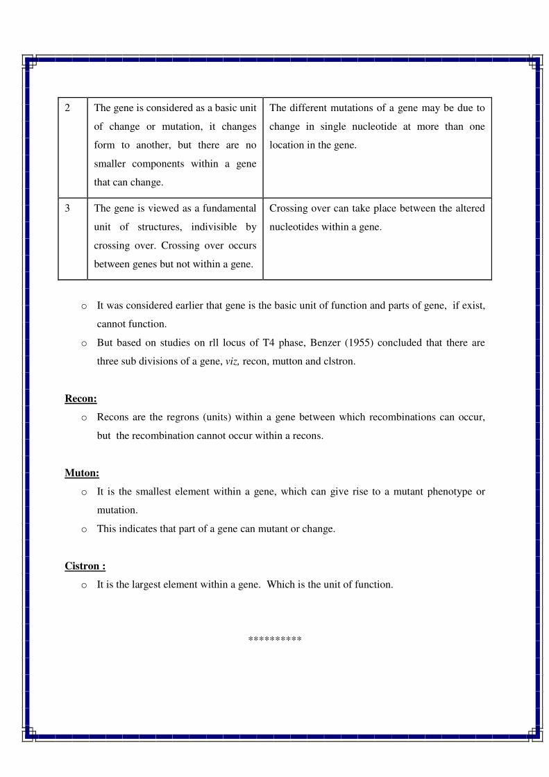
2 The gene is considered as a basic unit
of change or mutation, it changes
form to another, but there are no
smaller components within a gene
that can change.
The different mutations of a gene may be due to
change in single nucleotide at more than one
location in the gene.
3 The gene is viewed as a fundamental
unit of structures, indivisible by
crossing over. Crossing over occurs
between genes but not within a gene.
Crossing over can take place between the altered
nucleotides within a gene.
o It was considered earlier that gene is the basic unit of function and parts of gene, if exist,
cannot function.
o But based on studies on rll locus of T4 phase, Benzer (1955) concluded that there are
three sub divisions of a gene, viz, recon, mutton and clstron.
Recon:
o Recons are the regrons (units) within a gene between which recombinations can occur,
but the recombination cannot occur within a recons.
Muton:
o It is the smallest element within a gene, which can give rise to a mutant phenotype or
mutation.
o This indicates that part of a gene can mutant or change.
Cistron :
o It is the largest element within a gene. Which is the unit of function.
**********
Gene Structure & Gene Regulation
The hereditary units which are transmitted from one generation to the next generation are
called genes. A gene is the fundamental biologic unit, like the atom which is the fundamental
physical unit.
Mendel while explaining the result of his monohybrid and dihybrid crosses, first of all
conceived of the genes as particulate units and referred them by various names such as
hereditary factors or hereditary elements. But his concept about the gene was entirely
hypothetical and he remained ignorant about the physical and chemical nature of gene.
Earlier workers proposed various hypotheses to explain the nature of genes. For instance,
De Vries postulated one gene one character hypothesis according to which a particular trait of
an individual is controlled by a particular gene.
Bateson and Punnett proposed the presence or absence theory. According to them, in a
cross the character which dominates the other has a determiner, while, the recessive character has
no such determiner.
But all the theories were discarded by Morgan, who produced the particulate gene
theory in 1926. He considered genes as corpuscles, which are arranged in a linear order on
the chromosomes and appear like beads on a string. The particulate theory of gene was
widely accepted and supported by cytological observations.
But, the discovery of DNA molecule as a sole carrier of genetic informations base
altogether discarded the Morgan's theory. Therefore, before defining the gene it will be
advisable to consider both the classical as well as modern definitions of gene.
CHANGING CONCEPT OF GENE
The concept of gene has been the focal point of study from the beginning of twentieth
century to establish the basis of heredity. The gene has been examined from two main angles,
i.e., (1) genetic view, and (2) biochemical and molecular view. These aspects are briefly
described below:
(1) A Genetic View
The genetic view or perspective of gene is based mainly on the Mendelian inheritance,
chromosomal theory of inheritance and linkage studies.
� Mendel used the term factors for genes and reported that factors were responsible for
transmission of characters from parents to their offspring.
� Sutton and Boveri (1903) based on the study of mitosis and meiosis in higher plants
established parallel behaviour of chromosomes and genes. They reported that both
chromosomes and genes segregate and exhibit random assortment, which clearly
demonstrated that genes are located on chromosomes. The Sutton- Boveri hypothesis is
known as chromosome theory of inheritance.
� Morgan based on linkage studies in Drosophila reported that genes are located on the
chromosome in a linear fashion. Some genes do not assort independently because of
linkage between them. He suggested that recombinants are the result of crossing over.
The crossing over increases if the distance between two genes is more. The chromosome
theory and linkage studies reveal that genes are located on the chromosomes. This view is
sometimes called as bead theory.
Part of a Gene Can Function
It was considered earlier that gene is the basic unit of function and parts of gene, if exist,
cannot function. But this concept has been outdated now. Based on studies on rll locus of T4
phage, Banzer (1955) concluded that there are three sub divisions of a gene, viz., recon,
muton and cistron. These are briefly described below:
Recon
Recons are the regions (units) within a gene between which recombinations can
occur, but the recombination cannot occur within a recon. There is a minimum
recombination distance within a gene which separates recons. The map of a gene is completely
linear sequence of recons.
Muton
It is the smallest element within a gene, which can give rise to a mutant phenotype
or mutation. This indicates that part of a gene can mutate or change. This disproved the
bead theory according to which the entire gene was a mutate or change.
Cistron
It is the largest element within a gene which is the unit of function. This also nocked
down the bead theory according to which entire gene was the unit of function. The name cistron
has been derived from the test which is performed to know whether two mutants are within the
same cistron on in different cistrons. It is called cis-trans test.
(2) A Biochemical View
� It is now generally believed that a gene is a sequence of nucleotides in DNA which
controls a single polypeptide chain.
� The different mutations of a gene may be due to change in single nucleotide at more than
one location in the gene.
� Crossing over can take place between the altered nucleotides within a gene. Since the
mutant nucleotides are placed so close together, crossing over is expected within very
low frequency.
� When several different genes which affect the same trait are present so close that crossing
over is rare between them, the term complex locus is applied to them.
� Within the nucleotide sequence of DNA, which represents a gene, multiple alleles are due
to mutations at different points within the gene.
Fine Structure of Gene
Benzer, in 1955, divided the gene into recon, muton and cistron which are the units of
recombination, mutation and function within a gene. Several units of this type exist in a gene. In
other words, each gene consists of several units of function, mutation and recombination. The
fine structure of gene deals with mapping of individual gene locus. This is parallel to the
mapping of chromosomes. In chromosome mapping, various genes are assigned on a
chromosome, whereas in case of a gene several alleles are assigned to the same locus. The
individual gene maps are prepared with the help of intragenic recombination. Since the
frequency of intragenic recombination is extremely low, very large population has to be grown to
obtain such rare combination.
Genes can be classified in various ways. The classification of genes is generally done on
the basis of (1) dominance, (2) interaction, (3) character controlled, (4) effect on survival, (5)
location, (6) movement, (7) nucleotide sequence, (8) sex linkage, (9) operon model, and (10) role
in mutation. A brief classification of genes on the basis of above criteria is presented
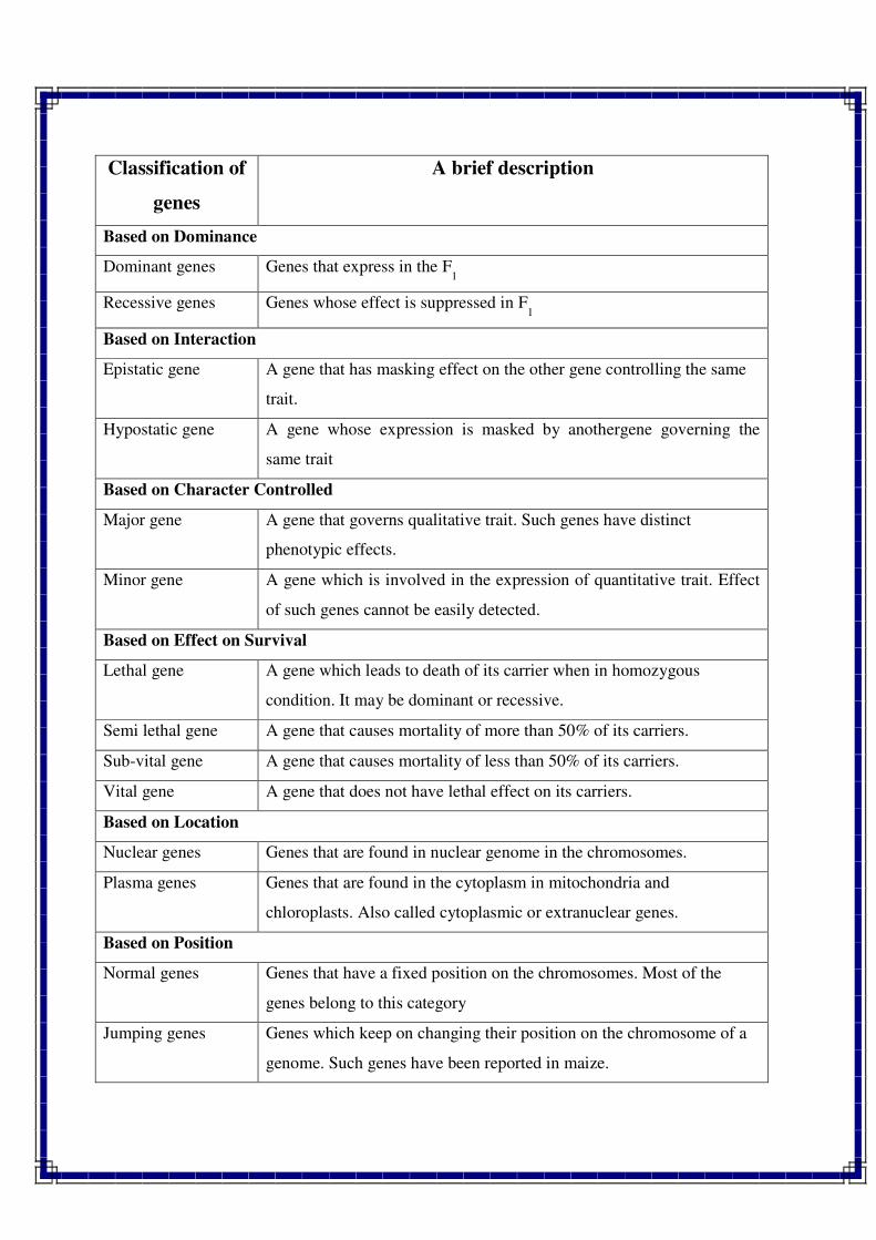
Classification of
genes
A brief description
Based on Dominance
Dominant genes Genes that express in the F1
Recessive genes Genes whose effect is suppressed in F1
Based on Interaction
Epistatic gene A gene that has masking effect on the other gene controlling the same
trait.
Hypostatic gene A gene whose expression is masked by anothergene governing the
same trait
Based on Character Controlled
Major gene
A gene that governs qualitative trait. Such genes have distinct
phenotypic effects.
Minor gene A gene which is involved in the expression of quantitative trait. Effect
of such genes cannot be easily detected.
Based on Effect on Survival
Lethal gene
A gene which leads to death of its carrier when in homozygous
condition. It may be dominant or recessive.
Semi lethal gene A gene that causes mortality of more than 50% of its carriers.
Sub-vital gene A gene that causes mortality of less than 50% of its carriers.
Vital gene A gene that does not have lethal effect on its carriers.
Based on Location
Nuclear genes Genes that are found in nuclear genome in the chromosomes.
Plasma genes Genes that are found in the cytoplasm in mitochondria and
chloroplasts. Also called cytoplasmic or extranuclear genes.
Based on Position
Normal genes
Genes that have a fixed position on the chromosomes. Most of the
genes belong to this category
Jumping genes Genes which keep on changing their position on the chromosome of a
genome. Such genes have been reported in maize.
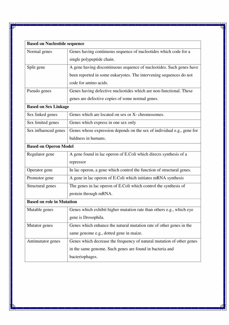
Based on Nucleotide sequence
Normal genes
Genes having continuous sequence of nucleotides which code for a
single polypeptide chain.
Split gene
A gene having discontinuous sequence of nucleotides. Such genes have
been reported in some eukaryotes. The intervening sequences do not
code for amino acids.
Pseudo genes Genes having defective nucleotides which are non-functional. These
genes are defective copies of some normal genes.
Based on Sex Linkage
Sex linked genes Genes which are located on sex or X- chromosomes.
Sex limited genes Genes which express in one sex only
Sex influenced genes Genes whose expression depends on the sex of individual e.g., gene for
baldness in humans.
Based on Operon Model
Regulator gene
A gene found in lac operon of E.Coli which directs synthesis of a
repressor
Operator gene In lac operon, a gene which control the function of structural genes.
Promotor gene A gene in lac operon of E.Coli which initiates mRNA synthesis
Structural genes The genes in lac operon of E.Coli which control the synthesis of
protein through mRNA.
Based on role in Mutation
Mutable genes
Genes which exhibit higher mutation rate than others e.g., which eye
gene is Drosophila.
Mutator genes
Genes which enhance the natural mutation rate of other genes in the
same genome e.g., dotted gene in maize.
Antimutator genes
Genes which decrease the frequency of natural mutation of other genes
in the same genome. Such genes are found in bacteria and
bacteriophages.
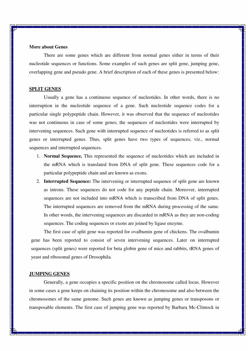
More about Genes
There are some genes which are different from normal genes either in terms of their
nucleotide sequences or functions. Some examples of such genes are split gene, jumping gene,
overlapping gene and pseudo gene. A brief description of each of these genes is presented below:
SPLIT GENES
Usually a gene has a continuous sequence of nucleotides. In other words, there is no
interruption in the nucleotide sequence of a gene. Such nucleotide sequence codes for a
particular single polypeptide chain. However, it was observed that the sequence of nucleotides
was not continuous in case of some genes; the sequences of nucleotides were interrupted by
intervening sequences. Such gene with interrupted sequence of nucleotides is referred to as split
genes or interrupted genes. Thus, split genes have two types of sequences, viz., normal
sequences and interrupted sequences.
1. Normal Sequence. This represented the sequence of nucleotides which are included in
the mRNA which is translated from DNA of split gene. These sequences code for a
particular polypeptide chain and are known as exons.
2. Interrupted Sequence: The intervening or interrupted sequence of split gene are known
as introns. These sequences do not code for any peptide chain. Moreover, interrupted
sequences are not included into mRNA which is transcribed from DNA of split genes.
The interrupted sequences are removed from the mRNA during processing of the same.
In other words, the intervening sequences are discarded in mRNA as they are non-coding
sequences. The coding sequences or exons are joined by ligase enzyme.
The first case of split gene was reported for ovalbumin gene of chickens. The ovalbumin
gene has been reported to consist of seven intervening sequences. Later on interrupted
sequences (split genes) were reported for beta globin gene of mice and rabbits, tRNA genes of
yeast and ribosomal genes of Drosophila.
JUMPING GENES
Generally, a gene occupies a specific position on the chromosome called locus. However
in some cases a gene keeps on chaining its position within the chromosome and also between the
chromosomes of the same genome. Such genes are known as jumping genes or transposons or
transposable elements. The first case of jumping gene was reported by Barbara Mc-Clintock in
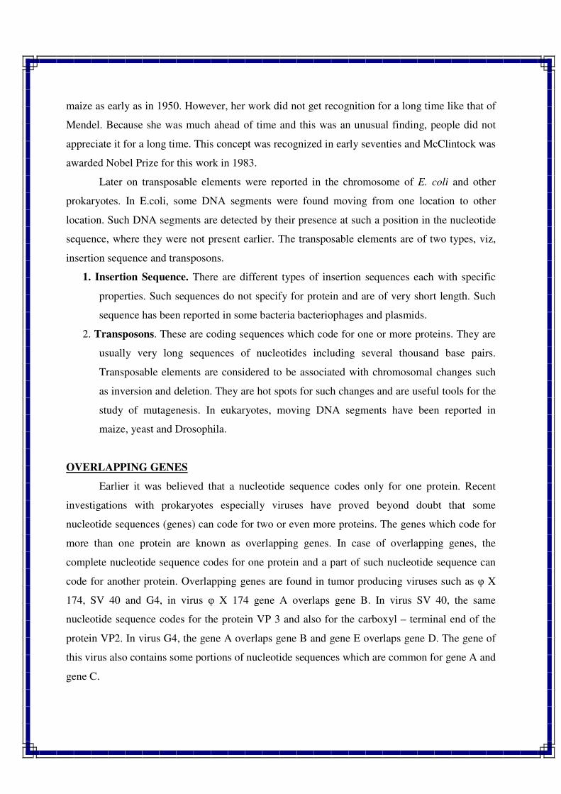
maize as early as in 1950. However, her work did not get recognition for a long time like that of
Mendel. Because she was much ahead of time and this was an unusual finding, people did not
appreciate it for a long time. This concept was recognized in early seventies and McClintock was
awarded Nobel Prize for this work in 1983.
Later on transposable elements were reported in the chromosome of E. coli and other
prokaryotes. In E.coli, some DNA segments were found moving from one location to other
location. Such DNA segments are detected by their presence at such a position in the nucleotide
sequence, where they were not present earlier. The transposable elements are of two types, viz,
insertion sequence and transposons.
1. Insertion Sequence. There are different types of insertion sequences each with specific
properties. Such sequences do not specify for protein and are of very short length. Such
sequence has been reported in some bacteria bacteriophages and plasmids.
2. Transposons. These are coding sequences which code for one or more proteins. They are
usually very long sequences of nucleotides including several thousand base pairs.
Transposable elements are considered to be associated with chromosomal changes such
as inversion and deletion. They are hot spots for such changes and are useful tools for the
study of mutagenesis. In eukaryotes, moving DNA segments have been reported in
maize, yeast and Drosophila.
OVERLAPPING GENES
Earlier it was believed that a nucleotide sequence codes only for one protein. Recent
investigations with prokaryotes especially viruses have proved beyond doubt that some
nucleotide sequences (genes) can code for two or even more proteins. The genes which code for
more than one protein are known as overlapping genes. In case of overlapping genes, the
complete nucleotide sequence codes for one protein and a part of such nucleotide sequence can
code for another protein. Overlapping genes are found in tumor producing viruses such as φ X
174, SV 40 and G4, in virus φ X 174 gene A overlaps gene B. In virus SV 40, the same
nucleotide sequence codes for the protein VP 3 and also for the carboxyl – terminal end of the
protein VP2. In virus G4, the gene A overlaps gene B and gene E overlaps gene D. The gene of
this virus also contains some portions of nucleotide sequences which are common for gene A and
gene C.
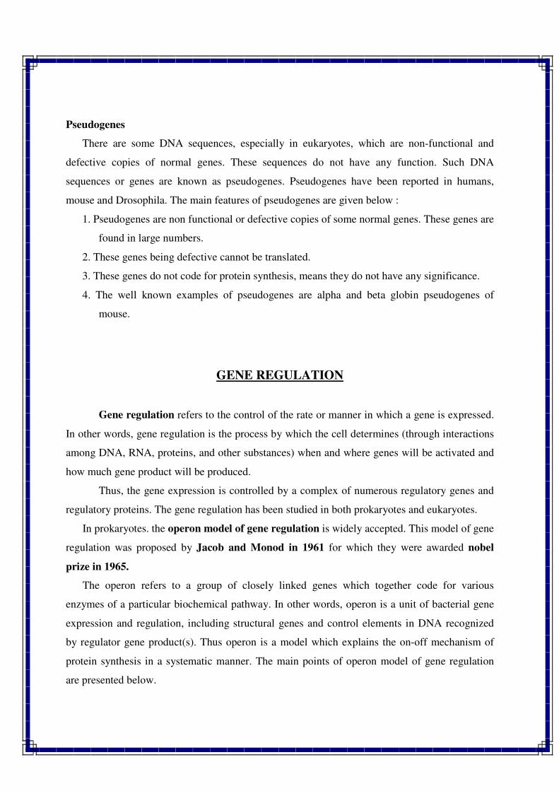
Pseudogenes
There are some DNA sequences, especially in eukaryotes, which are non-functional and
defective copies of normal genes. These sequences do not have any function. Such DNA
sequences or genes are known as pseudogenes. Pseudogenes have been reported in humans,
mouse and Drosophila. The main features of pseudogenes are given below :
1. Pseudogenes are non functional or defective copies of some normal genes. These genes are
found in large numbers.
2. These genes being defective cannot be translated.
3. These genes do not code for protein synthesis, means they do not have any significance.
4. The well known examples of pseudogenes are alpha and beta globin pseudogenes of
mouse.
GENE REGULATION
Gene regulation refers to the control of the rate or manner in which a gene is expressed.
In other words, gene regulation is the process by which the cell determines (through interactions
among DNA, RNA, proteins, and other substances) when and where genes will be activated and
how much gene product will be produced.
Thus, the gene expression is controlled by a complex of numerous regulatory genes and
regulatory proteins. The gene regulation has been studied in both prokaryotes and eukaryotes.
In prokaryotes. the operon model of gene regulation is widely accepted. This model of gene
regulation was proposed by Jacob and Monod in 1961 for which they were awarded nobel
prize in 1965.
The operon refers to a group of closely linked genes which together code for various
enzymes of a particular biochemical pathway. In other words, operon is a unit of bacterial gene
expression and regulation, including structural genes and control elements in DNA recognized
by regulator gene product(s). Thus operon is a model which explains the on-off mechanism of
protein synthesis in a systematic manner. The main points of operon model of gene regulation
are presented below.
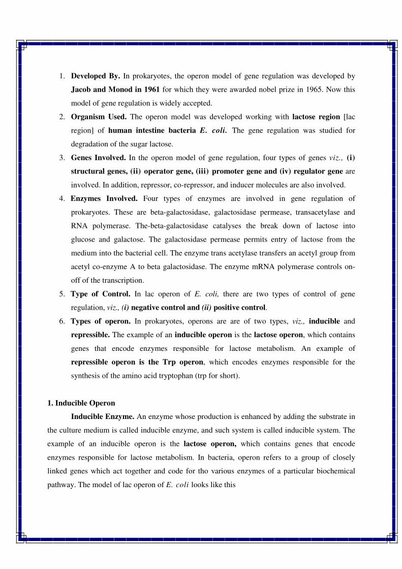
1. Developed By. In prokaryotes, the operon model of gene regulation was developed by
Jacob and Monod in 1961 for which they were awarded nobel prize in 1965. Now this
model of gene regulation is widely accepted.
2. Organism Used. The operon model was developed working with lactose region [lac
region] of human intestine bacteria E. coli. The gene regulation was studied for
degradation of the sugar lactose.
3. Genes Involved. In the operon model of gene regulation, four types of genes viz., (i)
structural genes, (ii) operator gene, (iii) promoter gene and (iv) regulator gene are
involved. In addition, repressor, co-repressor, and inducer molecules are also involved.
4. Enzymes Involved. Four types of enzymes are involved in gene regulation of
prokaryotes. These are beta-galactosidase, galactosidase permease, transacetylase and
RNA polymerase. The-beta-galactosidase catalyses the break down of lactose into
glucose and galactose. The galactosidase permease permits entry of lactose from the
medium into the bacterial cell. The enzyme trans acetylase transfers an acetyl group from
acetyl co-enzyme A to beta galactosidase. The enzyme mRNA polymerase controls on-
off of the transcription.
5. Type of Control. In lac operon of E. coli, there are two types of control of gene
regulation, viz., (i) negative control and (ii) positive control.
6. Types of operon. In prokaryotes, operons are are of two types, viz., inducible and
repressible. The example of an inducible operon is the lactose operon, which contains
genes that encode enzymes responsible for lactose metabolism. An example of
repressible operon is the Trp operon, which encodes enzymes responsible for the
synthesis of the amino acid tryptophan (trp for short).
1. Inducible Operon
Inducible Enzyme. An enzyme whose production is enhanced by adding the substrate in
the culture medium is called inducible enzyme, and such system is called inducible system. The
example of an inducible operon is the lactose operon, which contains genes that encode
enzymes responsible for lactose metabolism. In bacteria, operon refers to a group of closely
linked genes which act together and code for tho various enzymes of a particular biochemical
pathway. The model of lac operon of E. coli looks like this
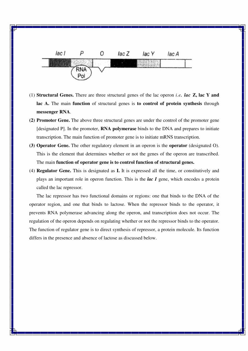
(1) Structural Genes. There are three structural genes of the lac operon i.e. lac Z, lac Y and
lac A. The main function of structural genes is to control of protein synthesis through
messenger RNA.
(2) Promoter Gene. The above three structural genes are under the control of the promoter gene
[designated P]. In the promoter, RNA polymerase binds to the DNA and prepares to initiate
transcription. The main function of promoter gene is to initiate mRNS transcription.
(3) Operator Gene. The other regulatory element in an operon is the operator (designated O).
This is the element that determines whether or not the genes of the operon are transcribed.
The main function of operator gene is to control function of structural genes.
(4) Regulator Gene. This is designated as I. It is expressed all the time, or constitutively and
plays an important role in operon function. This is the lac I gene, which encodes a protein
called the lac repressor.
The lac repressor has two functional domains or regions: one that binds to the DNA of the
operator region, and one that binds to lactose. When the repressor binds to the operator, it
prevents RNA polymerase advancing along the operon, and transcription does not occur. The
regulation of the operon depends on regulating whether or not the repressor binds to the operator.
The function of regulator gene is to direct synthesis of repressor, a protein molecule. Its function
differs in the presence and absence of lactose as discussed below.
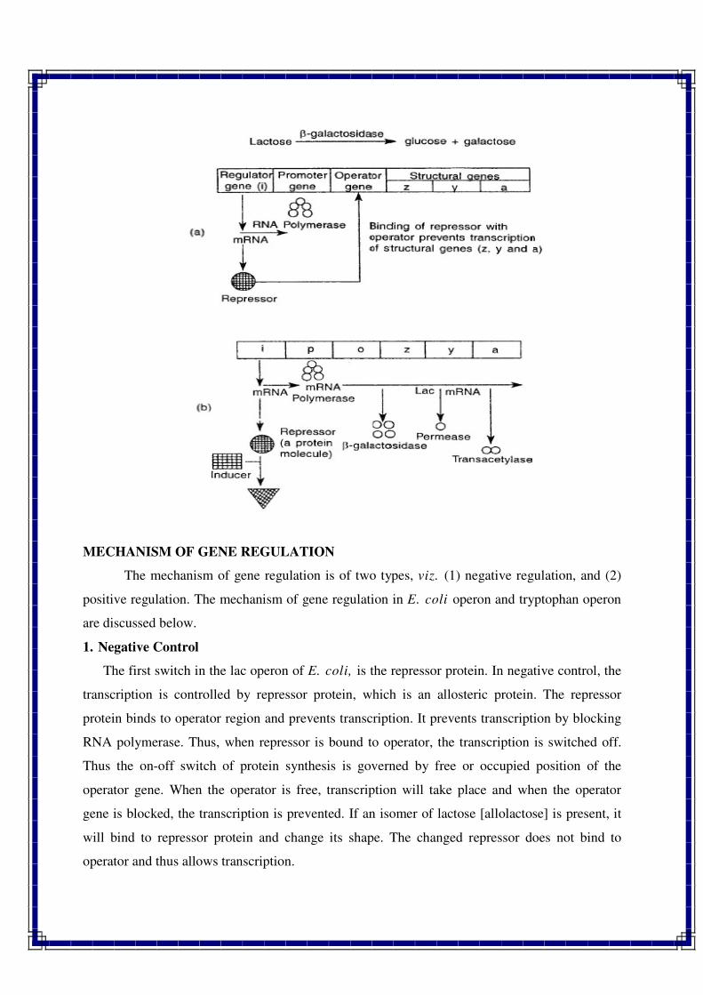
MECHANISM OF GENE REGULATION
The mechanism of gene regulation is of two types, viz. (1) negative regulation, and (2)
positive regulation. The mechanism of gene regulation in E. coli operon and tryptophan operon
are discussed below.
1. Negative Control
The first switch in the lac operon of E. coli, is the repressor protein. In negative control, the
transcription is controlled by repressor protein, which is an allosteric protein. The repressor
protein binds to operator region and prevents transcription. It prevents transcription by blocking
RNA polymerase. Thus, when repressor is bound to operator, the transcription is switched off.
Thus the on-off switch of protein synthesis is governed by free or occupied position of the
operator gene. When the operator is free, transcription will take place and when the operator
gene is blocked, the transcription is prevented. If an isomer of lactose [allolactose] is present, it
will bind to repressor protein and change its shape. The changed repressor does not bind to
operator and thus allows transcription.
2. Positive Control
The second switch in the lac operon of E. coli, is the catabolite activator protein [CAP].The
CAP is an allosteric protein. The CAP binds to DNA and small molecule called cyclic adenosine
mono phosphate [cAMP]. The CAP only binds to promoter region and stimulates transcription
when cAMP binds to allosteric site.
Tryptophan Operon
The tryptophan operon [in short trp operon] is regulated by trp, which is the product of the
metabolic pathway. The trp operon contains genes that makes enzymes in the biosynthetic
pathway for the production of amino acid tryptophan. In trp operon, the negative control is
associated with a repressor protein. However, the repressor protein only binds with operator gene
when an allosteric effector is bound to it. The tryptophan is an allosteric effector, which is called
a corepressor in trp operon also, the transcription is controlled by the free or occupied position of
repressor.
If the repressor protein doesn't bind with operator gene, transcription will take place. If
tryptophan is present, there is no need to synthesize enzymes. In such situation tryptophan binds
to repressor protein and both these [trp and repressor] bind to operator gene preventing
transcription. When trp is absent, the repressor will not bind to the operator, and transcription
will take place. In the negative control, repressor protein binds DNA and stops transcription. In
positive control, activator protein binds DNA and stimulates transcription. In the inducible
system, allosteric effector binds and releases repressor protein from DNA resulting in
transcription. In the repressible system, allosteric effector binds and causes repressor protein to
bind to DNA preventing transcription.
Merits of Operon Model
1. It is a very simple yet informative model of gene regulation in prokaryotes.
2. It is a very well understood model of gene regulation in prokaryotes.
3. This model is based on empirical results and has been studied on different prokaryotes.
4. This model is of two types, viz., (i) inducible operon and (ii) repressible.
//*//*//*//
Related Documents