The imme tem i high damic ad diere, hich eabe it to effectie protect a idiida from mer- o potetia pathogeic ecoter . Ear tdie that combied techiqe ch a eectro microcop ad immoforecece ieded aabe iformatio o the tatic ocatio ad orgaizatio of the ce of the imme tem, ad iferred their damic propertie (for a hitorica perpectie ad referece to origia artice ee REFS 1–3). Hoeer, it i ecear to coider the deeopmet of imme repoe i term of both itracear ad itercear iteractio i the appro- priate eirome t ad i rea time 4–8 . Ideed, the recet deeopmet of high-reotio imagig techiqe, icdig cofoca microcop, total internal reflection fluorescence microscopy ad intravital multiphoton micro- scopy, ha proided iight ito the patio-tempora damic of the moecar ad cear eet that derie imme repoe to pathogeic ifectio. Oe exampe of ch a iight i the moecar decriptio of the immoogica ape, a trctre that i com- mo aociated ith mphocte actiatio fooig the recogitio of a pecific atige 9–12 (BOX 1). B-ce actiatio i iitiated fooig egagemet of the B-ce receptor (BCR) b a pecific atige. B ce ca recogize ad repod to both obe ad membrae-aociated atige, athogh recet iight gget that membrae-aociated atige are more importat for B-ce actiatio in vivo 13,14 . Fooig atigeic timatio, B ce ca proce ad preet atige i aociatio ith MHC ca II moece, thereb recritig pecific CD4 + T-ce hep ad timatig B-ce pro iferatio ad dif- feretiatio 15,16 (BOX 2) . Athogh the precie factor that determie the fate of actiated B ce crret remai c ear, B ce ca dif feretiate aog to ditict patha. O the oe had, B ce ca dif- feretiate to form extrafo ic ar p amabat that are eetia for rapid atibod prodctio ad ear protectie imme repoe. O the other had, actiated B ce ca eter germinal centres, here the ca differetiate ito plasma cells, hich ca ecrete high-affiit atibod fooig affiit matratio, or memory B cells, hich cofer og-atig protectio from ecodar chaege ith atige 17,18 . I thi Re ie, e dic recet iight ito the ite at hich B ce ecoter atige in vivo, a e a the mechaim b hich thee iteractio occr (for exceet reie that icde compreheie decriptio of atige preetatio to T ce in vivo, e e REFS 2,19–21 ). It i o c ear that B ce ca ecoter ad repod to atige throgh ma dif- feret mechaim depedig o the atre ad ize of the atige itef, a e a o t he cear cotext ad ocatio i hich atige preetatio occr. Thi proide great eratiit i term of iitiatig repoe that are appropriate to the particar atige ad are therefore the mot effectie for the protectio of the hot. Lymphocyte Interaction Laboratory, Cancer Research UK London Research Institute, Lincoln’s Inn Fields Laboratories , 44 Lincoln’s Inn Fields, London WC2A 3PX, UK. Correspondence to F.D.B. e‑mail: facundo.batista@ cancer.org.uk doi:10.1038/nri2454 Published online 12 December 2008 Total internal reflection fluorescence microscopy A microscopy method that allows for the identification of fluorescence within 100–200 nm of the interface between cells and their substrate (for example, lipid bilayers or glass coverslips), thereby providing a high lateral and axial resolution at the cell–substrate interface and the ability to observe nanoscale movement of signalling molecules during cellular activation. The who, how and where of antigen presentation to B cells Facundo D. Batista and Naomi E. Harwood Abstract | A functional immune system depends on the appropriate activation of lymphocytes following antigen encounter. In this Review, we summarize studies that have used high-resolution imaging approaches to visualize antigen presentation to B cells in secondary lymphoid organs. These studies illustrate that encounters of B cells with antigen in these organs can be facilitated by diffusion of the antigen or by the presentation of antigen by macrophages, dendritic cells and follicular dendritic cells. We describe cell-surface molecules that might be important in mediating antigen presentation to B cells and also highlight the key role of B cells themselves in antigen transport. Data obtained from the studies discussed here highlight the predominance, importance and variety of the cell-mediated processes that are involved in presenting antigen to B cells in vivo. REVIEWS nATuRE REvIEws | Immunology vOluME 9 | jAnuARy 2009 | 15 © 2009 Macmillan Publishers Limited. All rights reserved

Welcome message from author
This document is posted to help you gain knowledge. Please leave a comment to let me know what you think about it! Share it to your friends and learn new things together.
Transcript

8/4/2019 The Who, How and Where of Antigen
http://slidepdf.com/reader/full/the-who-how-and-where-of-antigen 1/13
The imme tem i high damic ad diere, hicheabe it to effectie protect a idiida from mer-o potetia pathogeic ecoter. Ear tdie thatcombied techiqe ch a eectro microcop adimmoforecece ieded aabe iformatio othe tatic ocatio ad orgaizatio of the ce of theimme tem, ad iferred their damic propertie(for a hitorica perpectie ad referece to origiaartice ee REFS 1–3). Hoeer, it i ecear to coiderthe deeopmet of imme repoe i term of bothitracear ad itercear iteractio i the appro-priate eiromet ad i rea time4–8. Ideed, the recetdeeopmet of high-reotio imagig techiqe,icdig cofoca microcop, total internal reflection
fluorescence microscopy ad intravital multiphoton micro-
scopy, ha proided iight ito the patio-tempora
damic of the moecar ad cear eet thatderie imme repoe to pathogeic ifectio. Oeexampe of ch a iight i the moecar decriptioof the immoogica ape, a trctre that i com-mo aociated ith mphocte actiatio fooigthe recogitio of a pecific atige9–12 (BOX 1).
B-ce actiatio i iitiated fooig egagemetof the B-ce receptor (BCR) b a pecific atige.B ce ca recogize ad repod to both obead membrae-aociated atige, athogh recetiight gget that membrae-aociated atigeare more importat for B-ce actiatio in vivo13,14.Fooig atigeic timatio, B ce ca proce
ad preet atige i aociatio ith MHC ca IImoece, thereb recritig pecific CD4+ T-cehep ad timatig B-ce proiferatio ad dif-feretiatio15,16 (BOX 2). Athogh the precie factorthat determie the fate of actiated B ce crret remai cear, B ce ca dif feretiate aog toditict patha. O the oe had, B ce ca dif-feretiate to form extrafoicar pamabat thatare eetia for rapid atibod prodctio ad ear protectie imme repoe. O the other had,actiated B ce ca eter germinal centres, here the ca differetiate ito plasma cells, hich ca ecretehigh-affiit atibod fooig affiit matratio,or memory B cells, hich cofer og-atig protectiofrom ecodar chaege ith atige17,18.
I thi Reie, e dic recet iight ito the
ite at hich B ce ecoter atige in vivo, a ea the mechaim b hich thee iteractio occr(for exceet reie that icde compreheiedecriptio of atige preetatio to T ce in vivo,ee REFS 2,19–21). It i o cear that B ce caecoter ad repod to atige throgh ma dif-feret mechaim depedig o the atre ad izeof the atige itef, a e a o the cear cotextad ocatio i hich atige preetatio occr.Thi proide great eratiit i term of iitiatigrepoe that are appropriate to the particar atigead are therefore the mot effectie for the protectioof the hot.
Lymphocyte Interaction
Laboratory, Cancer Research
UK London Research
Institute, Lincoln’s Inn Fields
Laboratories, 44 Lincoln’s Inn
Fields, London WC2A 3PX, UK.
Correspondence to F.D.B.
e‑mail: facundo.batista@
cancer.org.uk
doi:10.1038/nri2454
Published online
12 December 2008
Total internal reflection
fluorescence microscopy
A microscopy method that
allows for the identification
of fluorescence within
100–200 nm of the interface
between cells and their
substrate (for example, lipid
bilayers or glass coverslips),
thereby providing a high lateral
and axial resolution at the
cell–substrate interface and
the ability to observe
nanoscale movement of
signalling molecules during
cellular activation.
The who, how and where of antigenpresentation to B cellsFacundo D. Batista and Naomi E. Harwood
Abstract | A functional immune system depends on the appropriate activation of
lymphocytes following antigen encounter. In this Review, we summarize studies that
have used high-resolution imaging approaches to visualize antigen presentation to
B cells in secondary lymphoid organs. These studies illustrate that encounters of B cells
with antigen in these organs can be facilitated by diffusion of the antigen or by thepresentation of antigen by macrophages, dendritic cells and follicular dendritic cells.
We describe cell-surface molecules that might be important in mediating antigen
presentation to B cells and also highlight the key role of B cells themselves in antigen
transport. Data obtained from the studies discussed here highlight the predominance,
importance and variety of the cell-mediated processes that are involved in presenting
antigen to B cells in vivo.
REVIEWS
nATuRE REvIEws | Immunology vOluME 9 | jAnuARy 2009 | 15
© 2009 Macmillan Publishers Limited. All rights reserved

8/4/2019 The Who, How and Where of Antigen
http://slidepdf.com/reader/full/the-who-how-and-where-of-antigen 2/13
Intravital multiphoton
microscopy
A microscopy method that
combines the advanced
optical techniques of
laser-scanning confocal
microscopy with
long-wavelength multiphoton
fluorescence excitation to
capture high-resolution,
three-dimensional images of
living cells and/or tissues that
have been labelled with
fluorophores. It provides a
greater tissue imaging depth
(up to 350 µm depending
on the tissue) and less
photobleaching and
phototoxicity than
conventional imaging
methods.
Germinal centre
A highly specialized and
dynamic microenvironment
that gives rise to secondaryB-cell follicles during an
immune response. It is the
main site of B-cell maturation,
during which memory B cells
and plasma cells that produce
high-affinity antibody are
generated.
Plasma cell
A non-dividing, terminally
differentiated, antibody-
secreting cell of the B-cell
lineage.
Memory B cell
An antigen-experienced B cell
that expresses high-affinity
antibodies and quickly
differentiates into a plasma
cell during antigen-recall
responses.
Alymphoplastic (aly/aly)
mice
Mice that are characterized
by the absence of lymph
nodes and Peyer’s patches.
Alymphoplasia is caused by a
spontaneous mutation in the
gene that encodes nuclear-
factor-κB-inducing kinase.
Sites of antigen encounter
A ce of the imme tem ca repod to a hgerage of potetia pathogeic chaege throghot thehoe bod, thee repoe mt be tight coordiatedto cofer effectie protectio. Oig to thi great ati-geic dierit, the eet of atige ecoter ith aatige-pecific mphocte that ca mot a rapid adappropriate repoe od eem ike. To maximizethe probabiit of ch a iteractio, thee eet occri defied ite, ch a the mph ode ad pee,hich are coectie ko a ecodar mphoidorga (slO). Thee ite poe a high orgaizedmicroarchitectre that i ecear for the compartme-taizatio of mero cear iteractio ad there-fore proide the optima eiromet for the iitiatioof imme repoe, icdig the determiatio of ce fate22. Frthermore, ithi 24 hor mphoctethat hae bee ccef i their earch for atigerecircate throghot the bod ad betee slO todramatica icreae the probabiit of ecoterigcogate atige3.
slO are extreme e coected to the bood admphatic tem, aoig them to cotia ampead cocetrate atige that are circatig throgh-ot the bod. The importace of slO i orgaizigad coordiatig imme repoe i eidet fromthe eere impairmet i the actiatio of aie m-phocte i alymphoplastic (aly/aly) mice ad apeic(homebox 11-deficiet) mice23. Accordig, a mea-igf ietigatio ito the patio-tempora damicad the mechaim b hich mphocte ecoteratige in vivo mt be coidered ithi ch high orgaized eiromet.
Lymph nodes. It i o ide dertood thatbeeath the protectie oter coageo cape, amph ode i diided ito three dicrete (bt o-rigid) regio that are defied b the expreio of pecific chemokie3,24 (FIG. 1a). Direct beeath thebcapar i i a macrophage-rich heet that r-rod the B-ce zoe, hich i ao termed the cor-tex. B ce i thi regio are orgaized ito aggregate,
Box 1 | The immunological synapse
Lymphocyte activation is initiated following the recognition of specific antigen by clonotypic immunoreceptors, such
as the B‑cell receptor (BCR) and the T‑cell receptor (TCR). The recognition of specific antigen on a spatially constrained
surface is associated with membrane reorganization that results in the formation of an immunological synapse9–12.
The structure of the mature immunological synapse is characterized by the spatial segregation of immunoreceptors into
a central supramolecular activation cluster (cSMAC) from the surrounding peripheral SMAC (pSMAC), which contains integrins,
such as lymphocyte function‑associated antigen 1 (LFA1) (see the figure). The early molecular events that underlie the formation
of the immunological synapse have been revealed by high‑resolution imaging techniques and are highly coordinated and tightly
regulated. Following the recognition of specific antigen and before the formation of the immunological synapse, B cells rapidly
spread over the antigen‑containing surface in a manner that is dependent on intracellular signalling and cytoskeletal
rearrangements120. During B‑cell spreading, antigen–BCR microclusters are continually assembled at the periphery of the
contact area between the cells14. As similar microclusters are crucial for sustained signalling in T cells121–123, we have proposed
that these microclusters form functional signalling units that are common to both types of lymphocyte 14.A comprehensive genetic dissection of the requirements for B‑cell spreading identified an important role for the sequential
recruitment of the kinases LYN and spleen tyrosine kinase (SYK) to antigen–BCR microclusters in the initiation of
BCR‑induced signalling124. Furthermore, this study visualized the formation of microsignalosomes through the recruitment of
signalling molecules and adaptors to the antigen–BCR microclusters and showed the importance of cooperation between
VAV and phospholipase Cγ2 in mediating the spreading response. The role of LYN and SYK in the initiation of BCR signalling
has been further supported through
fluorescence resonance energy transfer
(FRET)‑based assays125. B‑cell spreading
facilitates the propagation of signalling
through microclusters by allowing the BCR
to engage more antigen, which promotes
further localized signalling and cytoskeleton
rearrangements120. The spreading response
is followed by a prolonged contraction
phase, which results in the accumulationof antigen–BCR microclusters in the
cSMAC of the mature immunological
synapse. The cSMAC functions as a site for
internalization of antigen12,120, before it is
processed and presented in association
with MHC class II molecules; presentation
of antigen–MHC molecules mediates the
recruitment of the CD4+ T‑cell help that is
necessary for full B‑cell activation15,16. APC,
antigen‑presenting cell; C3, complement
component 3; CR, complement receptor;
FcR, Fc receptor.
APC
Antigen
BCR
Antibody
B cellMicrosignalosome
APC
B cell
B-cell contact area with APC(APC not shown)
Mature immunological synapse:
top view
Immunological synapse: side view
cSMAC pSMAC
Antigen–BCR microcluster: side view
pSMAC cSMAC
B-cellcontact area
FcR CR
Integrinreceptor
C3fragment
R E V I E W S
16 | jAnuARy 2009 | vOluME 9 www.atr./riw/i
© 2009 Macmillan Publishers Limited. All rights reserved
8/4/2019 The Who, How and Where of Antigen
http://slidepdf.com/reader/full/the-who-how-and-where-of-antigen 3/13
T-cell-dependent antigen
A protein antigen that needs to
be recognized by T helper cells
(in the context of MHC
molecules) and requires
cooperation between these
antigen-specific T cells and
B cells for a specific antibody
response to be generated.
Affinity maturation
A process whereby the
mutation of antibody
variable-region genes followed
by selection for higher-affinity
variants in the germinal centre
leads to an increase in average
antibody affinity for an antigen
as an immune response
progresses. The selection is
thought to be a competitive
process in which B cells
compete with free antibody
to capture decreasing amounts
of antigen.
µ mt − / − mice
A strain of mutant mice that
carry a stop codon in the first
membrane exon of the µ-chain
constant region. They lack
IgM+ B cells, and B-cell
development is arrested before
the differentiation stage at
which IgD can be expressed.
ko a foice, ad are the arget popatio of IgMmedIgDhiCD21medCD23hi B ce i the bod. Foiceare ao rich i radiatio-reitat foicar dedriticce (FDC) that expre high ee of the adheiomoece acar ce-adheio moece 1 (vCAM1)ad itercear adheio moece 1 (ICAM1), a ea compemet ad Fc receptor25–28. FDC are thoghtto be of meechma origi ad th form a pop-atio of ce that i ditict from caic DC29. Theprecie mechaim of FDC deeopmet i ot etf dertood, bt i ko to reqire the expre-io of ario chemokie receptor ad the preeceof B ce17,30. Fooig expore to atige, foicema ao cotai peciaized trctre, ko agermia cetre, hich coit of rapid proiferat-ig B ce ithi a etork of FDC. The formatioof germia cetre i importat drig the deeop-met of hmora imme repoe to T-cell-dependent
antigens, a the ere a ite for affinity maturation ad thegeeratio of og-atig memor B ce.
The T-ce zoe, or paracortex, hich i adacet to
the foice, cotai high edotheia ee (HEv)ad mero atige-preetig ce. The HEv arepeciaized capiarie that cotia pp the mphode ith ad drai it of mphocte from ad to theperipher. Frthermore, HEv i the paracortex ca oftemediate the recritmet of ce other tha mphocte,ch a atige-preetig ce that hae accmatedatige i the peripher. The paracortex ao cotaia etork of coageo codit fibre that ao thepaage of o-moecar-ma compoet of mphaticfid, icdig chemokie, from the bcapar ito HEv (reieed i REF. 31).
The meda, hich i the iermot area of themph ode, cotai both B ad T ce that are orga-ized ito medar cord ad i ao rich i DC admacrophage.
lmph ode are trategica poitioed at brachethroghot the mphatic tem to eabe exteieatigeic ampig of mphatic fid4. lmphatic fid,hich cotai ario chemokie ad atige thathae bee coected from the bod, eter the mphode throgh the afferet mph ee ad i be-qet fitered b the mph ode a it fo toardthe efferet mph ee. lmphatic fid i preetedfrom free diffig ito the iterior he it eterthe mph ode b the bcapar i; itead, ittrae to the meda throgh trctre ko atrabecar ie. I additio, compoet of m-phatic fid that hae a moecar eight of beo~70 kDa (eqiaet to a damic radi of ~5.5 m)are aoed to moe toard the HEv throgh the itri-cate mehork of fibrobatic reticar ce (FRC)-iedcoageo codit that are aociated ith DC32,33
(FIG. 1b). Accordig, ear oberatio idicated thatthe paracortex did ot cotai proceed atige,athogh ome atige cod be detected i DC34. Themphatic fid that exit the mph ode i erichedith mphocte35 ad therefore proide a mea forthee ce to retr to the circatio24.
Spleen. The orgaizatio of the mphoid tie of thepee i imiar to that of the mph ode, ith B-cefoice ad T-ce zoe that cotai etork of retic-ar ce (ao ko a the periarterioar mphoidheath) ad a imiar codit tem36 i the peic
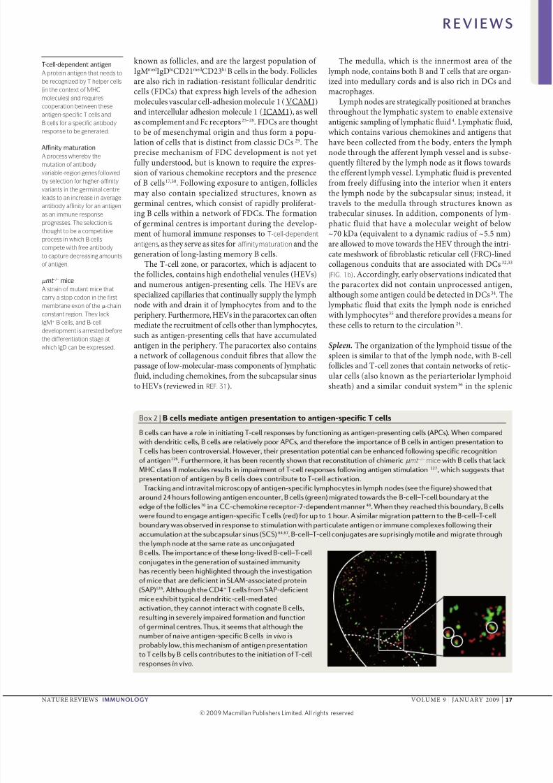
Box 2 | B cells mediate antigen presentation to antigen-specific T cells
B cells can have a role in initiating T‑cell responses by functioning as antigen‑presenting cells (APCs). When compared
with dendritic cells, B cells are relatively poor APCs, and therefore the importance of B cells in antigen presentation to
T cells has been controversial. However, their presentation potential can be enhanced following specific recognition
of antigen126. Furthermore, it has been recently shown that reconstitution of chimeric µ mt –/– mice with B cells that lack
MHC class II molecules results in impairment of T‑cell responses following antigen stimulation 127, which suggests that
presentation of antigen by B cells does contribute to T‑cell activation.
Tracking and intravital microscopy of antigen‑specific lymphocytes in lymph nodes (see the figure) showed that
around 24 hours following antigen encounter, B cells (green) migrated towards the B‑cell–T‑cell boundary at the
edge of the follicles70 in a CC‑chemokine receptor‑7‑dependent manner48. When they reached this boundary, B cells
were found to engage antigen‑specific T cells (red) for up to 1 hour. A similar migration pattern to the B‑cell–T‑cell
boundary was observed in response to stimulation with particulate antigen or immune complexes following their
accumulation at the subcapsular sinus (SCS)64,67. B‑cell–T‑cell conjugates are suprisingly motile and migrate through
the lymph node at the same rate as unconjugated
B cells. The importance of these long‑lived B‑cell–T‑cell
conjugates in the generation of sustained immunity
has recently been highlighted through the investigation
of mice that are deficient in SLAM‑associated protein
(SAP)128. Although the CD4+ T cells from SAP‑deficient
mice exhibit typical dendritic‑cell‑mediated
activation, they cannot interact with cognate B cells,
resulting in severely impaired formation and function
of germinal centres. Thus, it seems that although the
number of naive antigen‑specific B cells in vivo is
probably low, this mechanism of antigen presentation
to T cells by B cells contributes to the initiation of T‑cell
responses in vivo.
Follicle
Follicle
T-cellzone
SCS
R E V I E W S
nATuRE REvIEws | Immunology vOluME 9 | jAnuARy 2009 | 17
© 2009 Macmillan Publishers Limited. All rights reserved

8/4/2019 The Who, How and Where of Antigen
http://slidepdf.com/reader/full/the-who-how-and-where-of-antigen 4/13
T-cell-independent antigen
An antigen that directly
activates B cells.
hite pp37 (FIG. 1a). The pee ao cotai red pp,hich i rich ppied ith bood ad cotai arioatige-preetig ce, mphocte ad pama ce38.The oter imit of the hite pp i eparated from thered pp b a regio ko a the margia zoe 39.Thi area ha exteie acatre, ad bood thateter the pee throgh foicar arterioe brachei the margia i reache the margia zoe beforeit permeatio throgh the red pp40. The margiazoe ao cotai mero macrophage ad DC,ad a izabe popatio of o-recircatig B ce41.
Thee margia-zoe B ce form a popatio of cethat i ditict from foicar B ce, a arge propor-tio of hich are poreactie coe. Margia-zoeB ce hae a IgMhiIgDoCD21hiCD23o pheotpead are thoght to participate i the deeopmet of ear imme repoe to both T-ce-depedet adT-cell-independent antigens42. I cotrat to mph ode,the pee i ot ppied ith afferet mphatic fid(athogh it doe cotai efferet mphatic); itead,it i peciaized i motig imme repoe tobood-bore atige43.
Subcapsularsinus
Afferent lymph
Efferentlymph
HEV
Follicle
Arteriolebranch
Paracortex
PALS
Medulla
Conduit networkConduitnetwork
Conduit network
a
b c
Marginalzone
SpleenLymph node
Marginalsinus
Collagenbundles
FRC Associated DC
Red pulp
Whitepulp
Centralarteriole
T-cell zoneFollicle
Follicle
SCS
Trabecularsinus
Blood
Follicle
Figure 1 | T raizati ar pi ra. a | A schematic representation of secondary lymphoid organs
(SLOs). The lymph node is organized into three discrete (but non-rigid) regions: the medulla, the paracortex (also known as the
T-cell zone) and the follicles (also known as the B-cell zone). Antigen-laden lymphatic fluid flows from the afferent lymph vessel
into the subcapsular sinus and through the trabecular sinuses to the medulla, where it exits through the efferent lymph
vessel. Lymphatic fluid also flows through the conduit network in the paracortex, allowing passage of low-molecular-mass
components, including chemokines and antigen, from the subcapsular sinus to high endothelial venules (HEVs). HEVs in the
paracortex also allow for the entry and exit of lymphocytes to and from the lymph node. The white pulp of the spleen consists
of the paracortex (also known as the periarteriolar lymphoid sheath; PALS), B-cell follicles, a central arteriole and the marginal
zone. Blood arrives at the spleen at the marginal sinus through branches of the central arteriole and from there it flows to the
red pulp through the marginal zone or to the white pulp through the conduit network. b | The conduit network in the lymph
node is composed of a collagen core and is lined with fibroblastic reticular cells (FRCs). Dendritic cells (DCs) are often
associated with FRCs and can extend short protrusions into the conduits, possibly to sample the lymphatic fluid that is
transported through this network. | A snapshot of a video generated using multiphoton microscopy on the popliteal lymph
node. The distribution of seminaphtho-rhodafluor (SNARF)-labelled B cells (red) within the follicles (delineated by a dashed
line) beneath the subcapsular sinus (SCS; solid line) and of carboxyfluorescein diacetate succinimidyl ester (CFSE)-labelled
T cells (green) in the paracortex is shown in the absence of antigen. Scale bar represents 100 µm.
R E V I E W S
18 | jAnuARy 2009 | vOluME 9 www.atr./riw/i
© 2009 Macmillan Publishers Limited. All rights reserved

8/4/2019 The Who, How and Where of Antigen
http://slidepdf.com/reader/full/the-who-how-and-where-of-antigen 5/13
Lymphocyte behaviour in ‘resting’ SLOs
To etabih the ite ad mechaim of atige eco-ter ith mphocte in vivo, it i importat to dertadthe ocaizatio ad damic of thee ce i the retigmphoid orga. Athogh iformatio o ce dam-ic i the pat a iferred from tdie ig taticapproache1–3, iitia grodbreakig tdie that direct
iaized the patio-tempora damic of mphoctein vivo ere poibe becae of adace i the fied of mtiphoto microcop (reieed i REFS 5,19). Thimethod ao for the ietigatio of cear behaiori rea time ad to a depth i iig tie that i prohib-ited b techiqe ch a cofoca microcop 5. The ear-iet mtiphoto microcop ietigatio ere carriedot o expated or ioated mph ode44–46, ad ma of the origia oberatio hae ice bee cofirmed imph ode in vivo fooig miima rgica dirp-tio. Hoeer, high-reotio imagig of mphoctebehaior i peic tie i hampered b the techicachaege that are aociated ith the ioatio of thi
ita ad deep embedded orga6.
The firt rpriig oberatio from thee mti-photo microcop tdie a the high motie atreof mphocte ithi the retig mph ode45–48. B adT ce that ere high poarized i hape ere fodto moe ithi their retricted cear zoe (FIG. 1c) bt aog apparet radom traectorie, ith aerage
eocitie of arod 6 µm (for B ce) ad 12 µm (for Tce) per mite. Coer ipectio of thi moemet fo-oig the iaizatio of the codit etork proidedeidece for a order to thi proce. uig a eegattem ioig chimeric mice that expre gree fo-recet protei, Baéoff et al.49 cod iaize the FDCad FRC i the mph ode before the itrodctio of apopatio of abeed mphocte. Thi td hoedthat the T ce i the paracortex are i coe aociatioith FRC, hich ao the T ce to be gided aoga defied etork. A B ce eter the mph odethrogh HEv i the paracortex, it a ao ggetedthat thee ce cod migrate aog FRC i thi areabefore migratig i a chemokie-directed maer to thefoice, here a imiar etork compoed of FDC cabe ed to orchetrate their moemet. sch rapid moe-met acro thee etork gget that mphocte ithe mph ode are cotia caig their eiro-met ad are read to repod to atige a oo a iti ecotered. Iteretig, imiar ietigatio haeoffered iight ito the behaior of CD11c+ DC i the
paracortex50. I cotrat to mphocte, ad a expectedgie their potetia roe i trctra pport of the co-dit etork, thee DC ere fod to be eie, ho-ig moemet at a eocit of e tha 2 µm per mite.Hoeer, thi retrictio i motiit did ot preet therapid moemet of their dedritic procee, idicatigthat thee DC retai the capacit to ampe ad repodto eirometa iga.
Presentation of antigen to B cells
B ce ca prompt detect ad mot repoeto atige after immizatio. I the cae of ma obeatige, repoe ca be moted fooig a impe
diffio of atige ito the mphoid tie; ho-eer, thee ecoter are a mediated throghmacrophage, DC ad FDC (FIG. 2).
Encountering of soluble antigen. Atige ca be rap-id ppied to the mph ode throgh the afferetmph ee. Ideed, atige arria at the bcap-ar i ca be detected ithi mite of bctae-o admiitratio51. A mot B ce are mai ocatedi the foice, mph-bore atige mt gai acceto foicar B ce i a maer that i idepedetof the codit tem, hich i preet predomiat ithe paracortex.
Iteretig, eera eectro microcop td-ie hae idetified pore of ~0.1–1 µm diameter i theregio of the bcapar i that are adacet tothe mph-ode parechma52–54. Thee pore mightao ma obe atige, ch a o-moecar-matoxi, that eter the mph ode throgh the afferetmph ee to direct pa ito the foice ad gaiacce to B ce55 (FIG. 2a). Ideed, ig immohito-
chemitr of froze mph-ode ectio, it a hothat thi rapid diffio of atige a idepedet of DC ad did ot reqire migratio of B ce from theirfoicar ocatio. Thee oberatio are coitetith a preio td, hich hoed that immiza-tio of mice ith obe atige iitia reted i abtatia decreae i the motiit of atige-pecificB ce i the foice48. Frthermore, i the 24 horfooig immizatio thee B ce ere ho topreet atige i aociatio ith MHC ca II mo-ece o their rface (BOX 2) ad to migrate toardthe bodar betee the foice ad the paracortex.Therefore, ma o-moecar-ma atige ma gaiacce to ce i the foice ithot reqirig ce-mediated preetatio, throgh direct diffio frompore i the bcapar i. Hoeer the exiteceof thee pore remai cotroeria, a the ma imp be ite geerated b ce that hae recet migratedthrogh the i a. Frthermore, there i a dicrep-ac betee the dimeio of the obered poread the radi of the atige that hae bee excdedfrom acceig the foice b diffio. I ie of thi, iteem reaoabe to potate the exitece of a ater-atie mea of mediatig the moemet of maeratige ito the foice, perhap i a maer that iremiicet of diffio from the codit tem, aobered i the paracortex.
Macrophage-mediated presentation.Athogh diff-io of atige from the bcapar i proide oemechaim for expoig foicar B ce to ma obeatige, the acce of arger particate atige to B cei the foice i imited. It a therefore ggeted thatpeciaized ce, poib ocated i the bcapar i,might traport arge atige to the B-ce foice53,56–58.Ear microcop tdie reeaed a popatio of macro-phage that reide direct beeath the bcapari52,53 ad ca exted their procee throgh it to gaiacce to afferet mphatic fid53,56,58. A thee macro-phage hae bee ho to bid a obe recombiat
R E V I E W S
nATuRE REvIEws | Immunology vOluME 9 | jAnuARy 2009 | 19
© 2009 Macmillan Publishers Limited. All rights reserved

8/4/2019 The Who, How and Where of Antigen
http://slidepdf.com/reader/full/the-who-how-and-where-of-antigen 6/13
Metallophilic macrophage
A macrophage that is located
at the border of the white pulp
and the marginal zone of the
spleen. These macrophages
express high levels of CD169
but lack expression of the
mannose receptor.
protei that cotai domai of the maoe receptor,it ha bee ggeted that the ma captre ad coce-trate atige i a imiar a to metallophilic macrophages that reide i the peic margia zoe59–61.
The macrophage i the bcapar i are a di-tict popatio to thoe i the meda ad hae im-ited phagoctic actiit, hich eabe them to preet
itact atige o their ce rface56,58. Ideed, theemacrophage hae bee ho to captre ad retaiatige for p to 72 hor fooig the iitia atigeexpore62. Hoeer, hether thi ioe the imperetetio of atige o the macrophage rface or ati-ge iteraizatio ito o-degradatie itracearcompartmet before it i recced to the ce rface
Subcapsular sinus
SCS macrophage
HEV
Recentlymigrated DC
FRC
T cell
c
a
b
Resident DC
Antigen-specificB cell
Antigen-specificB cell
Folliculardendritic cell
Primary follicle
Small solubleantigen
Paracortex
Afferent lymph vessel
DC-SIGN
BCR
FcR
Antibody
Immunecomplex
C3 fragmentAntigen
SCS
Antigen-specific B cells
Antigen
Antigen non-specific B cells
MAC1
CR
Figure 2 | B- tr wit pii ati i t p . a | B cells in follicles have been found
to encounter small soluble antigens from the lymphatic fluid as they diffuse from the subcapsular sinus (SCS) to the
follicles55. Large antigens, immune complexes and viruses can be presented to follicular B cells at the macrophage-
rich SCS64,67,68. In addition, follicular B cells may recognize antigen that is presented on the surface of follicular dendritic
cells (FDCs). b | Snapshot of a video that was generated using multiphoton intravital microscopy on the popliteal lymph
node. The solid line identifies the position of the SCS. A magnified view of antigen–B-cell conjugates is shown, in which
conjugates appear yellow as a result of the merge of the red antigen-specific B cells and green antigen (P. Barral and F. B.,
unpublished observations). | Schematic view of the paracortex to illustrate where antigen-specific B cells encounter
antigen at this site. B cells entering the lymph node can encounter unprocessed antigen on the surface of resident or
recently migrated DCs, in close proximity to the high-endothelial venules (HEVs) 73,77–79. The conduit system, which is
lined with FRCs and DCs, transports low-molecular-mass components of the lymphatic fluid through the lymph node;B cells and T cells have been shown to migrate in association with the FRC network49. BCR, B-cell receptor; C3, complement
component 3; CR, complement receptor; DC-SIGN, DC-specific ICAM3-grabbing non-integrin; FcR, Fc receptor;
ICAM3, intercellular adhesion molecule 3; MAC1, macrophage receptor 1.
R E V I E W S
20 | jAnuARy 2009 | vOluME 9 www.atr./riw/i
© 2009 Macmillan Publishers Limited. All rights reserved

8/4/2019 The Who, How and Where of Antigen
http://slidepdf.com/reader/full/the-who-how-and-where-of-antigen 7/13
Clodronate-loaded liposome
A liposome that contains the
drug dichloromethylene
diphosphonate. These
liposomes are ingested by
macrophages, resulting in
cell death.
i ot cear. I additio, macrophage are ko to
expre a ide rage of ce-rface receptor thatcod participate i the preetatio of proceedatige, icdig compemet receptor, patter-recogitio receptor ad/or carbohdrate-bidigcaeger receptor63 (TABLE 1). Ideed, macrophagereceptor 1 (MAC1; ao ko a αMβ2 itegriad CD11b–CD18 dimer), hich i a receptor forcompemet compoet 3 (C3) that i expreed b macrophage, ha bee ggeted to cotribte to theretetio of atige o the ce rface64. Ateratie,the ihibitor o-affiit receptor for IgG (FcγRIIB)might mediate the iteraizatio ad reccig of IgG-cotaiig imme compexe to the macrophage cerface, a ha bee ho i DC65. Fia, the C-tpeecti DC-pecific ICAM3-grabbig o-itegri(DC-sIGn; ao ko a CD209) cod participatei the retetio of gcoated atige, hich i co-itet ith the oberatio that mice deficiet i themoe homooge of DC-sIGn, sIGnR1, fai to mothmora imme repoe fooig ifectio ithStreptococcus pneumoniae66.
A crcia ad e decribed roe for bcapar-i macrophage i the preetatio of atige tofoicar B ce ha bee recet etabihed b threeidepedet tdie64,67,68. Thee tdie idetified that,oo after atige admiitratio, bcapar-imacrophage are repoibe for the rapid accma-
tio of ario arger atige, ch a thoe fod iimme compexe (ith atibod ad/or compemetfragmet), particate, bacteria ad ire (FIG. 2a,b).Thee macrophage might faor the retetio of ati-ge o their rface throgh their expreio of -phated gcoprotei, ch a CD169, ad the ack of expreio of the maoe receptor, hich i a aociated ith phagoctoi of opoized atige68.Depetio of macrophage, icdig thoe i the b-capar i ad the meda, throgh treatmet ithclodronate-loaded liposomes redered mice abe tocaptre ad retai eicar tomatiti ir at the b-capar i, retig i temic diemiatio of
thi ir68. Frthermore, a the retetio of atige a
ot impaired fooig the depetio of C3, compemet-idepedet mechaim might ao cotribte to themaiteace of atige o the macrophage rface68.
The accmated atige i beqet preetedi it itact form b macrophage for recogitio b eighborig foicar B ce. Accordig, fooigadmiitratio of atige, it a ho that atige-pecific B ce i the foice exhibited redced migratiooer time ad made tabe cotact ith macrophagei the bcapar i64,67,68. A ICAM1 ad vCAM1are expreed i the bcapar i68, it i expectedthat the ma faciitate B-ce adheio i thi regio adthereb oer the threhod of atige that i reqiredfor B-ce actiatio13,69. Together, thee tdie proidethe firt cear demotratio of a roe for macrophagei the iitiatio of foicar B-ce repoe. The macro-phage–B-ce iteractio at the bcapar i aoatige-pecific B ce to acqire ad iteraize atigethrogh their BCR before their migratio to the B-ce–T-ce bodar 67, here the ma receie pecific T-cehep48,70 (BOX 2). Iteretig, it ha recet bee otedthat the repoe of B ce ad CD4+ T ce to arge irapartice i determiitica iked to their atige pe-cificitie71, ggetig that atige ptake b the B cema ot ioe iteraizatio of hoe ira pathogeat the bcapar i.
DC-mediated presentation. B ce eter the mph odethrogh the HEv, o the paracortex od eem the ideaite for the immediate preetatio of atige to theece. Frthermore, thi regio cotai both reidet DCi coe aociatio ith FRC ad recet migratedDC that hae coected atige from periphera ti-e. DC are ide coidered to be the mot efficietprofeioa atige-preetig ce ad therefore areparticar importat for the preetatio of peptidei a compex ith MHC moece to aie T ce72–74.Hoeer, B ce recogize atige i it proceedatie tate, ad coeqet preetatio to B ceod eceitate a mechaim hereb DC-accmated
Table 1 | Cell-surface molecules that are implicated in presenting antigen to B cells
Prti Rptr Ati prt Prtati trat
Macrophage MAC1 Complement-coated antigen Remains on the cell surface
FcγRIIB IgG-coated antigen Internalized in neutral endosomes and recycled?
DC-SIGN Carbohydrate-containing antigen Internalized in neutral endosomes and recycled?
DC FcγRIIB IgG-coated antigen Internalized in neutral endosomes and recycled?
DC-SIGN Carbohydrate-containing antigen Internalized in neutral endosomes and recycled?
FDC CR1 and CR2 Complement-coated antigen Remains on the cell surface
FcγRIIB IgG-coated antigen Internalized in neutral endosomes and recycled?
Marginal-zoneB cell
CR1 and CR2 Complement-coated antigen Remains on the cell surface
Follicular B cell CR1 and CR2 Complement-coated antigen Remains on the cell surface
FcγRIIB IgG-coated antigen Internalized in neutral endosomes and recycled?
CR, complement receptor; DC, dendritic cell; DC-SIGN, DC-specific ICAM3-grabbing non-integrin; FcγRIIB, low-affinity Fcreceptor for IgG; FDC, follicular DC; ICAM3, intercellular adhesion molecule 3; MAC1, macrophage receptor 1.
R E V I E W S
nATuRE REvIEws | Immunology vOluME 9 | jAnuARy 2009 | 21
© 2009 Macmillan Publishers Limited. All rights reserved

8/4/2019 The Who, How and Where of Antigen
http://slidepdf.com/reader/full/the-who-how-and-where-of-antigen 8/13
Endosome
A vacuolar compartment
where large molecules are
transported after being
engulfed by endocytosis. The
endosome can then mature
and fuse with lysosomes, which
contain degrading enzymes.
Endosomal and phagosomal
pathways are interconnected.
B-1 cell
An IgMhi
IgDlow
MAC1+
B220lowCD23– cell that is
dominant in the peritoneal and
pleural cavities. B-1-precursor
cells develop in the fetal liver
and omentum, and in adult
mice the size of the B-1-cell
population is kept constant
owing to the self-renewing
capacity of these cells. B-1
cells recognize self
components, as well as
common bacterial antigens,
and they secrete antibodies
that tend to have low affinity
and broad specificity.
atige i either tab dipaed o the ce rface or ireitat to itracear degradatio65,75 (TABLE 1). Ideed,it ha bee ho that FcγRIIB ca mediate the iter-aizatio of atige-cotaiig imme compexe itoo-degradatie itracear compartmet65, ad itha ao bee ggeted that DC-sIGn might ao theaccmatio of itact atige i etra endosomes iDC. Cocomitat ith thi hpothei, sIGnR1 habee impicated i the iteraizatio of HIv-1 ito ao-ooma compartmet of DC ad the beqetdeier of itact HIv-1 irio to slO76.
A popatio of DC i the paracortex that capreet itact atige to B ce ha bee idetified77–79.A detaied characterizatio of thi DC popatio b itraita mtiphoto microcop hoed that theece are mai ocated arod HEv o that migrat-ig B ce ca re their atigeic cotet79 (FIG. 2c).Fooig the admiitratio of atige-oaded DCto mice, recet migrated atige-pecific B ceho decreaed motiit ad a icreae i their rei-dec time o DC. Iteretig, thee DC retaied
the capacit to timate B ce fooig treatmetith proae (a mixtre of proteiae) to remoe ce-rface-expoed atige, ggetig that DC ca iter-aize ad the recce itact atige to their ce rface.The DC-mediated preetatio of atige to B cei the paracortex i the idea eiromet i hich toreceie the ecear T-ce hep for their maxima acti-
atio79. I pport of thi, DC, B ce ad T ce haebee ho to coocaize i thi regio of the mphode50. Thi DC-mediated actiatio of B ce ma gie rie to extrafoicar pama ce that mot ear atibod repoe to atige. Ateratie, fooigactiatio b CD4+ T ce, B ce might migrate to thefoice ad mediate germia-cetre formatio. It iao proe aabe to characterize the in vivo damicof a popatio of maoe-receptor-bidig DC thataccmate obe atige i the foice ad preet itto foicar B ce80.
Techica cotrait hae imited the appicatioof itraita mtiphoto microcop i the td of mphocte damic i the pee. Hoeer, a the co-dit tem i the hite pp36 ad the core orgaizatioof B- ad T-ce zoe i the pee are imiar to thoei the mph ode, the mechaim of atige pree-tatio to B ce might be imiar i both tie. Thepeic margia-zoe B-ce popatio ha bee hoto rapid prodce atibod i repoe to bood-bore
atige, ad therefore thee ce hae a importat roei the iitiatio of ear imme repoe81. Hoeer,the participatio of margia-zoe B ce at ater tageof the imme repoe ha bee e e character-ized. static in vitro imagig approache hae bee edto td DC-mediated atige preetatio to peicmargia-zoe B ce82 ad hae ho that thi actia-tio ead to the rapid geeratio of pama ce thatprodce IgM idepedet of T-ce hep, aoig forthe rapid ad ehaced formatio of atige-cotaiigimme compexe. Itrigig, a popatio of B ceith imiar characteritic to margia-zoe B ce habee detected i hma mph ode83.
FDC-mediated presentation. Caica, FDC haebee coidered to be the ce that are mai repo-ibe for atige preetatio to B ce i slO. Ear atoradiograph aai combied ith high-reotioeectro microcop hoed that extracear atige ia pama-membrae-aociated form i retaied i thefoice for prooged period after immizatio51,84.sbeqet, it a etabihed that FDC i the fo-ice are repoibe for mediatig the retetio of ati-ge i the form of imme compexe26,85,86. I additioto their roe i atige retetio ad preetatio, it iko that FDC fctio a potet acceor ce dr-ig B-ce actiatio, poib throgh mechaim thatioe the ecretio of chemokie ad/or the expre-io of FcγRIIB87.
The ditictie pheotpe of FDC pport theretetio of imme compexe i the foice throghto differet mechaim (TABLE 1). The firt mechaimdeped o the compemet tem85,88,89. FDC exprehigh ee of compemet receptor 1 (CR1) ad CR2 (ao ko a CD35 ad CD21, repectie), hich
bid to ario fragmet of C3. Chimeric mice that ack expreio of CR1 ad CR2 i the radioreitat troma-ce compartmet (icdig FDC) caot depoitatige o the rface of FDC90,91, ad FDC from micethat are abe to prodce CR2 igad caot preetatige to B ce92.
The ecod mechaim of atige depoitio ioethe retetio of imme compexe that cotai IgG fo-oig their bidig to Fc receptor, ch a FcγRIIB,that are expreed o the rface of FDC i germiacetre28,93. Coitet ith thi, FcγRIIB-deficiet miceho eere redced trappig of imme compexe ithe mph ode ad pee, ad FcγRIIB-deficiet micethat ere recotitted ith id-tpe, atige-pecificmphocte exhibit eere impaired reca repoeto imme compexe28.
Depoitio of atige o the rface of FDC throgheither or both of thee mechaim od therefore beexpected to icreae the iitiatio of imme repoe.Ideed, preformed imme compexe58,94 ad atigecoated ith C3d fragmet95 are both aociated ith thetimatio of B-ce actiatio, potetia a a ret of icreaed depoitio of atige o the rface of FDC96.Frthermore, fooig their bidig to atige, atraIgM atibodie that are prodced mai b peritoeaB-1 cells97 idce the formatio of imme compexead acceerate the depoitio of atige o the rface
of FDC27. Mice ith a targeted deetio i the carbox-termia tai of IgM fai to expre erm IgM ad hodeaed hmora imme repoe, ggetig a roefor FDC i the iitiatio of imme repoe98,99.
Depite the abiit of FDC to preet proceedatige, the importace of thi patha for the actiatioof aie B ce drig the iitiatio of primar immerepoe, a e a it derig mechaim, hae etto be firm etabihed. Iteretig, it ha bee pot-ated that FDC ma be particar importat for thepreetatio of microbia atige, i repoe to hichpre-exitig atra IgM atibodie cod faciitate therapid formatio of imme compexe, thereb retig
R E V I E W S
22 | jAnuARy 2009 | vOluME 9 www.atr./riw/i
© 2009 Macmillan Publishers Limited. All rights reserved

8/4/2019 The Who, How and Where of Antigen
http://slidepdf.com/reader/full/the-who-how-and-where-of-antigen 9/13
Somatic hypermutation
(SHM). A unique mutation
mechanism that is targeted
to the variable regions of rearranged immunoglobulin
gene segments. Combined with
selection for B cells that
produce high-affinity antibody,
SHM leads to affinity
maturation of B cells in
germinal centres.
Cobra venom factor
The complement-activating
glycoprotein component of
cobra venom, which is
functionally analogous to the
mammalian complement
factor C3b.
i microbia-atige preetatio b Fc receptor o therface of FDC100. Hoeer, it i ko that FDC iprimar foice do ot expre high ee of FcγRIIB28 ad therefore thi receptor caot hae a importatroe i atige retetio ad preetatio drig theiitiatio of imme repoe.
FDC ca ao act a atige ‘depot’ i the slOch that the ca preet atige ee after the iitia‘ae’ of atige ha paed26–28,101. Thi i importatfor the deeopmet of a effectie ad og-atigimme repoe, a the iitia ecoter of atigeith aie B ce i the slO i ike to be of oaffiit. uder thee coditio, actiated B ce theeter germia cetre, here the dergo affiit matratio17. Thi proce ao the eectio of B-cecoe ith higher affiit for atige drig a T-ce-depedet atibod repoe ad tart a fe da afterthe iitiatio of the repoe.
The caic mode that decribed the mechaim of atige preetatio to B ce i germia cetre abaed o eera biochemica ad hitoogica obera-
tio. I thi mode, the germia cetre a diidedito to fctioa ditict zoe17. The dark zoea thoght to cotai cetrobat ad to be the iteof rapid B-ce proiferatio, herea the FDC-rich ightzoe fctioed a the ite for the eectio of B-cecoe that expre BCR of the highet affiit foo-ig somatic hypermutation. Recet, three idepedettdie examiig the patio-tempora damic of B ce hae qetioed the origia mode ad the abo-te ‘diiio of abor’ betee the to zoe of thegermia cetre102–104. Iteretig, each of thee tdiehoed that 6 da after atige admiitratio B cemoed rapid ithi the germia cetre, hich idi-cated the did ot make prooged cotact ith atigepreeted o the rface of FDC. It a therefore g-geted that the mode of atige recogitio b B ceoccrrig drig the ater tage of a imme repoema operate differet to that obered drig ear atige ecoter. Ideed, thee tdie hoed thatgermia-cetre B ce adopted a a morphoog ith ma exteded procee102–104. Thee proceema be repoibe for mediatig atige iteraiza-tio a the B ce migrate aog the rface of the FDC.uder atige-imitig coditio, B ce expreigBCR ith higher affiit ca accmate more atigead coeqet compete more effectie to recritpecific T-ce hep for their deeopmet, ith o
the highet affiit coe beig eected for riai the germia cetre102,105. Hoeer, it i importat toote that CD4+ T ce reide excie ithi the ightzoe, o the compete abece of fctioa egregatioi the germia cetre i ike.
Atige preetatio b FDC throgh bidig of imme compexe to FcR or compemet receptor ma be importat for the deeopmet of high-affiit atibod ad og-atig memor repoe106. srpriig, ho-eer, the proce of affiit matratio ad the riaof memor B ce, athogh impaired, ca occr i theabece of detectabe atige depoitio o FDC107,108.Thi oberatio ed to the hpothei that i additio
to atige retetio, FDC might hae aother roedrig affiit matratio109. Hoeer, thee tdiedid ot excde the poibiit that competitio amogB ce for atige cod hae a roe i the eectio of high-affiit B-ce coe i the germia cetre. I
ie of thi, it od proe extreme iformatie tocharacterize the patio-tempora damic of ce thatretai atige ithi imme compexe, hich odetabih the precie roe of depoit of atige o FDCdrig the proce of affiit matratio.
How does antigen gain access to FDCs? A a coeqeceof the roe of FDC i mediatig atige preetatio toB ce, mch effort ha bee ieted i addreig theapparet aoma of ho atige i the form of immecompexe ca gai rapid acce to FDC, hich are co-fied to the B-ce foice. A the bcapar i adthe codit tem retrict the diffio of arge atigefrom the mphatic fid, a ce-mediated mechaimmt operate to traport atige to FDC.
I the pee, margia-zoe B ce hae bee impi-
cated i thi proce59,110 (FIG. 3a). The ocatio of B cei the margia zoe make them idea poitioedto ampe ad mot rapid repoe to bood-boreatige. Ideed, dipacemet of thee B ce b treat-met ith edotoxi, ch a ipopoaccharide, aaccompaied b impaired accmatio of immecompexe o the rface of FDC i the foice59,111,112.Moreoer, it ha bee ho that the accmatio of IgM-cotaiig imme compexe o FDC a abro-gated i the abece of fctioa margia-zoe B ce.Thi gget that margia-zoe B ce faciitate theadat actiit of IgM113.
Margia-zoe B ce expre compemet receptor(TABLE 1), ad thee ma be importat for their fctioi traportig atige to FDC. Ideed, ear ieti-gatio ig cobra venom factor ad atibodie agaitC3 hoed that the traport of imme compexefrom the margia zoe to the FDC deped o com-poet of the compemet tem85,89,114. I the abeceof CR2 expreio b margia-zoe B ce, immecompexe ere aociated predomiat ith macro-phage i the margia zoe, a the B ce faied totraport atige to the FDC115. The mechaim b hich margia-zoe B ce traport atige from themargia zoe to the foice a reeaed b a recettd ioig the treatmet of chimeric mice ith thephigoie 1-phophate receptor 1 (s1PR1) atagoit
FTy720 (REF. 116). Margia-zoe B ce ere fod tocotia htte to ad from the foice i a procethat depeded o the expreio of CXC-chemokiereceptor 5 (CXCR5) ad s1PR1, repectie. A mar-gia-zoe B ce ca bid imme compexe throghCR1 ad CR2, thi httig proce cod be the a i hich atige i effectie deiered from the boodto FDC. Iteretig, it ha bee potated that theacta trafer of imme compexe from margia-zoe B ce to FDC occr throgh proteoi of CR2o the rface of the margia-zoe B ce117. Hoeer,it i ot ko hether margia-zoe B ce ca pafree atige direct to foicar B ce.
R E V I E W S
nATuRE REvIEws | Immunology vOluME 9 | jAnuARy 2009 | 23
© 2009 Macmillan Publishers Limited. All rights reserved

8/4/2019 The Who, How and Where of Antigen
http://slidepdf.com/reader/full/the-who-how-and-where-of-antigen 10/13
simiar, it ha bee ggeted that foicar B cethemee fctio a atige traporter i the mphode ad pee89,118 (FIG. 3b). Thi actiit doe otdeped o atige-pecific recogitio ad thereforei mediated b receptor other tha the BCR (TABLE 1).It ha bee propoed that atige ca be acqired b other foicar B ce i the abece of atige-pecificB ce64,67, ad thi ma deped o the expreio of compemet receptor o the B-ce rface64. A FDCexpre higher ee of compemet receptor thafoicar B ce, it od be expected that the codcompete effectie ith B ce for atige bidig27.It remai to be determied hether other receptor of the iate imme tem hae a roe i the traportof atige b B ce ithi slO. CD23+ matre B cehae i fact bee ho to mediate the traport of IgE-cotaiig imme compexe ito the foice i aBCR-idepedet maer119. Athogh ch a itatioi ike to occr i phioogica coditio, it doeidicate that ateratie receptor might be ioed iatige traport.
Conclusions and perspectives
Oera, it i cear that B ce ca repod rapid toatigeic timatio throgh eera mechaim, adthe ao hae a iqe trategic roe i the traportof atige to FDC i the germia cetre. Thi ide
ariet i potetia atige-preetatio mechaim ipromoted b fctioa peciaizatio ithi slO adproide iaabe fexibiit i motig appropriateimme repoe to atige. A ch, the mechaimthat i ed to preet a pecific atige ca be effec-tie taiored both to the atige itef ad to the a the atige i deiered to the B ce. The predomiaceof ce-baed trategie for the preetatio of atige toB ce ao for exteie regatio ad coordiatioof the retig imme repoe.
I pite of igificat recet progre i character-izig the ite ad damic of atige preetatioto B ce in vivo, ome ie remai to be addreed.Thee icde a defiitie moecar decriptio of themechaim of atige preetatio to B ce, icd-ig the retetio ad iteraizatio trategie that are
Immunecomplex
Antigennon-specificBCR
CR
Marginal-zoneB cell
Marginal zone
a Splenic marginal-zone B cells transport antigen to FDCs b Follicular B cells in the lymph node transport antigen to FDCs
Primary follicle Primary follicle
SCSmacrophage
Folliculardendritic cell
FollicularB cell
?
?
Subcapsular sinusS1Phi
CXCL13low
S1Plow
CXCL13hi
C3fragment
FcR
DC-SIGN
Figure 3 | B iat ati traprt t iar riti i pt rptr.
a | Marginal-zone B cells in the spleen can bind to immune complexes that contain antigen and are coated in complement
fragments, using complement receptors (CR) in a manner that is independent of B-cell receptor (BCR) specificity. These
marginal-zone B cells can shuttle to the follicular region, which is rich in CXC-chemokine ligand 13 (CXCL13), in a CXC-
chemokine-receptor-5-dependent manner116. Follicular dendritic cells (FDCs) in the follicle, which express high levels of
CR, can then compete for binding to antigen that is presented by marginal-zone B cells. The marginal-zone B cells then
migrate back to the marginal zone, where there are high levels of sphingosine 1-phosphate (S1P), and this depends on
their expression of the receptors for S1P1 and S1P3. b | Follicular B cells in the lymph node can bind to particulate antigen
on the surface of subcapsular sinus (SCS) macrophages. This binding does not depend on the specificity of the BCR and is
mediated by CRs. It has been suggested that these follicular B cells can then mediate the transport of antigen to FDCs,
which express higher levels of CRs64,67. C3, complement component 3; DC-SIGN, DC-specific ICAM3-grabbing
non-integrin; FcR, Fc receptor; ICAM3, intercellular adhesion molecule 3.
R E V I E W S
24 | jAnuARy 2009 | vOluME 9 www.atr./riw/i
© 2009 Macmillan Publishers Limited. All rights reserved

8/4/2019 The Who, How and Where of Antigen
http://slidepdf.com/reader/full/the-who-how-and-where-of-antigen 11/13
ed b the atige-preetig ce. I additio, itod proe iformatie to track the fate of B ce fo-oig atigeic timatio, i term of the particardifferetiatio patha that i iitiated. sch progrema be poibe b improig the reotio that iachieabe b imagig techiqe, i combiatio iththe geeratio of aima mode that are deficiet i orhae atered expreio of the differet compoet thatma be ioed. Frthermore, adace i the fied of high-reotio itraita imagig ma ao for the
iaizatio of in vivo atige ecoter at greatertie depth ad for more exteded period of time.Thee deeopmet ma ao offer iight ito potetia
roe of the iteractio betee foicar B ce i theimme repoe ad ito the importace of FDC ithe preetatio of atige to aie B ce. Fia, it iimportat to etabih that the in vivo oberatio thathae bee coected o far i trageic aima-modetem refect thoe that occr drig the timatioof the atra repertoire of B ce drig ifectio orfooig acciatio.
Take together, a more compreheie decriptio of the mechaim b hich B ce ee ad repod toatige i hae impicatio for the ftre taiorig of
acciatio trategie ad ao i the ratioa deigof treatmet for ifectio ad atoimme dieae.
1. Bajénoff, M. & Germain, R. Seeing is believing: a focus
on the contribution of microscopic imaging to our
understanding of immune system function.
Eur. J. Immunol. 37, S18–S33 (2007).
2. Halin, C., Rodrigo Mora, J., Sumen, C. &
von Andrian, U. In vivo imaging of lymphocyte
trafficking. Annu. Rev. Cell Dev. Biol. 21, 581–603
(2005).
3. von Andrian, U. & Mempel, T. Homing and cellular
traffic in lymph nodes. Nature Rev. Immunol. 3,
867–878 (2003).
References 1–3 are three excellent reviews
detailing key historical insights into the function of
the immune system and have served as the
foundation for subsequent high-resolution imaging
investigations.
4. Catron, D., Itano, A., Pape, K., Mueller, D. & Jenkins, M.
Visualizing the first 50 hr of the primary immune
response to a soluble antigen. Immunity 21, 341–347
(2004).
5. Germain, R., Miller, M., Dustin, M. & Nussenzweig, M.
Dynamic imaging of the immune system: progress,
pitfalls and promise. Nature Rev. Immunol. 6,
497–507 (2006).6. Mempel, T., Scimone, M., Mora, J. & von Andrian, U.
In vivo imaging of leukocyte trafficking in blood vessels
and tissues. Curr. Opin. Immunol. 16, 406–417 (2004).
7. Cahalan, M. & Parker, I. Imaging the choreography of
lymphocyte trafficking and the immune response.Curr. Opin. Immunol. 18, 476–482 (2006).
8. Bousso, P. & Robey, E. Dynamic behavior of T cells
and thymocytes in lymphoid organs as revealed by
two-photon microscopy. Immunity 21, 349–355
(2004).9. Grakoui, A. et al. The immunological synapse: a
molecular machine controlling T cell activation.
Science 285, 221–227 (1999).
10. Krummel, M., Sjaastad, M., Wülfing, C. & Davis, M.
Differential clustering of CD4 and CD3z during T cell
recognition. Science 289, 1349–1352 (2000).
11. Monks, C., Freiberg, B., Kupfer, H., Sciaky, N. &
Kupfer, A. Three-dimensional segregation of
supramolecular activation clusters in T cells.
Nature 395, 82–86 (1998).
12. Batista, F., Iber, D. & Neuberger, M. B cells acquire
antigen from target cells after synapse formation.
Nature 411, 489–494 (2001).
This paper describes the formation of the
immunological synapse in response tomembrane-bound antigen and the necessity of
internalization of antigen through the central
supramolecular activation cluster for maximal
B-cell activation.
13. Carrasco, Y. R. & Batista, F. D. B cell recognition of
membrane-bound antigen: an exquisite way of
sensing ligands. Curr. Opin. Immunol. 18, 286–291
(2006).
14. Depoil, D. et al. CD19 is essential for B cell activation
by promoting B cell receptor-antigen microcluster
formation in response to membrane-bound ligand.
Nature Immunol. 9, 63–72 (2008).
This paper visualizes the formation of antigen–BCR
microclusters and, by identifying an essential role
for CD19 in B-cell activation by membrane-bound
antigen, it indicates the importance of the
recognition of this type of antigen in vivo during
the development of immune responses.
15. Lanzavecchia, A. Antigen-specific interaction
between T and B cells. Nature 314, 537–539
(1985).16. Rock, K., Benacerraf, B. & Abbas, A. Antigen
presentation by hapten-specific B lymphocytes. I. Role
of surface immunoglobulin receptors. J. Exp. Med.
160, 1102–1113 (1984).
17. MacLennan, I. Germinal centers. Annu. Rev. Immunol.
12, 117–139 (1994).
18. Rajewsky, K. Clonal selection and learning in the
antibody system. Nature 381, 751–758 (1996).
19. Cahalan, M. & Parker, I. Choreography of Cell. Motility
and interaction dynamics imaged by two-photon
microscopy in lymphoid organs. Annu. Rev. Immunol.
26, 585–626 (2008).
20. Celli, S., Garcia, Z., Beuneu, H. & Bousso, P.
Decoding the dynamics of T cell–dendritic cell
interactions in vivo. Immunol. Rev. 221, 182–187
(2008).
21. Germain, R. et al. Making friends in out-of-the-way
places: how cells of the immune system get together
and how they conduct their business as revealed by
intravital imaging. Immunol. Rev. 221, 163–181
(2008).22. Junt, T., Scandella, E. & Ludewig, B. Form follows
function: lymphoid tissue microarchitecture in
antimicrobial immune defence. Nature Rev. Immunol.
8, 764–775 (2008).
23. Karrer, U. et al. On the key role of secondarylymphoid organs in antiviral immune responses
studied in alymphoplastic (aly/aly) and spleenless
(Hox11–/–) mutant mice. J. Exp. Med. 185,
2157–2170 (1997).
24. Cyster, J. Chemokines, sphingosine-1-phosphate, and
cell migration in secondary lymphoid organs. Annu.
Rev. Immunol. 23, 127–159 (2005).
25. Klaus, G., Humphrey, J., Kunkl, A. & Dongworth, D.
The follicular dendritic cell: its role in antigen
presentation in the generation of immunological
memory. Immunol. Rev. 53, 3–28 (1980).
26. Mandel, T., Phipps, R., Abbot, A. & Tew, J. The
follicular dendritic cell: long term antigen retention
during immunity. Immunol. Rev. 53, 29–59 (1980).
References 25 and 26 are two excellent reviews
that summarize much of the early work that
characterized the role of FDCs in the presentation
and retention of antigen and described how FDCs
influence immune responses.
27. Carroll, M. The role of complement and complementreceptors in induction and regulation of immunity.
Annu. Rev. Immunol. 16, 545–568 (1998).
28. Qin, D. et al. Fcγ receptor IIB on follicular dendritic
cells regulates the B cell recall response. J. Immunol.
164, 6268–6275 (2000).
29. Cyster, J. et al. Follicular stromal cells and lymphocyte
homing to follicles. Immunol. Rev. 176, 181–193
(2000).
30. Fu, Y. & Chaplin, D. Development and maturation of
secondary lymphoid tissues. Annu. Rev. Immunol. 17,
399–433 (1999).
31. Gretz, J., Anderson, A. & Shaw, S. Cords, channels,
corridors and conduits: critical architectural elements
facilitating cell interactions in the lymph node cortex.
Immunol. Rev. 156, 11–24 (1997).
32. Gretz, J., Norbury, C., Anderson, A., Proudfoot, A. &
Shaw, S. Lymph-borne chemokines and other low
molecular weight molecules reach high endothelial
venules via specialized conduits while a functional
barrier limits access to the lymphocyte
microenvironments in lymph node cortex. J. Exp.
Med. 192, 1425–1440 (2000).
This paper examines the distribution of various
fluorescently labelled soluble antigens in the
draining lymph node following subcutaneous
administration, showing that low-molecular-mass
molecules, but not larger antigens, can gain access
to the interior of the lymph node by direct diffusion
from the conduit network.33. Sixt, M. et al. The conduit system transports soluble
antigens from the afferent lymph to resident dendritic
cells in the T cell area of the lymph node. Immunity
22, 19–29 (2005).
34. Miller, J. & Nossal, G. Antigens in immunity. VI. The
phagocytic reticulum of lymph node of follicles. J. Exp.
Med. 120, 1075–1086 (1964).
35. Gowans, J. The effect of the continuous re-infusion
of lymph and lymphocytes on the output of
lymphocytes from the thoracic duct of
unanaesthetized rats. Brit J. Exp. Pathol. 38,
67–78 (1957).
36. Nolte, M. et al. A conduit system distributes
chemokines and small blood-borne molecules through
the splenic white pulp. J. Exp. Med. 198, 505–512
(2003).
37. Cesta, M. Normal structure, function, and histology
of the spleen. Toxicol. Path. 34, 455–465 (2006).38. Saito, H. et al. Reticular meshwork of the spleen in
rats studied by electron microscopy. Am. J. Anat. 181,
235–252 (1988).
39. Kraal, G. Cells in the marginal zone of the spleen.
Int. Rev. Cytol. 132, 31–74 (1992).
40. Schmidt, E., MacDonald, I. & Groom, A. Comparative
aspects of splenic microcirculatory pathways in
mammals: the region bordering the white pulp.
Scanning Microsc. 7, 613–628 (1993).41. Kumararatne, D., Bazin, H. & MacLennan, I.
Marginal zones: the major B cell compartment of
rat spleens. Eur. J. Immunol. 11, 858–864
(1981).
42. Martin, F. & Kearney, J. Marginal-zone B cells.
Nature Rev. Immunol. 2, 323–335 (2002).
43. Bohnsack, J. & Brown, E. The role of the spleen in
resistance to infection. Annu. Rev. Med. 37, 49–59
(1986).
44. Bousso, P., Bhakta, N., Lewis, R. & Robey, E.
Dynamics of thymocyte–stromal cell interactionsvisualized by two-photon microscopy. Science 296,
1876–1880 (2002).
45. Miller, M., Wei, S., Parker, I. & Cahalan, M. Two-
photon imaging of lymphocyte motility and antigen
response in intact lymph node. Science 296,
1869–1873 (2002).46. Stoll, S., Delon, J., Brotz, T. & Germain, R. Dynamic
imaging of T cell–dendritic cell interactions in lymph
nodes. Science 296, 1873–1876 (2002).
References 44–46 are three key papers published
back-to-back that describe the first use of
two-photon microscopy for the investigation
of lymphocyte behaviour, visualizing the
unexpected and rapid migration of lymphocytes
in the absence of antigen.
47. Bousso, P. & Robey, E. Dynamics of CD8+ T cell
priming by dendritic cells in intact lymph nodes.
Nature Immunol. 4, 579–585 (2003).
R E V I E W S
nATuRE REvIEws | Immunology vOluME 9 | jAnuARy 2009 | 25
© 2009 Macmillan Publishers Limited. All rights reserved

8/4/2019 The Who, How and Where of Antigen
http://slidepdf.com/reader/full/the-who-how-and-where-of-antigen 12/13
48. Okada, T. et al. Antigen-engaged B cells undergo
chemotaxis toward the T zone and form motile
conjugates with helper T cells. PLoS Biol. 3, e150
(2005).
This paper uses multiphoton microscopy to observe
the dynamics of B cells and their interaction with
antigen-specific T cells within the lymph node
following antigen exposure.
49. Bajénoff, M. et al. Stromal cell networks regulate
lymphocyte entry, migration, and territoriality in
lymph nodes. Immunity 25, 989–1001 (2006).
In this paper, the authors develop an elegantchimeric system to visualize the highly organized
migration of B and T cells within lymph nodes
across networks of FDCs and FRCs, respectively.
50. Lindquist, R. et al. Visualizing dendritic cell networks
in vivo. Nature Immunol. 5, 1243–1250 (2004).51. Nossal, G., Abbot, A., Mitchell, J. & Lummus, Z.
Antigens in immunity. XV. Ultrastructural features of
antigen capture in primary and secondary lymphoid
follicles. J. Exp. Med. 127, 277–290 (1968).
52. Clark, S. The reticulum of lymph nodes in mice studied
with the electron microscope. Am. J. Anat. 110,
217–257 (1962).
53. Farr, A., Cho, Y. & De Bruyn, P. The structure of the
sinus wall of the lymph node relative to its endocytic
properties and transmural cell passage. Am. J. Anat.
157, 265–284 (1980).
54. van Ewijk, W., Brekelmans, P., Jacobs, R. & Wisse, E.
Lymphoid microenvironments in the thymus and
lymph node. Scanning Microsc. 2, 2129–2140
(1988).
55. Pape, K., Catron, D., Itano, A. & Jenkins, M. The
humoral immune response is initiated in lymph nodes
by B cells that acquire soluble antigen directly in the
follicles. Immunity 26, 491–502 (2007).
This study tracks the diffusion of a low-molecular-
mass fluorescent antigen from the subcapsular
sinus and shows the rapid acquisition of this
antigen by follicular B cells within minutes of
administration.56. Szakal, A., Holmes, K. & Tew, J. Transport of immune
complexes from the subcapsular sinus to lymph node
follicles on the surface of nonphagocytic cells,
including cells with dendritic morphology. J. Immunol.
131, 1714–1727 (1983).
57. Szakal, A., Kosco, M. & Tew, J. Microanatomy of
lymphoid tissue during humoral immune responses:
structure function relationships. Annu. Rev. Immunol.
7, 91–109 (1989).
58. Fossum, S. The architecture of rat lymph nodes. IV.
Distribution of ferritin and colloidal carbon in the
draining lymph nodes after foot-pad injection. Scand. J. Immunol. 12, 433–441 (1980).
59. Gray, D., Kumararatne, D., Lortan, J., Khan, M. &
MacLennan, I. Relation of intra-splenic migration of
marginal zone B cells to antigen localization on
follicular dendritic cells. Immunology 52, 659–669
(1984).
60. Humphrey, J. & Grennan, D. Different macrophage
populations distinguished by means of fluorescent
polysaccharides. Recognition and properties of
marginal-zone macrophages. Eur. J. Immunol. 11,
221–228 (1981).
61. Martínez-Pomares, L. et al. Fc chimeric protein
containing the cysteine-rich domain of the murine
mannose receptor binds to macrophages from splenic
marginal zone and lymph node subcapsular sinus and
to germinal centers. J. Exp. Med. 184, 1927–1937
(1996).
62. Unanue, E., Cerottini, J. & Bedford, M. Persistence of
antigen on the surface of macrophages. Nature 222,
1193–1195 (1969).63. Taylor, P. et al. Macrophage receptors and immune
recognition. Annu. Rev. Immunol. 23, 901–944
(2005).
64. Phan, T., Grigorova, I., Okada, T. & Cyster, J.
Subcapsular encounter and complement-dependent
transport of immune complexes by lymph node
B cells. Nature Immunol. 8, 992–1000 (2007).
65. Bergtold, A., Desai, D. D., Gavhane, A. & Clynes, R.
Cell surface recycling of internalized antigen permits
dendritic cell priming of B cells. Immunity 23,
503–514 (2005).
66. Koppel, E. et al. Specific ICAM-3 grabbing
nonintegrin-related 1 (SIGNR1) expressed by marginal
zone macrophages is essential for defense against
pulmonary Streptococcus pneumoniae infection.
Eur. J. Immunol. 35, 2962–2969 (2005).67. Carrasco, Y. & Batista, F. B cells acquire particulate
antigen in a macrophage-rich area at the boundary
between the follicle and the subcapsular sinus of the
lymph node. Immunity 27, 160–171 (2007).
68. Junt, T. et al. Subcapsular sinus macrophages in
lymph nodes clear lymph-borne viruses and present
them to antiviral B cells. Nature 450, 110–114
(2007).
References 64, 67 and 68 reveal a role for
macrophages within the subcapsular sinus in the
presentation of larger antigens, such as
particulates, immune complexes and viruses, to
follicular B cells, using multiphoton microscopy to
visualize the distribution of B cells in vivo
over timefollowing antigen administration.69. Carrasco, Y., Fleire, S., Cameron, T., Dustin, M. &
Batista, F. LFA-1/ICAM-1 interaction lowers the
threshold of B cell activation by facilitating B cell
adhesion and synapse formation. Immunity 20,
589–599 (2004).
70. Garside, P. et al. Visualization of specific B and T
lymphocyte interactions in the lymph node. Science
281, 96–99 (1998).
71. Sette, A. et al. Selective CD4+ T cell help for antibody
responses to a large viral pathogen: deterministic
linkage of specificities. Immunity 28, 847–858
(2008).
72. Delamarre, L., Pack, M., Chang, H., Mellman, I. &
Trombetta, E. Differential lysosomal proteolysis in
antigen-presenting cells determines antigen fate.
Science 307, 1630–1634 (2005).
73. Itano, A. et al. Distinct dendritic cell populations
sequentially present antigen to CD4 T cells and
stimulate different aspects of cell-mediated immunity.
Immunity 19, 47–57 (2003).
74. Dudziak, D. et al. Differential antigen processing by
dendritic cell subsets in vivo. Science 315, 107–111
(2007).
75. Huang, N., Han, S., Hwang, I. & Kehrl, J. B cells
productively engage soluble antigen-pulsed dendritic
cells: visualization of live-cell dynamics of B cell-
dendritic cell interactions. J. Immunol. 175,
7125–7134 (2005).
76. Kwon, D., Gregorio, G., Bitton, N., Hendrickson, W. &
Littman, D. DC-SIGN-mediated internalization of HIV
is required for trans-enhancement of T cell infection.
Immunity 16, 135–144 (2002).
77. Wykes, M., Pombo, A., Jenkins, C. & MacPherson, G.
Dendritic cells interact directly with naive B
lymphocytes to transfer antigen and initiate class
switching in a primary T-dependent response.
J. Immunol. 161, 1313–1319 (1998).
78. Colino, J., Shen, Y. & Snapper, C. Dendritic cells
pulsed with intact Streptococcus pneumoniae elicit
both protein- and polysaccharide-specificimmunoglobulin isotype responses in vivo through
distinct mechanisms. J. Exp. Med. 195, 1–13 (2002).
79. Qi, H., Egen, J. G., Huang, A. Y. & Germain, R. N.
Extrafollicular activation of lymph node B cells by
antigen-bearing dendritic cells. Science 312,
1672–1676 (2006).
This paper uses two-photon microscopy to visualize
the presentation of intact antigen by DCs to B cells
as they enter the lymph node.80. Berney, C. et al. A member of the dendritic cell family
that enters B cell follicles and stimulates primary
antibody responses identified by a mannose receptor
fusion protein. J. Exp. Med. 190, 851–860 (1999).
81. Lopes-Carvalho, T., Foote, J. & Kearney, J. Marginal
zone B cells in lymphocyte activation and regulation.
Curr. Opin. Immunol. 17, 244–250 (2005).
82. Balázs, M., Martin, F., Zhou, T. & Kearney, J. Blood
dendritic cells interact with splenic marginal zone
B cells to initiate T-independent immune responses.
Immunity 17, 341–352 (2002).83. Stein, H. et al. Immunohistologic analysis of the
organization of normal lymphoid tissue and non-
Hodgkin’s lymphomas. J. Histochem. Cytochem. 28,
746–760 (1980).
84. Mitchell, J. & Abbot, A. Ultrastructure of the antigen-
retaining reticulum of lymph node follicles as shown by
high-resolution autoradiography. Nature 208,
500–502 (1965).
85. Chen, L., Frank, A., Adams, J. & Steinman, R.
Distribution of horseradish peroxidase (HRP)–
anti-HRP immune complexes in mouse spleen with
special reference to follicular dendritic cells. J. Cell
Biol. 79, 184–199 (1978).
86. Tew, J., Phipps, R. & Mandel, T. The maintenance and
regulation of the humoral immune response:
persisting antigen and the role of follicular antigen-
binding dendritic cells as accessory cells. Immunol.
Rev. 53, 175–201 (1980).
87. Tew, J., Wu, J., Fakher, M., Szakal, A. & Qin, D.
Follicular dendritic cells: beyond the necessity of T-cell
help. Trends Immunol. 22, 361–367 (2001).
88. Klaus, G. & Humphrey, J. The generation of memory
cells. I. The role of C3 in the generation of B memory
cells. Immunology 33, 31–40 (1977).89. Papamichail, M. et al. Complement dependence of
localisation of aggregated IgG in germinal centres.
Scand. J. Immunol. 4, 343–347 (1975).
90. Fang, Y., Xu, C., Fu, Y., Holers, V. & Molina, H.
Expression of complement receptors 1 and 2 on
follicular dendritic cells is necessary for the generationof a strong antigen-specific IgG response. J. Immunol.
160, 5273–5279 (1998).
91. Barrington, R., Pozdnyakova, O., Zafari, M.,
Benjamin, C. & Carroll, M. B lymphocyte memory:
role of stromal cell complement and FcγRIIB receptors.
J. Exp. Med. 196, 1189–1199 (2002).
92. Qin, D. et al. Evidence for an important interaction
between a complement-derived CD21 ligand on
follicular dendritic cells and CD21 on B cells in the
initiation of IgG responses. J. Immunol. 161,
4549–4554 (1998).93. Yoshida, K., van den Berg, T. & Dijkstra, C. Two
functionally different follicular dendritic cells in
secondary lymphoid follicles of mouse spleen, as
revealed by CR1/2 and FcRγII-mediated immune-
complex trapping. Immunology 80, 34–39 (1993).
94. Nossal, G., Ada, G., Austin, C. & Pye, J. Antigens in
immunity. 8. Localization of 125-I-labelled antigens in
the secondary response. Immunology 9, 349–357
(1965).
95. Dempsey, P., Allison, M., Akkaraju, S., Goodnow, C. &
Fearon, D. C3d of complement as a molecular
adjuvant: bridging innate and acquired immunity.
Science 271, 348–350 (1996).
96. Heyman, B. Regulation of antibody responses via
antibodies, complement, and Fc receptors. Annu. Rev.
Immunol. 18, 709–737 (2000).
97. Herzenberg, L. et al. The Ly-1 B cell lineage. Immunol.
Rev. 93, 81–102 (1986).
98. Boes, M., Prodeus, A., Schmidt, T., Carroll, M. &
Chen, J. A critical role of natural immunoglobulin M
in immediate defense against systemic bacterial
infection. J. Exp. Med. 188, 2381–2386 (1998).
99. Ehrenstein, M., O’Keefe, T., Davies, S. & Neuberger, M.
Targeted gene disruption reveals a role for natural
secretory IgM in the maturation of the primary
immune response. Proc. Natl Acad. Sci. USA 95,
10089–10093 (1998).
100. MacLennan, I. Holding antigen where B cells can find
it. Nature Immunol. 8, 909–910 (2007).
101. Kosco-Vilbois, M. Are follicular dendritic cells reallygood for nothing? Nature Rev. Immunol. 3, 764–769
(2003).
102. Allen, C., Okada, T., Tang, H. & Cyster, J. Imaging of
germinal center selection events during affinity
maturation. Science 315, 528–531 (2007).
103. Hauser, A. et al. Definition of germinal-center B cell
migration in vivo reveals predominant intrazonal
circulation patterns. Immunity 26, 655–667
(2007).
104. Schwickert, T. et al. In vivo imaging of germinal
centres reveals a dynamic open structure. Nature
446, 83–87 (2007).
Studies 102–104, published concurrently, provide
a description of the cellular events in the germinal
centre, calling into question the classic light-zone
and dark-zone model of germinal-centre function.
105. Batista, F. & Neuberger, M. Affinity dependence of the
B cell response to antigen: a threshold, a ceiling, and
the importance of off-rate. Immunity 8, 751–759
(1998).106. Allen, C. & Cyster, J. Follicular dendritic cell networks
of primary follicles and germinal centers: phenotype
and function. Semin. Immunol. 20, 14–25 (2008).
107. Anderson, S., Hannum, L. & Shlomchik, M. Memory
B cell survival and function in the absence of secreted
antibody and immune complexes on follicular
dendritic cells. J. Immunol. 176, 4515–4519
(2006).
108. Hannum, L., Haberman, A., Anderson, S. &
Shlomchik, M. Germinal center initiation, variable
gene region hypermutation, and mutant B cell
selection without detectable immune complexes on
follicular dendritic cells. J. Exp. Med. 192, 931–942
(2000).
109. Haberman, A. & Shlomchik, M. Reassessing the
function of immune-complex retention by follicular
dendritic cells. Nature Rev. Immunol. 3, 757–764
(2003).
R E V I E W S
26 | jAnuARy 2009 | vOluME 9 www.atr./riw/i
© 2009 Macmillan Publishers Limited. All rights reserved

8/4/2019 The Who, How and Where of Antigen
http://slidepdf.com/reader/full/the-who-how-and-where-of-antigen 13/13
110. Brown, J., De Jesus, D., Holborow, E. & Harris, G.
Lymphocyte-mediated transport of aggregated human
γ-globulin into germinal centre areas of normal mouse
spleen. Nature 228, 367–369 (1970).
111. Groeneveld, P., Erich, T. & Kraal, G. In vivo effects of
LPS on B lymphocyte subpopulations. Migration of
marginal zone-lymphocytes and IgD-blast formation in
the mouse spleen. Immunobiology 170, 402–411
(1985).
112. Groeneveld, P., Erich, T. & Kraal, G. The differential
effects of bacterial lipopolysaccharide (LPS) on splenic
non-lymphoid cells demonstrated by monoclonalantibodies. Immunology 58, 285–290 (1986).113. Ferguson, A., Youd, M. & Corley, R. Marginal zone
B cells transport and deposit IgM-containing immune
complexes onto follicular dendritic cells. Int. Immunol.
16, 1411–1422 (2004).
114. Van den Berg, T., Döpp, E., Daha, M., Kraal, G. &
Dijkstra, C. Selective inhibition of immune complex
trapping by follicular dendritic cells with monoclonal
antibodies against rat C3. Eur. J. Immunol. 22,
957–962 (1992).
115. Youd, M., Ferguson, A. & Corley, R. Synergistic roles of
IgM and complement in antigen trapping and follicular
localization. Eur. J. Immunol. 32, 2328–2337 (2002).
116. Cinamon, G., Zachariah, M., Lam, O., Foss, F. &
Cyster, J. Follicular shuttling of marginal zone B cells
facilitates antigen transport. Nature Immunol. 9,
54–62 (2007).
In this paper, the authors demonstrate the continual
shuttling of splenic marginal-zone B cells into the
follicle and suggest that this is a mechanism for the
capture and delivery of antigen to follicular B cells.
117. Whipple, E., Shanahan, R., Ditto, A., Taylor, R. &
Lindorfer, M. Analyses of the in vivo trafficking of
stoichiometric doses of an anti-complement receptor
1/2 monoclonal antibody infused intravenously in
mice. J. Immunol. 173, 2297–2306 (2004).
118. Heinen, E. et al. Transfer of immune complexes
from lymphocytes to follicular dendritic cells.
Eur. J. Immunol. 16, 167–172 (1986).
119. Hjelm, F., Karlsson, M. & Heyman, B. A novel B cell-
mediated transport of IgE-immune complexes to the follicle
of the spleen. J. Immunol. 180, 6604–6610 (2008).
120. Fleire, S. et al. B cell ligand discrimination through aspreading and contraction response. Science 312,
738–741 (2006).
121. Bunnell, S. et al. T cell receptor ligation induces the
formation of dynamically regulated signaling
assemblies. J. Cell Biol. 158, 1263–1275 (2002).
122. Campi, G., Varma, R. & Dustin, M. Actin and agonist
MHC–peptide complex-dependent T cell receptor
microclusters as scaffolds for signaling. J. Exp. Med.
202, 1031–1036 (2005).
123. Yokosuka, T. et al. Newly generated T cell receptor
microclusters initiate and sustain T cell activation by
recruitment of Zap70 and SLP-76. Nature Immunol.
6, 1253–1262 (2005).
124. Weber, M. et al. Phospholipase C-γ2 and Vav
cooperate within signaling microclusters to propagate
B cell spreading in response to membrane-bound
antigen. J. Exp. Med. 205, 853–868 (2008).
125. Sohn, H. W., Tolar, P. & Pierce, S. K. Membrane
heterogeneities in the formation of B cell receptor–
Lyn kinase microclusters and the immune synapse.
J. Cell Biol. 182, 367–379 (2008).
126. Sallusto, F. & Lanzavecchia, A. Efficient presentation
of soluble antigen by cultured human dendritic cells
is maintained by granulocyte/macrophage colony-
stimulating factor plus interleukin 4 and downregulated
by tumor necrosis factor α. J. Exp. Med. 179,
1109–1118 (1994).127. Crawford, A., Macleod, M., Schumacher, T., Corlett, L.
& Gray, D. Primary T cell expansion and differentiation
in vivo requires antigen presentation by B cells.
J. Immunol. 176, 3498–3506 (2006).
128. Qi, H., Cannons, J. L., Klauschen, F., Schwartzberg, P. L.
& Germain, R. N. SAP-controlled T–B cell interactionsunderlie germinal centre formation. Nature 455,
764–769 (2008).
AcknowledgementsWe thank members of the Lymphocyte Interaction Laboratory
for critical reading of the manuscript, particularly P. Barral for
providing multiphoton microscopy images.
DATABASESEntrez Gene: http://www.ncbi.nlm.nih.gov/entrez/query.
fcgi?db=gene
C3 | CR1 | CR2 | CXCR5 | DC-SIGN | ICAM1 | MAC1 | S1PR1 |
VCAM1
FURTHER INFORMATIONFacundo Batista’s homepage: http://science.cancerresearchuk.
org/research/loc/london/lifch/batistaf
All lInks ARe AcTIve In The onlIne Pdf
R E V I E W S
Related Documents

![TITLE: Streptococcal Antigen Test for Pneumonia Detection ... · Streptococcal Antigen Test for Pneumonia Detection 2 [HAP].8,11,12 CAP is diagnosed in 5-12% of adults who go to their](https://static.cupdf.com/doc/110x72/5e711face377c1333416d52b/title-streptococcal-antigen-test-for-pneumonia-detection-streptococcal-antigen.jpg)