Review The Warburg and Crabtree effects: On the origin of cancer cell energy metabolism and of yeast glucose repression ☆ Rodrigo Diaz-Ruiz c , Michel Rigoulet a,b , Anne Devin a,b, ⁎ a Université Bordeaux 2, 1 rue Camille Saint Saëns, 33077 Bordeaux Cedex, France b Institute of Biochemistry and Genetics of the Cell (IBGC) du CNRS, 1 rue Camille Saint Saëns, 33077 Bordeaux Cedex, France c Oxygen Sensing and Cancer Laboratory, Ludwig Institute for Cancer Research Ltd., Karolinska Institute, Nobels väg 3, SE-171 77 Stockholm, Sweden abstract article info Article history: Received 25 June 2010 Received in revised form 12 August 2010 Accepted 15 August 2010 Available online 8 September 2010 Keywords: Energy metabolism Warburg Crabtree Mitochondria Glycolysis Oncogene During the last decades a considerable amount of research has been focused on cancer. Recently, tumor cell metabolism has been considered as a possible target for cancer therapy. It is widely accepted that tumors display enhanced glycolytic activity and impaired oxidative phosphorylation (Warburg effect). Therefore, it seems reasonable that disruption of glycolysis might be a promising candidate for specific anti-cancer therapy. Nevertheless, the concept of aerobic glycolysis as the paradigm of tumor cell metabolism has been challenged, as some tumor cells exhibit high rates of oxidative phosphorylation. Mitochondrial physiology in cancer cells is linked to the Warburg effect. Besides, its central role in apoptosis makes this organelle a promising “dual hit target” to selectively eliminate tumor cells. From a metabolic point of view, the fermenting yeast Saccharomyces cerevisiae and tumor cells share several features. In this paper we will review these common metabolic properties as well as the possible origins of the Crabtree and Warburg effects. This article is part of a Special Issue entitled: Bioenergetics of Cancer. © 2010 Elsevier B.V. All rights reserved. 1. The hallmarks of cancer cell energy metabolism: The Warburg effect and the Crabtree effect In order to proliferate, cells must comply with the energy demand imposed by vital processes such as macromolecule biosynthesis, DNA replication, ion gradients generation and cell structure maintenance. Mitochondria play an important role in energy metabolism as they synthesize most of the cellular ATP through oxidative phosphoryla- tion. However, it was suggested that cancer cells suppress mitochon- drial metabolism [1]. The early discoveries from O. Warburg pointed out that cancer cells display a decreased respiration along with an enhanced lactate production, suggesting that they depend mainly on fermentative metabolism for ATP generation [1]. In spite of the decrease in energy yield as a consequence of the “glycolytic phenotype” this seems to allow an increase in cell proliferation rate and be applicable to other fast growing cells [2]. Because the repression of oxidative metabolism occurs even in the presence of oxygen, this metabolic phenomenon is known as “aerobic glycolysis”, also known as the “Warburg effect”. The specific advantages that cancer cells acquire by undergoing this metabolic switch are unknown. Although it is possible that these cells use this mechanism in order to proliferate in hypoxic environments, such as conditions prevailing within solid tumors [3]. Another hypothesis is that the down-regulation of oxidative metabolism could help these cells to escape from apoptosis [4–6]. A correlation between the glycolytic phenotype and tumor invasiveness has also been suggested [7]. Nonetheless, there is a considerable body of evidence that challenges the paradigm of the purely “glycolytic” cancer cell [8]. It has been demonstrated that some glioma, hepatoma and breast cancer cell lines possess functional mitochondria and that they obtain their ATP mainly from oxidative phosphorylation [9–12]. Moreover, it has been demonstrated that some cancer cells can reversibly switch between fermentation and oxidative metabolism, depending on the absence or the presence of glucose and the environmental condi- tions [13–15]. Interestingly, a recent model proposed that “glycolytic” cells could establish a metabolic symbiosis with the “oxidative” ones through lactate shuttling [16]. This points out that the metabolic plasticity observed in vitro may have an impact on tumor physiology in vivo. Therefore, it is crucial to understand the mechanisms by which cancer cells can reversibly regulate their energy metabolism. Regarding this, a well-defined feature of some cancer cells is the glucose-induced suppression of respiration and oxidative phosphor- ylation [17,18]. This is a short-term and reversible event and is referred to as the “Crabtree effect”. This reversible shift might represent an advantage of cancer cells in vivo, as it would allow them to adapt their metabolism to the rather heterogeneous micro- environments in malignant solid overgrowths. Biochimica et Biophysica Acta 1807 (2011) 568–576 ☆ This article is part of a Special Issue entitled: Bioenergetics of Cancer. ⁎ Corresponding author. Institute of Biochemistry and Genetics of the Cell (IBGC) du CNRS, 1 rue Camille Saint Saëns, 33077 Bordeaux Cedex, France. Tel.: + 33 556999035; fax: + 33 556999040. E-mail address: [email protected] (A. Devin). 0005-2728/$ – see front matter © 2010 Elsevier B.V. All rights reserved. doi:10.1016/j.bbabio.2010.08.010 Contents lists available at ScienceDirect Biochimica et Biophysica Acta journal homepage: www.elsevier.com/locate/bbabio

Welcome message from author
This document is posted to help you gain knowledge. Please leave a comment to let me know what you think about it! Share it to your friends and learn new things together.
Transcript
Biochimica et Biophysica Acta 1807 (2011) 568–576
Contents lists available at ScienceDirect
Biochimica et Biophysica Acta
j ourna l homepage: www.e lsev ie r.com/ locate /bbab io
Review
The Warburg and Crabtree effects: On the origin of cancer cell energy metabolismand of yeast glucose repression☆
Rodrigo Diaz-Ruiz c, Michel Rigoulet a,b, Anne Devin a,b,⁎a Université Bordeaux 2, 1 rue Camille Saint Saëns, 33077 Bordeaux Cedex, Franceb Institute of Biochemistry and Genetics of the Cell (IBGC) du CNRS, 1 rue Camille Saint Saëns, 33077 Bordeaux Cedex, Francec Oxygen Sensing and Cancer Laboratory, Ludwig Institute for Cancer Research Ltd., Karolinska Institute, Nobels väg 3, SE-171 77 Stockholm, Sweden
☆ This article is part of a Special Issue entitled: Bioen⁎ Corresponding author. Institute of Biochemistry and
CNRS, 1 rue Camille Saint Saëns, 33077 Bordeaux Cedexfax: +33 556999040.
E-mail address: [email protected] (A. Devin).
0005-2728/$ – see front matter © 2010 Elsevier B.V. Adoi:10.1016/j.bbabio.2010.08.010
a b s t r a c t
a r t i c l e i n f oArticle history:Received 25 June 2010Received in revised form 12 August 2010Accepted 15 August 2010Available online 8 September 2010
Keywords:Energy metabolismWarburgCrabtreeMitochondriaGlycolysisOncogene
During the last decades a considerable amount of research has been focused on cancer. Recently, tumor cellmetabolism has been considered as a possible target for cancer therapy. It is widely accepted that tumorsdisplay enhanced glycolytic activity and impaired oxidative phosphorylation (Warburg effect). Therefore, itseems reasonable that disruption of glycolysis might be a promising candidate for specific anti-cancertherapy. Nevertheless, the concept of aerobic glycolysis as the paradigm of tumor cell metabolism has beenchallenged, as some tumor cells exhibit high rates of oxidative phosphorylation. Mitochondrial physiology incancer cells is linked to the Warburg effect. Besides, its central role in apoptosis makes this organelle apromising “dual hit target” to selectively eliminate tumor cells. From a metabolic point of view, thefermenting yeast Saccharomyces cerevisiae and tumor cells share several features. In this paper we willreview these common metabolic properties as well as the possible origins of the Crabtree and Warburgeffects. This article is part of a Special Issue entitled: Bioenergetics of Cancer.
ergetics of Cancer.Genetics of the Cell (IBGC) du
, France. Tel.: +33 556999035;
ll rights reserved.
© 2010 Elsevier B.V. All rights reserved.
1. The hallmarks of cancer cell energy metabolism: The Warburgeffect and the Crabtree effect
In order to proliferate, cells must comply with the energy demandimposed by vital processes such as macromolecule biosynthesis, DNAreplication, ion gradients generation and cell structure maintenance.Mitochondria play an important role in energy metabolism as theysynthesize most of the cellular ATP through oxidative phosphoryla-tion. However, it was suggested that cancer cells suppress mitochon-drial metabolism [1]. The early discoveries from O. Warburg pointedout that cancer cells display a decreased respiration along with anenhanced lactate production, suggesting that they depend mainly onfermentative metabolism for ATP generation [1]. In spite of thedecrease in energy yield as a consequence of the “glycolyticphenotype” this seems to allow an increase in cell proliferation rateand be applicable to other fast growing cells [2].
Because the repression of oxidative metabolism occurs even in thepresence of oxygen, this metabolic phenomenon is known as “aerobicglycolysis”, also known as the “Warburg effect”. The specificadvantages that cancer cells acquire by undergoing this metabolicswitch are unknown. Although it is possible that these cells use this
mechanism in order to proliferate in hypoxic environments, such asconditions prevailing within solid tumors [3]. Another hypothesis isthat the down-regulation of oxidative metabolism could help thesecells to escape from apoptosis [4–6]. A correlation between theglycolytic phenotype and tumor invasiveness has also been suggested[7].
Nonetheless, there is a considerable body of evidence thatchallenges the paradigm of the purely “glycolytic” cancer cell [8]. Ithas been demonstrated that some glioma, hepatoma and breastcancer cell lines possess functional mitochondria and that they obtaintheir ATP mainly from oxidative phosphorylation [9–12]. Moreover, ithas been demonstrated that some cancer cells can reversibly switchbetween fermentation and oxidative metabolism, depending on theabsence or the presence of glucose and the environmental condi-tions [13–15]. Interestingly, a recent model proposed that “glycolytic”cells could establish a metabolic symbiosis with the “oxidative” onesthrough lactate shuttling [16]. This points out that the metabolicplasticity observed in vitro may have an impact on tumor physiologyin vivo. Therefore, it is crucial to understand themechanisms bywhichcancer cells can reversibly regulate their energy metabolism.Regarding this, a well-defined feature of some cancer cells is theglucose-induced suppression of respiration and oxidative phosphor-ylation [17,18]. This is a short-term and reversible event and isreferred to as the “Crabtree effect”. This reversible shift mightrepresent an advantage of cancer cells in vivo, as it would allowthem to adapt their metabolism to the rather heterogeneous micro-environments in malignant solid overgrowths.
569R. Diaz-Ruiz et al. / Biochimica et Biophysica Acta 1807 (2011) 568–576
Recently, cancer cell energy metabolism has been suggested as apossible target in therapy [10,18] and much of the actual research inthe field is being addressed to this particular issue. It is thereforecrucial to clearly understand the long-termmetabolic reprogrammingof cancer cells (the Warburg effect) and the short-term adaptationmechanisms (the Crabtree effect) as the targeting of both would leadto much more effective therapeutic strategies.
2. Molecular mechanisms that may give rise to the Warburg effect
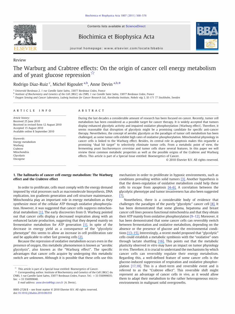
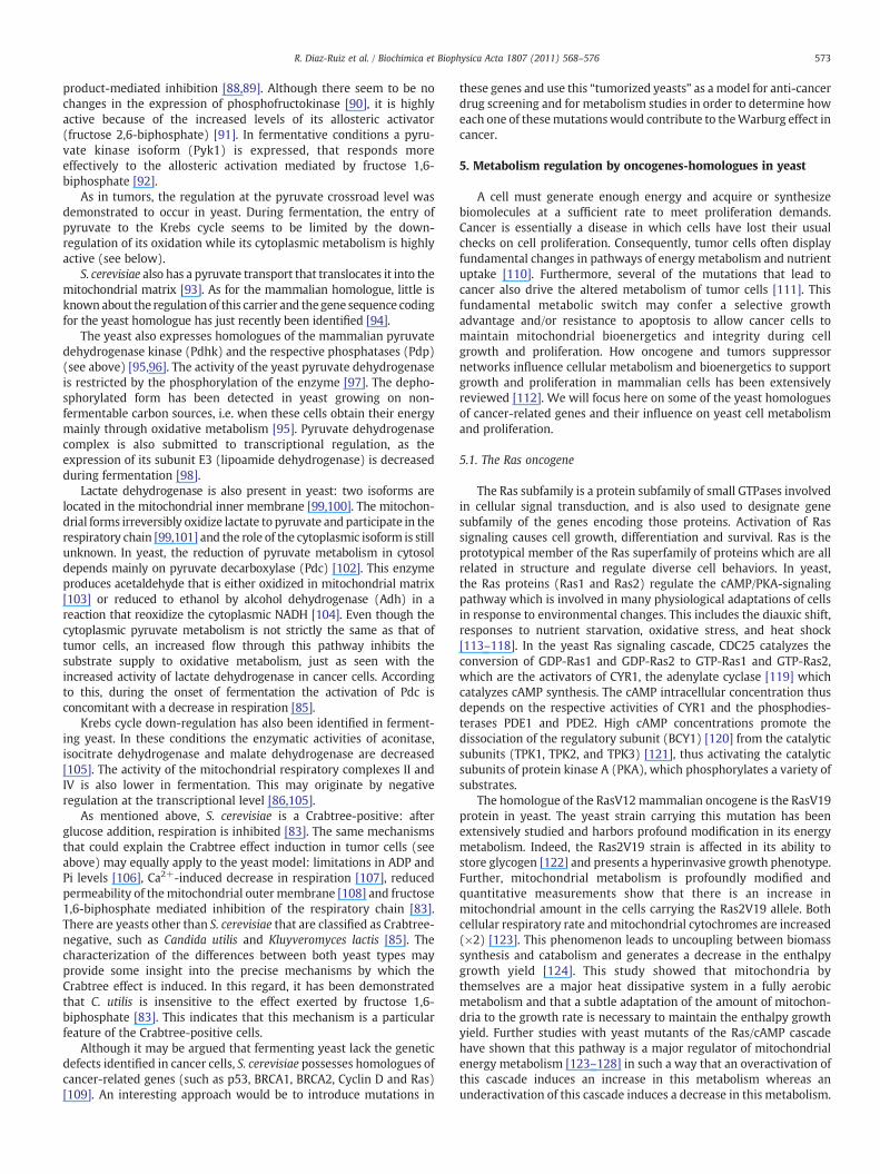
It is assumed that theWarburg effect originates from an increase inglucose uptake and glycolysis and/or a down-regulation of mitochon-drial metabolism (reviewed in [18]). There is experimental evidencethat fit into each one of these scenarios (Fig. 1). Here we brieflysummarize some of the molecular mechanisms that may give rise tothe aerobic glycolysis phenomenon. Nevertheless, care must be takenregarding interpretation of the data, as each particular cancer cellcarries its own mutations and there is no unique mechanism thatcould explain the Warburg effect.
2.1. Glucose transport and glycolysis
Commonly, the controlling step of a metabolic pathway is thesubstrate supply [19]. This has been shown to be the case for somecancer cell lines [20]. According to this, the overexpression of glucosetransporters has been proposed as one of the main determinantsfor the Warburg effect [21]. Results obtained using the positronemission tomography (PET scan) seem to support this proposal. Thistechnique is based on the use of glucose derivatives such as 18
fluoro-deoxyglucose as a tracer for imaging its uptake in tissues in vivo [22].Theoretically, as cancer cells have high glycolytic rates, they willavidly trap glucose and thus they could be visualized and distin-guished from normal tissue. FDG-PET has been used for diagnosticprocedures and it has been found to be applicable in a variety of
Glut1, Glut3Glucose Glucose
HK II
PFK (L and P Fructos
isoforms)
VDAC
CompleMITOCHONDRIA
Mitocmatrix
PFKB3
Glucose 6-phosphate
Fructose 6-phosphate
Fruct
Fig. 1. The mechanisms proposed to explain the aerobic glycolysis (Warburg effect) of cancerare overexpressed. Hexokinase isoform II (HK II) interacts with mitochondria through thebecause the predominance of its L- and P-isoforms. An overexpressed PFKB3 maintains hpyruvate kinase (PK) regulates the glycolytic flux and promotes metabolite accumulation in o(LDH) is overexpressed. Pyruvate transport into the mitochondrial matrix through its sphosphorylation-dependent mechanism mediated by an overexpressed pyruvate dehydrogThe levels of the mitochondrial respiratory complexes I, III and IV are decreased. Mitochon
cancers [22]. However, high glucose uptake does not necessarily meanthat mitochondrial metabolism is down-regulated and this must betaken into account for in vitro studies.
The high affinity glucose transporters (Glut1 and Glut3) areoverexpressed in several cancer cell lines [23]. The inhibition of theGlut transporters has been used to impair the growth of tumor cells invitro [24]. Furthermore, it has been demonstrated that glucosetransport could effectively be the rate-controlling step of glycolysisin hepatocarcinoma and HeLa cell lines and therefore it may be asuitable target for pharmacological anti-tumor agents [20].
High glycolytic fluxes may also be explained by the increasedactivity of the enzymes participating in the pathway. In this regard, ithas been found that each one of them is overexpressed and/orderegulated in several cancer cell lines [25]. Although an enhancedexpression cannot be strictly correlated with an increase of themetabolic flux, some studies have demonstrated that the activity ofeach one of the enzymes in the pathway is several-fold increasedcompared to their normal counterparts [26]. It is also important toconsider that the contribution to the overall metabolic flux is differentfor each enzyme [27]. Consequently, only the stimulation of the stepsthat kinetically control the glycolytic flux would have an impact forthe quantitative increase in glycolysis. Hexokinase (HK), phospho-fructokinase (PFK) and pyruvate kinase (PK) may be the rate-controlling steps and their regulation and expression patterns changein some tumors.
Hexokinase II (HKII) is themain isoform expressed in some tumors[28]. It is found to be associated with the mitochondrial outermembrane interacting with the voltage-dependent anionic channel(VDAC) [29]. This localization would allow HKII to immediately usethe ATP generated by oxidative phosphorylation and also it wouldhelp it to overcome the product-mediated inhibition that is normallyobserved for this enzyme [29]. In hepatoma and Ehrlich ascites, HKII isindeed the slowest step in the glycolytic sequence [26,30]. Thus, theHKII–mitochondria interaction would offer an explanation for the
e 1,6-biphosphate
PK, M2 isoform
LDH, M isoformPC
Pyruvate
Acetyl-CoAPDH
Succinate Fumaratex I
SDH
Citrate
ComplexIII
PLASMA MEMBRANE
ComplexIV
ATP synthase
hondrial
ose 2,6-biphosphate
Pyruvate
Lactate
Phosphoenolpyruvate
cells. The high-affinity glucose transporters (Glut1 and Glut3) in the plasma membranevoltage-dependent anionic channel (VDAC). Phosphofructokinase (PFK) is overactiveigher levels of fructose 2,6-biphosphate that further activate PFK. The M2 isoform ofrder to fulfill the biosynthetic needs of the cell. TheM-isoform of lactate dehydrogenasepecific carrier (PC) is decreased. Pyruvate dehydrogenase complex is inhibited in aenase kinase (PDHK). Mutations in succinate dehydrogenase (SDH) impair its activity.dria ATP synthase activity is restricted by the overexpression of its inhibitory subunit.
570 R. Diaz-Ruiz et al. / Biochimica et Biophysica Acta 1807 (2011) 568–576
elevated lactate production observed in these cases. However, thiswould not be the case for all tumors as it has been demonstrated thatglucose 6-phosphate effectively inhibits themitochondria-boundHKII[20,26]. Another drawback of this model is that the high glycolytic fluxwould be completely dependent on a functional mitochondrialmetabolism, which is assumed to be downregulated or impaired incancer cells (see below).
PFK activity is regulated according to the energy state of the cell,being inhibited when ATP demand decreases. Some alterations on theactivity of this enzyme have also been reported [31]. For instance, inleukemia and lymphoma cell lines, the L- and P-isoforms (mainlyexpressed in liver and platelets, respectively) are the predominantforms [32]. Interestingly, the allosteric properties of these isoformsallow the maximal activity of the enzyme even in low energy demandconditions, i.e. they respond less effectively to their inhibitors (citrateand ATP) while they are highly activated by lower concentrations offructose 2,6-biphosphate (F26bP) [31,32].
High levels of F26bP are also found on cancer cells [33]. It has beenshown that in malignant cells the levels of this intermediate dependon PFKB3 (ATP-dependent phosphofructokinase) [34]. This enzymebelongs to a family of enzymes that have a dual activity: theyphosphorylate fructose 6-phosphate (F6P) to F26bP and they are alsoable to revert this by dephosphorylation [34]. PFKB3 is an isoform thathas a low phosphatase activity and is overexpressed in cancer [35].This may explain the high activity of PFK that has been detected insome tumors [26].
Pyruvate kinase alterations have also been identified, as isoformM2 predominates in some cancer cell lines [35]. PK-M2 activitydepends on its quaternary structure: its tetrameric form ismore activethan the dimer [36]. Some evidences point out that this enzyme ispresent in its inactive form in tumors [35]. This is probably mediatedby PK-M2 phosphorylation, elicited by the activation of the tyrosinekinase signaling triggered by growth factors [37]. The phosphorylateddimeric form does not allow the binding of the allosteric activator ofthe enzyme (fructose 1,6-biphosphate) thereby leading to a loweractivity of PK [38,39]. However, the inhibition of PKM2 seemscontradictory with the high glycolytic flux measured in cancer cells.Furthermore, an inactive PK would severely impair cell energyproduction in the cells that depends mainly on glycolysis for ATPsynthesis. It is proposed that the low activity of PKM2would allow theaccumulation of the glycolysis metabolites that would eventuallyserve as precursors for biosynthesis [35].
2.2. The pyruvate crossroad
Pyruvate is located at the intersection between two of the maincatabolic pathways of the cell: glycolysis and the Krebs cycle. Thismetabolite is transported into the mitochondrial matrix in order to beconverted to acetyl CoA and afterwards enters the Krebs cycle to yieldreducing equivalents that will be used by the respiratory chain todrive oxidative phosphorylation. Alternatively, pyruvate can remainin the cytosol and be reduced by lactate dehydrogenase.
To explain the aerobic glycolysis, it has been proposed thatpyruvate could not be efficiently metabolized by mitochondriathereby deviating the metabolic flux into lactate production. Threeevents would account for this: a) the restriction of pyruvate transportinto mitochondrial matrix, b) the inhibition of the pyruvatedehydrogenase complex (PDH) activity, and c) the overactivation oflactate dehydrogenase.
Pyruvate can enter to mitochondrial matrix through a specificcarrier located in the inner membrane of this organelle [40]. Pyruvatetransport has been shown to be decreased in mitochondria isolatedfrom a hepatoma cell line [41,42]. Although this may well explain theaerobic glycolysis, there is evidence that showed an enhancedpyruvate uptake and oxidation in a different hepatoma cell line [43].
The pyruvate dehydrogenase complex (PDH) catalyzes thedecarboxylation of pyruvate and its condensation with coenzyme Ato produce acetyl-CoA. Its activity is regulated according to the energyavailability in the cell, being inactive when cellular energy supply ishigh [44]. PDH activity can also be modulated by reversiblephosphorylation. In this regard, the pyruvate dehydrogenase kinases(Pdhk) phosphorylate and inactivate the complex while the pyruvatedehydrogenase phosphatases (PDP) revert this inhibition [44]. Abalance between both processes would define the activity of PDH andhence the substrate supply to Krebs cycle. Disruption of theexpression of Pdhk1 decreased lactate production in head and necksquamous cancer cells suggesting an elimination of the Warburgeffect [45,46]. Moreover, in these cells the expression of Pdhk1 ismediated by the Hypoxia-Induced transcription Factor 1 (HIF-1)[45,46], which is also implicated in the overexpression of theglycolysis enzymes and the glucose transporters in cancer.
An alternative model for the inhibition of pyruvate dehydroge-nase activity is the production of acetoin, a by-product from thenon-oxidative decarboxylation of pyruvate [47]. This compoundinhibits pyruvate oxidation in isolated mitochondria [48]. Acetoinis found in significant amounts in tumors but not in normal cells,pointing out a restriction of pyruvate influx into the Krebs cyclein cancer cells [47]. Nonetheless, the acetoin-mediated inhibitionof PDH could be completely reverted in vitro by ADP and otherrespiratory substrates as α-ketoglutarate, malate and glutamate[48]. Thus, as these other substrates are normally present in thecell, it is unlikely that the entry of pyruvate to the Krebs cycle becompletely inhibited by acetoin.
Pyruvate is reduced in the cell cytosol by lactate dehydrogenasein order to keep a constant supply of NAD+ required to driveglycolysis. This enzyme is also overexpressed in a variety of cancercell lines [49] and the disruption of its expression stimulatesrespiration and decreases tumor cell viability in hypoxic condi-tions [50,51]. Lactate dehydrogenase is a homo- or hetero-tetramercomposed of H or M subunits. The latter is the predominant form inmuscle [52]. In several human tumors, an overexpression of the Msubunit was detected [49].
2.3. Krebs cycle and oxidative phosphorylation defects
Defects in the tricarboxylic acid cycle have also been proposed ashigh citrate efflux is detected on mitochondria isolated from ahepatoma cell line [53,54]. This seems to correlate with the lack ofstate 3 respiration in these mitochondria [53,54]. As citrate is oxidizedby isocitrate dehydrogenase in the cell cytoplasm in order to produceNADPH, these results could be interpreted as a deviation of carbonflux towards lipid synthesis as a consequence of an impaired Krebscycle. This feature seems to be restricted to this cell line as anotherstudy that used a different hepatoma cell line revealed no citrateefflux [43]. Another impairment of the Krebs cycle may be at the levelof succinate dehydrogenase (SDH), which also participates in themitochondrial respiratory chain as the complex II. Mutations on SDHcommonly occur in phaeochromocytomas and paragangliomas [55].
A decrease of ADP translocation to the mitochondrial matrix hasbeen reported to occur, as well as the inhibition of the ATP synthase[56,57]. Both events would evidently restrict ATP production inmitochondria and lead to a diminished respiratory rate andconsequently the cell would have to rely mostly on glycolysis-derivedATP. Reduced content and/or activity of the respiratory chaincomponents may occur. There are some reports demonstrating thedown-regulation of complexes I, III and IV [55,58,59]. As mentionedabove, these features could be particular for a specific cell line andthey could not be extrapolated to other tumors. In fact, other cell linespossess fully functional oxidative phosphorylation [9–12].
Another interesting feature of cancer cell energy metabolism istheir extensive consumption of glutamine [60]. Glutaminolysis is
571R. Diaz-Ruiz et al. / Biochimica et Biophysica Acta 1807 (2011) 568–576
indeed highly increased in cancer cells [61,62]. Glutamine is involvedin numerous anabolic pathways (such as nucleic acids) and can bedegraded in the Krebs cycle thereby generating ATP [63,64] throughboth substrate level phosphorylation and oxidative phosphorylation.Glutaminolysis generates i) malate which, through the malic enzyme,will give rise to NADPH that can be used to fuel lipid biosynthesis; ii)oxaloacetate, which will generate citrate, which is necessary for lipidbiosynthesis [65]. Consequently, glutaminolysis is an importantpathway mandatory for cancer cell proliferation and could be agood therapeutic target [66]. It has been shown that glutaminemetabolism can be targeted in humans using the glutamine analogueDON (6-Diazo-5-oxo-L-norleucine) [67]. However, toxicity can be anissue in attempts to target glutamine metabolism using DON [68].Recent studies suggest that the green tea polyphenol (EGCG) couldtarget glutamine metabolism by inhibiting glutamate dehydrogenaseunder low glucose conditions [69].
3. The Crabtree effect and its induction
Some cancer cells, in spite of possessing functional mitochondria,can switch between glycolytic and oxidative metabolism in areversible fashion (the Crabtree effect) [17,18]. Regarding the noveltherapies based on the inhibition of tumor cell energymetabolism, thesole inhibition of glycolysis would not be sufficient to eliminate allmalignant cells some of them may easily overcome the inhibition ofthe fermentative metabolism. This would bring as a consequence theincomplete elimination of cancer cells and it also would increase theprobability of reoccurrence after treatment. Because of this, it isimportant to precisely know the Crabtree effect and its underlyingcauses in order to properly target the cancer cells.
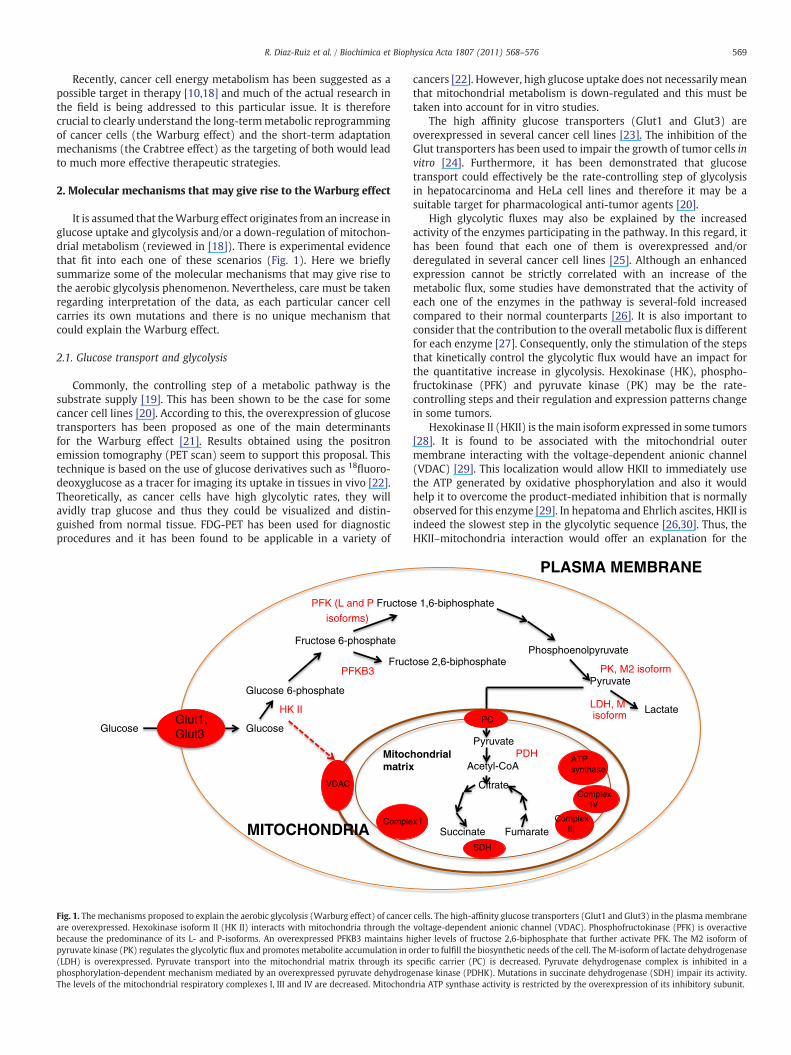
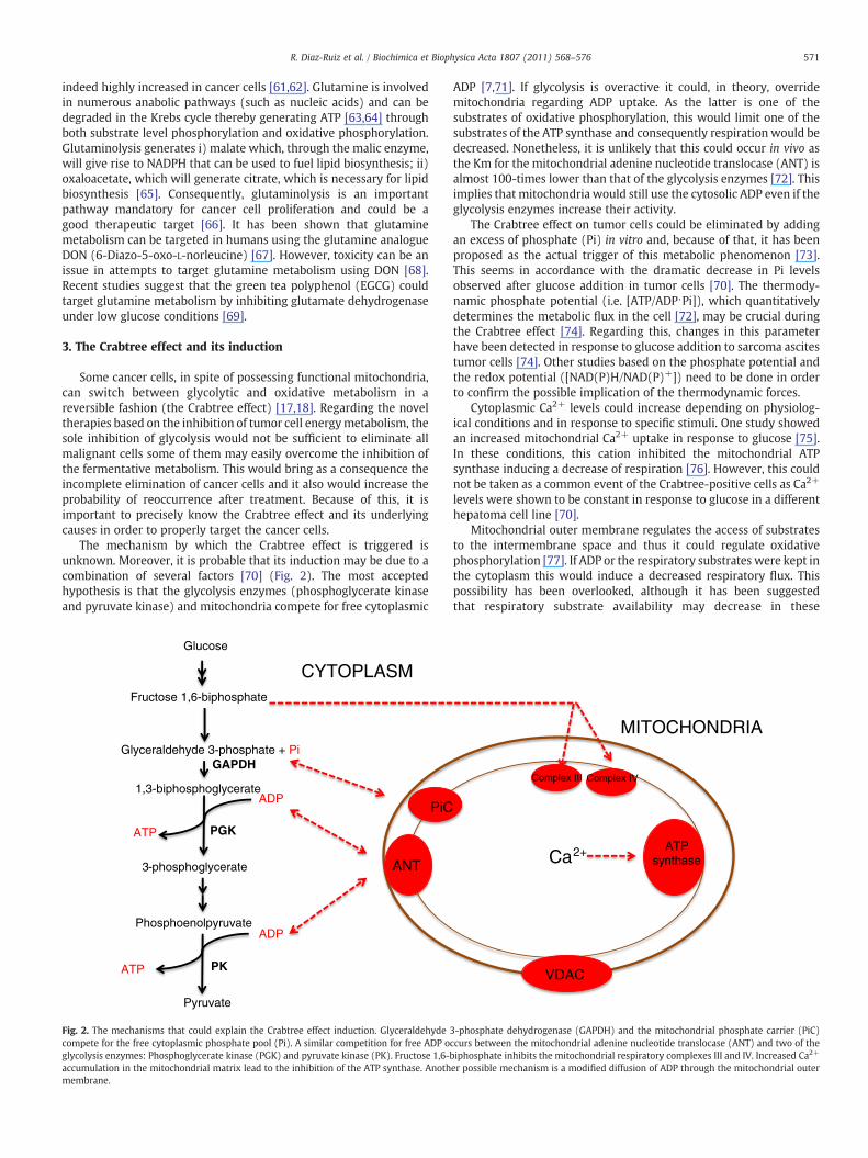
The mechanism by which the Crabtree effect is triggered isunknown. Moreover, it is probable that its induction may be due to acombination of several factors [70] (Fig. 2). The most acceptedhypothesis is that the glycolysis enzymes (phosphoglycerate kinaseand pyruvate kinase) and mitochondria compete for free cytoplasmic
Glucose
Glyceraldehyde 3-phosphate + Pi
1,3-biphosphoglycerate
3-phosphoglycerate
Phosphoenolpyruvate
Fructose 1,6-biphosphate
Pyruvate
ADP
ATP
ADP
ATP
ANT
PiC
CYTOPLASM
GAPDH
PGK
PK
Fig. 2. The mechanisms that could explain the Crabtree effect induction. Glyceraldehydecompete for the free cytoplasmic phosphate pool (Pi). A similar competition for free ADP oglycolysis enzymes: Phosphoglycerate kinase (PGK) and pyruvate kinase (PK). Fructose 1,6-baccumulation in the mitochondrial matrix lead to the inhibition of the ATP synthase. Anothmembrane.
ADP [7,71]. If glycolysis is overactive it could, in theory, overridemitochondria regarding ADP uptake. As the latter is one of thesubstrates of oxidative phosphorylation, this would limit one of thesubstrates of the ATP synthase and consequently respiration would bedecreased. Nonetheless, it is unlikely that this could occur in vivo asthe Km for the mitochondrial adenine nucleotide translocase (ANT) isalmost 100-times lower than that of the glycolysis enzymes [72]. Thisimplies that mitochondria would still use the cytosolic ADP even if theglycolysis enzymes increase their activity.
The Crabtree effect on tumor cells could be eliminated by addingan excess of phosphate (Pi) in vitro and, because of that, it has beenproposed as the actual trigger of this metabolic phenomenon [73].This seems in accordance with the dramatic decrease in Pi levelsobserved after glucose addition in tumor cells [70]. The thermody-namic phosphate potential (i.e. [ATP/ADP⋅Pi]), which quantitativelydetermines the metabolic flux in the cell [72], may be crucial duringthe Crabtree effect [74]. Regarding this, changes in this parameterhave been detected in response to glucose addition to sarcoma ascitestumor cells [74]. Other studies based on the phosphate potential andthe redox potential ([NAD(P)H/NAD(P)+]) need to be done in orderto confirm the possible implication of the thermodynamic forces.
Cytoplasmic Ca2+ levels could increase depending on physiolog-ical conditions and in response to specific stimuli. One study showedan increased mitochondrial Ca2+ uptake in response to glucose [75].In these conditions, this cation inhibited the mitochondrial ATPsynthase inducing a decrease of respiration [76]. However, this couldnot be taken as a common event of the Crabtree-positive cells as Ca2+
levels were shown to be constant in response to glucose in a differenthepatoma cell line [70].
Mitochondrial outer membrane regulates the access of substratesto the intermembrane space and thus it could regulate oxidativephosphorylation [77]. If ADP or the respiratory substrates were kept inthe cytoplasm this would induce a decreased respiratory flux. Thispossibility has been overlooked, although it has been suggestedthat respiratory substrate availability may decrease in these
VDAC
MITOCHONDRIA
Complex IVComplex III
ATP synthaseCa2+
3-phosphate dehydrogenase (GAPDH) and the mitochondrial phosphate carrier (PiC)ccurs between the mitochondrial adenine nucleotide translocase (ANT) and two of theiphosphate inhibits the mitochondrial respiratory complexes III and IV. Increased Ca2+
er possible mechanism is a modified diffusion of ADP through the mitochondrial outer
572 R. Diaz-Ruiz et al. / Biochimica et Biophysica Acta 1807 (2011) 568–576
conditions [78]. However, it has also been shown that in normal adultcardiomyocytes and HL-1 cardiac cell line, intracellular local restric-tions of diffusion of adenine nucleotides and metabolic feedbackregulation of respiration via phosphotransfer networks are different,most probably related to differences in structural organization ofthese cells [79]. Moreover, contrary to cardiomyocytes wheremitochondria and CaMgATPases are organized into tight complexeswhich ensure effective energy transfer and feedback signalingbetween these structures via specialized pathways mediated by CKand AK isoforms and direct adenine nucleotide channeling, thesecomplexes do not exist in HL-1 cells due to less organized energymetabolism [80]. In such cells the permeability of the outermembrane for ADP and other substrates is increased and themitochondrial compartment is very dynamic leading to an increasein ATP consumption [81,82].
One mechanism that links glycolysis acceleration to the inhibitionof respiration is fructose 1,6-biphosphate (F16bP) [83]. At physiolog-ical levels, F16bP induce a decrease on the activity of mitochondrialcomplexes III and IV [83]. An important finding was that the Crabtreeeffect could be induced onmitochondria isolated from normal rat liverby incubating them in the presence of F16bP concentrations similar tothose measured in hepatoma cells [83]. This demonstrates that theimpairment of the mitochondrial oxidative metabolism is not arequisite for the Crabtree effect induction and may explain itsreversible nature.
Glucose
Glucose 6-phosphate
Overxpressed. Bound to mitochondria.
Insensitive to G6P-mediated inhibition
HK
Fructose 6-phosphate
Fructose 1,6-biphosphate
Overexpressed and overactive. High levels of one of its allosteric
activators (F26bP)
PFK
Pyruvate
Lactate
Overexpressed and overactiveLDH
PDH
Acetyl-CoA
Inhibited by an overexpressed PDHK
Citrate
Succinate
Fumarate
SDH
Mutations in subunits D,B, and C
Respiratory chain:
Krebs cycle
Complex IV
ATP synthase
Decreased expression
Increased expression of its inhibitory subunit
Cancer cells
Mitochondrial matrix
Cytoplasm
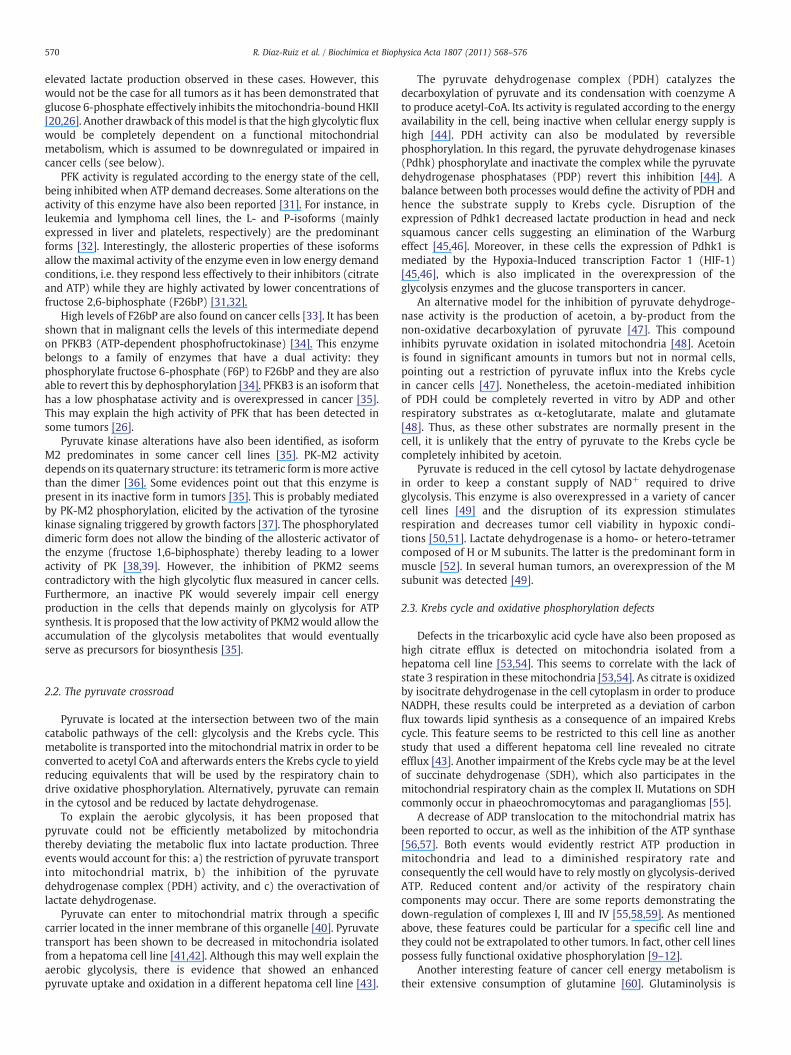
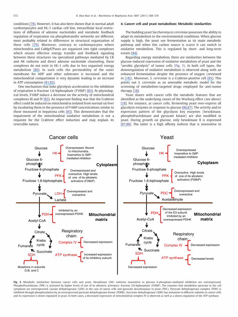
Fig. 3. Metabolic similarities between cancer cells and yeast. Hexokinase (HK) isofPhosphofructokinase (PFK) is activated by higher levels of one of its allosteric activators:cytoplasm are overexpressed: Lactate dehydrogenase (LDH) in the case of cancer cells aninhibited through phosphorylation by an overexpressed pyruvate dehydrogenase kinase (PDand its expression is down-regulated in yeast. In both cases, a decreased expression of mit
4. Cancer cell and yeast metabolism: Metabolic similarities
The budding yeast Saccharomyces cerevisiae possesses the ability toadapt its metabolism to the environmental conditions. When glucosesupply is high, the yeast use fermentation as its main metabolicpathway and when this carbon source is scarce it can switch tooxidative metabolism. This is regulated by short- and long-termevents [84].
Regarding energy metabolism, there are similarities between theglucose-induced repression of oxidative metabolism of yeast and the“aerobic glycolysis” of tumor cells (Fig. 3). In both cell types, thedownregulation of oxidative metabolism is observed along with anenhanced fermentation despite the presence of oxygen (reviewedin [18]). Moreover, S. cerevisiae is a Crabtree-positive cell [85]. Thispoints out S. cerevisiae as an amenable metabolic model for thescreening of metabolism-targeted drugs employed for anti-tumortherapy [18].
Yeast shares with cancer cells the metabolic features that areidentified as the underlying causes of the Warburg effect (see above)[18]. For instance, as cancer cells, fermenting yeast over-express allglycolysis enzymes in response to glucose [86,87]. The activity and/orexpression pattern of the glycolysis key enzymes (hexokinase,phosphofructokinase and pyruvate kinase) are also modified inyeast. During growth on glucose, only hexokinase II is expressed[87,88]. The latter is a high affinity isoform that is insensitive to
Glucose
Glucose 6-phosphate
Overexpressed. Insensitive to G6P-mediated inhibition
HK
Fructose 6-phosphate
Fructose 1,6-biphosphate
Overactive. High levels of one of its allosteric
activators (F26bP)PFK
Pyruvate
Acetaldehyde
Overexpressed andoveractivePDC
PDH
Acetyl-CoA
Decreased expression of the E3 subunit. Inhibited by an
overexpressed PDHK
Citrate
Succinate
Fumarate
SDH
Decreased expression
Krebs cycle
Respiratory chain:
Complex IV
ATP synthase
Decreased expression
Decreased levels
Yeast
Mitochondrialmatrix
Cytoplasm
orms insensitive to glucose 6-phosphate-mediated inhibition are overexpressed.fructose 2,6-biphosphate (F26bP). The enzymes that metabolize pyruvate in the celld pyruvate decarboxylase in yeast (PDC). Pyruvate dehydrogenase complex (PDH) isHK). Succinate dehydrogenase (SDH) has mutations in different subunits in cancer cellsochondrial complex IV is observed as well as a down-regulation of the ATP synthase.
573R. Diaz-Ruiz et al. / Biochimica et Biophysica Acta 1807 (2011) 568–576
product-mediated inhibition [88,89]. Although there seem to be nochanges in the expression of phosphofructokinase [90], it is highlyactive because of the increased levels of its allosteric activator(fructose 2,6-biphosphate) [91]. In fermentative conditions a pyru-vate kinase isoform (Pyk1) is expressed, that responds moreeffectively to the allosteric activation mediated by fructose 1,6-biphosphate [92].
As in tumors, the regulation at the pyruvate crossroad level wasdemonstrated to occur in yeast. During fermentation, the entry ofpyruvate to the Krebs cycle seems to be limited by the down-regulation of its oxidation while its cytoplasmic metabolism is highlyactive (see below).
S. cerevisiae also has a pyruvate transport that translocates it into themitochondrial matrix [93]. As for the mammalian homologue, little isknownabout the regulation of this carrier and thegene sequence codingfor the yeast homologue has just recently been identified [94].
The yeast also expresses homologues of the mammalian pyruvatedehydrogenase kinase (Pdhk) and the respective phosphatases (Pdp)(see above) [95,96]. The activity of the yeast pyruvate dehydrogenaseis restricted by the phosphorylation of the enzyme [97]. The depho-sphorylated form has been detected in yeast growing on non-fermentable carbon sources, i.e. when these cells obtain their energymainly through oxidative metabolism [95]. Pyruvate dehydrogenasecomplex is also submitted to transcriptional regulation, as theexpression of its subunit E3 (lipoamide dehydrogenase) is decreasedduring fermentation [98].
Lactate dehydrogenase is also present in yeast: two isoforms arelocated in the mitochondrial inner membrane [99,100]. The mitochon-drial forms irreversibly oxidize lactate to pyruvate and participate in therespiratory chain [99,101] and the role of the cytoplasmic isoform is stillunknown. In yeast, the reduction of pyruvate metabolism in cytosoldepends mainly on pyruvate decarboxylase (Pdc) [102]. This enzymeproduces acetaldehyde that is either oxidized in mitochondrial matrix[103] or reduced to ethanol by alcohol dehydrogenase (Adh) in areaction that reoxidize the cytoplasmic NADH [104]. Even though thecytoplasmic pyruvate metabolism is not strictly the same as that oftumor cells, an increased flow through this pathway inhibits thesubstrate supply to oxidative metabolism, just as seen with theincreased activity of lactate dehydrogenase in cancer cells. Accordingto this, during the onset of fermentation the activation of Pdc isconcomitant with a decrease in respiration [85].
Krebs cycle down-regulation has also been identified in ferment-ing yeast. In these conditions the enzymatic activities of aconitase,isocitrate dehydrogenase and malate dehydrogenase are decreased[105]. The activity of the mitochondrial respiratory complexes II andIV is also lower in fermentation. This may originate by negativeregulation at the transcriptional level [86,105].
As mentioned above, S. cerevisiae is a Crabtree-positive: afterglucose addition, respiration is inhibited [83]. The same mechanismsthat could explain the Crabtree effect induction in tumor cells (seeabove) may equally apply to the yeast model: limitations in ADP andPi levels [106], Ca2+-induced decrease in respiration [107], reducedpermeability of themitochondrial outermembrane [108] and fructose1,6-biphosphate mediated inhibition of the respiratory chain [83].There are yeasts other than S. cerevisiae that are classified as Crabtree-negative, such as Candida utilis and Kluyveromyces lactis [85]. Thecharacterization of the differences between both yeast types mayprovide some insight into the precise mechanisms by which theCrabtree effect is induced. In this regard, it has been demonstratedthat C. utilis is insensitive to the effect exerted by fructose 1,6-biphosphate [83]. This indicates that this mechanism is a particularfeature of the Crabtree-positive cells.
Although it may be argued that fermenting yeast lack the geneticdefects identified in cancer cells, S. cerevisiae possesses homologues ofcancer-related genes (such as p53, BRCA1, BRCA2, Cyclin D and Ras)[109]. An interesting approach would be to introduce mutations in
these genes and use this “tumorized yeasts” as a model for anti-cancerdrug screening and for metabolism studies in order to determine howeach one of thesemutationswould contribute to theWarburg effect incancer.
5. Metabolism regulation by oncogenes-homologues in yeast
A cell must generate enough energy and acquire or synthesizebiomolecules at a sufficient rate to meet proliferation demands.Cancer is essentially a disease in which cells have lost their usualchecks on cell proliferation. Consequently, tumor cells often displayfundamental changes in pathways of energy metabolism and nutrientuptake [110]. Furthermore, several of the mutations that lead tocancer also drive the altered metabolism of tumor cells [111]. Thisfundamental metabolic switch may confer a selective growthadvantage and/or resistance to apoptosis to allow cancer cells tomaintain mitochondrial bioenergetics and integrity during cellgrowth and proliferation. How oncogene and tumors suppressornetworks influence cellular metabolism and bioenergetics to supportgrowth and proliferation in mammalian cells has been extensivelyreviewed [112]. We will focus here on some of the yeast homologuesof cancer-related genes and their influence on yeast cell metabolismand proliferation.
5.1. The Ras oncogene
The Ras subfamily is a protein subfamily of small GTPases involvedin cellular signal transduction, and is also used to designate genesubfamily of the genes encoding those proteins. Activation of Rassignaling causes cell growth, differentiation and survival. Ras is theprototypical member of the Ras superfamily of proteins which are allrelated in structure and regulate diverse cell behaviors. In yeast,the Ras proteins (Ras1 and Ras2) regulate the cAMP/PKA-signalingpathway which is involved in many physiological adaptations of cellsin response to environmental changes. This includes the diauxic shift,responses to nutrient starvation, oxidative stress, and heat shock[113–118]. In the yeast Ras signaling cascade, CDC25 catalyzes theconversion of GDP-Ras1 and GDP-Ras2 to GTP-Ras1 and GTP-Ras2,which are the activators of CYR1, the adenylate cyclase [119] whichcatalyzes cAMP synthesis. The cAMP intracellular concentration thusdepends on the respective activities of CYR1 and the phosphodies-terases PDE1 and PDE2. High cAMP concentrations promote thedissociation of the regulatory subunit (BCY1) [120] from the catalyticsubunits (TPK1, TPK2, and TPK3) [121], thus activating the catalyticsubunits of protein kinase A (PKA), which phosphorylates a variety ofsubstrates.
The homologue of the RasV12 mammalian oncogene is the RasV19protein in yeast. The yeast strain carrying this mutation has beenextensively studied and harbors profound modification in its energymetabolism. Indeed, the Ras2V19 strain is affected in its ability tostore glycogen [122] and presents a hyperinvasive growth phenotype.Further, mitochondrial metabolism is profoundly modified andquantitative measurements show that there is an increase inmitochondrial amount in the cells carrying the Ras2V19 allele. Bothcellular respiratory rate andmitochondrial cytochromes are increased(×2) [123]. This phenomenon leads to uncoupling between biomasssynthesis and catabolism and generates a decrease in the enthalpygrowth yield [124]. This study showed that mitochondria bythemselves are a major heat dissipative system in a fully aerobicmetabolism and that a subtle adaptation of the amount of mitochon-dria to the growth rate is necessary to maintain the enthalpy growthyield. Further studies with yeast mutants of the Ras/cAMP cascadehave shown that this pathway is a major regulator of mitochondrialenergy metabolism [123–128] in such a way that an overactivation ofthis cascade induces an increase in this metabolism whereas anunderactivation of this cascade induces a decrease in this metabolism.
574 R. Diaz-Ruiz et al. / Biochimica et Biophysica Acta 1807 (2011) 568–576
Interestingly, it has been shown that the oncoprotein H-RasV12increases mitochondrial metabolism [129], further strengthening thefact that there is strong similarities between the regulation of yeastand mammalian cells energy metabolism by oncogene/oncogenehomologues.
5.2. Sch9, the yeast homologue of Akt
Growth factor-independent activation of the PI3K/Akt (a knownoncogene) pathway drives changes in cellular metabolism to promotecancer cell growth and proliferation [112]. The SCH9 gene of the yeastS. cerevisiae encodes a serine–threonine protein kinasewith a catalyticdomain very similar to that of human Akt1 [130]. There are severallines of evidence that Sch9p plays an important role in glucosesignaling in the budding yeast. Early work showed parallelism andcomplementarity of SCH9 signaling with the cyclic AMP-dependentprotein kinase (PKA) pathway, which signals hexose abundance [131–134]. It was also shown that Sch9p integrates nutrient signals with cellsize regulation. In fact, theΔsch9mutationwas one of themost potentmodifiers of cell size identified in a genome-wide screen for pathwayscoupling cell growth and division in yeast [135]. Sch9p is an activatorof ribosomal protein and ribosomal biogenesis regulons and isrequired for carbon source modulation of cell size [136]. Furthermore,it was recently shown that the Δsch9 mutation upregulates electrontransport chain gene expression and that this is associated with anincrease in mitochondrial respiration [137], further strengthening thefact that yeast homologues of oncogene play a role in the regulation ofcell metabolism.
It has recently been shown that yeast Sch9 is a central componentof a network that controls a common set of genes implicated in ametabolic switch from the TCA cycle and respiration to glycolysis andglycerol biosynthesis. During chronological survival, mutants lackingSCH9 depleted extracellular ethanol and reduced stored lipids, butsynthesized and released glycerol. Deletion of the glycerol biosyn-thesis genes GPD1, GPD2, or RHR2, among the most up-regulated inlong-lived Δsch9, Δtor1, and Δras2 mutants, was sufficient to reversechronological life span extension in Δsch9 mutants, suggesting thatglycerol production, in addition to the regulation of stress resistancesystems, optimizes life span extension [138].
5.2.1. ConclusionThe emergence of metabolic enzymes as important regulator of
cancer cell growth suggests that metabolic control is a key element oftumor progression. Understanding the pathways that regulate cancercell metabolism may lead to greater understanding of cancer celldevelopment and progression, and has the potential to open a newvista of metabolic therapy for cancer treatment. As shown in the lastparagraph of this review, there are strong similarities betweenmammalian and yeast cell metabolism regulation by oncogenes/oncogenes homologues. An interesting approach would be to use“tumorized yeasts” as a model for anti-cancer drug screening and formetabolism studies in order to determine how each one of thesemutations would contribute to the profound metabolic alterations incancer.
References
[1] O. Warburg, On the origin of cancer cells, Science 123 (1956) 309–314.[2] K. Brand, Aerobic glycolysis by proliferating cells: protection against oxidative
stress at the expense of energy yield, J. Bioenerg. Biomembr. 29 (1997) 355–364.[3] R.A. Gatenby, R.J. Gillies, Why do cancers have high aerobic glycolysis? Nat. Rev.
Cancer 4 (2004) 891–899.[4] S. Schlisio, Neuronal apoptosis by prolyl hydroxylation: implication in the
nervous system tumors and the Warburg conundrum, J. Cell. Mol. Med. 13(2009) 4104–4112.
[5] A. Tomiyama, S. Serizawa, K. Tachibana, K. Sakurada, H. Samejima, Y. Kuchino, C.Kitanaka, Critical role for mitochondrial oxidative phosphorylation in theactivation of tumor suppressors Bax and Bak, J. Natl. Cancer Inst. 98 (2006)1462–1473.
[6] V. Gogvadze, B. Zhivotovsky, S. Orrenius, The Warburg effect and mitochondrialstability, Mol. Aspects Med. 31 (2010) 60–74.
[7] S. Winehouse, Glycolysis, respiration and anomalous gene expression inexperimental hepatomas: G.H.A. Clawes memorial lecture, Cancer Res. 32(1972) 2007–2016.
[8] R. Moreno-Sánchez, S. Rodríguez-Enríquez, A. Marín-Hernández, E. Saavedra,Energy metabolism in tumor cells, FEBS J. 274 (2007) 1393–1418.
[9] M. Guppy, P. Leeman, X. Zu, V. Russel, Contribution by different fuels andmetabolic pathways to the total ATP turnover of proliferating MCF-7 breastcancer cells, Biochem. J. 364 (2002) 309–315.
[10] S. Rodríguez-Enríquez, P.A. Vital-González, F.L. Flores-Rodríguez, A. Marín-Hernández, L. Ruiz-Azuara, R. Moreno-Sanchez, Control of cellular proliferationby modulation, Toxicol. Appl. Pharmacol. 215 (2006) 208–217.
[11] M. Martin, B. Beauvoit, P.J. Voisin, P. Canioni, B. Guérin, M. Rigoulet, Energeticand morphologic plasticity of glioma cells grown on 3-D support: effect ontransient glutamine deprivation, J. Bioenerg. Biomembr. 30 (1998) 565–577.
[12] P. Pasdois, C. Devaud, P. Voisin, V. Bouchaud, M. Rigoulet, B. Beauvoit,Contribution of the phosphorylable complex I in the growth phase-dependentrespiration of C6 glioma cells in vitro, J. Bioenerg. Biomembr. 35 (2003)439–450.
[13] S. Rodríguez-Enríquez, J.C. Pérez-Gallardo, A. Avilés-Salas, A. Marín-Hernández,L. Carrreño-Fuentes, V. Maldonado-Lagunas, R. Moreno-Sánchez, Energymetabolism transition in multi-cellular human tumor spheroids, J. Cell. Physiol.216 (2008) 189–197.
[14] R. Rossignol, R. Gilkerson, R. Aggeler, K. Yamagata, S.J. Remington, R.A. Capaldi,Energy substrate modulates mitochondrial structure and oxidative capacity incancer cells, Cancer Res. 64 (2004) 985–993.
[15] K. Smolková, N. Bellance, F. Scandurra, E. Génot, E. Gnaiger, L. Plecitá-Hlavatá, P.Jezek, R. Rossignol, Mitochondrial bioenergetic adaptations of breast cancer cellsto aglycemia and hypoxia, J. Bioenerg. Biomembr. 42 (2010) 55–67.
[16] P. Sonveaux, F. Végran, T. Schroeder, M.C. Wergin, J. Verrax, Z.N. Rabbani, C.J. DeSaedeleer, K.M. Kennedy, C. Diepart, B.F. Jordan, M.J. Kelley, B. Gallez, M.L. Whal,O. Feron, M.W. Dewhirst, Targeting lactate-fueled respiration selectively killshypoxic tumor cells in mice, J. Clin. Invest. 118 (2008) 3930–3942.
[17] H.G. Crabtree, Observations on the carbohydrate metabolism of tumors,Biochem. J. 23 (1929) 536–545.
[18] R. Díaz-Ruiz, S. Uribe-Carvajal, A. Devin, M. Rigoulet, Tumor cell energymetabolism and its common features with yeast metabolism, Biochim. Biophys.Acta 1796 (2009) 252–265.
[19] Y. Kashiwaya, K. Sato, N. Tsuchiya, S. Thomas, D. Fell, R.L. Veech, J.V. Passonneau,Control of glucose utilisation in working perfused rat heart, J. Biol. Chem. 269(1994) 25502–25514.
[20] S. Rodríguez-Enriquez, A. Marín-Hernández, J.C. Pérez-Gallardo, R. Moreno-Sánchez, Kinetics of transport and phosphorylation of glucose in cancer cells, J.Cell. Physiol. 221 (2009) 552–559.
[21] T. Yamamoto, Y. Seino, H. Fukumoto, G. Koh, N. Inagaki, Y. Yamada, K. Inoue, T.Manabe, H. Imura, Over-expression in facilitative glucose transporter in humancancer, Biochem. Biophys. Res. Commun. 170 (1990) 223–230.
[22] J. Czernin, M.E. Phelps, Positron emission tomography scanning: current andfuture applications, Annu. Rev. Med. 53 (2002) 89–112.
[23] M.L. Macheda, S. Rogers, J.D. Best, Molecular and cellular regulation of glucosetransporter (GLUT) proteins in cancer, J. Cell. Physiol. 203 (2005) 654–662.
[24] X. Cao, L. Fang, S. Gibbs, Y. Huang, Z. Dai, P. Wen, X. Zheng, W. Sadee, D. Sun,Glucose uptake inhibitor sensitizes cancer cells to daunorubicin and overcomesdrug resistance in hypoxia, Cancer Chemother. Pharmacol. 59 (2007) 495–505.
[25] H. Pelicano, D.S. Martin, R.H. Xu, P. Huang, Glycolysis inhibition for anticancertreatment, Oncogene 25 (2006) 4633–4646.
[26] A. Marín-Hernández, S. Rodríguez-Enríquez, P.A. Vital-González, F.L. Flores-Rodríguez, M. Macías-Silva, M. Sosa-Garrocho, R. Moreno-Sánchez, Determiningand understanding the control of glycolysis in fast-growth tumor cells. Fluxcontrol by an over-expressed but strongly product-inhibited hexokinase, FEBS J.273 (2006) 1975–1988.
[27] D. Fell, Metabolic control analysis, Understanding the Control of Metabolism,Portland Press, London, UK, 1997, pp. 101–132.
[28] E. Bustamante, H.P. Morris, P.L. Pedersen, Energy metabolism of tumor cells.Requirement for a form of hexokinase with a propensity for mitochondrialbinding, J. Biol. Chem. 256 (1981) 8699–8704.
[29] P.L. Pedersen, Voltage dependent anion channels (VDACs): a brief introductionwithfocus on the outer mitochondrial compartment's roles together with hexokinase-2in the “Warburg effect” in cancer, J. Bioenerg. Biomembr. 40 (2008) 123–126.
[30] J. Schulz, A. Baufeld, E. Hofmann, T.A. Rapoport, R. Heinrich, S.M. Rapoport,Regulation of anaerobic glycolysis in Ehrlich ascites tumour cells, Acta Biol. Med.Ger. 36 (1977) 1379–1391.
[31] S. Vora, J.P. Halper, D.M. Knowles, Alterations in the activity and isoenzymicprofile of human phosphofructokinase during malignant transformation in vivoand in vitro: transformation- and progression-linked discriminants of malig-nancy, Cancer Res. 45 (1985) 2993–3001.
[32] S. Vora, C. Seaman, S. Durham, S. Piomelli, Isozymes of human phosphofructo-kinase: identification and subunit structural characterisation of a new system,Proc. Natl. Acad. Sci. USA 77 (1980) 62–66.
[33] L. Hue, G.G. Rousseau, Fructose 2, 6-biphosphate and the control of glycolysis bygrowth factors, tumor promoters and oncogenes, Adv. Enzyme Regul. 33 (1993)97–110.
[34] A. Yalcin, S. Telang, B. Clem, J. Chesney, Regulation of glucose metabolism by 6-phosphofructo-2-kinase/fructose2, 6-biphosphatases in cancer, Exp. Mol. Pathol.86 (2009) 174–179.
575R. Diaz-Ruiz et al. / Biochimica et Biophysica Acta 1807 (2011) 568–576
[35] T. Atsumi, J. Chesney, C. Metz, L. Leng, S. Donelly, Z. Makita, R. Mitchell, R. Bucala,High expression of inducible 6-phosphofructo-2-kinase/fructose-2, 6-bispho-sphatase (iPFK2; PFKB3) in human cancers, Cancer Res. 62 (2002) 5881–5887.
[36] S. Mazurek, C.B. Boscheck, F. Hugo, E. Eigenbrodt, Pyruvate kinase type M2 andits role in tumor growth and spreading, Semin. Cancer Biol. 15 (2005) 300–308.
[37] W. Zwerschke, S. Mazurek, P. Massimi, L. Banks, E. Eigenbrodt, P. Jansen-Dürr,Modulation of type M2 pyruvate kinase activity by the human papillomavirustype 16 E7 oncoprotein, Proc. Natl. Acad. Sci. USA 96 (1999) 1291–1296.
[38] D. Lüftner, S. Mazurek, P. Henschke, J. Masterham, S. Schildhauer, R. Geppert, K.D.Wernecke, K. Possinger, Plasma levels of HER/neu, tumor type M2 pyruvatekinase and its tyrosine-phosphorylated metabolite in advanced breast cancer,Anticancer Res. 23 (2003) 991–997.
[39] T. Hitosugi, S. Kang, M.G. Vander Heiden, T.W. Chung, S. Elf, K. Lythgoe, S. Dong, S.Lional, X.Wang, G.Z. Chen, J. Xie, T.L. Gu, R.D. Polakiewicz, J.L. Roesel, T.J. Bogon, F.R. Khuri, D.G. Gilliland, L.C. Cantley, J. Kaufman, J. Chen, Tyrosine phosphory-lation inhibits PKM2 to promote the Warburg effect and tumor growth, Sci.Signal. 2 (2009) ra73.
[40] A.P. Halestrap, The mitochondrial pyruvate carrier. Kinetics and specificity forsubstrates and inhibitors, Biochem. J. 148 (1975) 85–96.
[41] G. Paradies, F. Capuano, G. Palombini, T. Galeotti, S. Papa, Transport of pyruvatein mitochondria from different tumor cells, Cancer Res. 43 (1983) 5068–5071.
[42] M.L. Eboli, G. Paradies, T. Galeotti, S. Papa, Pyruvate transport in tumor-cellmitochondria, Biochim. Biophys. Acta 460 (1977) 183–187.
[43] D.J. Dietzen, E.J. Davis, Oxidation of pyruvate, malate, citrate, and cytosolicreducing equivalents by AS-30D hepatoma mitochondria, Arch. Biochem.Biophys. 305 (1993) 91–102.
[44] S. Strumilo, Short-term regulation of the mammalian pyruvate dehydrogenasecomplex, Acta Biochim. Pol. 52 (2005) 759–764.
[45] J.W. Kim, I. Tchernyshyov, G.L. Semenza, C.V. Dang, HIF1-mediated expression ofpyruvate dehydrogenase kinase: a metabolic switch required for cellularadaptation to hypoxia, Cell Metab. 3 (2006) 177–185.
[46] I. Papandreou, R.A. Cairns, L. Fontana, A.L. Lim, N.C. Denko, HIF-1 mediatesadaptation to hypoxia by actively down-regulating mitochondrial oxygenconsumption, Cell Metab. 3 (2006) 187–197.
[47] L.G. Baggetto, A.L. Lehninger, Formation and utilisation of acetoin, an unusualproduct of pyruvate metabolism by Ehrlich and AS30-D tumor mitochondria, J.Biol. Chem. 262 (1987) 9535–9541.
[48] L.G. Baggetto, A.L. Lehninger, Isolated tumoral pyruvate dehydrogenase cansynthesize acetoin which inhibits pyruvate oxidation as well as other aldehydes,Biochem. Biophys. Res. Commun. 145 (1987) 153–159.
[49] R.D. Goldman, N.O. Kaplan, T.C. Hall, Lactic dehydrogenase in human neoplastictissues, Cancer Res. 24 (1964) 389–399.
[50] V. Fantin, J. St-Pierre, P. Leder, Attenuation of LDH-A expression uncovers a linkbetween glycolysis, mitochondrial physiology and tumor maintenance, CancerCell 9 (2006) 425–434.
[51] A. Le, C.R. Cooper, A.M. Gouw, R. Dinavahi, L.M. Deck, R.E. Royer, D.L. Vander Jagt,G.L. Semenza, C.V. Dang, Inhibition of lactate dehydrogenase A induces oxidativestress and inhibits tumor progression, Proc. Natl. Acad. Sci. USA 107 (2010)2037–2042.
[52] D.M. Dawson, T.L. Goodfriend, N.O. Kaplan, Lactate dehydrogenases: function ofthe two types. Rates of synthesis of the two major forms can be correlated withmetabolic differentiation, Science 143 (1964) 929–933.
[53] R.A. Parlo, R.S. Coleman, Enhanced rate of citrate export from cholesterol-richhepatoma mitochondria. The truncated Krebs cycle and other metabolic ramifica-tions ofmitochondrialmembrane cholesterol, J. Biol. Chem. 259 (1984) 9997–10003.
[54] R.A. Parlo, P.S. Coleman, Continuous pyruvate carbon efflux to newly synthesizedcholesterol and the suppressed evolution of pyruvate generated CO2 in tumors:further evidence for a truncated Krebs cycle in hepatomas, Biochim. Biophys.Acta 886 (1986) 169–176.
[55] E. Gottlieb, I.P. Tomlinson, Mitochondrial tumor suppressors: a genetical andbiochemical update, Nat. Rev. Cancer 5 (2005) 857–866.
[56] S.H. Chan, R.L. Barbour, Adenine nucleotide transport in hepatomamitochondria.Characterization of factors influencing the kinetics of ADP and ATP uptake,Biochim. Biophys. Acta 723 (1983) 104–113.
[57] J.M. Cuezva, M. Sánchez-Aragó, S. Sola, A. Blanco-Rivero, A.D. Ortega, A messageemerging from development: the repression of mitochondrial beta F1-ATPaseexpression in cancer, J. Bioenerg. Biomembr. 39 (2007) 259–265.
[58] I. Ishikawa, K. Takenaga, M. Akimoto, N. Koshikawa, A. Yamaguchi, H. Imanishi, K.Nakada, Y. Honma, J. Hayashi, ROS-generated mitochondrial DNA mutations canregulate tumor cell metastasis, Science 320 (2008) 661–664.
[59] S.A. Sun, A.I. Cederbaum, Oxidoreductase activities in normal rat liver, tumor-bearing rat liver, and hepatoma HC-252, Cancer Res. 40 (1980) 4677–4681.
[60] D.M. Greenberg, M.E. Olson, M. Rabinovitz, Role of glutamine in proteinsynthesis by the Ehrlich ascites carcinoma, J. Biol. Chem. 222 (1956) 879–893.
[61] T. Matsuno, H. Hirai, Glutamine synthetase and glutaminase activities in varioushepatoma cells, Biochem. Int. 19 (1989) 219–225.
[62] T. Matsuno, I. Goto, Glutaminase and glutamine synthetase activities in humancirrhotic liver and hepatocellular carcinoma, Cancer Res. 52 (1992) 1192–1194.
[63] Z. Kovacevic, H.P. Morris, The role of glutamine in the oxidative metabolism ofmalignant cells, Cancer Res. 32 (1972) 326–333.
[64] L.J. Reitzer, B.M. Wice, D. Kenell, Evidence that glutamine, not sugar, is the majorenergy source for cultured HeLa cells, J. Biol. Chem. 254 (1979) 2669–2676.
[65] R.J. DeBerardinis, A. Mancuso, E. Daikhin, I. Nissim, M. Yudkoff, S. Wehrli, C.B.Thompson, Beyond aerobic glycolysis: transformed cells can engage inglutamine metabolism that exceeds the requirement for protein and nucleotidesynthesis, Proc. Natl. Acad. Sci. USA 104 (2007) 19345–19350.
[66] T.N. Seyfried, L.M. Shelton, Cancer as a metabolic disease, Nutr. Metab. (London)7 (2010) 7.
[67] S.C. Piscitelli, A. Thilbault, W.D. Figg, A. Thompkins, D. Headlee, R. Lieberman, D.Samid, C.E. Myers, Disposition of phenylbutyrate and its metabolites, phenyla-cetate and phenylacetylglutamine, J. Clin. Pharmacol. 35 (1995) 368–373.
[68] M. Molina, Glutamine and cancer, J. Nutr. 131 (2001) 2550S–2551S.[69] C. Yang, J. Sudderth, T. Dang, R.M. Bachoo, J.G. McDonald, R.J. DeBerardinis,
Glioblastoma cells require glutamate dehydrogenase to survive impairments ofglucose metabolism of Akt signaling, Cancer Res. 69 (2009) 7986–7993.
[70] S. Rodríguez-Enriquez, O. Juárez, J.S. Rodríguez-Zavala, R. Moreno-Sánchez,Multisite control of the Crabtree effect in ascites hepatoma cells, Eur. J. Biochem.268 (2001) 2512–2519.
[71] S. Gatt, E. Racker, Regulatory mechanisms in carbohydrate metabolism: I.Crabtree effect in reconstituted systems, J. Biol. Chem. 234 (1959) 1015–1023.
[72] R.L. Veech, J.W. Lawson, N.W. Cornell, H.A. Krebs, Cytosolic phosphorylationpotential, J. Biol. Chem. 254 (1979) 6538–6547.
[73] D.H. Koobs, Phosphate mediation of the Crabtree and Pasteur effects, Science 178(1972) 127–133.
[74] I. Sussman, M. Erecinska, D.F. Wilson, Regulation of cellular energy metabolism:the Crabtree effect, Biochim. Biophys. Acta 591 (1980) 209–223.
[75] L. Wojtczak, V.V. Teplova, K. Bogucka, A. Czyz, A. Makowska, M.R. Wieckowski, J.Duszynski, V. Evtodienko Yu, Effect of glucose and deoxyglucose on theredistribution of calcium in Ehrlich ascites tumor and Zajdela hepatoma cellsand its consequences for mitochondrial energetics. Further arguments for therole of Ca(2+) in the mechanism of the Crabtree effect, Eur. J. Biochem. 263(1999) 495–501.
[76] Yu.V. Evtodienko, V.V. Teplova, J. Duszynski, K. Bogucka, L. Wojtczak, The role ofcytoplasmic [Ca2+] in glucose-induced inhibition of respiration and oxidativephosphorylation in Ehrlich ascites tumor cells: a novel mechanism of theCrabtree effect, Cell Calcium 15 (1994) 439–446.
[77] V. Saks, Y. Belikova, E. Vasilyeva, A. Kuznetsov, E. Fontaine, C. Keriel, X. Leverve,Correlation between degree of rupture of outer mitochondrial membrane andchanges of kinetics of regulation of respiration by ADP in permeabilised heartand liver cells, Biochem. Biophys. Res. Commun. 208 (1995) 919–926.
[78] E.F. Greiner, M. Guppy, K. Brand, Glucose is essential for the proliferation andglycolytic enzyme induction that provokes a transition to glycolytic energyproduction, J. Biol. Chem. 269 (1994) 31484–31490.
[79] T. Anmann, R. Guzun, N. Beraud, S. Pelloux, A.V. Kuznetsov, L. Kogerman, T.Kaambre, P. Sikk, K. Paju, N. Peet, E. Seppet, C. Ojeda, Y. Tourneur, V. Saks,Different kinetics of the regulation of respiration in permeabilized cardiomyo-cytes and in HL-1 cardiac cells. Importance of cell structure/organization forrespiration regulation, Biochim. Biophys. Acta 1757 (2006) 1597–1606.
[80] M. Eimre, K. Paju, S. Pelloux, N. Beraud, M. Roosimaa, L. Kadaja, M. Gruno, N. Peet,E. Orlova, R. Remmelkoor, A. Piirsoo, V. Saks, E. Seppet, Distinct organization ofenergy metabolism in HL-1 cardiac cell line and cardiomyocytes, Biochim.Biophys. Acta 1777 (2008) 514–524.
[81] N. Beraud, S. Pelloux, Y. Usson, A.V. Kuznetsov, X. Ronot, Y. Tourneur, V. Saks,Mitochondrial dynamics in heart cells: very low amplitude high frequencyfluctuations in adult cardiomyocytes and flowmotion in non beating Hl-1 cells, J.Bioenerg. Biomembr. 41 (2009) 195–214.
[82] V. Saks, R. Guzun, N. Timohhina, K. Tepp, M. Varikmaa, C. Monge, N. Beraud, T.Kaambre, A. Kuznetsov, L. Kadaja, M. Eimre, E. Seppet, Structure–functionrelationships in feedback regulation of energy fluxes in vivo in health anddisease: mitochondrial interactosome, Biochim. Biophys. Acta 1797 (2010)678–697.
[83] R. Díaz-Ruiz, N. Avéret, D. Araiza, B. Pinson, S. Uribe-Carvajal, A. Devin, M.Rigoulet, Mitochondrial oxidative phosphorylation is regulated by fructose 1, 6-biphosphate: a possible role in Crabtree effect induction? J. Biol. Chem. 283(2008) 26948–26955.
[84] J.M. Thevelein, Signal transduction in yeast, Yeast 10 (1994) 1753–1790.[85] H. Van Urk, W.S. Voll, W.A. Scheffers, J.P. Van Dijken, Transient-state analysis of
metabolic fluxes in Crabtree-positive and Crabtree negative yeasts, Appl.Environ. Microbiol. 56 (1990) 281–287.
[86] M. Takeda, Glucose-induced inactivation of mitochondrial enzymes in the yeastSaccharomyces cerevisiae, Biochem. J. 198 (1981) 281–287.
[87] K.D. Entian, K.U. Frohlich, D. Mecke, Regulation of enzymes and isoenzymes ofcarbohydrate metabolism in the yeast Saccharomyces cerevisiae, Biochim.Biophys. Acta 799 (1984) 181–186.
[88] H. Muratsubaki, T. Katsume, Distribution of hexokinase isoenzymes dependingon a carbon source in Saccharomyces cerevisiae, Biochem. Biophys. Res. Commun.86 (1979) 1030–1036.
[89] S.P. Colowick, Group Transfer, Part A, Third Edition, in: P.D. Boyer (Ed.), TheEnzymes, vol. IX, Academic Press, New York, USA, 1973, pp. 1–48.
[90] J. Heinisch, K. Vogelsang, C.P. Hollenberg, Transcriptional control of yeastphosphofructokinase gene expression, FEBS Lett. 289 (1991) 77–82.
[91] D. Ribstein, J.A. den Hollander, S.J. Pilkis, R.G. Shulman, Studies on the regulationof yeast phosphofructo-1-kinase: its role in aerobic and anaerobic glycolysis,Biochemistry 25 (1986) 219–227.
[92] E. Boles, F. Schulte, T. Miosga, K. Freidel, E. Schlüter, F.K. Zimmermann, C.P.Hollenberg, J.J. Heinisch, Characterisation of a glucose-repressed pyruvate kinase(Pk2p) that is catalytically insensitive to fructose 1, 6-biphosphate, J. Bacteriol.179 (1997) 2987–2993.
[93] M. Briquet, Transport of pyruvate and lactate in yeast mitochondria, Biochim.Biophys. Acta 459 (1977) 290–299.
[94] J.C. Hildyard, A.P. Halestrap, Identification of the mitochondrial pyruvate carrierin Saccharomyces cerevisiae, Biochem. J. 374 (2002) 607–611.
576 R. Diaz-Ruiz et al. / Biochimica et Biophysica Acta 1807 (2011) 568–576
[95] U. Krause-Buchholz, U. Gey, J. Wünschmann, S. Becker, G. Rödel, YIL042c andYOR090c encode the kinase and phosphatase of the Saccharomyces cerevisiaepyruvate dehydrogenase complex, FEBS Lett. 580 (2006) 2553–2560.
[96] U. Gey, C. Czupalla, B. Hoflack, G. Rödel, U. Krause-Buchholz, Yeast pyruvatedehydrogenase complex is regulated by concerted activity of two kinases andtwo phosphatases, J. Biol. Chem. 283 (2008) 9759–9767.
[97] D.J. Uhlinger, C.Y. Yang, L.J. Reed, Phosphorylation–dephosphorylation ofpyruvate dehydrogenase from baker's yeast, Biochemistry 25 (1986)5673–5677.
[98] D.J. Roy, I.W. Dawes, Cloning and characterisation of the gene encodinglipoamide dehydrogenase in Saccharomyces cerevisiae, J. Gen. Microbiol. 133(1987) 925–933.
[99] M.L. Pallotta, D. Valenti, M. Iacovino, S. Passarella, Two separate pathways for D-lactate oxidation by Saccharomyces cerevisiae mitochondria which differ inenergy production and carrier involvement, Biochim. Biophys. Acta 1608 (2004)104–113.
[100] A. Chelstowska, Z. Liu, Y. Jia, D. Amberg, R.A. Buttow, Signaling betweenmitochondria and the nucleus regulates the expression of a new D-lactatedehydrogenase activity in yeast, Yeast 15 (1999) 1377–1391.
[101] A. Mourier, J. Vallortigara, E.D. Yoboue, M. Rigoulet, A. Devin, Kinetic activation ofyeast D-lactate dehydrogenase by carboxylic acids, Biochim. Biophys. Acta 1777(2008) 1283–1288.
[102] S. Hohmann, H. Cederberg, Autoregulation may control the expression of yeastpyruvate decarboxylase structural genes PDC1 and PDC5, Eur. J. Biochem. 188(1990) 615–621.
[103] S. Boubekeur, O. Bunoust, N. Camougrand, M. Castroviejo, M. Rigoulet, B. Guérin,A mitochondrial pyruvate dehydrogenase bypass in the yeast Saccharomycescerevisiae, J. Biol. Chem. 274 (1999) 21044–21048.
[104] M. Rigoulet, H. Aguilaniu, N. Avéret, O. Bunoust, N. Camougrand, X. Grandier-Vazeille, C. Larsson, I.L. Pahlman, S. Manon, L. Gustafsson, Organisation andregulation of cytosolic NADH metabolism in the yeast Saccharomyces cerevisiae,Mol. Cell. Biochem. 256–257 (2004) 73–81.
[105] P. Eraso, J.M. Gancedo, Catabolite repression in yeast is not associated with lowlevels of cAMP, Eur. J. Biochem. 141 (1984) 195–198.
[106] N. Sánchez, M. Calahorra, J.C. González-Hernández, A. Peña, Glycolytic sequenceand respiration of Debaryomices hansenii compared to Saccharomyces cerevisiae,Yeast 23 (2006) 361–374.
[107] V. Pérez-Vázquez, A. Saavedra-Molina, S. Uribe, In Saccharomyces cerevisiae,cations control the fate of the energy derived from oxidative metabolismthrough the opening and closing of the yeast mitochondrial unselective channel,J. Bioenerg. Biomembr. 35 (2003) 231–241.
[108] N. Avéret, V. Fitton, O. Bunoust, M. Rigoulet, B. Guérin, Yeast mitochondrialmetabolism: from in vitro to in situ quantitative study, Mol. Cell. Biochem. 184(1998) 67–79.
[109] L.H. Hartwell, P. Szankasi, C.J. Roberts, A.W. Murray, S.H. Friend, Integratinggenetic approaches into the discovery of anticancer drugs, Science 278 (1997)1064–1068.
[110] K. Garber, Energy deregulation: licensing tumors to grow, Science 312 (2006)1158–1159.
[111] B. Vogelstein, K.W. Kinzler, Cancer genes and the pathways they control, Nat.Med. 10 (2004) 789–799.
[112] R.G. Jones, C.B. Thompson, Tumor suppressors and cell metabolism: a recipe forcancer growth, Genes Dev. 23 (2009) 537–548.
[113] E. Boy-Marcotte, D. Tadi, M. Perrot, H. Boucherie, M. Jacquet, High cAMP levelsantagonize the reprogramming of gene expression that occurs at the diauxicshift in Saccharomyces cerevisiae, Microbiology 142 (1996) 459–467.
[114] Y. Jiang, C. Davis, J.R. Broach, Efficient transition to growth on fermentablecarbon sources in Saccharomyces cerevisiae requires signaling through the RASpathway, EMBO J. 17 (1998) 6942–6951.
[115] M. Russel, J. Bradshaw-Rouse, D. Markwardt, W. Heideman, Changes in geneexpression in the Ras/Adenylate cyclase system of Saccharomyces cerevisiae:correlation with cAMP levels and growth arrest, Mol. Cell. Biol. 4 (1993)757–765.
[116] K. Tatchell, L.C. Robinson, M. Breitenbach, RAS2 of Saccharomyces cerevisiae isrequired for gluconeogenic growth and proper response to nutrient limitation,Proc. Natl. Acad. Sci. USA 82 (1985) 3785–3789.
[117] J.M. Thevelein, J.H. de Winde, Novel sensing mechanisms and targets or thecAMP-protein kinase A pathway in the yeast Saccharomyces cerevisiae, Mol.Microbiol. 33 (1999) 904–918.
[118] O. Zaragoza, C. Lindley, J.M. Gancedo, Cyclic AMP can decrease expression of genesubjects to catabolite repression in Saccharomyces cerevisiae, J. Bacteriol. 181(1999) 2640–2642.
[119] T. Toda, I. Uno, T. Ishikawa, S. Powers, T. Kataoka, D. Broek, S. Cameron, J. Broach,K. Matsumoto, M. Wigler, In yeast, RAS protein are controlling elements ofadenylate cyclase, Cell 40 (1985) 27–36.
[120] T. Toda, S. Cameron, P. Sass, M. Zoller, J.D. Scott, B. McMullen, M. Hurwitz, E.G.Krebs, M. Wigler, Cloning and characterization of BCY1, a locus encoding aregulatory subunit of the cyclic AMP-dependent protein kinase in Saccharomycescerevisiae, Mol. Cell. Biol. 7 (1987) 1371–1377.
[121] T. Toda, S. Cameron, P. Sass, M. Zoller, M. Wigler, Three different genes in S.cerevisiae encode the catalytic subunits of the cAMP-dependent protein kinase,Cell 50 (1987) 27–36.
[122] M.S. Marshall, J.B. Gibbs, E.M. Scolnick, I.S. Sigal, Regulatory function of theSaccharomyces cerevisiae RAS C-terminus, Mol. Cell. Biol. 7 (1987) 2309–2315.
[123] L. Dejean, B. Beauvoit, O. Bunoust, B. Guérin, M. Rigoulet, Activation of Rascascade increases the mitochondrial enzyme content of respiratory competentyeast, Biochem. Biophys. Res. Commun. 24 (2002) 1383–1388.
[124] A. Devin, L. Dejean, B. Beauvoit, C. Chevtzoff, N. Avéret, O. Bunoust, M. Rigoulet,Growth yield homeostasis in respiring yeast is due to strict mitochondrialcontent adjustment, J. Biol. Chem. 281 (2006) 26779–26784.
[125] L. Dejean, B. Beauvoit, A.P. Alonso, O. Bunoust, B. Guérin, M. Rigoulet, cAMP-induced modulation of the growth yield of Saccharomyces cerevisiae duringrespiratory and respiro-fermentative metabolism, Biochim. Biophys. Acta 1554(2002) 159–169.
[126] C. Chevtzoff, J. Vallortigara, N. Avéret, M. Rigoulet, A. Devin, The yeast cAMPprotein kinase Tpk3p is involved in the regulation of mitochondrial enzymaticcontent during growth, Biochim. Biophys. Acta 1706 (2005) 117–125.
[127] C. Chevtzoff, E.D. Yoboue, A. Gallinier, L. Castella, B. Daignan-Fornier, M. Rigoulet,A. Devin, Reactive oxygen species-mediated regulation ofmitochondrial biogenesisin the yeast Saccharomyces cerevisiae, J. Biol. Chem. 285 (2010) 1733–1742.
[128] A. Devin, M. Rigoulet, Mechanisms of mitochondrial response to variations inenergydemand ineukaryotic cells, Am. J. Physiol. Cell Physiol. 292 (2006)C52–C58.
[129] S. Telang, A.N. Lane, K.K. Nelson, S. Arumugam, J. Chesney, The oncoprotein H-RasV12 increases mitochondrial metabolism, Mol. Cancer 6 (2007) 77.
[130] P. Fabrizio, F. Pozza, S.D. Pletcher, C.M. Gendron, V.D. Longo, Regulation oflongevity and stress resistance by Sch9 in yeast, Science 292 (2001) 288–290.
[131] L. Kraakman, K. Lemaire, P. Ma, A.W. Teunissen, M.C. Donaton, P. Van Dijck, J.Winderickx, J.H. de Winde, J.M. Thevelein, A Saccharomyces cerevisiae G-proteincoupled receptor, Gpr1, is specifically required for glucose activation of the cAMPpathway during the transition to growth on glucose, Mol. Microbiol. 32 (1999)1002–1012.
[132] M.C. Lorenz, X. Pan, T. Harashima, M.E. Cardenas, Y. Xue, J.P. Hirsch, J. Heitman, TheG protein-coupled receptor gpr1 is a nutrient sensor that regulates pseudohyphaldifferentiation in Saccharomyces cerevisiae, Genetics 154 (2000) 609–622.
[133] T. Toda, S. Cameron, P. Sass, M. Wigler, SCH9, a gene of Saccharomyces cerevisiaethat encodes a protein distinct from, but functionally and structurally related to,cAMP-dependent protein kinase catalytic subunits, Genes Dev. 2 (1988)517–527.
[134] Y. Xue, M. Batlle, J.P. Hirsch, GPR1 encodes a putative G protein-coupled receptorthat associates with the Gpa2p Galpha subunit and functions in a Ras-independent pathway, EMBO J. 17 (1998) 1996–2007.
[135] P. Jorgensen, J.L. Nishikawa, B.J. Breitkreutz, M. Tyers, Systematic identification ofpathways that couplecell growthanddivision inyeast, Science297(2002)395–400.
[136] P. Jorgensen, I. Rupes, J.R. Sharom, L. Schneper, J.R. Broach, M. Tyers, A dynamictranscriptional network communicates growth potential to ribosome synthesisand critical cell size, Genes Dev. 18 (2004) 2491–2505.
[137] H. Lavoie, M. Whiteway, Increased respiration in the sch9Delta mutant isrequired for increasing chronological life span but not replicative life span,Eukaryot. Cell 7 (2008) 1127–1135.
[138] M.Wei, P. Fabrizio, F. Madia, J. Hu, H. Ge, L.M. Li, V.D. Longo, Tor1/Sch9-regulatedcarbon source substitution is as effective as calorie restriction in life spanextension, PLoS Genet. 5 (2009) e1000467.
Related Documents