The Visual System

The Visual System. Figure 6.1 A cross-sectional view of the human eye Klein/Thorne: Biological Psychology © 2007 by Worth Publishers.
Dec 20, 2015
Welcome message from author
This document is posted to help you gain knowledge. Please leave a comment to let me know what you think about it! Share it to your friends and learn new things together.
Transcript

The Visual System

Figure 6.1 A cross-sectional view of the human eyeKlein/Thorne: Biological Psychology© 2007 by Worth Publishers

Vision = Perception of Light
• Light– Particles– Waves
• To humans, light = waves between 380-760 nanometers long
• Other wavelengths are visible to other animals

Figure 6.9 The electromagnetic spectrumKlein/Thorne: Biological Psychology© 2007 by Worth Publishers

The Visual System
• Retina– Photoreceptors
• Rods• Cones
– Bipolar cells– Ganglion cells

Figure 6.3 Cellular organization of the retinaKlein/Thorne: Biological Psychology© 2007 by Worth Publishers
The Retina

Figure 6.4 The convergence of rods and cones onto ganglion cellsKlein/Thorne: Biological Psychology© 2007 by Worth Publishers
Rods and Cones

Cones and Rods
RODS• Helps you see in the
dark (more sensitive to light)
• Not good for seeing color
• Not good for seeing details
CONES• Most active when
there is a lot of light• Helps you see color• Helps you see fine
details
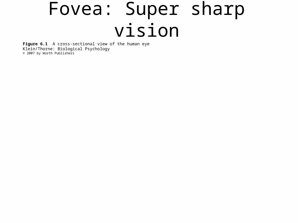
Figure 6.1 A cross-sectional view of the human eyeKlein/Thorne: Biological Psychology© 2007 by Worth Publishers
Fovea: Super sharp vision

Blind spot
• Visual system takes the info from the rest of the retina, and fills in the blank.
From the eye to the brain

• Fig. 6.6
• Fig. 6.8
Primary Visual Cortex
• Also known as the striate cortex• First stage of processing for visual information

Secondary Visual Cortex
• Transmits information to other areas of brain

Blindsight
• Damage to occipital lobe
• Person is able to respond to an object even though not consciously aware of seeing it
• Most often associated with object that is moving

The Case of D.B.
• Right occipital lobe removed• Things that are in left visual field are never
perceived• Therefore, D.B. had no awareness of seeing
things in left visual field• But…
– Could reach for things in left visual field– Could differentiate vertical line from horizontal or
diagonal line– Could differentiate letters “X” and “O”
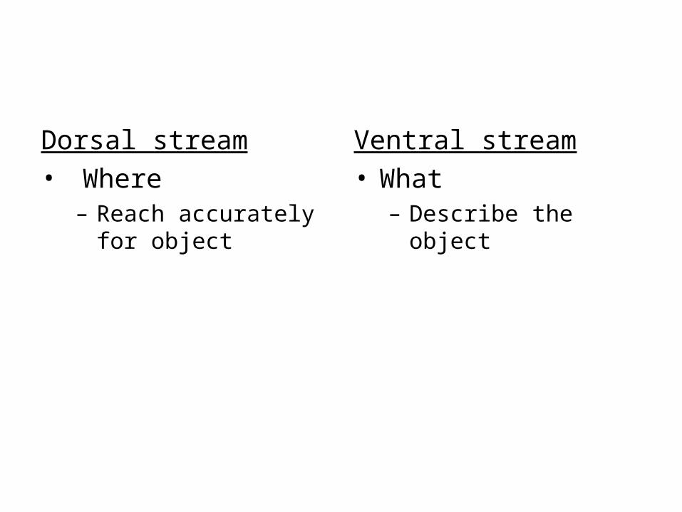
Dorsal stream• Where
– Reach accurately for object
Ventral stream• What
– Describe the object

Alternative explanation
Dorsal stream• Where
• Control of behavior
Ventral stream• What
• Conscious perception

Visual Agnosia
• Cannot name an object they see
• But can identify object if presented in a different mode (e.g., touch)
• Can see features of object but cannot integrate

Visual Prosopagnosia
• The inability to recognize faces

Depth Perception

Depth Perception
• Monocular depth cues– Able to gauge depth using only one eye

Monocular depth cues: Fig 6.23

Depth Perception
• Binocular depth cues– Retinal disparity

Color Perception

Young-HelmholtzTrichromatic Theory
• Three different types of color receptors• Each type is sensitive to different color
– Red– Green– Blue or violet– Fig. 6.26
Young-HelmholtzTrichromatic Theory
• The color that you see is the result of a mixture of activity of each type of color receptor• E.g., yellow = activation of green and red fibers
• Research suggests that there are in foact three types of cones
Hering’s Opponent Process Theory
• Perception of color is based on pairs of complementary colors
• Red-Green, Yellow-Blue, Black-White
• For example: cells signal the color red by being hyperpolarized, and signal green by being depolarized

Hering’s Opponent Process Theory
• Perception of color is based on pairs of complementary colors
• Red-Green, Yellow-Blue, Black-White
• For example: cells signal the color red by being hyperpolarized, and signal green by being depolarized

Opponent Process Theory
• Negative afterimage• Fig. 6.25

Opponent Process Theory
• Research has also found support for opponent process theory: – E.g., there are cells that are activated by red
and inhibited by green, and vice ersa

Opponent Process Theory
• May account for color defects, which occurs in pairs (e.g., blue-yellow)
Related Documents