The visual system Ch 6

The visual system Ch 6. In general, our visual system represents the world: a) Imperfectly b) Accurately c) Better than reality.
Dec 26, 2015
Welcome message from author
This document is posted to help you gain knowledge. Please leave a comment to let me know what you think about it! Share it to your friends and learn new things together.
Transcript

The visual system
Ch 6
In general, our visual system represents the world:
a) Imperfectly
b) Accurately
c) Better than reality

Light
Photons – discrete particles of energy– travel through space at 300,000
kilometers/sec (186,000 miles/sec)
Waves of electromagnetic energy– 380 to 760 nanometers in length

Electromagnetic spectrum
nm = nanometer

What other animals see…
Honeybees can see Ultraviolet light Rattlesnakes can see infrared light

Rats can see ultraviolet light
Properties of light and perception
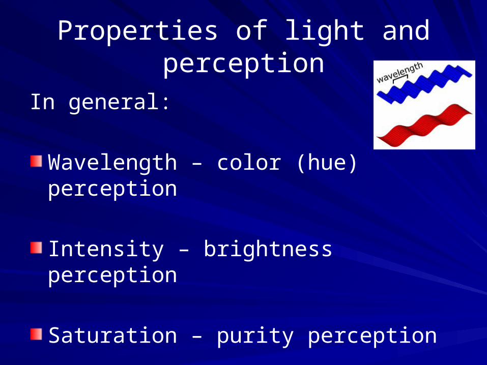
In general:
Wavelength – color (hue) perception
Intensity – brightness perception
Saturation – purity perception

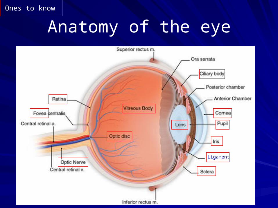
Light enters the eyethrough the pupil
size of the pupil
is regulated by the iris
The lens focuses light
on the retina
Note: that the
retinal image
is upside down.
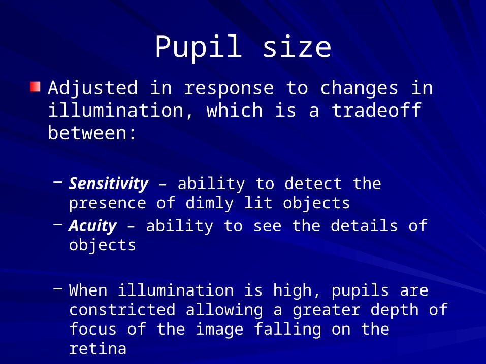
Pupil sizeAdjusted in response to changes in illumination, which is a tradeoff between:
– Sensitivity – ability to detect the presence of dimly lit objects
– Acuity – ability to see the details of objects
– When illumination is high, pupils are constricted allowing a greater depth of focus of the image falling on the retina
– When illumination is low, pupils dilate in response to low activation of receptors allowing more light to enter the eye but sacrificing acuity and depth of focus

AccomodationProcess of adjusting the configuration of the lens to bring images into focus on the retina– Focus on a near object
ciliary muscles contract
putting less tension on the ligaments
allowing the lens to take its natural cylindrical shape
thus increasing its ability to refract (bend) light– Focus on a distant object
Ciliary muscles relax
Increasing tension on the ligaments
flattens the lens
thus decreasing its ability to refract (bend) light

Binocular disparity
The difference in the positions of the same image on the two retinas– Is greater for close objects (eyes must
converge or turn slightly inward)– The degree of binocular disparity enables the
visual system to construct 3-D perception from two 2-D retinal images

The retina
Composed of 5 layers of neurons – Receptors (photoreceptors)
1 rod
3 cones
– Horizontal cells (2 subtypes)– Bipolar cells (10 subtypes)– Amacrine cells (25-30 subtypes)– Ganglion cells (10-15 subtypes)
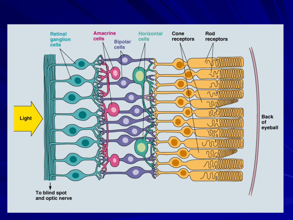
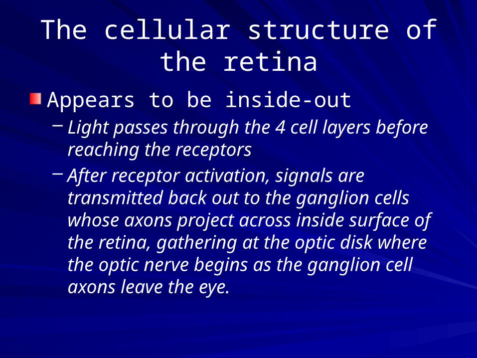
The cellular structure of the retina
Appears to be inside-out– Light passes through the 4 cell layers before
reaching the receptors– After receptor activation, signals are
transmitted back out to the ganglion cells whose axons project across inside surface of the retina, gathering at the optic disk where the optic nerve begins as the ganglion cell axons leave the eye.

Two visual problems
result from the inside out arrangement:
1. Incoming light is distorted as it passes through the cell layers
2. There is a blind spot (no receptors or cells) at the optic disk where the axons gather to exit the eye

Solutions
The fovea is an area (0.33 cm diameter) in the center of the retina where there is a thinning of the retinal ganglion cell layer.– Less distortion of light– Specialized for high-acuity vision
(seeing details)
Completion – the visual system uses information from receptors around the blind spot to fill in the gap in the retinal image.

photopic and scotopic vision
The two systems are “wired” differently
Cones – low degree of convergence (a single ganglion cell receives signals from a few cones).
Rods – high degree of convergence (a single ganglion cell receives signals from hundreds of rods).

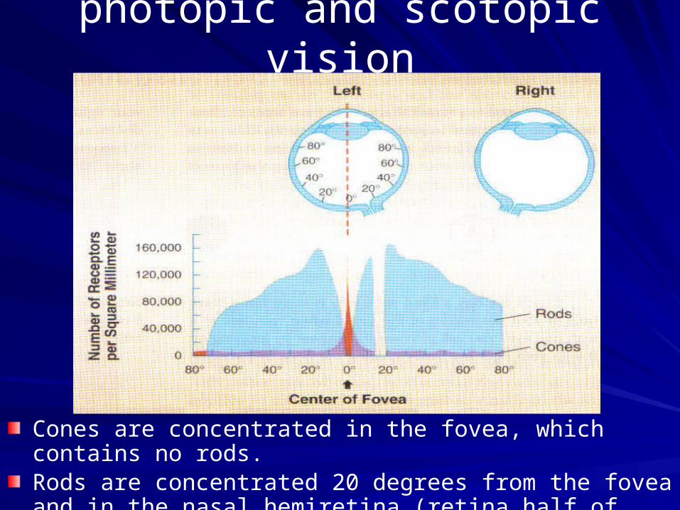
photopic and scotopic vision
Cones are concentrated in the fovea, which contains no rods.Rods are concentrated 20 degrees from the fovea and in the nasal hemiretina (retina half of both eyes near the nose).
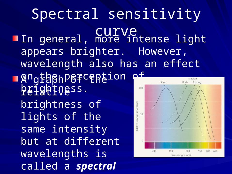
Spectral sensitivity curveIn general, more intense light appears brighter. However, wavelength also has an effect on the perception of brightness.
A graph of the relative brightness of lights of the same intensity but at different wavelengths is called a spectral sensitivity curve (see Pinel p. 138).
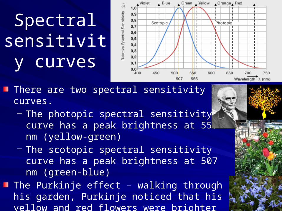
Spectral sensitivity
curves
There are two spectral sensitivity curves.– The photopic spectral sensitivity curve has a
peak brightness at 555 nm (yellow-green)– The scotopic spectral sensitivity curve has a
peak brightness at 507 nm (green-blue)
The Purkinje effect – walking through his garden, Purkinje noticed that his yellow and red flowers were brighter than the blues ones just before dusk; just a few minutes later the trend was reversed (blue flowers appeared as brighter greys).
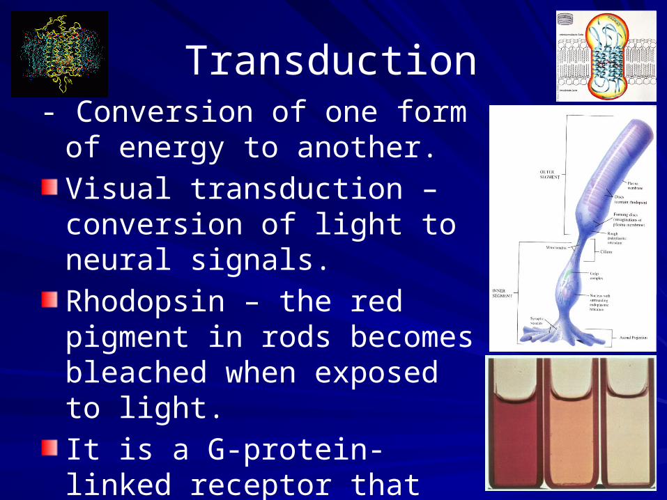
Transduction- Conversion of one form of
energy to another.
Visual transduction – conversion of light to neural signals.
Rhodopsin – the red pigment in rods becomes bleached when exposed to light.
It is a G-protein-linked receptor that responds to light.
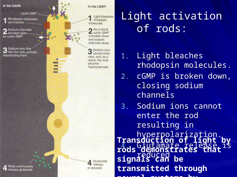
Light activation of rods:
1. Light bleaches rhodopsin molecules.
2. cGMP is broken down, closing sodium channels
3. Sodium ions cannot enter the rod resulting in hyperpolarization.
4. Glutamate release is reduced
Transduction of light by rods demonstrates that signals can be transmitted through neural systems by inhibition.

From retina to primary visual cortex
Pathway: retina lateral geniculate nucleus (LGN) primary visual cortex~90% of axons of retinal ganglion cells make up this pathwayLGN channels– Parvocellular (P layers) run through top 4 layers of
LGN – responsive to color and fine detail (input from cones)
– Magnocellular (M layers) run through bottom 2 layers of LGN – responsive to movement (input from rods)
Most LGN neurons that project to primary visual cortex (V1, striate cortex) terminate in the lower part of cortical layer IV
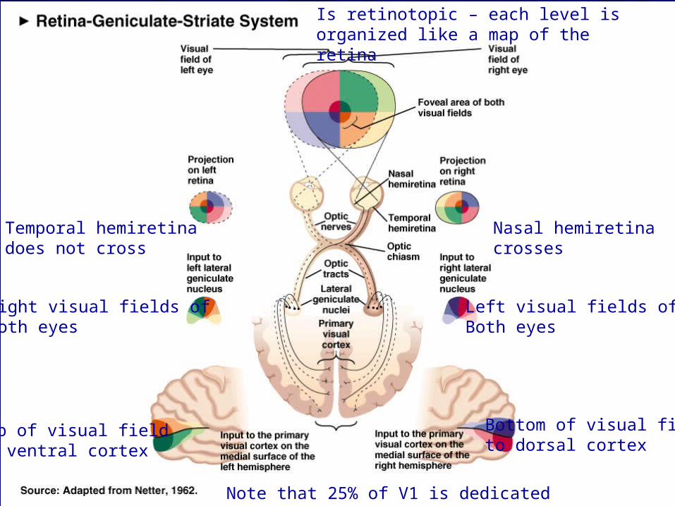
Temporal hemiretinadoes not cross
Nasal hemiretina crosses
Right visual fields ofBoth eyes
Left visual fields ofBoth eyes
Top of visual fieldto ventral cortex
Bottom of visual fieldto dorsal cortex
Is retinotopic – each level is organized like a map of the retina
Note that 25% of V1 is dedicated to fovea

Saccades
The eye continually scans the visual field and makes a series of brief fixations (3/sec) connected by quick eye movements called saccades. The fixations are integrated to produce greater color and detail than the restricted foveal region can produce if it remained stationarystabilized retinal images, projected from a contact lens that moves with the eye; image disappears in a few seconds.
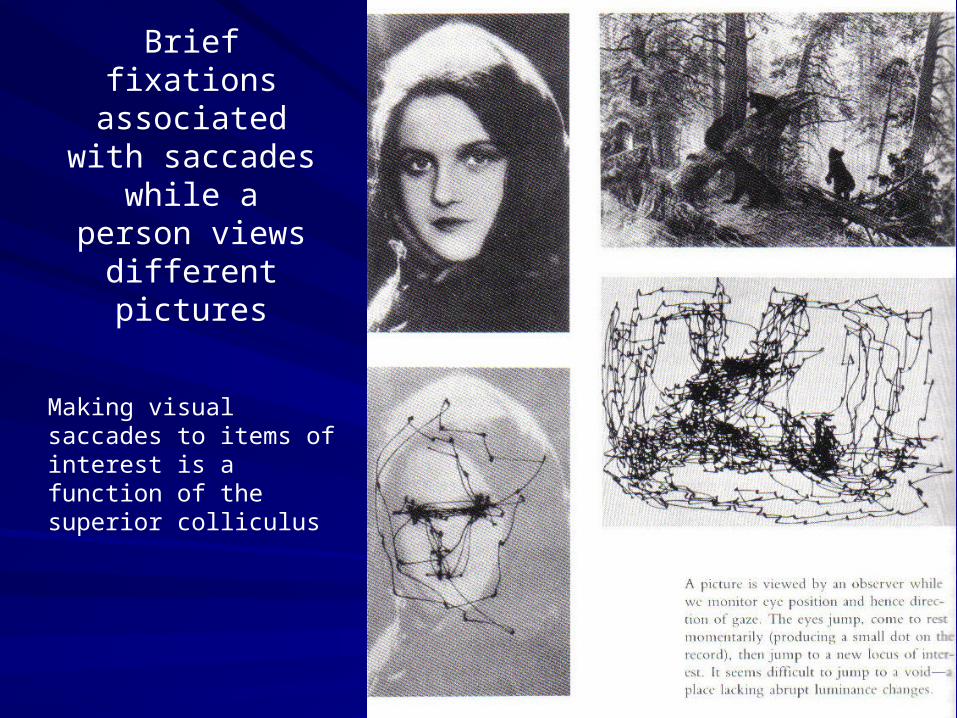
Brief fixations associated with
saccades while a person views
different pictures
Making visual saccades to items of interest is a function of the superior colliculus
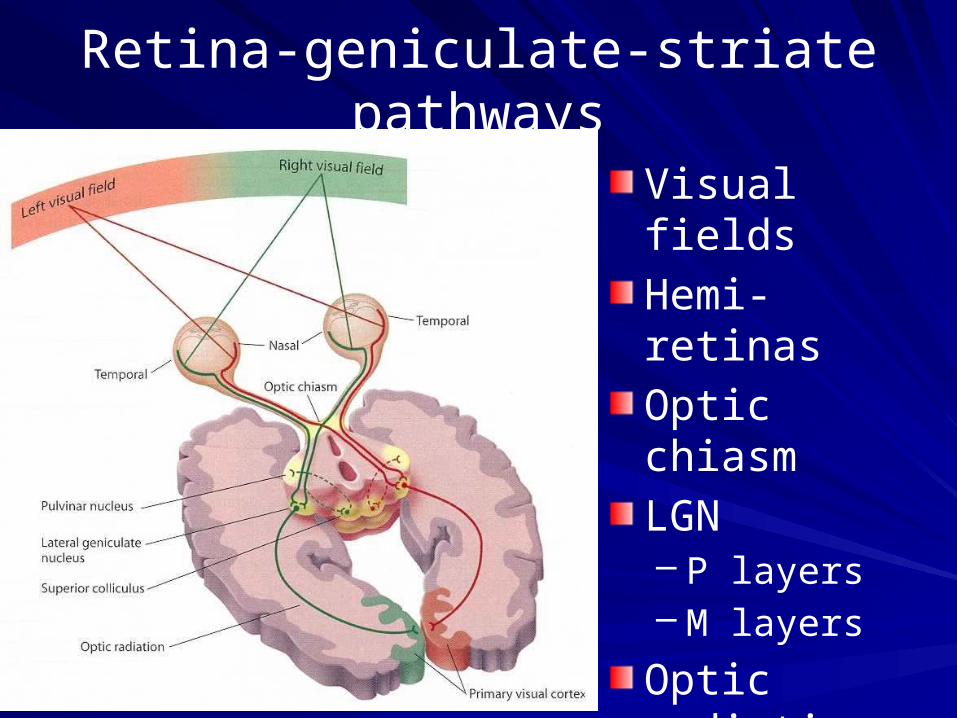
Retina-geniculate-striate pathways
Visual fields
Hemi-retinas
Optic chiasm
LGN– P layers– M layers
Optic radiations
Striate (primary visual) cortex

LGN

Hubel and Wiesel
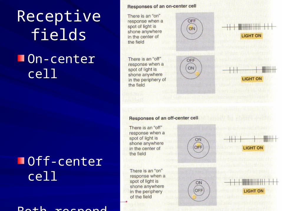
Receptive fields
On-center cell
Off-center cell
Both respond best to contrast

Both types of cells respond best to contrast
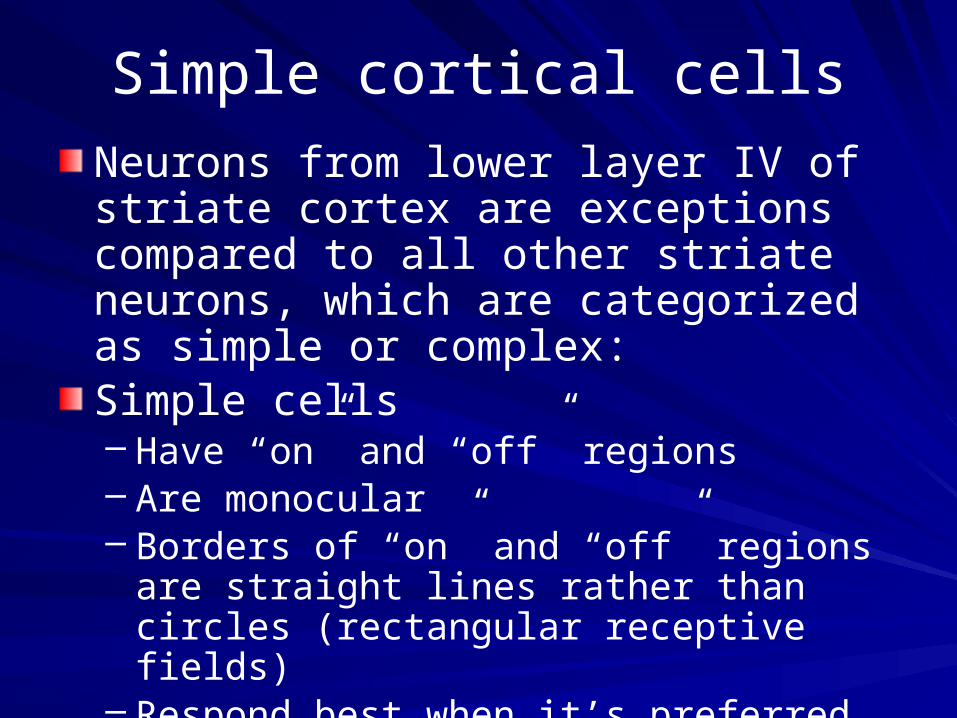
Simple cortical cells
Neurons from lower layer IV of striate cortex are exceptions compared to all other striate neurons, which are categorized as simple or complex:Simple cells– Have “on” and “off” regions – Are monocular– Borders of “on” and “off” regions are straight
lines rather than circles (rectangular receptive fields)
– Respond best when it’s preferred straight edge is in a particular orientation and position
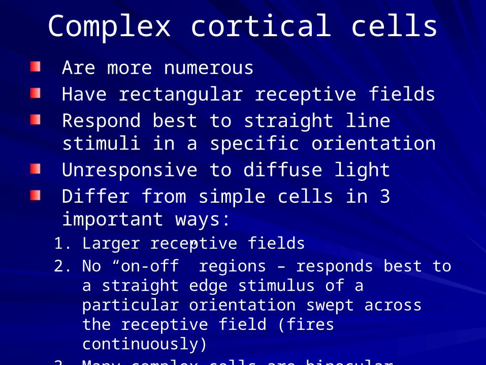
Complex cortical cellsAre more numerous
Have rectangular receptive fields
Respond best to straight line stimuli in a specific orientation
Unresponsive to diffuse light
Differ from simple cells in 3 important ways:1. Larger receptive fields
2. No “on-off” regions – responds best to a straight edge stimulus of a particular orientation swept across the receptive field (fires continuously)
3. Many complex cells are binocular (respond to stimulation of either eye and will respond more robustly to stimulation of both eyes simultaneously).

Columnar organization of V1
Vertical electrode tract
Horizontal electrode tract
1 right eye
2 right eye
3 right eye
4 right eye
1 right eye
2 right eye
3 left eye
4 left eye

Hubel & Wiesel’smodel of the columnar
organization of the primary visual cortex
Big block of tissue analyzes signals from one area of the visual field
Sub-blocks analyze signals from the left and right eyes
Slices of block prefer lines in a particular orientation

Spatial-frequency theory
Striate neurons respond even more robustly to sine-waves place at a particular angle compared to straight lines and edges

Component theory of color vision
Three kinds of color receptors (cones) each with a different spectral sensitivity
Color of a particular stimulus is determined by the ratio of activity in the three kinds of receptors
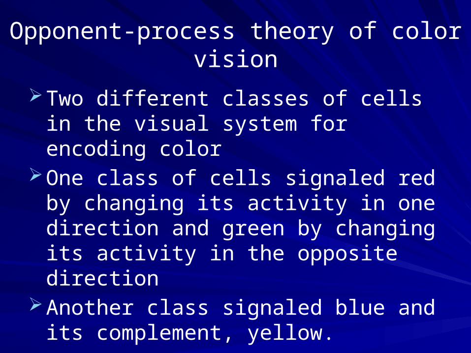
Opponent-process theory of color vision
Two different classes of cells in the visual system for encoding color
One class of cells signaled red by changing its activity in one direction and green by changing its activity in the opposite direction
Another class signaled blue and its complement, yellow.

Which theory is correct?
The Answer: both (and a third one)Cones code color on a purely
component basis (different photopigments maximally sensitive to low, medium and high wavelengths of light)
Opponent processing of color occurs at all other levels of the retina-geniculate-striate system
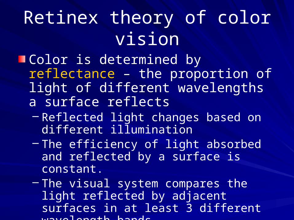
Retinex theory of color vision
Color is determined by reflectance – the proportion of light of different wavelengths a surface reflects– Reflected light changes based on different
illumination– The efficiency of light absorbed and reflected
by a surface is constant.– The visual system compares the light
reflected by adjacent surfaces in at least 3 different wavelength bands.

Principles of sensory system organization
Three different types of sensory cortex:
1. Primary sensory cortex – receives most of its input from thalamic relays
2. Secondary sensory cortex – receives most of its input from the primary sensory cortex of a system
3. Association cortex – receives input from more than one sensory system
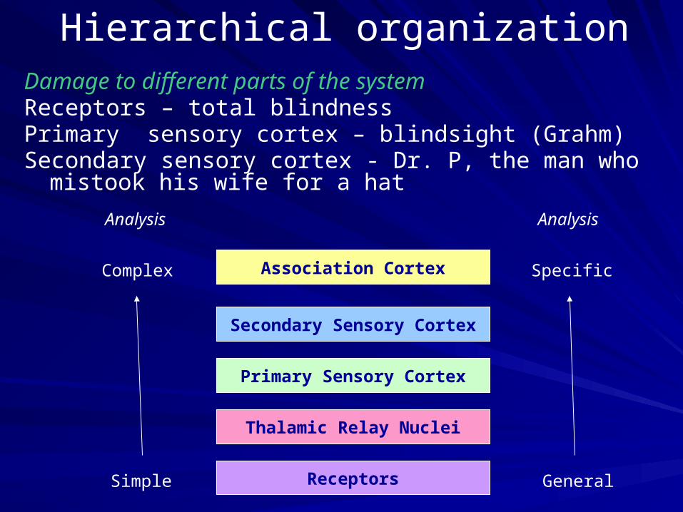
Hierarchical organizationDamage to different parts of the systemReceptors – total blindnessPrimary sensory cortex – blindsight (Grahm)Secondary sensory cortex - Dr. P, the man who mistook his
wife for a hat
Receptors
Thalamic Relay Nuclei
Primary Sensory Cortex
Secondary Sensory Cortex
Association Cortex
Simple
Complex
Analysis
General
Specific
Analysis

Sensory system organization
Receptors
Thalamic Relay Nuclei
Primary Sensory Cortex
Secondary Sensory Cortex
Association Cortex
R
TRN
PSC
SSC
AS
PSC
SSC
AS
TRN
PSC
SSC
AS
SSC
AS
SSCSSC
Former Model (1960s) Current Model
(functionally homogeneous and serial) (functionally segregated and parallel)

Two Visual Streams
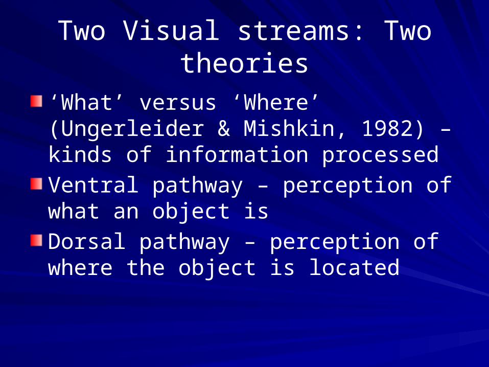
Two Visual streams: Two theories
‘What’ versus ‘Where’ (Ungerleider & Mishkin, 1982) – kinds of information processed
Ventral pathway – perception of what an object is
Dorsal pathway – perception of where the object is located
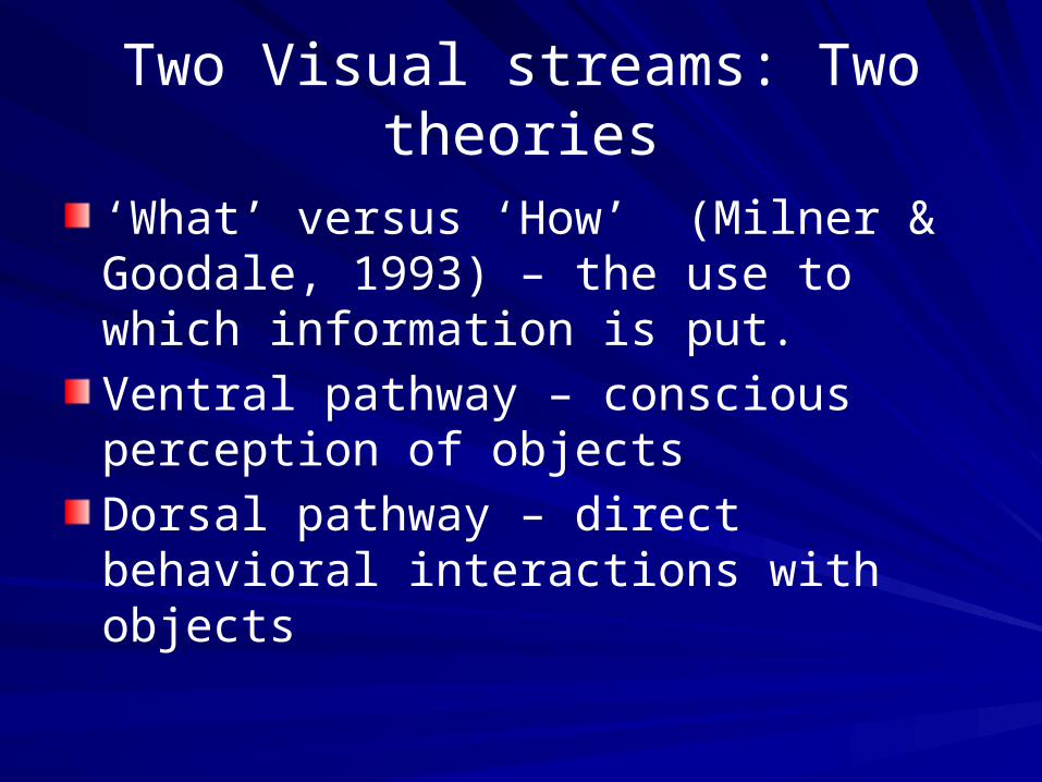
Two Visual streams: Two theories
‘What’ versus ‘How’ (Milner & Goodale, 1993) – the use to which information is put.
Ventral pathway – conscious perception of objects
Dorsal pathway – direct behavioral interactions with objects

Visual agnosia
Gnosis means “to know”
Visual agnosics can see stimuli but do not know what they are– Object agnosia– Color agnosia– Movement agnosia (akinetopsia)– Face agnosia (prosopagnosia)
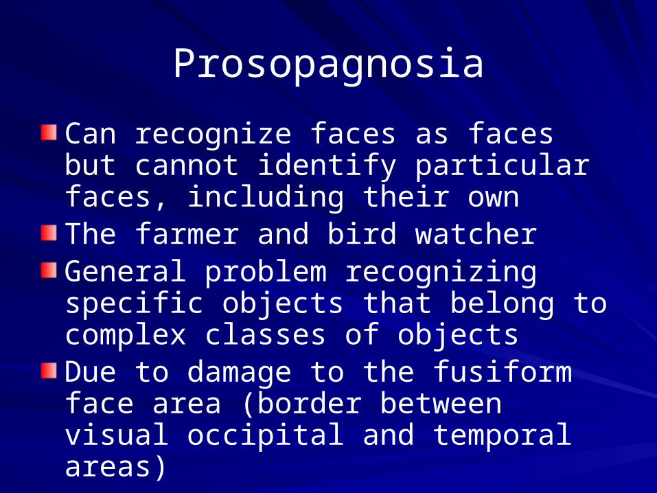
Prosopagnosia
Can recognize faces as faces but cannot identify particular faces, including their ownThe farmer and bird watcherGeneral problem recognizing specific objects that belong to complex classes of objectsDue to damage to the fusiform face area (border between visual occipital and temporal areas)
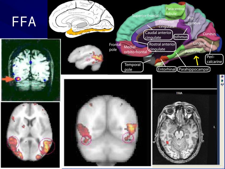
FFA

Area MT/V5fMRI – shows region is active when viewing movementTMS – inactivation produces motion blindnessLesions – uni- or bi-lateral damage results in akinetopsiaAkinetopsia – deficit in seeing movement (a.k.a. movement agnosia)

Magic Eye autostereogram created for NIH
http://www.magiceye.com/client/nih.html
Related Documents


