This is a pre-published version This is a pre-published version The use of Ilizarov external fixation in the treatment of distal tibial fractures Frankie LEUNG Senior Medical Officer, Department of Orthopaedic Surgery, Queen Mary Hospital, The University of Hong Kong, Hong Kong Hau Yan KWOK Medical Officer, Department of Orthopaedic Surgery, Queen Mary Hospital, The University of Hong Kong, Hong Kong Tsz Shing PUN Medical Officer, Department of Orthopaedic Surgery, Queen Mary Hospital, The University of Hong Kong, Hong Kong Shew Ping CHOW Professor, Department of Orthopaedic Surgery, Queen Mary Hospital, The University of Hong Kong, Hong Kong Correspondence and Request for reprints: Frankie LEUNG. Department of Orthopaedic Surgery, Queen Mary Hospital, The University of Hong Kong, Hong Kong. Tel: +852 2855 4654 Fax: +852 2817 4392 e mail: [email protected]

Welcome message from author
This document is posted to help you gain knowledge. Please leave a comment to let me know what you think about it! Share it to your friends and learn new things together.
Transcript
This is a pre-published versionThis is a pre-published version
The use of Ilizarov external fixation in the treatment of distal
tibial fractures
Frankie LEUNG Senior Medical Officer, Department of
Orthopaedic Surgery, Queen Mary Hospital, The
University of Hong Kong, Hong Kong
Hau Yan KWOK Medical Officer, Department of Orthopaedic
Surgery, Queen Mary Hospital, The University of
Hong Kong, Hong Kong
Tsz Shing PUN Medical Officer, Department of Orthopaedic
Surgery, Queen Mary Hospital, The University of
Hong Kong, Hong Kong
Shew Ping CHOW Professor, Department of Orthopaedic
Surgery, Queen Mary Hospital, The University of
Hong Kong, Hong Kong
Correspondence and Request for reprints:
Frankie LEUNG.
Department of Orthopaedic Surgery, Queen Mary Hospital, The University of
Hong Kong, Hong Kong. Tel: +852 2855 4654 Fax: +852 2817 4392
e mail: [email protected]
Summary
We reviewed 31 distal tibial fractures (sixteen involving tibial
plafond) treated with Ilizarov external fixation. The study population was
composed of 19 males and 12 females, with an average age of 54 (range, 13-
80 years). The fractures were classified according to the AO classification:
A1 (3), A2 (6), A3 (6), C1 (2), C2 (8), C3 (6). There were 6 open injuries.
In 14 of the pilon cases, open reduction of the intra-articular
fragments and bone grafting via a limited incision was performed. Clinical
follow up averaged 28 months (range, 18-42 months). All but one fractures
united with an average healing time of 13.9 weeks (range, 10-20 weeks).
All but one patient with AO Type A fracture had excellent or good
functional scores. The 14 cases of AO C2 and C3 group had 6 good results,
5 fair results and 3 poor results.
This method yielded satisfactory results comparable with previous
studies using open reduction and internal fixation while decreasing the
number of serious complications. Its usage can be recommended,
especially in fractures with severe soft tissue damage and in comminuted
fractures.
Introduction
Fractures of the distal tibia are among the most difficult fractures to treat. The
short distal segment presents difficulty for the orthopaedic surgeon in choosing
the appropriate fixation method. The intra-articular group of these injuries, the
pilon fracture, is due to high energy trauma and articular comminution is
frequently present. The greatest challenge to the orthopaedic surgeon lies in the
relatively tight soft tissue around the ankle. The conventional method of open
reduction follows by plate and screw fixation provides good outcomes only in
the fractures with less severe, lower energy trauma [5-8,10,12,13-15,18]. Once
complications including wound dehiscence and infection set in, patients will
have a lengthy stay in hospital. Often they need multiple operations, and may
even end up in having amputation [20]. As a result, it has been a recent
interest in treating these fractures with external fixation and limited internal
fixation [1-4,9,17,19].
The Ilizarov principle of circular external fixation has been applied to fracture
treatment. The advantage in using tensioned transfixion wires is that in a small
bone segment, multiple wires can be inserted and tightened, resulting in strong
fixation of the bone. This is particularly useful in fixing the short distal
fragment in distal tibial fractures. The transfixion wire can bypass the area with
poor soft tissue condition and the minimally invasive nature of the surgery can
avoid catastrophic wound complication that may follow the conventional open
fracture treatment. There is no need to extend the fixation device across the
ankle joint and early range of motion is possible. This is particularly important
in intra-articular fractures where joint motion is important for articular cartilage
healing and nourishment [16]. The elasticity of the wire allows micromotion
during weight bearing walking and that will facilitate fracture healing. This
study retrospectively evaluates the efficacy of this method in treating distal
tibial and pilon fractures.
Materials and Methods
Between July 1995 and March 1998, patients with distal tibial fractures or pilon
fractures admitted to our institute will be treated with the technique of circular
external fixation and limited internal fixation if necessary. All open fractures
were treated on an emergency basis in the operation theatre with irrigation and
debridement, followed by fracture fixation. The timing of operation for closed
fractures depended on the condition of soft tissues and the likelihood of a
limited open reduction, which was often dictated by the degree of articular
involvement (Fig. 1). The affected limbs were put in plaster slabs and elevated.
The definitive procedure for fixation was delayed for an average of 2 days in
the extra-articular group (AO type A) and 9 days in the intra-articular group
(AO type C).
The operation was performed with the patient in the supine position on a
radiolucent table. The device was Ilizarov external fixator (Smith & Nephew,
Richards Inc.). with tensioned wires for the distal bone fragment. Preassembly
of the frame was preferable, as this would greatly reduce the operation time.
The operation started with the placement of a transverse wire across the talus or
calcaneus. The pin should be fixed to a half ring for distraction of the fracture
site. The pre-assembled frame was then applied to the tibia. A hybrid kind of
fixation using 5mm half pins for the proximal fragment was used in order to
minimize soft tissue impalement. 1.8mm wires were used to fix the distal
fragment and were tensioned to 120kg. Olive wires were used for reduction of
wedge fragments or for better alignment. The frame usually included 3 rings (2
for proximal and 1 for distal). The use of conical washers in connecting the
threaded rods to the rings greatly facilitated the reduction of the fracture. In
most cases, 3 half pins were inserted to the proximal fragment and 3 wires were
inserted to the distal fragment. The effect of ligamentotaxis on reduction was
assessed with the use of a C-arm. In fourteen cases of the pilon fractures, open
reductions of the intra-articular fragments via a limited open approach were
performed prior to the stabilization of the metaphyseal portion of the fracture. A
5 to 6 cm long incision was made anteriorly over the major fragments. The
ankle joint was visualized and anatomical reduction of the articular surface was
possible. Lag screws were inserted to fix the articular fracture. Bone graft
harvested from the iliac crest was placed in 15 patients (3 had extra-articular
fractures) to fill up bony defects or to support the articular surface. The
metaphyseal fracture fragments were not exposed. The surgical wound was
closed first before proceeding to the insertion of wires. The fixation of the rest
of the tibia was then performed in the above-mentioned manner. In two patients
with comminuted C3 fractures, the wire through the hind foot with the ring was
left behind for another two weeks to maintain the reduction by temporarily
immobilizing the ankle joint. In all other patients, the wire at the hind foot was
removed at the end of the procedure and the ankle would be free for movement.
The average duration of the operation was 126 minutes (range, 65 to 150
minutes).
Usually patients with extra-articular fractures were allowed weight bearing
walking as tolerated, while those with pilon fractures were kept non-
weightbearing for at least 8 weeks (Fig. 2). All patients were encouraged to
have early range of motion exercises. Clinical and radiological assessment was
done every two weeks until the fracture united (Fig. 3a to 3e). The fixator was
removed at an average of 15 weeks post-operatively (range, 10 to 20 weeks).
Results
There were 31 patients treated under the protocol. The study population
comprised 19 males and 12 females. The average age of the population was 54
years (range, 13-80 years). The fractures were classified according to the AO
classification [11]: A1 (3), A2 (6), A3 (6), C1 (2), C2 (8), C3 (6). Partial
articular fractures (AO type B) were excluded from the study since a rigid
fixation of the diaphyseal-epiphyseal junction is not necessary. The fibula was
intact in 5 patients.
There were 6 open injuries, and in 19 patients (61%) the mechanism of injury
was high-energy trauma, including fall from height, direct crush by heavy
object and motor vehicle accident. The average injury severity score was 8.3.
Ten patients had multiple fractures which included second long bone fractures
(4 patients), spinal fractures (3 patients), pelvic fractures (2 patients) and one
ipsilateral talus fracture.
Union
Clinical follow-up averaged 28 months (range, 18-42 months). All but one
fractures united with an average healing time of 13.9 weeks (range, 10 to 20
weeks). The only non-union case was a patient complicated with infection and
he would be discussed later.
Clinical assessment
In order to evaluate the pain and function of the subjects, a clinical scoring
system based on the one used by Teeny and Wiss [18] was obtained in 30
patients, excluding the one who had an arthrodesis. The results were graded as
excellent, good, fair, or poor (Table 1). The presence of intra-articular
involvement greatly affected the result (Table 2). In the fifteen patients with
AO extra-articular types A1-A3 fractures, fourteen of them had excellent or
good results and one had fair results. There was no poor result. The two
fractures in the C1 group also had excellent or good results. However, in the 14
patients with C2 and C3 fractures, there were all together 6 good results, 5 fair
results and 3 poor results.
Radiological assessment
Patients returned for follow-up visits at least every three months for the first
year and every six months thereafter. Serial radiographs were made and
evaluated for the bone healing, fracture alignment and the development and
progression of osteoarthritic changes. Degenerative changes were rated as mild
(one millimeter of narrowing of joint space), moderate (two millimeters
narrowing with small periarticular cysts or spurs) and severe (complete loss of
joint space with osteophytes or cysts). Only patients with C2 and C3 fractures
had radiographic signs of degeneration (Fig.3d to 3e). Out of fourteen patients
in this group, four had no changes, five had mild changes, three had moderate
changes and two had severe changes. The presence of moderate to severe
degeneration correlates well with the clinical outcome as all of them having
either fair or poor clinical score results. However, despite radiographic signs of
degeneration, no patients had symptoms severe enough necessitating an ankle
arthrodesis up to the time of follow up.
Complications
There were nine (29%) pin tract infection in this series. All responded to oral
antibiotics and local pin care although six of them had an early removal of the
involved pin as fracture healing progressed. Most of these infections involved
the wires in the region of the ankle. There seemed to be no correlation with the
fracture type.
There was one patient complicated with deep infection after the operation. He
was a 42-year-old patient who had an open pilon and ipsilateral talus fracture.
He had circular external fixator put on as an emergency procedure. He
developed pain and swelling at the fracture site and discharge from the distal
transfixion wire two months after the external fixator. He was diagnosed to
have osteomyelitis of the distal tibia and repeated debridements were performed.
The infection was under control and the fracture healed eventually with
subsequent bone grafting. However, there was destruction of the ankle joint
and subsequently the patient needed an ankle arthrodesis.
There was one case of skin necrosis in a thirteen-year-old boy who had an A3
fracture. There was initially some abrasion of the skin around the fracture
region which after the fixation showed a necrotic patch. Retrospectively, the
necrotic skin was caused by the jeopardized blood supply at the time of
distraction during fracture reduction. The necrotic skin was debrided and the
wound finally healed with granulation.
Two patients were found to have unsatisfactory reduction on follow-up and had
frame adjustment under fluoscopic guidance in the operating theatre. One sixty-
two year old lady had fifteen degrees of varus angulation, but she refused
further adjustment. Despite she had a malunion, she had an excellent functional
score at the end of follow up.
Discussion
Distal tibial fractures are complex injuries, not only regarding the bony
component, but also in terms of the management of the soft tissue problem.
Failure to recognize this often resulted in repeated surgery and even
amputations. In 1969, Ruedi and Allgower [13] reported a 74 per cent excellent
or good functional result when they reviewed 84 pilon fractures treated with
open reduction and internal fixation. Thereafter grew a widespread enthusiasm
for such technique. The four principles that they advocated [13-15] were: (1)
restoration of fibula length, (2) reduction of articular surface, (3) cancellous
bone grafting of the metaphyseal defect, and (4) Stabilization with a medial
buttress plate. While some authors [5-8] shared the same good results as Ruedi
and Allgower, others have reported less favorable results, together with a high
rate of complications. Teeny and Wiss [18] reported eleven (37 per cent) of
thirty patients having deep infection, and McFerran et al.[10] reported twenty-
one (40 per cent) of fifty-two patients having a major complication. Wyrsch et
al.[20] reported three (16%) amputations of nineteen patients having open
reduction and internal fixation. These reports reflected the fact that with
extensive surgical dissection in achieving an anatomical reduction, the
vascularity of the bony fragments are often jeopardized and these
devascularized fragments will act as foci for infection. The insertion of a bulky
plate into the tight soft tissue envelope of the distal tibia also impaired wound
healing. The incidence of skin slough and wound dehiscence was as high as
27% in the Teeny and Wiss series [18].
While anatomical reduction is still crucial in the reconstruction of the articular
surface, it is less important regarding the metaphyseal fragments. The biology
of the bony fragments should always be preserved maximally. The concept of
external fixation, when combined with minimal internal fixation if necessary,
can deal with both mechanical and biological aspects for better fracture healing.
Reduction is achieved largely through ligamentotaxis and extensive surgical
dissection is obviated. External fixation device spanning across the ankle joint
had been used with promising result [1-4,9,17,19]. A randomized, prospective
study on pilon fractures performed by Wyrsch et al.[20] comparing open
reduction and internal fixation with external fixation with or without limited
internal fixation showed similar results in the two groups. However, the latter
group was associated with fewer and less severe complications than internal
fixation.
The Ilizarov circular external fixation system provides the advantage of sparing
the ankle joint and allows early motion. The usage of tensioned wires is an
effective way to fix a short bony segment. Minor readjustment of the frame can
also be done as an outpatient procedure. This option is not possible with cast
immobilization, rigid internal fixation or uniplanar external fixation.
The current study yielded good results concerning the use of this technique in
treating AO type A extra-articular fractures. Fractures extending distally to
within 4 to 5 cm of the ankle joint can be satisfactorily treated. There was no
nonunion although three patients with comminuted fractures had primary bone
grafting. Early weight bearing walking could also be achieved in most patients.
It is extremely difficult to compare the results for the sixteen cases of pilon
fractures in this series with those reported in the literature on the technique of
open reduction and internal fixation although the incidence of major
complications in our series appears much lower. Radiological changes of
moderate to severe degeneration were noticed in 36% of our patients with C2
and C3 fractures. This may reflect the intrinsic difficulty in treating an intra-
articular fracture of the ankle joint. However, no patient in this series had an
ankle arthrodesis because of pain in a degenerated ankle.
Although the procedure was time-consuming and the safety zones for wire
insertion posed limitation on fracture reduction, its usage can still be
recommended, especially in fractures with severe soft tissue damage and in
comminuted fractures. The issue of whether fibular fixation is needed has not
been looked into in our current study, although we think that rigid fixation on
the lateral side may predispose to varus alignment during fracture healing,
especially in the case of marked tibial metaphyseal comminution.
References
[1] Bonar SK, Marsh JL: Unilateral external fixation for severe pilon
fractures. Foot and Ankle 14:57-64, 1993
[2] Bonar SK, Marsh JL: Tibial plafond fractures: changing principles of
treatment. J Am Acad Orthop Surg 2:297-305, 1994
[3] Bone LB: Fractures of the tibial plafond. The pilon fracture. Orthop Clin
North America 18:95-104, 1987
[4] Bone LB, Stegemann P, McNamara K, Seibel R: External fixation of
severely comminuted and open tibial pilon fractures. Clin Orthop
292:101-107, 1993
[5] Bourne RB: Pylon fractures of the distal tibia. Clin Orthop 240:42-46,
1989
[6] Bourne RB, Rorabeck CH, MacNab I: Intraarticular fractures of the
distal tibia: The pilon fracture. J Trauma 23:591-596, 1983
[7] Helfet DL, Koval K, Pappas J, et al.: Intraarticular Pilon fracture of the
tibia. Clin Orthop 298:221-228, 1994
[8] Kellam JF, Waddell JP: Fractures of the distal metaphysis with
intraarticular extension: The distal tibia explosion fracture. J Trauma
19:593-601, 1979
[9] Marsh JL, Bonar S, Nepola JV, Decoster TA, Hurwitz SR: Use of an
articulated external fixator for fractures of the tibial plafond. J Bone
Joint Surg 77A:1498-1509, 1995
[10] McFerran MA, Smith SW, Boulas HJ, Schwartz HS: Complications
encountered in the treatment of pilon fractures. J Orthop Trauma 6: 195-
200, 1992
[11] Muller ME, Nazarian S, Koch P: The AO Classification of Fractures.
New York, Springer, 1988
[12] Ovadia DN, Beals RK: Fractures of the tibial plafond. J Bone Joint Surg
68A:543-551, 1986
[13] Ruedi TP, Allgower M: Fractures of the lower end of the tibia into the
ankle joint. Injury 1:92-99, 1969
[14] Ruedi TP, Allgower M: Fractures of the lower end of the tibia into the
ankle joint: Results 9 years after open reduction and internal fixation.
Injury 5:130-134, 1973
[15] Ruedi TP, Allgower M: The operative treatment of intraarticular
fractures of the lower end of the tibia. Clin Orthop 138:105-110, 1979
[16] Salter RB, Simmonds DF, Malcolm BW: The biologic effect of
continuous passive motion on the healing of full thickness defects in
articular cartilage. An experimental investigation in the rabbit. J Bone
Joint Surg 62A:1232-1251, 1980
[17] Saleh M, Shanahan MD, Fern ED: Intra-articular fractures of the distal
tibia: surgical management by limited internal fixation and articulated
distraction. Injury 24;1:37-40, 1993
[18] Teeny SM, Wiss DA: Open reduction and internal fixation of tibial
plafond fractures. Clin Orthop 292:108-117, 1993
[19] Tornetta P, Weiner L, Bergman M, et al: Pilon fractures: Treatment with
combined internal and external fixation. J Orthop Trauma 7:489-496,
1993
[20] Wyrsch B, McFerran MA, McAndrew M, Limbird TJ, Harper MC,
Johnson KD, Schwartz HS: Operative treatment of fractures of the tibial
plafond. A randomized, prospective study. J Bone Joint Surg 78A:1646-
1657, 1996
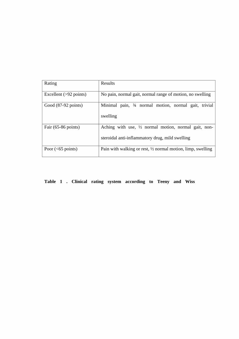
Rating Results
Excellent (>92 points) No pain, normal gait, normal range of motion, no swelling
Good (87-92 points) Minimal pain, ¾ normal motion, normal gait, trivial
swelling
Fair (65-86 points) Aching with use, ½ normal motion, normal gait, non-
steroidal anti-inflammatory drug, mild swelling
Poor (<65 points) Pain with walking or rest, ½ normal motion, limp, swelling
Table 1 . Clinical rating system according to Teeny and Wiss
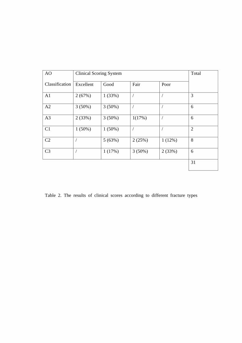
Clinical Scoring System AO
Classification Excellent Good Fair Poor
Total
A1 2 (67%) 1 (33%) / / 3
A2 3 (50%) 3 (50%) / / 6
A3 2 (33%) 3 (50%) 1(17%) / 6
C1 1 (50%) 1 (50%) / / 2
C2 / 5 (63%) 2 (25%) 1 (12%) 8
C3 / 1 (17%) 3 (50%) 2 (33%) 6
31
Table 2. The results of clinical scores according to different fracture types
Legends for illustrations
Fig. 1 Open reduction of articular fragments via a small incision. The
frame had been put on for ligamentotaxis
Fig. 2 Weight-bearing walking for extra-articular fractures
Fig. 3a A 36-year-old man sustained a C2 fracture showing the lateral
view
Fig. 3b Same patient showing the A-P view
Fig. 3c Post-fixation AP view
Fig. 3d 22 months after the injury. Lateral view showed mild
degeneration.
Fig. 3e AP view showing the same patient
Top Frankie LEUNG Fig. 1 Open reduction of articular fragments via a small incision. The frame had been put on for ligamentotaxis Top Frankie LEUNG Fig. 2 Weight-bearing walking for extra-articular fractures Top Frankie LEUNG Fig. 3a A 36-year-old man sustained a C2 fracture showing the lateral
view Top Frankie LEUNG Fig. 3b Same patient showing the A-P view Top Frankie LEUNG Fig. 3c Post-fixation AP view Top Frankie LEUNG Fig.3d 22 months after the injury. Lateral view showed mild degeneration. Top Frankie LEUNG Fig 3e AP view showing the same patient
Related Documents