ORIGINAL ARTICLE The Use of Biomedical Sensors to Monitor Capsule Formation Around Soft Tissue Implants J. W. Bender, PhD,‡ H. I. Friedman, MD, PhD,* V. Giurgiutiu, PhD,‡ C. Watson, MD,† M. Fitzmaurice, MD,† and M. L. Yost, PhD† Abstract: Piezoelectric sensors have been shown to respond repro- ducibly to changes in tissue mechanical properties surrounding an implant over a 4-month period. The vibrational amplitude at a frequency corresponding to the radial resonance shows a statistically significant change over time. The initial period of inflammation is marked by a significant reduction in amplitude, which is indicative of an increase in viscous dissipation of the tissue. As collagen displaces the cellular response, the amplitude continues to decrease. Finally, as the tissue matures, the capsule becomes stiffer, and the viscous dissipation lessens. These results are consistent with quali- tative assessments of explanted capsules. Strain gauges encased in a monolithic block of silicone exhibited a greater degree of variability, yet show similar trends over time. The strain increases in the initial 4-week period and remains rela- tively steady over the following 4 weeks. Beyond 8 weeks, the gauges begin to extrude from the animal or suffer a loss of electrical continuity. Steps are being taken to improve the strain sensor longevity in the animals. Key Words: biomedical sensors, capsule formation, soft tissue implants (Ann Plast Surg 2006;56: 72–77) B ioelectrical sensing devices have had a long history in monitoring and pacing heart rate in patients and even delivering a lifesaving shock if ventricular fibrillation is detected. 1–3 Monitoring biochemical concentrations in vivo using implanted devices has been the subject of intense research efforts, notably in the area of glucose measurement for diabetes management. 4–6 Still, the majority of implanted devices are programmed as continuous drug delivery systems (eg, Baclofen pump) or are externally located to deliver medication (insulin pump). In vivo biomechanical character- ization of soft tissues has focused on cartilage and on topical wounds, in which the sensing device is external. 7–12 There has been limited research performed on implanted mechani- cal sensing devices, and those that exist focus primarily on short-term studies. For example, Park et al 7 implanted pres- sure transducers at the interface of an artificial hip joint to measure pressure distributions across the surface and validate computational models. The development of long-term in vivo stress measurement or mechanical characterization of soft tissues could have tremendous application in decubitus ulcer prevention or in early detection of capsular contraction around an implant placed during reconstructive or cosmetic surgery. The former occurs in paraplegic patients when cap- illary closing pressure is applied to soft tissues for too long a period of time. The latter occurs as a natural course of the later stages of wound healing and can lead to poor clinical outcomes. The current investigation was undertaken to study the tissue response to 2 different small implantable sensing de- vices, piezoelectric wafer active sensors (PWAS) and strain gauge sensors (SGS). These sensors provide complementary information on the mechanical properties of tissue reactions to their presence. The piezoelectric sensor can quantify dif- ferences in local tissue viscoelasticity via the generation of high-frequency shear waves. The strain sensor deforms under static pressure. The response of the mammalian tissues to these sensors is felt to be the first step in developing devices for practical application to clinical situations. Therefore, in these experiments we have implanted these 2 types of sensors into rats and studied the sensor recordings to correlate them with the histology of the soft tissue reactions to their presence. Sensors Electromechanical Oscillation Piezoelectric materials are those that change dimen- sions upon applying an electric field. When driven with an AC electric field, devices such as the PWAS are oscillated over a spectrum of frequencies. At certain frequencies, the device will resonate and have a high amplitude of motion. Because the structure of the piezomaterial can be controlled, they can be made to possess characteristic motions (reso- nances) at specific frequencies. Figure 1 shows 2 modes of oscillation of the PWAS sensor used in these studies. There is Received August 25, 2005 and accepted for publication August 26, 2005. From the *Division of Plastic Surgery and †Department of Surgery, USC School of Medicine, Columbia, SC; and ‡School of Engineering, Uni- versity of South Carolina, Columbia, SC. Presented at the Annual Meeting of the Southeastern Society of Plastic and Reconstructive Surgeons, Paradise Island, The Bahamas, June 4 – 8, 2005. Reprints: Harold I. Friedman, MD, PhD, Division of Plastic Surgery, De- partment of Surgery, USC School of Medicine, Suite 402, #2 Medical Park Rd, Columbia, SC 29203. E-mail: [email protected]. Copyright © 2005 by Lippincott Williams & Wilkins ISSN: 0148-7043/06/5601-0072 DOI: 10.1097/01.sap.0000189620.45708.5f Annals of Plastic Surgery • Volume 56, Number 1, January 2006 72

Welcome message from author
This document is posted to help you gain knowledge. Please leave a comment to let me know what you think about it! Share it to your friends and learn new things together.
Transcript

ORIGINAL ARTICLE
The Use of Biomedical Sensors to Monitor CapsuleFormation Around Soft Tissue Implants
J. W. Bender, PhD,‡ H. I. Friedman, MD, PhD,* V. Giurgiutiu, PhD,‡ C. Watson, MD,†M. Fitzmaurice, MD,† and M. L. Yost, PhD†
Abstract: Piezoelectric sensors have been shown to respond repro-ducibly to changes in tissue mechanical properties surrounding animplant over a 4-month period. The vibrational amplitude at afrequency corresponding to the radial resonance shows a statisticallysignificant change over time. The initial period of inflammation ismarked by a significant reduction in amplitude, which is indicativeof an increase in viscous dissipation of the tissue. As collagendisplaces the cellular response, the amplitude continues to decrease.Finally, as the tissue matures, the capsule becomes stiffer, and theviscous dissipation lessens. These results are consistent with quali-tative assessments of explanted capsules.
Strain gauges encased in a monolithic block of silicone exhibiteda greater degree of variability, yet show similar trends over time.The strain increases in the initial 4-week period and remains rela-tively steady over the following 4 weeks. Beyond 8 weeks, thegauges begin to extrude from the animal or suffer a loss of electricalcontinuity. Steps are being taken to improve the strain sensorlongevity in the animals.
Key Words: biomedical sensors, capsule formation, soft tissueimplants
(Ann Plast Surg 2006;56: 72–77)
Bioelectrical sensing devices have had a long history inmonitoring and pacing heart rate in patients and even
delivering a lifesaving shock if ventricular fibrillation isdetected.1–3 Monitoring biochemical concentrations in vivousing implanted devices has been the subject of intenseresearch efforts, notably in the area of glucose measurementfor diabetes management.4–6 Still, the majority of implanteddevices are programmed as continuous drug delivery systems
(eg, Baclofen pump) or are externally located to delivermedication (insulin pump). In vivo biomechanical character-ization of soft tissues has focused on cartilage and on topicalwounds, in which the sensing device is external.7–12 Therehas been limited research performed on implanted mechani-cal sensing devices, and those that exist focus primarily onshort-term studies. For example, Park et al7 implanted pres-sure transducers at the interface of an artificial hip joint tomeasure pressure distributions across the surface and validatecomputational models. The development of long-term in vivostress measurement or mechanical characterization of softtissues could have tremendous application in decubitus ulcerprevention or in early detection of capsular contractionaround an implant placed during reconstructive or cosmeticsurgery. The former occurs in paraplegic patients when cap-illary closing pressure is applied to soft tissues for too long aperiod of time. The latter occurs as a natural course of thelater stages of wound healing and can lead to poor clinicaloutcomes.
The current investigation was undertaken to study thetissue response to 2 different small implantable sensing de-vices, piezoelectric wafer active sensors (PWAS) and straingauge sensors (SGS). These sensors provide complementaryinformation on the mechanical properties of tissue reactionsto their presence. The piezoelectric sensor can quantify dif-ferences in local tissue viscoelasticity via the generation ofhigh-frequency shear waves. The strain sensor deforms understatic pressure. The response of the mammalian tissues tothese sensors is felt to be the first step in developing devicesfor practical application to clinical situations. Therefore, inthese experiments we have implanted these 2 types of sensorsinto rats and studied the sensor recordings to correlate them withthe histology of the soft tissue reactions to their presence.
SensorsElectromechanical Oscillation
Piezoelectric materials are those that change dimen-sions upon applying an electric field. When driven with anAC electric field, devices such as the PWAS are oscillatedover a spectrum of frequencies. At certain frequencies, thedevice will resonate and have a high amplitude of motion.Because the structure of the piezomaterial can be controlled,they can be made to possess characteristic motions (reso-nances) at specific frequencies. Figure 1 shows 2 modes ofoscillation of the PWAS sensor used in these studies. There is
Received August 25, 2005 and accepted for publication August 26, 2005.From the *Division of Plastic Surgery and †Department of Surgery, USC
School of Medicine, Columbia, SC; and ‡School of Engineering, Uni-versity of South Carolina, Columbia, SC.
Presented at the Annual Meeting of the Southeastern Society of Plastic andReconstructive Surgeons, Paradise Island, The Bahamas, June 4–8,2005.
Reprints: Harold I. Friedman, MD, PhD, Division of Plastic Surgery, De-partment of Surgery, USC School of Medicine, Suite 402, #2 MedicalPark Rd, Columbia, SC 29203. E-mail: [email protected].
Copyright © 2005 by Lippincott Williams & WilkinsISSN: 0148-7043/06/5601-0072DOI: 10.1097/01.sap.0000189620.45708.5f
Annals of Plastic Surgery • Volume 56, Number 1, January 200672

a lower frequency resonance in which the sensor expands andcontracts radially and a higher frequency thickness vibration.
When the piezosensor is mechanically coupled with themedium, such as being attached to a rigid object or implantedin soft tissue, the amplitude of vibration changes over thefrequency domain and the frequencies at which the sensorresonates change. Together, these constitute an electrome-chanical impedance shift that is measured by an impedanceanalyzer. In these experiments, the impedance will change asthe mechanical properties of the tissue surrounding the sensorchange. The mathematics relating the measured impedance tothe mechanical impedance of the tissue is complex.13 Forcurrent purposes, we focus on simply the amplitude change ofthe PWAS at the radial resonance, and leave a more detailedanalysis for future work. As the PWAS is oscillated radially,shear waves are generated that propagate away from thesurface in an exponentially decaying magnitude. The rate ofdecay depends on the viscosity of the medium and thefrequency of oscillation. For these sensors at 100 kHz, theinteraction depth is calculated to be of order 50 �m assumingthe viscosity of water. Thus, the PWAS is most sensitive tovariations in mechanical properties of materials to this dis-tance. As the viscoelasticity of the tissue surrounding theimplant changes, the amplitude of the radial resonancechanges as well. As the tissue becomes more viscous itdissipates more of the energy of the PWAS, leading to adamp PWAS amplitude of vibration. In this sense, the PWAScan quantify the relative viscous nature of the tissue duringthe process of wound healing.
Strain GaugesStrain gauges are complementary to the PWAS sensor
in that they directly measure the contractile force generatedby the surrounding tissue. The resistance of a thin metalliccircuit changes as the circuit is compressed, expanded, orbent and is directly related to the compressive or bendingforce.
MATERIALS AND METHODSTwenty 250-g male Sprague-Dawley rats were each
anesthetized with 75 mg/kg ketamine, 7.5 mg/kg xylazine,and 1 mg/kg acepromazine, intraperitoneal (IP). Their backswere then shaved, prepped, and draped. A ventral incisionwas used to create a pocket for the placement of the sensorsand their connecting wires. The implants were placed beneaththe platysma muscle. One of each type of sensors (PWAS andSGS) was implanted. The PWAS consists of a 7-mm diam-eter, 0.2-mm-thick piezoelectric wafer (APC-850) withnickel surface electrodes and connecting wires. The PWAS
was coated with a thin layer of acrylic (Vishay Micro-Measure-ments, M-Coat D) to insulate the electrodes. The SGS (VishayMicro-Measurements, CEA-13-015UW-120) were encasedin a cured 2-part silicone of dimensions 10 mm by 7 mm by4 mm using a custom mold (Silpak, Inc: R-2374 A/B RT HeatCure Silicone). A baseline recording from each sensor wasobtained prior to closing the incision with a running 4-0Prolene suture. Subsequently, and sequentially at 2, 4, 8, 12,and 16 weeks, 4 of the animals were reanesthetized and theold incision was reopened. The wires were freed of the devel-oped capsule and new recordings were made. For the PWAS, aHewlett Packard 4194 Impedance Analyzer was used tomeasure the real and imaginary parts of the PWAS electricalimpedance over a wide frequency band. The frequency bandof primary interest here is the 100-900 kHz range, whichcovers the first and second resonance radial in-plane vibra-tions. For the SGS, a Wheatstone bridge (P-3500 StrainIndicator) was used to measure accumulated strain. Next, theimplants and a surrounding envelope of soft tissue weredissected free and immersed in formalin. They were main-tained at room temperature for several hours and then placedin the refrigerator for later preparation. The sensor was thendissected from the envelope of soft tissue, and the latter wasprocessed for routine histopathology. The recordings ob-tained from the sensors were correlated with the histologicobservations.
At 12 weeks, it became apparent that there was aproblem with many of the connections between the wire andthe sensor of the SGS devices. The wires were becomingdislocated from the sensor itself. This problem persisted withthe implants placed for 16 weeks. For this reason, a modifi-cation of the sensor connection was developed, and an addi-tional group of 4 animals was implanted with 3 SGS sensorseach for 4 months. In this case, serial recordings wereobtained from the same sensors at monthly intervals. That is,each animal had a sensor recording made at the time ofimplantation, and then the recordings were repeated eachmonth without removal of the SGS devices. Since the mate-rial of the sensors themselves had not changed from the firstset of experiments, the capsules around these sensors werenot analyzed morphologically.
The data overall were analyzed using analysis of vari-ance to determine statistical significance. Direct comparisonsbetween 2 time points were analyzed using a 2-tailed Studentt test.
RESULTSOf the several vibrational modes of the PWAS sensor,
the radial mode at 100 kHz appears to be the most sensitive
FIGURE 1. Principal modes of vi-bration of a circular piezoelectricwafer under oscillatory voltage ex-citation.
Annals of Plastic Surgery • Volume 56, Number 1, January 2006 Implantable Biomedical Sensors
© 2005 Lippincott Williams & Wilkins 73
to the wound healing process. The first graph demonstratesthe percent reduction in radial vibration amplitude during thestudy, while Figures 2 through 6 show the histology of thetissue surrounding the implant at the corresponding times.The initial reading is of the sensor amplitude immediatelyfollowing implantation, which is taken as a baseline at 100%.The error bars represent the total variability in readings of 4sensors, and analysis of variance indicates statistical differ-ences among the mean amplitude values to a confidence ofP � 0.001. Within 2 weeks, the amplitude has decreased toroughly 50% of its original value (P � 0.005). Histologically,at this time the sensors were surrounded by a predominantlycellular response (Fig. 2) consisting of polymorphonuclearleukocytes, mononuclear lymphocytes, and an occasionalmultinuclear giant cell. In addition, there was a large in-growth of capillaries in this inflammatory matrix.
At 4 weeks, little change has occurred in the PWASradial amplitude compared with the 2-week period (graph 1).During this time, the cellular response had matured somewhatwith the presence of more fibroblasts and the early applica-tion of collagen fibers around the implant (Fig. 3). Thecollagen bundles and cells are oriented parallel to the surfaceof the implant. As shown in graph 2, the increase in com-pressive strain generated by the tissue surrounding the SGS
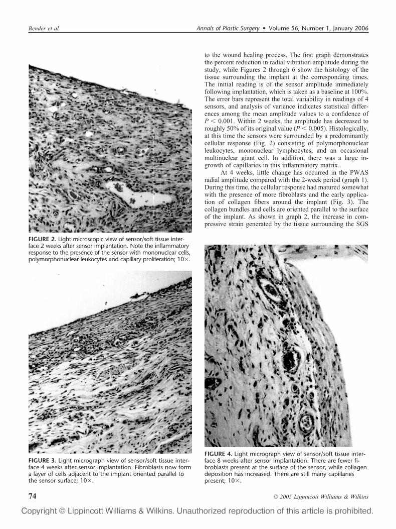
FIGURE 2. Light microscopic view of sensor/soft tissue inter-face 2 weeks after sensor implantation. Note the inflammatoryresponse to the presence of the sensor with mononuclear cells,polymorphonuclear leukocytes and capillary proliferation; 10�.
FIGURE 3. Light micrograph view of sensor/soft tissue inter-face 4 weeks after sensor implantation. Fibroblasts now forma layer of cells adjacent to the implant oriented parallel tothe sensor surface; 10�.
FIGURE 4. Light micrograph view of sensor/soft tissue inter-face 8 weeks after sensor implantation. There are fewer fi-broblasts present at the surface of the sensor, while collagendeposition has increased. There are still many capillariespresent; 10�.
Bender et al Annals of Plastic Surgery • Volume 56, Number 1, January 2006
© 2005 Lippincott Williams & Wilkins74

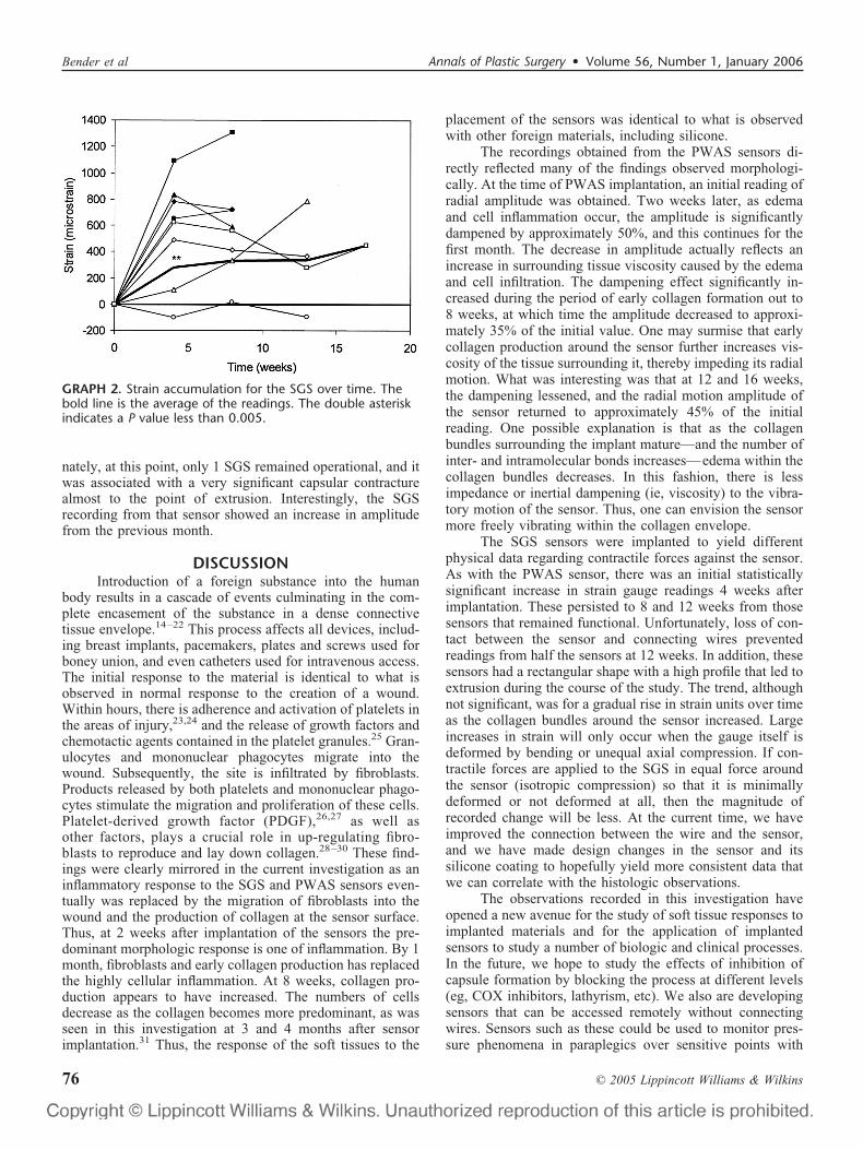
implant during the initial 4-week period was statisticallysignificant (P � 0.05), although the results are considerablymore scattered. Overall, analysis of variance of the SGS dataindicates a significant change in the mean value of the sensorsthat survived to 8 weeks (P � 0.005).
At 8 weeks, the PWAS radial amplitude decreasedsignificantly compared with that at 4 weeks (P � 0.005). Atthis same time, the remaining fibroblasts are gradually re-placed by a diffusely organized layer of collagen (Fig. 4). Incontrast, the SGS sensors showed no significant change overthe same time period. There are few inflammatory cells in thelayer of the capsule beneath the collagen envelope; however,a large number of capillaries persist.
At 12 weeks, there is an apparent increase in radialamplitude as measured by the PWAS sensors. Dense bundlesof collagen fibers with scattered fibroblasts surround theimplant (Fig. 5). Fewer capillaries are observed in the layerbeneath the collagen envelope. Half of the SGS sensors failedto generate data at this time point, and resistance changes inthose that survived are not statistically distinguishable. Thisfinding was attributable to continued challenges with bothmaintaining electrical continuity at the wire-sensor junctionand preventing extrusion of this more prominent sensorthrough the skin.
Finally, at 16 weeks, there is a further rise in amplitudeof the PWAS sensor, which is statistically distinguishable
from the readings at 8 weeks (P � 0.05). Histologically, thisis commensurate with the formation of an increasingly denseand structurally aligned collagen layer (Fig. 6). Unfortu-
GRAPH 1. PWAS vibration amplitude as a percentage of theinitial for the duration of the experiment. The double aster-isk indicates a P value less than 0.005, whereas a single as-terisk indicates a P value less than 0.05 compared with thevalues at 8 weeks. The error bars indicate the total variationin the date.
FIGURE 5. Light micrograph view of the sensor/soft tissueinterface 12 weeks after sensor implantation. The tissue adja-cent to the sensor is now predominantly collagen with veryfew mononuclear cells compared with the appearance of thecapsule earlier; 10�.
FIGURE 6. Light micrograph view of the sensor/soft tissueinterface 16 weeks after sensor implantation. At this time thecapsule has further matured and is almost all collagen withscattered cells and small capillaries; 10�.
Annals of Plastic Surgery • Volume 56, Number 1, January 2006 Implantable Biomedical Sensors
© 2005 Lippincott Williams & Wilkins 75
nately, at this point, only 1 SGS remained operational, and itwas associated with a very significant capsular contracturealmost to the point of extrusion. Interestingly, the SGSrecording from that sensor showed an increase in amplitudefrom the previous month.
DISCUSSIONIntroduction of a foreign substance into the human
body results in a cascade of events culminating in the com-plete encasement of the substance in a dense connectivetissue envelope.14–22 This process affects all devices, includ-ing breast implants, pacemakers, plates and screws used forboney union, and even catheters used for intravenous access.The initial response to the material is identical to what isobserved in normal response to the creation of a wound.Within hours, there is adherence and activation of platelets inthe areas of injury,23,24 and the release of growth factors andchemotactic agents contained in the platelet granules.25 Gran-ulocytes and mononuclear phagocytes migrate into thewound. Subsequently, the site is infiltrated by fibroblasts.Products released by both platelets and mononuclear phago-cytes stimulate the migration and proliferation of these cells.Platelet-derived growth factor (PDGF),26,27 as well asother factors, plays a crucial role in up-regulating fibro-blasts to reproduce and lay down collagen.28–30 These find-ings were clearly mirrored in the current investigation as aninflammatory response to the SGS and PWAS sensors even-tually was replaced by the migration of fibroblasts into thewound and the production of collagen at the sensor surface.Thus, at 2 weeks after implantation of the sensors the pre-dominant morphologic response is one of inflammation. By 1month, fibroblasts and early collagen production has replacedthe highly cellular inflammation. At 8 weeks, collagen pro-duction appears to have increased. The numbers of cellsdecrease as the collagen becomes more predominant, as wasseen in this investigation at 3 and 4 months after sensorimplantation.31 Thus, the response of the soft tissues to the
placement of the sensors was identical to what is observedwith other foreign materials, including silicone.
The recordings obtained from the PWAS sensors di-rectly reflected many of the findings observed morphologi-cally. At the time of PWAS implantation, an initial reading ofradial amplitude was obtained. Two weeks later, as edemaand cell inflammation occur, the amplitude is significantlydampened by approximately 50%, and this continues for thefirst month. The decrease in amplitude actually reflects anincrease in surrounding tissue viscosity caused by the edemaand cell infiltration. The dampening effect significantly in-creased during the period of early collagen formation out to8 weeks, at which time the amplitude decreased to approxi-mately 35% of the initial value. One may surmise that earlycollagen production around the sensor further increases vis-cosity of the tissue surrounding it, thereby impeding its radialmotion. What was interesting was that at 12 and 16 weeks,the dampening lessened, and the radial motion amplitude ofthe sensor returned to approximately 45% of the initialreading. One possible explanation is that as the collagenbundles surrounding the implant mature—and the number ofinter- and intramolecular bonds increases—edema within thecollagen bundles decreases. In this fashion, there is lessimpedance or inertial dampening (ie, viscosity) to the vibra-tory motion of the sensor. Thus, one can envision the sensormore freely vibrating within the collagen envelope.
The SGS sensors were implanted to yield differentphysical data regarding contractile forces against the sensor.As with the PWAS sensor, there was an initial statisticallysignificant increase in strain gauge readings 4 weeks afterimplantation. These persisted to 8 and 12 weeks from thosesensors that remained functional. Unfortunately, loss of con-tact between the sensor and connecting wires preventedreadings from half the sensors at 12 weeks. In addition, thesesensors had a rectangular shape with a high profile that led toextrusion during the course of the study. The trend, althoughnot significant, was for a gradual rise in strain units over timeas the collagen bundles around the sensor increased. Largeincreases in strain will only occur when the gauge itself isdeformed by bending or unequal axial compression. If con-tractile forces are applied to the SGS in equal force aroundthe sensor (isotropic compression) so that it is minimallydeformed or not deformed at all, then the magnitude ofrecorded change will be less. At the current time, we haveimproved the connection between the wire and the sensor,and we have made design changes in the sensor and itssilicone coating to hopefully yield more consistent data thatwe can correlate with the histologic observations.
The observations recorded in this investigation haveopened a new avenue for the study of soft tissue responses toimplanted materials and for the application of implantedsensors to study a number of biologic and clinical processes.In the future, we hope to study the effects of inhibition ofcapsule formation by blocking the process at different levels(eg, COX inhibitors, lathyrism, etc). We also are developingsensors that can be accessed remotely without connectingwires. Sensors such as these could be used to monitor pres-sure phenomena in paraplegics over sensitive points with
GRAPH 2. Strain accumulation for the SGS over time. Thebold line is the average of the readings. The double asteriskindicates a P value less than 0.005.
Bender et al Annals of Plastic Surgery • Volume 56, Number 1, January 2006
© 2005 Lippincott Williams & Wilkins76

warning signals emitted when capillary closing pressuresover bone have been exceeded for too long a period of time.Sensors could be incorporated into tissue implants to detectearly capsule contracture so that early intervention might befeasible.
REFERENCES1. Ellenbogen KA, Kay GN, Wilkoff BL, eds. Clinical Cardiac Pacing.
Philadelphia: WB Saunders; 1995.2. Norlin A, Pan J, Leygraf C. Investigation of electrochemical behavior of
stimulation/sensing materials for pacemaker electrode applications: I. Pt,Ti, and TiN coated electrodes. J Electrochem Soc. 2005;152:J7–J15.
3. Schoenfisch MH, Ovadia M, Pemberton JE. Covalent surface chemicalmodification of electrodes for cardiac pacing applications. J BiomedMater Res. 2000;51:209–215.
4. Pickup JC, Hussain F, Evans ND, et al. In vivo glucose monitoring: theclinical reality and the promise. Biosens Bioelectron. 2005;20:1897–1902.
5. Wickramasinghe Y, Yang Y, Spencer SA. Current problems and potentialtechniques in in vivo glucose monitoring. J Fluoresc. 2004;14:513–520.
6. Wilson GS, Hu Y. Enzyme-based biosensors for in vivo measurements.Chem Rev. 2000;100:2693–2704.
7. Park S, Krebs D, Mann RW. Hip muscle co-contraction: evidence fromconcurrent in vivo pressure measurement and force estimation. GaitPosture. 1999;10:211–222.
8. Gingrass M, Perry L, Hill D, et al. Nondisruptive, in vivo method forbiomechanical characterization of linear incision wound healing: pre-liminary report. Plast Reconstr Surg. 1998;102:801–806.
9. LaBerge M, Audet J, Drouin G, et al. Structural and in vivo mechanicalcharacterization of canine patellar cartilage: a closed chondromalaciapatellae model. J Invest Surg. 1993;6:105–116.
10. Wang J, Brienza DM, Yuan Y, et al. A compound sensor for biome-chanical analyses of buttock soft tissue in vivo. J Rehab Res Dev.2000;37:433–443.
11. Zalzal P, Papini M, Petruccelli D, et al. An in vivo biomechanicalanalysis of the soft-tissue envelope of osteoarthritic knees. J Arthro-plasty. 2004;19:217–223.
12. Han L, Noble J, Burcher M. A novel ultrasound indentation system formeasuring biomechanical properties of in vivo soft tissue. UltrasoundMed Biol. 2003;29:813–823.
13. Giurgiutiu V, Zagrai A. Characterization of piezoelectric wafer activesensors. J Intell Mater Syst Struct. 2000;11:959–976.
14. McLean AL, Malmor M, Harper A, et al. Expression of cyclooxygen-ase-2 in the periprosthetic capsule surrounding a silicone shell implant inthe rat. Ann Plast Surg. 2002;48:292–297.
15. Ksander GA, Vistnes LA. The incidence of experimental contracturevaries with the source of the prosthesis. Plast Reconstr Surg 1985;75:668–676.
16. Carpaneda CA. Inflammatory reaction and capsular contracture aroundsmooth silicone implants. Aesthetic Plast Surg 1997;21:110–114.
17. Granchi D, Cavedagna D, Ciapetti G, et al. Silicone breast implants: therole of the immune system on capsular contracture formation. J BiomedMater Res. 1995;29:197–202.
18. Rubino C, Mazzarello V, Farace F, et al. Ultrastructural anatomy ofcontracted capsules around textured implants in augmented breasts. AnnPlast Surg. 2001;46:95–102.
19. Barone FE, Perry L, Keller T, et al. The biochemical and histopathologiceffects of surface texturing with silicone and polyurethane in tissueimplantation and expansion. Plast Reconstr Surg. 1992;90:77–86.
20. Maxwell GP, Perry L. The capsule in various types of breast implants.Plast Reconstr Surg. 1995;95:937–938.
21. Raso DS, Greene WB. Silicone breast implants: pathology. UltrastructPathol. 1997;21:263–271.
22. Friedman HI, Friedman A, Carson K. The fate of the fibrous capsulefollowing saline implant removal. Ann Plast Surg. 2001;46:215–221.
23. Barnes MJ, Bailey AJ, Gordon JL, et al. Platelet aggregation by base-ment membrane-associated collagens. Thromb Res. 1980;18:375–388.
24. Legrand YJ, Fauvel F, Arbeille B, et al. Activation of platelets bymicrofibrils and collagen: a comparative study. Lab Invest. 1986;54:566–573.
25. Weksler BB. Platelets. Gallin OM, Goldstein IM, Snyderman R, eds.Inflammation: Basic Principles and Clinical Correlates. New York:Raven Press; 1988:543–557.
26. Kohler N, Lipton A. Platelets as a source of fibroblast growth-promotingactivity. Exp Cell Res. 1974;87:297–301.
27. Ross R, Raines EW, Bpwen-Pope DF. The biology of platelet-derivedgrowth factor. Cell. 1986;46:155–169.
28. Carpenter G. Receptors for epidermal growth factor and other polypep-tide mitogens. Ann Rev Biochem. 1987;56:881–914.
29. Wharton W, Leoff EB, Olashaw NE, et al. Mitogenic response toepidermal growth factor is modulated by platelet derived growth factor.Exp Cell Res. 1983;147:443–448.
30. Clemmons DR, Van Wyk JJ. Evidence for a functional role of endog-enously produced somatomedin-like peptides in the regulation of DNAsynthesis in cultured human fibroblasts and porcine smooth musclescells. J Clin Invest. 1985;75:1914–1918.
31. Pierce GF, Mustoe TA, Lingelbach J, et al. Transforming growth factorB reverses the glucocorticoid-induced wound-healing deficit in rats:possible regulation in macrophages by platelet-derived growth factor.Proc Natl Acad Sci U S A. 1989;86:2229–2233.
Annals of Plastic Surgery • Volume 56, Number 1, January 2006 Implantable Biomedical Sensors
© 2005 Lippincott Williams & Wilkins 77
Related Documents


