Dalton Transactions PAPER Cite this: Dalton Trans., 2016, 45, 5629 Received 4th December 2015, Accepted 15th February 2016 DOI: 10.1039/c5dt04747a www.rsc.org/dalton The unusual metal ion binding ability of histidyl tags and their mutated derivatives† Davide Brasili,‡ a Joanna Watly,‡ b Eyal Simonovsky, c,d Remo Guerrini, a Nuno A. Barbosa, b Robert Wieczorek, b Maurizio Remelli,* a Henryk Kozlowski* b and Yifat Miller* c,d Polyhistidine-tags are often used for the affinity purification of polyhistidine-tagged recombinant proteins. These sequences are also found in nature and are often highly conserved across different species. However, their exact role in the biological systems is not clear. The purpose of this work is to shed light on the behavior of poly-His sequences in their interactions with metal ions. This work illustrates the first study of novel poly-(His–Ala) peptides that bind Cu(II) applying both experimental techniques and exten- sive computational tools. The studied novel peptides are analogues of the short protected fragment of the pHpG (EDDH 9 GVG 10 ) peptide, which was found in the venom of Atheris squamigera. Our study pre- sents the properties of metal ion binding-histidine tag complexes and their mutated derivatives. The Cu(II) binding ability in pHG (Ac-EDDH 9 G-NH 2 ) is more efficient than in the mutated derivatives, although the number of imidazoles that bind to Cu(II) ions are similar. Finally, the formation of an α-helical structure is observed in pHG and in one of the mutated derivatives, indicating the importance of the sequence in the poly-(His-Ala) tags. Introduction Nature has developed histidine rich proteins (HRPs) in many living organisms. 1 They are found in bacterial chaperones, 2 snake venoms 3 and human genome. 4 Often, they play a crucial role in many life functions. 5 A special group of HRPs are sequences with His-tag motifs and in the last few decades around 700 of such natural peptides have been discovered. 1,4 The His-tag is an excellent ligand for metal ions, but its func- tional mechanisms are more complicated than that of a simple chelating agent and therefore have not been yet fully elucidated. Chelating properties of histidine rich proteins are the basis of one of the most effective methods of protein puri- fication. Immobilized Metal Ion Affinity Chromatography (IMAC) makes use of the strong complex-formation capabilities of natural or artificial sequences of histidines (usually His 6 - tag). 6,7 The imidazole nitrogens of histidines play a role as donor groups that have the ability to form coordination bonds with transition metals immobilized onto a chromatographic support such as Ni(II), Cu(II), Co(II), Zn(II). 8 Recently, the interest in HRP studies has been increased, especially for a large class of proteins with poly-His–poly-Gly (pHpG) sequences, which are found in the venom of snakes. 3,9 For example, the EDDHHHHHHHHHGVGGGGGGGGGG (pHpG) sequence appears in the venom of Atheris squamigera, an African viper. It has been proposed that such peptides may block the metalloproteinases contained in the poison, and therefore do not cause damage to the venom gland. 3,10 Recently, we have studied the short protected fragment of the pHpG peptide from the Atheris squamigera venom: Ac- EDDHHHHHHHHHG-NH 2 ( pHG) using experimental tech- niques and computational tools. 11 We have shown that the multiple histidine residues in this peptide fragment are very efficient metal ion chelators with very high affinities toward Cu(II), Ni(II) and Zn(II) ions in comparison with other histidine rich proteins. Furthermore, the interactions of metal ions with the pHG peptide have shown a very unusual behavior of this system. 11 First, various sets of three imidazoles from the His 9 - tag in pHG can bind metal ions in different ways along the sequence and consequently form polymorphic binding states. Second, the formation of a regular α-helical structure has been observed when a metal binds to the peptide molecule. Poly- morphism allows metal ions to ‘move along’ the polyhistidine † Electronic supplementary information (ESI) available: Spectroscopy measure- ment, DFT and MD data. See DOI: 10.1039/c5dt04747a ‡ These authors contributed equally to this work. a Department of Chemical and Pharmaceutical Sciences, University of Ferrara, via Fossato di Mortara 17, I-44121 Ferrara, Italy. E-mail: [email protected] b Department of Chemistry, University of Wroclaw, F. Joliot-Curie 14, 50-383 Wroclaw, Poland. E-mail: [email protected] c Department of Chemistry, Ben Gurion University of the Negev, Beer-Sheva 84105, Israel d Ilse Katz Institute for Nanoscale Science and Technology, Ben Gurion University of the Negev, Beer-Sheva 84105, Israel. E-mail: [email protected] This journal is © The Royal Society of Chemistry 2016 Dalton Trans. , 2016, 45, 5629–5639 | 5629 Open Access Article. Published on 17 February 2016. Downloaded on 12/10/2021 12:33:19 PM. This article is licensed under a Creative Commons Attribution 3.0 Unported Licence. View Article Online View Journal | View Issue

Welcome message from author
This document is posted to help you gain knowledge. Please leave a comment to let me know what you think about it! Share it to your friends and learn new things together.
Transcript
DaltonTransactions
PAPER
Cite this: Dalton Trans., 2016, 45,5629
Received 4th December 2015,Accepted 15th February 2016
DOI: 10.1039/c5dt04747a
www.rsc.org/dalton
The unusual metal ion binding ability of histidyltags and their mutated derivatives†
Davide Brasili,‡a Joanna Watly,‡b Eyal Simonovsky,c,d Remo Guerrini,a
Nuno A. Barbosa,b Robert Wieczorek,b Maurizio Remelli,*a Henryk Kozlowski*b andYifat Miller*c,d
Polyhistidine-tags are often used for the affinity purification of polyhistidine-tagged recombinant proteins.
These sequences are also found in nature and are often highly conserved across different species.
However, their exact role in the biological systems is not clear. The purpose of this work is to shed light
on the behavior of poly-His sequences in their interactions with metal ions. This work illustrates the first
study of novel poly-(His–Ala) peptides that bind Cu(II) applying both experimental techniques and exten-
sive computational tools. The studied novel peptides are analogues of the short protected fragment of
the pHpG (EDDH9GVG10) peptide, which was found in the venom of Atheris squamigera. Our study pre-
sents the properties of metal ion binding-histidine tag complexes and their mutated derivatives. The Cu(II)
binding ability in pHG (Ac-EDDH9G-NH2) is more efficient than in the mutated derivatives, although the
number of imidazoles that bind to Cu(II) ions are similar. Finally, the formation of an α-helical structure is
observed in pHG and in one of the mutated derivatives, indicating the importance of the sequence in the
poly-(His-Ala) tags.
Introduction
Nature has developed histidine rich proteins (HRPs) in manyliving organisms.1 They are found in bacterial chaperones,2
snake venoms3 and human genome.4 Often, they play a crucialrole in many life functions.5 A special group of HRPs aresequences with His-tag motifs and in the last few decadesaround 700 of such natural peptides have been discovered.1,4
The His-tag is an excellent ligand for metal ions, but its func-tional mechanisms are more complicated than that of asimple chelating agent and therefore have not been yet fullyelucidated. Chelating properties of histidine rich proteins arethe basis of one of the most effective methods of protein puri-fication. Immobilized Metal Ion Affinity Chromatography(IMAC) makes use of the strong complex-formation capabilitiesof natural or artificial sequences of histidines (usually His6-
tag).6,7 The imidazole nitrogens of histidines play a role asdonor groups that have the ability to form coordination bondswith transition metals immobilized onto a chromatographicsupport such as Ni(II), Cu(II), Co(II), Zn(II).8
Recently, the interest in HRP studies has been increased,especially for a large class of proteins with poly-His–poly-Gly(pHpG) sequences, which are found in the venom of snakes.3,9
For example, the EDDHHHHHHHHHGVGGGGGGGGGG(pHpG) sequence appears in the venom of Atheris squamigera,an African viper. It has been proposed that such peptides mayblock the metalloproteinases contained in the poison, andtherefore do not cause damage to the venom gland.3,10
Recently, we have studied the short protected fragment ofthe pHpG peptide from the Atheris squamigera venom: Ac-EDDHHHHHHHHHG-NH2 (pHG) using experimental tech-niques and computational tools.11 We have shown that themultiple histidine residues in this peptide fragment are veryefficient metal ion chelators with very high affinities towardCu(II), Ni(II) and Zn(II) ions in comparison with other histidinerich proteins. Furthermore, the interactions of metal ions withthe pHG peptide have shown a very unusual behavior of thissystem.11 First, various sets of three imidazoles from the His9-tag in pHG can bind metal ions in different ways along thesequence and consequently form polymorphic binding states.Second, the formation of a regular α-helical structure has beenobserved when a metal binds to the peptide molecule. Poly-morphism allows metal ions to ‘move along’ the polyhistidine
†Electronic supplementary information (ESI) available: Spectroscopy measure-ment, DFT and MD data. See DOI: 10.1039/c5dt04747a‡These authors contributed equally to this work.
aDepartment of Chemical and Pharmaceutical Sciences, University of Ferrara, via
Fossato di Mortara 17, I-44121 Ferrara, Italy. E-mail: [email protected] of Chemistry, University of Wroclaw, F. Joliot-Curie 14, 50-383
Wroclaw, Poland. E-mail: [email protected] of Chemistry, Ben Gurion University of the Negev, Beer-Sheva 84105,
IsraeldIlse Katz Institute for Nanoscale Science and Technology, Ben Gurion University of
the Negev, Beer-Sheva 84105, Israel. E-mail: [email protected]
This journal is © The Royal Society of Chemistry 2016 Dalton Trans., 2016, 45, 5629–5639 | 5629
Ope
n A
cces
s A
rtic
le. P
ublis
hed
on 1
7 Fe
brua
ry 2
016.
Dow
nloa
ded
on 1
2/10
/202
1 12
:33:
19 P
M.
Thi
s ar
ticle
is li
cens
ed u
nder
a C
reat
ive
Com
mon
s A
ttrib
utio
n 3.
0 U
npor
ted
Lic
ence
.
View Article OnlineView Journal | View Issue
fragments, which serve as a metal ion transporting pathway.Our previous studies on copper complexes with the His6-taghave shown similar results: the presence of polymorphicbinding states (with the participation of maximum two Hisresidues in metal ion binding) and the formation of helicalstructures for some binding modes.12,13
Since our recent studies on the specificity of the metal ioninteractions with His-tags containing 6 or 9 His residues haveshown very unusual behavior and incredible efficiency ofbinding metal ions,11,12,14 this work extends our studies tonew analogues in which the His residues mutated to Ala resi-dues. Therefore the aim of this study is to examine the pro-perties and the structural characterization of these new poly-(His–Ala) analogues of the pHG peptide.
To provide an insight into the mechanisms through whichthe Cu(II) binds non-adjacent His or alternate His residues inpoly-His peptides, this study is focused on two new poly-Hispeptides that are derived from mutations of Ala of the pHGpeptide. The first peptide consists of four His residues alter-nating with five Ala residues: Ac-EDDAHAHAHAHAG-NH2 (L1)and the second peptide consists of five His residues alternat-ing with four Ala residues: Ac-EDDHAHAHAHAHG-NH2 (L2).These two analogues contain a lower number of histidineswith respect to the original peptide pHG; but, more impor-tantly, histidines are not consecutive. In fact, it has beenalready reported in the literature that not only the number ofhistidines in the sequence, but also their relative positioninfluences the stability and the structure of the complexes.15–18
This work presents a combination of experimental tech-niques and computational tools used to investigate the inter-actions of Cu(II) with each one of these two peptides. This isthe first study that shows the coordination abilities of mutatedsequences of pHG peptide derivatives towards the Cu(II) ionand the comparison of their properties to the originalsequence with consecutive histidines.
Results and discussionPeptide L1 consists of seven groups of acidic/basic sites andpeptide L2 consists of eight groups of acidic/basic sites
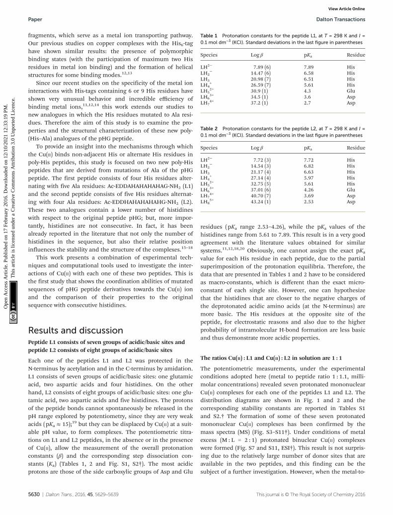
Each one of the peptides L1 and L2 was protected in theN-terminus by acetylation and in the C-terminus by amidation.L1 consists of seven groups of acidic/basic sites: one glutamicacid, two aspartic acids and four histidines. On the otherhand, L2 consists of eight groups of acidic/basic sites: one glu-tamic acid, two aspartic acids and five histidines. The protonsof the peptide bonds cannot spontaneously be released in thepH range explored by potentiometry, since they are very weakacids (pKa ≈ 15);19 but they can be displaced by Cu(II) at a suit-able pH value, to form complexes. The potentiometric titra-tions on L1 and L2 peptides, in the absence or in the presenceof Cu(II), allow the measurement of the overall protonationconstants (β) and the corresponding step dissociation con-stants (Ka) (Tables 1, 2 and Fig. S1, S2†). The most acidicprotons are those of the side carboxylic groups of Asp and Glu
residues (pKa range 2.53–4.26), while the pKa values of thehistidines range from 5.61 to 7.89. This result is in a very goodagreement with the literature values obtained for similarsystems.11,12,18,20 Obviously, one cannot assign the exact pKa
value for each His residue in each peptide, due to the partialsuperimposition of the protonation equilibria. Therefore, thedata that are presented in Tables 1 and 2 have to be consideredas macro-constants, which is different than the exact micro-constant of each single site. However, one can hypothesizethat the histidines that are closer to the negative charges ofthe deprotonated acidic amino acids (at the N-terminus) aremore basic. The His residues at the opposite site of thepeptide, for electrostatic reasons and also due to the higherprobability of intramolecular H-bond formation are less basicand thus demonstrate more acidic properties.
The ratios Cu(II) : L1 and Cu(II) : L2 in solution are 1 : 1
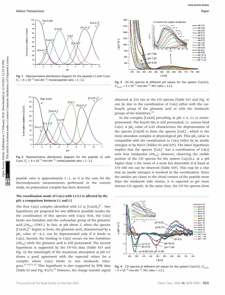
The potentiometric measurements, under the experimentalconditions adopted here (metal to peptide ratio 1 : 1.1, milli-molar concentrations) revealed seven protonated mononuclearCu(II) complexes for each one of the peptides L1 and L2. Thedistribution diagrams are shown in Fig. 1 and 2 and thecorresponding stability constants are reported in Tables S1and S2.† The formation of some of these seven protonatedmononuclear Cu(II) complexes has been confirmed by themass spectra (MS) (Fig. S3–S11†). Under conditions of metalexcess (M : L = 2 : 1) protonated binuclear Cu(II) complexeswere formed (Fig. S7 and S11, ESI†). This result is not surpris-ing due to the relatively large number of donor sites that areavailable in the two peptides, and this finding can be thesubject of a further investigation. However, when the metal-to-
Table 1 Protonation constants for the peptide L1, at T = 298 K and I =0.1 mol dm−3 (KCl). Standard deviations in the last figure in parentheses
Species Log β pKa Residue
LH2− 7.89 (6) 7.89 HisLH2
− 14.47 (6) 6.58 HisLH3 20.98 (7) 6.51 HisLH4
+ 26.59 (7) 5.61 HisLH5
2+ 30.9 (1) 4.3 GluLH6
3+ 34.5 (1) 3.6 AspLH7
4+ 37.2 (1) 2.7 Asp
Table 2 Protonation constants for the peptide L2, at T = 298 K and I =0.1 mol dm−3 (KCl). Standard deviations in the last figure in parentheses
Species Log β pKa Residue
LH2− 7.72 (3) 7.72 HisLH2
− 14.54 (3) 6.82 HisLH3 21.17 (4) 6.63 HisLH4
+ 27.14 (4) 5.97 HisLH5
2+ 32.75 (5) 5.61 HisLH6
3+ 37.01 (6) 4.26 GluLH7
4+ 40.70 (7) 3.69 AspLH8
5+ 43.24 (1) 2.53 Asp
Paper Dalton Transactions
5630 | Dalton Trans., 2016, 45, 5629–5639 This journal is © The Royal Society of Chemistry 2016
Ope
n A
cces
s A
rtic
le. P
ublis
hed
on 1
7 Fe
brua
ry 2
016.
Dow
nloa
ded
on 1
2/10
/202
1 12
:33:
19 P
M.
Thi
s ar
ticle
is li
cens
ed u
nder
a C
reat
ive
Com
mon
s A
ttrib
utio
n 3.
0 U
npor
ted
Lic
ence
.View Article Online
peptide ratio is approximately 1 : 1, as it is the case for thethermodynamic measurements performed in the currentstudy, no polynuclear complex has been detected.
The coordination mode of Cu(II) with L1/L2 is affected by thepH: a comparison between L1 and L2
The first Cu(II) complex identified with L1 is [CuLH3]2+. Two
hypotheses are proposed for two different possible modes forthe coordination of this species with Cu(II): first, the Cu(II)binds one histidine and the carboxylate group of the glutamicacid (1Nim; COO–). In fact, at pH about 3, when the species[CuLH3]
2+ begins to form, the glutamic acid, characterized by apKa value of ∼4.3, can be deprotonated only if it binds toCu(II). Second, the binding to Cu(II) occurs via two histidines(2Nim) while the glutamic acid is still protonated. The secondhypothesis is supported by the UV-Vis data (Table S3† andFig. 3): the wavelength of the maximum absorption at pH 4.5shows a good agreement with the expected values for acomplex where Cu(II) binds to two imidazole nitro-gens.11,19,21,22 This hypothesis is also supported by EPR data(Table S3 and Fig. S12†).23 However, the charge transfer signal
observed at 254 nm in the CD spectra (Table S3† and Fig. 4)can be due to the coordination of Cu(II) either with the car-boxylic group of the glutamic acid or with the imidazolegroups of the histidines.24
In the complex [CuLH] prevailing at pH ≈ 6, L1 is mono-protonated. The fourth His is still protonated, i.e. cannot bindCu(II). A pKa value of 6.82 characterizes the deprotonation ofthe species [CuLH] to form the species [CuL]−, which is themost abundant complex at physiological pH. This pKa value iscompatible with the coordination to Cu(II) either by an amidenitrogen or by His11 (Tables S1 and S2†). The latter hypothesisimplies that the species [CuL]− has a coordination of Cu(II)with four imidazoles (4Nim). However, observing the visibleportion of the CD spectra for the system Cu(II)/L1, at a pHhigher than 5 the onset of a weak but detectable d–d band at570–580 nm can be observed (Table S4†). This may be a cluethat an amide nitrogen is involved in the coordination. Sincethe amides are closer to the chiral centers of the peptide morethan the imidazole side chains, it is expected to get moreintense CD signals. At the same time, the UV-Vis spectra show
Fig. 1 Representative distribution diagram for the peptide L1 with Cu(II);C°
L = 6 × 10⁻⁴ mol dm−3; metal/peptide ratio = 1 : 1.1.
Fig. 2 Representative distribution diagram for the peptide L2 withCu(II); C°
L = 6 × 10⁻⁴ mol dm−3; metal/peptide ratio = 1 : 1.1.
Fig. 3 UV-Vis spectra at different pH values for the system Cu(II)/L1;C°
CuðIIÞ = 5 × 10⁻⁴ mol dm−3; M/L ratio = 1.1.1.
Fig. 4 CD spectra at different pH values for the system Cu(II)/L1; C°CuðIIÞ
= 5 × 10⁻⁴ mol dm−3; M/L ratio = 1.1.1.
Dalton Transactions Paper
This journal is © The Royal Society of Chemistry 2016 Dalton Trans., 2016, 45, 5629–5639 | 5631
Ope
n A
cces
s A
rtic
le. P
ublis
hed
on 1
7 Fe
brua
ry 2
016.
Dow
nloa
ded
on 1
2/10
/202
1 12
:33:
19 P
M.
Thi
s ar
ticle
is li
cens
ed u
nder
a C
reat
ive
Com
mon
s A
ttrib
utio
n 3.
0 U
npor
ted
Lic
ence
.View Article Online
a further blue-shift of the absorption band to 603 nm(pH 7.2–7.9) and the EPR AII value increases from 173 to186 G. Therefore, the coordination mode proposed for thespecies [CuL]− of L1 is (3Nim, N−), where N− refers to thedeprotonated nitrogen of a peptide bond. It is therefore pro-posed that the fourth His residue should remain protonated.The deprotonation of [CuL]−, leading to the formation of thesubsequent [CuLH−1]
2− species, is characterized by a pKa valueof 7.87 which is very close to the value that is measured forHis11 in the free peptide (7.89, Table 1). This value suggeststhat no interaction occurs between this imidazole and Cu(II)and that the complex geometry remains with three imidazolesand a nitrogen (3Nim, N−). Finally, at higher pH values thespecies [CuLH−2]
3− and [CuLH−3]4− are formed. Spectroscopic
data suggest that at these higher pH values the deprotonatedamides gradually substitute imidazole nitrogens, forming a(2Nim, 2N
−) and (Nim, 3N−) complex, respectively.
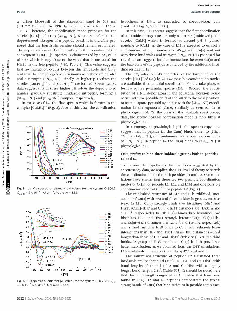
In the case of L2, the first species which is formed is thecomplex [CuLH4]
3+ (Fig. 2). Also in this case, the coordination
hypothesis is 2Nim, as suggested by spectroscopic data(Table S4,† Fig. 5, 6 and S13†).
In this case, CD spectra suggest that the first coordinationof an amide nitrogen occurs only at pH 8.5 (Table S4†). Thespecies [CuLH] which is formed at around pH 5 (corres-ponding to [CuL]− in the case of L1) is expected to exhibit acoordination of four imidazoles (4Nim) with Cu(II) and notwith three imidazoles and nitrogen (3Nim, N
−), as proposed forL1. This can suggest that the interactions between Cu(II) andthe backbone of the peptide is shielded by the additional histi-dine residue in L2.
The pKa value of 6.43 characterizes the formation of thespecies [CuL]− of L2 (Fig. 2). Two possible coordination modesare available: first, an axial coordination should take place, toform a square pyramidal species (5Nim). Second, the substi-tution of a Nim donor atom in the equatorial position wouldoccur, with the possible shift of the latter to the axial position,to form a square pyramid again but with the (3Nim, N
−) coordi-nation in the equatorial plane, similarly as seen for L1 atphysiological pH. On the basis of the available spectroscopydata, the second possible coordination mode is more likely atphysiological pH.
In summary, at physiological pH, the spectroscopy datasuggest that in peptide L1 the Cu(II) binds either to (2Nim,2N−) or (3Nim, N
−), in a preference to the coordination modeof (3Nim, N
−). In peptide L2 the Cu(II) binds to (3Nim, N−) at
physiological pH.
Cu(II) prefers to bind three imidazole groups both in peptidesL1 and L2
To examine the hypotheses that had been suggested by thespectroscopy data, we applied the DFT level of theory to searchthe coordination mode for both peptides L1 and L2. Our calcu-lations have shown that there are two possible coordinationmodes of Cu(II) for peptide L1 (L1a and L1b) and one possiblecoordination mode of Cu(II) for peptide L2 (Fig. 7).
The minimized structures of L1a and L1b exhibited inter-actions of Cu(II) with two and three imidazole groups, respect-ively. In L1a, Cu(II) strongly binds two histidines: His7 andHis11 (Cu(II)–His7 and Cu(II)–His11 distances are: 1.832 Å and1.835 Å, respectively). In L1b, Cu(II) binds three histidines: twohistidines His7 and His11 strongly interact Cu(II) (Cu(II)–His7and Cu(II)–His11 distances are: 1.849 Å and 1.841 Å, respectively)and a third histidine His5 binds to Cu(II) with relatively lowerinteractions than His7 and His11 (Cu(II)–His5 distance is ∼0.5 Ålonger than those of His7 and His11) (Table S5†). Yet, the thirdimidazole group of His5 that binds Cu(II) in L1b provides abetter stabilization, as we obtained from the DFT calculations:L1b is relatively more stable than L1a by 47.2 kcal mol−1.
The minimized structure of peptide L2 illustrated threeimidazole groups that bind Cu(II): Cu–His4 and Cu–His10 withbond lengths of around 1.9 Å and Cu–His6 with a slightlylonger bond length: 2.1 Å (Table S6†). It should be noted herethat the bond length ranges of all Cu(II)–His that have beenfound in L1a, L1b and L2 peptides demonstrate the typicalstrong bonds of Cu(II) that bind residues in peptide complexes,
Fig. 5 UV-Vis spectra at different pH values for the system Cu(II)/L2;C°
CuðIIÞ = 5 × 10⁻⁴ mol dm−3; M/L ratio = 1.1.1.
Fig. 6 CD spectra at different pH values for the system Cu(II)/L2; C°CuðIIÞ
= 5 × 10⁻⁴ mol dm−3; M/L ratio = 1.1.1.
Paper Dalton Transactions
5632 | Dalton Trans., 2016, 45, 5629–5639 This journal is © The Royal Society of Chemistry 2016
Ope
n A
cces
s A
rtic
le. P
ublis
hed
on 1
7 Fe
brua
ry 2
016.
Dow
nloa
ded
on 1
2/10
/202
1 12
:33:
19 P
M.
Thi
s ar
ticle
is li
cens
ed u
nder
a C
reat
ive
Com
mon
s A
ttrib
utio
n 3.
0 U
npor
ted
Lic
ence
.View Article Online
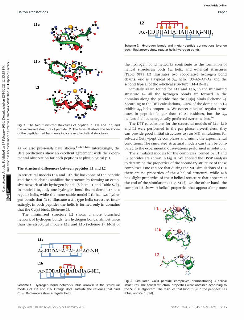
as we also previously have shown.11,12,14,25 Interestingly, theDFT predictions show an excellent agreement with the experi-mental observation for both peptides at physiological pH.
The structural differences between peptides L1 and L2
In structural models L1a and L1b the backbone of the peptideand the side chains stabilize the structure by forming an exten-sive network of six hydrogen bonds (Scheme 1 and Table S7†).In model L1a, only one hydrogen bond fits to demonstrate aregular helix, while the more stable model L1b has two hydro-gen bonds that fit to illustrate a 310 type helix structure. Inter-estingly, in both peptides the helix is formed only in domainsthat the Cu(II) binds (Scheme 1).
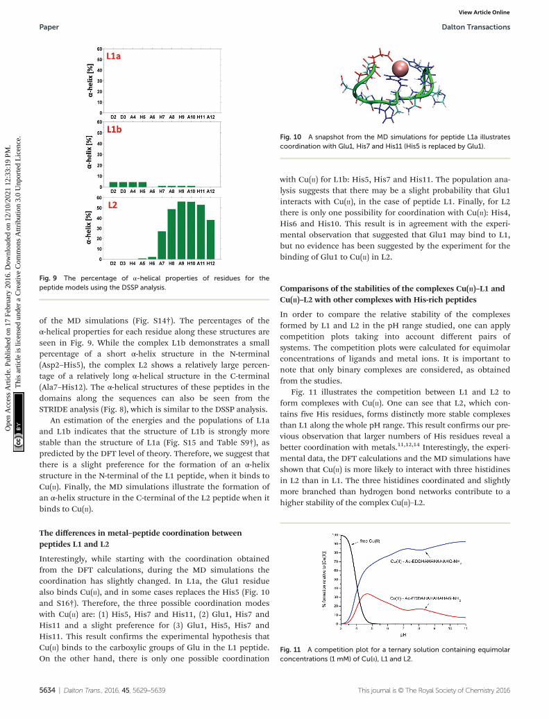
The minimized structure L2 shows a more branchednetwork of hydrogen bonds: ten hydrogen bonds, almost twicethan the structural models L1a and L1b (Scheme 2). Most of
the hydrogen bond networks contribute to the formation ofhelical structures: both 310 helix and α-helical structures(Table S8†). L2 illustrates two cooperative hydrogen bondchains: one is a typical of 310 helix: D3–A5–A7–A9 and thesecond typical of the α-helical structure: H4–H6–H8.
Similarly as we found for L1a and L1b, in the minimizedstructure L2 all the hydrogen bonds are formed in thedomains along the peptide that the Cu(II) binds (Scheme 2).According to the DFT calculations, ∼50% of the domains in L2exhibit 310 helix properties. We expect α-helical regular struc-tures in peptides longer than 19–21 residues, but the 310helices shall be energetically preferred over α-helixes.26
The DFT calculations for the structural models of L1a, L1band L2 were performed in the gas phase; nevertheless, theycan provide good initial structures to run MD simulations forsolvated Cu(II)–peptide complexes and mimic the experimentalconditions. The simulated structural models can then be com-pared to the experimental observations performed in solution.
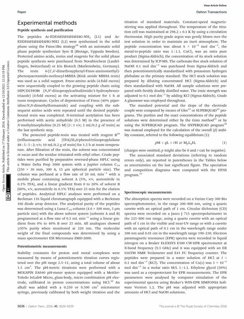
The simulated models for the complexes formed by L1 andL2 peptides are shown in Fig. 8. We applied the DSSP analysisto determine the properties of the secondary structure of thesecomplexes. One can see that during the MD simulations of L1athere are no properties of the α-helical structure, while L1bhas slight properties of the α-helical structure that appears atthe end of the simulations (Fig. S14†). On the other hand, thecomplex L2 shows α-helical properties that appear along most
Fig. 7 The two minimized structures of peptide L1: L1a and L1b, andthe minimized structure of peptide L2. The tubes illustrate the backboneof the peptides; red fragments indicate regular helical structures.
Scheme 1 Hydrogen bond networks (blue arrows) in the structuralmodels of L1a and L1b. Orange dots illustrate the residues that bindCu(II). Red arrows show a regular helix.
Scheme 2 Hydrogen bonds and metal–peptide connections (orangedots). Red arrows show regular helix hydrogen bonds.
Fig. 8 Simulated Cu(II)–peptide complexes demonstrating α-helicalstructures. The helical structural properties were obtained according tothe STRIDE algorithm. The residues that bind Cu(II) in the peptides: His(blue) and Glu1 (red).
Dalton Transactions Paper
This journal is © The Royal Society of Chemistry 2016 Dalton Trans., 2016, 45, 5629–5639 | 5633
Ope
n A
cces
s A
rtic
le. P
ublis
hed
on 1
7 Fe
brua
ry 2
016.
Dow
nloa
ded
on 1
2/10
/202
1 12
:33:
19 P
M.
Thi
s ar
ticle
is li
cens
ed u
nder
a C
reat
ive
Com
mon
s A
ttrib
utio
n 3.
0 U
npor
ted
Lic
ence
.View Article Online
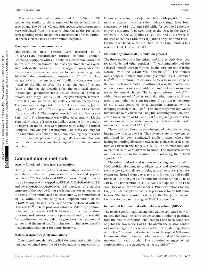
of the MD simulations (Fig. S14†). The percentages of theα-helical properties for each residue along these structures areseen in Fig. 9. While the complex L1b demonstrates a smallpercentage of a short α-helix structure in the N-terminal(Asp2–His5), the complex L2 shows a relatively large percen-tage of a relatively long α-helical structure in the C-terminal(Ala7–His12). The α-helical structures of these peptides in thedomains along the sequences can also be seen from theSTRIDE analysis (Fig. 8), which is similar to the DSSP analysis.
An estimation of the energies and the populations of L1aand L1b indicates that the structure of L1b is strongly morestable than the structure of L1a (Fig. S15 and Table S9†), aspredicted by the DFT level of theory. Therefore, we suggest thatthere is a slight preference for the formation of an α-helixstructure in the N-terminal of the L1 peptide, when it binds toCu(II). Finally, the MD simulations illustrate the formation ofan α-helix structure in the C-terminal of the L2 peptide when itbinds to Cu(II).
The differences in metal–peptide coordination betweenpeptides L1 and L2
Interestingly, while starting with the coordination obtainedfrom the DFT calculations, during the MD simulations thecoordination has slightly changed. In L1a, the Glu1 residuealso binds Cu(II), and in some cases replaces the His5 (Fig. 10and S16†). Therefore, the three possible coordination modeswith Cu(II) are: (1) His5, His7 and His11, (2) Glu1, His7 andHis11 and a slight preference for (3) Glu1, His5, His7 andHis11. This result confirms the experimental hypothesis thatCu(II) binds to the carboxylic groups of Glu in the L1 peptide.On the other hand, there is only one possible coordination
with Cu(II) for L1b: His5, His7 and His11. The population ana-lysis suggests that there may be a slight probability that Glu1interacts with Cu(II), in the case of peptide L1. Finally, for L2there is only one possibility for coordination with Cu(II): His4,His6 and His10. This result is in agreement with the experi-mental observation that suggested that Glu1 may bind to L1,but no evidence has been suggested by the experiment for thebinding of Glu1 to Cu(II) in L2.
Comparisons of the stabilities of the complexes Cu(II)–L1 andCu(II)–L2 with other complexes with His-rich peptides
In order to compare the relative stability of the complexesformed by L1 and L2 in the pH range studied, one can applycompetition plots taking into account different pairs ofsystems. The competition plots were calculated for equimolarconcentrations of ligands and metal ions. It is important tonote that only binary complexes are considered, as obtainedfrom the studies.
Fig. 11 illustrates the competition between L1 and L2 toform complexes with Cu(II). One can see that L2, which con-tains five His residues, forms distinctly more stable complexesthan L1 along the whole pH range. This result confirms our pre-vious observation that larger numbers of His residues reveal abetter coordination with metals.11,12,14 Interestingly, the experi-mental data, the DFT calculations and the MD simulations haveshown that Cu(II) is more likely to interact with three histidinesin L2 than in L1. The three histidines coordinated and slightlymore branched than hydrogen bond networks contribute to ahigher stability of the complex Cu(II)–L2.
Fig. 10 A snapshot from the MD simulations for peptide L1a illustratescoordination with Glu1, His7 and His11 (His5 is replaced by Glu1).
Fig. 11 A competition plot for a ternary solution containing equimolarconcentrations (1 mM) of Cu(II), L1 and L2.
Fig. 9 The percentage of α-helical properties of residues for thepeptide models using the DSSP analysis.
Paper Dalton Transactions
5634 | Dalton Trans., 2016, 45, 5629–5639 This journal is © The Royal Society of Chemistry 2016
Ope
n A
cces
s A
rtic
le. P
ublis
hed
on 1
7 Fe
brua
ry 2
016.
Dow
nloa
ded
on 1
2/10
/202
1 12
:33:
19 P
M.
Thi
s ar
ticle
is li
cens
ed u
nder
a C
reat
ive
Com
mon
s A
ttrib
utio
n 3.
0 U
npor
ted
Lic
ence
.View Article Online

In order to better shed light on the role of the number ofHis residues in the ability of Cu(II) to bind the peptide, wefurther investigated the competition between each one of thestudied peptides L1 and L2 with other similar peptides withpoly-His sequences. Four poly-His peptides were studied: (i)the peptide Ac-THHHHAHGG-NH2, which is a protected frag-ment of the Hpn protein that is secreted by Helicobacterpylori18 and consists of five His residues, four of which areconsecutive (Fig. S18†); (ii) a fragment of the prion protein ofzebrafish (zp-PrP63-87),27 which consists of seven His residues,six of which are separated by two amino acids (Fig. S19†); (iii)a pHG peptide,11 which consists of nine consecutive His resi-dues (His9-tag) (Fig. 12) and (iv) a His6-tag,
12 commonly usedin IMAC chromatography (Fig. S20†).
One can see from Fig. 12 and S18–S20† that the peptideswith a larger number of histidines in the sequence have abetter ability to bind the Cu(II) ion. For example, the compe-tition plots that compare the pHG peptide with peptides L1and L2 show that pHG binds Cu(II) ions more effectively thanL1 and L2 (Fig. 12). It is important to note that in pHG with L1and L2 peptides Cu(II) binds a maximum of three imidazolegroups. Interestingly, in all peptides metal ions induce the for-mation of α-helix. The surprising difference in the efficiency ofmetal ion binding may be due to the occurrence of poly-morphic states in the case of Cu(II)–pHG (twelve of variousstructures) and a much better defined secondary structure ofthe α-helix, stabilized by the richer network of hydrogen bondsbetween the imidazole rings in most of these complexes.11
Similar effects are observed when comparing L1 and L2complexes with the Cu(II)–His6-tag system (Fig. S20†). Thepeptide with six histidines is the most effective, but more
interestingly, in these complexes we have differences in coordi-nation modes before involvement of the amide nitrogen in themetal ion binding: {2Nim} (His6-tag) and {3Nim} (L1 and L2).The MD simulations and DFT calculations have shown anensemble of polymorphic states for the Cu(II)–His6-tagcomplex and a preference for the formation of the α-helixstructure.12
Finally, for peptides with a similar number of His residues,the alternate sequence of His and Ala as in peptide L2 providesa more stable complex than the non-alternating sequence Hisand Ala, as seen in peptide Ac-THHHHAHGG-NH2
(Fig. S18b†). It is likely that in the peptide Ac-THHHHAHGG-NH2 the His residues are too close to eachother to contemporarily bind Cu(II), while in L2 the His resi-dues that bind Cu(II) are not close to each other, i.e. notcontinuous.
Conclusions
This work illustrates two novel Ala mutated analogue peptidesof the short protected pHpG peptide from Atheris squamigeravenom: Ac-EDDHHHHHHHHHG-NH2 (pHG) using extensiveexperimental techniques and computational tools. Our studyleads to several conclusions. First, peptides that consist ofrepeated sequences of alternating histidines and alanines havethe ability to strongly bind Cu(II). Second, the number of histi-dines and their location along the sequence affect the stabilityof the peptide complexes. Peptides with a relatively largenumber of His residues show a relatively better stability, i.e.they bind the Cu(II) stronger than peptides with a smallernumber of His residues. Third, the binding ability of the Histag of pHG that consists of nine consecutive His residues isstrongly more effective than the two novel Ala mutated ana-logue peptides that have been investigated here, although thenumber of imidazoles that coordinate to Cu(II) is similar.Fourth, the metal ion binding to the two Ala mutated analoguepeptides induces the formation of the α-helical structure. Yet,the formation of the α-helical structure in the original pHGpeptide is more remarkable compared with these two peptides.This can be explained due to the relatively small number ofhydrogen-bonding networks in the two peptides comparedwith the pHG peptide. These differences could be critical forcomplex stability.
On the basis of the obtained results we suggest that thereare strong dependencies between the number and positions ofhistidines along the histidine-rich sequences and their abilityto coordinate metal ions and the stability of the complexes.Furthermore, in longer sequences of poly-His peptides thequantity of possible polymorphic states and the possibility toinduce the formation of helical structures are increased.Finally, we propose that the extremely efficient metal ionbinding properties that induce the formation of a regularhelical structure in the unstructured His-tag peptides may beimportant for the biological behavior of peptides and proteinscontaining such sequences.
Fig. 12 Competition plot for a ternary solution containing equimolarconcentrations (1 mM) of: (a) Cu(II), L1 and Ac-EDDH9G-NH2 (pHG); (b)Cu(II), L2 and Ac-EDDH9G-NH2 (pHG).
Dalton Transactions Paper
This journal is © The Royal Society of Chemistry 2016 Dalton Trans., 2016, 45, 5629–5639 | 5635
Ope
n A
cces
s A
rtic
le. P
ublis
hed
on 1
7 Fe
brua
ry 2
016.
Dow
nloa
ded
on 1
2/10
/202
1 12
:33:
19 P
M.
Thi
s ar
ticle
is li
cens
ed u
nder
a C
reat
ive
Com
mon
s A
ttrib
utio
n 3.
0 U
npor
ted
Lic
ence
.View Article Online
Experimental methodsPeptide synthesis and purification
The peptides Ac-EDDAHAHAHAHAG-NH2 (L1) and Ac-EDDHAHAHAHAHG-NH2 (L2) were synthesized in the solidphase using the Fmoc/tBu strategy28 with an automatic solidphase peptide synthesizer Syro II (Biotage, Uppsala Sweden).Protected amino acids, resins and reagents for the solid phasepeptide synthesis were purchased from Novabiochem (Laufel-fingen, Switzerland) or Iris Biotech (Marktredwitz, Germany).The resin 4-(2′,4′-dimethoxyphenyl-Fmoc-aminomethyl)-phenoxyacetamido-norleucyl-MBHA (Rink amide MBHA resin)was used as a solid support. Fmoc-amino acids (4-fold excess)were sequentially coupled to the growing peptide chain usingDIPCDI/HOBt (N,N′-diisopropylcarbodiimide/1-hydroxybenzo-triazole) (4-fold excess) as the activating mixture for 1 h atroom temperature. Cycles of deprotection of Fmoc (40% piper-idine/N,N-dimethylformamide) and coupling with the sub-sequent amino acids were repeated until the desired peptide-bound resin was completed. N-terminal acetylation has beenperformed with acetic anhydride (0.5 M) in the presence ofN-methylmorpholine (0.25 M) (3 : 1 v/v; 2 mL/0.2 g of resin) asthe last synthetic step.
The protected peptide-resin was treated with reagent B29
(trifluoroacetic acid (TFA)/H20/phenol/triisopropylsilane88 : 5 : 5 : 2; v/v; 10 mL/0.2 g of resin) for 1.5 h at room tempera-ture. After filtration of the resin, the solvent was concentratedin vacuo and the residue triturated with ethyl ether. Crude pep-tides were purified by preparative reversed-phase HPLC usinga Water Delta Prep 3000 system with a Jupiter column C18
(250 × 30 mm, 300 A, 15 μm spherical particle size). Thecolumn was perfused at a flow rate of 20 mL min−1 with amobile phase containing solvent A (5%, v/v, acetonitrile in0.1% TFA), and a linear gradient from 0 to 20% of solvent B(60%, v/v, acetonitrile in 0.1% TFA) over 25 min for the elutionof peptides. Analytical HPLC analyses were performed on aBeckman 116 liquid chromatograph equipped with a Beckman166 diode array detector. The analytical purity of the peptideswas determined using a Luna C18 column (4.6 × 100 mm, 3 μmparticle size) with the above solvent system (solvents A and B)programmed at a flow rate of 0.5 mL min−1 using a linear gra-dient from 0% to 40% B over 25 min. All analogues showed≥95% purity when monitored at 220 nm. The molecularweight of the final compounds was determined by using amass spectrometer ESI Micromass ZMD-2000.
Potentiometric measurements
Stability constants for proton and metal complexes weremeasured by means of potentiometric titration curves regis-tered over the pH range 2.5–11, using a total volume of about1.5 cm3. The pH-metric titrations were performed with aMOLSPIN EA940 pH-meter system equipped with a Mettler-Toledo InLab® Micro, glass-body, micro combination pH elec-trode, calibrated in proton concentrations using HCl.30 Analkali was added with a 0.250 or 0.500 cm3 micrometersyringe, previously calibrated by both weight titration and the
titration of standard materials. Constant-speed magnetic-stirring was applied throughout. The temperature of the titra-tion cell was maintained at 298.2 ± 0.1 K by using a circulationthermostat. High purity grade argon was gently blown over thetest solution in order to maintain an inert atmosphere. Thepeptide concentration was about 6 × 10⁻⁴ mol dm−3, themetal-to-peptide ratio was 1 : 1.1. CuCl2 was an extra pureproduct (Sigma-Aldrich); the concentration of its stock solutionwas determined by ICP-MS. The carbonate-free stock solution ofNaOH 0.1 mol dm−3 was purchased from Sigma-Aldrich andthen potentiometrically standardized with potassium hydrogenphthalate as the primary standard. The HCl stock solution wasprepared by diluting concentrated HCl (Sigma-Aldrich) andthen standardized with NaOH. All sample solutions were pre-pared with freshly doubly distilled water. The ionic strength wasadjusted to 0.1 mol dm−3 by adding KCl (Sigma-Aldrich). GradeA glassware was employed throughout.
The standard potential and the slope of the electrodecouple were computed by means of Glee31 or SUPERQUAD32 pro-grams. The purities and the exact concentrations of the peptidesolutions were determined either by the Gran method33 or byusing the SUPERQUAD program. The HYPERQUAD program34
was instead employed for the calculation of the overall (β) stabi-lity constant, referred to the following equilibrium (1):
pMþ qLþ rH ⇄ MpLqHr ð1Þ(charges were omitted; p might also be 0 and r can be negative).
The associated standard deviations (referring to randomerrors only), are reported in parentheses in the Tables belowas uncertainties on the last significant figure. The speciationand competition diagrams were computed with the HYSSprogram.35
Spectroscopic measurements
The absorption spectra were recorded on a Varian Cary 300 Biospectrophotometer, in the range 200–800 nm, using a quartzcuvette with an optical path of 1 cm. Circular dichroism (CD)spectra were recorded on a Jasco J 715 spectropolarimeter inthe 225–800 nm range, using a quartz cuvette with an opticalpath of 1 cm in the visible and near-UV range or with a cuvettewith an optical path of 0.1 cm in the wavelength range under300 nm and 0.01 cm in the wavelength range 190–230. Electronparamagnetic resonance (EPR) spectra were recorded in liquidnitrogen on a Bruker ELEXSYS E500 CW-EPR spectrometer atX-band frequency (9.5 GHz) and it was equipped with an ER036TM NMR Teslameter and E41 FC frequency counter. Thepeptides were prepared in a water solution of HCl at I =0.1 mol dm−3 (KCl). The concentration of Cu(II) was 1 × 10⁻³
mol dm−3 in a molar ratio M/L 1 : 1.1. Ethylene glycol (30%)was used as a cryoprotectant for EPR measurements. The EPRparameters were analysed by computer simulation of theexperimental spectra using Bruker’s WIN-EPR SIMFONIA Soft-ware Version 1.2. The pH was adjusted with appropriateamounts of HCl and NaOH solutions.
Paper Dalton Transactions
5636 | Dalton Trans., 2016, 45, 5629–5639 This journal is © The Royal Society of Chemistry 2016
Ope
n A
cces
s A
rtic
le. P
ublis
hed
on 1
7 Fe
brua
ry 2
016.
Dow
nloa
ded
on 1
2/10
/202
1 12
:33:
19 P
M.
Thi
s ar
ticle
is li
cens
ed u
nder
a C
reat
ive
Com
mon
s A
ttrib
utio
n 3.
0 U
npor
ted
Lic
ence
.View Article Online
The concentration of solutions used for UV-Vis and CDstudies was similar to those employed in the potentiometricexperiment. The UV-Vis, CD and EPR spectroscopic parameterswere calculated from the spectra obtained at the pH valuescorresponding to the maximum concentration of each particu-lar species, on the basis of distribution diagrams.
Mass spectrometric measurements
High-resolution mass spectra were recorded on aBrukerQ-FTMS spectrometer (Bruker Daltonik, Bremen,Germany), equipped with an Apollo II electrospray ionizationsource with an ion funnel. The mass spectrometer was oper-ated both in the positive and in the negative ion modes. Theinstrumental parameters were as follows: scan range m/z400–1600, dry gas-nitrogen, temperature 170 °C, capillaryvoltage 4500 V, ion energy 5 eV. Capillary voltage was opti-mized to the highest S/N. The small changes of voltage(±500 V) did not significantly affect the optimized spectra.Instrumental parameters for a Bruker MicrOTOF-Q were asfollows: scan range m/z 250–2000, dry gas-nitrogen, tempera-ture 200 °C, ion source voltage 4500 V, collision energy 10 eV.The samples (metal/peptide in a 1 : 1.1 stoichiometry, [pepti-de]tot = 5 × 10⁻⁴ mol dm−3) were prepared in a 1 : 1 MeOH–H2Omixture at pH 4.5. The samples were infused at a flow rate of3 μL min−1. The instrument was calibrated externally with theTunemix™ mixture (Bruker Daltonik, Germany) in the quadra-tic regression mode. Data were processed by using the BrukerCompass Data Analysis 4.0 program. The mass accuracy forthe calibration was better than 5 ppm, enabling together withthe true isotopic pattern (using SigmaFit) an unambiguousconfirmation of the elemental composition of the obtainedcomplex.
Computational methodsDensity functional theory (DFT) calculations
Density functional theory has been successfully used to investi-gate the structure and properties of peptides and peptidecomplexes.36–41 We performed DFT studies on Cu(II) cations inthe 1 : 1 complex with capped Ac-EDDAHAHAHAHAG-NH2 (L1)and Ac-EDDHAHAHAHAHG-NH2 (L2) peptides. The startingstructure of the peptide for DFT calculations was generated onthe basis of the amino acid sequence after 75 ps simulation at300 K, without cutoffs using BIO+ implementation of theCHARMM force field. All calculations were performed with theGaussian 09 42 suite of programs using the M06 43 hybrid func-tional and the triple-zeta 6-311G basis set. In the peptide struc-tures imidazole nitrogens are not protonated and thus availablefor coordination, while amide nitrogens bear their proton andcannot bind the metal ion. This situation is similar to that of ametal/peptide solution at pH approximately 5.
Molecular dynamics (MD) simulations
Constructed models. We applied the structural models thathad been obtained from the DFT calculations to run MD simu-
lations: concerning the Cu(II) complexes with peptide L1, twomain structures involving side imidazole rings have beensuggested by DFT (L1a and L1b) while for peptide L2 there isonly one structure (L2). According to the DFT, in the case ofstructure L1a, the Cu(II) binds His5, His7 and His11 while inthe case of complex L1b, the Cu(II) binds only His7 and His11.On the other hand, in the structure L2, the Cu(II) binds 3 Hisresidues: His4, His6 and His10.
Molecular dynamics (MD) simulation protocol
The three models were first minimized as previously describedfor amyloids and other peptides.44–50 MD simulations of thesolvated models were performed in the NPT ensemble usingNAMD51 with the CHARMM27 force-field.52,53 The modelswere energy minimized and explicitly solvated in a TIP3P waterbox54,55 with a minimum distance of 15 Å from each edge ofthe box. Each water molecule within 2.5 Å of the models wasremoved. Counter ions were added at random locations to neu-tralize the model charge. The Langevin piston method56,57
with a decay period of 100 fs and a damping time of 50 fs wasused to maintain a constant pressure of 1 atm. A temperatureof 310 K was controlled by a Langevin thermostat with adamping coefficient of 10 ps.51 The short-range van der Waalsinteractions were calculated using the switching function, witha twin range cut-off of 10.0 and 12.0 Å. Long-range electrostaticinteractions were calculated using the particle mesh Ewaldmethod with a cutoff of 12.0 Å.58,59
The equations of motion were integrated using the leapfrogintegrator with a step of 1 fs. The solvated systems were energyminimized for 2000 conjugated gradient steps, where thehydrogen bonding distance between the β-sheet in each oligo-mer was fixed in the range 2.2–2.5 Å. The counter ions andwater molecules were allowed to move. The hydrogen atomswere constrained to the equilibrium bond using the SHAKEalgorithm.60
The minimized solvated systems were energy minimized for5000 additional conjugate gradient steps and 20 000 heatingsteps at 250 K, with all atoms being allowed to move. Then, thesystem was heated from 250 K to 310 K for 300 ps and equili-brated at 310 K for 300 ps. All simulations were run for 30 ns at310 K. The temperature of 310 K had been applied to test thestabilities of all the variant models. Parameterizations for theCu(II)–peptide complexes had been performed for all MD simu-lations. The force constant values for the Cu(II)–N atom andCu(II)–O atom are in the range of 25–50 kcal mol−1 Å−2.
Generalized born method with molecular volume (GBMV)
The relative conformational energies can be compared only formodels that have the same sequence and number of peptides,thus the relative conformational energies had been computedonly for the two models of L1. To obtain the relative confor-mational energies of these two models, the model trajectoriesof the last 5 ns were first extracted from the explicit MD simu-lations excluding the water molecules – a total of 500 confor-mations for each model. The solvation energies of allconformations were calculated using the GBMV.61,62
Dalton Transactions Paper
This journal is © The Royal Society of Chemistry 2016 Dalton Trans., 2016, 45, 5629–5639 | 5637
Ope
n A
cces
s A
rtic
le. P
ublis
hed
on 1
7 Fe
brua
ry 2
016.
Dow
nloa
ded
on 1
2/10
/202
1 12
:33:
19 P
M.
Thi
s ar
ticle
is li
cens
ed u
nder
a C
reat
ive
Com
mon
s A
ttrib
utio
n 3.
0 U
npor
ted
Lic
ence
.View Article Online
In the GBMV calculations, the dielectric constant of waterwas set to 80. The hydrophobic solvent-accessible surface area(SASA) term factor was set to 0.00592 kcal (mol Å)−1. Each con-formation was minimized using 1000 cycles, and the confor-mational energy was evaluated by grid-based GBMV.
A total of 1000 conformations (500 for each model) wereused to construct the energy landscapes of the two models andto evaluate the conformer probabilities by using Monte Carlo(MC) simulations. In the first step, one conformation of con-former i and one conformation of conformer j were randomlyselected. Then, the Boltzmann factor was computed ase−(Ej−Ei)/kT, where Ei and Ej are the conformational energiesevaluated using the GBMV calculations for conformationsi and j, respectively, k is the Boltzmann constant and T is theabsolute temperature (298 K used here). If the value of theBoltzmann factor was larger than the random number, thenthe move from conformation i to conformation j was allowed.After 1 million steps, the conformations ‘visited’ for each con-former were counted. Finally, the relative probability of modeln was evaluated as Pn = Nn/Ntotal, where Pn is the population ofmodel n, Nn is the total number of conformations visited formodel n, and Ntotal is the total steps. The advantages of usingMC simulations to estimate conformer probability lie in theirgood numerical stability and the control that they allow fortransition probabilities among several conformers.
A total of 1000 conformations of these two models (500 con-formations for each model) were used to construct the energylandscape (Table S9†). The pair of these two models is likely topresent may be only a very small percentage of the ensemble.Nevertheless, the carefully selected models cover the mostlikely structures.
Assigning a secondary structure to amino acids by the DSSPalgorithm
The DSSP algorithm is the standard method for assigning thesecondary structure to the amino acids of a protein or apeptide, given the atomic-resolution coordinates of the proteinor the peptide. It does this by reading the position of theatoms in a protein (the ATOM records in a PDB file) followedby calculation of the hydrogen bond energy between all atoms.The best two hydrogen bonds for each atom are then used todetermine the most likely class of the secondary structure foreach residue in the protein or the peptide. We applied theDSSP algorithm that is embedded in the CHARMM software.63
The DSSP algorithm provides information on specificdomains that illustrate an α-helix and a β-sheet. It does notdistinguish between various types of α-helixes, such as the 310helix, α-helix, etc.
Acknowledgements
This research was supported by grant no. 2011128 from theUnited States-Israel Binational Science Foundation (BSF) andby MNiSW project 4478/E-344/S/2014. All simulations were per-formed using the high-performance computational facilities of
the Miller lab in the BGU HPC computational center. Thesupport of the BGU HPC computational center staff is greatlyappreciated. Financial support from the University of Ferrara(FAR 2013) is gratefully acknowledged.
References
1 M. Rowinska-Zyrek, D. Witkowska, S. Potocki, M. Remelliand H. Kozlowski, New J. Chem., 2013, 37, 58–70.
2 D. Witkowska, M. Rowinska-Zyrek, G. Valensin andH. Kozlowski, Coord. Chem. Rev., 2012, 256, 133–148.
3 S. C. Wagstaff, P. Favreau, O. Cheneval, G. D. Laing,M. C. Wilkinson, R. L. Miller, R. Stoecklin and R. A. Harrison,Biochem. Biophys. Res. Commun., 2008, 365, 650–656.
4 E. Salichs, A. Ledda, L. Mularoni, M. M. Alba and S. de laLuna, PLoS Genet., 2009, 5.
5 T. Cheng, W. Xia, P. Wang, F. Huang, J. Wang and H. Sun,Metallomics, 2013, 5, 1423–1429.
6 K. Terpe, Appl. Microbiol. Biotechnol., 2003, 60, 523–533;K. Terpe, Appl. Microbiol. Biotechnol., 2003, 60, 523–533.
7 D. S. Waugh, Trends Biotechnol., 2005, 23, 316–320.8 H. L. Liu, Y. Ho and C. M. Hsu, J. Biomol. Struct. Dyn.,
2003, 21, 31–41.9 P. Favreau, O. Cheneval, L. Menin, S. Michalet, H. Gaertner,
F. Principaud, R. Thai, A. Menez, P. Bulet and R. Stocklin,Rapid Commun. Mass Spectrom., 2007, 21, 406–412.
10 S. C. Wagstaff, L. Sanz, P. Juarez, R. A. Harrison andJ. J. Calvete, J. Proteomics, 2009, 71, 609–623.
11 J. Watly, E. Simonovsky, N. Barbosa, M. Spodzieja,R. Wieczorek, S. Rodziewicz-Motowidlo, Y. Miller andH. Kozlowski, Inorg. Chem., 2015, 54, 7692–7702.
12 J. Watly, E. Simonovsky, R. Wieczorek, N. Barbosa, Y. Millerand H. Kozlowski, Inorg. Chem., 2014, 53, 6675–6683.
13 E. Simonovsky, H. Kozlowski and Y. Miller, RSC Adv., 2015,5, 104551–104555.
14 F. Pontecchiani, E. Simonovsky, R. Wieczorek, N. Barbosa,M. Rowinska-Zyrek, S. Potocki, M. Remelli, Y. Miller andH. Kozlowski, Dalton Trans., 2014, 43, 16680–16689.
15 D. Valensin, L. Szyrwiel, F. Camponeschi, M. Rowinska-Zyrek, E. Molteni, E. Jankowska, A. Szymanska, E. Gaggelli,G. Valensin and H. Kozlowski, Inorg. Chem., 2009, 48,7330–7340.
16 A. Janicka-Klos, P. Juszczyk, Z. Grzonka and H. Kozlowski,Polyhedron, 2008, 27, 1511–1516.
17 M. Orfei, M. C. Alcaro, G. Marcon, M. Chelli,M. Ginanneschi, H. Kozlowski, J. Brasun and L. Messori,J. Inorg. Biochem., 2003, 97, 299–307.
18 D. Witkowska, R. Politano, M. Rowinska-Zyrek, R. Guerrini,M. Remelli and H. Kozlowski, Chem – Eur. J., 2012, 18,11088–11099.
19 H. Sigel and R. B. Martin, Chem. Rev., 1982, 82, 385–426.20 L. D. Pettit and H. K. J. Powell, The IUPAC Stability Con-
stants Database, Royal Society of Chemistry, London,1992–2000.
21 E. J. Billo, Inorg. Nucl. Chem. Lett., 1974, 10, 613–617.
Paper Dalton Transactions
5638 | Dalton Trans., 2016, 45, 5629–5639 This journal is © The Royal Society of Chemistry 2016
Ope
n A
cces
s A
rtic
le. P
ublis
hed
on 1
7 Fe
brua
ry 2
016.
Dow
nloa
ded
on 1
2/10
/202
1 12
:33:
19 P
M.
Thi
s ar
ticle
is li
cens
ed u
nder
a C
reat
ive
Com
mon
s A
ttrib
utio
n 3.
0 U
npor
ted
Lic
ence
.View Article Online
22 E. Prenesti, P. G. Daniele, M. Prencipe and G. Ostacoli,Polyhedron, 1999, 18, 3233–3241.
23 J. Peisach and W. E. Blumberg, Arch. Biochem. Biophys.,1974, 165, 691–708.
24 P. G. Daniele, E. Prenesti and G. Ostacoli, J. Chem. Soc.,Dalton Trans., 1996, 3269–3275.
25 N. M. Chiera, M. Rowinska-Zyrek, R. Wieczorek,R. Guerrini, D. Witkowska, M. Remelli and H. Kozlowski,Metallomics, 2013, 5, 214–221.
26 R. Wieczorek and J. J. Dannenberg, J. Am. Chem. Soc., 2004,126, 14198–14205.
27 L. Szyrwiel, E. Jankowska, A. Janicka-Klos, Z. Szewczuk,D. Valensin and H. Kozlowski, Dalton Trans., 2008, 6117–6120.
28 N. L. Benoiton, Chemistry of Peptide Synthesis, Taylor &Francis, London, 2005, pp. 125–154.
29 N. A. Solé and G. Barany, J. Org. Chem., 1992, 57, 5399–5403.
30 H. M. Irving, M. G. Miles and L. D. Pettit, Anal. Chim. Acta,1967, 38, 475–488.
31 P. Gans and B. O’Sullivan, Talanta, 2000, 51, 33–37.32 P. Gans, A. Sabatini and A. Vacca, J. Chem. Soc., Dalton
Trans., 1985, 1195–1200.33 G. Gran, Acta Chem. Scand., 1950, 4, 559–577.34 P. Gans, A. Sabatini and A. Vacca, Talanta, 1996, 43, 1739–
1753.35 L. Alderighi, P. Gans, A. Ienco, D. Peters, A. Sabatini and
A. Vacca, Coord. Chem. Rev., 1999, 184, 311–318.36 A. Masson, M. Z. Kamrath, M. A. S. Perez, M. S. Glover,
U. Rothlisberger, D. E. Clemmer and T. R. Rizzo, J. Am. Soc.Mass Spectrom., 2015, 26, 1444–1454.
37 R. Walesa, D. Man, G. Engel, D. Siodlak, T. Kupka, T. Ptakand M. A. Broda, Chem. Biodiversity, 2015, 12, 1007–1024.
38 E. Gumienna-Kontecka, G. Berthon, I. O. Fritsky,R. Wieczorek, Z. Latajka and H. Kozlowski, J. Chem. Soc.,Dalton Trans., 2000, 4201–4208.
39 P. Salvador, R. Wieczorek and J. J. Dannenberg, J. Phys.Chem. B, 2007, 111, 2398–2403.
40 M. Rudowska, R. Wieczorek, A. Kluczyk, P. Stefanowicz andZ. Szewczuk, J. Am. Soc. Mass Spectrom., 2013, 24, 846–856.
41 G. Pohl, A. Asensio and J. J. Dannenberg, Biochemistry,2014, 53, 617–623.
42 J. Frisch, G. W. Trucks, H. B. Schlegel, G. E. Scuseria,M. A. Robb, J. R. Cheeseman, G. Scalmani, V. Barone,B. Mennucci, G. A. Petersson, H. Nakatsuji, M. Caricato,X. Li, H. P. Hratchian, A. F. Izmaylov, J. Bloino, G. Zheng,J. L. Sonnenberg, M. Hada, M. Ehara, K. Toyota, R. Fukuda,J. Hasegawa, M. Ishida, T. Nakajima, Y. Honda, O. Kitao,H. Nakai, T. Vreven, J. A. Montgomery Jr., J. E. Peralta,F. Ogliaro, M. Bearpark, J. J. Heyd, E. Brothers, K. N. Kudin,V. N. Staroverov, R. Kobayashi, J. Normand, K. Raghavachari,A. Rendell, J. C. Burant, S. S. Iyengar, J. Tomasi, M. Cossi,N. Rega, J. M. Millam, M. Klene, J. E. Knox, J. B. Cross,V. Bakken, C. Adamo, J. Jaramillo, R. Gomperts,R. E. Stratmann, O. Yazyev, A. J. Austin, R. Cammi,C. Pomelli, J. W. Ochterski, R. L. Martin, K. Morokuma,V. G. Zakrzewski, G. A. Voth, P. Salvador, J. J. Dannenberg,
S. Dapprich, A. D. Daniels, Ö. Farkas, J. B. Foresman,J. V. Ortiz, J. Cioslowski and D. J. Fox, Gaussian 09, RevisionC.01, Gaussian, Inc., Wallingford CT, 2009.
43 Y. Zhao and D. G. Truhlar, Theor. Chem. Acc., 2008, 120,215–241.
44 V. Wineman-Fisher, R. Simkovitch, S. Shomer,R. Gepshtein, D. Huppert, M. Saif, K. Kallio,S. J. Remington and Y. Miller, Phys. Chem. Chem. Phys.,2014, 16, 11196–11208.
45 N. Zeytuni, R. Uebe, M. Maes, G. Davidov, M. Baram,O. Raschdorf, M. Nadav-Tsubery, S. Kolusheva, R. Bitton,G. Goobes, A. Friedler, Y. Miller, D. Schueler andR. Zarivach, PLoS One, 2014, 9.
46 Y. Raz, B. Rubinov, M. Matmor, H. Rapaport, G. Ashkenasyand Y. Miller, Chem. Commun., 2013, 49, 6561–6563.
47 Y. Raz and Y. Miller, PLoS One, 2013, 8.48 Y. Raz, J. Adler, A. Vogel, H. A. Scheidt, T. Haupl, B. Abel,
D. Huster and Y. Miller, Phys. Chem. Chem. Phys., 2014, 16,7710–7717.
49 V. Wineman-Fisher, Y. Atsmon-Raz and Y. Miller, Biomacro-molecules, 2014, 16, 156–165.
50 Y. Miller, B. Y. Ma and R. Nussinov, Coord. Chem. Rev.,2012, 256, 2245–2252.
51 L. Kale, R. Skeel, M. Bhandarkar, R. Brunner, A. Gursoy,N. Krawetz, J. Phillips, A. Shinozaki, K. Varadarajan andK. Schulten, J. Comput. Phys., 1999, 151, 283–312.
52 A. D. MacKerell, D. Bashford, M. Bellott, R. L. Dunbrack,J. D. Evanseck, M. J. Field, S. Fischer, J. Gao, H. Guo, S. Ha,D. Joseph-McCarthy, L. Kuchnir, K. Kuczera, F. T. K. Lau,C. Mattos, S. Michnick, T. Ngo, D. T. Nguyen, B. Prodhom,W. E. Reiher, B. Roux, M. Schlenkrich, J. C. Smith, R. Stote,J. Straub, M. Watanabe, J. Wiorkiewicz-Kuczera, D. Yin andM. Karplus, J. Phys. Chem. B, 1998, 102, 3586–3616.
53 B. R. Brooks, R. E. Bruccoleri, B. D. Olafson, D. J. States,S. Swaminathan and M. Karplus, J. Comput. Chem., 1983, 4,187–217.
54 M. W. Mahoney and W. L. Jorgensen, J. Chem. Phys., 2000,112, 8910–8922.
55 W. L. Jorgensen, J. Chandrasekhar, J. D. Madura, R. W. Impeyand M. L. Klein, J. Chem. Phys., 1983, 79, 926–935.
56 K. Tu, D. J. Tobias and M. L. Klein, Biophys. J., 1995, 69,2558–2562.
57 S. E. Feller, Y. H. Zhang, R. W. Pastor and B. R. Brooks,J. Chem. Phys., 1995, 103, 4613–4621.
58 U. Essmann, L. Perera, M. L. Berkowitz, T. Darden, H. Leeand L. G. Pedersen, J. Chem. Phys., 1995, 103, 8577–8593.
59 T. Darden, D. York and L. Pedersen, J. Chem. Phys., 1993,98, 10089–10092.
60 J. P. Ryckaert, G. Ciccotti and H. J. C. Berendsen, J. Comput.Phys., 1977, 23, 327–341.
61 M. S. Lee, F. R. Salsbury and C. L. Brooks, J. Chem. Phys.,2002, 116, 10606–10614.
62 M. S. Lee, M. Feig, F. R. Salsbury and C. L. Brooks,J. Comput. Chem., 2003, 24, 1348–1356.
63 W. Kabsch and C. Sander, Biopolymers, 1983, 22, 2577–2637.
Dalton Transactions Paper
This journal is © The Royal Society of Chemistry 2016 Dalton Trans., 2016, 45, 5629–5639 | 5639
Ope
n A
cces
s A
rtic
le. P
ublis
hed
on 1
7 Fe
brua
ry 2
016.
Dow
nloa
ded
on 1
2/10
/202
1 12
:33:
19 P
M.
Thi
s ar
ticle
is li
cens
ed u
nder
a C
reat
ive
Com
mon
s A
ttrib
utio
n 3.
0 U
npor
ted
Lic
ence
.View Article Online
Related Documents