1. Introduction In an incisive article, J Goldstein, the 1985 Nobel laureate for the regulation of cholesterol metabolism (together with M Brown) and Chair for the Jury for the Lasker awards, laments the fact that it is hard to pick out truly original dis- coveries among the plethora of scientific publications (Goldstein 2004). He suggests that there are two ways to analyse a scientific discovery which, for ease of understand- ing, can be categorized into two groups: “top to bottom” or “bottom to top.” In the first category are papers that are pub- lished in highly ranked journals and often arrive with lots of media coverage but are soon forgotten, i.e. they start at the top but end up at the bottom. On the other hand, it is harder to spot a “bottom to top” discovery as it starts off at the bot- tom and its arrival is unheralded. However, with the passage of time, the significance of this discovery is increasingly appreciated, resulting in its emergence at the top with true impact! The discovery of protein ubiquitination is a fine example of a “bottom to top” discovery and its role in ‘big’ biological processes, e.g. transcription, cell cycle, antigen processing, cellular defense, signalling etc. is now well established (Ciechanover and Iwai 2004; Varshavsky 2005). During the early days in the field of cytosolic protein degradation, cell biologists were intrigued by the requirement of ATP in this process as it is well known that peptide bond hydrolysis does not require metabolic energy. The strategy used by A Hershko and colleagues involved biochemical fractionation of reticulocytes (terminally differentiated red blood cells lacking lysosomes), purification, identification of the components involved and reconstitution of the ATP- dependent cytosolic protein degradation in vitro. These stud- ies were supported by genetic studies and data from other groups on the in vivo roles of cytosolic protein degradation in different biological processes. The steps that led to the dis- covery and appreciation of the ubiquitin-proteasome system (UPS) are highlighted in table 1. For their pioneering studies on the role of ubiquitination of proteins during cytosolic pro- tein degradation, Avram Hershko, Aaron Ciechanover and Irwin Rose were awarded the 2004 Nobel Prize in Chemistry. Review The ubiquitin-proteasome system DIPANKAR NANDI*, P ANKAJ T AHILIANI, ANUJITH KUMAR and DILIP CHANDU Department of Biochemistry, Indian Institute of Science, Bangalore 560 012, India *Corresponding author (Fax, 91-80-23600814; Email, [email protected]) The 2004 Nobel Prize in chemistry for the discovery of protein ubiquitination has led to the recognition of cellular proteolysis as a central area of research in biology. Eukaryotic proteins targeted for degradation by this pathway are first ‘tagged’ by multimers of a protein known as ubiquitin and are later proteolyzed by a giant enzyme known as the proteasome. This article recounts the key observations that led to the discovery of ubiquitin-proteasome system (UPS). In addition, different aspects of proteasome biology are highlighted. Finally, some key roles of the UPS in different areas of biology and the use of inhibitors of this pathway as possible drug targets are discussed. [Nandi D, Tahiliani P, Kumar A and Chandu D 2006 The ubiquitin-proteasome system; J. Biosci. 31 137–155] http://www.ias.ac.in/jbiosci Abbreviations used: AAA-ATPase, ATPases associated with various cellular activities-ATPase; BrAAP, branched chain amino acids pre- ferring; CSN, COP9 signalosome; ER, endoplasmic reticulum; HECT, homologous to E6-AP carboxyl terminus; IFN, interferon; MHC, major histocompatibility complex; NTN, N-terminal nucleophile; PA, proteasome activator; PAC, proteasome assembling chaperone; PAN, proteasome-activating nucleotidase; PI, proteasome inhibitor; POMP, proteasome maturing protein; RING, really interesting new gene; Rpn, regulatory particle non-ATPase; Rpt, regulatory particle tripleA-ATPase; SCF, Skp-Cullin-F box; SNAAP, small neutral amino acids pre- ferring; UBH, ubiquitin hydrolase; UBP, ubiqutin processing; UMP1, ubiquitin mediated proteolysis-1; UPS, ubiquitin-proteasome system Keywords. Cellular proteolysis; proteasomes; protein degradation; ubiquitination. J. Biosci. 31(1), March 2006, 137–155, Indian Academy of Sciences 137

Welcome message from author
This document is posted to help you gain knowledge. Please leave a comment to let me know what you think about it! Share it to your friends and learn new things together.
Transcript

1. Introduction
In an incisive article, J Goldstein, the 1985 Nobel laureatefor the regulation of cholesterol metabolism (together withM Brown) and Chair for the Jury for the Lasker awards,laments the fact that it is hard to pick out truly original dis-coveries among the plethora of scientific publications(Goldstein 2004). He suggests that there are two ways toanalyse a scientific discovery which, for ease of understand-ing, can be categorized into two groups: “top to bottom” or“bottom to top.” In the first category are papers that are pub-lished in highly ranked journals and often arrive with lots ofmedia coverage but are soon forgotten, i.e. they start at thetop but end up at the bottom. On the other hand, it is harderto spot a “bottom to top” discovery as it starts off at the bot-tom and its arrival is unheralded. However, with the passageof time, the significance of this discovery is increasinglyappreciated, resulting in its emergence at the top with trueimpact! The discovery of protein ubiquitination is a fineexample of a “bottom to top” discovery and its role in ‘big’
biological processes, e.g. transcription, cell cycle, antigenprocessing, cellular defense, signalling etc. is now wellestablished (Ciechanover and Iwai 2004; Varshavsky 2005).
During the early days in the field of cytosolic proteindegradation, cell biologists were intrigued by the requirementof ATP in this process as it is well known that peptide bondhydrolysis does not require metabolic energy. The strategyused by A Hershko and colleagues involved biochemicalfractionation of reticulocytes (terminally differentiated redblood cells lacking lysosomes), purification, identification ofthe components involved and reconstitution of the ATP-dependent cytosolic protein degradation in vitro. These stud-ies were supported by genetic studies and data from othergroups on the in vivo roles of cytosolic protein degradation indifferent biological processes. The steps that led to the dis-covery and appreciation of the ubiquitin-proteasome system(UPS) are highlighted in table 1. For their pioneering studieson the role of ubiquitination of proteins during cytosolic pro-tein degradation, Avram Hershko, Aaron Ciechanover andIrwin Rose were awarded the 2004 Nobel Prize in Chemistry.
Review
The ubiquitin-proteasome system
DIPANKAR NANDI*, PANKAJ TAHILIANI, ANUJITH KUMAR and DILIP CHANDUDepartment of Biochemistry, Indian Institute of Science, Bangalore 560 012, India
*Corresponding author (Fax, 91-80-23600814; Email, [email protected])
The 2004 Nobel Prize in chemistry for the discovery of protein ubiquitination has led to the recognition of cellularproteolysis as a central area of research in biology. Eukaryotic proteins targeted for degradation by this pathway arefirst ‘tagged’ by multimers of a protein known as ubiquitin and are later proteolyzed by a giant enzyme known as theproteasome. This article recounts the key observations that led to the discovery of ubiquitin-proteasome system(UPS). In addition, different aspects of proteasome biology are highlighted. Finally, some key roles of the UPS indifferent areas of biology and the use of inhibitors of this pathway as possible drug targets are discussed.
[Nandi D, Tahiliani P, Kumar A and Chandu D 2006 The ubiquitin-proteasome system; J. Biosci. 31 137–155]
http://www.ias.ac.in/jbiosci
Abbreviations used: AAA-ATPase, ATPases associated with various cellular activities-ATPase; BrAAP, branched chain amino acids pre-ferring; CSN, COP9 signalosome; ER, endoplasmic reticulum; HECT, homologous to E6-AP carboxyl terminus; IFN, interferon; MHC,major histocompatibility complex; NTN, N-terminal nucleophile; PA, proteasome activator; PAC, proteasome assembling chaperone; PAN,proteasome-activating nucleotidase; PI, proteasome inhibitor; POMP, proteasome maturing protein; RING, really interesting new gene; Rpn,regulatory particle non-ATPase; Rpt, regulatory particle tripleA-ATPase; SCF, Skp-Cullin-F box; SNAAP, small neutral amino acids pre-ferring; UBH, ubiquitin hydrolase; UBP, ubiqutin processing; UMP1, ubiquitin mediated proteolysis-1; UPS, ubiquitin-proteasome system
Keywords. Cellular proteolysis; proteasomes; protein degradation; ubiquitination.
J. Biosci. 31(1), March 2006, 137–155, Indian Academy of Sciences 137

Cellular protein degradation occurs in different cellularcompartments: cytosol, membrane, lysosome and endoplas-mic reticulum. In addition, regulatory intramembrane prote-olysis is emerging as a major player in several criticalprocesses, e.g. cellular differentiation, lipid metabolism, theunfolded protein response, Alzeimer’s disease etc. (Brownet al 2000). Lysosomal degradation is, primarily, mediatedby cathepsins, belonging to the cysteine or aspartate familyof proteases. Patients lacking cathepsin K suffer from pycn-odysostosis, a disorder in bone remodelling (Turk et al2001). Calpains belong to the group of Ca2+ dependant cys-teine proteases, which are involved in adhesion, locomo-tion, cytoskeletal rearrangements, inflammation and apop-tosis. Typically, calpains consist of a large ~ 80 kDA sub-unit together with a small ~ 30 kDa subunit. Disruption ofthe small subunit that is common to all calpains, results inembryonic lethality (Arthur et al 2000) and deficiency in
calpain 3 leads to muscular dystrophy (Richard et al 2000).The majority of cytosolic protein degradation in eukaryotesoccurs via the UPS, the main focus of this review. In thisprocess, cellular proteins targeted for degradation aretagged by multimers of an evolutionarily conserved proteinknown as ubiquitin and are degraded by the 26S protea-some, a giant cytosolic protease.
The steady state levels of cellular proteins depend ontheir rate of synthesis and degradation. How are cellularproteins targeted for cytosolic protein degradation? The ini-tial events are not completely understood and are dependenton multiple factors: the presence of specific sequences (e.g.the destruction box in cyclins) or the amino-terminalresidue may be important (also known as the N-end rule),for e.g. proteins with a basic amino acid at the amino-ter-minus are less stable compared to those containing smallamino acids. In addition, aged proteins may display a
Dipankar Nandi et al138
J. Biosci. 31(1), March 2006
Table 1. Key landmarks leading to the discovery of the ubiquitin-proteasome system.
Key landmarks Contributors
Development of a rabbit reticulolysate system to study non-lysosomal and ATP-dependent Etlinger and Goldberg 1977protein degradationFractionation of reticulolysate led to the identification of two fractions, active principle of Ciechanover et al 1978fraction (APF)-I and APF-II. The combination of APF-I and APF-II reconstituted protein degradationAPF-II was subfractionated into two fractions: APF-IIa and APF-IIb. APF-IIb contained the Hershko et al 1979E1-E3 ubiquitin conjugating enzymesAPF-IIa was later shown to contain proteasomes Hough et al 1986APF-I identified as ubiquitin Wilkinson et al 1980High molecular conjugates of ubiquitin and substrate proteins were formed in the presence of Hershko et al 1980conjugating enzymes and ATPDeubiquitinating enzyme activity identified that was capable of recycling ubiquitin bound to Hershko et al 1980substrate proteinsThe carboxyl terminal glycine of ubiquitin was found to be activated by the E1 enzyme Hershko et al 1981The amount of ubiquitin-protein conjugates increased in reticulocytes during the formation of Hershko et al 1982abnormal proteins, demonstrating a link between ubiqutination and protein degradationPurification and identification of all three ubiquitin conjugating enzymes, E1, E2 and E3 Hershko et al 1983Genetic experiments revealed physiological roles of the UPS. E1 enzyme was responsible for Finley et al 1984the lack of growth of the mutant mammary cell line ts85 in non permissive temperature, suggesting the involvement of ubiquitination during cell cycle progressionThe degradation of short-lived proteins in ts85 cell line was inhibited at the non-permissive Ciechanover et al 1984temperatureAn E3 ligase was found to demonstrate substrate specificity Hershko et al 1986Several groups were involved in the characterization of a large molecular weight, neutral Wilk and Orlowski 1983protease from different sources: bovine pituitary, rat liver and the archaebacterium, Tanaka et al 1986Thermoplasma acidophilum Dahlmann et al 1989This protease was initially called as 'pro-some' as it was thought to be associated with RNA Schmid et al 1984Interestingly, a large molecular weight complex consisting of small subunits was detected using Monaco and McDevitt 1984an allo-MHC serum. Several years later, genes encoding two IFN-γ-inducible proteasome subunits, β1i and β5i, were found to be in the MHC locus in mammalsCharacterization of a high molecular protease that degraded ubiquitin adducts of protein Hough et al 1986but not untagged proteinThe term proteasome was coined to indicate its proteolytic and particulate nature Arrigo et al 1988Active 26S proteasomes were generated on mixing purified 20S proteasomes and 19S Hoffman et al 1992regulators in the presence of ATP
hydrophobic patch due to denaturation that are recognizedand routed for degradation. More importantly, cellular sig-nalling events target proteins by post translation modifica-tions (e.g. phosphorylation or oxidation) that undergodegradation. It is this selective degradation of proteins thatplay crucial roles during cellular decision making and ren-der this process important (Varshavsky 2005).
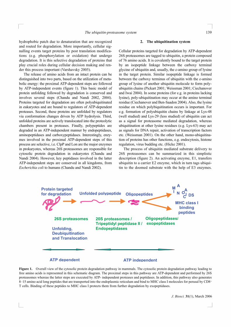
The release of amino acids from an intact protein can bedistinguished into two parts, based on the utilization of meta-bolic energy: the proximal ATP-dependent steps are followedby ATP-independent events (figure 1). This basic model ofprotein unfolding followed by degradation is conserved andinvolves several steps (Chandu and Nandi 2002, 2004).Proteins targeted for degradation are often polyubiquitinatedin eukaryotes and are bound to regulators of ATP-dependentproteases. Second, these proteins are unfolded by regulatorsvia conformation changes driven by ATP hydrolysis. Third,unfolded proteins are actively translocated into the proteolyticchambers present in proteases. Finally, polypeptides aredegraded in an ATP-independent manner by endopeptidases,aminopeptidases and carboxypeptidases. Interestingly, enzy-mes involved in the proximal ATP-dependent steps of thisprocess are selective, i.e. ClpP and Lon are the major enzymesin prokaryotes, whereas 26S proteasomes are responsible forcytosolic protein degradation in eukaryotes (Chandu andNandi 2004). However, key peptidases involved in the latterATP-independent steps are conserved in all kingdoms, fromEscherichia coli to humans (Chandu and Nandi 2002).
2. The ubiquitination system
Cellular proteins targeted for degradation by ATP-dependent26S proteasomes are tagged to ubiquitin, a protein composedof 76 amino acids. It is covalently bound to the target proteinby an isopeptide linkage between the carboxy terminalglycine of ubiquitin and, usually, the ε-amino group of lysinein the target protein. Similar isopeptide linkage is formedbetween the carboxy terminus of ubiquitin with the ε-aminogroup of lysine of another ubiquitin molecule to form poly-ubiquitin chains (Pickart 2001; Weissman 2001; Ciechanoverand Iwai 2004). In some proteins (for e.g. in proteins lackinglysine), poly-ubiquitination may occur at the amino terminalresidue (Ciechanover and Ben-Saadon 2004). Also, the lysineresidue on which polybiquitination occurs is important. Fore.g. formation of polyubiquitin chains by linkage at Lys-48(well studied) and Lys-29 (less studied) of ubiquitin can actas a signal for proteasome mediated degradation, whereasubiquitination at other lysine residues (e.g. Lys-63) may actas signals for DNA repair, activation of transcription factorsetc. (Weissman 2001). On the other hand, mono-ubiquitina-tion of proteins has other functions, e.g. endocytosis, histoneregulation, virus budding etc. (Hicke 2001).
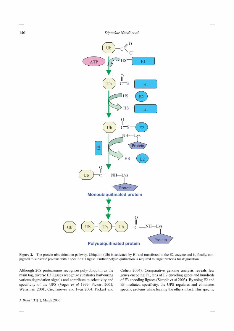
The process of ubiquitin mediated substrate delivery to26S proteasomes can be summarized in this simplisticdescription (figure 2). An activating enzyme, E1, transfersubiquitin to a carrier E2 enzyme, which in turn tags ubiqui-tin to the doomed substrate with the help of E3 enzymes.
The ubiquitin-proteasome system 139
J. Biosci. 31(1), March 2006
Unfolding, Deubiquitination
and Translocation
26S proteasomes
Unfolded polypeptide
ATP independent
Oligopeptides
MHC class I binding
peptides
Protein targeted
for degradation
20S proteasomes / Tripeptidyl peptidase II /
Endopeptidases
IN
A OI
C DS
Oligopeptidases/
exopeptidases
ATP dependent
A M
Figure 1. Overall view of the cytosolic protein degradation pathway in mammals. The cytosolic protein degradation pathway leading tofree amino acids is represented in this schematic diagram. The proximal steps in this pathway are ATP-dependent and performed by 26Sproteasomes whereas the latter steps are executed by ATP- independent proteases and peptidases. In addition, this pathway also generates8–15 amino acid long peptides that are transported into the endoplasmic reticulum and bind to MHC class I molecules for perusal by CD8+
T cells. Binding of these peptides to MHC class I protects them from further degradation by exopeptidases.
Although 26S proteasomes recognize poly-ubiquitin as themain tag, diverse E3 ligases recognize substrates harbouringvarious degradation signals and contribute to selectivity andspecificity of the UPS (Voges et al 1999; Pickart 2001;Weissman 2001; Ciechanover and Iwai 2004; Pickart and
Cohen 2004). Comparative genome analysis reveals fewgenes encoding E1, tens of E2 encoding genes and hundredsof E3 encoding ligases (Semple et al 2003). By using E2 andE3 mediated specificity, the UPS regulates and eliminatesspecific proteins while leaving the others intact. This specific
Dipankar Nandi et al140
J. Biosci. 31(1), March 2006
C
C
O
Ub C
O
E1 HS
Ub C O
O-
ATP
Ub E1 S
E1 HS
HS E2
Ub
O
S E2
HS E2
Monoubiquitinated protein
Polyubiquitinated protein
NH2—Lys
NH—Lys
Ub Ub Ub Ub C
O
NH—Lys
Protein
E3 Protein
Protein
Figure 2. The protein ubiquitination pathway. Ubiquitin (Ub) is activated by E1 and transferred to the E2 enzyme and is, finally, con-jugated to substrate proteins with a specific E3 ligase. Further polyubiquitination is required to target proteins for degradation.

regulation of protein degradation enables cells to regulatecellular ‘decisions’ in a dynamic fashion (Varshavsky 2005).
The activation of ubiquitin is carried out by the E1 enzymein the presence of ATP, resulting in the formation of ubiquitin-AMP as intermediate and ubiquitin-E1 thiol ester as the finalproduct. The E1-ubiquitin thiol ester is recognized by multipleE2s, to which ubiquitin is transferred by another thiol esterlinkage. E2 enzymes aid in carrying the activated ubiquitinfrom E1 to the substrate and hence, are often called as ubiqui-tin-conjugating or ubiquitin-carrier proteins. All E2 enzymesharbour a conserved core that is utilized, along with supportfrom the termini, in E2-E3 binding. E2 enzymes associatewith E3 enzymes in a specific manner although each E2 canbind more than one E3. Importantly, E3 enzymes are respon-sible for the final target selection and specificity. E3 enzymesbelong to two distinct families: (i) E3s that harbour the ~ 350aa homologous to E6-AP carboxyl terminus (HECT) domainwith the conserved catalytic cysteine residue. The E6-AP pro-tein is encoded by the human papilloma virus and is responsi-ble for targeting certain proteins (e.g. p53) for ubiquitination.HECT domain proteins form a covalent (thiolester) bond withthe ubiquitin before transferring it to the substrate. (ii) On theother hand, E3 enzymes that harbour the really interesting newgene (RING) domain contain cysteines and histidines aminoacids that co-ordinate two metal atoms. Many observationsfacilitated identification of this group including the fact that allE3s lacking HECT domain harbour a RING finger domain.Ubiquitination via RING finger E3s involves binding to E2-ubiquitin complex and facilitating direct transfer of ubiquitinto the targeted protein without the additional thiol ester for-mation as observed in the HECT family of E3 ligases (Pickart2001; Weissman 2001; Ciechanover and Iwai 2004). A subsetof E3 ligases are the Skp-Cullin-F box (SCF) proteins that arecharacterized by: Skp–1 or Skp–1-like protein that is involvedin substrate recognition, Cullin subunit e.g. Cul–1, an F-boxcontaining protein and the catalytic RING finger containingprotein, Rbx–1. Modification of Cullins by an ubiquitin-likeprotein Nedd/Eub–1, results in neddylation and activation ofSCF-ligases due to increased recruitment of E2 enzymes(Cardozo and Pagano 2004). The conventional ubiquitinationenzymes E1, E2, and E3 add only limited number of ubiquitinmoieties to protein substrates and polyubiquitination isrequired to act as a degradation signal. E4 enzymes (e.g. Ufd2)support the formation of multi-ubiquitin conjugates (Hoppe2005) and Saccharomyces cerevisiae lacking ufd2 are defec-tive in proteolysis of ubiquitin-substrates (Koegl et al 1999).Ufd2a is involved in the degradation of pathological forms ofataxin type 3 that are responsible for a neurodegenerative dis-ease known as spinocerebellar ataxia type 3 (Matsumoto et al2004).
Ubiquitin needs to be removed from tagged proteinsbefore they enter the proteolytic core of proteasomes. Theclassical deubiquitinaing enzymes belong to ubiquitin
processing (UBP) and ubiquitin carboxy-terminal hydrolases(UBH) families. In general, UBPs remove ubiquitin frompoly-ubiquitinated proteins whereas UBHs remove smalladducts from ubquitin and regenerate free monomeric ubiq-uitin. Also, ubiquitin genes are transcribed and translated as apolyubiquitin chain, which then needs to be acted upon byUBHs to release single ubiquitin moieties (Weissman 2001;Kim et al 2003). As ubiquitin is bound to enzymes by thioester linkage, deubiquitinating enzymes are thiol proteases.Genome analysis has revealed that genes encoding UBPs arehigher in number compared to UBHs in most organisms.Also, the crystal structures of two UBH enzymes revealstructural similarities with papain-like cysteine proteases,especially in the active site (Kim et al 2003). Other noveldeubiquitinating enzymes may also exist. For e.g. the deu-biquitinating enzyme identified to be a part of the PA700 or19S regulator complex is an ATP-dependent metalloproteasewith a JAMM motif (Rpn11; see table 2) (Berndt et al 2002)and is essential for 26S proteasomal degradation (Verma et al2002; Yao and Cohen 2002). The functional role of the Rpn11subunit demonstrates the close relationship between deubiq-uitination and proteasome mediated protein degradation.
3. PA700, the 19S regulator
The process of tagging with multi-ubiquitin marks proteinsfor degradation by 26S proteasome, a huge cytosolic proteasecomplex. This structure consists of the 20S proteasome, har-bouring the proteolytic core, bound to a 19S cap also knownas PA700 (figure 3). 20S proteasomes degrade unfolded orloosely folded proteins and peptides in an ATP-independentmanner; however, they cannot degrade ubiquitin-protein con-jugates. The proteolytic active sites of 20S proteasome aresequestered within the lumen of this cylindrical complex, toavoid non-specific degradation of cellular proteins. PA700binds to either or both ends of 20S proteasomes, in the pres-ence of ATP, to channelize ubiquitinated proteins into thecentral active site chamber for degradation (Glickman et al1998; Voges et al 1999; Pickart and Cohen 2004).
PA700 is a large complex comprising several subunits,which impart the complex with diverse activities: ATPase,ubiquitin-binding, deubiquitinating, reverse chaperone etc.PA700 from S. cerevisiae harbours at least 17 subunits, regu-latory particle non-ATPase (Rpn)1–12 and regulatory particletripleA-ATPase (Rpt)1–6. PA700 can be dissociated into twosub-complexes, base consisting of nine subunits and lid con-sisting of eight subunits. The base harbours six essentialATPases and three nonATPase subunits, including the polyu-biquitin-interacting protein S5a (table 2). These ATPasesbelong to the ATPases associated with various cellular activi-ties (AAA)-ATPase family. By utilizing its ATPase activity,the base complex acts as a reverse chaperone to unfold targetproteins and also facilitates opening up the narrow pore of 20S
The ubiquitin-proteasome system 141
J. Biosci. 31(1), March 2006
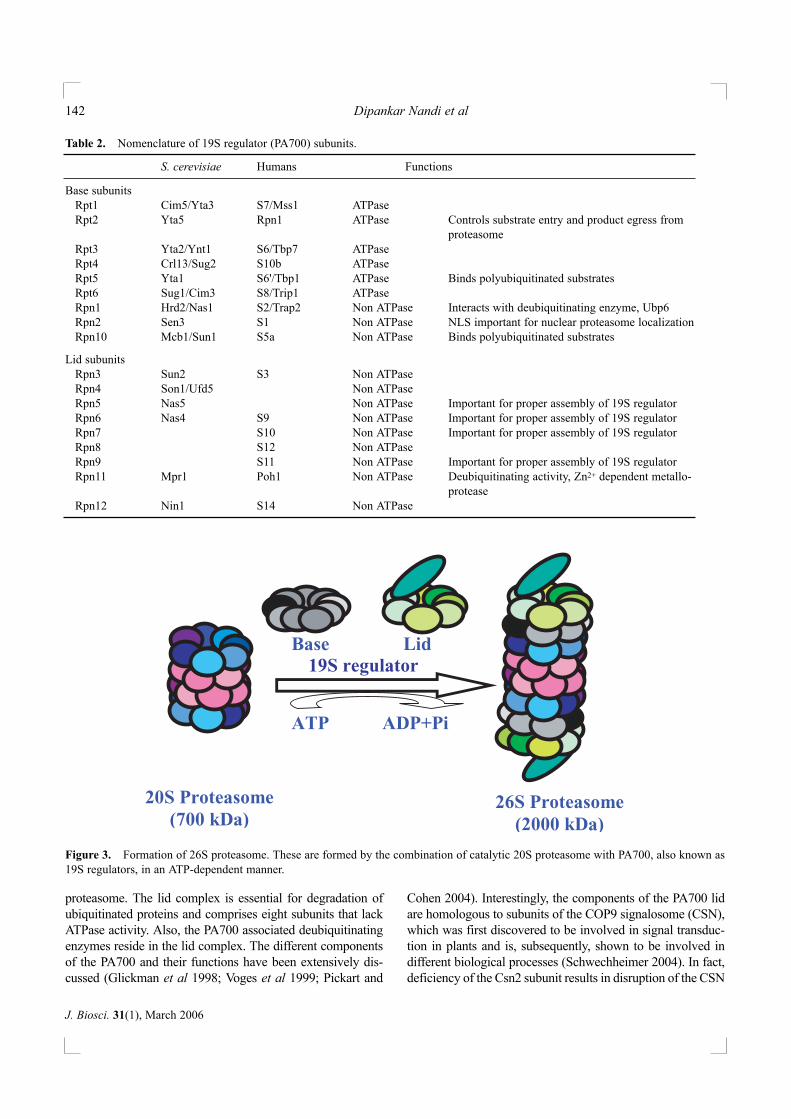
proteasome. The lid complex is essential for degradation ofubiquitinated proteins and comprises eight subunits that lackATPase activity. Also, the PA700 associated deubiquitinatingenzymes reside in the lid complex. The different componentsof the PA700 and their functions have been extensively dis-cussed (Glickman et al 1998; Voges et al 1999; Pickart and
Cohen 2004). Interestingly, the components of the PA700 lidare homologous to subunits of the COP9 signalosome (CSN),which was first discovered to be involved in signal transduc-tion in plants and is, subsequently, shown to be involved indifferent biological processes (Schwechheimer 2004). In fact,deficiency of the Csn2 subunit results in disruption of the CSN
Dipankar Nandi et al142
J. Biosci. 31(1), March 2006
Table 2. Nomenclature of 19S regulator (PA700) subunits.
S. cerevisiae Humans Functions
Base subunitsRpt1 Cim5/Yta3 S7/Mss1 ATPaseRpt2 Yta5 Rpn1 ATPase Controls substrate entry and product egress from
proteasomeRpt3 Yta2/Ynt1 S6/Tbp7 ATPaseRpt4 Crl13/Sug2 S10b ATPaseRpt5 Yta1 S6'/Tbp1 ATPase Binds polyubiquitinated substratesRpt6 Sug1/Cim3 S8/Trip1 ATPaseRpn1 Hrd2/Nas1 S2/Trap2 Non ATPase Interacts with deubiquitinating enzyme, Ubp6Rpn2 Sen3 S1 Non ATPase NLS important for nuclear proteasome localizationRpn10 Mcb1/Sun1 S5a Non ATPase Binds polyubiquitinated substrates
Lid subunitsRpn3 Sun2 S3 Non ATPaseRpn4 Son1/Ufd5 Non ATPaseRpn5 Nas5 Non ATPase Important for proper assembly of 19S regulatorRpn6 Nas4 S9 Non ATPase Important for proper assembly of 19S regulatorRpn7 S10 Non ATPase Important for proper assembly of 19S regulatorRpn8 S12 Non ATPaseRpn9 S11 Non ATPase Important for proper assembly of 19S regulatorRpn11 Mpr1 Poh1 Non ATPase Deubiquitinating activity, Zn2+ dependent metallo-
proteaseRpn12 Nin1 S14 Non ATPase
26S Proteasome (2000 kDa)
20S Proteasome (700 kDa)
Base Lid
ATP ADP+Pi
19S regulator
Figure 3. Formation of 26S proteasome. These are formed by the combination of catalytic 20S proteasome with PA700, also known as19S regulators, in an ATP-dependent manner.
complex, increased levels of Cyclin E and p53, resulting inarrest of embryo development in mice (Lykke-Andersen et al2003). The CSN directly interacts with 26S proteasomes andcompetes with the lid for assembly. In addition, the CSN mod-ifies cullin subunits by cleaving off the Nedd8 (i.e. deneddy-lation) and modulating the assembly and activity of SCF-E3ligases. Additionally, CSN regulates proteolysis by associat-ing with protein kinases and deubiquitinating enzymes(Schwechheimer 2004).
Eubacterial enzymes involved in cytosolic protein degra-dation also associate with ATPase regulators, for e.g. ClpPbinds to ClpA or ClpX (Chandu and Nandi 2004). Althoughthere is no evidence of protein-ubiquitination in archaea andeubacteria, 20S proteasomes present in some bacteria in thesekingdoms may bind to ATPase regulators. 20S proteasomesfrom some archeaebacteria, e.g. Methanococcus jannaschii,are known to interact with the proteasome-activatingnucleotidase (PAN) complex which is homologous to PA700present in 26S proteasomes complex (Zwickl et al 1999).PAN (~ 650 kDa) is an oligomeric complex with a subunitsize of 50 kDa and increases the protein degradation (but notpeptide degradation) ability of proteasomes. Substrate bind-ing to PAN activates nucleotidase activity, which in turn isutilized for substrate unfolding, 20S proteasome terminalpore opening and protein translocation (Benaroudj et al2003). Although ATP is most efficient in hydrolyzing pro-teins, other nucleotides could also significantly supporthydrolysis and hence the name, nucleotidase (Zwickl et al1999). Further light scattering and fluorescence experimentsdemonstrated that PAN inhibits protein aggregation and
facilitates protein unfolding in an ATP-dependent manner.PAN, similar to PA700 ATPases, acts as a reverse-chaperonethat can facilitate degradation by unfolding targeted proteins(Benaroudj and Goldberg 2000). Also, AAA-ATPase ineubacteria form homo-hexameric ring shaped complexes(ARC). In fact, the proteasomal ATPase in Mycobacteriumtuberculosis is important in resisting host-encoded nitrosativestress (Darwin et al 2003, 2005).
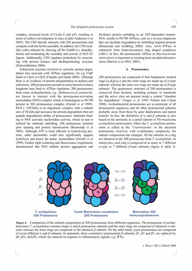
4. Proteasomes
20S proteasomes are composed of four heptameric stackedrings (α7β7β7α7) and the outer rings are made up of α-typesubunits whereas the inner two rings are made up of β-typesubunits. The quarternary structure of 20S proteasomes isconserved from bacteria, including archaea, to mammalsand the active sites are present inside a central “chamberfor degradation” (Voges et al 1999; Pickart and Cohen2004). Archaebacterial proteasomes act as prototype of allproteasomal sequences and all other proteasomal subunitsprobably arose from these by gene duplications and lateraltransfer. In fact, the definition of α and β subunits is alsobased on the similarity to α and β subunit of Thermoplasmaacidophilum proteasomes. Often the T. acidophilum protea-some is called as the ‘Urproteasome’ or the ancestralproteasome; however, with evolutionary complexity, thesubunit composition has changed. All the subunits in a ringare identical in the 20S proteasome from T. acidophilum. Ineukaryotes, each ring is composed of as many as 7 differentα-type or 7 different β-type subunits (figure 4; table 3).
The ubiquitin-proteasome system 143
J. Biosci. 31(1), March 2006
ß
ß1
ß2
ß2
ß5
ß5 ß1i
ß1i
ß2i
ß2i
ß5i
ß5i
Yeast/ Mammalian constitutive
20S Proteasome
Mammalian 20S
T. acidophilum
20S Proteasome Immunoproteasome
1
ß1– ß7
ß1– ß7 ß7
ß7
α1– α7
α1– α7
α7
α7
Figure 4. Comparison of the subunit composition of 20S proteasomes from different organisms. The proteasome of archae-bacterium T. acidophilum contains single α and β proteasome subunits and the outer rings are composed of identical α sub-units whereas the inner rings are composed of the identical β subunit. On the other hand, yeast proteasomes are composedof seven different α and β subunits. In mammals, three constitutive proteasomal β subunits, β1, β2 and β5, are replaced byβli, β2i, and β5i, which are induced in response to inflammatory signals, e.g. IFNγ.
Functionally, proteasomes from T. acidophilum are essentialonly during heat shock (Ruepp et al 1998). However, out of14 proteasome subunits in S. cerevisiae, 13 are essential forviability and an extra copy of α4/Pre6 subunit can substitutefor the α3/Pre9 subunit (Velichutina et al 2004). 20S pro-teasomes are absent in E. coli and the complete sequencesof various eubacterial genomes revealed the presence of sin-gle α and β proteasome subunit encoding genes in the orderactinomycetales of eubacteria (Lupas et al 1994).Interestingly, 20S proteasomes in Mycobacterium smegma-tis are dispensable (Knipfer and Shrader 1997) but areessential in the pathogen Mycobacterium tuberculosis(Sassetti et al 2003).
The α subunits are more conserved than the β subunitsand form a selective barrier between the catalytic chamberand the cytoplasm. They are the sites for the binding of var-ious regulatory particles, entry and exit of substrates. On theother hand, β subunits harbour the catalytic site. Most βsubunits have a prosequence that is cleaved off to expose athreonine residue at the N terminus. This Thr-1 is consid-ered as a critical part of the active site of the proteasome,there by making it a distinct member of a new class of pro-teases known as N-terminal nucleophile (NTN) hydrolasefamily. This N-terminal threonine is physiologically impor-tant as replacement with serine results in reduced cleavageof peptide bonds (Kisselev et al 2000). All the β subunits ofT. acidophilum possess this N-terminal threonine. Ineukaryotes, only three of the seven β subunits (β1, β2, andβ5) in each ring are processed to expose the N terminal thre-onine (Seemuller et al 1995). However, the crystal structuredetermination of 20S proteasomes from bovine liver hasrevealed processing of another β subunit (β7) with anexposed N-terminal Thr-1 (Unno et al 2002).
Originally described as a complex of multiple peptidaseactivities, studies with inhibitors and specific substrates havedefined important activities of this enzyme. Eukaryotic pro-teasomes display three major peptidase activities, based oncleavage of fluorogenic peptides that can be easily assayed:chymotrypsin-like activity (cleavage after hydrophobicamino acids), trypsin-like activity (cleavage after the basicamino acids) and caspase-like activity (cleavage after acidicamino acids). In addition, two activities, e.g. cleavage afterbranched chain amino acids (BrAAP) and small neutralamino acids (SNAAP) are also known. The three major activ-ities of yeast 20S proteasomes, caspase-like, trypsin-like andchymotrypsin-like activities can be correlated directly to thethree subunits β1 (Pre3), β2 (Pup1) and β5 (Pre2), respec-tively, as demonstrated by the mutation studies (Heinemeyeret al 1997; Groll et al 1999). The other four subunits eitherhave unprocessed (β3 or Pup3, β4 or C11) or partiallyprocessed (β6 or Pre7, β7 or Pre4) propeptides (Groll et al1999). Studies of human 20S proteasomes on peptidelibraries demonstrated that proteasomes can cleave peptidebonds at the P1 position (the amino acid immediately proxi-mal to the peptide bond that is cleaved) of most amino acids,with a preference for leucine and alanine. Also, amino acidsproximal to the P1 position, i.e. at P3 and P4 positions, glut-amine, valine, isoleucine, leucine and asparagine influencepeptide cleavage by 20S proteasomes (Harris et al 2001).Interestingly, no major difference in the size of peptides gen-erated after cleavage of proteins was observed from protea-somes from T. acidophilum, which contain 14 active sites,and humans, which contain 6 well defined active sites.Peptides generated vary in length from 3–25 amino acids anddecreasing amounts of peptides are produced with increasein size (Kisselev et al 1998, 1999). The data suggests that
Dipankar Nandi et al144
J. Biosci. 31(1), March 2006
Table 3. Nomenclature of 20S proteasome subunits.
Mammals
Subunit T. acidophilum S. cerevisiae Constitutive Immunoproteasomes
α1 α Prs2/C7 PSMA6/ Iota/ LMP11 PSMA6/ Iota/ LMP11α2 α Y7 PSMA2/ C3/ LMP8 PSMA2/ C3/ LMP8α3 α Y13 PSMA4/ C9/ LMP14 PSMA4/ C9/ LMP14α4 α Pre6 PSMA7/ C6/ XAPC7/ LMP16 PSMA7/ C6/ XAPC7/ LMP16α5 α Pup2 PSMA5/ Zeta/ LMP1 PSMA5/ Zeta/ LMP1α6 α Pre5 PSMA1/ C2/ LMP13 PSMA1/ C2/ LMP13α7 α Prs1/C1 PSMA3/ C8/ LMP18 PSMA3/ C8/ LMP18β1 β Pre3 PSMB6/ Y/ delta/ LMP19 PSMB9/ β 1i/ LMP2β2 β Pup1 PSMB7/ Z/ LMP9/ PSMB10/ β 2i/ LMP10/ MECL1β3 β Pup3 PSMB3/ C10/ LMP5 PSMB3/ C10/ LMP5β4 β Pre1/C11 PSMB2/ C7/ LMP6 PSMB2/ C7/ LMP6β5 β Pre2/Doa3 PSMB5/ X/ MB1/ LMP17 PSMB8/ β 5i/ LMP7β6 β Prs3/C5 PSMB1/ C5/ LMP15 PSMB1/ C5/ LMP15β7 β Pre4 PSMB4/ N3/ beta/ LMP3 PSMB4/ N3/ beta/ LMP3
IFNγ-inducible β subunits present in immunoproteasomes are indicated in bold.
proteasomes process peptides until they are small enough todiffuse out of the proteolytic chamber.
In addition to the ‘constitutive’ proteasome described sofar, there also exists the ‘immunoproteasome’, which is pri-marily induced by microbial infections and the immuno-modulatory cytokine, IFNγ (figure 4). This cytokine inducesthe expression of three mammalian proteasome subunits, β1i(LMP2), β5i (LMP7) and β2i (MECL-1), which are incor-porated in the place of the constitutive β subunits (Y, X andZ respectively) to form immunoproteasomes (Frentzel et al1994; Nandi et al 1997; Griffin et al 1998). Interestingly, β1iand β1 are mutually exclusive and belong to the same sub-group as is the case with β2i and β2 or β5i and β5 (table 3),implying that the IFNγ-inducible β subunits in mammalsprobably arose by gene duplication. The presence of genesencoding β1i and β5i in the MHC locus of mammals impliesthat the immune system has utilized the services of alreadyexisting set of genes by imparting specialized functions. Thisis further corroborated by the observation that yeast protea-somes, that lack the three IFNγ-inducible β proteasome sub-units, hydrolyze proteins and generate peptides that can beloaded on to MHC I molecules (Niedermann et al 1997).Immunoproteasomes are present in hematopoietic cells inmammals, unlike the house keeping proteasomes, which arefound in all cells. Apart from IFNγ, other cytokines, fore.g. IFNβ and tumour necrosis factor-α also induce thesesubunits. In fact, it has been recently demonstrated that theconstitutive levels of immnoproteasome subunits are IFNγindependent and IFNγ is essential only for the induction ofthese subunits and incorporation into proteasomes in vivo(Barton et al 2002). Interestingly, microbial infections mod-ulate proteasomal subunit expression, resulting in significantreplacement of constitutive proteasomes by immunoprotea-somes (Khan et al 2001; Barton et al 2002). The importanceof the immunoproteasome subunits is reflected in mice lack-ing these subunits. Mice lacking β1i or β5i display differ-ences in peptidase activities and are defective in someimmune responses. β1i-/- and β5i-/- mice display diminishedCD8+ T cells and MHC class I expression, respectively.These mice also display defects in processing some, but notall, antigens e.g. influenza virus by β1i-/- mice and H-Y anti-gen by β5i-/- mice (Fehling et al 1994; Van Kaer et al 1994).β1i and β5i are polymorphic and are associated with somediseases, e.g. acute anterior uveitis, juvenile rheumatoidarthritis etc. (Nandi et al 1998).
5. Structure and mechanism of action of proteasomes
The X-ray crystallographic analysis of 20S proteasomesfrom T. acidophilum (Lowe et al 1995), S. cerevisiae (Grollet al 1997) and bovine liver (Unno et al 2002) revealedsimilarity in size, shape, active sites, catalytic mechanism,binding to inhibitors etc., despite the complex subunit
composition in eukaryotes. T. acidophilum 20S proteasomesare 148 Å × 113 Å, where as bovine 20S proteasomes are150 Å × 115 Å in size. These crystal structures confirmedprevious electron micrographic findings that the fourrings of proteasome form a cylindrical structure with a nar-row channel harbouring a catalytic chamber with a maxi-mum diameter of ~ 53 Å. Although the three active site har-bouring β subunits (β1, β2 and β5) are conserved in bovine20S proteasomes, another subunit, β7 displayed propeptideprocessing at Thr1 including the presence of residuesimportant for catalysis in correct positions and may displaySNAAP activity. However, Thr1 of bovine β7 is placed in acompletely different orientation and close to the α ring-βring junction, unlike Thr1 of other active β subunits, whichstay in the chamber formed by two β rings. The comparisonof predicted structure of immunoproteasomes with that ofthe crystal structure of bovine constitutive proteasomesrevealed that the active center of immunoproteasomes isapolar, where as it is positively charged in the constitutiveproteasomes (Unno et al 2002).
20S proteasomes from T. acidophilum allow entry of thesubstrates to the active sites through the central channel asthe outer rings made of α subunits harbour an entry port of13 Å suggesting that translocation may require prior unfold-ing of the substrate. However, the hydrolytic chamber inS. cerevisiae and bovine liver proteasomes does not haveeasy access from the axial pore as it is surrounded by theN-termini of the α subunits. Thus, eukaryotic proteasomesdisplay low levels of in vitro activity, also termed as latent;however, activity increases in the presence of low amountsof SDS and poly-lysine (Tanaka et al 1989) or addition ofnatively unstable protein substrates, e.g. the CDK inhibitorp21cip1 and α-synuclein (Liu et al 2003). The most likelyexplanation is that low amounts of SDS, poly-lysine etc.enhance pore opening leading to increased proteasomalactivity. A series of structures of 20S proteasomes bound toactivators revealed that the proteasome activators PA700,PA28, PA200 and PAN, induce opening of the axial gates ofα rings by outward displacement of a set of turns surround-ing the gate that allows for better access of substrates intothe catalytic chamber of proteasomes (Whitby et al 2000;Forster et al 2003, 2005). Together, these structures demon-strate that opening the gate pore by activators is critical forthe 20S proteasome ability to perform proteolysis.
The structures of 20S proteasomes from T. acidophilumand S. cerevisiae bound to an inhibitor indicated the pres-ence of Thr1, Glu/Asp17, Lys33, Ser129, Asp166 andSer169 to be in the vicinity of active sites. Further, the keyplayers are: the amino group of Thr1 which acts as a protondonor, the Thr1Oγ which acts as a nucleophile, Glu17 isimportant in proper orientation of Lys33 and the positivecharge of Lys33 lowers the pKa of Thr1 to enhance itsnucleophilicity. The mechanism of cleaving of peptide bond
The ubiquitin-proteasome system 145
J. Biosci. 31(1), March 2006

by the N-terminal threonine of proteasome involves the ini-tial formation of an acyl-enzyme intermediate by thehydroxyl group of the active site Thr1 forming a bond withthe carbonyl of the substrate. Further, activated water mole-cules act as the general base and acid and hydrolyzes theacyl-enzyme intermediate to free enzyme and product(Lowe et al 1995; Seemuller et al 1995; Groll et al 1997).
6. Proteasome biogenesis
The general model of proteasome assembly involves theformation of a ring containing α subunits followed byincorporation of β subunits into another ring. These ‘half’proteasomes undergo dimerization followed by cleavage ofthe prosequences in the β subunits, most likely by auto-catalysis (Kruger et al 2001). T. acidophilum proteasomesover-expressed in E. coli assemble into complexes indistin-guishable from native 20S proteasomes. Over-expressed αsubunits form a ring by themselves; however, β subunitscannot form a ring by themselves and require preformed αrings for their assembly. The processing of the β subunit toexpose the N-terminal active site threonine is autocatalyticand is dependent on the presence of the catalytic Thr-1 andLys-33. In addition, this processing occurs after the assem-bly of complete proteasome complex to prevent non-specif-ic hydrolysis of cellular proteins (Zwickl et al 1994).
Although the broad pathway is similar, there are somedifferences between the assembly and processing of T. aci-dophilum and eukaryotic proteasomes. The formation of a αring is followed by binding of β subunits and formation ofthe tetrameric ring complex followed by the processing ofpropeptides of β subunits harbouring active sites. As men-tioned before, the S. cerevisiae proteasome harbours 7 dif-ferent α and 7 different β subunits. However, only three βsubunits undergo processing of propeptides after assemblyand hence, S. cerevisiae proteasome harbours only threeactive sites per β ring, unlike the T. acidophilum proteasomewhich harbours seven active sites per ring. Also, propep-tides of β subunits in T. acidophilum are dispensable, andfully assembled proteasomes can be dissociated andreassembled (Grziwa et al 1994; Zwickl et al 1994).Remarkably, propeptides of yeast β subunits are specific toeach subunit; for e.g. the β5 (Doa3) propeptide is indispen-sable, and the subunit lacking propeptide cannot be incor-porated into the proteasome complex. However, the β5(Doa3) propeptide expressed separately (i.e. in trans) canfacilitate its incorporation into the proteasome. Also, the β5(Doa3) subunit responsible for the “chymotrypsin” like-activity in yeast 20S proteasome cannot undergo processingif a mutation disrupts its contact with the complementaryinactive subunit β4 (Pre1) from other ring (Chen andHochstrasser 1996). Also, β2 (Pup1) in the opposite ring isresponsible for the processing of β6 (Pre7) and β7 (Pre4)
into their final forms. The β5 propeptide plays a greater roleduring proteasome assembly and activity, which is essentialin S. cerevisiae, compared to β2 and β1 propeptides (Arendtand Hochstrasser 1999; Groll et al 1999; Jager et al 1999).
The assembly pathway of mammalian proteasomes hasbeen delineated using antibodies specific for precursor andprocessed subunits. Interestingly, intermediate forms of pro-teasome (~ 300 kDa) have been observed, which harbours theprecursor subunits and probably correspond to half protea-somes. It has been proposed that following the formation of aring consisting of α subunits, the first β subunits that becomea part of these precursor proteasomes are unprocessed β sub-units lacking the catalytic threonine, β4 (C7) and β3 (C10).Interestingly, the other precursor β subunits are incorporatedat different times and undergo processing of propeptides atdifferent rates. Perhaps, the incorporation of one β facilitatesthe incorporation of another β as the localization of differentsubunits within the proteasome ring is fixed. Two half protea-somes containing a ring of α subunits and another ring of pre-cusor β subunits dimerize followed by the autocatalytic pro-cessing of β subunits (Frentzel et al 1994; Nandi et al 1997).Mammalian proteasomes display displacement of the active βsubunits, β1 (Y/delta), β2 (Z) and β5 (X) by interferon (IFN)-γ inducible subunits β1i (LMP2), β2i (MECL) and β5i(LMP7) respectively. A model has been proposed to explainthe preferential incorporation of these IFNγ-inducible subunitsinto immunoproteasomes. Here β1i (LMP2) and β2i (LMP10)are incorporated in ‘early’ proteasomes and the incorporationof β5i (LMP7) results in formation of active immunoprotea-somes (Nandi et al 1997, Griffin et al 1998). Propeptides playkey roles in this process, for e.g. the β5i (LMP7) propeptide ismore efficient in incorporation into proteasomes compared toits counterpart, i.e. the β5 (X) propeptide (Kingsbury et al2000). Similarly, β2i (MECL) containing the propeptide of β2(Z) is incorporated in precursor proteasomes containing con-stitutive subunits, β5 (X) and β1 (Y) (De et al 2003). Theseexperiments clearly demonstrate the key roles of propeptidesduring proteasome assembly in eukaryotes. Propeptides playmultiple roles and support the proper folding and assembly ofβ subunits; in addition, they protect the N-terminal threoninefrom acetylation mediated inhibition (Arendt andHochstrasser 1999; Groll et al 1999; Jager et al 1999).
Studies have also demonstrated a role of ubiquitin medi-ated proteolysis-1 (UMP1), encoding an ~ 17 kDa protein,for proper proteasome maturation in S. cerevisiae.Remarkably, Ump1p binds to precursor proteasomes andgets degraded by the same proteasome after maturation(Ramos et al 1998). Homologues of UMP1 known as pro-tassemblin have been identified in mouse (Griffin et al2000), and in humans known as hUMP1 (Burri et al 2000),or the proteasome maturation protein (POMP) (Witt et al2000). POMP has been shown to be up-regulated in cellstreated with IFNγ (Burri et al 2000) or proteasome
Dipankar Nandi et al146
J. Biosci. 31(1), March 2006
inhibitors. In fact, it has been observed that proteasomalinhibition leads to increased expression of proteasomalgenes, facilitating formation of new functional proteasomesto compensate for the loss (Meiners et al 2003). Althoughthe function of S. cerevisiae and mammalian UMP1 appearsto be similar, mammalian UMP1 cannot complement theloss of yeast UMP1 perhaps due to differences in the primarysequence. Mutation studies performed with human UMP1identified the proteasome-interacting domain to residues68–72 (RNIQG), as deletion of these five amino acidsseverely diminishes its precursor proteasome-binding ability(Burri et al 2000). POMP is induced by IFNγ and binds toβ5i (LMP7) more efficiently than β5 (X), resulting in accel-erated formation of immunoproteasomes. In fact, reducedPOMP expression results in lower formation of proteasomes,reduced MHC class I and induction of apoptosis (Heink et al2005). Recent studies have identified a proteasome assem-bling complex (PAC) consisting of two chaperones, PAC1and PAC2, that binds to α subunits and ensures proper for-mation and stability of the ring of α subunits (Hirano et al2005). Therefore, UMP1 and the PAC complex play distinctroles during mammalian proteasome assembly.
7. Cellular localization of proteasomes
In mammalian cells, proteasomes are primarily localized incytosol but also display significant association withcytoskeletal elements, ER, nucleus and plasma membrane;however the ratios of proteasomes associated with differentorganelles varies in different cells (Wojcik and DeMartino2003). Studies performed with GFP-tagged β1i (LMP2) in ahuman cell line revealed that proteasomes are distributed inboth nucleus and cytoplasm, though they are excluded fromnucleolus and ER lumen. These experiments demonstratedthat preformed proteasomes move from cytosol to nucleusin a slow and unidirectional manner. It is also possible thatproteasomes from nucleus and cytosol mix after the break-down of the nuclear envelope after mitosis (Reits et al1997). In S. cerevisiae, proteasomes are mainly localized inthe nuclear membrane-ER network (Enenkel et al 1998).However, localization of proteasomes at different times isdependent on the physiological state of the cell. In fissionyeast S. pombe, proteasomes are localized at the nuclearperiphery through out mitosis and is dispersed in the nucle-us during the first meiotic division. However, proteasomesare observed at the interface between the two nuclei duringthe second meiotic division (Wilkinson et al 1998).Proteasomes are responsible for protein degradation in thenucleus. Some of the α subunits harbour nuclear localiza-tion signals (NLS), which probably aid in nuclear targetingof proteasomes. In fact, T. acidophilum proteasomes whenexpressed in HeLa and 3T3 cells can translocate into nucle-us (Wang et al 1997).
Although proteasomes are distributed throughout thecells, it is possible that they play important roles in selectedproteolytic centers within cells, e.g. the centrosome, a peri-nuclear structure that plays important roles in organizing themitotic spindle to separate chromosomes. Recent evidencesdemonstrate that cellular proteins targeted for degradationare delivered to the centrosome via the nocadozole sensitivemicrotubule mediated transport system. On treatment ofcells with proteasomal inhibitors, an accumulation of proteinaggregates, known as aggresomes, consisting of chaperones,ubiquitinated proteins, together with components of the 26Sproteasomes occurs. The assembly of aggresomes serves asa site for recruitment and concentration of the unfolded pro-tein response (Johnston et al 1998; Wigley et al 1999;Fabunmi et al 2000). The identification of the roles of cen-trosomes in cell division and protein degradation reinforcesthe close interrelationship between these two processes.
8. Additional modulators of proteasomes
In addition to binding to PA700, 20S proteasomes can asso-ciate in an ATP-independent manner with another activator,PA28 or the 11S regulator, to form complexes that enhanceproteasomal activity. In fact, PA28 and PA700 can simulta-neously bind a 20S proteasome molecule (Hendil et al1998). There are two forms of PA28: it can exist as a hetero-heptameric complex of 180–200 kDa, comprising of twoIFNγ inducible subunits, α and β, or a homohepatmericcomplex of PA28γ subunits, also known as Ki antigen(Rechsteiner and Hill 2005). Incubation of human protea-somes with PA28αβ greatly enhances activity against allamino acids whereas modest increase in proteasomal activi-ty against basic amino acids is observed on incubation withPA28γ (Harris et al 2001). Functional studies performed onPA28αβ revealed the importance of C-terminus in the pro-teasome activation. PA28 undergoes carboxypeptidase Bmediated inactivation (Ping et al 1993) and site directedmutagenesis studies revealed the importance of the C-termi-nal 14 amino acids of each PA28 subunit in activating theproteasome (Zhang et al 1998a). Furthermore, chimericPA28α containing the distal C-terminal residues of PA28β ismore efficient at activating proteasomes compared to PA28αcontaining the distal C terminal residues of PA28γ (Li et al2000). The crystal structure of PA28 revealed a narrow porein the middle of the heptameric ring and suggested that itinduces changes in the conformation of proteasomes facili-tating access for substrates into the active site channel(Knowlton et al 1997; Forster et al 2005). Further, structurebased mutations revealed the importance of the loop at thebase of the PA28 in activating proteasome. A single mutationof Asn146 in the PA28α loop interferes in activation of pro-teasomes although binding to proteasomes is not affected.Mutation in the corresponding aspargines of PA28β
The ubiquitin-proteasome system 147
J. Biosci. 31(1), March 2006

(N135Y) and PA28γ (N151Y) also display similar inactiva-tion (Zhang et al 1998b). PA28 modulates the proteasomalcleavage of proteins, e.g. cytomegalovirus pp89, to releasepeptides that can bind MHC class I molecules (Dick et al1996). Both the subunits of PA28 are distributed only inorganisms with adaptive immune response and are uniform-ly localized in the cytosol and nucleus of the cell. In fact,mice lacking both subunits of PA28 display 15–25% reduc-tion in ATP-dependent protein degradation suggesting theimportance of complexes harbouring PA28 in proteolyticfunction. It is possible that PA28 is important in processingof some antigens; processing of an epitope from tyrosinaserelated protein, an antigen of B16 melanoma tumour, butnot ovalbumin was severely defective in the Pa28-/- mice(Murata et al 2001). PA28αβ is present mainly in the cytosolwhereas PA28γ is predominantly localized in the nucleus(Soza et al 1997). PA28γ is a regulator of cell proliferationand mice lacking this subunit are smaller in size (Murata etal 1999). In addition, Pa28γ-/- mice display reduced numbersof CD8+ T cells and are inefficient in processing some anti-gens (Barton et al 2004). Together, PA28 is involved inimproving the antigen processing ability of proteasomes, bymodulating the quantity and quality of peptides released.
Recently, another proteasome activator, PA200 was iden-tified from bovine testis. PA200 adopts a solenoid structure,usually implicated in protein-protein interaction, suggestingthat PA200 may act as an adaptor complex. PA200 stimu-lated the peptidase activities of proteasome by 2–10-fold,with higher folds of stimulations for hydrolysis after acidicresidues. However, PA200 did not display any significantstimulation in hydrolysis of protein substrates. PA200 islocalized primarily to the nucleus in HeLa cells under nor-mal conditions, although upon IR irradiation PA200 local-izes to certain nuclear foci. A mutation in yeast PA200resulted in sensitivity to bleomycin, suggesting a probablerole in DNA repair (Ustrell et al 2002). PA200 is inducedunder conditions of muscle wasting, although functionalimplications in mammals have not been delineated(Rechsteiner and Hill 2005).
In a search to detect native proteins that can modulate pro-teasome function, an inhibitor of ~ 31 kDa, PI31 (for protea-some inhibitor of 31 kDa), was identified along with PA28and PA700 from bovine red blood cells (Chu-Ping et al 1992).Over-expressed mouse PI31 inhibits chymotryptic and trypticactivities, but has only marginal influence on the caspase-likeactivity of proteasomes. Further kinetic experiments revealedthat PI31 acts as a competitive inhibitor of PA28 and PA700and efficiently inhibits the degradation of large peptides (Zaisset al 1999; McCutchen-Maloney et al 2000). PI31 hindersmaturation of immunoproteasomes and processing of anepitope. Interestingly, induction of two MHC class I allelesH2-Kb as well as H2-Db is also hampered in PI31 over-expressing cells (Zaiss et al 2002), which probably is the
result of proteasome inhibition. It would be interesting tounderstand the regulation of PI31 in future and the conditionsin which it associates with proteasomes, considering the factthat proteasomes are essential for survival in eukaryotes.Other proteinaceous proteasome inhibitors from varioussources have also been described: δ-Aminolevulinic aciddehydratase, an enzyme involved in heme biosynthesis, isidentical to a 240 kDa proteasome inhibitor (subunit size~ 40 kDa) isolated from human blood (Guo et al 1994).Certain virus encoded proteins, e.g. HIV encoded Tat (Apcheret al 2003) and Hepatitis B virus encoded X protein (Hu et al1999), also inhibit proteasome activity. Interestingly, aggre-gated proteins that occur in cells due to aging, oxidative stressor during disease progression, especially neuronal disease, etc.may be physiologically important as the binding of largeamounts of aggregated protein to proteasomes results information of protein aggregates that are difficult to unfold andprevents the degradation of normal ubiquitinated proteins. Asa result, high levels of aggregated proteins may act as domi-nant negative regulators of proteasome activity that maylead to cell death (Grune et al 2004). Finally, an antibacterialpeptide PR39, which is rich in proline and arginine aminoacids, binds to the α7 subunit, causes structural alterations andacts as a non-competative inhibitor of proteasome activity(Gaczynska et al 2003). This peptide reduces pancreatitis andmyocardial infarctions in disease models by reducing NF-kBactivation (Gao et al 2000).
9. Chemical inhibitors of proteasomes
Most protease inhibitors are peptide based ligand mimeticsor transition state analogs of enzyme catalyzed reactions.They inhibit enzymes reversibly or irreversibly by forma-tion of a covalent adduct between the inhibitor and the cat-alytic site of the enzyme. Inhibitors targeting proteasomalfunction (table 4) are attractive drug targets due to theimportance of the UPS in numerous biological processes(Adams 2003; Groll and Huber 2004; Rajkumar et al 2005).The first discovered proteasome inhibitors were peptidealdehydes, e.g. N-acetyl-Leu-Leu-Norleucinal, also calledCalpain inhibitor I. These compounds reversibly inhibit 20Sproteasomes by modifying the catalytic hydroxyl group ofthreonine by forming a hemiacetal bond. They can rapidlydissociate from proteasomes and are rapidly oxidized intoinactive acids. Calpain inhibitor I was found to primarilyinhibit the chymotryptic-like activity of proteasomes andgreatly reduce the rate of cytosolic protein degradation(Rock et al 1994). Peptide boronates are much more potentinhibitors of the proteasome compared to aldehydes and dis-sociate more slowly from the proteasome. The boronatesalso share the mode of inhibition with peptide aldehydes, byforming a tetrahedral adduct with the active site N-terminalthreonine. Peptide vinyl sulfones are synthetic irreversible
Dipankar Nandi et al148
J. Biosci. 31(1), March 2006
inhibitors of proteasome that covalently modify the cata-lytic Thr1 present in all active β proteasome subunits.Lactacystin, the first natural proteasomal inhibitor identi-fied, inhibits the chymotryptic activity to a great extent bycovalently binding to the N-terminal Thr1 of the β5 protea-some subunit via an ester bond (Fenteany et al 1995; Grollet al 1997). Epoxymycin is probably the most selective as itdoes not inhibit any other cellular enzymes. Epoxymycinreacts with the catalytic N-terminal threonine of the protea-some to form an irreversible morpholino derivative (Grollet al 2000) and exhibits anti-tumour activity and anti-inflammatory activity in vivo (Meng et al 1999).
Numerous reports have demonstrated the anti-tumourability of bortezomib (a boronic dipeptide; also known asvelcade or PS–341), a reversible inhibitor of proteasomes.This compound has become the first proteasome inhibitorevaluated in human trials and has been approved to treatpatients suffering from multiple myeloma (Rajkumar et al2005). Proteasome inhibition can cause cellular apoptosisby modulating the levels of various short-lived proteins andinhibition of NF-kB activity. Consequently, one has to becautious about the widespread use of proteasome inhibitorsfor therapeutic purposes. A smarter strategy may be to tar-get different E3 ligases as they posses specialized functions.Each E3 ligase is specific for a small set of proteins; hence,inhibitors of E3 ligases can inhibit ubiquitination and 26S
proteasome mediated degradation of specific cellularprocesses. Nutlin-3 is a cis-imidazoline analog, which bindsto Mdm2, a Ring finger E3 ligase and modulator of tumoursuppressor protein p53, and inhibits its association withp53. Consequently, high levels of p53 lead to apoptosis ofcells in an experimental tumour model (Vassilev et al 2004).
10. Major roles of the UPS
The UPS play major roles in several biological processesand only key ones are listed below to reinforce its impor-tance to the uninitiated reader.
(i) Regulation of the cell cycle: The levels of regulatoryproteins (e.g. cyclin B, CDK inhibitor p27kip1) are modulat-ed at different phases of the cell cycle and the UPS is essen-tial for cells to exit mitosis. The two major classes of E3 lig-ases that are involved in this process are the SCF complexesand the anaphase promoting complex/cyclosome. In general,SCF regulate entry into S phase and recognizes substratespost-phosphorylation. The anaphase promoting complex isimportant for sister chromatid separation, exit from mitosisand degrades cell cycle regulators containing a nine aminoacid motif known as the destruction box (Murray 2004). (ii) Cancer and cell survival: The tumour suppressor p53plays a key role in regulating cell cycle arrest, DNA repairand apoptosis. Under normal conditions, p53 levels are lowdue to binding to Mdm2, an E3 ubiquitin ligase. After DNAdamage, p53 is phosphorylated resulting in reduced interac-tion with Mdm2, and induction of cell death. Not surprising-ly, mutations in p53 are often associated with different humancancers. Interestingly, the human papilloma virus encodes aE3 ligase (E6-AP) which degrades p53 and is involved ingeneration of cervical tumours (Ciechanover and Iwai 2004).(iii) Inflammatory responses: NF-κB is a key transcriptionfactor involved in the inflammatory response. NF-κB isbound to inhibitor-κB and is found in the cytosol. On appro-priate stimulation, inhibitor-κB is phosphorylated anddegraded by the UPS. Free NF-κB enters into the nucleusand induces the expression of several genes involved in theinflammatory response (Karin and Ben-Neriah 2000). (iv) Immune response: MHC class I molecules presentpeptides to CD8+ T cells. This process involves the diges-tion of self or microbial proteins into peptides by the UPSthat are presented on MHC class I. Inhibition of UPS leadsto the impairment of the biogenesis of MHC class I mole-cules (Kloetzel 2004).(v) Protein misfolding: The UPS interacts with membersof the heat shock family and cofactors to eliminate misfold-ed proteins. A direct relation between protein unfolding anddegradation is via CHIP, an E3 ligase and a Hsc70 interact-ing protein (McDonough and Patterson 2003). (vi) ER associated degradation: The UPS is also involved inthe degradation of misfolded proteins in the ER which
The ubiquitin-proteasome system 149
J. Biosci. 31(1), March 2006
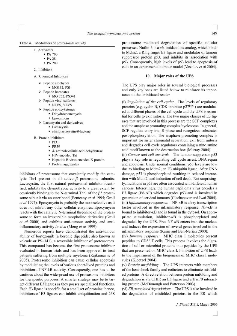
Table 4. Modulators of proteasomal activity
1. ActivatorsPA 700PA 28PA 200
2. Inhibitors
A. Chemical Inhibitors
Peptide aldehydesMG132, PSI
Peptide boronatesMG 262, PS341
Peptide vinyl sulfonesNLVS, YLVS
Peptide epoxyketonesDihydroeponemycinEpoximicin
Lactacystin and derivativesLactacystinclastolactacystin-β-lactone
B. Protein InhibitorsPI31PR39δ- aminolevulinic acid dehydrataseHIV encoded TatHepatitis B virus encoded X proteinProtein aggregates
involves retro-translocation of misfolded proteins from the ERto cytoplasm via the Sec61 translocon. Other components ofthis pathway also include a cytosolic N-glycanase, ATPases,e.g. Cdc48p/p97/valosin-containing protein, and associatedcofactors (Kostova and Wolf 2003). A clinical manifestationof this is observed in patients suffering from cystic fibrosiswhere the mutant ∆F508 CFTR protein is retained exclusive-ly in the ER and degraded by the UPS (Ward et al 1995). Therole of chaperones and factors involved in ER-associateddegradation is an active area of investigation. (vii) Disease progression: Angelman syndrome is char-acterized by severe phenotypic defects including mentalretardation, seizures and abnormal gait. Mutations in the E3ligase, E6-AP cause Angelman syndrome, the first humandisorder to be identified with a defect in the UPS (Kishinoet al 1997). Another E3 ligase which contains the HECTdomain, NEDD4, regulates the number of sodium channelson the cell surface. Mutations in NEDD4 cause hyperten-sion associated with hypokalemic metabolic alkalosis, lowplasma renin activity, and suppressed aldosterone secretion,together termed as the Liddle syndrome (Staub et al 1997).
11. Summary and future directions
It has been a remarkable journey of the UPS from what wasconsidered to be an esoteric post translation protein modifica-tion to appreciation of this pathway in different biologicalprocesses. Much has been learned in the past few decadesabout the genetics, biochemistry and structural aspects ofcomponents of the UPS; certainly more will be learned in thecoming years. Not only is the UPS involved in the overall non-lysosomal degradation of cellular proteins but, more impor-tantly, plays an important role in modulating cellular decisionmaking. The UPS is able to perform this task, after receivingappropriate signals, by reducing levels of important proteins(e.g. cyclins) or partially degrading selective proteins, i.e.degrading of the C-terminal of a cohesion subunit which isimportant during chromosome segregation (Rao et al 2001).In fact, regulated protein degradation has challenged the pre-dominant view that major cellular decisions are made at tran-scriptional and translational levels (Varshavsky 2005).
The roles of cellular enzymes involved in degradation ofproteasomes needs to be understood. Inhibition of lysoso-mal enzymes revealed that proteasomes are present withinthe lumen of lysosomes and it is likely that they are degrad-ed by lysosomal enzymes as a consequence of autophagy(Cuervo et al 1995). Interestingly, proteasomal activity isreduced in cells undergoing death because proteasome sub-units are cleaved in a caspase-dependent manner (Adrain etal 2004; Sun et al 2004). This relationship between caspaseactivity and proteasomes is important because lowered pro-teasomal activity is often associated with cell death and fur-ther studies in this area will be important.
The UPS continues to remain attractive area of researchwith the human genome encoding multiple enzymesinvolved in ubiquitination and deubiquitination (Sempleet al 2003). In addition, the 26S proteasome is composed ofproteins encoded by at least 34 independent genes. With somany genes directly involved in the UPS, systematic inacti-vation of genes in mice may identify their physiologicalfunctions during different cellular processes; in addition, thechallenge will be to decipher their cellular substrates. Thisis reinforced by the fact that enzymes involved in ubiquiti-nation may be specific to different cellular processes; fore.g. E2–C is involved in cell cycle regulation (Hershko et al1994), where as another E2 enzyme known as UbcM4 isinvolved in placental development (Harbers et al 1996).
In fact, there may be an underestimation of proteinsinvolved in the UPS as some bind to PA700 in sub-stoichio-metric amounts. Affinity-purified yeast proteasomes areassociated with three proteins that are not observed in pro-teasomes purified by conventional purification methods,probably due to the high salt concentrations used in the lat-ter procedures. It has been established that Ecm29, one of theassociated proteins, is involved in the association of 20Sproteasome and PA700. Hul5 and Ubp6 are the other twoproteins associated with affinity-purified proteasomes. Ubp6is responsible for hydrolysis of majority of the deubiquiti-nating activity associated with proteasomes (as detected byUb-AMC hydrolysis) and is probably responsible for recy-cling of the proteasome-associated ubiquitin to the cellularpool (Leggett et al 2002). In fact, similar analysis of proteinsassociated with affinity purified proteasomes performed pre-viously has also identified Ubp6, along with Hsp70 andHsp82 and other regulatory complex proteins (Verma et al2000). These proteasome associated proteins may playimportant and diverse roles. Yin6, a yeast homologue of Int6(involved in breast cancer formation), has been identified tointeract with proteasomal subunit Rpn5 and mediate thelocalization of proteasomes. S. pombe lacking yin6 has beendemonstrated to harbour mislocalized proteasomes andthereby, improper degradation of proteins involved in mitot-ic regulation (Yen et al 2003). Recently, a proteasomalATPase associated factor has been shown to interact withATPases present in PA700 and inhibit proteasomal activity(Park et al 2005). Another interesting protein that interactswith proteasomes is Rad23, a component of nucleotide exci-sion repair. Rad23 harbours a ubiquitin like domain, supportsthe formation of multiubiquitin chain and facilitates proteindegradation. It has been demonstrated that Rad23 interactswith proteasomes via N-terminal domain and with DNArepair proteins via C-terminal domain (Schauber et al 1998).Also, the Cdc48/p97/valosin containing protein family ofATPases that unfold proteins are involved in docking ubiq-uitinated proteins to 26S proteasomes (Elsasser and Finley2005). The identification of 26S proteasome-interacting
Dipankar Nandi et al150
J. Biosci. 31(1), March 2006
proteins will be of increasing importance in the future as theymay shed light on how the UPS recruits additional proteins tomodulate specific cellular responses. Further information onsuch types of protein interactions may lead to the generationof small molecule drugs that inhibit specific interactions thatmay ameliorate UPS-associated processes and diseases.
Acknowledgements
We have attempted to survey a large field of research andcommunicate its flavour to readers of this journal. In theprocess, only key references have been cited for the sake ofbrevity and we apologize to authors whose work was notcited. We thank Dr U Nath, Dr B Gopal and members of theDpN laboratory for their comments on the manuscript.Funding from the Department of Science and Technology,New Delhi for our studies on cytosolic protein degradationis greatly appreciated.
References
Adams J 2003 Potential for proteasome inhibition in the treatmentof cancer; Drug Discovery Today 8 307–315
Adrain C, Creagh E M, Cullen S P and Martin S J 2004 Caspase-dependent inactivation of proteasome function during pro-grammed cell death in Drosophila and man; J. Biol. Chem. 27936923–36930
Apcher G S, Heink S, Zantopf D, Kloetzel P M, Schmid H P,Mayer R J and Kruger E 2003 Human immunodeficiency virus-1 Tat protein interacts with distinct proteasomal alpha and betasubunits; FEBS Lett. 553 200–204
Arendt C S and Hochstrasser M 1999 Eukaryotic 20S proteasomecatalytic subunit propeptides prevent active site inactivation byN-terminal acetylation and promote particle assembly; EMBO J.18 3575–3585
Arrigo A P, Tanaka K, Goldberg A L and Welch W J 1988 Identityof the 19S ‘prosome’ particle with the large multifunctional pro-tease complex of mammalian cells (the proteasome); Nature(London) 331 192–204
Arthur J S, Elce J S, Hegadorn C, Williams K and Greer P A 2000Disruption of the murine calpain small subunit gene, Capn4:calpain is essential for embryonic development but not for cellgrowth and division; Mol. Cell. Biol. 20 4474–4481.
Barton L F, Cruz M, Rangwala R, Deepe G S Jr and Monaco J J2002 Regulation of immunoproteasome subunit expression invivo following pathogenic fungal infection; J. Immunol. 1693046–3052
Barton L F, Runnels H A, Schell T D, Cho Y, Gibbons R,Tevethia S S, Deepe G S Jr and Monaco J J 2004 Immunedefects in 28-kDa proteasome activator gamma-deficient mice;J. Immunol. 172 3948–3954.
Benaroudj N and Goldberg A L 2000 PAN, the proteasome-acti-vating nucleotidase from archaebacteria, is a protein-unfoldingmolecular chaperone; Nat. Cell. Biol. 2 833–839
Benaroudj N, Zwickl P, Seemuller E, Baumeister W and GoldbergA L 2003 ATP hydrolysis by the proteasome regulatory
complex PAN serves multiple functions in protein degradation;Mol. Cell 11 69–78
Berndt C, Bech-Otschir D, Dubiel W and Seeger M 2002Ubiquitin System: JAMMing in the Name of the Lid; Curr. Biol.12 R815–R817
Brown M S, Ye J, Rawson R B and Goldstein J L 2000 Regulatedintramembrane proteolysis: a control mechanism conservedfrom bacteria to humans; Cell 100 391–398
Burri L, Hockendorff J, Boehm U, Klamp T, Dohmen R J andLevy F 2000 Identification and characterization of a mam-malian protein interacting with 20S proteasome precursors;Proc. Natl. Acad. Sci. USA 97 10348–10353
Cardozo T and Pagano M 2004 The SCF ubiquitin ligase: insightsinto a molecular machine; Nat. Rev. Mol. Cell. Biol. 5 739–751
Chu-Ping M, Slaughter C A and Demartino G N 1992 Purificationand characterization of a protein inhibitor of the 20S protea-some (macropain); Biochem. Biophys. Acta 1119 303–311
Chandu D and Nandi D 2002 From proteins to peptides to aminoacids: comparative genomics of enzymes involved in down-stream events during cytosolic protein degradation; Appl.Genom. Proteom. 4 235–252
Chandu D and Nandi D 2004 Comparative genomics and func-tional roles of the ATP-dependent proteases Lon and Clp duringcytosolic protein degradation; Res. Microbiol. 155 710–719
Chen P and Hochstrasser M 1996 Autocatalytic subunit processingcouples active site formation in the 20S proteasome to comple-tion of assembly; Cell 86 961–972
Ciechanover A, Hod Y and Hershko A 1978 A heat-stable polypep-tide component of an ATP-dependent proteolytic system fromreticulocytes; Biochem. Biophys. Res. Commun. 81 1100–1105
Ciechanover A, Finley D and Varshavsky A 1984 Ubiquitindependence of selective protein degradation demonstrated in themammalian cell cycle mutant ts85; Cell 37 57–66
Ciechanover A and Ben-Saadon R 2004 N-terminal ubiquitina-tion: more protein substrates join in; Trends Cell Biol. 14103–106
Ciechanover A and Iwai K 2004 The ubiquitin system: from basicmechanisms to the patient bed; IUBMB Life 56 193–201
Cuervo A M, Palmer A, Rivett A J and Knecht E 1995Degradation of proteasomes by lysosomes in rat liver; Eur. J.Biochem. 227 792–800
Dahlmann B, Kopp F, Kuehn L, Niedel B, Pfeifer G, Hegerl R andBaumeister W 1989 The multicatalytic proteinase (prosome) isubiquitous from eukaryotes to archaebacteria; FEBS Lett. 251125–131
Darwin K H, Ehrt S, Gutierrez-Ramos J C, Weich N and NathanC F 2003 The proteasome of Mycobacterium tuberculosis isrequired for resistance to nitric oxide; Science 302 1963–1966
Darwin K H, Lin G, Chen Z, Li H and Nathan C F 2005Characterization of a Mycobacterium tuberculosis proteasomalATPase homologue; Mol. Microbiol. 55 561–571
De M, Jayarapu K, Elenich L, Monaco J J, Colbert R A and GriffinT A 2003 Beta 2 subunit propeptides influence cooperative pro-teasome assembly; J. Biol. Chem. 278 6153–6159
Dick T P, Ruppert T, Groettrup M, Kloetzel P M, Kuehn L,Koszinowski U H, Stevanovic S, Schild H and Rammensee H G1996 Coordinated dual cleavages induced by the proteasomeregulator PA28 lead to dominant MHC ligands; Cell 86 253–262
The ubiquitin-proteasome system 151
J. Biosci. 31(1), March 2006

Etlinger J D and Goldberg A L 1977 A soluble ATP-dependentproteolytic system responsible for the degradation of abnormalproteins in reticulocytes; Proc. Natl. Acad. Sci. USA. 74 54–58
Enenkel C, Lehmann A and Kloetzel P M 1998 Subcellular distri-bution of proteasomes implicates a major location of proteindegradation in the nuclear envelope-ER network in yeast;EMBO J. 17 6144–6154
Elsasser S and Finley D 2005 Delivery of ubiquitinated substratesto protein-unfolding machines; Nat. Cell Biol. 7 742–749
Fabunmi R P, Wigley W C, Thomas P J and DeMartino G N 2000Activity and regulation of the centrosome-associated protea-some; J. Biol. Chem. 275 409–413
Fehling H J, Swat W, Laplace C, Kuhn R, Rajewsky K, Muller Uand von Boehmer H 1994 MHC class I expression in mice lack-ing the proteasome subunit LMP-7; Science 265 1234–1237
Fenteany G, Standaert R F, Lane W S, Choi S, Corey E J andSchreiber S L 1995 Inhibition of proteasome activities and sub-unit-specific amino-terminal threonine modification by lacta-cystin; Science 268 726–731
Finley D, Ciechanover A and Varshavsky A 1984 Thermolabilityof ubiquitin-activating enzyme from the mammalian cell cyclemutant ts85; Cell 37 43–55
Förster A, Whitby F G and Hill C P 2003 The pore of activated20S proteasomes has an ordered 7-fold symmetric conforma-tion; EMBO J. 22 4356–4364
Förster A, Masters E I, Whitby F G, Robinson H and Hill C P 2005The 1.9 Å structure of a proteasome-11S activator complex andimplications for proteasome-PAN/PA700 interactions; Mol. Cell18 589–599
Frentzel S, Pesold-Hurt B, Seelig A and Kloetzel P M 1994 20 Sproteasomes are assembled via distinct precursor complexes.Processing of LMP2 and LMP7 proproteins takes place in 13-16S preproteasome complexes; J. Mol. Biol. 236 975–981
Gaczynska M, Osmulski P A, Gao Y, Post M J and Simons M2003 Proline- and arginine-rich peptides constitute a novel classof allosteric inhibitors of proteasome activity; Biochemistry 428663–8670.
Gao Y, Lecker S, Post M J, Hietaranta A J, Li J, Volk R, Li M,Sato K, Saluja A K, Steer M L, Goldberg A L and Simons M2000 Inhibition of ubiquitin-proteasome pathway-mediated Ikappa B alpha degradation by a naturally occurring antibacteri-al peptide; J. Clin. Invest. 106 439–448.
Glickman M H, Rubin D M, Fried V A and Finley D 1998 Theregulatory particle of the Saccharomyces cerevisiae proteasome;Mol. Cell. Biol. 18 3149–3162
Goldstein J L 2004 Towering science: an ounce of creativity isworth a ton of impact; Nat. Med. 10 1015–1016
Griffin T A, Nandi D, Cruz M, Fehling H J, Kaer L V, Monaco J Jand Colbert R A 1998 Immunoproteasome assembly: coopera-tive incorporation of interferon gamma (IFN-gamma)-induciblesubunits; J. Exp. Med. 187 97–104
Griffin T A, Slack J P, McCluskey T S, Monaco J J and Colbert RA 2000 Identification of proteassemblin, a mammalian homo-logue of the yeast protein, Ump1p, that is required for normalproteasome assembly; Mol. Cell. Biol. Res. Commun. 3 212–217
Groll M, Ditzel L, Lowe J, Stock D, Bochtler M, Bartunik H Dand Huber R 1997 Structure of 20S Proteasome from yeast at2.4Å resolution; Nature (London) 386 463–470
Groll M, Heinemeyer W, Jager S, Ullrich T, Bochtler M, Wolf D Hand Huber R 1999 The catalytic sites of 20S proteasomes andtheir role in subunit maturation: a mutational and crystallo-graphic study; Proc. Natl. Acad. Sci. USA 96 10976–10983
Groll M, Kim K B, Kairies N, Huber R and Crews C M 2000Crystal structure of epoxomicin : 20S proteasome reveals amolecular bassifor selectivity of alpha ‘beta’ – epoxyketoneproteasome inhibitors; J. Am. Chem. Soc. 122 1237–1238
Groll M and Huber R 2004 Inhibitors of the eukaryotic 20S pro-teasome core particle: a structural approach; Biochim. Biophys.Acta 1695 33–44
Grune T, Jung T, Merker K and Davies K J 2004 Decreased prote-olysis caused by protein aggregates, inclusion bodies, plaques,lipofuscin, ceroid, and 'aggresomes' during oxidative stress,aging, and disease; Int. J. Biochem. Cell. Biol. 36 2519–2530
Grziwa A, Maack S, Puhler G, Wiegand G, Baumeister W andJaenicke R 1994 Dissociation and reconstitution of theThermoplasma proteasome; Eur. J. Biochem. 223 1061–1067
Guo G G, Gu M and Etlinger J D 1994 240-kDa proteasomeinhibitor (CF-2) is identical to delta-aminolevulinic acid dehy-dratase; J. Biol. Chem. 269 12399–12402
Harbers K, Muller U, Grams A, Li E, Jaenisch R and Franz T1996 Provirus integration into a gene encoding a ubiquitin-con-jugating enzyme results in a placental defect and embryoniclethality; Proc. Natl. Acad. Sci. USA 93 12412–12417
Harris J L, Alper P B, Li J, Rechsteiner M and Backes B J 2001Substrate specificity of the human proteasome; Chem. Biol. 81131–1141
Heink S, Ludwig D, Kloetzel P and Kruger E 2005 IFNγ-inducedimmune adaptation of the proteasome system is an accelerated andtransient response; Proc. Natl. Acad. Sci. USA. 102 9241–9246
Hendil K B, Khan S and Tanaka K 1998 Simultaneous binding ofPA28 and PA700 activators to 20S proteasomes; Biochem.J. 332 749–754
Hershko A, Ciechanover A and Rose I A 1979 Resolutionof the ATP-dependent proteolytic system from reticulocytes: acomponent that interacts with ATP; Proc. Natl. Acad. Sci. USA76 3107–3110
Hershko A, Ciechanover A, Heller H, Haas A L and Rose I A 1980Proposed role of ATP in protein breakdown: conjugation of pro-tein with multiple chains of the polypeptide of ATP-dependentproteolysis; Proc. Natl. Acad. Sci. USA 77 1783–1786
Hershko A, Ciechanover A and Rose I A 1981 Identification of theactive amino acid residue of the polypeptide of ATP-dependentprotein breakdown; J. Biol. Chem. 256 1525–1528
Hershko A, Eytan E, Ciechanover A and Haas A L 1982Immunochemical analysis of the turnover of ubiquitin-proteinconjugates in intact cells. Relationship to the breakdown ofabnormal proteins; J. Biol. Chem. 257 13964–13970
Hershko A, Heller H, Elias S and Ciechanover A 1983Components of ubiquitin-protein ligase system. Resolution,affinity purification, and role in protein breakdown; J. Biol.Chem 258 8206–8214
Hershko A, Helen H, Eytane and Ressy 1986 The Proteinsubstrate binding site of the Ubiquitin-Protein ligase system;J. Biol. Chem. 261 11982–11989
Hershko A, Ganoth D, Sudakin V, Dahan A, Cohen L H,Luca F C, Ruderman J V and Eytan E 1994 Components of a
Dipankar Nandi et al152
J. Biosci. 31(1), March 2006
system that ligates cyclin to ubiquitin and their regulation by theprotein kinase cdc2; J. Biol. Chem. 269 4940–4946
Heinemeyer W, Fischer M, Krimmer T, Stachon U and Wolf D H1997 The active sites of the eukaryotic 20 S proteasome andtheir involvement in subunit precursor processing; J. Biol.Chem. 272 25200–25209
Hicke L 2001 Protein regulation by monoubiquitin; Nat. Rev. Mol.Cell. Biol. 2 195–201
Hirano Y, Hendil K B, Yashiroda H, Iemura S, Nagane R, Hioki Y,Natsume T, Tanaka K and Murata S 2005 A heterodimeric com-plex that promotes the assembly of mammalian 20S protea-somes; Nature (London) 437 1381–1385
Hoffman L, Pratt G and Rechsteiner M 1992 Multiple forms of the 20S multicatalytic and the 26 S ubiquitin/ATP-dependent proteasesfrom rabbit reticulocyte lysate; J. Biol. Chem. 267 22362–22368
Hoppe T 2005 Multiubiquitylation by E4 enzymes: 'one size'doesn't fit all; Trends Biochem. Sci. 30 183–187
Hough R, Pratt G and Rechsteiner M 1986 Ubiquitin-lysozymeconjugates. Identification and characterization of an ATP-dependent protease from rabbit reticulocyte lysates; J. Biol.Chem. 261 2400–2408
Hu Z, Zhang Z, Doo E, Coux O, Goldberg A L and Liang T J 1999Hepatitis B virus X protein is both a substrate and a potentialinhibitor of the proteasome complex; J. Virol. 73 7231–7240
Jager S, Groll M, Huber R, Wolf D H and Heinemeyer W 1999Proteasome beta-type subunits: unequal roles of propeptides incore particle maturation and a hierarchy of active site function;J. Mol. Biol. 291 997–1013
Johnston J A, Ward C L and Kopito R R 1998 Aggresomes: a cellu-lar response to misfolded proteins; J. Cell. Biol. 143 1883–1898
Karin M and Ben-Neriah Y 2000 Phosphorylation meets ubiquiti-nation: the control of NF-[kappa]B activity; Annu. Rev.Immunol. 18 621–663
Khan S, van den Broek M, Schwarz K, de Giuli R, Diener P A andGroettrup M 2001 Immunoproteasomes largely replace consti-tutive proteasomes during an antiviral and antibacterial immuneresponse in the liver; J. Immunol. 167 6859–6868
Kim J H, Park K C, Jung S S, Bang O and Chung C H 2003Deubiquitinating enzymes as cellular regulators; J. Biochem.134 9–18
Kingsbury D J, Griffin T A and Colbert R A 2000 Novel propep-tide function in 20 S proteasome assembly influences beta sub-unit composition; J. Biol. Chem. 275 24156–24162
Kishino T, Lalande M and Wagstaff J. 1997 UBE3A/E6-AP muta-tions cause Angelman syndrome; Nat. Genet. 15 70–73
Kisselev A F, Akopian T N and Goldberg A L 1998 Range of sizesof peptide products generated during degradation of different pro-teins by archaeal proteasomes; J. Biol. Chem. 273 1982–1989
Kisselev A F, Akopian T N, Woo K M and Goldberg A L 1999The sizes of peptides generated from protein by mammalian26 and 20 S proteasomes. Implications for understanding thedegradative mechanism and antigen presentation; J. Biol. Chem.274 3363–3371
Kisselev A F, Songyang Z and Goldberg A L 2000 Why does thre-onine, and not serine, function as the active site nucleophile inproteasomes?; J. Biol. Chem. 275 14831–14837
Kloetzel P M 2004 The proteasome and MHC class I antigen pro-cessing; Biochim. Biophys. Acta 1695 225–233
Knipfer N and Shrader T E 1997 Inactivation of the 20S proteasomein Mycobacterium smegmatis; Mol. Microbiol. 25 375–383
Knowlton J R, Johnston S C, Whitby F G, Realini C, Zhang Z,Rechsteiner M and Hill C P 1997 Structure of the proteasomeactivator REGalpha (PA28alpha); Nature (London) 390 639–643
Koegl M, Hoppe T, Schlenker S, Ulrich H D, Mayer T U andJentsch S 1999 A novel ubiquitination factor, E4, is involved inmultiubiquitin chain assembly; Cell 96 635–644
Kostova Z and Wolf D H 2003 For whom the bell tolls: proteinquality control of the endoplasmic reticulum and the ubiquitin-proteasome connection; EMBO J. 22 2309–2317
Kruger E, Kloetzel P M and Enenkel C 2001 20S proteasome bio-genesis; Biochimie 83 289–293
Leggett D S, Hanna J, Borodovsky A, Crosas B, Schmidt M,Baker R T, Walz T, Ploegh H and Finley D 2002 Multiple asso-ciated proteins regulate proteasome structure and function; Mol.Cell. 10 495–507
Li J, Gao X, Joss L and Rechsteiner M 2000 The proteasome acti-vator 11 S REG or PA28: chimeras implicate carboxyl-terminalsequences in oligomerization and proteasome binding but not inthe activation of specific proteasome catalytic subunits; J. Mol.Biol. 299 641–654
Liu C W, Corboy M J, DeMartino G N and Thomas P J 2003Endoproteolytic Activity of the Proteasome; Science 299 408–411
Lowe J, Stock D, Jap B, Zwick P, Baumeister W and Huber R1995 Crystal structure of the 20S Proteasome from the ArchaeonT. acidophilum at 3.4Å resolution; Science 268 533–539
Lupas A, Zwickl P and Baumeister W 1994 Proteasome sequencesin eubacteria; Trends Biochem. Sci. 19 533–534
Lykke-Andersen K, Schaefer L, Menon S, Deng X W, Miller J Band Wei N 2003 Disruption of the COP9 signalosome Csn2 sub-unit in mice causes deficient cell proliferation, accumulation ofp53 and cyclin E, and early embryonic death; Mol. Cell Biol. 236790–6797
Matsumoto M, Yada M, Hatakeyama S, Ishimoto H, Tanimura T,Tsuji S, Kakizuka A, Kitagawa M and Nakayama K I 2004Molecular clearance of ataxin-3 is regulated by a mammalianE4; EMBO J. 23 659–669
McCutchen-Maloney S L, Matsuda K, Shimbara N, Binns D D,Tanaka K, Slaughter C A and DeMartino G N 2000 cDNA clon-ing, expression, and functional characterization of PI31, a proline-rich inhibitor of the proteasome; J. Biol. Chem. 275 18557–18565
McDonough H and Patterson C 2003 CHIP: a link between thechaperone and proteasome systems; Cell Stress Chaperones 8303–308
Meiners S, Heyken D, Weller A, Ludwig A, Stangl K, Kloetzel P Mand Kruger E 2003 Inhibition of proteasome activity inducesconcerted expression of proteasome genes and de novo formationof mammalian proteasomes; J. Biol. Chem. 278 21517–21525
Meng L, Mohan R, Kwok B H, Elofsson M, Sin N and Crews C M1999 Epoxomicin, a potent and selective proteasome inhibitor,exhibits in vivo antiinflammatory activity; Proc. Natl. Acad.Sci. USA 96 10403–10408
Monaco J J and McDevitt H O 1984 H-2-linked low-molecularweight polypeptide antigens assemble into an unusual macro-molecular complex; Nature (London) 309 797–799
Murata S, Kawahara H, Tohma S, Yamamoto K, Kasahara M,Nabeshima Y, Tanaka K and Chiba T 1999 Growth retardation
The ubiquitin-proteasome system 153
J. Biosci. 31(1), March 2006

in mice lacking the proteasome activator PA28gamma; J. Biol.Chem. 274 38211–38215
Murata S, Udono H, Tanahashi N, Hamada N, Watanabe K,Adachi K, Yamano T, Yui K, Kobayashi N, Kasahara M,Tanaka K and Chiba T 2001 Immunoproteasome assembly andantigen presentation in mice lacking both PA28alpha andPA28beta; EMBO J. 20 5898–5907
Murray A W 2004 Recycling the cell cycle: cyclins revisited; Cell116 221–234
Nandi D, Woodward E, Ginsburg D B and Monaco J J 1997Intermediates in the formation of mouse 20S proteasomes:implications for the assembly of precursor beta subunits;EMBO J. 16 5363–5375
Nandi D, Marusina K and Monaco J J 1998 How do endogenousproteins become peptides and reach the endoplasmic reticulum;Curr. Top. Microbiol. Immunol. 232 15–47
Niedermann G, Grimm R, Geier E, Maurer M, Realini C,Gartmann C, Soll J, Omura S, Rechsteiner M C, Baumeister Wand Eichmann K 1997 Potential immunocompetence of prote-olytic fragments produced by proteasomes before evolution ofthe vertebrate immune system; J. Exp. Med. 186 209–220
Park Y, Hwang Y P, Lee J S, Seo S H, Yoon S K and Yoon J B2005 Proteasomal ATPase-associated factor 1 negatively reg-ulates proteasome activity by interacting with proteasomalATPases; Mol. Cell. Biol. 25 3842–3853
Pickart C M 2001 Mechanisms underlying ubiquitination; Annu.Rev. Biochem. 70 503–533
Pickart C M and Cohen R E 2004 Proteasomes and their kin: pro-teases in the machine age; Nat. Rev. Mol. Cell. Biol. 5 177–187
Ping M C, Willy P J, Slaughter C A and DeMartino G N 1993PA28, an activator of the 20 S proteasome, is inactivated by pro-teolytic modification at its carboxyl terminus; J. Biol. Chem. 26822514–22519
Rajkumar S V, Richardson P G, Hideshima T and Anderson K C2005 Proteasome inhibition as a novel therapeutic target inhuman cancer; J. Clin. Oncol. 23 630–639
Ramos P C, Hockendorff J, Johnson E S, Varshavsky A andDohmen R J 1998 Ump1p is required for proper maturation ofthe 20S proteasome and becomes its substrate upon completionof the assembly; Cell 92 489–499
Rao H, Uhlmann F, Nasmyth K and Varshavsky A 2001 Degradationof a cohesin subunit by the N-end rule pathway is essential forchromosome stability; Nature (London) 410 955–959
Rechsteiner M and Hill C P 2005 Mobilizing the proteolyticmachine: cell biological roles of proteasome activators andinhibitors; Trends Cell. Biol. 15 7–33
Reits E A, Benham A M, Plougastel B, Neefjes J and Trowsdale J1997 Dynamics of proteasome distribution in living cells;EMBO J. 16 6087–6094
Richard I, Roudaut C, Marchand S, Baghdiguian S, Herasse M,Stockholm D, Ono Y, Suel L, Bourg N, Sorimachi H, Lefranc G,Fardeau M, Sebille A and Beckmann J S 2000 Loss of calpain 3proteolytic activity leads to muscular dystrophy and to apopto-sis-associated IkappaBalpha/nuclear factor kappaB pathwayperturbation in mice; J. Cell. Biol. 151 1583–1590
Rock K L, Gramm C, Rothstein L, Clark K, Stein R, Dick L,Hwang D and Goldberg A L 1994 Inhibitors of the proteasomeblock the degradation of most cell proteins and the generation
of peptides presented on MHC class I molecules; Cell 78761–771
Ruepp A, Eckerskorn C, Bogyo M and Baumeister W 1998Proteasome function is dispensable under normal but not underheat shock conditions in Thermoplasma acidophilum; FEBSLett. 425 87–90
Sassetti C M, Boyd D H and Rubin E J 2003 Genes required formycobacterial growth defined by high density mutagenesis;Mol. Microbiol. 48 77–84
Schauber C, Chen L, Tongaonkar P, Vega I, Lambertson D,Potts W and Madura K 1998 Rad23 links DNA repair to theubiquitin/proteasome pathway; Nature (London) 391 715–718
Schmid H P, Akhayat O, Martins De Sa C, Puvion F, Koehler Kand Scherrer K 1984 The prosome: an ubiquitous morphologi-cally distinct RNP particle associated with repressed mRNPsand containing specific ScRNA and a characteristic set of pro-teins; EMBO J. 3 29–34
Schwechheimer C 2004 The COP9 signalosome (CSN): an evolu-tionary conserved proteolysis regulator in eukaryotic develop-ment; Biochim. Biophys. Acta 1695 45–54
Seemuller E, Lupas A, Stock D, Lowe J, Huber R and BaumeisterW 1995 Proteasome from Thermoplasma acidophilum: a threo-nine protease; Science 268 579–582
Semple C A, RIKEN GER Group and GSL Members 2003 Thecomparative proteomics of ubiquitination in mouse; GenomeRes. 13 1389–1394
Soza A, Knuehl C, Groettrup M, Henklein P, Tanaka K and KloetzelP M 1997 Expression and subcellular localization of mouse 20Sproteasome activator complex PA28; FEBS Lett. 413 27–34
Staub O, Gautschi I, Ishikawa T, Breitschopf K, Ciechanover A,Schild L and Rotin D 1997 Regulation of stability and functionof the epithelial Na+ channel (ENaC) by ubiquitination; EMBOJ. 16 6325–6336
Sun X M, Butterworth M, MacFarlane M, Dubiel W, CiechanoverA and Cohen G M 2004 Caspase activation inhibits proteasomefunction during apoptosis; Mol. Cell. 14 81–93
Tanaka K, Ii K, Ichihara A, Waxman L and Goldberg A L 1986 Ahigh molecular weight protease in the cytosol of rat liver. I.Purification, enzymological properties, and tissue distribution;J. Biol. Chem. 261 15197–15203
Tanaka K, Yoshimura T and Ichihara A 1989 Role of substrate inreversible activation of proteasomes (multi-protease complexes)by sodium dodecyl sulfate; J. Biochem. (Tokyo) 106 495–500
Turk V, Turk B and Turk D 2001 Lysosomal cysteine proteases:facts and opportunities; EMBO. J. 20 4629–4633
Unno M, Mizushima T, Morimoto Y, Tomisugi Y, Tanaka K,Yasuoka N and Tsukihara T 2002 The structure of the mam-malian 20S proteasome at 2.75 Å resolution; Structure(Camb).10 609–618
Ustrell V, Hoffman L, Pratt G and Rechsteiner M 2002 PA200, anuclear proteasome activator involved in DNA repair; EMBO J.21 3516–3525
Van Kaer L, Ashton-Rickardt P G, Eichelberger M, Gaczynska M,Nagashima K, Rock K L, Goldberg A L, Doherty P C andTonegawa S 1994 Altered peptidase and viral-specific T cellresponse in LMP2 mutant mice; Immunity 1 533–541
Varshavsky A 2005 Regulated protein degradation; TrendsBiochem Sci. 30 283–286
Dipankar Nandi et al154
J. Biosci. 31(1), March 2006

Vassilev L T, Vu B T, Graves B, Carvajal D, Podlaski F,Filipovic Z, Kong N, Kammlott U, Lukacs C, Klein C,Fotouhi N and Liu E A 2004 In vivo activation of the p53pathway by small-molecule antagonists of MDM2; Science 303844–848
Velichutina I, Connerly P L, Arendt C S, Li X and Hochstrasser M2004 Plasticity in eucaryotic 20S proteasome ring assemblyrevealed by a subunit deletion in yeast; EMBO J. 23 500–510
Verma R, Chen S, Feldman R, Schieltz D, Yates J, Dohmen J andDeshaies R J 2000 Proteasomal proteomics: identification ofnucleotide-sensitive proteasome-interacting proteins by massspectrometric analysis of affinity-purified proteasomes; Mol.Biol. Cell. 11 3425–3439
Verma R, Aravind L, Oania R, McDonald W H, Yates J R 3rd,Koonin E V and Deshaies R J 2002 Role of Rpn11 metallopro-tease in deubiquitination and degradation by the 26S protea-some; Science 298 611–615
Voges D, Zwickl P and Baumeister W 1999 The 26S proteasome:a molecular machine designed for controlled proteolysis; Annu.Rev. Biochem. 68 1015–10168
Wang H R, Kania M, Baumeister W and Nederlof P M 1997Import of human and Thermoplasma 20S proteasomes intonuclei of HeLA cells requires functional NLS sequences; Eur.J. Cell. Biol. 73 105–113
Ward C L, Omura S and Kopito R R 1995 Degradation of CFTRby the ubiquitin-proteasome pathway; Cell 83 121–127
Weissman A M 2001 Themes and variations on ubiquitylation;Nat. Rev. Mol. Cell. Biol. 2 169–178
Whitby F G, Masters E I, Kramer L, Knowlton J R, Yao Y,Wang C C and Hill C P 2000 Structural basis for the activationof 20S proteasomes by 11S regulators; Nature (London) 408115–120
Wigley W C, Fabunmi R P, Lee M G, Marino C R, Muallem S,DeMartino G N and Thomas P J 1999 Dynamic association ofproteasomal machinery with the centrosome; J. Cell. Biol. 145481–490
Wilk S and Orlowski M 1983 Evidence that pituitary cation-sensi-tive neutral endopeptidase is a multicatalytic protease complex;J. Neurochem. 40 842–849
Wilkinson K D, Urban M K and Haas A L 1980 Ubiquitin is theATP-dependent proteolysis factor I of rabbit reticulocytes; J.Biol. Chem. 255 7529–7532
Wilkinson C R, Wallace M, Morphew M, Perry P, Allshire R,Javerzat J P, McIntosh J R and Gordon C 1998 Localization ofthe 26S proteasome during mitosis and meiosis in fission yeast;EMBO. J. 17 6465–6476
Witt E, Zantopf D, Schmidt M, Kraft R, Kloetzel P M andKruger E 2000 Characterisation of the newly identified humanUmp1 homologue POMP and analysis of LMP7 (β5i) incorpo-ration into 20S proteasomes; J. Mol. Biol. 30 1–9
Wojcik C and DeMartino G N 2003 Intracellular localization ofproteasomes; Int. J. Biochem. Cell. Biol. 35 579–589
Yao T and Cohen R E 2002 A cryptic protease couples deubiquiti-nation and degradation by the proteasome; Nature (London) 419403–407
Yen H C, Gordon C and Chang E C 2003 Schizosaccharomycespombe Int6 and Ras homologs regulate cell division and mitot-ic fidelity via the proteasome; Cell 112 207–217
Zaiss D M, Standera S, Holzhutter H, Kloetzel P and Sijts A J 1999The proteasome inhibitor PI31 competes with PA28 for bindingto 20S proteasomes; FEBS Lett. 457 333–338
Zaiss D M, Standera S, Kloetzel P M and Sijts A J 2002 PI31 is amodulator of proteasome formation and antigen processing;Proc. Natl. Acad. Sci. USA 99 14344–14349
Zhang Z, Clawson A and Rechsteiner M 1998a The proteasomeactivator 11 S regulator or PA28. Contribution By both alphaand beta subunits to proteasome activation; J. Biol. Chem. 27330660–30668
Zhang Z, Clawson A, Realini C, Jensen C C, Knowlton J R,Hill C P and Rechsteiner M 1998b Identification of an activa-tion region in the proteasome activator REGalpha; Proc. Natl.Acad. Sci. USA 95 2807–2811
Zwickl P, Kleinz J and Baumeister W 1994 Critical elements inproteasome assembly; Nat. Struct. Biol. 1 765–770
Zwickl P, Ng D, Woo K M, Klenk H P and Goldberg A L 1999 Anarchaebacterial ATPase, homologous to ATPases in the eukary-otic 26 S proteasome, activates protein breakdown by 20 S pro-teasomes; J. Biol. Chem. 274 26008–26016
The ubiquitin-proteasome system 155
J. Biosci. 31(1), March 2006
MS received 15 October 2005; accepted 20 December 2005
ePublication: 30 January 2006
Corresponding editor: VINEETA BAL
Related Documents