The transcriptional landscape and small RNAs of Salmonella enterica serovar Typhimurium Carsten Kröger a , Shane C. Dillon a , Andrew D. S. Cameron a , Kai Papenfort b , Sathesh K. Sivasankaran a , Karsten Hokamp c , Yanjie Chao b , Alexandra Sittka d , Magali Hébrard a , Kristian Händler a , Aoife Colgan a , Pimlapas Leekitcharoenphon e,f , Gemma C. Langridge g , Amanda J. Lohan h , Brendan Loftus h , Sacha Lucchini i , David W. Ussery e , Charles J. Dorman a , Nicholas R. Thomson g , Jörg Vogel b , and Jay C. D. Hinton a,1 a Department of Microbiology, School of Genetics and Microbiology, Moyne Institute of Preventive Medicine, and c Department of Genetics, School of Genetics and Microbiology, Smurfit Institute of Genetics, Trinity College Dublin, Dublin 2, Ireland; b Institute for Molecular Infection Biology, University of Würzburg, 97080 Würzburg, Germany; d Molecular Pulmonology, Universities of Giessen and Marburg Lung Center, German Center for Lung Research, Philipps University, 35043 Marburg, Germany; e Department of Systems Biology, Center for Biological Sequence Analysis, and f National Food Institute, Technical University of Denmark, 2800 Kongens Lyngby, Denmark; g The Wellcome Trust Sanger Institute, Hinxton, Cambridge CB10 1SA, United Kingdom; h School of Medicine and Medical Science, Conway Institute, University College Dublin, Dublin 4, Ireland; and i Institute of Food Research, Colney, Norwich NR4 7UA, United Kingdom Edited* by Stanley Falkow, Stanford University, Stanford, CA, and approved March 9, 2012 (received for review January 19, 2012) More than 50 y of research have provided great insight into the physiology, metabolism, and molecular biology of Salmonella enterica serovar Typhimurium (S. Typhimurium), but important gaps in our knowledge remain. It is clear that a precise choreog- raphy of gene expression is required for Salmonella infection, but basic genetic information such as the global locations of transcrip- tion start sites (TSSs) has been lacking. We combined three RNA- sequencing techniques and two sequencing platforms to generate a robust picture of transcription in S. Typhimurium. Differential RNA sequencing identified 1,873 TSSs on the chromosome of S. Typhimurium SL1344 and 13% of these TSSs initiated antisense transcripts. Unique findings include the TSSs of the virulence reg- ulators phoP, slyA, and invF. Chromatin immunoprecipitation revealed that RNA polymerase was bound to 70% of the TSSs, and two-thirds of these TSSs were associated with σ 70 (including phoP, slyA, and invF ) from which we identified the -10 and -35 motifs of σ 70 -dependent S. Typhimurium gene promoters. Overall, we corrected the location of important genes and discovered 18 times more promoters than identified previously. S. Typhimurium expresses 140 small regulatory RNAs (sRNAs) at early stationary phase, including 60 newly identified sRNAs. Almost half of the experimentally verified sRNAs were found to be unique to the Salmonella genus, and <20% were found throughout the Enter- obacteriaceae. This description of the transcriptional map of SL1344 advances our understanding of S. Typhimurium, arguably the most important bacterial infection model. transcriptional mapping | noncoding RNA | posttranscriptional regulation | pathogenicity | genome sequence L arge numbers of human deaths are caused by Salmonella bacteria, particularly in developing countries. Typhoidal serovars kill ∼244,000 people (1), and nontyphoidal serovars kill ∼155,000 people each year (2). The number of cases of human Salmonellosis in the United States remains at 17.6 per 100,000 people, a rate that is as high today as it was a decade ago (3). Indeed, half of the recent outbreaks of food-borne disease in England and Wales were caused by Salmonella enterica, more than any other pathogen (4). The S. enterica species is divided into >2,300 serovars that can be distinguished on the basis of surface-exposed lipopolysaccharide and flagellin molecules (5). One serovar, Salmonella Typhimurium, causes a considerable level of human disease in developed nations, and variants of S. Typhimurium have arisen in Africa that cause a highly invasive form of nontyphoidal Salmonellosis (6, 7). After ingestion by a mammalian host, S. Typhimurium pro- gresses through the diverse environments of the gastrointestinal tract and subsequently crosses the intestinal epithelial barrier. Its ability to persist within macrophages as well as the gall bladder makes it a formidable pathogen that causes both acute and chronic infections (8). The ease of genetic manipulation coupled with a detailed understanding of core metabolism has made S. Typhimurium the preeminent model for studying host–pathogen interactions and intracellular survival (9). Unfortunately, re- liance upon an Escherichia coli archetype and the paucity of well- annotated genome sequences of virulent S. Typhimurium strains have limited the analysis of regulatory functions in relation to S. Typhimurium infection. The majority of gene regulatory studies have focused on the Salmonella pathogenicity islands (SPI)1 and SPI2, but it has become clear from transcriptomic analyses that additional global changes in metabolic and physiological pro- cesses are required for adaptation to host environments (10). To gain insight into host–pathogen interactions we must characterize the genetic regulatory programs that allow S. Typhimurium to cause infection. Despite a decade of intensive research and the beginning of systems-level analysis (11), we still have many unanswered questions about the global transcriptional networks of S. Typhimurium. For example, where are gene promoters lo- cated? Is antisense transcription widespread? What is the com- plement of small regulatory RNAs expressed by S. Typhimurium? To answer these questions, we defined the global transcriptional map of the virulent S. Typhimurium strain SL1344. Until recently, transcriptomic analysis of S. Typhimurium has relied upon DNA microarray-based technology (12). Now, RNA sequencing (RNA-seq) has become the ideal technique for visu- alizing transcription at the genomic level (13–15). As well as allowing comparative gene expression, RNA-seq can also identify novel transcripts at the single-nucleotide level. Individual −10 and −35 promoter motifs can be found by characterizing the first Author contributions: C.K., J.V., and J.C.D.H. designed research; C.K., S.C.D., A.D.S.C., K.P., S.K.S., K. Hokamp, Y.C., A.S., K. Händler, A.C., P.L., G.C.L., A.J.L., S.L., and N.R.T. performed research; K. Hokamp, M.H., K. Händler, A.C., A.J.L., B.L., S.L., D.W.U., and N.R.T. contrib- uted new reagents/analytic tools; C.K., S.C.D., A.D.C., K.P., S.K.S., K. Hokamp, Y.C., A.S., P.L., G.C.L., S.L., D.W.U., N.R.T., J.V., and J.C.D.H. analyzed data; and C.K., S.C.D., A.D.S.C., K.P., C.J.D., N.R.T., J.V., and J.C.D.H. wrote the paper. The authors declare no conflict of interest. *This Direct Submission article had a prearranged editor. Freely available online through the PNAS open access option. Data deposition: The S. Typhimurium SL1344 genome and plasmid sequences reported in this paper have been deposited in the European Molecular Biology Laboratory database, www.ebi.ac.uk/embl/ (accession nos. FQ312003, HE654724, HE654725, and HE654726) and microarray data have been deposited in the Gene Expression Omnibus (GEO) database, www.ncbi.nlm.nih.gov/geo (accession no. GSE35827). 1 To whom correspondence should be addressed. E-mail: [email protected]. See Author Summary on page 7606 (volume 109, number 20). This article contains supporting information online at www.pnas.org/lookup/suppl/doi:10. 1073/pnas.1201061109/-/DCSupplemental. www.pnas.org/cgi/doi/10.1073/pnas.1201061109 PNAS | Published online April 25, 2012 | E1277–E1286 MICROBIOLOGY PNAS PLUS Downloaded by guest on January 21, 2022

Welcome message from author
This document is posted to help you gain knowledge. Please leave a comment to let me know what you think about it! Share it to your friends and learn new things together.
Transcript
The transcriptional landscape and small RNAs ofSalmonella enterica serovar TyphimuriumCarsten Krögera, Shane C. Dillona, Andrew D. S. Camerona, Kai Papenfortb, Sathesh K. Sivasankarana,Karsten Hokampc, Yanjie Chaob, Alexandra Sittkad, Magali Hébrarda, Kristian Händlera, Aoife Colgana,Pimlapas Leekitcharoenphone,f, Gemma C. Langridgeg, Amanda J. Lohanh, Brendan Loftush, Sacha Lucchinii,David W. Usserye, Charles J. Dormana, Nicholas R. Thomsong, Jörg Vogelb, and Jay C. D. Hintona,1
aDepartment of Microbiology, School of Genetics and Microbiology, Moyne Institute of Preventive Medicine, and cDepartment of Genetics, School of Geneticsand Microbiology, Smurfit Institute of Genetics, Trinity College Dublin, Dublin 2, Ireland; bInstitute for Molecular Infection Biology, University of Würzburg,97080 Würzburg, Germany; dMolecular Pulmonology, Universities of Giessen and Marburg Lung Center, German Center for Lung Research, PhilippsUniversity, 35043 Marburg, Germany; eDepartment of Systems Biology, Center for Biological Sequence Analysis, and fNational Food Institute, TechnicalUniversity of Denmark, 2800 Kongens Lyngby, Denmark; gThe Wellcome Trust Sanger Institute, Hinxton, Cambridge CB10 1SA, United Kingdom; hSchool ofMedicine and Medical Science, Conway Institute, University College Dublin, Dublin 4, Ireland; and iInstitute of Food Research, Colney, Norwich NR4 7UA,United Kingdom
Edited* by Stanley Falkow, Stanford University, Stanford, CA, and approved March 9, 2012 (received for review January 19, 2012)
More than 50 y of research have provided great insight into thephysiology, metabolism, and molecular biology of Salmonellaenterica serovar Typhimurium (S. Typhimurium), but importantgaps in our knowledge remain. It is clear that a precise choreog-raphy of gene expression is required for Salmonella infection, butbasic genetic information such as the global locations of transcrip-tion start sites (TSSs) has been lacking. We combined three RNA-sequencing techniques and two sequencing platforms to generatea robust picture of transcription in S. Typhimurium. DifferentialRNA sequencing identified 1,873 TSSs on the chromosome of S.Typhimurium SL1344 and 13% of these TSSs initiated antisensetranscripts. Unique findings include the TSSs of the virulence reg-ulators phoP, slyA, and invF. Chromatin immunoprecipitationrevealed that RNA polymerase was bound to 70% of the TSSs,and two-thirds of these TSSs were associated with σ70 (includingphoP, slyA, and invF) from which we identified the −10 and −35motifs of σ70-dependent S. Typhimurium gene promoters. Overall,we corrected the location of important genes and discovered 18times more promoters than identified previously. S. Typhimuriumexpresses 140 small regulatory RNAs (sRNAs) at early stationaryphase, including 60 newly identified sRNAs. Almost half of theexperimentally verified sRNAs were found to be unique to theSalmonella genus, and <20% were found throughout the Enter-obacteriaceae. This description of the transcriptional map of SL1344advances our understanding of S. Typhimurium, arguably the mostimportant bacterial infection model.
transcriptional mapping | noncoding RNA | posttranscriptional regulation |pathogenicity | genome sequence
Large numbers of human deaths are caused by Salmonellabacteria, particularly in developing countries. Typhoidal
serovars kill ∼244,000 people (1), and nontyphoidal serovars kill∼155,000 people each year (2). The number of cases of humanSalmonellosis in the United States remains at 17.6 per 100,000people, a rate that is as high today as it was a decade ago (3).Indeed, half of the recent outbreaks of food-borne disease inEngland and Wales were caused by Salmonella enterica, morethan any other pathogen (4). The S. enterica species is dividedinto >2,300 serovars that can be distinguished on the basis ofsurface-exposed lipopolysaccharide and flagellin molecules (5).One serovar, Salmonella Typhimurium, causes a considerablelevel of human disease in developed nations, and variants ofS. Typhimurium have arisen in Africa that cause a highly invasiveform of nontyphoidal Salmonellosis (6, 7).After ingestion by a mammalian host, S. Typhimurium pro-
gresses through the diverse environments of the gastrointestinaltract and subsequently crosses the intestinal epithelial barrier. Itsability to persist within macrophages as well as the gall bladder
makes it a formidable pathogen that causes both acute andchronic infections (8). The ease of genetic manipulation coupledwith a detailed understanding of core metabolism has made S.Typhimurium the preeminent model for studying host–pathogeninteractions and intracellular survival (9). Unfortunately, re-liance upon an Escherichia coli archetype and the paucity of well-annotated genome sequences of virulent S. Typhimurium strainshave limited the analysis of regulatory functions in relation to S.Typhimurium infection. The majority of gene regulatory studieshave focused on the Salmonella pathogenicity islands (SPI)1 andSPI2, but it has become clear from transcriptomic analyses thatadditional global changes in metabolic and physiological pro-cesses are required for adaptation to host environments (10). Togain insight into host–pathogen interactions we must characterizethe genetic regulatory programs that allow S. Typhimurium tocause infection. Despite a decade of intensive research and thebeginning of systems-level analysis (11), we still have manyunanswered questions about the global transcriptional networksof S. Typhimurium. For example, where are gene promoters lo-cated? Is antisense transcription widespread? What is the com-plement of small regulatory RNAs expressed by S. Typhimurium?To answer these questions, we defined the global transcriptionalmap of the virulent S. Typhimurium strain SL1344.Until recently, transcriptomic analysis of S. Typhimurium has
relied upon DNA microarray-based technology (12). Now, RNAsequencing (RNA-seq) has become the ideal technique for visu-alizing transcription at the genomic level (13–15). As well asallowing comparative gene expression, RNA-seq can also identifynovel transcripts at the single-nucleotide level. Individual −10and −35 promoter motifs can be found by characterizing the first
Author contributions: C.K., J.V., and J.C.D.H. designed research; C.K., S.C.D., A.D.S.C., K.P.,S.K.S., K. Hokamp, Y.C., A.S., K. Händler, A.C., P.L., G.C.L., A.J.L., S.L., and N.R.T. performedresearch; K. Hokamp, M.H., K. Händler, A.C., A.J.L., B.L., S.L., D.W.U., and N.R.T. contrib-uted new reagents/analytic tools; C.K., S.C.D., A.D.C., K.P., S.K.S., K. Hokamp, Y.C., A.S.,P.L., G.C.L., S.L., D.W.U., N.R.T., J.V., and J.C.D.H. analyzed data; and C.K., S.C.D., A.D.S.C.,K.P., C.J.D., N.R.T., J.V., and J.C.D.H. wrote the paper.
The authors declare no conflict of interest.
*This Direct Submission article had a prearranged editor.
Freely available online through the PNAS open access option.
Data deposition: The S. Typhimurium SL1344 genome and plasmid sequences reported inthis paper have been deposited in the European Molecular Biology Laboratory database,www.ebi.ac.uk/embl/ (accession nos. FQ312003, HE654724, HE654725, and HE654726) andmicroarray data have been deposited in the Gene Expression Omnibus (GEO) database,www.ncbi.nlm.nih.gov/geo (accession no. GSE35827).1To whom correspondence should be addressed. E-mail: [email protected].
See Author Summary on page 7606 (volume 109, number 20).
This article contains supporting information online at www.pnas.org/lookup/suppl/doi:10.1073/pnas.1201061109/-/DCSupplemental.
www.pnas.org/cgi/doi/10.1073/pnas.1201061109 PNAS | Published online April 25, 2012 | E1277–E1286
MICRO
BIOLO
GY
PNASPL
US
Dow
nloa
ded
by g
uest
on
Janu
ary
21, 2
022
nucleotide of a transcript, termed the transcriptional start site(TSS). Recently, a novel differential RNA-sequencing (dRNA-seq) approach was developed to discover TSSs at a genome-widescale (16). It uses the 5′-monophosphate–dependent terminatorexonuclease (TEX) that specifically degrades 5′-monophosphor-ylated RNA species such as processed RNA including maturerRNA and tRNA, whereas 5′-triphosphorylated RNA species(primary transcripts) are protected and remain intact. This ap-proach results in an enrichment of primary transcripts, allowingthe TSSs to be identified by comparison of the TEX-treated withuntreated libraries.We used a combination of chromatin immunoprecipitation
coupled with microarray hybridization (ChIP-chip), RNA-seq,dRNA-seq, and Hfq coimmunoprecipitation coupled with RNA-seq (Hfq-coIP-seq) to generate a robust and comprehensivepicture of the transcriptional organization of the genome of S.enterica. Key insights include the identification of the 832 σ70-associated promoters in S. Typhimurium, as well as the discoveryof 60 small RNAs.
Results and DiscussionThe SL1344 Genome. S. Typhimurium strain SL1344 has played animportant role in the analysis of Salmonella infection, startingwith its use as a virulent Salmonella strain for vaccine research.The ancestral strain ST4/74 was originally isolated from the bowelof a calf with Salmonellosis (17) and used by Bruce Stocker togenerate a histidine auxotroph named SL1344 (18).Here, we report the complete and annotated genome sequence
of S. Typhimurium SL1344 (Fig. S1A and Dataset S1). SL1344shares a similar GC ratio with other S. enterica serovars, 52.3%,which is significantly higher than that of other enteric species likeE. coli (19). The SL1344 genome contains 4,742 protein-codinggenes (Dataset S1). A total of 4,530 of these genes are present onthe chromosome, and 212 genes are encoded by three plasmids,pSLTSL1344, pCol1B9SL1344 (also known as p2), andpRSF1010SL1344. The plasmid pCol1B9SL1344 is responsible forhorizontal gene transfer via conjugation to E. coli during infectionof themurine gut (20). The relatively high proportion of regulatoryand metabolic genes in S. Typhimurium contributes to the physi-ological versatility of this robust pathogen (Dataset S1) (21).
Comparative Genomics of SL1344. To put the gene content ofSL1344 into a broader context, we performed an iterative BLASTanalysis against 31 sequenced enterobacterial genomes. The an-notation of the resulting BLAST Atlas shows the 13 SPIs and fiveprophages present in the SL1344 genome and their conservationwithin the chromosomes of six S. Typhimurium strains and 13other Salmonella serovars (Fig. S1B). The 13 SPI regions are ab-sent from E. coliK12, from three other E. coli pathovars, and fromfour more disparate members of the Enterobacteriaceae (Shigella,Pectobacterium, Yersinia, and Serratia).The first S. Typhimurium genome sequence was published in
2001 for the attenuated type strain, LT2 (22). The attenuation ofstrain LT2 is largely due to suboptimal translation of the RpoS(σ38) sigma factor (23). The SL1344 genome sequence confirmedthat the rpoS coding sequence of SL1344 begins with an optimalATG translational start at location 3,088,055. Comparison of theLT2 and SL1344 genome sequences identified 260 genes that arenot present in LT2. The largest difference in gene complement isexplained by the absence of the plasmids pCol1B9SL1344 andpRSF1010SL1344 and the phages Gifsy-2 and Fels-2 from LT2 andseveral other S. Typhimurium strains (Fig. S1B).
Identification of Transcriptional Start Sites Under Infection-RelevantConditions. A promoter is defined as a DNA sequence that bindsRNA polymerase (RNAP) to initiate the transcription of RNA.To understand the transcriptional control of S. Typhimuriumvirulence genes that are required for infection we must de-
termine the precise location of promoter regions. This processwill allow transcriptional regulatory networks to be assembledand allow the DNA-binding motifs of different transcriptionfactors to be identified. Promoter identification was previouslydone “one gene at a time,” and up to now promoters have beenassigned to only 2% of S. Typhimurium genes (Dataset S2).We used RNA-seq–based approaches to globally define the
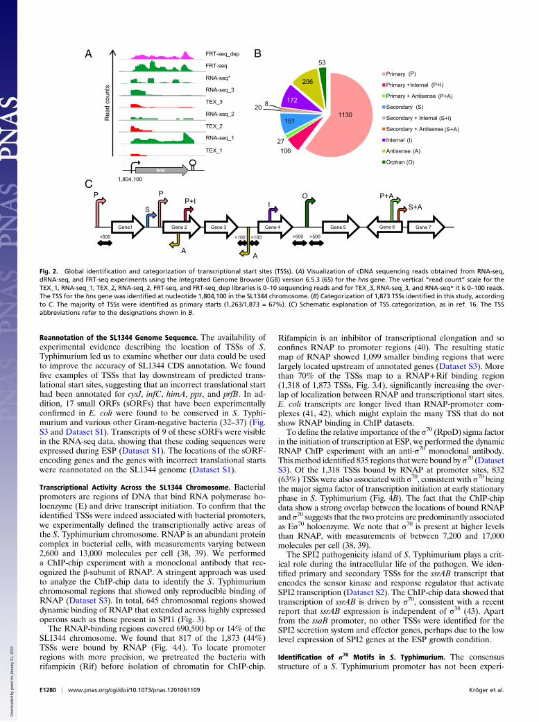
TSSs of S. Typhimurium grown to early stationary phase (ESP)(Fig. 1 and Dataset S2). ESP is an infection-relevant growthcondition associated with high levels of expression of the SPI1virulence genes that are responsible for invasion of epithelialcells (24). To ensure that the identified TSSs were robust andreproducible, we used five biological replicates of RNA-seq(including three dRNA-seq replicates) and a combination of 454and Illumina-based sequence platforms (Figs. 1 and 2A). Theidentification of small regulatory RNAs was aided by the en-richment of one of the RNA samples for small RNA fragments(<500 nt). We complemented the standard RNA-seq protocol byusing the flow cell reverse transcription sequencing (FRT-seq)approach; this method involved the synthesis of cDNA on thesequencing flow cell to improve cDNA library representation(25). The dRNA-seq technique identified examples of processedtranscripts, such as the small RNAs ArcZ and RprA, and suc-ceeded in precisely localizing the TSS to a single nucleotide (26,27). Two FRT-seq sequencing reactions were conducted on oneof the biological replicates, one of which was depleted for rRNA(Fig. 1A) (25). The sequencing statistics and the number of bi-ological replicates are shown in Fig. 1B and Dataset S1. Wemapped >12 million sequence reads uniquely to the S. Typhi-murium SL1344 genome, amounting to 120-fold coverage. Atotal of ∼3.5 million (23%) of all sequenced reads mapped to theannotated coding sequences (CDS), whereas just ∼200,000 reads(1.75%) mapped antisense to CDS.The dRNA-seq data often confirmed the TSSs that were al-
ready clearly apparent from RNA-seq and FRT-seq, as seen forthe hns gene (Fig. 2A). When the location of the start of tran-scripts was not clear from RNA-seq and FRT-seq, the dRNA-seqbecame more important. A conservative approach was used toidentify the precise nucleotide used for transcriptional initiation:The same “+1” nucleotide of each TSS was identified in at leasttwo biological replicates using dRNA-seq. A total of 1,873 TSSswere classified into eight promoter categories (16) (Fig. 2 B andC). We assigned primary starts to 1,130 protein-coding genes ofS. Typhimurium and 87 transcriptional starts were assigned toknown or newly identified small RNAs (see below). We observed206 TSSs for transcripts located antisense to ORFs and 172 in-ternal starts, highlighting the complexity of transcription andgene expression in Salmonella.
Validation of S. Typhimurium TSSs. The dRNA-seq approach hasalready been validated in Helicobacter, Synechocystis, and otherorganisms (16, 28–31), but not in S. Typhimurium. It was im-portant to put our global TSS approach into the context of thewealth of the Salmonella literature. We found publications thatdescribed the TSSs of 57 genes. Fig. S2 and Dataset S2 show theoverlap between 37 of the published transcriptional start sitesthat are present within our dRNA-seq dataset. Thirty-one of 37transcriptional starts lie within ± 2nt of the published start, with15 starts matching exactly.To corroborate our approach, we performed a series of 5′-
RACE experiments that unambiguously identified TSSs for 10genes, namely invF, hilD, ompA, osmC, phoP, prgH, slyA, sodB,yfgE, and yibP (Fig. S2). The 5′-RACE data were in completeagreement with the TSSs, confirming that the 1,873 TSSsrepresent a robust database that describes the transcription ofS. Typhimurium genes at ESP.
E1278 | www.pnas.org/cgi/doi/10.1073/pnas.1201061109 Kröger et al.
Dow
nloa
ded
by g
uest
on
Janu
ary
21, 2
022

Fig. 1. RNA-seq and ChIP-chip–based strategy to identify promoters, transcribed regions, and small RNAs. (A) The different cDNA libraries that were gen-erated and sequenced in this study. (B) The distribution of mapped reads to different genome locations (IGR, intergenic region). Names of the 11 sequencedatasets are defined in Dataset S1. The three libraries marked with TEX were treated with terminator exonuclease (TEX) to enrich the cDNA libraries forprimary transcripts that carried a 5′ triphosphate (dRNA-seq). The suffix numbers link the “RNA-seq” (TEX-untreated) libraries with the appropriate TEX--enriched libraries. The RNA-seq* library sample was enriched for small RNA species (mirVana) before cDNA library generation. The FRT-seq_dep sample wasdepleted of rRNA, and both FRT-seq and FRT-seq_dep were done with RNA from the same biological replicate (Materials and Methods). (C) The workflow thatuses the dRNA-seq, RNA-seq, and ChIP-chip data to identify small RNAs, TSSs, promoters, and transcribed regions throughout the chromosome ofS. Typhimurium SL1344 (Rif, rifampicin; RNAP, RNA polymerase; TSS, transcriptional start site).
Kröger et al. PNAS | Published online April 25, 2012 | E1279
MICRO
BIOLO
GY
PNASPL
US
Dow
nloa
ded
by g
uest
on
Janu
ary
21, 2
022
Reannotation of the SL1344 Genome Sequence. The availability ofexperimental evidence describing the location of TSSs of S.Typhimurium led us to examine whether our data could be usedto improve the accuracy of SL1344 CDS annotation. We foundfive examples of TSSs that lay downstream of predicted trans-lational start sites, suggesting that an incorrect translational starthad been annotated for cysJ, infC, himA, pps, and prfB. In ad-dition, 17 small ORFs (sORFs) that have been experimentallyconfirmed in E. coli were found to be conserved in S. Typhi-murium and various other Gram-negative bacteria (32–37) (Fig.S3 and Dataset S1). Transcripts of 9 of these sORFs were visiblein the RNA-seq data, showing that these coding sequences wereexpressed during ESP (Dataset S1). The locations of the sORF-encoding genes and the genes with incorrect translational startswere reannotated on the SL1344 genome (Dataset S1).
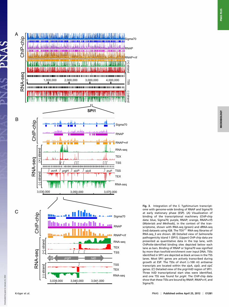
Transcriptional Activity Across the SL1344 Chromosome. Bacterialpromoters are regions of DNA that bind RNA polymerase ho-loenzyme (E) and drive transcript initiation. To confirm that theidentified TSSs were indeed associated with bacterial promoters,we experimentally defined the transcriptionally active areas ofthe S. Typhimurium chromosome. RNAP is an abundant proteincomplex in bacterial cells, with measurements varying between2,600 and 13,000 molecules per cell (38, 39). We performeda ChIP-chip experiment with a monoclonal antibody that rec-ognized the β-subunit of RNAP. A stringent approach was usedto analyze the ChIP-chip data to identify the S. Typhimuriumchromosomal regions that showed only reproducible binding ofRNAP (Dataset S3). In total, 645 chromosomal regions showeddynamic binding of RNAP that extended across highly expressedoperons such as those present in SPI1 (Fig. 3).The RNAP-binding regions covered 690,500 bp or 14% of the
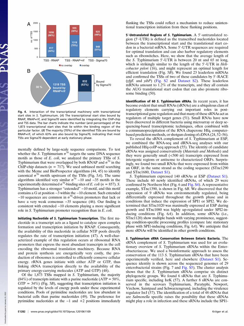
SL1344 chromosome. We found that 817 of the 1,873 (44%)TSSs were bound by RNAP (Fig. 4A). To locate promoterregions with more precision, we pretreated the bacteria withrifampicin (Rif) before isolation of chromatin for ChIP-chip.
Rifampicin is an inhibitor of transcriptional elongation and soconfines RNAP to promoter regions (40). The resulting staticmap of RNAP showed 1,099 smaller binding regions that werelargely located upstream of annotated genes (Dataset S3). Morethan 70% of the TSSs map to a RNAP+Rif binding region(1,318 of 1,873 TSSs, Fig. 3A), significantly increasing the over-lap of localization between RNAP and transcriptional start sites.E. coli transcripts are longer lived than RNAP-promoter com-plexes (41, 42), which might explain the many TSS that do notshow RNAP binding in ChIP datasets.To define the relative importance of the σ70 (RpoD) sigma factor
in the initiation of transcription at ESP, we performed the dynamicRNAP ChIP experiment with an anti-σ70 monoclonal antibody.This method identified 835 regions that were bound by σ70 (DatasetS3). Of the 1,318 TSSs bound by RNAP at promoter sites, 832(63%) TSSs were also associated with σ70, consistent with σ70 beingthe major sigma factor of transcription initiation at early stationaryphase in S. Typhimurium (Fig. 4B). The fact that the ChIP-chipdata show a strong overlap between the locations of bound RNAPand σ70 suggests that the two proteins are predominantly associatedas Eσ70 holoenzyme. We note that σ70 is present at higher levelsthan RNAP, with measurements of between 7,200 and 17,000molecules per cell (38, 39).The SPI2 pathogenicity island of S. Typhimurium plays a crit-
ical role during the intracellular life of the pathogen. We iden-tified primary and secondary TSSs for the ssrAB transcript thatencodes the sensor kinase and response regulator that activateSPI2 transcription (Dataset S2). The ChIP-chip data showed thattranscription of ssrAB is driven by σ70, consistent with a recentreport that ssrAB expression is independent of σ38 (43). Apartfrom the ssaB promoter, no other TSSs were identified for theSPI2 secretion system and effector genes, perhaps due to the lowlevel expression of SPI2 genes at the ESP growth condition.
Identification of σ70 Motifs in S. Typhimurium. The consensusstructure of a S. Typhimurium promoter has not been experi-
1130
10627
151
20 8 172
206
53
Primary
Primary +Internal
Primary + Antisense
Secondary
Secondary + Internal
Secondary + Antisense
Internal
Antisense
Orphan
TEX_1
RNA-seq_1
TEX_2
RNA-seq_2
TEX_3
RNA-seq_3
RNA-seq*
FRT-seq
FRT-seq_dep
Rea
d co
unts
hns
1,804,100
Gene1 Gene 2 Gene 3 Gene 4 Gene 5
P P
S
AA
IO
P+I
<500 <100 <100 >500 >500
Gene 7Gene 6
S+AP+A
C
A B
(P)
(P+I)
(P+A)
(S)
(S+I)
(S+A)
(I)
(A)
(O)
Fig. 2. Global identification and categorization of transcriptional start sites (TSSs). (A) Visualization of cDNA sequencing reads obtained from RNA-seq,dRNA-seq, and FRT-seq experiments using the Integrated Genome Browser (IGB) version 6.5.3 (65) for the hns gene. The vertical “read count” scale for theTEX_1, RNA-seq_1, TEX_2, RNA-seq_2, FRT-seq, and FRT-seq_dep libraries is 0–10 sequencing reads and for TEX_3, RNA-seq_3, and RNA-seq* it is 0–100 reads.The TSS for the hns gene was identified at nucleotide 1,804,100 in the SL1344 chromosome. (B) Categorization of 1,873 TSSs identified in this study, accordingto C. The majority of TSSs were identified as primary starts (1,263/1,873 = 67%). (C) Schematic explanation of TSS categorization, as in ref. 16. The TSSabbreviations refer to the designations shown in B.
E1280 | www.pnas.org/cgi/doi/10.1073/pnas.1201061109 Kröger et al.
Dow
nloa
ded
by g
uest
on
Janu
ary
21, 2
022
0 1,000,000 2,000,000 3,000,000 4,000,000
ChI
P-c
hip
RN
A-s
eq
(+) strand(- ) strand
TSS
Sigma70
RNAP
RNAP+rif
SPI1
prgH sicPavrA sicA invF
Sigma70
RNAP
RNAP+rif
RNA-seq
TEX
TSS
TSS
TEX
RNA-seq
(+) s
trand
( - ) s
trand
ChI
P-c
hip
RN
A -se
q
3,030,000 3,050,000 3,070,000
hilD
prgI
prgI prgH
3,039,000 3,040,000 3,041,000
(+) s
trand
(-) s
trandRN
A -se
qC
hIP
-chi
p
Sigma70
RNAP
RNAP+rif
RNA-seq
TEX
TSS
TSS
TEX
RNA-seq
B
A
CFig. 3. Integration of the S. Typhimurium transcript-ome with genome-wide binding of RNAP and Sigma70at early stationary phase (ESP). (A) Visualization ofbinding of the transcriptional machinery (ChIP-chipdata: blue, Sigma70; purple, RNAP; orange, RNAP+rif)(Materials and Methods), in the context of the tran-scriptome, shown with RNA-seq (green) and dRNA-seq(red) datasets using IGB. The TEX+/− RNA-seq libraries ofRNA-seq_3 are shown. (B) Detailed view of Salmonellapathogenicity island 1 (SPI1). (Upper) ChIP-chip data arepresented as quantitative data in the top lane, withChIPotle-identified binding sites depicted below eachlane as bars. Binding of RNAP or Sigma70 was signifiedby more than twofold enrichment over input DNA. TSSsidentified in SPI1 are depicted as black arrows in the TSSlanes. Most SPI1 genes are actively transcribed duringgrowth at ESP. The TSSs of short (<100 nt) antisensetranscripts are located within the sipA, sipD, and sipCgenes. (C) Detailed view of the prgI-hilD region of SPI1.Three hilD transcriptional start sites were identified,and one TSS was found for prgH. The ChIP-chip datashow that these TSSs are bound by RNAP, RNAP+rif, andSigma70.
Kröger et al. PNAS | Published online April 25, 2012 | E1281
MICRO
BIOLO
GY
PNASPL
US
Dow
nloa
ded
by g
uest
on
Janu
ary
21, 2
022
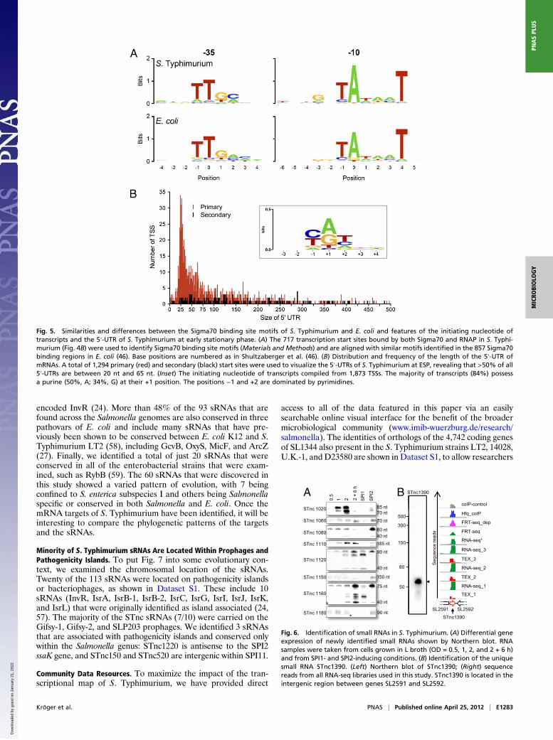
mentally defined by large-scale sequence comparisons. To testwhether the S. Typhimurium σ70 targets the same DNA sequencemotifs as those of E. coli, we analyzed the primary TSSs of S.Typhimurium that were overlapped by both RNAP and σ70 in theChIP-chip datasets (n = 717). We used unbiased motif searchingwith the Meme and BioProspector algorithms (44, 45) to identifycanonical σ70 motifs upstream of the TSSs (Fig. 5A). The samealgorithms identified very similar σ70 −10 and −35 motifs in theexperimentally determined σ70-binding sites of E. coli (n= 857). S.Typhimurium has a stronger “extended” −10 motif, and this motifcontains a G at position −3 within the −10 element. Such extended−10 sequences are common in σ70-driven promoters that lack orhave a very weak consensus −35 sequence (46). Our finding isconsistent with extended −10 elements playing a more significantrole in S. Typhimurium promoter recognition than in E. coli.
Initiating Nucleotide of S. Typhimurium Transcription. The first nu-cleotide in a transcript acts as a ligand to catalyze open complexformation and transcription initiation by RNAP. Consequently,the availability of this nucleotide in cellular NTP pools directlyregulates the rate of transcription initiation (47). A well-char-acterized example of this regulation occurs at ribosomal RNApromoters that express the most abundant transcripts in the cellencoding the ribosomal translation machinery. Because RNAand protein synthesis are energetically very costly, the pro-duction of ribosomes is controlled to efficiently conserve cellularenergy. rRNA genes initiate with either ATP or GTP, thuslinking rRNA transcription directly to the availability of theprimary energy-carrying molecules (ATP and GTP) (48).Of the 1,873 TSSs mapped in S. Typhimurium, the majority
(84%) of transcripts initiate with a purine nucleotide (ATP = 50%,GTP = 34%) (Fig. 5B), suggesting that transcription initiation isregulated by the levels of energy pools under these experimentalconditions. Pools of pyrimidine nucleotides are less abundant inbacterial cells than purine nucleotides (49). The preference forpyrimidine nucleotides at the −1 and +2 positions immediately
flanking the TSSs could reflect a mechanism to reduce uninten-tional transcription initiation from these flanking positions.
5′-Untranslated Regions of S. Typhimurium. A 5′-untranslated re-gion (5′-UTR) is defined as the transcribed nucleotides locatedbetween the transcriptional start and the translational start co-don in a bacterial mRNA. Some 5′-UTR sequences are requiredfor optimal translation and can also harbor regulatory elementssuch as riboswitches. Here, we show that the average length ofthe S. Typhimurium 5′-UTR is between 20 nt and 65 nt long,which is strikingly similar to the length of the 5′-UTR in Heli-cobacter pylori (16), and might represent an optimal length forefficient translation (Fig. 5B). We found 23 leaderless mRNAsand confirmed the TSSs of two of these candidates by 5′-RACE(yfgE and yibP) (Fig. S2 and Dataset S2). These leaderlessmRNAs amount to 1.2% of the transcripts, and they all containthe AUG translational start codon that can also promote ribo-some binding (50).
Identification of 60 S. Typhimurium sRNAs. In recent years, it hasbecome evident that small RNAs (sRNAs) are a ubiquitous class ofregulatory elements carrying out important roles in post-transcriptional gene regulation and thatmany of these sRNAsact asregulators of multiple target genes (51). Small RNAs have nowbeen discovered in different bacteria using microarray or deep se-quencing-based transcriptomic techniques, often combined witha coimmunoprecipitation of the RNA chaperone Hfq, computer-based predictionmethods, or shotgun cloning of cDNA (24, 52–56).To reveal the sRNA complement of S. Typhimurium at ESP,
we combined the RNA-seq and dRNA-seq analyses with ourpublished Hfq-coIP-seq approach (55). The identity of candidatesRNAs was assigned conservatively (Materials and Methods) andthey were generally small (<500 nt) transcripts expressed fromintergenic regions or antisense to characterized ORFs. Surpris-ingly, we found two small RNAs that were expressed from withinan ORF, in the same strand as the coding sequence (STnc1290and STnc1680, Dataset S1).S. Typhimurium expressed 140 sRNAs at ESP (Dataset S1).
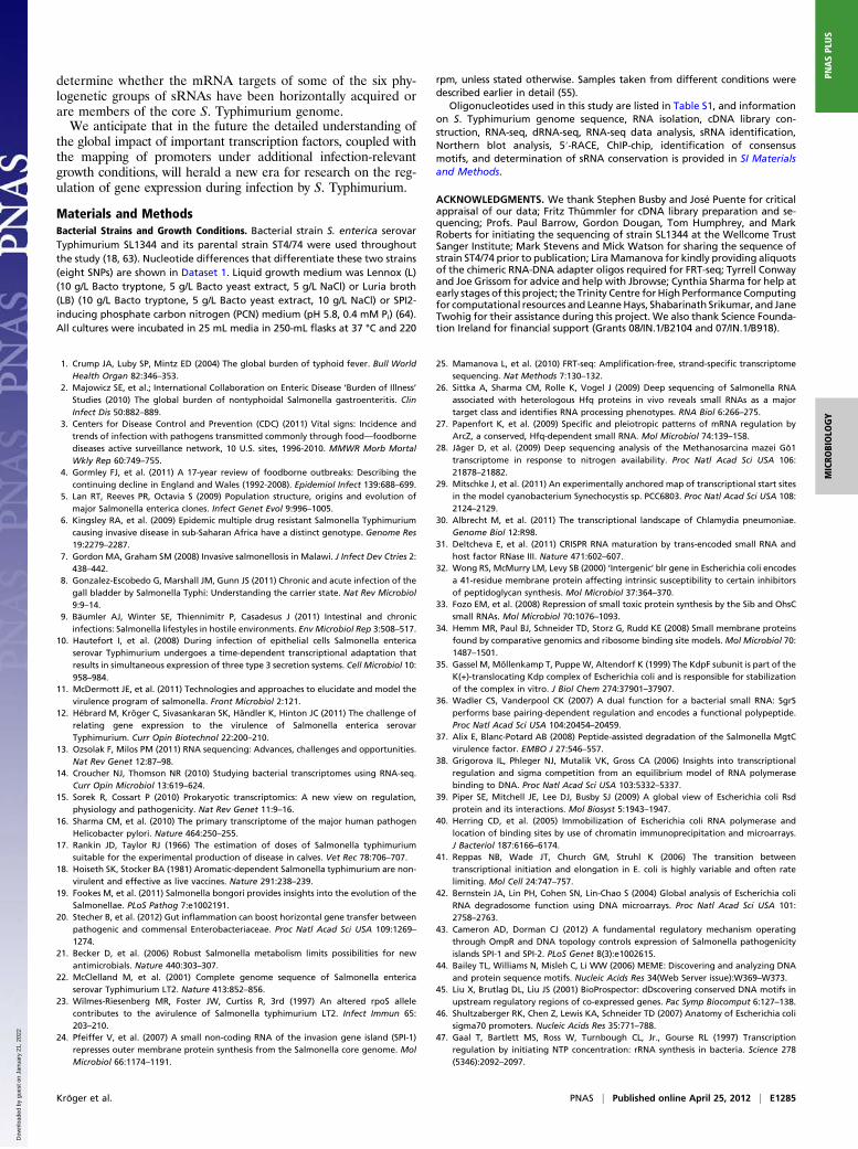
These include 60 newly identified sRNAs, of which 29 wereconfirmed by Northern blot (Fig. 6 and Fig. S4). A representativeexample, STnc1390, is shown in Fig. 6B. We discovered that theexpression of 9 sRNAs was environmentally regulated, beingdifferentially expressed throughout the growth phase and inconditions that induce the expression of SPI1 or SPI2. We de-termined that STnc1020 was maximally expressed at ESP duringgrowth and STnc1080 was highly up-regulated under SPI2-in-ducing conditions (Fig. 6A). In addition, some sRNAs (i.e.,STnc1120) show multiple bands with varying prominence, suggest-ing condition-specific processing profiles (compare late stationaryphase with SPI1-inducing conditions, Fig. 6A). We anticipate thatmore sRNAs will be identified in other growth conditions.
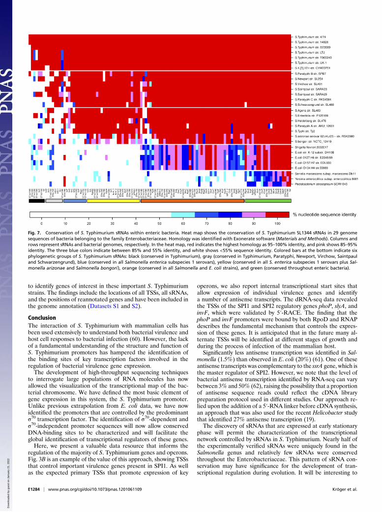
S. Typhimurium sRNA Conservation Between Enteric Bacteria. ThesRNA complement of S. Typhimurium was used for an evolu-tionary overview of S. Typhimurium sRNAs within the Enter-obacteriaceae. We used a bioinformatic approach to assess theconservation of the 113 S. Typhimurium sRNAs that have beenexperimentally verified, here and elsewhere (Dataset S1). Se-quence identity is shown across the sequenced genomes of 29enterobacterial strains (Fig. 7 and Fig. S5). The cluster analysisshows that the S. Typhimurium sRNAs comprise six distinctphylogenetic groups. We found 6 sRNAs that are S. Typhimu-rium specific, including IsrK (57). A further 8 sRNAs are con-served in the serovars Typhimurium, Paratyphi, Newport,Virchow, Saintpaul and Schwarzengrund, including the virulenceregulator IsrJ (57). The identification of a total of 48 sRNAs thatare Salmonella specific raises the possibility that these sRNAsmight play a role in infection and these sRNAs include the SPI1-
0
500
1000
1500
2000
TSS - RNAP TSS - RNAP+rif TSS - Sigma70
no overlap overlap
no. o
f TS
S
817(44%)
1056(66%)
555(30%)
1318(70%) 948
(51%)
925(49%)
A
B
1318 TSSbound byRNAP+rif
30 %
Boundby Sigma70
832(63%)
Fig. 4. Interaction of the transcriptional machinery with transcriptionalstart sites in S. Typhimurium. (A) The transcriptional start sites bound byRNAP, RNAP+rif, and Sigma70 were identified by integrating the ChIP-chipand TSS data. The bar charts indicate the number (and percentages) of the1,873 transcriptional start sites that lie within the binding region of theparticular factor. (B) The majority (70%) of the identified TSSs are bound byRNAP+rif, of which 63% are also bound by Sigma70, indicating that mostTSSs are Sigma70 dependent at early stationary phase.
E1282 | www.pnas.org/cgi/doi/10.1073/pnas.1201061109 Kröger et al.
Dow
nloa
ded
by g
uest
on
Janu
ary
21, 2
022
encoded InvR (24). More than 48% of the 93 sRNAs that arefound across the Salmonella genomes are also conserved in threepathovars of E. coli and include many sRNAs that have pre-viously been shown to be conserved between E. coli K12 and S.Typhimurium LT2 (58), including GcvB, OxyS, MicF, and ArcZ(27). Finally, we identified a total of just 20 sRNAs that wereconserved in all of the enterobacterial strains that were exam-ined, such as RybB (59). The 60 sRNAs that were discovered inthis study showed a varied pattern of evolution, with 7 beingconfined to S. enterica subspecies I and others being Salmonellaspecific or conserved in both Salmonella and E. coli. Once themRNA targets of S. Typhimurium have been identified, it will beinteresting to compare the phylogenetic patterns of the targetsand the sRNAs.
Minority of S. Typhimurium sRNAs Are Located Within Prophages andPathogenicity Islands. To put Fig. 7 into some evolutionary con-text, we examined the chromosomal location of the sRNAs.Twenty of the 113 sRNAs were located on pathogenicity islandsor bacteriophages, as shown in Dataset S1. These include 10sRNAs (InvR, IsrA, IsrB-1, IsrB-2, IsrC, IsrG, IsrI, IsrJ, IsrK,and IsrL) that were originally identified as island associated (24,57). The majority of the STnc sRNAs (7/10) were carried on theGifsy-1, Gifsy-2, and SLP203 prophages. We identified 3 sRNAsthat are associated with pathogenicity islands and conserved onlywithin the Salmonella genus: STnc1220 is antisense to the SPI2ssaK gene, and STnc150 and STnc520 are intergenic within SPI11.
Community Data Resources. To maximize the impact of the tran-scriptional map of S. Typhimurium, we have provided direct
access to all of the data featured in this paper via an easilysearchable online visual interface for the benefit of the broadermicrobiological community (www.imib-wuerzburg.de/research/salmonella). The identities of orthologs of the 4,742 coding genesof SL1344 also present in the S. Typhimurium strains LT2, 14028,U.K.-1, and D23580 are shown in Dataset S1, to allow researchers
Fig. 5. Similarities and differences between the Sigma70 binding site motifs of S. Typhimurium and E. coli and features of the initiating nucleotide oftranscripts and the 5′-UTR of S. Typhimurium at early stationary phase. (A) The 717 transcription start sites bound by both Sigma70 and RNAP in S. Typhi-murium (Fig. 4B) were used to identify Sigma70 binding site motifs (Materials and Methods) and are aligned with similar motifs identified in the 857 Sigma70binding regions in E. coli (46). Base positions are numbered as in Shultzaberger et al. (46). (B) Distribution and frequency of the length of the 5′-UTR ofmRNAs. A total of 1,294 primary (red) and secondary (black) start sites were used to visualize the 5′-UTRs of S. Typhimurium at ESP, revealing that >50% of all5′-UTRs are between 20 nt and 65 nt. (Inset) The initiating nucleotide of transcripts compiled from 1,873 TSSs. The majority of transcripts (84%) possessa purine (50%, A; 34%, G) at their +1 position. The positions −1 and +2 are dominated by pyrimidines.
SPI1
SPI2
85 nt70 nt
70 nt
60 nt
90 nt
75 nt
90 nt
STnc 1020
STnc 1060
185 nt
STnc 1080
STnc 1110
150 nt
STnc 1120
STnc 1150
STnc 1160
STnc 1180
0.5
1 2 2 +
6 h
40 nt
40 nt
40 nt
150
80
50
300
500
STnc1390
Sequ
ence
read
s
A B
SL2591
STnc1390
SL2592
coIP-control
Hfq_coIP
FRT-seq_dep
FRT-seq
RNA-seq*
RNA-seq_3
TEX_3
RNA-seq_2
TEX_2
RNA-seq_1
TEX_1
Fig. 6. Identification of small RNAs in S. Typhimurium. (A) Differential geneexpression of newly identified small RNAs shown by Northern blot. RNAsamples were taken from cells grown in L broth (OD = 0.5, 1, 2, and 2 + 6 h)and from SPI1- and SPI2-inducing conditions. (B) Identification of the uniquesmall RNA STnc1390. (Left) Northern blot of STnc1390; (Right) sequencereads from all RNA-seq libraries used in this study. STnc1390 is located in theintergenic region between genes SL2591 and SL2592.
Kröger et al. PNAS | Published online April 25, 2012 | E1283
MICRO
BIOLO
GY
PNASPL
US
Dow
nloa
ded
by g
uest
on
Janu
ary
21, 2
022
to identify genes of interest in these important S. Typhimuriumstrains. The findings include the locations of all TSSs, all sRNAs,and the positions of reannotated genes and have been included inthe genome annotation (Datasets S1 and S2).
ConclusionThe interaction of S. Typhimurium with mammalian cells hasbeen used extensively to understand both bacterial virulence andhost cell responses to bacterial infection (60). However, the lackof a fundamental understanding of the structure and function ofS. Typhimurium promoters has hampered the identification ofthe binding sites of key transcription factors involved in theregulation of bacterial virulence gene expression.The development of high-throughput sequencing techniques
to interrogate large populations of RNA molecules has nowallowed the visualization of the transcriptional map of the bac-terial chromosome. We have defined the most basic element ofgene expression in this system, the S. Typhimurium promoter.Unlike previous extrapolation from E. coli data, we have nowidentified the promoters that are controlled by the predominantσ70 transcription factor. The identification of σ70-dependent andσ70-independent promoter sequences will now allow conservedDNA-binding sites to be characterized and will facilitate theglobal identification of transcriptional regulators of these genes.Here, we present a valuable data resource that informs the
regulation of the majority of S. Typhimurium genes and operons.Fig. 3B is an example of the value of this approach, showing TSSsthat control important virulence genes present in SPI1. As wellas the expected primary TSSs that promote expression of key
operons, we also report internal transcriptional start sites thatallow expression of individual virulence genes and identifya number of antisense transcripts. The dRNA-seq data revealedthe TSSs of the SPI1 and SPI2 regulatory genes phoP, slyA, andinvF, which were validated by 5′-RACE. The finding that thephoP and invF promoters were bound by both RpoD and RNAPdescribes the fundamental mechanism that controls the expres-sion of these genes. It is anticipated that in the future many al-ternate TSSs will be identified at different stages of growth andduring the process of infection of the mammalian host.Significantly less antisense transcription was identified in Sal-
monella (1.5%) than observed in E. coli (20%) (61). One of theseantisense transcripts was complementary to the ssrA gene, which isthe master regulator of SPI2. However, we note that the level ofbacterial antisense transcription identified by RNA-seq can varybetween 3% and 50% (62), raising the possibility that a proportionof antisense sequence reads could reflect the cDNA librarypreparation protocol used in different studies. Our approach re-lied upon the addition of a 5′-RNA linker before cDNA synthesis,an approach that was also used for the recent Helicobacter studythat identified 27% antisense transcription (19).The discovery of sRNAs that are expressed at early stationary
phase will permit the characterization of the transcriptionalnetwork controlled by sRNAs in S. Typhimurium. Nearly half ofthe experimentally verified sRNAs were uniquely found in theSalmonella genus and relatively few sRNAs were conservedthroughout the Enterobacteriaceae. This pattern of sRNA con-servation may have significance for the development of tran-scriptional regulation during evolution. It will be interesting to
Fig. 7. Conservation of S. Typhimurium sRNAs within enteric bacteria. Heat map shows the conservation of S. Typhimurium SL1344 sRNAs in 29 genomesequences of bacteria belonging to the family Enterobacteriaceae. Homology was identified with Exonerate software (Materials and Methods). Columns androws represent sRNAs and bacterial genomes, respectively. In the heat map, red indicates the highest homology as 95–100% identity, and pink shows 85–95%identity. The three blue colors indicate between 85% and 55% identity, and white shows <55% sequence identity. Colored bars at the bottom indicate sixphylogenetic groups of S. Typhimurium sRNAs: black (conserved in Typhimurium), gray (conserved in Typhimurium, Paratyphi, Newport, Virchow, Saintpauland Schwarzengrund), blue (conserved in all Salmonella enterica subspecies 1 serovars), yellow (conserved in all S. enterica subspecies 1 serovars plus Sal-monella arizonae and Salmonella bongori), orange (conserved in all Salmonella and E. coli strains), and green (conserved throughout enteric bacteria).
E1284 | www.pnas.org/cgi/doi/10.1073/pnas.1201061109 Kröger et al.
Dow
nloa
ded
by g
uest
on
Janu
ary
21, 2
022
determine whether the mRNA targets of some of the six phy-logenetic groups of sRNAs have been horizontally acquired orare members of the core S. Typhimurium genome.We anticipate that in the future the detailed understanding of
the global impact of important transcription factors, coupled withthe mapping of promoters under additional infection-relevantgrowth conditions, will herald a new era for research on the reg-ulation of gene expression during infection by S. Typhimurium.
Materials and MethodsBacterial Strains and Growth Conditions. Bacterial strain S. enterica serovarTyphimurium SL1344 and its parental strain ST4/74 were used throughoutthe study (18, 63). Nucleotide differences that differentiate these two strains(eight SNPs) are shown in Dataset 1. Liquid growth medium was Lennox (L)(10 g/L Bacto tryptone, 5 g/L Bacto yeast extract, 5 g/L NaCl) or Luria broth(LB) (10 g/L Bacto tryptone, 5 g/L Bacto yeast extract, 10 g/L NaCl) or SPI2-inducing phosphate carbon nitrogen (PCN) medium (pH 5.8, 0.4 mM Pi) (64).All cultures were incubated in 25 mL media in 250-mL flasks at 37 °C and 220
rpm, unless stated otherwise. Samples taken from different conditions weredescribed earlier in detail (55).
Oligonucleotides used in this study are listed in Table S1, and informationon S. Typhimurium genome sequence, RNA isolation, cDNA library con-struction, RNA-seq, dRNA-seq, RNA-seq data analysis, sRNA identification,Northern blot analysis, 5′-RACE, ChIP-chip, identification of consensusmotifs, and determination of sRNA conservation is provided in SI Materialsand Methods.
ACKNOWLEDGMENTS. We thank Stephen Busby and José Puente for criticalappraisal of our data; Fritz Thümmler for cDNA library preparation and se-quencing; Profs. Paul Barrow, Gordon Dougan, Tom Humphrey, and MarkRoberts for initiating the sequencing of strain SL1344 at the Wellcome TrustSanger Institute; Mark Stevens and Mick Watson for sharing the sequence ofstrain ST4/74 prior to publication; LiraMamanova for kindly providing aliquotsof the chimeric RNA-DNA adapter oligos required for FRT-seq; Tyrrell Conwayand Joe Grissom for advice and help with Jbrowse; Cynthia Sharma for help atearly stages of this project; the Trinity Centre forHigh Performance Computingfor computational resources and LeanneHays, Shabarinath Srikumar, and JaneTwohig for their assistance during this project. We also thank Science Founda-tion Ireland for financial support (Grants 08/IN.1/B2104 and 07/IN.1/B918).
1. Crump JA, Luby SP, Mintz ED (2004) The global burden of typhoid fever. Bull WorldHealth Organ 82:346–353.
2. Majowicz SE, et al.; International Collaboration on Enteric Disease ‘Burden of Illness’Studies (2010) The global burden of nontyphoidal Salmonella gastroenteritis. ClinInfect Dis 50:882–889.
3. Centers for Disease Control and Prevention (CDC) (2011) Vital signs: Incidence andtrends of infection with pathogens transmitted commonly through food—foodbornediseases active surveillance network, 10 U.S. sites, 1996-2010. MMWR Morb MortalWkly Rep 60:749–755.
4. Gormley FJ, et al. (2011) A 17-year review of foodborne outbreaks: Describing thecontinuing decline in England and Wales (1992-2008). Epidemiol Infect 139:688–699.
5. Lan RT, Reeves PR, Octavia S (2009) Population structure, origins and evolution ofmajor Salmonella enterica clones. Infect Genet Evol 9:996–1005.
6. Kingsley RA, et al. (2009) Epidemic multiple drug resistant Salmonella Typhimuriumcausing invasive disease in sub-Saharan Africa have a distinct genotype. Genome Res19:2279–2287.
7. Gordon MA, Graham SM (2008) Invasive salmonellosis in Malawi. J Infect Dev Ctries 2:438–442.
8. Gonzalez-Escobedo G, Marshall JM, Gunn JS (2011) Chronic and acute infection of thegall bladder by Salmonella Typhi: Understanding the carrier state. Nat Rev Microbiol9:9–14.
9. Bäumler AJ, Winter SE, Thiennimitr P, Casadesus J (2011) Intestinal and chronicinfections: Salmonella lifestyles in hostile environments. Env Microbiol Rep 3:508–517.
10. Hautefort I, et al. (2008) During infection of epithelial cells Salmonella entericaserovar Typhimurium undergoes a time-dependent transcriptional adaptation thatresults in simultaneous expression of three type 3 secretion systems. Cell Microbiol 10:958–984.
11. McDermott JE, et al. (2011) Technologies and approaches to elucidate and model thevirulence program of salmonella. Front Microbiol 2:121.
12. Hébrard M, Kröger C, Sivasankaran SK, Händler K, Hinton JC (2011) The challenge ofrelating gene expression to the virulence of Salmonella enterica serovarTyphimurium. Curr Opin Biotechnol 22:200–210.
13. Ozsolak F, Milos PM (2011) RNA sequencing: Advances, challenges and opportunities.Nat Rev Genet 12:87–98.
14. Croucher NJ, Thomson NR (2010) Studying bacterial transcriptomes using RNA-seq.Curr Opin Microbiol 13:619–624.
15. Sorek R, Cossart P (2010) Prokaryotic transcriptomics: A new view on regulation,physiology and pathogenicity. Nat Rev Genet 11:9–16.
16. Sharma CM, et al. (2010) The primary transcriptome of the major human pathogenHelicobacter pylori. Nature 464:250–255.
17. Rankin JD, Taylor RJ (1966) The estimation of doses of Salmonella typhimuriumsuitable for the experimental production of disease in calves. Vet Rec 78:706–707.
18. Hoiseth SK, Stocker BA (1981) Aromatic-dependent Salmonella typhimurium are non-virulent and effective as live vaccines. Nature 291:238–239.
19. Fookes M, et al. (2011) Salmonella bongori provides insights into the evolution of theSalmonellae. PLoS Pathog 7:e1002191.
20. Stecher B, et al. (2012) Gut inflammation can boost horizontal gene transfer betweenpathogenic and commensal Enterobacteriaceae. Proc Natl Acad Sci USA 109:1269–1274.
21. Becker D, et al. (2006) Robust Salmonella metabolism limits possibilities for newantimicrobials. Nature 440:303–307.
22. McClelland M, et al. (2001) Complete genome sequence of Salmonella entericaserovar Typhimurium LT2. Nature 413:852–856.
23. Wilmes-Riesenberg MR, Foster JW, Curtiss R, 3rd (1997) An altered rpoS allelecontributes to the avirulence of Salmonella typhimurium LT2. Infect Immun 65:203–210.
24. Pfeiffer V, et al. (2007) A small non-coding RNA of the invasion gene island (SPI-1)represses outer membrane protein synthesis from the Salmonella core genome. MolMicrobiol 66:1174–1191.
25. Mamanova L, et al. (2010) FRT-seq: Amplification-free, strand-specific transcriptomesequencing. Nat Methods 7:130–132.
26. Sittka A, Sharma CM, Rolle K, Vogel J (2009) Deep sequencing of Salmonella RNAassociated with heterologous Hfq proteins in vivo reveals small RNAs as a majortarget class and identifies RNA processing phenotypes. RNA Biol 6:266–275.
27. Papenfort K, et al. (2009) Specific and pleiotropic patterns of mRNA regulation byArcZ, a conserved, Hfq-dependent small RNA. Mol Microbiol 74:139–158.
28. Jäger D, et al. (2009) Deep sequencing analysis of the Methanosarcina mazei Gö1transcriptome in response to nitrogen availability. Proc Natl Acad Sci USA 106:21878–21882.
29. Mitschke J, et al. (2011) An experimentally anchored map of transcriptional start sitesin the model cyanobacterium Synechocystis sp. PCC6803. Proc Natl Acad Sci USA 108:2124–2129.
30. Albrecht M, et al. (2011) The transcriptional landscape of Chlamydia pneumoniae.Genome Biol 12:R98.
31. Deltcheva E, et al. (2011) CRISPR RNA maturation by trans-encoded small RNA andhost factor RNase III. Nature 471:602–607.
32. Wong RS, McMurry LM, Levy SB (2000) ‘Intergenic’ blr gene in Escherichia coli encodesa 41-residue membrane protein affecting intrinsic susceptibility to certain inhibitorsof peptidoglycan synthesis. Mol Microbiol 37:364–370.
33. Fozo EM, et al. (2008) Repression of small toxic protein synthesis by the Sib and OhsCsmall RNAs. Mol Microbiol 70:1076–1093.
34. Hemm MR, Paul BJ, Schneider TD, Storz G, Rudd KE (2008) Small membrane proteinsfound by comparative genomics and ribosome binding site models. Mol Microbiol 70:1487–1501.
35. Gassel M, Möllenkamp T, PuppeW, Altendorf K (1999) The KdpF subunit is part of theK(+)-translocating Kdp complex of Escherichia coli and is responsible for stabilizationof the complex in vitro. J Biol Chem 274:37901–37907.
36. Wadler CS, Vanderpool CK (2007) A dual function for a bacterial small RNA: SgrSperforms base pairing-dependent regulation and encodes a functional polypeptide.Proc Natl Acad Sci USA 104:20454–20459.
37. Alix E, Blanc-Potard AB (2008) Peptide-assisted degradation of the Salmonella MgtCvirulence factor. EMBO J 27:546–557.
38. Grigorova IL, Phleger NJ, Mutalik VK, Gross CA (2006) Insights into transcriptionalregulation and sigma competition from an equilibrium model of RNA polymerasebinding to DNA. Proc Natl Acad Sci USA 103:5332–5337.
39. Piper SE, Mitchell JE, Lee DJ, Busby SJ (2009) A global view of Escherichia coli Rsdprotein and its interactions. Mol Biosyst 5:1943–1947.
40. Herring CD, et al. (2005) Immobilization of Escherichia coli RNA polymerase andlocation of binding sites by use of chromatin immunoprecipitation and microarrays.J Bacteriol 187:6166–6174.
41. Reppas NB, Wade JT, Church GM, Struhl K (2006) The transition betweentranscriptional initiation and elongation in E. coli is highly variable and often ratelimiting. Mol Cell 24:747–757.
42. Bernstein JA, Lin PH, Cohen SN, Lin-Chao S (2004) Global analysis of Escherichia coliRNA degradosome function using DNA microarrays. Proc Natl Acad Sci USA 101:2758–2763.
43. Cameron AD, Dorman CJ (2012) A fundamental regulatory mechanism operatingthrough OmpR and DNA topology controls expression of Salmonella pathogenicityislands SPI-1 and SPI-2. PLoS Genet 8(3):e1002615.
44. Bailey TL, Williams N, Misleh C, Li WW (2006) MEME: Discovering and analyzing DNAand protein sequence motifs. Nucleic Acids Res 34(Web Server issue):W369–W373.
45. Liu X, Brutlag DL, Liu JS (2001) BioProspector: dDscovering conserved DNA motifs inupstream regulatory regions of co-expressed genes. Pac Symp Biocomput 6:127–138.
46. Shultzaberger RK, Chen Z, Lewis KA, Schneider TD (2007) Anatomy of Escherichia colisigma70 promoters. Nucleic Acids Res 35:771–788.
47. Gaal T, Bartlett MS, Ross W, Turnbough CL, Jr., Gourse RL (1997) Transcriptionregulation by initiating NTP concentration: rRNA synthesis in bacteria. Science 278(5346):2092–2097.
Kröger et al. PNAS | Published online April 25, 2012 | E1285
MICRO
BIOLO
GY
PNASPL
US
Dow
nloa
ded
by g
uest
on
Janu
ary
21, 2
022
48. Murray HD, Schneider DA, Gourse RL (2003) Control of rRNA expression by smallmolecules is dynamic and nonredundant. Mol Cell 12:125–134.
49. Buckstein MH, He J, Rubin H (2008) Characterization of nucleotide pools as a functionof physiological state in Escherichia coli. J Bacteriol 190:718–726.
50. Brock JE, Pourshahian S, Giliberti J, Limbach PA, Janssen GR (2008) Ribosomes bindleaderless mRNA in Escherichia coli through recognition of their 5′-terminal AUG.RNA 14:2159–2169.
51. Papenfort K, Vogel J (2010) Regulatory RNA in bacterial pathogens. Cell Host Microbe8:116–127.
52. Vogel J, et al. (2003) RNomics in Escherichia coli detects new sRNA species andindicates parallel transcriptional output in bacteria. Nucleic Acids Res 31:6435–6443.
53. Zhang A, et al. (2003) Global analysis of small RNA and mRNA targets of Hfq. MolMicrobiol 50:1111–1124.
54. WassarmanKM, Repoila F, RosenowC, StorzG,Gottesman S (2001) Identificationofnovelsmall RNAs using comparative genomics and microarrays. Genes Dev 15:1637–1651.
55. Sittka A, et al. (2008) Deep sequencing analysis of small noncoding RNA and mRNAtargets of the global post-transcriptional regulator, Hfq. PLoS Genet 4:e1000163.
56. Sridhar J, et al. (2010) sRNAscanner: A computational tool for intergenic small RNAdetection in bacterial genomes. PLoS ONE 5:e11970.
57. Padalon-Brauch G, et al. (2008) Small RNAs encoded within genetic islands ofSalmonella typhimurium show host-induced expression and role in virulence. NucleicAcids Res 36:1913–1927.
58. Hershberg R, Altuvia S, Margalit H (2003) A survey of small RNA-encoding genes in
Escherichia coli. Nucleic Acids Res 31:1813–1820.59. Papenfort K, et al. (2006) SigmaE-dependent small RNAs of Salmonella respond to
membrane stress by accelerating global omp mRNA decay. Mol Microbiol 62:
1674–1688.60. Tsolis RM, Xavier MN, Santos RL, Bäumler AJ (2011) How to become a top model:
Impact of animal experimentation on human Salmonella disease research. Infect
Immun 79:1806–1814.61. Georg J, Hess WR (2011) cis-antisense RNA, another level of gene regulation in
bacteria. Microbiol Mol Biol Rev 75:286–300.62. Lasa I, et al. (2011) Genome-wide antisense transcription drives mRNA processing in
bacteria. Proc Natl Acad Sci USA 108:20172–20177.63. Richardson EJ, et al. (2011) Genome sequences of Salmonella enterica serovar
typhimurium, Choleraesuis, Dublin, and Gallinarum strains of well- defined virulence
in food-producing animals. J Bacteriol 193:3162–3163.64. Löber S, Jäckel D, Kaiser N, Hensel M (2006) Regulation of Salmonella pathogenicity
island 2 genes by independent environmental signals. Int J Med Microbiol 296:
435–447.65. Nicol JW, Helt GA, Blanchard SG, Jr., Raja A, Loraine AE (2009) The Integrated Genome
Browser: Free software for distribution and exploration of genome-scale datasets.
Bioinformatics 25:2730–2731.
E1286 | www.pnas.org/cgi/doi/10.1073/pnas.1201061109 Kröger et al.
Dow
nloa
ded
by g
uest
on
Janu
ary
21, 2
022
Related Documents