Cellular/Molecular The Transcriptional Activator Hypoxia Inducible Factor 2 (HIF-2/EPAS-1) Regulates the Oxygen-Dependent Expression of Erythropoietin in Cortical Astrocytes Juan C. Chavez, 1,2 Oxana Baranova, 1 Janice Lin, 1 and Paola Pichiule 3 1 Burke Medical Research Institute, White Plains, New York 10605, 2 Department of Neurology and Neuroscience, Weil Medical College of Cornell University, New York, New York 10021, and 3 Department of Pediatrics, Morgan Stanley Children’s Hospital, Columbia University, New York, New York 10032 In the ischemic or hypoxic brain, astrocytes appear to be one of the main sources of erythropoietin (EPO). In this study, we investigated the differential contribution of hypoxia inducible factor (HIF) isoforms to the regulation of hypoxic EPO expression in cultured astro- cytes. In addition, using an in vitro model of oxygen-glucose deprivation (OGD), we studied the role of HIF-1 and HIF-2 in the generation of paracrine protective signals by astrocytes that modulate the survival of neurons exposed to OGD. Expression of HIF-1 or HIF-2 was abrogated by infecting astrocytes with lentiviral particles encoding small interference RNA specific for HIF-1 or HIF-2 (siHIF-1 or siHIF-2). Astrocytes infected with siHIF-1 showed abrogated hypoxic induction of vascular endothelial growth factor (VEGF) and lactate dehydrogenase (LDH) but normal EPO induction. In contrast, reduction of HIF-2 expression by siHIF-2 led to a drastic decrease of EPO hypoxic expression, but it did not affect LDH or VEGF upregulation. To further test whether HIF-2 is sufficient to drive EPO upregulation, we expressed oxygen-insensitive mutant forms of HIF-1 (mtHIF-1) (P402A/P577A) and HIF-2 (mtHIF-2) (P405A/P530A). Expression of mtHIF-2 but not mtHIF-1 in normoxic astrocytes resulted in a significant upregulation of EPO mRNA and protein. Accordingly, HIF-2 but not HIF-1 was found to be associated with the EPO hypoxia-response element by a chromatin immunoprecipitation assay. Interestingly, conditioned medium from astrocytes challenged by sublethal OGD improved neuronal sur- vival to OGD; however, this effect was abolished during the downregulation of astrocytic HIF-2 using siHIF-2. These results indicate that HIF-2 mediates the transcriptional activation of EPO expression in astrocytes, and this pathway may promote astrocytic paracrine- dependent neuronal survival during ischemia. Key words: hypoxia; transcription; gene expression; EPO; astrocytes; HIF Introduction Erythropoietin (EPO) is a cytokine that regulates hematopoiesis, and, at the systemic level, it is produced mainly by interstitial fibroblasts in the kidneys of the adult and in hepatocytes in the fetus. Released into circulation, it reaches bone marrow in which it acts on Epo receptors (EPOR) promoting proliferation and differentiation of erythroid progenitors. Both EPO and EPOR are endogenously expressed in the CNS. In the developing brain, EPO and EPOR appear to play a role in early embryonic neuro- genesis (Tsai et al., 2006). Accordingly, in vitro studies reveal that EPO promotes neural stem cell proliferation, survival, and differ- entiation (Studer et al., 2000; Shingo et al., 2001). In the adult brain, the expression of EPO and EPOR persists in several re- gions, and hypoxic/ischemic insults trigger a robust augmenta- tion of EPO expression (Digicaylioglu et al., 1995; Marti et al., 1996, 1997; Morishita et al., 1997; Bernaudin et al., 2000; Chi- kuma et al., 2000). Several studies have shown the EPO has potent neuroprotective properties in vivo and in vitro. EPO appears to have several biological effects, including direct protection of neu- rons and modulation of the angiogenic response. It has also been proposed that EPO signaling is important for adult neurogenesis and neuroblast migration to ischemic regions in vivo (Wang et al., 2004a,b; Tsai et al., 2006). At the cellular level, EPO expression is detected mainly in astrocytes and neurons. However, astrocytes appear to be the main source of EPO in the CNS because its expression both at the mRNA and protein level is far more robust compared with neurons (Masuda et al., 1994; Ruscher et al., 2002). The hypoxia-induced upregulation of EPO expression is reg- ulated at the transcriptional level by the hypoxia inducible factor (HIF) (Pugh et al., 1991; Maxwell et al., 1993; Bunn et al., 1998; Semenza, 1998; Ebert and Bunn, 1999; Fandrey, 2004). HIF is a heterodimer of HIF- and HIF- subunits, which belong to the Received June 8, 2006; revised July 31, 2006; accepted Aug. 1, 2006. This work was supported in part by The W. Masterson Burke Foundation. J.C.C. is a recipient of a Transition to Independence Award from the Horace W. Goldsmith Foundation and Scientist Developing Award 0635556T from the Northeast affiliate of the American Heart Association. P.P. was supported by postdoctoral fellowship 0525870T from the Heritage affiliate of the American Heart Association. We thank Dr. Rajiv Ratan and Dr. Jin Son for their invaluable support throughout this study, Dr. Ricardo Caceda (Emory University, Atlanta, GA) for his help with lentiviral vectors, Dr. M. Celeste Simon (University of Pennsylvania, Philadelphia, PA) for the generous gift of the expression vectors encoding stable mutant forms of mouse HIF-1 (P402A/P577A) and HIF-2 (P405A/P530A), and Dr. Randall Johnson (University of California, San Diego, La Jolla, CA) for providing the HIF-1 conditional knock-out mice. Correspondence should be addressed to Dr. Juan C. Chavez, Burke Medical Research Institute, 785 Mamaroneck Avenue, White Plains, NY 10605. E-mail: [email protected] or [email protected]. DOI:10.1523/JNEUROSCI.2838-06.2006 Copyright © 2006 Society for Neuroscience 0270-6474/06/269471-11$15.00/0 The Journal of Neuroscience, September 13, 2006 • 26(37):9471–9481 • 9471

Welcome message from author
This document is posted to help you gain knowledge. Please leave a comment to let me know what you think about it! Share it to your friends and learn new things together.
Transcript

Cellular/Molecular
The Transcriptional Activator Hypoxia Inducible Factor 2(HIF-2/EPAS-1) Regulates the Oxygen-DependentExpression of Erythropoietin in Cortical Astrocytes
Juan C. Chavez,1,2 Oxana Baranova,1 Janice Lin,1 and Paola Pichiule3
1Burke Medical Research Institute, White Plains, New York 10605, 2Department of Neurology and Neuroscience, Weil Medical College of Cornell University,New York, New York 10021, and 3Department of Pediatrics, Morgan Stanley Children’s Hospital, Columbia University, New York, New York 10032
In the ischemic or hypoxic brain, astrocytes appear to be one of the main sources of erythropoietin (EPO). In this study, we investigatedthe differential contribution of hypoxia inducible factor (HIF) isoforms to the regulation of hypoxic EPO expression in cultured astro-cytes. In addition, using an in vitro model of oxygen-glucose deprivation (OGD), we studied the role of HIF-1� and HIF-2� in thegeneration of paracrine protective signals by astrocytes that modulate the survival of neurons exposed to OGD. Expression of HIF-1� orHIF-2� was abrogated by infecting astrocytes with lentiviral particles encoding small interference RNA specific for HIF-1� or HIF-2�(siHIF-1� or siHIF-2�). Astrocytes infected with siHIF-1� showed abrogated hypoxic induction of vascular endothelial growth factor(VEGF) and lactate dehydrogenase (LDH) but normal EPO induction. In contrast, reduction of HIF-2� expression by siHIF-2� led to adrastic decrease of EPO hypoxic expression, but it did not affect LDH or VEGF upregulation. To further test whether HIF-2 is sufficient todrive EPO upregulation, we expressed oxygen-insensitive mutant forms of HIF-1� (mtHIF-1�) (P402A/P577A) and HIF-2� (mtHIF-2�)(P405A/P530A). Expression of mtHIF-2� but not mtHIF-1� in normoxic astrocytes resulted in a significant upregulation of EPO mRNAand protein. Accordingly, HIF-2� but not HIF-1� was found to be associated with the EPO hypoxia-response element by a chromatinimmunoprecipitation assay. Interestingly, conditioned medium from astrocytes challenged by sublethal OGD improved neuronal sur-vival to OGD; however, this effect was abolished during the downregulation of astrocytic HIF-2� using siHIF-2�. These results indicatethat HIF-2� mediates the transcriptional activation of EPO expression in astrocytes, and this pathway may promote astrocytic paracrine-dependent neuronal survival during ischemia.
Key words: hypoxia; transcription; gene expression; EPO; astrocytes; HIF
IntroductionErythropoietin (EPO) is a cytokine that regulates hematopoiesis,and, at the systemic level, it is produced mainly by interstitialfibroblasts in the kidneys of the adult and in hepatocytes in thefetus. Released into circulation, it reaches bone marrow in whichit acts on Epo receptors (EPOR) promoting proliferation anddifferentiation of erythroid progenitors. Both EPO and EPOR areendogenously expressed in the CNS. In the developing brain,EPO and EPOR appear to play a role in early embryonic neuro-genesis (Tsai et al., 2006). Accordingly, in vitro studies reveal that
EPO promotes neural stem cell proliferation, survival, and differ-entiation (Studer et al., 2000; Shingo et al., 2001). In the adultbrain, the expression of EPO and EPOR persists in several re-gions, and hypoxic/ischemic insults trigger a robust augmenta-tion of EPO expression (Digicaylioglu et al., 1995; Marti et al.,1996, 1997; Morishita et al., 1997; Bernaudin et al., 2000; Chi-kuma et al., 2000). Several studies have shown the EPO has potentneuroprotective properties in vivo and in vitro. EPO appears tohave several biological effects, including direct protection of neu-rons and modulation of the angiogenic response. It has also beenproposed that EPO signaling is important for adult neurogenesisand neuroblast migration to ischemic regions in vivo (Wang et al.,2004a,b; Tsai et al., 2006). At the cellular level, EPO expression isdetected mainly in astrocytes and neurons. However, astrocytesappear to be the main source of EPO in the CNS because itsexpression both at the mRNA and protein level is far more robustcompared with neurons (Masuda et al., 1994; Ruscher et al.,2002).
The hypoxia-induced upregulation of EPO expression is reg-ulated at the transcriptional level by the hypoxia inducible factor(HIF) (Pugh et al., 1991; Maxwell et al., 1993; Bunn et al., 1998;Semenza, 1998; Ebert and Bunn, 1999; Fandrey, 2004). HIF is aheterodimer of HIF-� and HIF-� subunits, which belong to the
Received June 8, 2006; revised July 31, 2006; accepted Aug. 1, 2006.This work was supported in part by The W. Masterson Burke Foundation. J.C.C. is a recipient of a Transition to
Independence Award from the Horace W. Goldsmith Foundation and Scientist Developing Award 0635556T from theNortheast affiliate of the American Heart Association. P.P. was supported by postdoctoral fellowship 0525870Tfrom the Heritage affiliate of the American Heart Association. We thank Dr. Rajiv Ratan and Dr. Jin Son for theirinvaluable support throughout this study, Dr. Ricardo Caceda (Emory University, Atlanta, GA) for his help withlentiviral vectors, Dr. M. Celeste Simon (University of Pennsylvania, Philadelphia, PA) for the generous gift of theexpression vectors encoding stable mutant forms of mouse HIF-1� (P402A/P577A) and HIF-2� (P405A/P530A),and Dr. Randall Johnson (University of California, San Diego, La Jolla, CA) for providing the HIF-1� conditionalknock-out mice.
Correspondence should be addressed to Dr. Juan C. Chavez, Burke Medical Research Institute, 785 MamaroneckAvenue, White Plains, NY 10605. E-mail: [email protected] or [email protected].
DOI:10.1523/JNEUROSCI.2838-06.2006Copyright © 2006 Society for Neuroscience 0270-6474/06/269471-11$15.00/0
The Journal of Neuroscience, September 13, 2006 • 26(37):9471–9481 • 9471

PAS (Per, Arnt, Sim) family of basic helix–loop– helix (bHLH)transcription factors (Wang et al., 1995; Semenza et al., 1997; Iyeret al., 1998b). The expression of the HIF-� subunit is regulated byoxygen levels, whereas the expression of the HIF-� subunit, alsoknown as arylhydrocarbon receptor nuclear translocator(ARNT), is constitutive and dimerizes with other bHLH–PAStranscription factors (Jiang et al., 1996; Pugh et al., 1997; Iyer etal., 1998a; Bruick, 2003). Currently, three HIF-� subunits [HIF-1�, HIF-2�/endothelial PAS domain protein 1 (EPAS-1), andHIF-3�], as well as three HIF-� subunits (HIF-1�/ARNT1,ARNT2, and ARNT3) are known. The most widely expressed �subunit in mammalian tissues is HIF-1�. The other HIF-� sub-units appear to have more specialized and tissue-specific func-tions (Semenza, 2000). Recent studies have suggested thatHIF-2� rather than HIF-1� might be responsible for hypoxia-induced EPO expression in the kidney as well as in hepatoma andneuroblastoma cell lines (Warnecke et al., 2004; Eckardt andKurtz, 2005). In this study, we sought to determine the differen-tial roles of HIF-1� and HIF-2� in the regulation of the hypoxia-induced expression of EPO as well as other oxygen-regulatedgenes in cultured cortical astrocytes.
In hypoxic astrocytes, EPO expression was downregulated bya small interference (si) RNA targeting HIF-2� but not by ansiRNA targeting HIF-1�. By chromatin immunoprecipitation(ChIP) assay, we found that HIF-2� but not HIF-1� was associ-ated with the hypoxia-response element (HRE) of the Epo gene inhypoxic astrocytes. Moreover, the expression of a stable mutantform of HIF-2� (mtHIF-2�) (P405A/P530A) was sufficient toactivate EPO expression in normoxic astrocytes. As reported pre-viously, conditioned medium from astrocytes (ACM) subjectedto sublethal oxygen glucose deprivation (OGD) increased neuro-nal survival after a lethal OGD exposure; however, this effect wasabolished during downregulation of astrocytic HIF-2� usingsiHIF-2�. Together, our results indicate that HIF-2� mediatesthe transcriptional activation of EPO expression in astrocytes,and this pathway may contribute to neuronal survival duringischemia via an astrocytic paracrine-dependent mechanism.
Materials and MethodsAnimals. Mice carrying conditional HIF-1� floxed alleles (HIF-1� F/F)were generously provided by Dr. Randal Johnson (University of Califor-nia, San Diego, La Jolla, CA). These animals (C57BL/6 genetic back-ground) were generated by engineering LoxP sites flanking exon 2 of theHIF-1� gene as described previously (Ryan et al., 2000). Age-matchedwild-type black C57BL/6J mice were used as controls (The Jackson Lab-oratory, Bar Harbor, ME).
Neuronal cultures. Primary neuronal cultures were prepared from ce-rebral cortices of wild-type C57BL/6J or homozygote HIF-1� condi-tional knock-out (HIF-1� F/F) mouse embryos [embryonic day 15(E15)], as described previously (Chavez and LaManna, 2002; Siddiq etal., 2005). Dissected cortices were dissociated at 37°C in Earl’s balancedsalt solution (EBSS) containing papain (50 U/ml) and DNase I (100U/ml). The cell suspension was preplated in an uncoated T75 flask for2 h. The suspension was counted using the trypan blue exclusion method,and cells were plated at a density of 2 � 10 6 cells/cm 2 in poly-D-lysine-coated plates (50 mg/ml) under serum-free conditions using Neurobasalmedium supplemented with B27, glutamine (2 mM), glutamate (25 �M)(Invitrogen, Carlsbad, CA) and 25 mM �-mercapthoethanol. On thefourth day of plating, one-half of the medium was replaced withglutamate-free B27/Neurobasal medium, and subsequently onlyglutamate-free medium was used to feed the cultures every 4 d. Experi-ments were performed in cells at day in vitro (DIV) 12. These culturescontained �90% neurons as revealed by microtubule-associatedprotein-2 cytochemistry.
Astrocyte cultures. Primary cultures of astrocytes were prepared from
cerebral cortices of 2-d-old C57BL/6J or homozygote HIF-1� condi-tional knock-out (HIF-1� F/F) mice following the same dissociation pro-tocol described for neuronal cultures. Dissected cerebral cortices weredissociated at 37°C in EBSS containing papain (50 U/ml) and DNase I(100 U/ml). Cells were plated at a density of 1 � 10 6 cells/cm 2 in 75 cm 2
flasks coated with poly-D-lysine (10 �g/ml) in minimal essential mediasupplemented with 10% fetal bovine serum, 5% horse serum, glutamine(2 mM), and sodium bicarbonate (25 mM). At confluency (DIV 7), glialcultures were shaken for 8 h at 200 rpm in a temperature-controlledincubator (37°C; Barnstead, Dubuque, IA) to dislodge cells (i.e., micro-glia) that were loosely attached to the astrocyte monolayer. Cultures weremaintained for an additional 3 d, detached with 0.05% trypsin/EDTA(Invitrogen), plated in poly-D-lysine-coated (10 �g/ml) six-well dishes,and used at DIV 15. These cultures contained �95% astrocytes as re-vealed by the positive reaction of cells to an antibody against glial fibril-lary acidic protein (GFAP).
Hypoxia and oxygen-glucose deprivation. For this purpose, we used acustom-made temperature-controlled hypoxic/anaerobic glove box(Coy Laboratories, Grass Lake, MI). For hypoxia treatments, the systemwas set up at 37°C with an atmosphere of 0.5% O2, 5% CO2, and 94% N2,and all solutions were preequilibrated for at least 12 h before each exper-iment. Cells were transferred into the chamber, washed with PBS, andincubated with the appropriate media for up to 24 h in a humidifiedinternal incubator. For OGD, the glove-box system was set up at 37°Cwith an atmosphere of 5% CO2, 5% H2, and 90% N2 (anaerobic). Allsolutions were equilibrated for at least 12 h. Cells were transferred intothe chamber, washed with PBS, and incubated with a preequilibratedglucose-free balanced salt solution for up to 2 h. At the end of the proce-dure, cells were removed from the chamber, fresh Neurobasal mediawere added (reperfusion), and the cultures were returned to a regularincubator. Cell death was assayed at 24 h after OGD measuring lactatedehydrogenase (LDH) released into the media. For experimental con-trols, we used cultures that were subjected to the same procedures butmaintained with glucose-containing media at normoxia in a standardcell-culture incubator. The glove-box system used in this study wasequipped with an inverted microscope that allowed us to visually inspectcell viability before terminating each experiment. All manipulations, in-cluding cell harvesting and cell lysis, were performed within the chamber.
Cell viability/cell death assays. Cell viability was assessed by propidiumiodide (PI) uptake and retention of calcein (Invitrogen) using a Zeiss(Oberkochen, Germany)Axiovert 200M inverted epifluorescence micro-scope. Cultured neurons were rinsed with HBSS and incubated with 1�g/ml calcein-AM and 10 �g/ml PI in the same buffer at 37°C for 20 min.For cell counting, cells were rinsed with PBS and analyzed using a 20�objective lens. Calcein fluorescence was visualized using FITC filters (ex-citation, 488 nm; emission, 515 nm), and PI fluorescence was visualizedusing rhodamine filters (excitation, 536 nm; emission, 645 nm). Imageswere collected using a black and white Axiocam CCD camera. In a blindmanner, a total of 10 randomly selected fields per condition werecounted using the Axiovision Software (Zeiss). The magnitude of neuro-nal death was calculated by dividing the number of PI-positive cells bythe sum of calcein-positive and PI-positive cells. In parallel experiments,cell viability was assessed by measuring release of LDH into the culturemedium using the CytoTox 96 Non-Radioactive Cytotoxicity Assay kit(Promega, Madison, WI) following the protocol of the manufacturer.
siRNA-mediated gene silencing. For these experiments, we used a self-inactivating lentiviral vector system. Short RNA hairpins were clonedinto the pLLV3.7 vector (American Type Culture Collection, Manassas,VA) under the control of the mouse polymerase-III U6-RNA gene pro-moter. This vector contains a cytomegalovirus (CMV)– enhanced greenfluorescent protein (EGFP) expression cassette downstream of the U6promoter that is used as a reporter gene to monitor infection efficiency(see Fig. 4 A). RNA sequences targeting HIF-1�, HIF-2�, and�-galactosidase (�-Gal) genes were designed as described previously andare listed in Table 1 (Aminova et al., 2005). Primary astrocytes wereinfected using media containing 50 ng/ml of the p24 antigen at DIV 15and used 5 d later for all experiments.
Generation and titration of lentiviral particles. HEK293T cells weretransfected with the pLL3.7 plasmid and packaging vectors using Lipo-
9472 • J. Neurosci., September 13, 2006 • 26(37):9471–9481 Chavez et al. • Transcriptional Regulation of EPO

fectamine 2000 (Caceda et al., 2005). The second-generation humanimmunodeficiency virus (HIV)-based lentiviral packing vectors usedwere pCMV-dR8.91 and pVSV-G (generously provided by Dr. R.Caceda, Emory University, Atlanta, GA). The first plasmid (pCMV–dR8.91) carries most of the HIV genome, except for the deleted accessoryproteins. The second plasmid (pVSV-G) encodes a pseudotyped vesicu-lar stomatitis virus capsule that confers extended tropism for any mam-malian cell. After 36 h, the supernatant was collected, the viral particleswere recovered after ultracentrifugation [25,000 � g for 1.5 h in a Beck-man Instruments (Fullerton, CA) SW28 rotor], and they were resus-pended in PBS containing 10% glycerol (�200 ml). Titers were deter-mined by infecting 3T3 cells with serial dilutions of concentratedlentivirus and calculated by quantification of the p24 antigen (Retro-TekHIV-1 p24 Antigen ELISA; Zeptometrix Corporation, Buffalo, NY). Fora typical preparation, the titer was an average 6 � 10 8 infectious units permilliliter.
Cre recombinase-mediated HIF-1� deletion. Cortical astrocytes derivedfrom wild-type or homozygote HIF-1� F/F mice were prepared accordingto the procedure described above. At DIV 15, cells were infected withadenovirus encoding Cre recombinase under the control of the CMVpromoter (Ad-Cre) or with an adenovirus encoding EGFP (Ad-GFP).Complete deletion of the floxed HIF-1� alleles was accomplished after4 d after infection as determined by Southern blotting and PCR. Thefollowing primers were use for PCR: forward, 5� AGCTACTGTAAATT-TGGGGATGAA 3�; and reverse, 5� TGTTGGGGCAGTACTGGAAA-GATG 3�. For all experiments, HIF-1� null cells were used at DIV 20. Foradditional control experiments, cells isolated from wild-type mice wereinfected with Ad-Cre or Ad-GFP.
Preparation of whole-cell lysates and nuclear extracts. Cells were ho-mogenized with a Dounce glass homogenizer using ice-cold lysis radio-immunoprecipitation assay buffer (1� PBS, 1% Nonidet P-40, 0.5%sodium deoxycholate, and 0.1% SDS) supplemented with protease in-hibitors (Complete; Roche, Indianapolis, IN). Lysates were centrifugedat 10,000 � g for 10 min at 4°C, and supernatants were collected. Crudenuclear extracts were prepared as described previously (Chavez and La-Manna, 2002) with minor modifications. Cells were harvested, washedwith PBS, and centrifuged (2000 � g for 10 min). The cell pellet wasresuspended in 4 packed cell volume of buffer A (in mM: 10 Tris-HCl, pH7.5, 1.5 MgCl2, and 10 KCl), incubated on ice for 10 min, and homoge-nized with a glass Dounce homogenizer. Nuclei were pelleted by centrif-ugation (10,000 � g for 10 min at 4°C) and resuspended in 3 packednuclear volume of buffer C (0.42 M KCl, 20 mM Tris-HCl, pH 7.5, 20% v/vglycerol, and 1.5 mM MgCl2). After rotation for 30 min (4°C), sampleswere centrifuged for 30 min at 15,000 � g (4°C), and the supernatant(nuclear extract) was collected. Buffers A and C were freshly supple-mented with DTT, NaVO4, and a protease inhibitor mixture (Complete;Roche). In all cases, protein concentrations were determined by a Brad-ford protein assay with bovine serum albumin used as standards (Bio-Rad, Hercules, CA).
Western blot and ELISA analysis. Crude nuclear extracts (10 �g ofprotein) or cell lysates (25 �g of protein) were electrophoresed on SDS-PAGE under reducing conditions and transferred to a nitrocellulosemembrane (Bio-Rad) by standard procedures. Membranes were blockedwith 5% nonfat dried milk and then incubated with the following pri-mary antibodies: anti-HIF-1�, anti-HIF-1�, and anti-HIF-2� (NovusBiologicals, Littleton, CO); EPO and vascular endothelial growth factor(VEGF) (Santa Cruz Biotechnology, Santa Cruz, CA); neurofilament-68(NF-68) and �-tubulin (Chemicon, Temecula, CA); and �-actin andGFAP (Sigma, St. Louis, MO). After washing, membranes were incu-bated with the appropriate horseradish peroxidase-conjugated second-
ary antibodies. Antigen–antibody complexes were visualized by en-hanced chemiluminescence detection (ECL; Amersham Biosciences,Piscataway, NJ). Membranes were stripped and reprobed as needed.
Real-time PCR analysis. After indicated treatments, cells were har-vested and total RNA was extracted using an RNA extraction kit (RN-gents; Promega). Complementary DNA was synthesized from 2.5 �g oftotal RNA using the Superscript III system with oligo-dT (18) primer(Invitrogen). Real-time PCR analysis was performed with 0.5 �l of thefinal cDNA synthesis mix using mouse-specific Taq-Man-based geneexpression assays (Applied Biosystems, Foster City, CA). The followingassays were used: VEGF (catalog #Mm00437304_m1), Epo (catalog#Mm00433126_m1), LDH (catalog #Mm00495282_g1), glucosetransporter-1 (Glut-1) (catalog #Mm00441473_m1), and �-actin (cata-log #Mm00607939_s1). The PCR reaction was performed in an ABI 7500real-time PCR thermocycler (Applied Biosystems). All reactions wereperformed in duplicate using �-actin as an endogenous control. Experi-ments were independently repeated at least three times.
DNA electrophoretic mobility shift assay. Crude nuclear extracts (5 �gof proteins) were used for electrophoretic mobility shift assay (EMSA) todetect HIF-1 and HIF-2 DNA binding activity, as described previously(Aminova et al., 2005). Custom oligonucleotide probes (Invitrogen) cor-responding to the hypoxia responsive element of the 3� enhancer regionof the Epo gene were annealed and radiolabeled with [�- 32P]ATP usingT4 polynucleotide kinase. After incubation with nuclear extracts, theDNA/protein complexes were resolved in 5% polyacrylamide gels, andthe signal was visualized using a PhosphorImager (Fujifilm, Tokyo,Japan).
Chromatin immunoprecipitation assay and PCR amplification. Formal-dehyde cross-linking and chromatin immunoprecipitation were per-formed as described previously (Orlando et al., 1997). Approximately2 � 10 8 cells were used in each reaction. Cells were fixed in 10 ml of PBScontaining 1% formaldehyde for 15 min at room temperature. Afterwashing three times with ice-cold PBS containing 1� protease inhibitors(Roche) and 1� phosphatase inhibitors (Sigma), cells were harvestedand lysed in 2500 �l of a solution containing 0.1% SDS, 0.5% TritonX-100, 20 mM Tris-HCl, pH 8.1, and 150 mM NaCl supplemented withprotease and phosphatase inhibitors. The cross-linked chromatin sus-pension was sonicated 10 times (10 s pulses with 30 s rest periods) usinga Sonicator 3000 (Mosonix, Farmingdale, NY) at output level 2 to gen-erate 0.5– 0.8 kb DNA fragments. The homogenates were centrifuged (5min at 14,000 � g), and supernatants were collected and used for subse-quent IP, followed by PCR analysis. For each sample, 100 �l of superna-tant was saved to determine proper DNA fragmentation by gel electro-phoresis and to quantify the DNA input. The IP procedure wasperformed using a rotating mixer at 4°C. Briefly, samples were preclearedwith normal rabbit IgG and protein G-magnetic beads (New EnglandBiolabs, Beverly, MA) for 1 h. IP was performed by mixing 500 �l ofprecleared samples with 2.5 �g of the following monoclonal antibodies:HIF-1� and HIF-2� (Novus Biologicals) and ARNT2 (Santa Cruz Bio-technology), followed by an overnight incubation. DNA–protein–anti-body complexes were trapped by incubating each sample with protein Gmagnetic beads and separated using a magnetic stand. The resultingpellets were sequentially washed in lysis solution containing 2 mM EDTA(two times for 10 min), Li buffer containing 0.25 mM LiCl, 1% NP-40, 1%deoxycholate, 1 mM EDTA, 10 mM Tris-HCl, pH 8.1 (two times for 10min), and Tris-EDTA buffer, pH 8 (two times for 10 min). The protein Gmagnetic beads were released using a solution containing 1% SDS and0.1 M NaHCO3 and separated by centrifugation (2500 � g for 5 min).DNA–protein cross-linking was reversed by adding 10 ml of 5 M NaCl toeach sample, followed by an overnight incubation at 65°C. DNA wasisolated by phenol/chloroform/isoamyl alcohol (25:24:1) extraction, fol-lowed by ethanol precipitation, resuspended in 0.1 M Tris-HCl, pH 8, andsubjected to PCR analysis using primers indicated in Table 2.
Expression of stable forms of murine HIF-1� and HIF-2�. The pcDNA3plasmids encoding myc-tagged stable mutant forms of mouse HIF-1�(mtHIF-1�) (P402A/P577A) and HIF-2� (mtHIF-2�) (P405A/P530A)were generously provided by M. Celeste Simon (University of Pennsyl-vania, Philadelphia, PA). A pcDNA3.1 vector encoding the EGFP wasused as a control. Mouse cortical astrocytes grown as described above for
Table 1. Hairpin oligonucleotide sequences for constructing siRNA specific forHIF-1�, HIF-2�, and � -galactosidase
Gene Sequence
HIF-1� 5�-TTGTGAGCTCACATCTTGATTTCAAGAGAATCAAGATGTGAGCTCACATTTTTT-3�HIF-2� 5�-TCCTGTCCTTTGATGCCTGACATTCAAGAGATGTGAGGCATCAAAGGACAGGTTTTTT-3��-Gal 5�-TAAGGCCAGACGCGAATTATTTTCAAGAGAAATAATTCGCGTCTGGCCTTTTTTTT-3�
Target sequences are underlined.
Chavez et al. • Transcriptional Regulation of EPO J. Neurosci., September 13, 2006 • 26(37):9471–9481 • 9473

�12 DIV were trypsinized and seeded on six-well tissue culture platescoated with poly-D-lysine (10 �g/ml) at a density of 50,000 cells/cm 2.After 24 h, subconfluent astrocytes (70 – 80% confluency) were rinsedtwice with Dulbecco’s PBS and transfected with 8 �g of endotoxin-freeplasmids (Qiagen, Hilden, Germany) using Lipofectamine 2000 and Op-tiMEM (Invitrogen), following the protocol of the manufacturer. At 48 hlater, the media were collected, and cells were harvested and used forELISA, RNA isolation, and real-time PCR analysis. This protocol resultedin a transfection efficiency of �60%.
Retroviral transduction of astrocytes. Murine leukemia virus-based ret-roviruses were generated in HEK293T cells (Harvard Gene Therapy Ini-tiative, Boston, MA) by transfecting plasmids encoding a fusion proteinof amino acids 1–529 of HIF-1� and the herpes simplex virus VP16transactivation domain (pBABE-puro-HIF-1�–VP16) and a controlplasmid encoding only VP16 (pBABE-puro-VP16) as described previ-ously (Aminova et al., 2005). Retroviral transduction of wild-type corti-cal astrocyte cultures was accomplished by infecting cells plated in six-well plates for 8 h in the presence of 4 �g/ml polybrene (Sigma). Forty-eight hours after infection, transduced astrocytes were selected with 2�g/ml puromycin for an additional 48 h. One day later, the media werecollected and cells were harvested and used for ELISA, RNA isolation,and real-time PCR analysis, respectively.
Immunohistochemistry. Wild-type 2-month-old male C57BL/6J mice(The Jackson Laboratory) were exposed to hypoxia (8% O2) for 12 h in aPlexiglas chamber as described previously (Chavez et al., 2000; Chavezand LaManna, 2002). Littermate controls were kept under normoxia in asimilar chamber for the duration of the experiment. Mice were killedwith an overdose of pentobarbital, and brains were quickly removedfrom the skull and frozen in dry-ice chilled (�70°C) isopentane. Coronalsections (25 �m) were cut in a Leica (Nussloch, Germany) cryostat andmounted on Superfrost slides (VWR Scientific, West Chester, PA). Sec-tions were fixed with ice-cold methanol (�20°C) and incubated withPBS containing 0.4% (v/v) Triton X-100 and 1% (w/v) ELISA gradebovine serum albumin for 1 h. Subsequently, sections were incubatedovernight at 4°C with a rabbit polyclonal antibody against HIF-2� (1:200; Novus Biologicals). HIF-2�-positive cells were visualized with abiotinylated secondary anti-rabbit antibody, streptavidin-conjugatedwith horseradish peroxidase (HRP) and 3�diaminobenzidine (VectorLaboratories, Burlingame, CA). Sections were subsequently stained forthe endothelial cell-specific marker CD31/PECAM-1 (platelet/endothe-lial cell adhesion molecule-1) using a rat anti-mouse CD31 antibody(1:100; eBioscience, San Diego CA). The signal was developed using abiotinylated anti-rat antibody, streptavidin–HRP, and NovaRED sub-strate (Vector Laboratories). An additional group of sections werestained for EPO using a polyclonal rabbit anti-EPO antibody (1:200;Santa Cruz Biotechnology) that was detected using an anti-rabbit anti-body conjugated with Alexa-Fluor 564 (1:100; Invitrogen). Subse-quently, sections were double stained for HIF-2� using an anti-mouseantibody conjugated with Alexa Fluor 482 (1:100; Invitrogen). The stain-ing was analyzed and documented using a Nikon (Tokyo, Japan) Eclipse80i microscope.
Statistical analysis. Data are presented as mean � SD. Statistical com-parisons among groups were made using a one-way ANOVA test withTukey’s correction. A p � 0.05 was considered statistically significant.
ResultsExpression of EPO in cultured cortical astrocytes and neuronssubjected to hypoxia and hypoxia-mimetic agentsPrimary cultures of neurons and astrocytes were exposed to nor-moxia, hypoxia (0.5% O2), or treated with the pharmacological
HIF inducer desferrioxamine mesylate (DFO) (100 �M) for 24 h.Evaluation of EPO and VEGF mRNA levels by quantitative re-verse transcription (RT)-PCR showed that both transcripts wereupregulated by hypoxia or DFO in neurons and astrocytes com-pared with normoxic controls. However, induction of these tran-scripts was more prominent in astrocytes (Fig. 1A). We nextassessed whether increased EPO and VEGF mRNA levels resultedin a parallel increase of the corresponding proteins. ELISA anal-ysis showed almost a fivefold increase of secreted EPO and VEGFin astrocytes exposed to hypoxia or DFO compared with the two-fold to threefold increase found in neurons (Fig. 1B). In addition,immunoblot analysis showed enhanced levels of EPO and VEGFprotein levels in whole-cell lysates of astrocytes exposed to hyp-oxia or DFO compared with normoxia, but this induction wasless prominent in neurons (Fig. 1C). Thus, at least in culturedconditions, astrocytes showed higher upregulation of VEGF and
Figure 1. Differential expression of EPO and VEGF in neurons and astrocytes. A, Levels of Epoand VEGF mRNA were assessed in primary cultures of astrocytes and neurons exposed to nor-moxia, 0.5% O2, or DFO (100 �M) for 24 h by real-time RT-PCR. Results were expressed as foldinduction compared with cells exposed to normoxia and normalized to �-actin mRNA. B, ELISAanalysis of secreted EPO and VEGF in the conditioned media of neurons and astrocytes exposedfor 24 h to the same experimental conditions as described in A. C, Representative immunoblotsof Epo and VEGF in cell lysates derived from primary astrocytes and neurons exposed to nor-moxia (C), 0.5% O2 (H), or desferrioxamine (Df; 100 �M) for 24 h. Data are expressed as themean � SD from three to four independent experiments. *p � 0.05 compared with normoxia.D, Cell viability was assessed in neurons and astrocytes at different time points during exposureto normoxia, hypoxia, or desferrioxamine using the LDH release assay.
Table 2. Genomic primer sequences used for amplification of immunoprecipitated DNA samples from ChIP assays
Gene HIF binding site Forward primer Reverse primer Product
EPO 5� ttgggccctacgtgctgcctcg 3� 5� agtgccctgccagaactgtaacca 3� 5� caagcccagaggggtcaagaggt 3� 469 bpVEGF 5� cagtgcatacgtgggtttcca 3� 5� ctgggcagctggcctacctacctt 3� 5� gttagtcagtggcggggagtgaga 3� 266 bpLDH-A 5� ttcccgcacgtccgctgggctcccac 3� 5� cgggctcctgcttctgaggctgag 3� 5� cccacgcgtgcccaaatctgaac 3� 400 bpGlut-1 5� caggcgtgccgtctgacacgcatc 3� 5� cctgcctgcgccccttacatc 3� 5� catacccaaacccgcccctaccta 3� 469 bp
These sequences were designed to amplify the HRE-containing region of the corresponding genes.
9474 • J. Neurosci., September 13, 2006 • 26(37):9471–9481 Chavez et al. • Transcriptional Regulation of EPO

Epo than neurons in response to hypoxia or DFO. These findingscould not be explained by differences in cell death rate becauseboth cell types showed �95% viability after 24 h of hypoxia orDFO treatments (Fig. 1D). Immunoblot analysis for GFAP andNF-68 revealed that neuronal cultures had low levels of glial cellspresent, whereas astrocyte cultures did not show evidence of neu-ronal presence (Fig. 1C).
Distinct expression of HIF-1� and HIF-2� protein in corticalastrocytes and neurons subjected to hypoxia and hypoxia-mimetic agentsConsidering that both VEGF and EPO are targets of the family ofHIF transcription factors, we proceeded to compare the expres-sion of HIF-1� and HIF-2� in astrocytes and neurons. HIF-1�and HIF-2� protein accumulation was analyzed in crude nuclearextracts prepared from astrocytes and neurons incubated undernormoxic and hypoxic conditions or treated with the hypoxia-mimetic agent DFO (100 �M) for 4 h. In neurons, protein levels ofHIF-1� and to a lesser degree HIF-2� increased in response tohypoxia (1% O2, 24 h) or DFO (100 �M, 24 h). Conversely, inastrocytes exposed to hypoxia or DFO, induction of HIF-2� wasmore prominent than HIF-1� induction. The constitutive ex-pression of HIF-� (ARNT2) was not affected in either neurons orastrocytes (Fig. 2A).
HIF DNA binding activity in cultured cortical astrocytes andneurons subjected to hypoxia and hypoxia-mimetic agentsTo analyze HIF-specific DNA biding activity, we performedEMSA using nuclear extracts prepared from neurons or astro-cytes exposed to hypoxia for 4 h and incubated with a 32P-radiolabeled 35 bp probe containing the HRE of the mouse Epogene (Fig. 2B). EMSA analysis showed hypoxia-inducible bind-ing activity in both cell types, as well as a constitutive bindingactivity present in both normoxic and hypoxic samples. Theidentity of the hypoxia-inducible complex was investigated bySupershift assays using antibodies raised against HIF-1�, HIF-2�, ARNT2, or ARNT1. Supershift analysis revealed that, in neu-rons, the principal binding species appeared to be HIF-1� andARNT2, with apparently a lesser amount of HIF-2� (Fig 2B,compare lanes 9, 10). In contrast, in astrocytes, the major bindingspecies seems to be HIF-2� and ARNT2, whereas HIF-1� waspresent in a lesser amount (compare lanes 3, 4). Together, thesedata suggest that astrocytes might rely preferentially on HIF-2�to drive hypoxia-dependent EPO expression.
Cre recombinase-mediated deletion of HIF-1� does not affecthypoxia-induced EPO expression in cortical astrocytesBecause astrocytes are the major sources of VEGF and EPO pro-duction in the hypoxic/ischemic brain, we wanted to further ex-plore the differential role of HIF isoforms in the regulation of theexpression of these genes. For this purpose, the expression ofHIF-1� or HIF-2� was abrogated using different approaches,including Cre-mediated deletion (for HIF-1� only) and siRNA.In the first approach, primary cultures of astrocytes were isolatedfrom homozygous mice harboring lox-P sites flanking exon 2 ofthe HIF-1� gene (HIF-1� F/F) and transduced with Ad-Cre. Un-treated astrocytes or astrocytes infected with Ad-GFP were usedas controls. Preliminary experiments revealed that, at 2 d afterinfection, the majority of Ad-GFP-infected-cells expressed sub-stantial levels of GFP (data not shown). In addition, RT-PCRanalysis showed substantial Cre recombinase expression also at2 d after transduction with Ad-Cre (data not shown). Infection ofHIF-1� F/F astrocytes with Ad-Cre resulted in a time-dependent
decline of the floxed HIF-1� allele and a parallel increase of theCre-mediated excision product (HIF-1��), as revealed by PCRanalysis of genomic DNA performed at 1– 4 d after infection (Fig3A). Efficient downregulation of HIF-1� mRNA was confirmed
Figure 2. Expression of HIF-1�, HIF-2�, and ARNT2 in cultured neurons and astrocytes. A,Western blot analysis of HIF-1�, HIF-2�, and ARNT2 in total cell lysates from primary culturesof neurons and astrocytes exposed to normoxia (C), hypoxia (H; 0.5%O2), or desferrioxamine (D;100 �M) for 4 h. �-Tubulin confirms equal amounts of protein loading. B, Nuclear extracts fromneurons and astrocytes exposed to normoxia or hypoxia (4 h, 0.5% O2) were incubated with32P-labeled oligonucleotides harboring Epo HREs. The resulting DNA-binding complexes wereanalyzed by EMSA. For Supershift assays, antibodies against HIF-1� (lanes 3, 9), HIF-1� (lanes4, 10), ARNT1 (lanes 5, 11), or ARNT2 (lanes 6, 12) were added to the binding reaction. C,Constitutive binding activity; NS, nonspecific binding; FP, free probe; SS, Supershift.
Chavez et al. • Transcriptional Regulation of EPO J. Neurosci., September 13, 2006 • 26(37):9471–9481 • 9475

by RT-PCR (Fig. 3B, top) at 5 d after infection in Ad-Cre-infectedastrocytes. In addition, immunoblot analysis of Ad-Cre-infectedastrocytes subjected to hypoxia (24 h, 0.5% O2) demonstratedsignificant abrogation of hypoxic HIF-1� induction (Fig 3B, bot-tom). Conversely, uninfected or Ad-GFP-infected astrocytesshowed normal HIF-1� mRNA levels and normal hypoxic induc-tion of the HIF-1� protein (Fig. 3B). As expected, HIF-2� mRNAlevels or the hypoxic induction of the HIF-2� protein was notaffected by Ad-Cre treatment. Interestingly, although the down-regulation of HIF-1� by Ad-Cre infection led to a drastic atten-uation of the hypoxic induction of VEGF and LDH mRNA, twoclassic HIF-1 target genes, hypoxic induction of EPO mRNA dur-ing hypoxia was not affected (Fig 3C).
HIF-2�/EPAS-1 knockdown by RNA interference preventshypoxia-induced EPO expression in cortical astrocytesUsing a second approach to abrogate HIF, downregulation ofHIF-1� or HIF-2� by RNA interference was achieved by infect-ing wild-type astrocytes at DIV 15 with a lentiviral vector encod-ing an HIF-1�- or an HIF-2�-specific siRNA (siHIF-1� or siHIF-
2�) (Fig. 4A). Efficient downregulation of the expression ofHIF-1� or HIF-2� was confirmed by RT-PCR and Western blotanalysis of infected cells exposed to hypoxia (0.5% O2, 24 h).Importantly, siHIF-1� did not affect the hypoxic expression ofHIF-2�, whereas siHIF-2� did not affect HIF-1� hypoxic expres-sion (Fig. 4B,C). The infection of astrocytes with control lentivi-ral particles encoding an siRNA specific for �-galactosidase (si�-Gal) did not affect hypoxic induction of HIF-1� or HIF-2�. Inagreement with our findings with Cre-mediated deletion of HIF-1�, astrocytes infected with siHIF-1� showed abrogated hypoxicinduction of VEGF and LDH mRNA levels but normal EPO in-duction. Conversely, reduction of HIF-2� expression bysiHIF-2� led to a drastic decrease of hypoxic EPO mRNA levels,but it did not affect LDH or VEGF hypoxic upregulation (Fig.4D). Together, our data indicate that HIF-2� is part of a mecha-nism governing the transcriptional activation of EPO in astro-cytes, whereas HIF-1� controls LDH and VEGF expression.
HIF-2 binds to the EPO promoter in cultured astrocytesThe preceding results show that, in astrocytes, HIF-2� but notHIF-1� likely controls EPO expression during hypoxia. To di-rectly test which isoform binds to the HRE of the endogenous Epogene, we performed ChIP on wild-type (HIF-1� F/F) or HIF-1�-deficient astrocytes (HIF-1��/�) subjected to different periods ofhypoxia (0 –24 h). Abrogation of HIF-1� expression wasachieved by infecting astrocytes derived from HIF-1� F/F animals
Figure 3. Cre recombinase-mediated deletion of the HIF-1� allele in cultured astrocytesderived from double-floxed HIF-1� transgenic mice. A, PCR analysis of the occurrence of Cre-mediated deletion of HIF-1� in astrocytes isolated from HIF-1� F/F mice. At various time pointsafter infection of astrocytes with Ad-Cre, DNA was extracted and PCR was performed with theprimers indicated in the diagram. The amplified fragments correspond to the floxed HIF-1�allele (HIF-1� F) and the HIF1�-deleted allele (HIF-1� �). B, HIF-1� F/F astrocytes were in-fected with Ad-GFP or Ad-Cre and 5 d later exposed to normoxia (N) or hypoxia (H) (0.5% O2 for24 h). Total RNA was extracted for RT-PCR analysis of Cre recombinase, HIF-1�, HIF-2�, andglyceraldehyde-3-phosphate dehydrogenase (loading control) expression. In parallel experi-ments, nuclear extracts were prepared for immunoblot analysis of HIF-1�, HIF-2�, and�-tubulin (loading control). C, HIF-1� F/F cortical astrocytes were subjected to the same exper-imental conditions as described in B, and real-time RT-PCR was performed for EPO, LDH, VEGF,and �-actin (loading control). Data are expressed as the mean � SD from three to four inde-pendent experiments. *p � 0.05 compared with non-infected hypoxic samples. Non-infectedcells were denoted as Control.
Figure 4. Downregulation of HIF-1� and HIF-2� by RNA interference. Cells were infectedwith lentivirus encoding siRNA against HIF-1�, HIF-2�, or �-gal. A, A diagram of the pLL3.7vector containing the EGFP reporter. At 5 d after infection, cells were exposed to normoxia or0.5% O2 for 24 h, and total RNA or whole-cell lysates were prepared. B, C, The expression ofHIF-1� and HIF-2� was evaluated by RT-PCR (B) and immunoblot analysis (C). D, The effect ofsiRNA-mediated downregulation of HIF-1� and HIF-2� on the expression of EPO, LDH, andVEGF was evaluated by real-time RT-PCR. Data are expressed as the mean � SD from three tofour independent experiments. *p � 0.05 compared with non-infected hypoxic samples. Non-infected cells were denoted as Control. N, Normoxia; H, hypoxia.
9476 • J. Neurosci., September 13, 2006 • 26(37):9471–9481 Chavez et al. • Transcriptional Regulation of EPO

with Ad-Cre as described above. Immunoprecipitation of cross-linked DNA–protein complexes was performed using anti-HIF-1� or anti-HIF-2� antibodies. The amount of cross-linkedHRE-containing genomic DNA fragments derived from theEPO, VEGF, LDH, and Glut-1 enhancer/promoters regions thatcoprecipitated with either HIF-1� or HIF-2� was determined byPCR analysis. ChIP/PCR analysis revealed that, in astrocytesmaintained under normoxic conditions, neither HIF-1� norHIF-2� was significantly associated with the HRE region of theEpo gene. In contrast, in astrocytes exposed to 0.5% O2 for severaltime points up to 24 h, increasing amounts of genomic DNAfragments containing the Epo HRE were detected only when ananti-HIF-2� antibody was used for immunoprecipitation of theDNA/protein complexes. When an anti-HIF-1� antibody wasused, no significant association of HIF-1� with the Epo HREregion was observed. When we analyzed the promoter region ofother hypoxia-responsive genes such as LDH, VEGF, and Glut-1,we observed the opposite result, a time-dependent recruitment ofHIF-1� rather than HIF-2� to their corresponding HREs (Fig. 5).The specificity of this signal was confirmed by the absence of PCRamplicons in samples obtained from HIF-1� null astrocytes(HIF-1��/�) that were subjected to immunoprecipitation usingthe anti-HIF-1� antibody. Moreover, equivalent levels of PCRamplicon corresponding to the mouse Epo HRE were detected insamples obtained from hypoxic wild-type or HIF-1� null (HIF-1��/�) astrocytes after immunoprecipitation with the anti-HIF-2� antibody. As expected, ARNT2 protein, a binding part-ner for both HIFs, was found to interact with all of the targetgenes tested (Fig. 5, ). Together, the ChIP analysis demon-strated that HIF-2�/EPAS-1 preferentially binds the astrocyteEpo 3� enhancer during hypoxia.
Constitutive activation of HIF-2� is sufficient to induceEPO expressionTo further confirm the role of HIF-2� in the transcriptional reg-ulation of EPO, we investigated the effect of overexpressing
oxygen-insensitive mutant forms ofHIF-� on EPO expression. These mutantproteins, named mtHIF-1� (P402A/P577A) and mtHIF-2� (P405A/P530A),are constitutively expressed because ofmutations in key proline residues requiredfor oxygen-dependent degradation. Tran-sient transfection of astrocytes with ex-pression vectors encoding mtHIF-1�,mtHIF-2�, or GFP (control) under nor-moxic conditions resulted in the specificexpression of the corresponding exoge-nous proteins (Fig. 6E). In agreement withour previous results, forced expression ofmtHIF-2� but not mtHIF-1� resulted inan induction of EPO mRNA and proteinlevels, comparable with the effect of hyp-oxia (Fig. 6A,B).
To gain some insight into the molecu-lar mechanism that prevents HIF-1, butnot HIF-2, from binding to the EPO HREand activating transcription, we did someexperiments with astrocytes transducedwith retroviral vectors encoding eitherVP16 alone or an HIF-1�–VP16 fusionprotein. This fusion protein contains thehuman HIF-1� DNA binding domain (1–
529 amino acids) and the herpes simplex virus VP16 transactiva-tion domain. The HIF-1�–VP16 protein is stable under nor-moxia (Fig. 6E) because it lacks a prolyl residue at position 564(equivalent of P577 in mouse), which is required for oxygen-dependent degradation. In contrast to the results obtained withmtHIF-1�, retroviral-mediated expression of HIF-1�–VP16 innormoxic astrocytes led to induction of EPO mRNA and protein,comparable with the effect of hypoxia alone (Fig. 6C,D).
Cellular localization of HIF-2� and EPO in the hypoxic brainWe examined the cellular localization of HIF-2� and EPO in themouse brain during hypoxia (8% O2 for 24 h). Immunohisto-chemical analysis showed no detectable expression of HIF-2�(Fig. 7A) and EPO expression (data not shown) in normoxiccontrol brains. In the hypoxic brain, nuclear staining of HIF-2�was evident throughout the brain (Fig. 7B). Expression of HIF-2�was localized to nonvascular cells (Fig. 7C,E) and microvessels asdemonstrated by double immunostaining with the endothelial-specific antigen CD31 (Fig. 7D,F, red). In agreement with previ-ous reports (Bernaudin et al., 2000), expression of EPO was evi-dent throughout the hypoxic brain and was mainly detected inastrocytes and microvessels (Fig. 8A). Interestingly, we foundsubstantial colocalization of EPO-positive astrocytes and vesselswith HIF-2� (Fig. 8B–D). This finding supports an in vivo role ofHIF-2� in the control of EPO expression in astrocytes duringhypoxia; however, it remains to be determined whether this isalso true in endothelial cells.
HIF-2-dependent factors secreted by astrocytes affect thesurvival of neurons exposed to oxygen-glucose deprivationThe secretion of EPO or other growth factors from astrocytesduring ischemia can modulate the capacity of neurons to survivethis stress (Ruscher et al., 2002). To address the role of astroglialHIFs in this protective cell– cell signaling mechanism, we testedthe effect of conditioned medium from ischemic HIF null astro-cytes on the survival of neurons exposed to OGD. We used a
Figure 5. Endogenous HIF-2� protein occupies the HREs of Epo in primary astrocytes as shown by ChIP assay. Anti-HIF-1� andanti-HIF-2� antibodies were used to precipitate the HIF-� proteins in nuclear extracts of cross-linked hypoxic HIF-1�F/F andHIF-1��/� cells (0.5% O2 , 0 –24 h). Coprecipitated DNA fragments were detected using PCR with primers specific for HREs of Epo,VEGF, LDH, and Glut-1. IP with anti-ARNT2 was performed in hypoxic HIF-1�F/F astrocytes as a positive control (). HIF-1��/�
astrocytes were generated by Ad-Cre infection as described in Materials and Methods.
Chavez et al. • Transcriptional Regulation of EPO J. Neurosci., September 13, 2006 • 26(37):9471–9481 • 9477

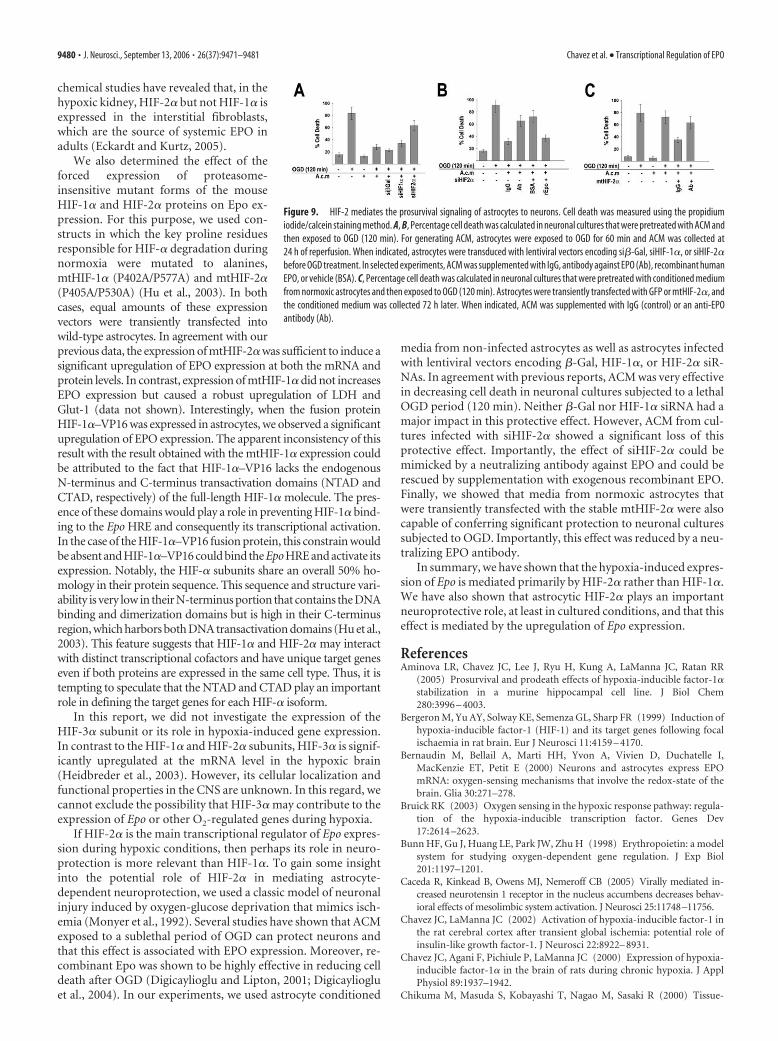
lentivirus-based strategy to downregulate HIF-1� or HIF-2� byRNA interference in primary astrocytes (as described in Fig. 4).At 5 d after transduction, astrocytes were subjected to sublethalOGD (60 min), and conditioned medium (ACM) was collected at24 h of recovery. Preliminary experiments showed that this OGDcondition did not affect the viability of wild-type or HIF nullastrocytes (data not shown). Next, we tested whether ACM fromOGD-stressed astrocytes could influence neuronal survival. Forthis purpose, neurons were pretreated with ACM for 6 h and thensubjected to 120 min OGD. Figure 9 shows that OGD in un-treated neurons led to �80% cell death, as measured by pro-pidium iodide/calcein staining 24 h after the insult. Treatment ofneurons with ACM derived from non-infected or lentivirus–si�-Gal-infected astrocytes resulted in the reduction of neuronaldeath (�60% reduction) (Fig. 9A). Not surprisingly, ACM de-rived from astrocytes with abrogated HIF-2� expression (siHIF-2�) showed a diminished efficacy preventing neuronal death(�15% reduction) (Fig. 9A). In comparison, ACM from siHIF-1�-infected astrocytes was still efficient in reducing neuronalmortality (50% reduction) (Fig. 9A). Previously, EPO has beenidentified as a component of the paracrine protective signalingpathway occurring from astrocytes to neurons (Ruscher et al.,2002). Accordingly, the addition of a neutralizing antibodyagainst EPO to ACM significantly reduced its neuronal protective
properties (Fig. 9B). Conversely, supplementation of ACM de-rived from HIF-2� null astrocytes with recombinant human EPO(100 U/ml) significantly improved neuronal survival. To furtherexplore the role of astroglial HIF-2� in this protective paracrinesignaling, we tested the effect of ACM derived from astrocytesexpressing mtHIF-2� (as described in Fig. 6). Because mtHIF-2�is constitutively expressed regardless of the oxygenation status,transfected astrocytes were cultured under normoxic conditionsfor 72 h, and ACM was used for the treatment of neuronal cul-tures before exposure to OGD. Control astrocytes were trans-fected with a vector encoding GFP. Figure 9C shows that ACMfrom astrocytes transfected with mtHIF-2� improved survival ofOGD-treated neurons (�50% reduction of neuronal death) (Fig.9C); however, addition of a neutralizing antibody against EPOreduced this prosurvival effect (Fig. 9C). ACM from GFP-transfected astrocytes had no effect on neuronal survival. Theexperiments described in Figure 9 were also repeated using theLDH release assay to assess neuronal death. This alternative ap-proach confirmed the results obtained with the PI/calcein stain-ing method (data not shown).
DiscussionIt is well established that astrocytes support neuronal functionand survival through multiple mechanisms. For instance, astro-cytes release trophic factors that can protect neurons against sev-eral forms of injury (Ransom et al., 2003; Christopherson et al.,2005; Dienel and Hertz, 2005; Griffin et al., 2005; Hertz andDienel, 2005; Nedergaard and Dirnagl, 2005). A key mediator ofthis paracrine neuroprotection is EPO, which acts via its cognateneuronal receptor (Ruscher et al., 2002). In cultured models,EPO-induced neuroprotection is effective within minutes and
Figure 6. HIF-2� is sufficient to activate EPO expression in astrocytes. A, B, Astrocytes weretransiently transfected with expression vectors encoding mutant forms of HIF-1� (mtHIF-1�,P402A/P577A) and HIF-2� (mtHIF-2�, P405A/P530A) or GFP and maintained under normoxicconditions. C, D, Astrocytes were infected with retroviruses encoding either VP16 alone orHIF-1�–VP16 fusion protein. For comparison, nontransfected or non-infected astrocytes wereexposed to normoxia (N) or 0.5% O2 (H) for 24 h. EPO mRNA and secreted EPO protein levelswere evaluated by real-time RT-PCR (A, C) and ELISA (B, D), respectively. RT-PCR data of non-transfected normoxia (N) was used as a baseline. E, Immunoblot analysis with antibodiesagainst HIF-1�, HIF-2�, GFP, and VP16 confirmed the expression of transfected vectors. Dataare expressed as the mean � SD from three to four independent experiments.
Figure 7. HIF-2� immunohistochemistry in the hypoxic brain. A, B, Nuclear staining ofHIF-2� was detected in the hypoxic (B) but not in the normoxic (A) mouse brain. D, F, Doublestaining with the vessel marker CD31 (red) showed HIF-2�-positive nuclei associated withvessels (arrows). C, E, Nonvascular cells also showed HIF-2� expression (arrowheads). Scalebars: A, B, 25 �m; C, D, 10 �m; E, F, 5 �m.
9478 • J. Neurosci., September 13, 2006 • 26(37):9471–9481 Chavez et al. • Transcriptional Regulation of EPO

can be sustained for many hours with continued EPO exposure.In addition, several studies with animal models have shown thatEPO in the neonate and adult brain contributes significantly toneuroprotection and ischemic tolerance (Prass et al., 2002, 2003;Ruscher et al., 2002; Diaz et al., 2005; Hoke and Keswani, 2005;Liu et al., 2005). As expected, our data show that cultured neu-rons and astrocytes exposed to hypoxia are able to significantlyupregulate EPO mRNA and protein levels. However, the upregu-lation of EPO mRNA and the secretion of the EPO protein intothe media were significantly higher in astrocytes compared withneurons exposed to equal oxygen levels (0.5% O2). Astrocytesalso showed a more robust upregulation of VEGF in response tohypoxia compared with neurons. This disparity seems to fit therole ascribed to astrocytes as a source of trophic support forneurons.
In the adult hypoxic brain, the most ubiquitously expressed �
subunit is HIF-1�. Several reports have confirmed the expressionof HIF-1� in neurons, astrocytes, endothelial cells, microglia, andoligodendrocytes (Bergeron et al., 1999; Chavez et al., 2000;Stroka et al., 2001). In contrast, as confirmed in this study,HIF-2� expression is restricted to endothelial cells, astrocytes,and a subpopulation of neurons such as Purkinje cells (data notshown). Unlike in vivo, in cultured conditions, Western blotanalysis showed that both the HIF-1� and HIF-2� proteins arepresent in cortical neurons and astrocytes, although their relativeabundance appears to be different. HIF-1� protein expression ismore prominent in neurons, whereas HIF-2� protein levels arehigher in astrocytes. This discrepancy might be related to thedevelopmental stage of the cultured neurons because they arederived from embryos (E15). Consistent with this finding, EMSAanalysis revealed that, in astrocytes and neurons, both complexes,HIF-1�/ARNT2 (HIF-1) and HIF-2�/ARNT2 (HIF-2), appear tobind efficiently to the synthetic oligonucleotide containing theHIF binding sequence derived from the 3� enhancer of the mouseEpo gene. Unlike neurons, in astrocytes, there was a preferentialbinding of HIF-2 rather than HIF-1. However, it is important toconsider that this assay does not take into consideration the com-plexities of the endogenous DNA enhancer region or the coacti-vators that interact with the HIF complexes that are likely toinfluence their DNA binding specificity in vivo.
The contribution of HIFs to the regulation of hypoxia-induced gene expression has not being extensively studied in thecontext of the CNS. Despite Epo being considered one of the bestcharacterized hypoxia-inducible genes, the specific contributionof HIF-1� and HIF-2� to the regulation of EPO expression re-mains unknown. In the present report, we explored this issue incortical primary astrocytes, which are an important source ofEPO production in vivo. We used several approaches to manip-ulate the expression of HIF-1� and HIF-2�. First, we used corti-cal astrocytes isolated from a conditional knock-out mousemodel in which exon 2 of the HIF-1� gene is flanked by LoxP sites(Ryan et al., 2000). When these cells were infected with an ade-novirus encoding Cre recombinase, HIF-1� mRNA levels weresignificantly decreased and HIF-1� protein accumulation wasnot detectable. In these HIF-1� null astrocytes, the expression ofEpo in response to hypoxia was maintained, whereas thehypoxia-driven upregulation of two known HIF-1� target genes,LDH and VEGF, was decreased. In agreement with this finding,wild-type astrocytes that were infected with a lentiviral vectorencoding an siRNA targeting HIF-1� retained their ability toupregulate EPO expression during hypoxia, whereas the expres-sion of VEGF and LDH was markedly reduced. In contrast,siRNA-mediated downregulation of HIF-2�/EPAS-1 expressionin astrocytes subjected to hypoxia caused a significant reductionof EPO mRNA levels, whereas VEGF and LDH mRNA upregula-tion remained unaffected. These experiments provide strong ev-idence that, in cortical astrocytes, HIF-2�/EPAS-1, but not HIF-1�, is the main regulator of EPO expression during hypoxia.Additional support for the role of HIF-2 on EPO expression wasprovided by a ChIP assay, which indicated that, in hypoxic astro-cytes, HIF-2� rather than HIF-1� is recruited to the 3� enhancerregion of the Epo gene that contains the HRE. These results are inagreement with previous studies that have provided evidence thatEpo is an HIF-2 target gene (Warnecke et al., 2004; Eckardt andKurtz, 2005). These studies relied also on siRNA technology todownregulate HIF-1� and HIF-2� expression in hepatoma(Hep3B and HepG2) and neuroblastoma (Kelly) cells that areknow to secrete substantial amounts of EPO when challengedwith hypoxia (Stolze et al., 2002). Furthermore, immunohisto-
Figure 8. Colocalization of EPO and HIF-2� expression. A, Immunohistochemical analysis ofEPO in the mouse brain at 24 h of hypoxia (red). B–D, Merged images showing colocalization ofHIF-2�-positive nuclei (green) and EPO-positive astrocytes and capillaries (red). Scale bars: A,B, 25 �m; C, D, 5 �m.
Chavez et al. • Transcriptional Regulation of EPO J. Neurosci., September 13, 2006 • 26(37):9471–9481 • 9479
chemical studies have revealed that, in thehypoxic kidney, HIF-2� but not HIF-1� isexpressed in the interstitial fibroblasts,which are the source of systemic EPO inadults (Eckardt and Kurtz, 2005).
We also determined the effect of theforced expression of proteasome-insensitive mutant forms of the mouseHIF-1� and HIF-2� proteins on Epo ex-pression. For this purpose, we used con-structs in which the key proline residuesresponsible for HIF-� degradation duringnormoxia were mutated to alanines,mtHIF-1� (P402A/P577A) and mtHIF-2�(P405A/P530A) (Hu et al., 2003). In bothcases, equal amounts of these expressionvectors were transiently transfected intowild-type astrocytes. In agreement with ourprevious data, the expression of mtHIF-2� was sufficient to induce asignificant upregulation of EPO expression at both the mRNA andprotein levels. In contrast, expression of mtHIF-1� did not increasesEPO expression but caused a robust upregulation of LDH andGlut-1 (data not shown). Interestingly, when the fusion proteinHIF-1�–VP16 was expressed in astrocytes, we observed a significantupregulation of EPO expression. The apparent inconsistency of thisresult with the result obtained with the mtHIF-1� expression couldbe attributed to the fact that HIF-1�–VP16 lacks the endogenousN-terminus and C-terminus transactivation domains (NTAD andCTAD, respectively) of the full-length HIF-1� molecule. The pres-ence of these domains would play a role in preventing HIF-1� bind-ing to the Epo HRE and consequently its transcriptional activation.In the case of the HIF-1�–VP16 fusion protein, this constrain wouldbe absent and HIF-1�–VP16 could bind the Epo HRE and activate itsexpression. Notably, the HIF-� subunits share an overall 50% ho-mology in their protein sequence. This sequence and structure vari-ability is very low in their N-terminus portion that contains the DNAbinding and dimerization domains but is high in their C-terminusregion, which harbors both DNA transactivation domains (Hu et al.,2003). This feature suggests that HIF-1� and HIF-2� may interactwith distinct transcriptional cofactors and have unique target geneseven if both proteins are expressed in the same cell type. Thus, it istempting to speculate that the NTAD and CTAD play an importantrole in defining the target genes for each HIF-� isoform.
In this report, we did not investigate the expression of theHIF-3� subunit or its role in hypoxia-induced gene expression.In contrast to the HIF-1� and HIF-2� subunits, HIF-3� is signif-icantly upregulated at the mRNA level in the hypoxic brain(Heidbreder et al., 2003). However, its cellular localization andfunctional properties in the CNS are unknown. In this regard, wecannot exclude the possibility that HIF-3� may contribute to theexpression of Epo or other O2-regulated genes during hypoxia.
If HIF-2� is the main transcriptional regulator of Epo expres-sion during hypoxic conditions, then perhaps its role in neuro-protection is more relevant than HIF-1�. To gain some insightinto the potential role of HIF-2� in mediating astrocyte-dependent neuroprotection, we used a classic model of neuronalinjury induced by oxygen-glucose deprivation that mimics isch-emia (Monyer et al., 1992). Several studies have shown that ACMexposed to a sublethal period of OGD can protect neurons andthat this effect is associated with EPO expression. Moreover, re-combinant Epo was shown to be highly effective in reducing celldeath after OGD (Digicaylioglu and Lipton, 2001; Digicayliogluet al., 2004). In our experiments, we used astrocyte conditioned
media from non-infected astrocytes as well as astrocytes infectedwith lentiviral vectors encoding �-Gal, HIF-1�, or HIF-2� siR-NAs. In agreement with previous reports, ACM was very effectivein decreasing cell death in neuronal cultures subjected to a lethalOGD period (120 min). Neither �-Gal nor HIF-1� siRNA had amajor impact in this protective effect. However, ACM from cul-tures infected with siHIF-2� showed a significant loss of thisprotective effect. Importantly, the effect of siHIF-2� could bemimicked by a neutralizing antibody against EPO and could berescued by supplementation with exogenous recombinant EPO.Finally, we showed that media from normoxic astrocytes thatwere transiently transfected with the stable mtHIF-2� were alsocapable of conferring significant protection to neuronal culturessubjected to OGD. Importantly, this effect was reduced by a neu-tralizing EPO antibody.
In summary, we have shown that the hypoxia-induced expres-sion of Epo is mediated primarily by HIF-2� rather than HIF-1�.We have also shown that astrocytic HIF-2� plays an importantneuroprotective role, at least in cultured conditions, and that thiseffect is mediated by the upregulation of Epo expression.
ReferencesAminova LR, Chavez JC, Lee J, Ryu H, Kung A, LaManna JC, Ratan RR
(2005) Prosurvival and prodeath effects of hypoxia-inducible factor-1�stabilization in a murine hippocampal cell line. J Biol Chem280:3996 – 4003.
Bergeron M, Yu AY, Solway KE, Semenza GL, Sharp FR (1999) Induction ofhypoxia-inducible factor-1 (HIF-1) and its target genes following focalischaemia in rat brain. Eur J Neurosci 11:4159 – 4170.
Bernaudin M, Bellail A, Marti HH, Yvon A, Vivien D, Duchatelle I,MacKenzie ET, Petit E (2000) Neurons and astrocytes express EPOmRNA: oxygen-sensing mechanisms that involve the redox-state of thebrain. Glia 30:271–278.
Bruick RK (2003) Oxygen sensing in the hypoxic response pathway: regula-tion of the hypoxia-inducible transcription factor. Genes Dev17:2614 –2623.
Bunn HF, Gu J, Huang LE, Park JW, Zhu H (1998) Erythropoietin: a modelsystem for studying oxygen-dependent gene regulation. J Exp Biol201:1197–1201.
Caceda R, Kinkead B, Owens MJ, Nemeroff CB (2005) Virally mediated in-creased neurotensin 1 receptor in the nucleus accumbens decreases behav-ioral effects of mesolimbic system activation. J Neurosci 25:11748–11756.
Chavez JC, LaManna JC (2002) Activation of hypoxia-inducible factor-1 inthe rat cerebral cortex after transient global ischemia: potential role ofinsulin-like growth factor-1. J Neurosci 22:8922– 8931.
Chavez JC, Agani F, Pichiule P, LaManna JC (2000) Expression of hypoxia-inducible factor-1� in the brain of rats during chronic hypoxia. J ApplPhysiol 89:1937–1942.
Chikuma M, Masuda S, Kobayashi T, Nagao M, Sasaki R (2000) Tissue-
Figure 9. HIF-2 mediates the prosurvival signaling of astrocytes to neurons. Cell death was measured using the propidiumiodide/calcein staining method. A, B, Percentage cell death was calculated in neuronal cultures that were pretreated with ACM andthen exposed to OGD (120 min). For generating ACM, astrocytes were exposed to OGD for 60 min and ACM was collected at24 h of reperfusion. When indicated, astrocytes were transduced with lentiviral vectors encoding si�-Gal, siHIF-1�, or siHIF-2�before OGD treatment. In selected experiments, ACM was supplemented with IgG, antibody against EPO (Ab), recombinant humanEPO, or vehicle (BSA). C, Percentage cell death was calculated in neuronal cultures that were pretreated with conditioned mediumfrom normoxic astrocytes and then exposed to OGD (120 min). Astrocytes were transiently transfected with GFP or mtHIF-2�, andthe conditioned medium was collected 72 h later. When indicated, ACM was supplemented with IgG (control) or an anti-EPOantibody (Ab).
9480 • J. Neurosci., September 13, 2006 • 26(37):9471–9481 Chavez et al. • Transcriptional Regulation of EPO

specific regulation of erythropoietin production in the murine kidney,brain, and uterus. Am J Physiol Endocrinol Metab 279:E1242–E1248.
Christopherson KS, Ullian EM, Stokes CC, Mullowney CE, Hell JW, Agah A,Lawler J, Mosher DF, Bornstein P, Barres BA (2005) Thrombospondinsare astrocyte-secreted proteins that promote CNS synaptogenesis. Cell120:421– 433.
Diaz Z, Assaraf MI, Miller Jr WH, Schipper HM (2005) Astroglial cytopro-tection by erythropoietin pre-conditioning: implications for ischemicand degenerative CNS disorders. J Neurochem 93:392– 402.
Dienel GA, Hertz L (2005) Astrocytic contributions to bioenergetics of ce-rebral ischemia. Glia 50:362–388.
Digicaylioglu M, Lipton SA (2001) Erythropoietin-mediated neuroprotec-tion involves cross-talk between Jak2 and NF-�B signalling cascades. Na-ture 412:641– 647.
Digicaylioglu M, Bichet S, Marti HH, Wenger RH, Rivas LA, Bauer C,Gassmann M (1995) Localization of specific erythropoietin bindingsites in defined areas of the mouse brain. Proc Natl Acad Sci USA92:3717–3720.
Digicaylioglu M, Garden G, Timberlake S, Fletcher L, Lipton SA (2004)Acute neuroprotective synergy of erythropoietin and insulin-like growthfactor I. Proc Natl Acad Sci USA 101:9855–9860.
Ebert BL, Bunn HF (1999) Regulation of the erythropoietin gene. Blood94:1864 –1877.
Eckardt KU, Kurtz A (2005) Regulation of erythropoietin production. EurJ Clin Invest 35 [Suppl 3]:13–19.
Fandrey J (2004) Oxygen-dependent and tissue-specific regulation of eryth-ropoietin gene expression. Am J Physiol Regul Integr Comp Physiol286:R977–R988.
Griffin S, Clark JB, Canevari L (2005) Astrocyte-neurone communicationfollowing oxygen-glucose deprivation. J Neurochem 95:1015–1022.
Heidbreder M, Frohlich F, Johren O, Dendorfer A, Qadri F, Dominiak P(2003) Hypoxia rapidly activates HIF-3� mRNA expression. FASEB J17:1541–1553.
Hertz L, Dienel GA (2005) Lactate transport and transporters: general prin-ciples and functional roles in brain cells. J Neurosci Res 79:11–18.
Hoke A, Keswani SC (2005) Neuroprotection in the PNS: erythropoietinand immunophilin ligands. Ann NY Acad Sci 1053:491–501.
Hu CJ, Wang LY, Chodosh LA, Keith B, Simon MC (2003) Differential rolesof hypoxia-inducible factor 1� (HIF-1�) and HIF-2� in hypoxic generegulation. Mol Cell Biol 23:9361–9374.
Iyer NV, Kotch LE, Agani F, Leung SW, Laughner E, Wenger RH, GassmannM, Gearhart JD, Lawler AM, Yu AY, Semenza GL (1998a) Cellular anddevelopmental control of O2 homeostasis by hypoxia-inducible factor 1�.Genes Dev 12:149 –162.
Iyer NV, Leung SW, Semenza GL (1998b) The human hypoxia-induciblefactor 1alpha gene: HIF1A structure and evolutionary conservation.Genomics 52:159 –165.
Jiang BH, Rue E, Wang GL, Roe R, Semenza GL (1996) Dimerization, DNAbinding, and transactivation properties of hypoxia-inducible factor 1.J Biol Chem 271:17771–17778.
Liu J, Narasimhan P, Yu F, Chan PH (2005) Neuroprotection by hypoxicpreconditioning involves oxidative stress-mediated expression ofhypoxia-inducible factor and erythropoietin. Stroke 36:1264 –1269.
Marti HH, Wenger RH, Rivas LA, Straumann U, Digicaylioglu M, Henn V,Yonekawa Y, Bauer C, Gassmann M (1996) Erythropoietin gene expres-sion in human, monkey and murine brain. Eur J Neurosci 8:666 – 676.
Marti HH, Gassmann M, Wenger RH, Kvietikova I, Morganti-KossmannMC, Kossmann T, Trentz O, Bauer C (1997) Detection of erythropoie-tin in human liquor: intrinsic erythropoietin production in the brain.Kidney Int 51:416 – 418.
Masuda S, Okano M, Yamagishi K, Nagao M, Ueda M, Sasaki R (1994) Anovel site of erythropoietin production. Oxygen-dependent productionin cultured rat astrocytes. J Biol Chem 269:19488 –19493.
Maxwell PH, Pugh CW, Ratcliffe PJ (1993) Inducible operation of the eryth-ropoietin 3� enhancer in multiple cell lines: evidence for a widespreadoxygen-sensing mechanism. Proc Natl Acad Sci USA 90:2423–2427.
Monyer H, Giffard RG, Hartley DM, Dugan LL, Goldberg MP, Choi DW(1992) Oxygen or glucose deprivation-induced neuronal injury in corti-cal cell cultures is reduced by tetanus toxin. Neuron 8:967–973.
Morishita E, Masuda S, Nagao M, Yasuda Y, Sasaki R (1997) Erythropoietinreceptor is expressed in rat hippocampal and cerebral cortical neurons,
and erythropoietin prevents in vitro glutamate-induced neuronal death.Neuroscience 76:105–116.
Nedergaard M, Dirnagl U (2005) Role of glial cells in cerebral ischemia. Glia50:281–286.
Orlando V, Strutt H, Paro R (1997) Analysis of chromatin structure by invivo formaldehyde cross-linking. Methods 11:205–214.
Prass K, Ruscher K, Karsch M, Isaev N, Megow D, Priller J, Scharff A, DirnaglU, Meisel A (2002) Desferrioxamine induces delayed tolerance againstcerebral ischemia in vivo and in vitro. J Cereb Blood Flow Metab22:520 –525.
Prass K, Scharff A, Ruscher K, Lowl D, Muselmann C, Victorov I, Kapinya K,Dirnagl U, Meisel A (2003) Hypoxia-induced stroke tolerance in themouse is mediated by erythropoietin. Stroke 34:1981–1986.
Pugh CW, Tan CC, Jones RW, Ratcliffe PJ (1991) Functional analysis of anoxygen-regulated transcriptional enhancer lying 3� to the mouse erythro-poietin gene. Proc Natl Acad Sci USA 88:10553–10557.
Pugh CW, O’Rourke JF, Nagao M, Gleadle JM, Ratcliffe PJ (1997) Activa-tion of hypoxia-inducible factor-1; definition of regulatory domainswithin the alpha subunit. J Biol Chem 272:11205–11214.
Ransom B, Behar T, Nedergaard M (2003) New roles for astrocytes (stars atlast). Trends Neurosci 26:520 –522.
Ruscher K, Freyer D, Karsch M, Isaev N, Megow D, Sawitzki B, Priller J,Dirnagl U, Meisel A (2002) Erythropoietin is a paracrine mediator ofischemic tolerance in the brain: evidence from an in vitro model. J Neu-rosci 22:10291–10301.
Ryan HE, Poloni M, McNulty W, Elson D, Gassmann M, Arbeit JM, JohnsonRS (2000) Hypoxia-inducible factor-1� is a positive factor in solid tu-mor growth. Cancer Res 60:4010 – 4015.
Semenza GL (1998) Hypoxia-inducible factor 1: master regulator of O2 ho-meostasis. Curr Opin Genet Dev 8:588 –594.
Semenza GL (2000) Oxygen-regulated transcription factors and their role inpulmonary disease. Respir Res 1:159 –162.
Semenza GL, Agani F, Booth G, Forsythe J, Iyer N, Jiang BH, Leung S, Roe R,Wiener C, Yu A (1997) Structural and functional analysis of hypoxia-inducible factor 1. Kidney Int 51:553–555.
Shingo T, Sorokan ST, Shimazaki T, Weiss S (2001) Erythropoietin regu-lates the in vitro and in vivo production of neuronal progenitors by mam-malian forebrain neural stem cells. J Neurosci 21:9733–9743.
Siddiq A, Ayoub IA, Chavez JC, Aminova L, Shah S, LaManna JC, Patton SM,Connor JR, Cherny RA, Volitakis I, Bush AI, Langsetmo I, Seeley T,Gunzler V, Ratan RR (2005) Hypoxia-inducible factor prolyl4-hydroxylase inhibition. A target for neuroprotection in the central ner-vous system. J Biol Chem 280:41732– 41743.
Stolze I, Berchner-Pfannschmidt U, Freitag P, Wotzlaw C, Rossler J, Frede S,Acker H, Fandrey J (2002) Hypoxia-inducible erythropoietin gene ex-pression in human neuroblastoma cells. Blood 100:2623–2628.
Stroka DM, Burkhardt T, Desbaillets I, Wenger RH, Neil DA, Bauer C,Gassmann M, Candinas D (2001) HIF-1 is expressed in normoxic tissueand displays an organ-specific regulation under systemic hypoxia. FASEBJ 15:2445–2453.
Studer L, Csete M, Lee SH, Kabbani N, Walikonis J, Wold B, McKay R (2000)Enhanced proliferation, survival, and dopaminergic differentiation ofCNS precursors in lowered oxygen. J Neurosci 20:7377–7383.
Tsai PT, Ohab JJ, Kertesz N, Groszer M, Matter C, Gao J, Liu X, Wu H,Carmichael ST (2006) A critical role of erythropoietin receptor in neu-rogenesis and post-stroke recovery. J Neurosci 26:1269 –1274.
Wang GL, Jiang BH, Rue EA, Semenza GL (1995) Hypoxia-inducible factor1 is a basic-helix-loop-helix-PAS heterodimer regulated by cellular O2
tension. Proc Natl Acad Sci USA 92:5510 –5514.Wang L, Zhang Z, Wang Y, Zhang R, Chopp M (2004a) Treatment of stroke
with erythropoietin enhances neurogenesis and angiogenesis and im-proves neurological function in rats. Stroke 35:1732–1737.
Wang L, Zhang Z, Zhang R, Hafner MS, Wong HK, Jiao Z, Chopp M (2004b)Erythropoietin up-regulates SOCS2 in neuronal progenitor cells derivedfrom SVZ of adult rat. NeuroReport 15:1225–1229.
Warnecke C, Zaborowska Z, Kurreck J, Erdmann VA, Frei U, Wiesener M,Eckardt KU (2004) Differentiating the functional role of hypoxia-inducible factor (HIF)-1� and HIF-2� (EPAS-1) by the use of RNA in-terference: erythropoietin is a HIF-2� target gene in Hep3B and Kellycells. FASEB J 18:1462–1464.
Chavez et al. • Transcriptional Regulation of EPO J. Neurosci., September 13, 2006 • 26(37):9471–9481 • 9481
Related Documents