The synergistic effects of 3-D porous silk fibroin matrix scaffold properties and hydrodynamic environment in cartilage tissue regeneration Yun Wang a , Erika Bella b , Christopher S.D. Lee a , Claudio Migliaresi b , Linda Pelcastre a , Zvi Schwartz a , Barbara D. Boyan a, * , Antonella Motta b a Wallace H. Coulter Department of Biomedical Engineering, Georgia Institute of Technology, 315 Ferst Drive, NW, Atlanta, GA 30332-0363, USA b Department of Materials Engineering and Industrial Technologies, Biotech Research Center, University of Trento, Via Mesiano 77, Trento 38050, Italy article info Article history: Received 17 October 2009 Accepted 2 February 2010 Available online 19 March 2010 Keywords: Silk fibroin Scaffolds Chondrocyte Hydrodynamic environment Cartilage tissue engineering abstract Autologous cell-based tissue engineering using three-dimensional porous scaffolds has provided a good option for the repair of cartilage defects. Silk fibroin-based scaffolds are naturally degradable materials with excellent biocompatibility and robust mechanical properties, indicating potential applications in cartilage tissue engineering. In this study, silk fibroin scaffolds prepared by freeze-drying (FD) and salt- leaching (SL300 and SL500) were fully characterized and used to study the effects of silk fibroin scaffold properties on chondrocyte attachment, proliferation and differentiation. The synergistic effects of scaf- fold properties and hydrodynamic environment generated by in vitro rocking culture were also inves- tigated using static cultures as control. FD scaffolds with small pore size and lower porosity increased cell attachment but inhibited cell penetration and limited cell proliferation and differentiation. In contrast, SL scaffolds displaying a bigger pore size, higher porosity and crystallinity resulted in homogenous cell distribution, increasing cell proliferation and advanced chondrocyte differentiation in terms of their spherical morphology, predominant chondrogenic gene expression and abundant cartilaginous extra- cellular matrix production. A hydrodynamic environment was beneficial to chondrocyte proliferation, differentiation, and integrin gene expression in a pore size dependent manner with superior cartilage matrix production but limited hypertrophic differentiation obtained using chondrocyte-seeded SL500 scaffolds. Integrin a5b1 might mediate these effects. Chondrocyte/SL500 silk fibroin constructs obtained under in vitro rocking culture might serve as an excellent implant for in vivo cartilage defect reparation. Ó 2010 Elsevier Ltd. All rights reserved. 1. Introduction Tissue engineering has been proposed as an alternative to reconstruct or replace craniofacial and articular cartilage. Polymer scaffolds are used to promote cell attachment and proliferation, while maintaining a rounded morphology typical of hyaline chondrocytes and facilitating production of a cartilaginous extra- cellular matrix (ECM). Porous or fibrous 3-D matrices made from natural or synthetic materials have been proposed in order to produce an environment and a structure similar to those of natural ECM. While synthetic polymers offer clear advantages in terms of easy material reproducibility and fabrication, natural polymers possess intrinsic properties that can be favorably exploited. Silk fibroin has recently gained attention as a scaffold for orthopaedic tissue engineering because of its versatile processing, slow biodegradation, and adjustable mechanical and biochemical properties [1e4]. As a result, mesenchymal stem cells and chon- drocytes have been implanted into silk fibroin sponges in vitro and in vivo and their potential applications in cell-based skeletal tissue engineering evaluated [5e7]. Depending on the method of preparation, silk fibroin can assume different conformations, which lead to different mechan- ical and biological properties. Secondary structure, molecular weight and amino acid composition depend on the silk source and processing conditions, and in turn, these properties can modulate biological functions such as platelet adhesion and production of inflammatory mediators [8]. Pore size of woven hybrid polymers has also been shown to influence chondrogenesis [9]. Studies of silk fibroin modified poly (D,L-lactic acid) suggest that the fibroin can enhance the cell affinity of a biomaterial [10] and silk fibroin can be modified with peptide sequences to enhance cell adhesion [11e 13]. Bioreactor cultivation is commonly used to enhance ECM deposition in tissue engineered constructs by improving mass transfer of nutrients and signaling factors while providing mechanical cues, including hydrodynamic forces [14,15]. Under- standing cellular attachment to different silk fibroin scaffolds * Corresponding author. Tel.: þ1 404 385 4108; fax: þ1 404 894 2291. E-mail address: [email protected] (A. Boyan). Contents lists available at ScienceDirect Biomaterials journal homepage: www.elsevier.com/locate/biomaterials 0142-9612/$ e see front matter Ó 2010 Elsevier Ltd. All rights reserved. doi:10.1016/j.biomaterials.2010.02.006 Biomaterials 31 (2010) 4672e4681

Welcome message from author
This document is posted to help you gain knowledge. Please leave a comment to let me know what you think about it! Share it to your friends and learn new things together.
Transcript

lable at ScienceDirect
Biomaterials 31 (2010) 4672e4681
Contents lists avai
Biomaterials
journal homepage: www.elsevier .com/locate/biomateria ls
The synergistic effects of 3-D porous silk fibroin matrix scaffold propertiesand hydrodynamic environment in cartilage tissue regeneration
Yun Wang a, Erika Bella b, Christopher S.D. Lee a, Claudio Migliaresi b, Linda Pelcastre a, Zvi Schwartz a,Barbara D. Boyan a,*, Antonella Motta b
aWallace H. Coulter Department of Biomedical Engineering, Georgia Institute of Technology, 315 Ferst Drive, NW, Atlanta, GA 30332-0363, USAbDepartment of Materials Engineering and Industrial Technologies, Biotech Research Center, University of Trento, Via Mesiano 77, Trento 38050, Italy
a r t i c l e i n f o
Article history:Received 17 October 2009Accepted 2 February 2010Available online 19 March 2010
Keywords:Silk fibroinScaffoldsChondrocyteHydrodynamic environmentCartilage tissue engineering
* Corresponding author. Tel.: þ1 404 385 4108; faxE-mail address: [email protected] (A
0142-9612/$ e see front matter � 2010 Elsevier Ltd.doi:10.1016/j.biomaterials.2010.02.006
a b s t r a c t
Autologous cell-based tissue engineering using three-dimensional porous scaffolds has provided a goodoption for the repair of cartilage defects. Silk fibroin-based scaffolds are naturally degradable materialswith excellent biocompatibility and robust mechanical properties, indicating potential applications incartilage tissue engineering. In this study, silk fibroin scaffolds prepared by freeze-drying (FD) and salt-leaching (SL300 and SL500) were fully characterized and used to study the effects of silk fibroin scaffoldproperties on chondrocyte attachment, proliferation and differentiation. The synergistic effects of scaf-fold properties and hydrodynamic environment generated by in vitro rocking culture were also inves-tigated using static cultures as control. FD scaffolds with small pore size and lower porosity increased cellattachment but inhibited cell penetration and limited cell proliferation and differentiation. In contrast, SLscaffolds displaying a bigger pore size, higher porosity and crystallinity resulted in homogenous celldistribution, increasing cell proliferation and advanced chondrocyte differentiation in terms of theirspherical morphology, predominant chondrogenic gene expression and abundant cartilaginous extra-cellular matrix production. A hydrodynamic environment was beneficial to chondrocyte proliferation,differentiation, and integrin gene expression in a pore size dependent manner with superior cartilagematrix production but limited hypertrophic differentiation obtained using chondrocyte-seeded SL500scaffolds. Integrin a5b1 might mediate these effects. Chondrocyte/SL500 silk fibroin constructs obtainedunder in vitro rocking culture might serve as an excellent implant for in vivo cartilage defect reparation.
� 2010 Elsevier Ltd. All rights reserved.
1. Introduction
Tissue engineering has been proposed as an alternative toreconstruct or replace craniofacial and articular cartilage. Polymerscaffolds are used to promote cell attachment and proliferation,while maintaining a rounded morphology typical of hyalinechondrocytes and facilitating production of a cartilaginous extra-cellular matrix (ECM). Porous or fibrous 3-D matrices made fromnatural or synthetic materials have been proposed in order toproduce an environment and a structure similar to those of naturalECM. While synthetic polymers offer clear advantages in terms ofeasy material reproducibility and fabrication, natural polymerspossess intrinsic properties that can be favorably exploited. Silkfibroin has recently gained attention as a scaffold for orthopaedictissue engineering because of its versatile processing, slowbiodegradation, and adjustable mechanical and biochemical
: þ1 404 894 2291.. Boyan).
All rights reserved.
properties [1e4]. As a result, mesenchymal stem cells and chon-drocytes have been implanted into silk fibroin sponges in vitro andin vivo and their potential applications in cell-based skeletal tissueengineering evaluated [5e7].
Depending on the method of preparation, silk fibroin canassume different conformations, which lead to different mechan-ical and biological properties. Secondary structure, molecularweight and amino acid composition depend on the silk source andprocessing conditions, and in turn, these properties can modulatebiological functions such as platelet adhesion and production ofinflammatory mediators [8]. Pore size of woven hybrid polymershas also been shown to influence chondrogenesis [9]. Studies of silkfibroin modified poly (D,L-lactic acid) suggest that the fibroin canenhance the cell affinity of a biomaterial [10] and silk fibroin can bemodified with peptide sequences to enhance cell adhesion [11e13].
Bioreactor cultivation is commonly used to enhance ECMdeposition in tissue engineered constructs by improving masstransfer of nutrients and signaling factors while providingmechanical cues, including hydrodynamic forces [14,15]. Under-standing cellular attachment to different silk fibroin scaffolds

Y. Wang et al. / Biomaterials 31 (2010) 4672e4681 4673
during hydrodynamic culture is important for optimizing scaffoldfabrication and culture techniques for tissue engineeringapplications. However, few studies though have thoroughly inves-tigated how bioreactor hydrodynamics affect scaffold seedingefficiency, cellular attachment, and subsequent proliferation, inaddition to ECM production.
Cells interact with their ECM via integrin binding and sensedifference in mechanical stresses through integrin signaling [16].Integrins been implicated in chondrocytic mechanotransduction[17], suggesting that they may be involved both in cell attachmentto silk fibroin scaffolds and in the cellular response to mechanicalstresses during bioreactor culture. Whether integrin expression inchondrocyte cultures is sensitive to scaffold design and/or hydro-dynamic loading in a bioreactor is not known. The objective of thisstudy was to test the hypothesis that the properties of silk fibroinscaffolds such as porosity and pore size interact with the hydro-dynamic environment of a bioreactor to influence integrinexpression and chondrocyte behavior. The material properties ofdifferent silk fibroin scaffolds were extensively characterized andcell attachment, proliferation, gene expression, and ECM synthesiswere assessed.
2. Materials and methods
2.1. Material preparation
Bombyx mori polyhybrid cross silkworms' white cocoons selected by CentroSperimentale di Gelsibachicoltura, in Como, Italy, were used. Cocoons weredegummed by boiling twice, 1 h each, in a water solution of Na2CO3 (1.1 and 0.4 g/L,respectively), in order to remove sericins. Degummed silk fibres were then washedseveral times in distilled water, dried at room temperature, dissolved in 9.3 m LiBr(Fluka Chemical, Steinheim, Germany) water solution (20% w/v) at 65 �C for 3 h, anddialyzed against distilled water to remove LiBr by using a cellulose membrane(Pierce, Slide-A-Lyzer, MWCO 3500 Da). After dialysis the solution was filtered(Duran Group, glass filter, maximum pore size 10e16 mm), and the concentration offibroin water solution was adjusted to 7% w/v.
Scaffolds with different porosities, pore sizes, and properties were made byfreeze-drying and salt-leaching. Three different types of samples with differentpores size were produced. Freeze-dried samples were prepared keeping the fibroinwater solution for 2 h at �20 �C, followed by drying in a freeze-dryer. Resultingsamples were stabilized in water vapour under vacuum for 24 h (sample code: FD)and dried in air at room temperature. For salt-leaching samples, 4 g of granular NaCl,particle size 300e500 mm (sample code: SL300) and 500e1100 mm (sample code:SL500), were added to 2mL of fibroinwater solutions, and kept at room temperatureuntil gelation occurred (about 2 days). Gels were then washed in distilled water forthree days in order to remove the salt, and then dried in air at room temperature.The resulting sponges were stable in water.
2.2. Material characterization
2.2.1. Amino acid compositionSilk fibroin was hydrolyzed in 6 M HCl at 120 �C for 24 h. The hydrolysate was
diluted with MilliQ water (Millipore, Billerica, MA, USA) to a concentration of10�7 mol/L and subjected to Waters AccQ.Tag� amino acid analysis (WatersCorporation, New Castle, Delaware, USA) to determine amino acid composition.
2.2.2. Gel permeation chromatographyThe weight-average molecular weight (MW) and polydispersity index (PDI) of
fibroin were determined by gel permeation chromatography (GPC) by using fibroinsolutions prepared following the above reported method. 20 mg of each sample wasrapidly dissolved in LiBr water solution and dialyzed against distilled water.Molecular weight analysis was performed on fibroin in water solution using anHPLC/GPC system equipped with a Shodex Protein KW-804 column (Shodex, Tokyo,Japan), a Jasco 1570 UV/Vis detector (Jasco, Tokyo, Japan) set at 274 nm and a SpectraSystem P4000 isocratic pump (Thermo Fisher Scientific Inc., Waltham, MA, USA).The calibration curve was obtained using protein standards (Gel Filtration Calibra-tion Kit, GE Healthcare, Pittsburgh, PA, USA).
2.2.3. Morphology: low vacuum environmental scanning microscopySilk fibroin sponge morphology was investigated with an environmental scan-
ning electron microscope (E-SEM, Philips XL 30 ESEM, Eindhoven, NL) in lowvacuum mode. Freeze-dried silk scaffolds were fractured in liquid nitrogen usinga surgical blade and observed without coating with gold at 10 kV accelerationvoltages at vacuum pressure levels in the range of 0.9e4.0 Torr and at a constant
temperature of 5 �C. The mean pore size of the scaffolds was determined by analysisof the SEM images of the scaffold cross-sections by importing data to Image-Jsoftware (available from NIH Image). Ten apparent pores per frame (30 frames persample) were measured for each type of scaffold.
2.2.4. Thermal analysis: differential scanning calorimetryThermal analysis was conducted by means of a differential scanning calorimeter
(DSC) (Mettler, Model DSC30, Columbus, OH, USA) with N2 gas flow, at a heating rateof 10 �C/min from 0 �C to 350 �C.
2.2.5. FTIR-ATR analysisInfrared spectroscopy in attenuated total internal reflection mode (ATR) was
performed by using an FTIR Spectrum One (Perking Elmer, Waltham, MA, USA) witha zinc selenide crystal. Each spectrum was the mean of 4 acquisitions (between4000 cm�1 and 650 cm�1) with a spectral resolution of 4 cm�1.
2.2.6. Porosity evaluationThe porosity of the sponges was evaluated by helium picnometer (ASTM Stan-
dard D6226e05) and hexane displacement [18]. Based on sample geometry andporosity, total polymer volume was calculated and densities of silk fibroin scaffoldswere also evaluated by normalizing total polymer volume to sample weight.Measurements were performed on n ¼ 3 samples for each kind of scaffold andresults were expressed as the mean � standard deviation.
2.2.7. Water uptake evaluationTo measure water uptake, three samples of each material were weighed after
drying in vacuum at 40 �C, then put inwater, and then blotted with paper in order toremove the excess of water from the surface and re-weighed at selected timeintervals until constant weight was reached. The water uptake is reported as waterweight/sponge dry weight ¼ (Wwet � Wdry)/Wdry, Wdry and Wwet being theweights of the sponges before and after the water absorption.
2.2.8. Mechanical propertiesCompressive mechanical properties were measured at room temperature on
wet fibroin sponges discs, 10 mm in diameter and 3 mm in thickness, by using anInstron 4502 mechanical tester machine (Instron, Nordwood, MA, USA) witha crosshead speed of 1 mm/min. Three samples were used for each type of scaffold.
2.3. In vitro culture and evaluation
2.3.1. Chondrocyte culturesChondrocytes were isolated from the resting zone of costochondral cartilage
growth plate from 125 g SpragueeDawley rats [19,20]. The resting zone is a hyaline-like cartilage, with chondrocytes being dispersed in a type II collagen, sulfatedproteoglycan-rich extracellular matrix. They retain their in vivo phenotype throughfour passages in culture. Chondrocytes were plated at a density of 25,000 cells/cm2
and cultured in Dulbecco's modified Eagle's medium (DMEM) containing 10% fetalbovine serum (FBS), 1% penicillinestreptomycin, and 50 mg/mL sodium ascorbate inan atmosphere of 5% CO2 and 100% humidity at 37 �C. Third passage confluent cellswere harvested for seeding on scaffolds.
2.3.2. Media loading into silk scaffoldsDry silk scaffolds were presoaked in distilled water for 2 h and punched into
cylinders (6 mm in diameter and 3 mm in thickness) using a biopsy punch (Miltex,London, UK). Scaffolds in aqueous suspension were sterilized by autoclave at 121 �Cfor 15 min. After cooling to room temperature, scaffolds were then conditioned withculture medium over night. Before cell seeding, the scaffolds were transferred intoa 20mL syringewith 5mLmedia. Vacuumpressurewas used for 2min to loadmediaand remove air bubbles in the scaffolds. These prewetted silk scaffolds were thenblotted dry and placed in the center of each well of a 12-well culture plate and weresurrounded by 0.5 mL culture medium to keep the construct wet during thefollowing incubation.
2.3.3. Cell seeding and culture of silk scaffold/chondrocyte constructsEach scaffold was seeded by adding 0.5 � 106 cells in 20 mL culture medium.
After 4 h of incubation, 2.5 mL additional media were added to each scaffold. After24 h static culture, these silk scaffold/chondrocyte constructs were either main-tained under static culture or subjected to rocking culture with rocking speed at15 rpm on a VWR Signature� Rocking Platform Shaker (VWR International, Inc., SanDiego, CA) in a humidified tissue culture incubator at 37 �C with 5% CO2. Culturemedium was changed every two days until harvest.
2.3.4. Cell seeding efficiencyIn order to evaluate the appropriate time for cells to attach on scaffolds, cell-
seeded scaffolds were maintained under static culture for 0 h, 4 h or 10 h andthen were transferred to rocking culture until harvest. The scaffolds under staticculture for 24 h without rocking culture served as control. By the end of 24 h ofculture, unincorporated cell numbers were evaluated by measuring the cellsremaining in the plates and media using Beckman Coulter Cell Counter (Beckman
Y. Wang et al. / Biomaterials 31 (2010) 4672e46814674
Coulter, Inc., Miami, FL). Cell seeding efficiency was then calculated using theformula:
Cell seeding efficiencyð%Þ ¼�1� Unincorporated cell numbers
Initial cell seeding numbers
�� 100%
2.3.5. Cell proliferation assayCell proliferation in scaffolds was evaluated based on the relative number of
metabolically active chondrocytes within each seeded scaffold determined by thealamarBlue� assay (AbD Serotec, Raleigh, NC) according to the manufacturer'sinstructions. Briefly, seeded scaffolds were incubated with 1 mL of 10%alamarBlue� reagent in culture medium for 2 h at 37 �C with 5% CO2. At the end ofincubation, 100 mL of the supernatant were then transferred to 96-well plates andquantified for fluorescence intensity with a microtiter plate reader (SpectromaxGemini XS, Molecular Devices, Sunnyvale, CA) using an excitation wavelength of565nm and an emissionwavelength of 595 nm. Non-seeded scaffolds were used asnegative control and their fluorescence was subtracted from the same type seededscaffolds. Cell numbers were then calculated using the standard curve obtained atthe same time.
2.3.6. Cell viability assayThe viability of cells in the scaffolds was examined by a Live/Dead� assay
(Invitrogen Corporation, Carlsbad, CA) as described in the literature [5]. In order toevaluate the cell distribution inside the scaffolds, the scaffolds were sliced in themiddle after staining, and the images were taken from both the surface and themiddle of scaffolds using a confocal microcopy (excitation/emission: 495 nm/515 nm).
2.3.7. DNA and sulfated glycosaminoglycan assaysAt harvest, scaffolds were collected and digested in papain digestion buffer for
24 h at 60 �C, using 1 mL of the papain solution per scaffold. DNA content wasdetermined fluorimetrically following the binding of PicoGreen dye (InvitrogenCorporation) using calf thymus type I DNA as a standard. Glycosaminoglycan (GAG)content was determined spectrophotometrically at 520 nm following the reactionwith dimethylmethylene blue dye (SigmaeAldrich, St. Louis, MO) and normalized toDNA content [5].
2.3.8. Real-time RT-PCRTotal RNA was isolated from the chondrocyte/silk constructs using RNeasy Kit
(Qiagen, Inc., Valencia, CA) according to the manufacturer's instructions. 1 mg of RNAwas then reverse-transcribed into cDNA according to protocols from the OmniscriptRT kit (Qiagen, Inc.). Quantitative real-time RT-PCR amplifications of chondrogenicmarker genes (Sox9, collagen 2a1 and aggrecan), hypertrophic marker genes (col-lagen10a1 and alkaline phosphatase), dedifferentiation maker gene (collagen1a1)and integrin subunits (a2, a5, b1 and b3) were performed in an iCycler real-time PCRmachine using iQ SYBR Green supermix (both from Bio-Rad Laboratories, Hercules,CA) according to the manufacturer's instructions. No-template (water) reactionmixture was prepared as a negative control. The quantity of mRNA of each gene wasnormalized using the CT (threshold cycle) value obtained for RPS18 mRNAamplifications. The sequences for individual primers are shown in SupplementalTable 1.
2.3.9. MicroCTBased on the results from the experiments described above, we only used the
SL300 and SL500 scaffolds for long term experiments (30 days of culture) in order tofurther confirm the production of cartilaginous ECM using microCT and immuno-fluorescence assay. Immediately after harvest, samples from each group werewashed twice in 10% phosphate buffered saline with 1% proteinase inhibitor(Cocktail I, CalBiochem, San Diego, CA) and then incubated in 2 mL of 100% Hexabrixcontrast agent (Mallinckrodt, Hazelwood, MO) and 1% proteinase inhibitor at 37 �C.After 24 h, the samples were patted dry and transferred to the microCT system forscanning. All the samples were scanned in air using a mCT 40 (Scanco Medical,Bassersdorf, Switzerland) at 45 kkVp, 177 mA, 200-m integration time, and a voxelsize of 16 mm. Image processing was performed with the accompanying Scancomedical software. Three-dimensional color images based on the X-ray attenuationwere created in order to visualize location of proteoglycan content. Consistent withthe inverse partitioning between proteoglycan concentration and contrast agent,low X-ray attenuation (green) corresponded to regions of high proteoglycanconcentration and high X-ray attenuation (red) indicated regions of low proteo-glycan concentration [21].
2.3.10. ImmunohistochemistrySamples were fixed in 10% neutral buffered formalin, embedded in paraffin and
sectioned (5 mm thick). The sections were immuno-labeled with rabbit polyclonalantibodies against rat type I and type II collagen (Abcam, Inc., Cambridge, MA) andaggrecan (Santa Cruz Biotechnology, Inc. Santa Cruz, CA). The primary antibody wasfollowed by incubation with secondary antibodies (Alexia 488 goat anti-rabbit IgG)(Invitrogen Corporation). Sections were mounted in Fluoro-Gel (Electron Micros-copy Sciences, Hatfield, PA). Slides were examined using Nikon Eclipse E600
Fluorescence Microscope/Image System (Nikon, Japan). At least three sections fromone scaffold and six different scaffolds from the same group were viewed. Repre-sentative areas were photographed.
2.4. Statistical analysis
All measurements for in vitro experiments were collected with N ¼ 6 inde-pendent determinations per data point and expressed as mean � standard errors.Statistical analysis was determined using GraphPad Prism 5.0 (GraphPad Software,Inc., La Jolla, CA). Data were analyzed by two-way ANOVA and Tukey post-hoctesting to determine the statistical significance. Differences between groups withp < 0.05 were considered statistically significant.
3. Results
3.1. Material characterization
In order to assess any potential changes in properties, materialswere characterized both before and after sterilization treatment.However, no significant changes, in terms of molecular weight,porosity, pore size and crystallinity, were observed afterautoclaving. Therefore, only the data obtained before the scaffoldsterilization are presented below.
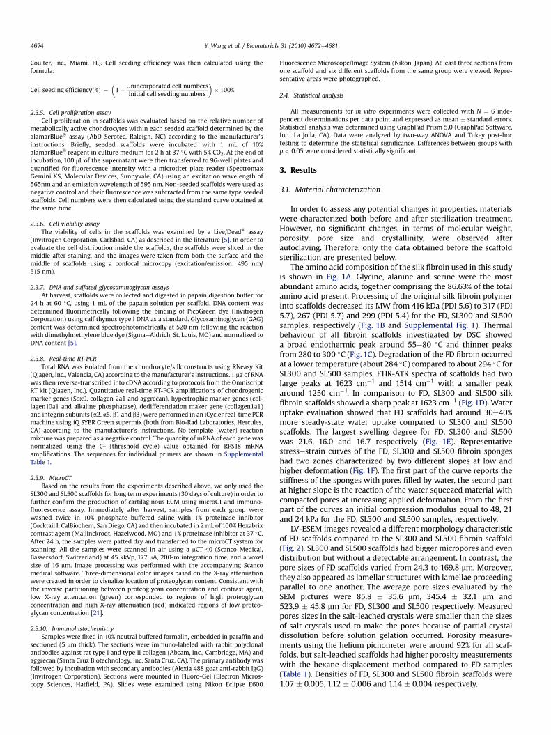
The amino acid composition of the silk fibroin used in this studyis shown in Fig. 1A. Glycine, alanine and serine were the mostabundant amino acids, together comprising the 86.63% of the totalamino acid present. Processing of the original silk fibroin polymerinto scaffolds decreased its MW from 416 kDa (PDI 5.6) to 317 (PDI5.7), 267 (PDI 5.7) and 299 (PDI 5.4) for the FD, SL300 and SL500samples, respectively (Fig. 1B and Supplemental Fig. 1). Thermalbehaviour of all fibroin scaffolds investigated by DSC showeda broad endothermic peak around 55e80 �C and thinner peaksfrom 280 to 300 �C (Fig. 1C). Degradation of the FD fibroin occurredat a lower temperature (about 284 �C) compared to about 294 �C forSL300 and SL500 samples. FTIR-ATR spectra of scaffolds had twolarge peaks at 1623 cm�1 and 1514 cm�1 with a smaller peakaround 1250 cm�1. In comparison to FD, SL300 and SL500 silkfibroin scaffolds showed a sharp peak at 1623 cm�1 (Fig. 1D). Wateruptake evaluation showed that FD scaffolds had around 30e40%more steady-state water uptake compared to SL300 and SL500scaffolds. The largest swelling degree for FD, SL300 and SL500was 21.6, 16.0 and 16.7 respectively (Fig. 1E). Representativestressestrain curves of the FD, SL300 and SL500 fibroin spongeshad two zones characterized by two different slopes at low andhigher deformation (Fig. 1F). The first part of the curve reports thestiffness of the sponges with pores filled by water, the second partat higher slope is the reaction of the water squeezed material withcompacted pores at increasing applied deformation. From the firstpart of the curves an initial compression modulus equal to 48, 21and 24 kPa for the FD, SL300 and SL500 samples, respectively.
LV-ESEM images revealed a different morphology characteristicof FD scaffolds compared to the SL300 and SL500 fibroin scaffold(Fig. 2). SL300 and SL500 scaffolds had bigger micropores and evendistribution but without a detectable arrangement. In contrast, thepore sizes of FD scaffolds varied from 24.3 to 169.8 mm. Moreover,they also appeared as lamellar structures with lamellae proceedingparallel to one another. The average pore sizes evaluated by theSEM pictures were 85.8 � 35.6 mm, 345.4 � 32.1 mm and523.9 � 45.8 mm for FD, SL300 and SL500 respectively. Measuredpores sizes in the salt-leached crystals were smaller than the sizesof salt crystals used to make the pores because of partial crystaldissolution before solution gelation occurred. Porosity measure-ments using the helium picnometer were around 92% for all scaf-folds, but salt-leached scaffolds had higher porosity measurementswith the hexane displacement method compared to FD samples(Table 1). Densities of FD, SL300 and SL500 fibroin scaffolds were1.07 � 0.005, 1.12 � 0.006 and 1.14 � 0.004 respectively.
Fig. 1. Characterization of fibroin scaffolds: Amino acid composition of original silk fibroin from Bomyx mori cocoons (A); molecular weights measured by GPC (B); DSC curves (C);FTIR-ATR spectra (D); water uptake curve (E); and stressestrain curves (F) obtained from fibroin sponges prepared by different protocols. Light dark dashed line represents FDscaffolds, heavy dark dotted line represents SL300 scaffolds, and grey solid line represents SL500 scaffolds.
Y. Wang et al. / Biomaterials 31 (2010) 4672e4681 4675
3.2. Cell loading efficiency
Starting rocking culture without pre-static culture resulted invery low cell loading efficiencies with only 38.3%, 24.9% and 13.7%of loaded cells being attached to FD, SL300 and SL500 scaffolds,respectively (Fig. 3A). Maintaining constructs in static culture aftercell seeding led to time-dependent significant increases in cellloading efficiency. A cell seeding retention of 56.5% was reachedafter 24 h of static culture in FD scaffolds, followed by 51.3% in
SL300 scaffolds, and 24.6% in SL500. Therefore, static loading for24 h was applied in subsequent experiments.
3.3. Cell proliferation
Chondrocytes proliferated slowly during the first 7 days ofculture regardless of the scaffold type or culture condition, but asthe proliferation rate increased from day 9 to day 15, differencesamong the groups became apparent (Fig. 3B). In general, cell

Fig. 2. LV-ESEM images of different fibroin scaffolds. Scale ¼ 500 mm.
Table 1Porosity of the fibroin sponges determined by hexane displacement.
Fibrion sponge types Hexane displacement (%)
FD 67.3 � 4.2SL300 84.9 � 4.5a
SL500 89.1 � 3.1a
N ¼ 3.a p < 0.05 vs. FD scaffolds.
Y. Wang et al. / Biomaterials 31 (2010) 4672e46814676
proliferation on SL300 and SL500 scaffolds was significantlyaccelerated under rocking culture compared to static culture, butcell proliferation was not influenced on FD scaffolds. By the end ofday 15, the increases in cell number under static culture versusrocking culture were 4.3 vs. 2.0 fold, 4.5 vs. 4.8 fold, and 5.7 vs. 8.6fold for FD, SL300, and SL500 scaffolds, respectively.
Fig. 3. Effects of culture conditions on cell loading efficiency and cell proliferation onsilk fibroin scaffolds. Chondrocyte/silk scaffold constructs were maintained understatic culture for 0 h, 4 h, 10 h or 24 h before they were transferred to rocking culture.Cell seeding efficiency was then measured by the end of 24 h of different culture (A).Cell proliferation in scaffolds was monitored every 2 days using alamarBlue� assayover the course of 15 days of culture (B). Each point represents the mean and standarddeviation of N ¼ 6 independent cultures.
3.4. Cell viability and cell distribution
After 15 days in culture, most cells presented green fluorescenceindicating no significant cell death inside all types of scaffoldsunder both static and rocking culture (Fig. 4). However, differentcell distributions were observed among these scaffolds. Celldistribution was restricted to the top surface of FD scaffolds withvery few to no cells presenting in the central region regardless ofstatic culture or rocking culture. In contrast, more homogenous cell
distributions were found in SL300 and SL500 scaffolds, withslightly less cells in the center compared to the top surface ofscaffolds. In addition, more cells were detected in FD scaffoldsunder static culture than rocking culture, whereas the oppositetendency was observed in SL300 and SL500 scaffolds.
3.5. Chondrocyte differentiation
No significant differences in transcript levels of chondrogenicgenes (Sox9, collagen 2a1 and aggrecan) were found among threetypes of scaffolds under static culture (Fig. 5). However, the data
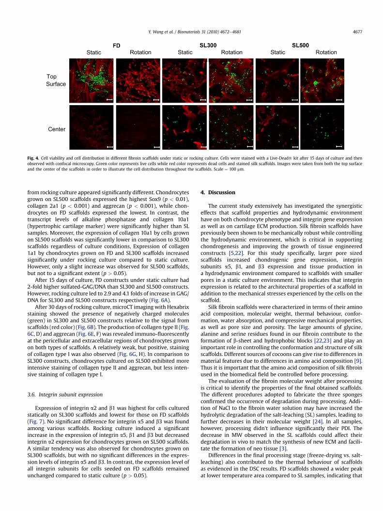
Fig. 4. Cell viability and cell distribution in different fibroin scaffolds under static or rocking culture. Cells were stained with a Live-Dead� kit after 15 days of culture and thenobserved with confocal microscopy. Green color represents live cells while red color represents dead cells and stained silk scaffolds. Images were taken from both the top surfaceand the center of the scaffolds in order to illustrate the cell distribution throughout the scaffolds. Scale ¼ 100 mm.
Y. Wang et al. / Biomaterials 31 (2010) 4672e4681 4677
from rocking culture appeared significantly different. Chondrocytesgrown on SL500 scaffolds expressed the highest Sox9 (p < 0.01),collagen 2a1 (p < 0.001) and aggrecan (p < 0.001), while chon-drocytes on FD scaffolds expressed the lowest. In contrast, thetranscript levels of alkaline phosphatase and collagen 10a1(hypertrophic cartilage marker) were significantly higher than SLsamples. Moreover, the expression of collagen 10a1 by cells grownon SL500 scaffolds was significantly lower in comparison to SL300scaffolds regardless of culture conditions, Expression of collagen1a1 by chondrocytes grown on FD and SL300 scaffolds increasedsignificantly under rocking culture compared to static culture.However, only a slight increase was observed for SL500 scaffolds,but not to a significant extent (p > 0.05).
After 15 days of culture, FD constructs under static culture had2-fold higher sulfated-GAG/DNA than SL300 and SL500 constructs.However, rocking culture led to 2.9 and 4.1 folds of increase in GAG/DNA for SL300 and SL500 constructs respectively (Fig. 6A).
After 30 days of rocking culture, microCT imaging with Hexabrixstaining showed the presence of negatively charged molecules(green) in SL300 and SL500 constructs relative to the signal fromscaffolds (red color) (Fig. 6B). The production of collagen type II (Fig.6C, D) and aggrecan (Fig. 6E, F) was revealed immuno-fluorescentlyat the pericellular and extracellular regions of chondrocytes grownon both types of scaffolds. A relatively weak, but positive, stainingof collagen type I was also observed (Fig. 6G, H). In comparison toSL300 constructs, chondrocytes cultured on SL500 exhibited moreintensive staining of collagen type II and aggrecan, but less inten-sive staining of collagen type I.
3.6. Integrin subunit expression
Expression of integrin a2 and b1 was highest for cells culturedstatically on SL300 scaffolds and lowest for those on FD scaffolds(Fig. 7). No significant difference for integrin a5 and b3 was foundamong various scaffolds. Rocking culture induced a significantincrease in the expression of integrin a5, b1 and b3 but decreasedintegrin a2 expression for chondrocytes grown on SL500 scaffolds.A similar tendency was also observed for chondrocytes grown onSL300 scaffolds, but with no significant differences in the expres-sion levels of integrin a5 and b3. In contrast, the expression level ofall integrin subunits for cells seeded on FD scaffolds remainedunchanged compared to static culture (p > 0.05).
4. Discussion
The current study extensively has investigated the synergisticeffects that scaffold properties and hydrodynamic environmenthave on both chondrocyte phenotype and integrin gene expressionas well as on cartilage ECM production. Silk fibroin scaffolds havepreviously been shown to be mechanically robust while controllingthe hydrodynamic environment, which is critical in supportingchondrogenesis and improving the growth of tissue engineeredconstructs [5,22]. For this study specifically, larger pore sizedscaffolds increased chondrogenic gene expression, integrinsubunits a5, b1, and b3 expression and tissue production ina hydrodynamic environment compared to scaffolds with smallerpores in a static culture environment. This indicates that integrinexpression is related to the architectural properties of a scaffold inaddition to the mechanical stresses experienced by the cells on thescaffold.
Silk fibroin scaffolds were characterized in terms of their aminoacid composition, molecular weight, thermal behaviour, confor-mation, water absorption, and compressive mechanical properties,as well as pore size and porosity. The large amounts of glycine,alanine and serine residues found in our fibroin contribute to theformation of b-sheet and hydrophobic blocks [22,23] and play animportant role in controlling the conformation and structure of silkscaffolds. Different sources of cocoons can give rise to differences inmaterial features due to differences in amino acid composition [9].Thus it is important that the amino acid composition of silk fibroinused in the biomedical field be controlled before processing.
The evaluation of the fibroin molecular weight after processingis critical to identify the properties of the final obtained scaffolds.The different procedures adopted to fabricate the three spongesconfirmed the occurrence of degradation during processing. Addi-tion of NaCl to the fibroin water solution may have increased thehydrolytic degradation of the salt-leaching (SL) samples, leading tofurther decreases in their molecular weight [24]. In all samples,however, processing didn't influence significantly their PDI. Thedecrease in MW observed in the SL scaffolds could affect theirdegradation in vivo to match the synthesis of new ECM and facili-tate the formation of neo tissue [3].
Differences in the final processing stage (freeze-drying vs. salt-leaching) also contributed to the thermal behaviour of scaffoldsas evidenced in the DSC results. FD scaffolds showed a wider peakat lower temperature area compared to SL samples, indicating that
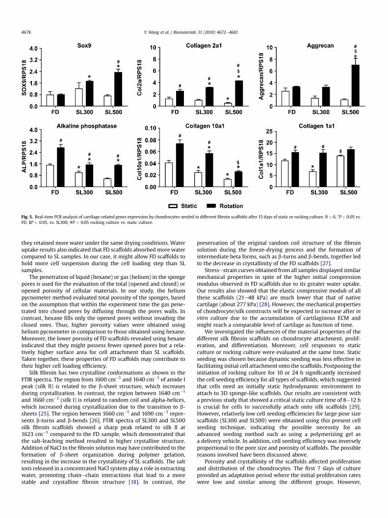
Fig. 5. Real-time PCR analysis of cartilage-related genes expression by chondrocytes seeded in different fibroin scaffolds after 15 days of static or rocking culture. N ¼ 6, *P < 0.05 vs.FD, $P < 0.05, vs. SL300, #P < 0.05 rocking culture vs. static culture.
Y. Wang et al. / Biomaterials 31 (2010) 4672e46814678
they retained more water under the same drying conditions. Wateruptake results also indicated that FD scaffolds absorbedmorewatercompared to SL samples. In our case, it might allow FD scaffolds tohold more cell suspension during the cell loading step than SLsamples.
The penetration of liquid (hexane) or gas (helium) in the spongepores is used for the evaluation of the total (opened and closed) oropened porosity of cellular materials. In our study, the heliumpycnometer method evaluated total porosity of the sponges, basedon the assumption that within the experiment time the gas pene-trated into closed pores by diffusing through the pores walls. Incontrast, hexane fills only the opened pores without invading theclosed ones. Thus, higher porosity values were obtained usinghelium pycnometer in comparison to those obtained using hexane.Moreover, the lower porosity of FD scaffolds revealed using hexaneindicated that they might possess fewer opened pores but a rela-tively higher surface area for cell attachment than SL scaffolds.Taken together, these properties of FD scaffolds may contribute totheir higher cell loading efficiency.
Silk fibroin has two crystalline conformations as shown in theFTIR spectra. The region from 1600 cm�1 and 1640 cm�1 of amide Ipeak (silk II) is related to the b-sheet structure, which increasesduring crystallization. In contrast, the region between 1640 cm�1
and 1660 cm�1 (silk I) is related to random coil and alpha-helices,which increased during crystallization due to the transition to b-sheets [25]. The region between 1660 cm�1 and 1690 cm�1 repre-sents b-turns and b-bends [26]. FTIR spectra of SL300 and SL500silk fibroin scaffolds showed a sharp peak related to silk II at1623 cm�1 compared to the FD sample, which demonstrated thatthe salt-leaching method resulted in higher crystalline structure.Addition of NaCl to the fibroin solutionmay have contributed to theformation of b-sheet organization during polymer gelation,resulting in the increase in the crystallinity of SL scaffolds. The saltions released in a concentrated NaCl system play a role in extractingwater, promoting chainechain interactions that lead to a morestable and crystalline fibroin structure [18]. In contrast, the
preservation of the original random coil structure of the fibroinsolution during the freeze-drying process and the formation ofintermediate beta forms, such as b-turns and b-bends, together ledto the decrease in crystallinity of the FD scaffolds [27].
Stressestrain curves obtained from all samples displayed similarmechanical properties in spite of the higher initial compressionmodulus observed in FD scaffolds due to its greater water uptake.Our results also showed that the elastic compressive moduli of allthese scaffolds (21e48 kPa) are much lower that that of nativecartilage (about 277 kPa) [28]. However, the mechanical propertiesof chondrocyte/silk constructs will be expected to increase after invitro culture due to the accumulation of cartilaginous ECM andmight reach a comparable level of cartilage as function of time.
We investigated the influences of the material properties of thedifferent silk fibroin scaffolds on chondrocyte attachment, prolif-eration, and differentiation. Moreover, cell responses to staticculture or rocking culture were evaluated at the same time. Staticseeding was chosen because dynamic seeding was less effective infacilitating initial cell attachment onto the scaffolds. Postponing theinitiation of rocking culture for 10 or 24 h significantly increasedthe cell seeding efficiency for all types of scaffolds, which suggestedthat cells need an initially static hydrodynamic environment toattach to 3D sponge-like scaffolds. Our results are consistent witha previous study that showed a critical static culture time of 8e12 his crucial for cells to successfully attach onto silk scaffolds [29].However, relatively low cell seeding efficiencies for large pose sizescaffolds (SL300 and SL500) were obtained using this present cellseeding technique, indicating the possible necessity for anadvanced seeding method such as using a polymerizing gel asa delivery vehicle. In addition, cell seeding efficiency was inverselyproportional to the pore size and porosity of scaffolds. The possiblereasons involved have been discussed above.
Porosity and crystallinity of the scaffolds affected proliferationand distribution of the chondrocytes. The first 7 days of cultureprovided an adaptation period where the initial proliferation rateswere low and similar among the different groups. However,
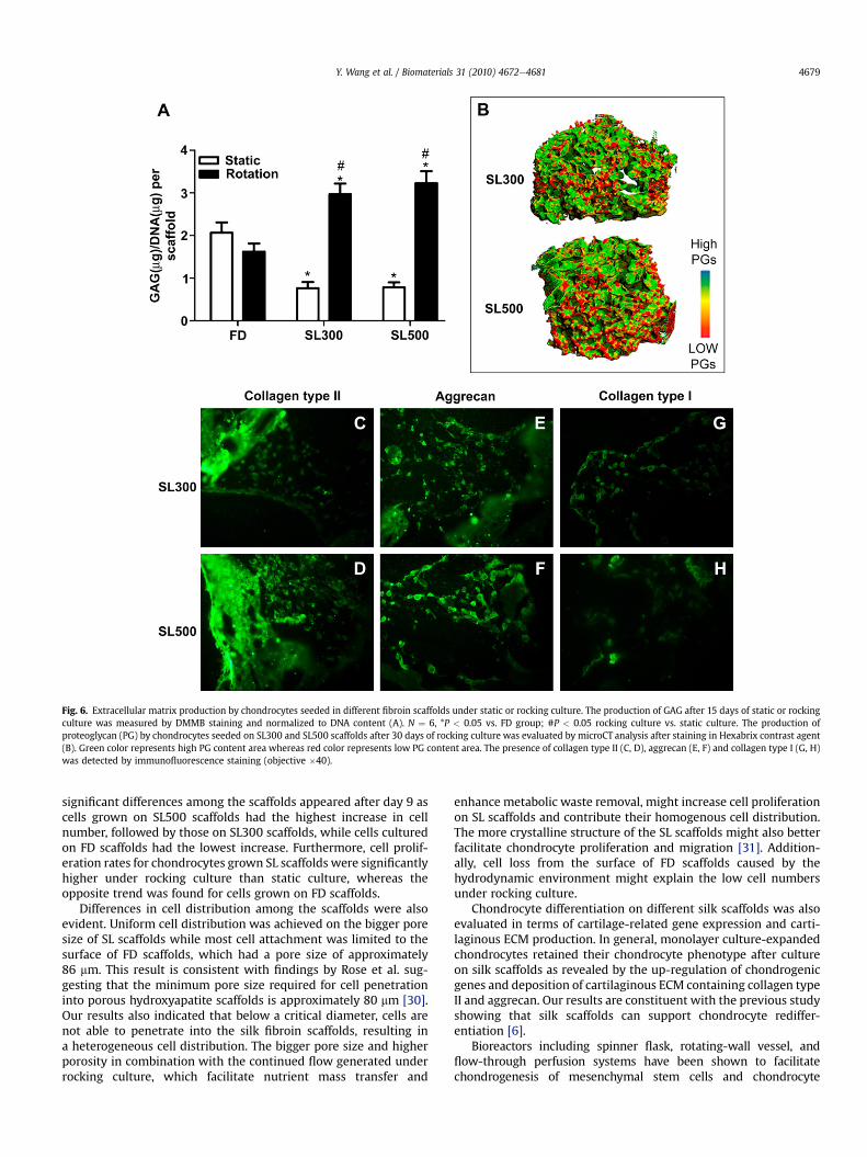
Fig. 6. Extracellular matrix production by chondrocytes seeded in different fibroin scaffolds under static or rocking culture. The production of GAG after 15 days of static or rockingculture was measured by DMMB staining and normalized to DNA content (A). N ¼ 6, *P < 0.05 vs. FD group; #P < 0.05 rocking culture vs. static culture. The production ofproteoglycan (PG) by chondrocytes seeded on SL300 and SL500 scaffolds after 30 days of rocking culture was evaluated by microCT analysis after staining in Hexabrix contrast agent(B). Green color represents high PG content area whereas red color represents low PG content area. The presence of collagen type II (C, D), aggrecan (E, F) and collagen type I (G, H)was detected by immunofluorescence staining (objective �40).
Y. Wang et al. / Biomaterials 31 (2010) 4672e4681 4679
significant differences among the scaffolds appeared after day 9 ascells grown on SL500 scaffolds had the highest increase in cellnumber, followed by those on SL300 scaffolds, while cells culturedon FD scaffolds had the lowest increase. Furthermore, cell prolif-eration rates for chondrocytes grown SL scaffolds were significantlyhigher under rocking culture than static culture, whereas theopposite trend was found for cells grown on FD scaffolds.
Differences in cell distribution among the scaffolds were alsoevident. Uniform cell distribution was achieved on the bigger poresize of SL scaffolds while most cell attachment was limited to thesurface of FD scaffolds, which had a pore size of approximately86 mm. This result is consistent with findings by Rose et al. sug-gesting that the minimum pore size required for cell penetrationinto porous hydroxyapatite scaffolds is approximately 80 mm [30].Our results also indicated that below a critical diameter, cells arenot able to penetrate into the silk fibroin scaffolds, resulting ina heterogeneous cell distribution. The bigger pore size and higherporosity in combination with the continued flow generated underrocking culture, which facilitate nutrient mass transfer and
enhance metabolic waste removal, might increase cell proliferationon SL scaffolds and contribute their homogenous cell distribution.The more crystalline structure of the SL scaffolds might also betterfacilitate chondrocyte proliferation and migration [31]. Addition-ally, cell loss from the surface of FD scaffolds caused by thehydrodynamic environment might explain the low cell numbersunder rocking culture.
Chondrocyte differentiation on different silk scaffolds was alsoevaluated in terms of cartilage-related gene expression and carti-laginous ECM production. In general, monolayer culture-expandedchondrocytes retained their chondrocyte phenotype after cultureon silk scaffolds as revealed by the up-regulation of chondrogenicgenes and deposition of cartilaginous ECM containing collagen typeII and aggrecan. Our results are constituent with the previous studyshowing that silk scaffolds can support chondrocyte rediffer-entiation [6].
Bioreactors including spinner flask, rotating-wall vessel, andflow-through perfusion systems have been shown to facilitatechondrogenesis of mesenchymal stem cells and chondrocyte
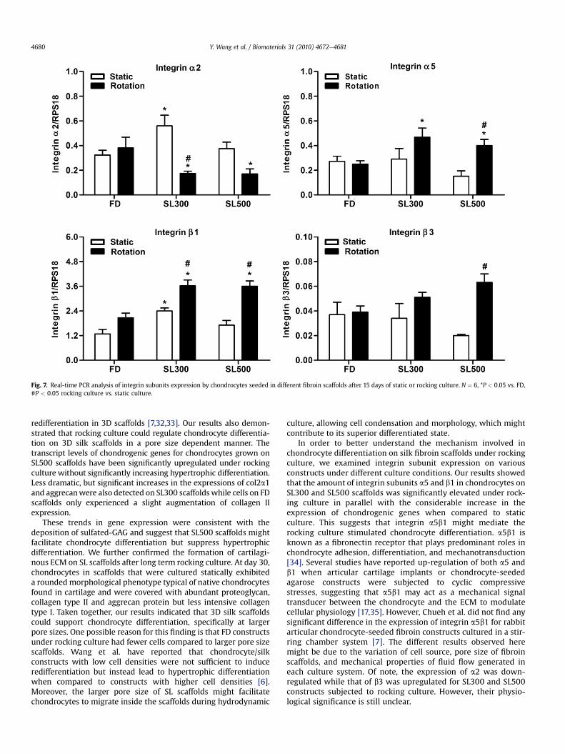
Fig. 7. Real-time PCR analysis of integrin subunits expression by chondrocytes seeded in different fibroin scaffolds after 15 days of static or rocking culture. N ¼ 6, *P < 0.05 vs. FD,#P < 0.05 rocking culture vs. static culture.
Y. Wang et al. / Biomaterials 31 (2010) 4672e46814680
redifferentiation in 3D scaffolds [7,32,33]. Our results also demon-strated that rocking culture could regulate chondrocyte differentia-tion on 3D silk scaffolds in a pore size dependent manner. Thetranscript levels of chondrogenic genes for chondrocytes grown onSL500 scaffolds have been significantly upregulated under rockingculture without significantly increasing hypertrophic differentiation.Less dramatic, but significant increases in the expressions of col2a1and aggrecanwere also detected on SL300 scaffolds while cells on FDscaffolds only experienced a slight augmentation of collagen IIexpression.
These trends in gene expression were consistent with thedeposition of sulfated-GAG and suggest that SL500 scaffolds mightfacilitate chondrocyte differentiation but suppress hypertrophicdifferentiation. We further confirmed the formation of cartilagi-nous ECM on SL scaffolds after long term rocking culture. At day 30,chondrocytes in scaffolds that were cultured statically exhibiteda roundedmorphological phenotype typical of native chondrocytesfound in cartilage and were covered with abundant proteoglycan,collagen type II and aggrecan protein but less intensive collagentype I. Taken together, our results indicated that 3D silk scaffoldscould support chondrocyte differentiation, specifically at largerpore sizes. One possible reason for this finding is that FD constructsunder rocking culture had fewer cells compared to larger pore sizescaffolds. Wang et al. have reported that chondrocyte/silkconstructs with low cell densities were not sufficient to induceredifferentiation but instead lead to hypertrophic differentiationwhen compared to constructs with higher cell densities [6].Moreover, the larger pore size of SL scaffolds might facilitatechondrocytes to migrate inside the scaffolds during hydrodynamic
culture, allowing cell condensation and morphology, which mightcontribute to its superior differentiated state.
In order to better understand the mechanism involved inchondrocyte differentiation on silk fibroin scaffolds under rockingculture, we examined integrin subunit expression on variousconstructs under different culture conditions. Our results showedthat the amount of integrin subunits a5 and b1 in chondrocytes onSL300 and SL500 scaffolds was significantly elevated under rock-ing culture in parallel with the considerable increase in theexpression of chondrogenic genes when compared to staticculture. This suggests that integrin a5b1 might mediate therocking culture stimulated chondrocyte differentiation. a5b1 isknown as a fibronectin receptor that plays predominant roles inchondrocyte adhesion, differentiation, and mechanotransduction[34]. Several studies have reported up-regulation of both a5 andb1 when articular cartilage implants or chondrocyte-seededagarose constructs were subjected to cyclic compressivestresses, suggesting that a5b1 may act as a mechanical signaltransducer between the chondrocyte and the ECM to modulatecellular physiology [17,35]. However, Chueh et al. did not find anysignificant difference in the expression of integrin a5b1 for rabbitarticular chondrocyte-seeded fibroin constructs cultured in a stir-ring chamber system [7]. The different results observed heremight be due to the variation of cell source, pore size of fibroinscaffolds, and mechanical properties of fluid flow generated ineach culture system. Of note, the expression of a2 was down-regulated while that of b3 was upregulated for SL300 and SL500constructs subjected to rocking culture. However, their physio-logical significance is still unclear.

Y. Wang et al. / Biomaterials 31 (2010) 4672e4681 4681
5. Conclusion
The current study demonstrated that chondrocytes are sensitiveto scaffold architecture in addition tomechanical stress. Bigger poresize and higher porosity silk scaffolds prepared by salt-leachingsuccessfully supported chondrocyte differentiation in terms oftheir spherical morphology, chondrogenic gene expression andcartilaginous ECM production. The hydrodynamic environmentgenerated by rocking culture exerted synergistic effects with scaf-fold structure on chondrocyte proliferation, differentiation, andintegrin gene expression. SL500 scaffolds seem to be the mostadvantageous silk fibroin scaffolds encouraging chondrogenicdifferentiation but limiting further hypertrophic differentiation.Chondrocyte/SL500 silk fibroin constructs obtained under in vitrorocking culture might serve as an excellent implant for in vivocartilage defect reparation. Scaffold-dependent differences inintegrin expression may play a role in mediating chondrocyteresponse to scaffold design and mechanical stress.
Future studies should investigate what other material proper-ties of silk fibroin scaffolds are important for chondrocyte differ-entiation and ECM synthesis. Additionally, the role of the a5, b1,and b3 subunits in cell adhesion to silk scaffolds and chondro-genesis should be further investigated. Using an advance cell-seeding method such as cell delivery via a polymerizing gelshould be further investigated in order to achieve more efficient,rapid and spatially uniform seeding that better mimic native tissue.Moreover, future in vivo animal studies will be necessary to fullyevaluate their clinical application.
Acknowledgements
The authors thank Mr. Lorenzo Moschini (DIMTI, University ofTrento, Italy) for GPC and HPLC measurements and Ms GiovannaSalice (Sociolario Center, Como, Itlay) for providing selected silk-worm cocoons. This project was supported by Children's Health-care of Atlanta, the Price Gilbert, Jr. Foundation and the USDepartment of Defense.
Appendix. Supplementary data
Supplementary data associated with this article can be found, inthe online version, at doi:10.1016/j.biomaterials.2010.02.006.
Appendix
Figures with essential color discrimination. Certain figures inthis article, in particular Figs. 4 and 6, are difficult to interpret inblack and white. The full color images can be found in the onlineversion, at doi:10.1016/j.biomaterials.2010.02.006.
References
[1] Altman GH, Horan RL, Lu HH, Moreau J, Martin I, Richmond JC, et al. Silkmatrix for tissue engineered anterior cruciate ligaments. Biomaterials 2002;23:4131e41.
[2] Chen J, Altman GH, Karageorgiou V, Horan R, Collette A, Volloch V, et al.Human bone marrow stromal cell and ligament fibroblast responses on RGD-modified silk fibers. J Biomed Mater Res A 2003;67:559e70.
[3] Wang Y, Rudym DD, Walsh A, Abrahamsen L, Kim HJ, Kim HS, et al. In vivodegradation of three-dimensional silk fibroin scaffolds. Biomaterials2008;29:3415e28.
[4] Marolt D, Augst A, Freed LE, Vepari C, Fajardo R, Patel N, et al. Bone andcartilage tissue constructs grown using human bone marrow stromal cells, silkscaffolds and rotating bioreactors. Biomaterials 2006;27:6138e49.
[5] Wang Y, Kim UJ, Blasioli DJ, Kim HJ, Kaplan DL. In vitro cartilage tissue engi-neering with 3D porous aqueous-derived silk scaffolds and mesenchymalstem cells. Biomaterials 2005;26:7082e94.
[6] Wang Y, Blasioli DJ, Kim HJ, KimHS, Kaplan DL. Cartilage tissue engineering withsilk scaffolds and human articular chondrocytes. Biomaterials 2006;27:4434e42.
[7] Shangkai C, Naohide T, Koji Y, Yasuji H, Masaaki N, Tomohiro T, et al. Trans-plantation of allogeneic chondrocytes cultured in fibroin sponge and stirringchamber to promote cartilage regeneration. Tissue Eng 2007;13:483e92.
[8] Yamane S, Iwasaki N, Kasahara Y, Harada K, Majima T, Monde K, et al. Effect ofpore size on in vitro cartilage formation using chitosan-based hyaluronic acidhybrid polymer fibers. J Biomed Mater Res A 2007;81:586e93.
[9] Motta A, Maniglio D, Migliaresi C, Kim HJ, Wang XY, Hu X, et al. Silk fibroinprocessing and thrombogenic responses. J Biomater Sci Polym Ed 2009;20:1875e97.
[10] Cai KY, Yao KD, Cui YL, Yang ZM, Li XQ, Xie HQ, et al. Influence of differentsurface modification treatments on poly(D,L -lactic acid) with silk fibroin andtheir effects on the culture of osteoblast in vitro. Biomaterials 2002;23:1603e11.
[11] Bini E, Foo CW, Huang J, Karageorgiou V, Kitchel B, Kaplan DL. RGD-func-tionalized bioengineered spider dragline silk biomaterial. Biomacromolecules2006;7:3139e45.
[12] Kardestuncer T, McCarthy MB, Karageorgiou V, Kaplan D, Gronowicz G. RGD-tethered silk substrate stimulates the differentiation of human tendon cells.Clin Orthop Relat Res 2006;448:234e9.
[13] Morgan AW, Roskov KE, Lin-Gibson S, Kaplan DL, Becker ML, Simon CG.Characterization and optimization of RGD-containing silk blends to supportosteoblastic differentiation. Biomaterials 2008;29:2556e63.
[14] Mauck RL, Byers BA, Yuan X, Tuan RS. Regulation of cartilaginous ECM genetranscription by chondrocytes and MSCs in 3D culture in response to dynamicloading. Biomech Model Mechanobiol 2007;6:113e25.
[15] Portner R, Nagel-Heyer S, Goepfert C, Adamietz P, Meenen NM. Bioreactordesign for tissue engineering. J Biosci Bioeng 2005;100:235e45.
[16] Knudson W, Loeser RF. CD44 and integrin matrix receptors participate incartilage homeostasis. Cell Mol Life Sci 2002;59:36e44.
[17] Lucchinetti E, Bhargava MM, Torzilli PA. The effect of mechanical load onintegrin subunits alpha5 and beta1 in chondrocytes from mature and imma-ture cartilage explants. Cell Tissue Res 2004;315:385e91.
[18] Kim UJ, Park J, Kim HJ, Wada M, Kaplan DL. Three-dimensional aqueous-derivedbiomaterial scaffolds from silk fibroin. Biomaterials 2005;26:2775e85.
[19] Lohmann CH, Schwartz Z, Niederauer GG, Carnes Jr DL, Dean DD, Boyan BD.Pretreatment with platelet derived growth factor-BB modulates the ability ofcostochondral resting zone chondrocytes incorporated into PLA/PGA scaffoldsto form new cartilage in vivo. Biomaterials 2000;21:49e61.
[20] Boyan BD, Wong KL, Wang L, Yao H, Guldberg RE, Drab M, et al. Regulation ofgrowth plate chondrocytes by 1,25-dihydroxyvitamin D3 requires caveolaeand caveolin-1. J Bone Miner Res 2006;21:1637e47.
[21] Palmer AW, Guldberg RE, Levenston ME. Analysis of cartilage matrix fixedcharge density and three-dimensional morphology via contrast-enhancedmicrocomputed tomography. Proc Natl Acad Sci U S A 2006;103:19255e60.
[22] Inoue S, Tanaka K, Arisaka F, Kimura S, Ohtomo K, Mizuno S. Silk fibroin ofBombyx mori is secreted, assembling a high molecular mass elementary unitconsisting of H-chain, L-chain, and P25, with a 6:6:1 molar ratio. J Biol Chem2000;275:40517e28.
[23] Jin HJ, Kaplan DL. Mechanism of silk processing in insects and spiders. Nature2003;424:1057e61.
[24] Penninger JM, Kersten R, Baur H. Hydrolysis of diphenylether in supercriticalwater: effects of dissolved NaCl. J Supercritical Fluid 2000;17:215e26.
[25] Servoli E, Maniglio D, Motta A, Predazzer R, Migliaresi C. Surface properties ofsilk fibroin films and their interaction with fibroblasts. Macromol Biosci2005;5:1175e83.
[26] Wilson D, Valluzzi R, Kaplan D. Conformational transitions in model silkpeptides. Biophys J 2000;78:2690e701.
[27] Asakura T, Kuzuhara A, Tabeta R, Saito H. Conformation characterization ofBombyx mori silk fibroin in the solid state by high-frequency 13C crosspolarization-magic angle spinning NMR, X-ray diffraction, and infrared spec-troscopy. Macromolecules 1985;18:1841e5.
[28] Hung CT, Mauck RL, Wang CC, Lima EG, Ateshian GA. A paradigm for func-tional tissue engineering of articular cartilage via applied physiologic defor-mational loading. Ann Biomed Eng 2004;32:35e49.
[29] Yamamoto K, Tomita N, Fukuda Y, Suzuki S, Igarashi N, Suguro T, et al. Time-dependent changes in adhesive force between chondrocytes and silk fibroinsubstrate. Biomaterials 2007;28:1838e46.
[30] Rose FR, Cyster LA, Grant DM, Scotchford CA, Howdle SM, Shakesheff KM. Invitro assessment of cell penetration into porous hydroxyapatite scaffolds witha central aligned channel. Biomaterials 2004;25:5507e14.
[31] Kotov NA, Liu Y, Wang S, Cumming C, Eghtedari M, Vargas G, et al. Invertedcolloidal crystals as three-dimensional cell scaffolds. Langmuir 2004;20:7887e92.
[32] Augst A, Marolt D, Freed LE, Vepari C, Meinel L, Farley M, et al. Effects ofchondrogenic and osteogenic regulatory factors on composite constructsgrown using human mesenchymal stem cells, silk scaffolds and bioreactors.J R Soc Interface 2008;5:929e39.
[33] Ohyabu Y, Tanaka J, Ikada Y, Uemura T. Cartilage tissue regeneration from bonemarrow cells by RWV bioreactor using collagen sponge scaffold. Mater Sci EngC 2009;4:1150e5.
[34] Schwartz MA, DeSimone DW. Cell adhesion receptors in mechano-transduction. Curr Opin Cell Biol 2008;20:551e6.
[35] Chowdhury TT, Appleby RN, Salter DM, Bader DA, Lee DA. Integrin-mediatedmechanotransduction in IL-1 beta stimulated chondrocytes. Biomech ModelMechanobiol 2006;5:192e201.
Related Documents