BioMed Central Page 1 of 14 (page number not for citation purposes) Cerebrospinal Fluid Research Open Access Research The subcommissural organ of the rat secretes Reissner's fiber glycoproteins and CSF-soluble proteins reaching the internal and external CSF compartments Karin Vio, Sara Rodríguez, Carlos R Yulis, Cristian Oliver and Esteban M Rodríguez* Address: Instituto de Anatomía, Histología y Patología, Facultad de Medicina, Universidad Austral de Chile, Valdivia, Chile Email: Karin Vio - [email protected]; Sara Rodríguez - [email protected]; Carlos R Yulis - [email protected]; Cristian Oliver - [email protected]; Esteban M Rodríguez* - [email protected] * Corresponding author Abstract Background: The subcommissural organ (SCO) is a highly conserved brain gland present throughout the vertebrate phylum; it secretes glycoproteins into the cerebrospinal fluid (CSF), where they aggregate to form Reissner's fiber (RF). SCO-spondin is the major constituent protein of RF. Evidence exists that the SCO also secretes proteins that remain soluble in the CSF. The aims of the present investigation were: (i) to identify and partially characterize the SCO-secretory compounds present in the SCO gland itself and in the RF of the Sprague-Dawley rat and non- hydrocephalic hyh mouse, and in the CSF of rat; (ii) to make a comparative analysis of the proteins present in these three compartments; (iii) to identify the proteins secreted by the SCO into the CSF at different developmental periods. Methods: The proteins of the SCO secreted into the CSF were studied (i) by injecting specific antibodies into ventricular CSF in vivo; (ii) by immunoblots of SCO, RF and CSF samples, using specific antibodies against the SCO secretory proteins (AFRU and anti-P15). In addition, the glycosylated nature of SCO-compounds was analysed by concanavalin A and wheat germ agglutinin binding. To analyse RF-glycoproteins, RF was extracted from the central canal of juvenile rats and mice; to investigate the CSF-soluble proteins secreted by the SCO, CSF samples were collected from the cisterna magna of rats at different stages of development (from E18 to PN30). Results: Five glycoproteins were identified in the rat SCO with apparent molecular weights of 630, 450, 390, 320 and 200 kDa. With the exception of the 200-kDa compound, all other compounds present in the rat SCO were also present in the mouse SCO. The 630 and 390 kDa compounds of the rat SCO have affinity for concanavalin A but not for wheat germ agglutinin, suggesting that they correspond to precursor forms. Four of the AFRU-immunoreactive compounds present in the SCO (630, 450, 390, 320 kDa) were absent from the RF and CSF. These may be precursor and/or partially processed forms. Two other compounds (200, 63 kDa) were present in SCO, RF and CSF and may be processed forms. The presence of these proteins in both, RF and CSF suggests a steady- state RF/CSF equilibrium for these compounds. Eight AFRU-immunoreactive bands were consistently found in CSF samples from rats at E18, E20 and PN1. Only four of these compounds were detected in the cisternal CSF of PN30 rats. The 200 kDa compound appears to be a key Published: 24 January 2008 Cerebrospinal Fluid Research 2008, 5:3 doi:10.1186/1743-8454-5-3 Received: 3 October 2007 Accepted: 24 January 2008 This article is available from: http://www.cerebrospinalfluidresearch.com/content/5/1/3 © 2008 Vio et al; licensee BioMed Central Ltd. This is an Open Access article distributed under the terms of the Creative Commons Attribution License (http://creativecommons.org/licenses/by/2.0 ), which permits unrestricted use, distribution, and reproduction in any medium, provided the original work is properly cited.

Welcome message from author
This document is posted to help you gain knowledge. Please leave a comment to let me know what you think about it! Share it to your friends and learn new things together.
Transcript
BioMed CentralCerebrospinal Fluid Research
ss
Open AcceResearchThe subcommissural organ of the rat secretes Reissner's fiber glycoproteins and CSF-soluble proteins reaching the internal and external CSF compartmentsKarin Vio, Sara Rodríguez, Carlos R Yulis, Cristian Oliver and Esteban M Rodríguez*Address: Instituto de Anatomía, Histología y Patología, Facultad de Medicina, Universidad Austral de Chile, Valdivia, Chile
Email: Karin Vio - [email protected]; Sara Rodríguez - [email protected]; Carlos R Yulis - [email protected]; Cristian Oliver - [email protected]; Esteban M Rodríguez* - [email protected]
* Corresponding author
AbstractBackground: The subcommissural organ (SCO) is a highly conserved brain gland presentthroughout the vertebrate phylum; it secretes glycoproteins into the cerebrospinal fluid (CSF),where they aggregate to form Reissner's fiber (RF). SCO-spondin is the major constituent proteinof RF. Evidence exists that the SCO also secretes proteins that remain soluble in the CSF. The aimsof the present investigation were: (i) to identify and partially characterize the SCO-secretorycompounds present in the SCO gland itself and in the RF of the Sprague-Dawley rat and non-hydrocephalic hyh mouse, and in the CSF of rat; (ii) to make a comparative analysis of the proteinspresent in these three compartments; (iii) to identify the proteins secreted by the SCO into theCSF at different developmental periods.
Methods: The proteins of the SCO secreted into the CSF were studied (i) by injecting specificantibodies into ventricular CSF in vivo; (ii) by immunoblots of SCO, RF and CSF samples, usingspecific antibodies against the SCO secretory proteins (AFRU and anti-P15). In addition, theglycosylated nature of SCO-compounds was analysed by concanavalin A and wheat germ agglutininbinding. To analyse RF-glycoproteins, RF was extracted from the central canal of juvenile rats andmice; to investigate the CSF-soluble proteins secreted by the SCO, CSF samples were collectedfrom the cisterna magna of rats at different stages of development (from E18 to PN30).
Results: Five glycoproteins were identified in the rat SCO with apparent molecular weights of 630,450, 390, 320 and 200 kDa. With the exception of the 200-kDa compound, all other compoundspresent in the rat SCO were also present in the mouse SCO. The 630 and 390 kDa compounds ofthe rat SCO have affinity for concanavalin A but not for wheat germ agglutinin, suggesting that theycorrespond to precursor forms. Four of the AFRU-immunoreactive compounds present in theSCO (630, 450, 390, 320 kDa) were absent from the RF and CSF. These may be precursor and/orpartially processed forms. Two other compounds (200, 63 kDa) were present in SCO, RF and CSFand may be processed forms. The presence of these proteins in both, RF and CSF suggests a steady-state RF/CSF equilibrium for these compounds. Eight AFRU-immunoreactive bands wereconsistently found in CSF samples from rats at E18, E20 and PN1. Only four of these compoundswere detected in the cisternal CSF of PN30 rats. The 200 kDa compound appears to be a key
Published: 24 January 2008
Cerebrospinal Fluid Research 2008, 5:3 doi:10.1186/1743-8454-5-3
Received: 3 October 2007Accepted: 24 January 2008
This article is available from: http://www.cerebrospinalfluidresearch.com/content/5/1/3
© 2008 Vio et al; licensee BioMed Central Ltd. This is an Open Access article distributed under the terms of the Creative Commons Attribution License (http://creativecommons.org/licenses/by/2.0), which permits unrestricted use, distribution, and reproduction in any medium, provided the original work is properly cited.
Page 1 of 14(page number not for citation purposes)
Cerebrospinal Fluid Research 2008, 5:3 http://www.cerebrospinalfluidresearch.com/content/5/1/3
compound in rats since it was consistently found in all samples of SCO, RF and embryonic andjuvenile CSF.
Conclusion: It is concluded that (i) during the late embryonic life, the rat SCO secretescompounds that remain soluble in the CSF and reach the subarachnoid space; (ii) during postnatallife, there is a reduction in the number and concentration of CSF-soluble proteins secreted by theSCO. The molecular structure and functional significance of these proteins remain to be elucidated.The possibility they are involved in brain development has been discussed.
BackgroundThe subcommissural organ (SCO) differentiates at anearly stage of ontogenetic development in all vertebrates[1] and, with the exception of a few species (anthropoidsand bats) [2], it remains fully active throughout life. Thesecretion of this gland is released into the ventricular cer-ebrospinal fluid (CSF) where most of it condenses to forma filamentous structure named after Reissner [3] as Reiss-ner's fiber (RF). RF is formed by the assembly of complex-type, high molecular weight glycoproteins secreted by theSCO into the cerebral aqueduct; it is a dynamic structurethat continuously grows caudally by the addition of newlyreleased molecules to its cephalic end [2,4]; it extendsthroughout the aqueduct of Sylvius, fourth ventricle andcentral canal of the spinal cord [4,5]. When arriving at theterminal ventricle of the central canal at the filum, RF glyc-oproteins undergo chemical modification, disassemblyand passage into neighbouring vessels [6,7]. RF has thecapacity to bind and transport away compounds such asdopamine, L-DOPA and serotonin, thus contributing tothe clearance of these compounds from the CSF [8,9].
The identification and characterization of the secretorycompounds of the SCO have been the subject of numer-ous investigations and have contributed to partial clarifi-cation of its function. Immunoblot analyses of bovineSCO using antibodies against RF glycoproteins have led tothe identification of high molecular weight glycoproteinsof 540, 450, 320 and 190 kDa. Evidence has beenobtained indicating that the 540 and the 320 kDa com-pounds would correspond to precursor forms [10-12]. Inthe SCO of chick embryos, del Brio et al. have determinedthe presence of three compounds of 540, 320 and 230kDa [13]. In the SCO of the elasmobranch Scyliorhinuscanicula, five compounds of 600, 475, 400, 145 and 35kDa have been identified [14]. Antibodies raised againstthe precursor form of 540 kDa and the processed form of450 kDa synthesized by the bovine SCO, when used toimmunostain the bovine and rat SCO, react with thebovine SCO but not with the rat SCO. At variance, theantibody against the bovine SCO 320 kDa band reactswith both, bovine and rat SCO [11,12]. This is the onlyinformation available concerning the secretory com-pounds of the SCO of murine species.
Ontogenetic studies have revealed that the SCO starts toexpress a secretory activity much earlier than the appear-ance of the first RF [1,15]. In the rat, the SCO is well devel-oped and immunoreactive with the anti-RF antibodies atembryonic day 14 (E14). However, aggregated secretorymaterial and a RF proper first appear during the first post-natal week [1]. These findings suggest that the embryonicSCO secretes compounds that remain soluble in the CSF,thus differing from RF proteins, which aggregate. Theexistence of CSF-soluble compounds secreted by the SCOgained support by immunochemical studies in humanand rabbit CSF [2,16]. Recently, Hoyo-Becerra et al. [17]have described the presence in the CSF from chickembryos of proteins that are reactive with anti-RF antise-rum (AFRU). Efforts to detect SCO glycoproteins in thebovine CSF have failed [18,19]. None of previous studieshave dealt with the characterization of the SCO secretoryglycoproteins present in the gland itself, in the CSF and inthe RF of the same animal species and using immunoblotmethodology; this has prevented a comparative analysisof the secretory proteins before they are released and afterthey either assemble into RF or remain soluble in the CSF.Thus, whether the SCO proteins forming RF and the SCOproteins solubilized in the CSF are the same, or similar orunrelated compounds has yet to be determined.
The primary structure of the major constituent of bovineRF, SCO-spondin, has been fully established as a large N-glycosylated protein (450 kDa) [20,21]. Several lines ofevidence indicate that SCO-spondin plays a role in CNSdevelopment. SCO-spondin belongs to a protein super-family exhibiting conserved motifs of the thrombospon-din type 1 repeat [21]. Proteins of this family are stronglyexpressed during mammalian CNS development and areinvolved in mechanisms of cellular adhesion and axonalpathfinding [22]. It has not yet been established whetherSCO-spondin itself or its processed products are presentin and circulate through the CSF compartments. Neitherhas it been established whether SCO-spondin or relatedcompounds are actually secreted into the embryonic CSF.This information is essential when interpreting the poten-tial role of SCO-spondin in CNS development.
The present investigation was designed to investigate therat and mouse SCO glycoproteins in order (i) to identify
Page 2 of 14(page number not for citation purposes)
Cerebrospinal Fluid Research 2008, 5:3 http://www.cerebrospinalfluidresearch.com/content/5/1/3
and partially characterize the secretory compoundspresent in the gland itself, in the RF and in the CSF (ratonly); (ii) to make a comparative analysis of the proteinspresent in these three compartments, in order to establishto what extent they share gel migration, immunoreactiveand lectin-binding properties; and (iii) to identify the pro-teins secreted by the SCO into the CSF at different devel-opmental periods. The identification of the CSF-solublesecretion of the SCO during embryonic and juvenile lifemay throw some light on the function of this gland.Indeed, such CSF-soluble proteins would circulatethroughout both the internal and external CSF compart-ments and could reach distant targets. Furthermore, differ-ences in the type or concentration of these CSF-solubleproteins at different developmental stages may open newavenues for SCO research.
MethodsAnimalsSprague-Dawley rats and hyh mice: Sprague-Dawley ratsand hyh mice were obtained from The Jackson Laboratory(Bar Harbor, ME, USA) and bred in two colonies (Fac-ultad de Medicina, Universidad Austral de Chile, Valdivia,Chile). Housing, handling, care and processing of animalswere carried out according to the regulations approved bythe council of the American Physiological Society. A localuniversity committee has approved the experimental pro-tocol. Animals were fed ad libitum with rodent food andmaintained under a constant 12-h light/dark photoperiodand at a constant temperature. Female rats were checkedfor the presence of a vaginal plug after overnight matingwith a male. At days 18, 19 and 20 of pregnancy, pregnantrats were weighed and anesthetized with an intraperito-neal injection of ketamine (40 mg/kg) and acepromazine(100 mg/kg); embryos were removed and CSF was col-lected from the cisterna magna.
For mice, the hyh mutation arose spontaneously on theC57BL/10J. They were then outcrossed onto a B6C3Fe-a/a background (C57BL/6J × C3HeB/FeJ-a/a). Hence, thegenetic background of hyh mice strain (B6C3Fe a/a-Napahyh/J) is C57BL/6J × C3HeB/FeJ-a/a. In the presentstudy only non-hydrocephalic mice were used. No differ-ences between wild type and heterozygous mice weredetected with the methods used.
ImmunocytochemistryThe brains of 10 Sprague-Dawley rats, PN2, were fixed byintravascular perfusion with Bouin fixative and embeddedin paraffin. Sections were processed by the immunoperox-idase method of Sternberger et al. [23]. The following pri-mary antibodies were used: (i) An antiserum developed inrabbits against bovine RF-glycoproteins extracted in amedium containing urea (AFRU, A = antiserum, FR =Fiber of Reissner, U = urea; [24]), (ii) An antiserum devel-
oped in rabbits against a synthetic 15-aminoacid peptidewith a sequence deduced from a region of the SCO-spon-din protein that does not correspond to any of the repeatspresent in this molecule (anti-P15; [21,25]). This peptidesequence has no homology with any of the proteinsrecorded in the Gene Bank; hence anti-P15 can beregarded as a specific antibody for SCO-spondin [25]. Sec-tions were sequentially incubated in: (i) AFRU, dilution1:1000, or anti-P15, dilution 1:500, for 18 h; (ii) second-ary antibody (anti-rabbit IgG, raised in our laboratory),diluted 1:15, for 30 min; (iii) rabbit PAP (Dako, Carpin-teria, CA, USA), diluted 1:75, for 30 min. All immunocy-tochemical reactions were visualized by the histochemicaldetection of peroxidase using hydrogen peroxide (Merck,Darmstadt, Germany) and 3-3'diaminobenzidine tetrahy-drochloride (DAB, Sigma, Madrid, Spain). Omission ofthe incubation in the primary antibody was used as a con-trol.
Injection of antibodies into the rat CSFEight PN2 Sprague-Dawley pups were anesthetized withan intraperitoneal injection of ketamine (40 mg/kg) andacepromazine (100 mg/kg); anesthesia lasted throughoutthe experiment. The head of the pup was immobilized byplacing it into a paraffin cast specially adapted to the headof PN2 rats. Using a dissecting microscope, a 27-gaugecannula connected to a perfusion pump, was inserted intothe left lateral ventricle; 3 μl of undiluted AFRU (n = 4) oranti-P15 (n = 4) sera were infused over 5 min. Two hoursafter antibody administration, the brain was dissected outand fixed by immersion in Bouin's fixative for 2 d. Afterdehydration in alcohols, the brains were embedded inparaffin. For the demonstration of binding sites of theinjected antibodies, sections were sequentially incubatedwith anti-rabbit IgG, diluted 1:15, for 30 min, and rabbitPAP (Dako, Carpinteria, CA, USA), diluted 1:75, for 30min. This was followed by the diaminobenzidine reac-tion.
Reissner's fiber extractsBovine spinal cord from adult cows was obtained asdescribed previously [24] and RF was collected by perfus-ing the central canal of the spinal cord with saline. RF of10 juvenile rats (PN30) and 8 juvenile mice (PN30) wascollected in the following way: after euthanasia with keta-mine, the spinal cord was dissected out, immersed in buff-ered saline and divided into several segments by makingtransverse cuts with a razor blade. Under a dissectingmicroscope, a cylinder of the grey matter surrounding thecentral canal was obtained by doing four longitudinalcuts; this block of tissue contained the central canal andthe corresponding stretch of RF. The bovine and murineRFs were extracted in 50 mM ammonium bicarbonate, pH7,4, containing 0.5 mM phenyl-methylsulfonyl fluoride(PMSF). The protein concentration of bovine RF extract
Page 3 of 14(page number not for citation purposes)
Cerebrospinal Fluid Research 2008, 5:3 http://www.cerebrospinalfluidresearch.com/content/5/1/3
[26] was 0.8 μg/μl, rat central canal/RF extracts rangedfrom 8.0 – 8.6 μg/μl and mouse central canal/RF extractsranged from 5.0 – 6.4 μg/μl. Samples were stored at -70°Cuntil used.
SCO extractsFifty PN30 rats and thirty PN30 mice were used. The ani-mals were sacrificed under ketamine anesthesia, the brainwas dissected out and a block of tissue containing theSCO and the posterior commissure was obtained under adissecting microscope; post-mortem interval was 2–5min. Six SCOs were pooled to make one sample. Eachsample was extracted in 300 μl of 50 mM ammoniumbicarbonate, pH 7,5, and 0,5 mM phenyl-methylsulfonylfluoride (PMSF), homogenized, sonicated in ice for 10 sand centrifuged at 12.000 g, for 45 min, at 4°C. Threealiquots of 100 μl, each containing the equivalent of twoSCOs, were obtained from each extracted sample. Proteincontent of rat SCO extracts ranged from 1.5 to 2.2 μg/μland of mouse SCO extracts from 1.2 – 1.3 μg/μl. Extractswere stored at -70°C. The bovine SCO of adult animalswas collected and extracted (protein concentration 1.2 –1.35 μg/μl) as previously reported [12].
Cerebrospinal fluid collectionRats: E18 (n = 18), E19 (n = 22) and E20 (n = 20) ratembryos were used for CSF collection. CSF samples wereobtained through a 30-gauge needle inserted into the cis-terna magna. About 10–15 μl were obtained from eachembryo. Pups at PN1 (n = 12) and PN7 (n = 20) wereanesthetized with ketamine (40 mg/kg) and acepro-mazine (100 mg/kg), the head flexed and a 27-gauge nee-dle inserted into the cisterna magna. In PN30 rats (n = 22)CSF was collected from the cisterna magna according toRodríguez et al. [9]. Occasionally, severence of a bloodvessel caused contamination of the samples with bloodand these samples were discarded. About 25–50 μl of CSFwere obtained from each pup and up to 100 μl from eachPN30 rat. CSF samples were collected into Eppendorftubes and centrifuged twice to remove cells or cell debris.Average protein concentrations of E18, E20, PN1, PN7and PN30 CSF samples were: 2.4, 1.8, 1.5, 1.0 and 0.4 μg/μl, respectively. Samples were stored at -70°C.
Immunoblot analysesThe following samples were used for blotting: (i) 50 μlsamples of SCO extracts, containing the equivalent of0.02 bovine SCO, 1 mouse SCO and 1 rat SCO; (ii) 50 μlsamples of RF extracts containing the equivalent of 0.001bovine RF, 0.1 mouse RF and 0.1 rat RF; (iii) 15 μl of undi-luted/non concentrated CSF from embryonic and postna-tal rats. Samples were subjected to SDS-PAGE using a 5–15% polyacrylamide linear gradient. Proteins were trans-ferred to nitrocellulose membranes [27]; to block non-specific binding, blots were saturated with 5% non-fat
milk in 0.1 M PBS containing 0.15 mM NaCl and 0.1%Tween-20 (Sigma, Madrid, Spain), for 90 min. Two pri-mary antisera were used for immunoreaction of blots ofSCO and RF: (i) AFRU, 1:25,000 dilution, for 2 h; (ii) anti-P15 [24], 1:1,000 dilution, for 2 h. Anti-rabbit IgG-HRP(Pierce, Rockford, IL, USA) was used at 1:25,000 dilution,for 1.5 h. For immunoblotting of CSF samples, the follow-ing protocol was used: (i) AFRU, at 1:5,000 dilution andanti-rabbit IgG-HRP at 1:25,000 dilution, for 2 h; (ii) anti-P15, at 1:1,000 dilution and anti-rabbit IgG-HRP at1:60,000 dilution, for 2 h. Incubations were at room tem-perature and in darkness. Immunoreactive polypeptideswere detected by using an enhanced chemiluminescence(ECL) system (Super Signal, Pierce, Rockford, IL, USA) asinstructed by the manufacturer. Molecular weight stand-ards in the range of 10–250 kDa were used (Bio-Rad, Her-cules, CA, USA). Control blots were processed as abovewithout the primary antibody. Immunoblotting of sam-ples was repeated as follows: SCO of rat and mouse × 10;RF of rat and mouse × 5; rat CSF of E18 and E20 embryos× 5; rat CSF collected at PN1, PN7 and PN30 × 10. Immu-noblots were digitized (n = 4 for each condition analysed)and linear densitograms were obtained using the UN –SCAN-IT software (Silk Scientific, Orem, UT, USA). Statis-tical analyses were performed using the Prism software(GraphPad Software, San Diego, CA, USA) applying the 1-way Anova and Tukey's test.
Lectin bindingBlots of bovine, rat and mouse SCO in parallel to thoseused for immunoblotting, were used to test for lectinbinding. Blots were sequentially incubated with (i) oxi-dized bovine serum albumin (BSA, Winkler, Santiago,Chile), (ii) concanavalin A (Con A; affinity = mannose,glucose; Sigma, St. Louis, MO, USA), 0.2 μg/ml for 1 h;(iii) anti-Con A developed in rabbits (Sigma, St. Louis,MO, USA), 1:10,000 dilution for 1 h at room temperature;(iii) anti-rabbit IgG-HRP (Pierce, Rockford, IL, USA)1:25,000, for 1.5 h; (iv) binding was detected by using anenhanced chemiluminescence system (see above). Thesame protocol was used for binding of wheat germ agglu-tinin (WGA; affinity = glucosamine, sialic acid, Sigma, St.Louis, MO, USA). WGA was used at a concentration of 0.5μg/ml and anti-WGA (Sigma, St. Louis, MO, USA) wasused at a 1:5,000 dilution.
Con A has affinity for internal, and preferentially for ter-minal, mannose residues; this makes it a good marker fornewly synthesised glycoproteins that have not yet beenprocessed through the Golgi apparatus (precursor forms).At variance, WGA has affinity for two sugar residues (glu-cosamine, sialic acid) added to the glycoprotein by theGolgi apparatus, thus being a good marker of post-Golgicompounds (processed forms) [28,29].
Page 4 of 14(page number not for citation purposes)

Cerebrospinal Fluid Research 2008, 5:3 http://www.cerebrospinalfluidresearch.com/content/5/1/3
Page 5 of 14(page number not for citation purposes)
A, C, E: Paraffin sagittal sections through the subcommissural organ (SCO) of a PN2 rat after a single injection of AFRU into a lateral ventricle (inset 1C, labeled Ab)Figure 1A, C, E: Paraffin sagittal sections through the subcommissural organ (SCO) of a PN2 rat after a single injection of AFRU into a lateral ventricle (inset 1C, labeled Ab). A: Immunostaining using AFRU (raised in rabbits) as primary antibody. The SCO is immunoreactive. Arrow points to RF-material. Insert: Detailed magnification of RF fibrils immunoreactive with AFRU (arrows). C: Adjacent section to that shown in A, immunostained using anti-rabbit IgG as primary antibody to reveal the antibody (AFRU) administered in vivo. The latter appears exclusively bound to the newly released secretory material aggregated on the surface of the SCO to form the pre-RF (square) and to aggregated RF-material lying in the ventricle (arrow). E: Detailed mag-nification of area framed in C showing the location of the antigen-antibody complexes formed in vivo at the pre-RF layer on the SCO surface and at the aggregated RF-material. B, D, F: Paraffin sagittal sections through the SCO of a PN2 rat after a single injection of anti-P15 into a lateral ventricle. B: Sagittal section through the SCO immunostained with anti-P15. The ependymal cells of the SCO appear immunostained (arrow). Lower insert: detailed magnification of the secretory ependyma (E) showing the location of the immunoreactive material in the supranuclear cytoplasm. n: nuclei. Upper insert: detailed magnification of RF fibrils immunoreactive with anti-P15 (arrows). D: Section immunostained with anti-rabbit IgG as primary antibody to reveal the location of the antibody administered in vivo at pre-RF (arrow). F: Detailed magnification of area framed in previous figure showing the location of the antigen-antibody complexes formed in vivo at the pre-RF (arrows). IIIV: third ventricle. Most sec-tions counterstained with hematoxilin.
SCO
IIIV
AFRU
SCO
IIIV
IIIV
SCO
IIIV
Anti-IgG
IIIV
SCO
SCO
AFRU
RF-material
Ab
A
C D
E F
200μm
200μm 200μm
20μm20μm
pre-RF
pre-RF
Anti-IgG
Anti-IgG Anti-IgG
IIIV
SCO
B 200μm
Anti-P15
50μm 40μm
40μm
E
n
Cerebrospinal Fluid Research 2008, 5:3 http://www.cerebrospinalfluidresearch.com/content/5/1/3
ResultsImmunocytochemistryThe compounds secreted by rat SCO were identified usingtwo antibodies. One is directed against bovine RF consti-tutive proteins (AFRU), whilst anti-P15 is directed againsta 15-aminoacid sequence of SCO-spondin, the main con-stitutive protein of RF. Both antibodies are highly specificfor the rat SCO secretion, located either intracellularly orextracellularly in the form of aggregated RF-material (Fig.1A, B, 2). The SCO-specificity of AFRU and anti-P15 isalso evident when used in vivo. After the intraventricularadministration of these antibodies, they bound exclu-sively to the SCO secretory material that had been releasedinto the ventricle and that had aggregated into the form ofpre-RF or RF [4] (Fig. 1C–F, 2A).
Analysis of rat and mouse SCOextractsThe secretory compounds of the SCO of PN30 rats andmice were analysed in parallel with the well known secre-tory glycoproteins of the bovine SCO [11,12]. Despite thesmall size of the rat and mouse SCO, the use of a high-sen-sitivity Western blot system based on enhanced chemilu-minescence enabled demonstration of the secretorycompounds using samples containing the equivalent ofone SCO. As reported previously [12], the Western blotanalysis of the bovine SCO, using AFRU as primary anti-body, revealed polypeptides of 540, 450, and 320 kDa(Fig. 2C). The high sensitivity of the method used in thepresent study, allowed detection of an additional immu-noreactive band of 390 kDa (Fig. 2C). The rat and mouseSCO displayed a pattern of AFRU-immunoreactive bandsthat differed from that of the bovine SCO (Fig. 2C). The540-kDa polypeptide present in the bovine SCO was notdetected in rat and mouse SCO (Fig. 2C). The rat SCO dis-played AFRU-immunoreactive bands of 630, 450, 390,320 and 200 kDa (Fig. 2C, Tables 1, 2). In a few blots,bands of 63 and 45 kDa were also detected (Figs. 2D, 3A,Tables 2, 3). The compounds of 630 and 200 kDa wereabsent from the bovine SCO.
With the exception of the 200-kDa compound, all highmolecular mass compounds present in the rat SCO werealso present in the mouse SCO (Fig. 2C).
Analysis of central canal/Reissner's fiber extractsImmunoblotting of a tissue extract that included the cen-tral canal of the spinal cord and RF of mice and rats atPN30, revealed AFRU-immunoreactive bands most likelycorresponding to RF-glycoproteins. In the rat RF, com-pounds of 200, 120 and 45 kDa were detected in all blots(Fig. 2D, Tables 2, 3). Compounds of 63, 50 and 25 kDawere detected in 2 or 3 of the 5 blots (Tables 2, 3). In themouse RF the 200-kDa compound was detected in allblots; compounds of 63 and 45 kDa were present in most
blots; a polypeptide of 120 kDa was never detected (Fig.2D).
Analysis of CSF samplesIn order to identify those compounds secreted by the SCOinto the CSF and that, at variance with RF-glycoproteins,remain soluble in the CSF, samples of this fluid wereobtained from embryo, early postnatal and juvenile ratsand analysed by immunoblotting using AFRU and anti-P15.
CSF samples collected from the cisterna magna during thelate embryonic life (E18, E19, E20) and the early postna-tal life (PN1, PN7) displayed a very similar pattern ofAFRU-immunoreactive bands (Fig. 2E). The latter were of200, 180, 164, 145, 120, 63, 50 and 25 kDa (Fig. 2E, Table2, 3). This band pattern was found in all blots. There were,however, some differences of intensity of immunoreac-tion among bands of samples collected at different ages(E18 to PN7; Fig. 2E, F). The 50 and 25 kDa compoundswere more strongly reactive in fetal CSF, whilst the 200and 120 kDa polypeptides were more reactive in PN1 andPN7 samples (Fig. 2E, F).
A marked difference was observed in the cisternal CSFobtained from PN30 rats; the following bands present inprevious stages were absent: 180, 164, 145 (P < 0.05) and120 kDa (P < 0.001) and only bands for 200, 63, 50, and25 kDa were seen (Fig. 2E, F, Table 2, 3).
Comparative analysis of immunoreactive secretory proteins in the rat SCO, RF and CSFThe high sensitivity of the Western blot system has ena-bled for the first time within the same species, detectionof the AFRU-immunoreactive polypeptides present in theSCO, RF and CSF. This, in turn, has made possible the fol-lowing comparative analysis: (i) the four AFRU-immuno-reactive compounds with the largest molecular mass (630,450,390, 320 kDa) were present in the SCO and missingfrom RF and CSF (Table 2, 3). These were possibly precur-sor or partially processed forms. (ii) Two other com-pounds (200, 63 kDa) were present in SCO, RF and CSF(Tables 2, 3), possibly processed forms; (iii) three com-pounds (200, 120, 63 kDa) were present in RF and CSF(Table 2) and possibly in a steady-state equilibrium; (iv)CSF of rat embryos had five bands (180, 164, 145, 50, 25kDa) not detected in the rat SCO or RF at PN30 (Table 2,compare Figs. 2D and 2E); (v) CSF of PN30 rats had twobands (50, 25 kDa) not detected in the rat SCO andinconsistently detected in RF at PN30 (Table 2). The 200-kDa compound was consistently found in all blots ofSCO, RF and CSF from rats (Table 2).
Page 6 of 14(page number not for citation purposes)
Cerebrospinal Fluid Research 2008, 5:3 http://www.cerebrospinalfluidresearch.com/content/5/1/3
Page 7 of 14(page number not for citation purposes)
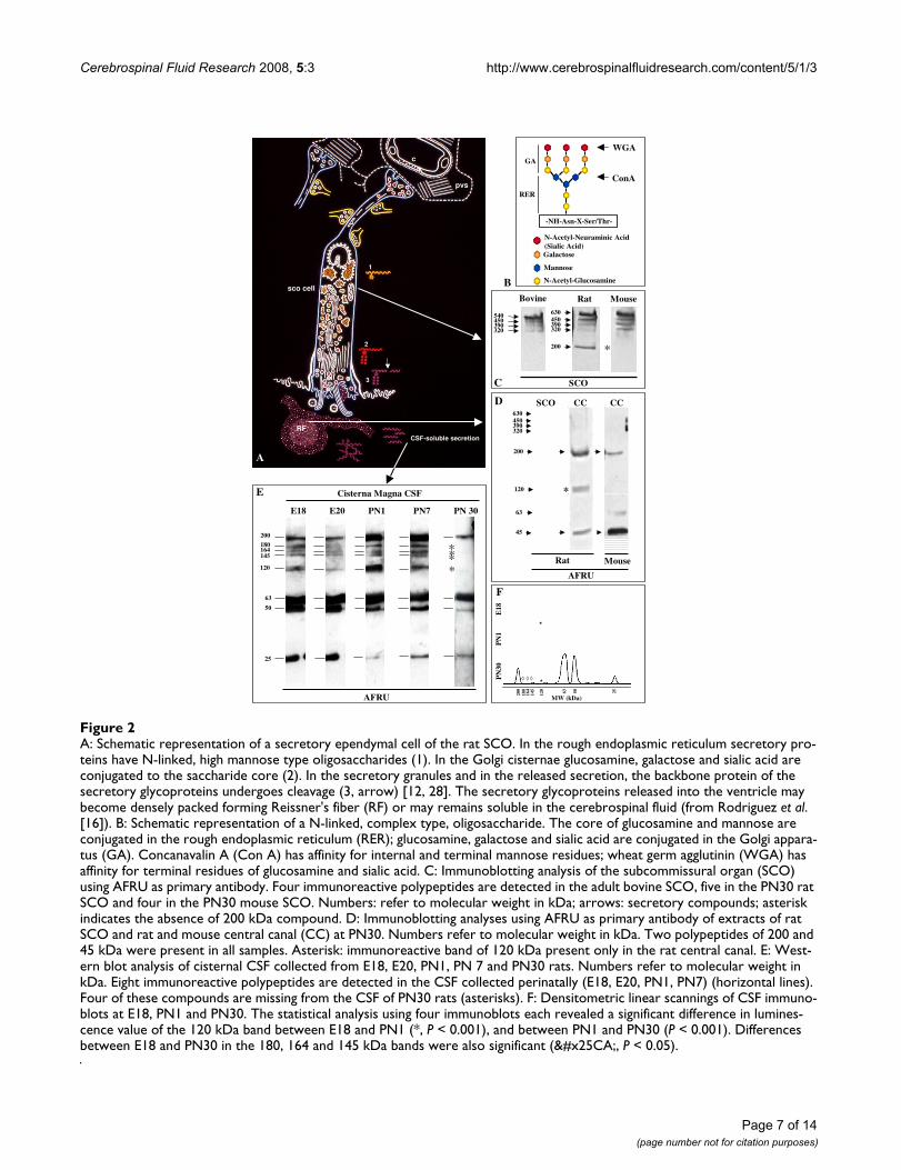
A: Schematic representation of a secretory ependymal cell of the rat SCOFigure 2A: Schematic representation of a secretory ependymal cell of the rat SCO. In the rough endoplasmic reticulum secretory pro-teins have N-linked, high mannose type oligosaccharides (1). In the Golgi cisternae glucosamine, galactose and sialic acid are conjugated to the saccharide core (2). In the secretory granules and in the released secretion, the backbone protein of the secretory glycoproteins undergoes cleavage (3, arrow) [12, 28]. The secretory glycoproteins released into the ventricle may become densely packed forming Reissner's fiber (RF) or may remains soluble in the cerebrospinal fluid (from Rodriguez et al. [16]). B: Schematic representation of a N-linked, complex type, oligosaccharide. The core of glucosamine and mannose are conjugated in the rough endoplasmic reticulum (RER); glucosamine, galactose and sialic acid are conjugated in the Golgi appara-tus (GA). Concanavalin A (Con A) has affinity for internal and terminal mannose residues; wheat germ agglutinin (WGA) has affinity for terminal residues of glucosamine and sialic acid. C: Immunoblotting analysis of the subcommissural organ (SCO) using AFRU as primary antibody. Four immunoreactive polypeptides are detected in the adult bovine SCO, five in the PN30 rat SCO and four in the PN30 mouse SCO. Numbers: refer to molecular weight in kDa; arrows: secretory compounds; asterisk indicates the absence of 200 kDa compound. D: Immunoblotting analyses using AFRU as primary antibody of extracts of rat SCO and rat and mouse central canal (CC) at PN30. Numbers refer to molecular weight in kDa. Two polypeptides of 200 and 45 kDa were present in all samples. Asterisk: immunoreactive band of 120 kDa present only in the rat central canal. E: West-ern blot analysis of cisternal CSF collected from E18, E20, PN1, PN 7 and PN30 rats. Numbers refer to molecular weight in kDa. Eight immunoreactive polypeptides are detected in the CSF collected perinatally (E18, E20, PN1, PN7) (horizontal lines). Four of these compounds are missing from the CSF of PN30 rats (asterisks). F: Densitometric linear scannings of CSF immuno-blots at E18, PN1 and PN30. The statistical analysis using four immunoblots each revealed a significant difference in lumines-cence value of the 120 kDa band between E18 and PN1 (*, P < 0.001), and between PN1 and PN30 (P < 0.001). Differences between E18 and PN30 in the 180, 164 and 145 kDa bands were also significant (◊, P < 0.05).
E18 E20 PN1 PN7 PN 30
200
180164145
120
Cisterna Magna CSF
63
50
25
****
AFRU
E
WGA
ConA
-NH-Asn-X-Ser/Thr-
N-Acetyl-Glucosamine
Mannose
Galactose
N-Acetyl-Neuraminic Acid (Sialic Acid)
RER
GA
BBovine
SCO C
450
Rat Mouse
*200
630450390320
540
390320
RF
sco cell
c
pvs
1
2
3
A
CSF-soluble secretion
200
180
164
145
120 5063 25
PN
30P
N1
E18
MW (kDa)
F
*
*◊ ◊ ◊
AFRU
SCO CC CC
200
630450390320
45
Rat Mouse
63
D
*120
Cerebrospinal Fluid Research 2008, 5:3 http://www.cerebrospinalfluidresearch.com/content/5/1/3
Comparison of immunoblot analysis of SCO, RF and CSF using AFRU and anti-P15Blots of rat SCO run in parallel and immunoreacted withAFRU and anti-P15 showed: (i) out of the four com-pounds of large molecular weight revealed by AFRU, onlythe 320 kDa reacted with anti-P15 (Fig. 3A, Table 3); (ii)the bands of 63 and 45 kDa reacted with both antibodies(Fig. 3A, Table 3); (iii) bands of 80, 37 and 32 kDa onlyreacted with anti-P15 (Fig. 3A, Table 3).
Blots of rat RF run in parallel and immunoreacted withAFRU and anti-P15 showed: (i) both antibodies reactedwith the 200, 120 and 45 kDa compounds (Table 3); (ii)bands of 320, 164, 145, 80 and 32 kDa only reacted withanti-P15 (Table 3).
Blots of CSF of E18, E20 and PN7 rats run in parallel andimmunoreacted with AFRU and anti-P15 showed: (i) outof the eight bands revealed by AFRU (200 to 25 kDa), only
the 200, 180, 145 and 63 kDa reacted with anti-P15 (Fig.3B, Table 3).
Evidence for the existence of two precursor forms of the secretory proteins in the rat and mouse SCOProteins extracted from the rat, mouse and bovine SCOwere blotted and run in parallel for immunoreaction withAFRU and Con A and WGA binding (Fig. 2B, 4; Table 1).In bovine SCO, four bands that immunoreacted withAFRU (540, 450, 390 and 320 kDa) bound Con A (Fig.4A, Table 1). The five polypeptides detected with AFRU inthe rat SCO (630, 450, 390, 320, 200 kDa) bound Con A,but only the compounds of 450, 320 and 200 kDa boundWGA (Fig. 4B, Table 1).
The four polypeptides detected with AFRU in the mouseSCO (630, 450, 390, 320 kDa) were Con A-positive (Fig.
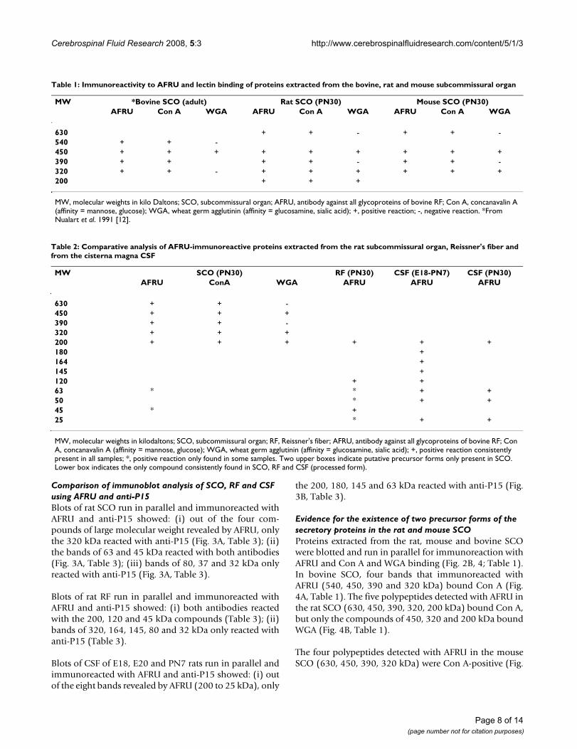
Table 1: Immunoreactivity to AFRU and lectin binding of proteins extracted from the bovine, rat and mouse subcommissural organ
MW *Bovine SCO (adult) Rat SCO (PN30) Mouse SCO (PN30)AFRU Con A WGA AFRU Con A WGA AFRU Con A WGA
630 + + - + + -540 + + -450 + + + + + + + + +390 + + + + - + + -320 + + - + + + + + +200 + + +
MW, molecular weights in kilo Daltons; SCO, subcommissural organ; AFRU, antibody against all glycoproteins of bovine RF; Con A, concanavalin A (affinity = mannose, glucose); WGA, wheat germ agglutinin (affinity = glucosamine, sialic acid); +, positive reaction; -, negative reaction. *From Nualart et al. 1991 [12].
Table 2: Comparative analysis of AFRU-immunoreactive proteins extracted from the rat subcommissural organ, Reissner's fiber and from the cisterna magna CSF
MW SCO (PN30) RF (PN30) CSF (E18-PN7) CSF (PN30)AFRU ConA WGA AFRU AFRU AFRU
630 + + -450 + + +390 + + -320 + + +200 + + + + + +180 +164 +145 +120 + +63 * * + +50 * + +45 * +25 * + +
MW, molecular weights in kilodaltons; SCO, subcommissural organ; RF, Reissner's fiber; AFRU, antibody against all glycoproteins of bovine RF; Con A, concanavalin A (affinity = mannose, glucose); WGA, wheat germ agglutinin (affinity = glucosamine, sialic acid); +, positive reaction consistently present in all samples; *, positive reaction only found in some samples. Two upper boxes indicate putative precursor forms only present in SCO. Lower box indicates the only compound consistently found in SCO, RF and CSF (processed form).
Page 8 of 14(page number not for citation purposes)
Cerebrospinal Fluid Research 2008, 5:3 http://www.cerebrospinalfluidresearch.com/content/5/1/3
4C); only the 450 and 320 kDa compounds bound WGA(Fig. 4C, Table 1).
DiscussionIdentification of the precursor and processed forms of the glycoproteins secreted by the rat and mouse SCOReissner's fiber results from the assembly of large molecu-lar weight proteins released by the SCO into the CSF cir-culating through the cerebral aqueduct. The majorconstituent protein of RF is SCO-spondin. The aminoacidsequence of bovine SCO-spondin has been fully estab-lished by Meiniel [21]. In the bovine SCO, three majorcompounds of 540, 450 and 320 kDa, reacting with anantiserum raised against the constitutive proteins of RF(AFRU), have been described [12]. Due to the lectin bind-ing properties and the subcellular localization of thesethree compounds, Nualart et al. [25] have selected a 15-aminoacid sequence from a region of SCO-spondin thatwas used to raise antibodies in rabbits. This antibody(anti-P15) specifically reacts with the bovine and rat SCOand RF and can be regarded as a specific marker of SCO-spondin [25]. In immunoblots of bovine SCO, anti-P15reacts with the 540 and 450 kDa bands, indicating thatboth bands correspond to SCO-spondin, with the formerbeing the precursor and the latter a processed form. Themolecular weight deduced from the aminoacid sequenceof bovine SCO-spondin is 540 kDa [30]. The nature of the320 kDa protein has yet to be investigated.
In the rat and mouse SCO, the 540 kDa compound wasabsent; instead an AFRU-immunoreactive compound of630 kDa was seen. This compound has immunoreactiveand lectin-binding properties similar to the bovine 540kDa SCO-spondin, indicating that it corresponds to amurine SCO-spondin. The sequence of the mouse SCO-spondin contains 4981 aminoacids with a theoretical pro-tein mass of 535 kDa. When compared to the bovineSCO-spondin there is a 66.8% homology [30].
Considering the large number of cryptic sites of N-glyco-sylaton present in the sequence of SCO-spondin, thelarger molecular mass of rat and mouse SCO-spondincould be due to a higher degree of glycosylation.
According to the lectin binding properties of all AFRU-immunoreactive glycoproteins present in the rat andmouse SCO (Table 1), the 630 and 390 kDa compoundswould correspond to precursor forms present in the roughendoplasmic reticulum of the SCO secretory cells. Indeed,they bound Con A, a lectin with affinity for mannose res-idues added to the nascent protein in the rough endoplas-mic reticulum, but did not bind WGA, a lectin specific fortwo sugar residues (glucosamine, sialic acid) added to theglycoprotein by the Golgi apparatus [2,4]. At variance, the450, 320 and 200 kDa proteins did bind WGA, indicating
that they are post-Golgi compounds located, most likely,in the secretory granules. The presence of the 200 kDaglycoprotein in RF is strongly supportive of the suggestionthat this compound corresponds to a processed form.
Out of the four compounds of large molecular weightrevealed by AFRU in the rat SCO, only the 320 kDareacted with anti-P15 (Table 3). Since this antibody is spe-cific for SCO-spondin, it may be postulated that the 320kDa protein corresponds to SCO-spondin. However, anti-P15 did not reveal the 630 kDa compound that mostlikely corresponds to the precursor of the rat SCO-spon-din. A likely explanation for this result is that anti-P15,reacting with a 15-aminoacid sequence of a protein thatconsists of about 4,500 amino acids, might not haveaccess to its epitopes when rat SCO-spondin is in its pre-cursor form.
Out of the eight bands consistently revealed by AFRU inthe embryonic CSF, only four of them (200, 180, 145, 63kDa) reacted with anti-P15. This suggests that the lattercompounds would be derived from the SCO-spondin pre-cursor (630 kDa) and the AFRU-positive, anti-P15-nega-tive compounds derived from the second putativeprecursor (390 kDa).
Are CSF-soluble compounds secreted by the SCO intracellularly processed and released into CSF, or "released" from RF?The 200 kDa appears as a key protein, since it was presentin the SCO, RF and embryonic and juvenile CSF of rats,and immunoreacted with AFRU and anti-P15. Thisindicates that this protein is a processed form of SCO-spondin released by the SCO cells into the CSF. In the rat,the 200 kDa compound represented the major constitu-ent protein of RF. A steady-state RF/CSF equilibrium forthis protein might explain the permanent presence of thiscompound in both, RF and CSF. On the other hand, thepresence of the 200 kDa protein in the embryonic CSF,before the first RF has formed, and in the juvenile cisternalCSF, a compartment where RF is absent, indicates that thisprotein is secreted as a CSF-soluble protein and remainssoluble in the circulating CSF.
The 63 kDa protein detected in all CSF samples and insome samples of SCO and RF, might also be regarded as aprocessed form secreted by the SCO to form RF as well asto be CSF-soluble.
The two proteins of 180 and 164 kDa, found in theembryonic CSF but absent from the juvenile SCO, CSFand RF, would correspond to processed forms released bythe SCO during late embryonic life. Since immunoblotanalysis of the embryonic SCO has not been performed,the possibility that these compounds were derived from a
Page 9 of 14(page number not for citation purposes)

Cerebrospinal Fluid Research 2008, 5:3 http://www.cerebrospinalfluidresearch.com/content/5/1/3
source other than the SCO has to be considered. Indeed,from E15 to PN1 the rat floor plate, that extends along theventral midline of the spinal cord and hindbrain, secretesproteins immunoreactive with AFRU [31]. Although floorplate explants release these proteins to culture medium[32], there is no evidence that they are actually releasedinto the embryonic CSF.
There were AFRU and anti-P15 immunoreactive proteinspresent in the rat RF and/or juvenile CSF (120, 50, 25kDa) that were absent from the SCO. Since these com-pounds were consistently detected in all blots, it seemsunlikely they result from artefactual degradation of largerproteins during handling and processing of the samples.The possibility that these proteins result from a post-release cleavage of compounds secreted by the SCO intothe CSF has to be considered and investigated. A similarphenomenon has been described in bovines, where sev-eral AFRU-immunoreactive polypeptides forming RF arenot present in the SCO [11,12,33]. The possibility of apost-release processing of high molecular mass glycopro-teins released into the CSF of chick embryos has beensuggested [17]. In the bovine, the wall of the centralcanal, especially at the lumbo-sacral level, containsAFRU-reactive cells [34]. These cells have not beendetected in the rat and mouse central canal. However,considering that in the present investigation, rat RF wasextracted together with the wall of the central canal, thepossibility should be considered that AFRU-immunore-active proteins present in the central canal/RF extract but
absent in the SCO extract, may have originated from cellslining the central canal.
The CSF-soluble compounds secreted by the rat SCO circulate in the internal and external CSF compartmentsAfter the intraventricular injection of an antibody againstRF-proteins, such an antibody immunoreacts with theproteins newly released by the SCO, forming insolubleantigen-antibody complexes on the surface of this glandthat were detected by immunocytochemistry using anti-IgG as primary antibody [35]. The same procedure hasbeen used in the present investigation. AFRU and anti-P15injected into the ventricular CSF formed insoluble anti-gen-antibody complexes exclusively located on the surfaceof the SCO, indicating that the rat SCO does secrete AFRUand anti-P15 reactive compounds into the third ventricle.Since AFRU and anti-P15 reactive compounds are alsopresent in the cisternal CSF (see above), it may be con-cluded that the CSF-soluble proteins secreted by the SCOinto the third ventricle circulate through the aqueduct ofSylvius, fourth ventricle and subarachnoid space.
In the fetal CSF the number and concentration of SCO secretory products is higher than in the CSF of juvenile ratsThe developing CNS of various vertebrate species hasbeen investigated using methods to visualize RF-proteins.Two structures, the floor plate and the SCO, have beenshown to share the property to synthesize RF-like material[36,37]. The use of anti-RF sera has confirmed that bothstructures secrete proteins reacting with these antibodies[31,37,38]. It has also been shown that both, the floor
Table 3: Immunoreactivity to AFRU and anti-P15 of proteins extracted from the rat subcommissural organ, Riessner's fiber and cisterna magna CSF
MW SCO (PN30) RF (PN30) CSF (E18-PN7)AFRU Anti-P15 AFRU Anti-P15 AFRU Anti-P15
630 +450 +390 +320 + + +200 + + + + +180 + +164 + +145 + + +120 + + +80 + +63 * * * * + *50 * * +45 * * + +37 +32 + +25 * +
MW, molecular weights in kilodaltons; SCO, subcommissural organ; RF, Reissner's fiber; AFRU, antibody against all glycoproteins of bovine RF; anti-P15, antibody specific for SCO-spondin; +, positive reaction consistently present in all samples; *, positive reaction only found in some samples. Horizontal box indicates 200 kDa compound consistently found in all samples. The eight AFRU-immunoreactive compounds detected in embryonic CSF were consistently found in all samples; only three of them react with anti-P15.
Page 10 of 14(page number not for citation purposes)

Cerebrospinal Fluid Research 2008, 5:3 http://www.cerebrospinalfluidresearch.com/content/5/1/3
plate and SCO of bovine embryos, express the SCO-spon-din gene and synthesize the 540 kDa protein [39]. The ratfloor plate synthesizes AFRU-immunoreactive proteinsfrom E15 to PN1, with the highest activity around E18[31]. In vitro studies have shown that the bovine floorplate cells release AFRU-reactive proteins of 540 and 60kDa into the culture medium [32]. The rat SCO starts tosecrete AFRU-reactive material at E14; by E18 it is fullydeveloped and displaying a high secretory activity [1].Since RF starts to form at PN1, and a RF proper is first seenin the central canal at PN7, it has been assumed that thecompounds secreted by the fetal SCO remain soluble inthe CSF [1].
In the present investigation, eight AFRU-immunoreactivecompounds were consistently found in all CSF samplescollected from E18 to PN1. Only four of these compounds(200, 63, 50, 25 kDa) were also detected in the CSF ofjuvenile rats. It may be assumed that the source of theseproteins is the SCO since in the juvenile rat this gland isthe only brain structure secreting AFRU-reactive proteins.On the other hand, the four AFRU-immunoreactive pro-
teins present only in the CSF collected from E18 to PN1could be secreted by the SCO, the floor plate or by bothglands. Five AFRU-immunoreactive proteins have beenshown to occur in the CSF of chick embryos [17]. Theauthors obtained evidence to suggest that in chickembryos, these proteins may be derived from both theSCO and floor plate.
Functional significance of the existence of CSF-soluble proteins secreted by the SCOThe embryonic CSF may be regarded as the main compo-nent of the milieu of stem cells and progenitor cells of thegerminal zone [40]. In rat embryos, the flowing CSF is inpermanent and direct contact with the ventricular andsubventricular zones and, after E17, it reaches the sub-arachnoid space to bathe the marginal zone of the braincortex [41]. The CSF is an efficient route to convey signalsbetween different regions of the brain [41,42]. In the adultrat, tracers injected into the ventricles move freely into thebrain parenchyma through the ependymal lining [43-45];similarly, tracers injected into the subarachnoid spaceenter the brain parenchyma through the pial surface[44,46-48]. However, the functional relationship betweenthe ventricular and subrachnoid compartments and theparenchyma of the embryonic brain is not clear. There isa significant body of evidence indicating that CSF carriesvital signal molecules to the germinal epithelium of thedeveloping brain cortex [41,49,50]. CSF from ratembryos, used as culture medium, has the capacity to sus-tain the proliferative activity of the embryonic cerebralcortex [51]. Rat CSF collected at E19 has the highest capac-ity to induce neuronal proliferation, compared to CSFobtained at other developmental periods, indicating thatthe CSF composition changes during development [51].Rat CSF promotes the neuronal differentiation from neu-rospheres obtained from stem cells of the subventricularzone of the lateral ventricle [52]. Several factors known toparticipate in brain development have been detected inthe embryonic CSF, namely, fibroblast growth factor(FGF2), epidermal growth factor (EGF), transforminggrowth factor TGF-b), neural growth factor (NGF), brain-derived growth factor (BDGF), transthyretin [53-62]. Thesource of most of these signals in not known.
The eight AFRU-immunoreactive compounds detected inthe CSF of rat embryos (present report), especially thosethat also react with anti-P15 (a SCO-spondin marker), aregood candidates to participate in brain development. Thispossibility is supported by previous observations. Theimmunological blockage of the SCO during developmentby maternal transfer of antibodies leads to brain abnor-malities [63]. There is strong evidence indicating thatSCO-spondin promotes neuronal growth and differentia-tion [64]. The fact that the eight AFRU-immunoreactivecompounds have been detected in the cisterna magna CSF
Immunoblots of rat SCO at PN30 (panel A) and cisternal CSF at PN7 (panel B), using AFRU and anti-P15 as primary anti-bodiesFigure 3Immunoblots of rat SCO at PN30 (panel A) and cisternal CSF at PN7 (panel B), using AFRU and anti-P15 as primary anti-bodies. The four AFRU-immunoreactive compounds with the largest molecular mass (630, 450,390, 320 kDa) are present in the SCO and missing from the CSF. Two other com-pounds (200, 63 kDa) are present in the SCO and CSF (proc-essed forms?). The CSF has four bands (180, 164, 145, 120 kDa) not seen in the SCO. The SCO has only one large molecular mass compound reacting with anti-P15 (specific for SCO-spondin) and several bands of lower molecular masses. The CSF compounds of 200, 180, 145 and 63 kDa, reacting with both antibodies are most likely to be SCO-spondin derivatives.
SCO
200
63
630450390320
AFRU Anti-P15
A
45
63
80
320
45
3732
50
180200
145
AFRU Anti-P15
B
CSF
50
25
120
180164145
200
63 63
Page 11 of 14(page number not for citation purposes)

Cerebrospinal Fluid Research 2008, 5:3 http://www.cerebrospinalfluidresearch.com/content/5/1/3
indicates they follow the flow of the subarachnoidal CSFand may gain access to the marginal zone of the develop-ing brain cortex. Worth mentioning is the fact that F-spon-din, secreted by the floor plate, SCO-spondin secreted bythe SCO and reelin, secreted by the Cajal-Retzius cells ofthe developing cortex, display regions of similarities intheir aminoacid sequences and all of them are glycopro-teins secreted into the extracellular space [21,65].
The four AFRU-immunoreactive compounds that con-tinue to be present in the CSF of juvenile rats may also beinvolved in neurogenesis, specially that occurring in thesubventricular zone of the lateral ventricle. Transplanta-tion of SCO explants to a lateral ventricle of juvenile ratsstimulates neuroblast proliferation and migration [66].
ConclusionThe present investigation has shown that (i) during lateembryonic life, the SCO secretes compounds that remainsoluble in the CSF and reach the subarachnoid space; (ii)during postnatal life, there is a reduction in the numberand concentration of CSF-soluble proteins secreted by theSCO. The molecular structure and functional significanceof these proteins remain to be elucidated. A glycoproteinof 200 kDa, found in all rat samples, may be a key com-pound for SCO function.
Competing interestsThe author(s) declare that they have no competing inter-ests.
Authors' contributionsKV and ER initiated and designed the study. KV, SR, CYand ER analysed and interpreted all the data. KV and COcollected data or carried out experiments for the study. KVand ER contributed to the preparation of the m.s. Allauthors have read and approved the final version of themanuscript.
AcknowledgementsSupported by Grants from Fondecyt 1030265 and 1070241, Chile to EMR and from Universidad Austral de Chile DID S-2006-72 to KV.
References1. Schoebitz K, Rodriguez EM, Garrido O, Del Brio Leon MA: Ontoge-
netic development of the subcommissural organ with refer-ence to the flexural organ. In The Subcommissural Organ AnEpendymal Brain Gland Edited by: Oksche A, Rodriguez EM, Fernan-dez-Llebrez P. Berlin, Heidelberg, New York: Springer; 1993:41-49.
2. Rodriguez EM, Oksche A, Montecinos H: Human subcommissuralorgan, with particular emphasis on its secretory activity dur-ing the fetal life. Microsc Res Tech 2001, 52:573-590.
3. Reissner E: Beiträge zur Kenntnis vom Bau des Rüchenmarksvon Petromyzon fluviatilis L. Arch anat physiol 1860, 77:545-588.
4. Rodriguez EM, Oksche A, Hein S, Yulis CR: Cell biology of the sub-commissural organ. Int Rev Cytol 1992, 135:39-121.
5. Rodriguez EM, Rodriguez S, Hein S: The subcommissural organ.Microsc Res Tech 1998, 41:98-123.
6. Peruzzo B, Rodriguez S, Delannoy L, Hein S, Rodriguez EM, OkscheA: Ultrastructural immunocytochemical study of the massacaudalis of the subcommissural organ-Reissner's fiber com-plex in lamprey larvae (Geotria australis): evidence for a ter-minal vascular route of secretory material. Cell Tissue Res 1987,247:367-376.
7. Rodriguez S, Rodriguez PA, Bance P, Rodriguez EM, Oksche A: Reiss-ner's fiber, massa caudalis and ampulla caudalis in the spinalcord of lamprey larvae (Geotria australis). Light-micro-scopic immunocytochemical and lectin-histochemical stud-ies. Cell Tissue Res 1987, 247:359-366.
8. Caprile T, Hein S, Rodriguez S, Montecinos H, Rodriguez E: Reissnerfiber binds and transports away monoamines present in thecerebrospinal fluid. Brain Res Mol Brain Res 2003, 110:177-192.
9. Rodriguez S, Vio K, Wagner C, Barria M, Navarrete EH, Ramirez VD,Perez-Figares JM, Rodriguez EM: Changes in the cerebrospinal-fluid monoamines in rats with an immunoneutralization ofthe subcommissural organ-Reissner's fiber complex bymaternal delivery of antibodies. Exp Brain Res 1999,128:278-290.
10. Hein S, Nualart F, Rodriguez EM, Oksche A: Partial characteriza-tion of the secretory products of the subcommisural organ.In The Subcommissural Organ An Ependymal Brain Gland Edited by:Oksche A, Rodriguez EM, Fernandez-Llebrez P. Berlin, Heidelberg,New York: Springer; 1993:78-88.
11. Nualart F, Hein S: Biosynthesis and molecular biology of thesecretory proteins of the subcommissural organ. Microsc ResTech 2001, 52:468-483.
12. Nualart F, Hein S, Rodriguez EM, Oksche A: Identification and par-tial characterization of the secretory glycoproteins of thebovine subcommissural organ-Reissner's fiber complex. Evi-dence for the existence of two precursor forms. Brain Res MolBrain Res 1991, 11:227-238.
13. del Brio MA, Riera P, Munoz RI, Montecinos H, Rodriguez EM: Themetencephalic floor plate of chick embryos expresses twosecretory glycoproteins homologous with the two glycopro-teins secreted by the subcommissural organ. Histochem CellBiol 2000, 113:415-426.
14. Lopez-Avalos MD, Perez J, Perez-Figares JM, Peruzzo B, GrondonaJM, Rodriguez EM: Secretory glycoproteins of the subcommis-sural organ of the dogfish (Scyliorhinus canicula): evidencefor the existence of precursor and processed forms. Cell Tis-sue Res 1996, 283:75-84.
15. Schoebitz K, Garrido O, Heinrichs M, Speer L, Rodriguez EM:Ontogenetical development of the chick and duck subcom-
Immunoblots of SCO extracts of bovine (adult, lanes 1–3), rat (PN30, lanes 4–6) and mouse (PN30, lanes 7–9)Figure 4Immunoblots of SCO extracts of bovine (adult, lanes 1–3), rat (PN30, lanes 4–6) and mouse (PN30, lanes 7–9). Lanes 1, 4, 7: immunoblots using AFRU. Lanes 2, 5, 8: concanavalin A (Con A) binding. Lanes 6, 9: wheat germ agglutinin (WGA) binding. Lane 3: concanavalin A blocked with mannose prior to binding. No binding is detected (asterisk). Numbers: molecular weight in kDa. Dotted lines: compounds not bind-ing WGA.
AFRU ConA WGA
1 2 3
Bovine Rat Mouse
AFRU ConA ConA AFRU ConA WGA
*
4 5 6
200
7 8 9 630450390320
630450390320
540450390320
A B C
Page 12 of 14(page number not for citation purposes)
http://www.ncbi.nlm.nih.gov/entrez/query.fcgi?cmd=Retrieve&db=PubMed&dopt=Abstract&list_uids=1618609
http://www.ncbi.nlm.nih.gov/entrez/query.fcgi?cmd=Retrieve&db=PubMed&dopt=Abstract&list_uids=1618609
http://www.ncbi.nlm.nih.gov/entrez/query.fcgi?cmd=Retrieve&db=PubMed&dopt=Abstract&list_uids=9579598
http://www.ncbi.nlm.nih.gov/entrez/query.fcgi?cmd=Retrieve&db=PubMed&dopt=Abstract&list_uids=1661820
http://www.ncbi.nlm.nih.gov/entrez/query.fcgi?cmd=Retrieve&db=PubMed&dopt=Abstract&list_uids=1661820
http://www.ncbi.nlm.nih.gov/entrez/query.fcgi?cmd=Retrieve&db=PubMed&dopt=Abstract&list_uids=1661820
Cerebrospinal Fluid Research 2008, 5:3 http://www.cerebrospinalfluidresearch.com/content/5/1/3
missural organ. An immunocytochemical study. Histochemis-try 1986, 84:31-40.
16. Rodriguez EM, Jara P, Richter H, Montecinos H, Flandes B, WiegandR, Oksche A: Evidence for the release of CSF-soluble secre-tory material from the subcommissural organ, with particu-lar reference to the situation in the human. In TheSubcommissural Organ An Ependymal Brain Gland Edited by: Oksche A,Rodriguez EM, Fernandez-Llebrez P. Berlin, Heidelberg, New York:Springer; 1993:121-131.
17. Hoyo-Becerra C, Lopez-Avalos MD, Perez J, Miranda E, Rojas-Rios P,Fernandez-Llebrez P, Grondona JM: Continuous delivery of amonoclonal antibody against Reissner's fiber into CSFreveals CSF-soluble material immunorelated to the sub-commissural organ in early chick embryos. Cell Tissue Res2006, 326:771-786.
18. Estivill-Torrus G, Cifuentes M, Grondona JM, Miranda E, Bermudez-Silva FJ, Fernandez-Llebrez P, Perez J: Quantification of the secre-tory glycoproteins of the subcommissural organ by a sensi-tive sandwich ELISA with a polyclonal antibody and a set ofmonoclonal antibodies against the bovine Reissner's fiber.Cell Tissue Res 1998, 294:407-413.
19. Fernandez-Llebrez P, Miranda E, Estivill-Torrus G, Cifuentes M, Gron-dona JM, Lopez-Avalos MD, Perez-Martin M, Perez J: Analysis andquantification of the secretory products of the subcommis-sural organ by use of monoclonal antibodies. Microsc Res Tech2001, 52:510-519.
20. Gobron S, Creveaux I, Meiniel R, Didier R, Herbet A, Bamdad M, ElBitar F, Dastugue B, Meiniel A: Subcommissural organ/Reissner'sfiber complex: characterization of SCO-spondin, a glycopro-tein with potent activity on neurite outgrowth. Glia 2000,32:177-191.
21. Meiniel A: SCO-spondin, a glycoprotein of the subcommis-sural organ/Reissner's fiber complex: evidence of a potentactivity on neuronal development in primary cell cultures.Microsc Res Tech 2001, 52:484-495.
22. Adams JC, Tucker RP: The thrombospondin type 1 repeat(TSR) superfamily: diverse proteins with related roles inneuronal development. Dev Dyn 2000, 218:280-299.
23. Sternberger LA, Hardy PH Jr, Cuculis JJ, Meyer HG: The unlabeledantibody enzyme method of immunohistochemistry: prepa-ration and properties of soluble antigen-antibody complex(horseradish peroxidase-antihorseradish peroxidase) and itsuse in identification of spirochetes. J Histochem Cytochem 1970,18:315-333.
24. Rodriguez EM, Oksche A, Hein S, Rodriguez S, Yulis R: Compara-tive immunocytochemical study of the subcommissuralorgan. Cell Tissue Res 1984, 237:427-441.
25. Nualart F, Hein S, Yulis CR, Zarraga AM, Araya A, Rodriguez EM:Partial sequencing of Reissner's fiber glycoprotein I (RF-GlyI). Cell Tissue Res 1998, 292:239-250.
26. Bradford MM: A rapid and sensitive method for the quantita-tion of microgram quantities of protein utilizing the princi-ple of protein-dye binding. Anal Biochem 1976, 72:248-254.
27. Towbin H, Staehelin T, Gordon J: Electrophoretic transfer ofproteins from polyacrylamide gels to nitrocellulose sheets:procedure and some applications. Proc Natl Acad Sci USA 1979,76:4350-4354.
28. Herrera H, Rodriguez EM: Secretory glycoproteins of the ratsubcommissural organ are N-linked complex-type glycopro-teins. Demonstration by combined use of lectins and specificglycosidases, and by the administration of Tunicamycin. His-tochemistry 1990, 93:607-615.
29. Peruzzo B, Rodriguez EM: Light and electron microscopicaldemonstration of concanavalin A and wheat-germ agglutininbinding sites by use of antibodies against the lectin or itslabel (peroxidase). Histochemistry 1989, 92:505-513.
30. Goncalves-Mendes N, Simon-Chazottes D, Creveaux I, Meiniel A,Guenet JL, Meiniel R: Mouse SCO-spondin, a gene of the throm-bospondin type 1 repeat (TSR) superfamily expressed in thebrain. Gene 2003, 312:263-270.
31. Rodriguez EM, del Brio Leon MA, Riera P, Menendez J, Schoebitz K:The floor plate of the hindbrain is a highly specialized gland.Immunocytochemical and ultrastructural characteristics.Brain Res Dev Brain Res 1996, 97:153-168.
32. Guinazu MF, Richter HG, Rodriguez EM: Bovine floor plateexplants secrete SCO-spondin. Cell Tissue Res 2002,308:177-191.
33. Meiniel A, Meiniel R, Didier R, Creveaux I, Gobron S, Monnerie H,Dastugue B: The subcommissural organ and Reissner's fibercomplex. An enigma in the central nervous system? Prog His-tochem Cytochem 1996, 30:1-66.
34. Rodriguez S, Hein S, Yulis R, Delannoy L, Siegmund I, Rodriguez E:Reissner's fiber and the wall of the central canal in thelumbo-sacral region of the bovine spinal cord. Comparativeimmunocytochemical and ultrastructural study. Cell TissueRes 1985, 240:649-662.
35. Rodriguez S, Rodriguez EM, Jara P, Peruzzo B, Oksche A: Singleinjection into the cerebrospinal fluid of antibodies againstthe secretory material of the subcommissural organ revers-ibly blocks formation of Reissner's fiber: immunocytochemi-cal investigations in the rat. Exp Brain Res 1990, 81:113-124.
36. Olsson R: Reissner's fiber mechanisms: some commondenominators. In The Subcommissural Organ An Ependymal BrainGland Edited by: Oksche A, Rodriguez EM, Fernandez-Llebrez P. Ber-lin, Heidelberg, New York: Springer; 1993:33-39.
37. Yulis CR, Mota MD, Andrades JA, Rodriguez S, Peruzzo B, ManceraJM, Ramirez P, Garrido M, Perez-Figarez JM, Fernandez-Llebrez P,Rodriguez EM: Floor plate and the subcommissural organ arethe source of secretory compounds of related nature: com-parative immunocytochemical study. J Comp Neurol 1998,392:19-34.
38. Rodriguez EM, Garrido O, Oksche A: Lectin histochemistry ofthe human fetal subcommissural organ. Cell Tissue Res 1990,262:105-113.
39. Richter HG, Munoz RI, Millan CS, Guinazu MF, Yulis CR, RodriguezEM: The floor plate cells from bovines express the mRNAencoding for SCO-spondin and its translation products. BrainRes Mol Brain Res 2001, 93:137-147.
40. Owen-Lynch PJ, Draper CE, Mashayekhi F, Bannister CM, Miyan JA:Defective cell cycle control underlies abnormal corticaldevelopment in the hydrocephalic Texas rat. Brain 2003,126:623-631.
41. Miyan JA, Nabiyouni M, Zendah M: Development of the brain: avital role for cerebrospinal fluid. Can J Physiol Pharmacol 2003,81:317-328.
42. Rodriguez EM: The cerebrospinal fluid as a pathway in neu-roendocrine integration. J Endocrinol 1976, 71:407-443.
43. Brightman MW: The distribution within the brain of ferritininjected into cerebrospinal fluid compartments. I. Ependy-mal distribution. J Cell Biol 1965, 26:99-123.
44. Brightman MW, Reese TS: Junctions between intimatelyapposed cell membranes in the vertebrate brain. J Cell Biol1969, 40:648-677.
45. Cifuentes M, Fernandez LP, Perez J, Perez-Figares JM, Rodriguez EM:Distribution of intraventricularly injected horseradish per-oxidase in cerebrospinal fluid compartments of the rat spinalcord. Cell Tissue Res 1992, 270:485-494.
46. Brightman MW: The distribution within the brain of ferritininjected into cerebrospinal fluid compartments. II. Paren-chymal distribution. Am J Anat 1965, 117:193-219.
47. Rennels ML, Gregory TF, Blaumanis OR, Fujimoto K, Grady PA: Evi-dence for a 'paravascular' fluid circulation in the mammaliancentral nervous system, provided by the rapid distribution oftracer protein throughout the brain from the subarachnoidspace. Brain Res 1985, 326:47-63.
48. Pena P, Rodriguez EM, Dellmann HD, Schoebitz K: Effects of colch-icine on the hypothalamo-neurohypophysial system ofchronically salt-loaded rats. Neuroendocrinology 1988,47:217-224.
49. Miyan JA, Mashayekhi F, Bannister CM: Developmental abnormal-ities in early-onset hydrocephalus: clues to signalling. SympSoc Exp Biol 2001:91-106.
50. Gato A, Moro JA, Alonso MI, Bueno D, De La Mano A, Martin C:Embryonic cerebrospinal fluid regulates neuroepithelial sur-vival, proliferation, and neurogenesis in chick embryos. AnatRec A Discov Mol Cell Evol Biol 2005, 284:475-484.
51. Miyan JA, Zendah M, Mashayekhi F, Owen-Lynch PJ: Cerebrospinalfluid supports viability and proliferation of cortical cells invitro, mirroring in vivo development. Cerebrospinal Fluid Res2006, 3:2.
Page 13 of 14(page number not for citation purposes)
http://www.ncbi.nlm.nih.gov/entrez/query.fcgi?cmd=Retrieve&db=PubMed&dopt=Abstract&list_uids=2420757
http://www.ncbi.nlm.nih.gov/entrez/query.fcgi?cmd=Retrieve&db=PubMed&dopt=Abstract&list_uids=9799457
http://www.ncbi.nlm.nih.gov/entrez/query.fcgi?cmd=Retrieve&db=PubMed&dopt=Abstract&list_uids=9799457
http://www.ncbi.nlm.nih.gov/entrez/query.fcgi?cmd=Retrieve&db=PubMed&dopt=Abstract&list_uids=4192899
http://www.ncbi.nlm.nih.gov/entrez/query.fcgi?cmd=Retrieve&db=PubMed&dopt=Abstract&list_uids=4192899
http://www.ncbi.nlm.nih.gov/entrez/query.fcgi?cmd=Retrieve&db=PubMed&dopt=Abstract&list_uids=4192899
http://www.ncbi.nlm.nih.gov/entrez/query.fcgi?cmd=Retrieve&db=PubMed&dopt=Abstract&list_uids=6435876
http://www.ncbi.nlm.nih.gov/entrez/query.fcgi?cmd=Retrieve&db=PubMed&dopt=Abstract&list_uids=6435876
http://www.ncbi.nlm.nih.gov/entrez/query.fcgi?cmd=Retrieve&db=PubMed&dopt=Abstract&list_uids=6435876
http://www.ncbi.nlm.nih.gov/entrez/query.fcgi?cmd=Retrieve&db=PubMed&dopt=Abstract&list_uids=9560467
http://www.ncbi.nlm.nih.gov/entrez/query.fcgi?cmd=Retrieve&db=PubMed&dopt=Abstract&list_uids=9560467
http://www.ncbi.nlm.nih.gov/entrez/query.fcgi?cmd=Retrieve&db=PubMed&dopt=Abstract&list_uids=9560467
http://www.ncbi.nlm.nih.gov/entrez/query.fcgi?cmd=Retrieve&db=PubMed&dopt=Abstract&list_uids=2329058
http://www.ncbi.nlm.nih.gov/entrez/query.fcgi?cmd=Retrieve&db=PubMed&dopt=Abstract&list_uids=2329058
http://www.ncbi.nlm.nih.gov/entrez/query.fcgi?cmd=Retrieve&db=PubMed&dopt=Abstract&list_uids=2329058
http://www.ncbi.nlm.nih.gov/entrez/query.fcgi?cmd=Retrieve&db=PubMed&dopt=Abstract&list_uids=2478508
http://www.ncbi.nlm.nih.gov/entrez/query.fcgi?cmd=Retrieve&db=PubMed&dopt=Abstract&list_uids=2478508
http://www.ncbi.nlm.nih.gov/entrez/query.fcgi?cmd=Retrieve&db=PubMed&dopt=Abstract&list_uids=2478508
http://www.ncbi.nlm.nih.gov/entrez/query.fcgi?cmd=Retrieve&db=PubMed&dopt=Abstract&list_uids=8997500
http://www.ncbi.nlm.nih.gov/entrez/query.fcgi?cmd=Retrieve&db=PubMed&dopt=Abstract&list_uids=8997500
http://www.ncbi.nlm.nih.gov/entrez/query.fcgi?cmd=Retrieve&db=PubMed&dopt=Abstract&list_uids=8824845
http://www.ncbi.nlm.nih.gov/entrez/query.fcgi?cmd=Retrieve&db=PubMed&dopt=Abstract&list_uids=8824845
http://www.ncbi.nlm.nih.gov/entrez/query.fcgi?cmd=Retrieve&db=PubMed&dopt=Abstract&list_uids=2410129
http://www.ncbi.nlm.nih.gov/entrez/query.fcgi?cmd=Retrieve&db=PubMed&dopt=Abstract&list_uids=2410129
http://www.ncbi.nlm.nih.gov/entrez/query.fcgi?cmd=Retrieve&db=PubMed&dopt=Abstract&list_uids=2410129
http://www.ncbi.nlm.nih.gov/entrez/query.fcgi?cmd=Retrieve&db=PubMed&dopt=Abstract&list_uids=2394219
http://www.ncbi.nlm.nih.gov/entrez/query.fcgi?cmd=Retrieve&db=PubMed&dopt=Abstract&list_uids=2394219
http://www.ncbi.nlm.nih.gov/entrez/query.fcgi?cmd=Retrieve&db=PubMed&dopt=Abstract&list_uids=2394219
http://www.ncbi.nlm.nih.gov/entrez/query.fcgi?cmd=Retrieve&db=PubMed&dopt=Abstract&list_uids=9482230
http://www.ncbi.nlm.nih.gov/entrez/query.fcgi?cmd=Retrieve&db=PubMed&dopt=Abstract&list_uids=9482230
http://www.ncbi.nlm.nih.gov/entrez/query.fcgi?cmd=Retrieve&db=PubMed&dopt=Abstract&list_uids=9482230
http://www.ncbi.nlm.nih.gov/entrez/query.fcgi?cmd=Retrieve&db=PubMed&dopt=Abstract&list_uids=2257605
http://www.ncbi.nlm.nih.gov/entrez/query.fcgi?cmd=Retrieve&db=PubMed&dopt=Abstract&list_uids=2257605
http://www.ncbi.nlm.nih.gov/entrez/query.fcgi?cmd=Retrieve&db=PubMed&dopt=Abstract&list_uids=5859025
http://www.ncbi.nlm.nih.gov/entrez/query.fcgi?cmd=Retrieve&db=PubMed&dopt=Abstract&list_uids=5859025
http://www.ncbi.nlm.nih.gov/entrez/query.fcgi?cmd=Retrieve&db=PubMed&dopt=Abstract&list_uids=5859025
http://www.ncbi.nlm.nih.gov/entrez/query.fcgi?cmd=Retrieve&db=PubMed&dopt=Abstract&list_uids=5765759
http://www.ncbi.nlm.nih.gov/entrez/query.fcgi?cmd=Retrieve&db=PubMed&dopt=Abstract&list_uids=5765759
http://www.ncbi.nlm.nih.gov/entrez/query.fcgi?cmd=Retrieve&db=PubMed&dopt=Abstract&list_uids=1486601
http://www.ncbi.nlm.nih.gov/entrez/query.fcgi?cmd=Retrieve&db=PubMed&dopt=Abstract&list_uids=1486601
http://www.ncbi.nlm.nih.gov/entrez/query.fcgi?cmd=Retrieve&db=PubMed&dopt=Abstract&list_uids=1486601
http://www.ncbi.nlm.nih.gov/entrez/query.fcgi?cmd=Retrieve&db=PubMed&dopt=Abstract&list_uids=5883189
http://www.ncbi.nlm.nih.gov/entrez/query.fcgi?cmd=Retrieve&db=PubMed&dopt=Abstract&list_uids=5883189
http://www.ncbi.nlm.nih.gov/entrez/query.fcgi?cmd=Retrieve&db=PubMed&dopt=Abstract&list_uids=5883189
http://www.ncbi.nlm.nih.gov/entrez/query.fcgi?cmd=Retrieve&db=PubMed&dopt=Abstract&list_uids=3971148
http://www.ncbi.nlm.nih.gov/entrez/query.fcgi?cmd=Retrieve&db=PubMed&dopt=Abstract&list_uids=3971148
http://www.ncbi.nlm.nih.gov/entrez/query.fcgi?cmd=Retrieve&db=PubMed&dopt=Abstract&list_uids=3971148
http://www.ncbi.nlm.nih.gov/entrez/query.fcgi?cmd=Retrieve&db=PubMed&dopt=Abstract&list_uids=3362303
http://www.ncbi.nlm.nih.gov/entrez/query.fcgi?cmd=Retrieve&db=PubMed&dopt=Abstract&list_uids=3362303
Cerebrospinal Fluid Research 2008, 5:3 http://www.cerebrospinalfluidresearch.com/content/5/1/3
Publish with BioMed Central and every scientist can read your work free of charge
"BioMed Central will be the most significant development for disseminating the results of biomedical research in our lifetime."
Sir Paul Nurse, Cancer Research UK
Your research papers will be:
available free of charge to the entire biomedical community
peer reviewed and published immediately upon acceptance
cited in PubMed and archived on PubMed Central
yours — you keep the copyright
Submit your manuscript here:http://www.biomedcentral.com/info/publishing_adv.asp
BioMedcentral
52. Gonzalez C, Vio K, Muñoz RI, Rodriguez EM: The CSF of normalH-Tx rats promotes neuronal differentiation from neuro-spheres but CSF of hydrocephalic H-Tx rats does not. Cere-brospinal Fluid Res 2006, 3:s10.
53. Doublier S, Duyckaerts C, Seurin D, Binoux M: Impaired braindevelopment and hydrocephalus in a line of transgenic micewith liver-specific expression of human insulin-like growthfactor binding protein-1. Growth Horm IGF Res 2000, 10:267-274.
54. Kasaian MT, Neet KE: Nerve growth factor in human amnioticand cerebrospinal fluid. Biofactors 1989, 2:99-104.
55. Johnson MD, Gold LI, Moses HL: Evidence for transforminggrowth factor-beta expression in human leptomeningealcells and transforming growth factor-beta-like activity inhuman cerebrospinal fluid. Lab Invest 1992, 67:360-368.
56. Mogi M, Harada M, Narabayashi H, Inagaki H, Minami M, Nagatsu T:Interleukin (IL)-1 beta, IL-2, IL-4, IL-6 and transforminggrowth factor-alpha levels are elevated in ventricular cere-brospinal fluid in juvenile parkinsonism and Parkinson's dis-ease. Neurosci Lett 1996, 211:13-16.
57. Heinze E, Boker M, Blum W, Behnisch W, Schulz A, Urban J, MauchE: GH, IGF-I, IGFBP-3 and IGFBP-2 in cerebrospinal fluid ofinfants, during puberty and in adults. Exp Clin Endocrinol Diabetes1998, 106:197-202.
58. Arnold PM, Ma JY, Citron BA, Festoff BW: Insulin-like growth fac-tor binding proteins in cerebrospinal fluid during humandevelopment and aging. Biochem Biophys Res Commun 1999,264:652-656.
59. Johanson CE, Szmydynger-Chodobska J, Chodobski A, Baird A,McMillan P, Stopa EG: Altered formation and bulk absorption ofcerebrospinal fluid in FGF-2-induced hydrocephalus. Am JPhysiol 1999, 277:R263-271.
60. Riikonen R, Somer M, Turpeinen U: Low insulin-like growth fac-tor (IGF-1) in the cerebrospinal fluid of children with pro-gressive encephalopathy, hypsarrhythmia, and optic atrophy(PEHO) syndrome and cerebellar degeneration. Epilepsia1999, 40:1642-1648.
61. Moinuddin SM, Tada T: Study of cerebrospinal fluid flow dynam-ics in TGF-beta 1 induced chronic hydrocephalic mice. NeurolRes 2000, 22:215-222.
62. Montecinos HA, Richter H, Caprile T, Rodriguez EM: Synthesis oftransthyretin by the ependymal cells of the subcommissuralorgan. Cell Tissue Res 2005, 320:487-499.
63. Vio K, Rodriguez S, Navarrete EH, Perez-Figares JM, Jimenez AJ, Rod-riguez EM: Hydrocephalus induced by immunological block-age of the subcommissural organ-Reissner's fiber (RF)complex by maternal transfer of anti-RF antibodies. Exp BrainRes 2000, 135:41-52.
64. Monnerie H, Dastugue B, Meiniel A: Effect of synthetic peptidesderived from SCO-spondin conserved domains on chick cor-tical and spinal-cord neurons in cell cultures. Cell Tissue Res1998, 293:407-418.
65. Rice DS, Curran T: Role of the reelin signaling pathway in cen-tral nervous system development. Annu Rev Neurosci 2001,24:1005-1039.
66. Gonzalez C: Participación del órgano subcomisural y el líquidocefalorraquídeo en la neurogénesis postnatal. In PhD ThesisUniversidad Austral de Chile, Institute of Anatomy, Histology andPathology, Faculty of Medicine; 2007.
Publish with BioMed Central and every scientist can read your work free of charge
"BioMed Central will be the most significant development for disseminating the results of biomedical research in our lifetime."
Sir Paul Nurse, Cancer Research UK
Your research papers will be:
available free of charge to the entire biomedical community
peer reviewed and published immediately upon acceptance
cited in PubMed and archived on PubMed Central
yours — you keep the copyright
Submit your manuscript here:http://www.biomedcentral.com/info/publishing_adv.asp
BioMedcentral
Page 14 of 14(page number not for citation purposes)
http://www.ncbi.nlm.nih.gov/entrez/query.fcgi?cmd=Retrieve&db=PubMed&dopt=Abstract&list_uids=2696479
http://www.ncbi.nlm.nih.gov/entrez/query.fcgi?cmd=Retrieve&db=PubMed&dopt=Abstract&list_uids=2696479
http://www.ncbi.nlm.nih.gov/entrez/query.fcgi?cmd=Retrieve&db=PubMed&dopt=Abstract&list_uids=1328762
http://www.ncbi.nlm.nih.gov/entrez/query.fcgi?cmd=Retrieve&db=PubMed&dopt=Abstract&list_uids=1328762
http://www.ncbi.nlm.nih.gov/entrez/query.fcgi?cmd=Retrieve&db=PubMed&dopt=Abstract&list_uids=1328762
http://www.ncbi.nlm.nih.gov/entrez/query.fcgi?cmd=Retrieve&db=PubMed&dopt=Abstract&list_uids=8809836
http://www.ncbi.nlm.nih.gov/entrez/query.fcgi?cmd=Retrieve&db=PubMed&dopt=Abstract&list_uids=8809836
http://www.ncbi.nlm.nih.gov/entrez/query.fcgi?cmd=Retrieve&db=PubMed&dopt=Abstract&list_uids=8809836
http://www.ncbi.nlm.nih.gov/entrez/query.fcgi?cmd=Retrieve&db=PubMed&dopt=Abstract&list_uids=9710360
http://www.ncbi.nlm.nih.gov/entrez/query.fcgi?cmd=Retrieve&db=PubMed&dopt=Abstract&list_uids=9710360
http://www.ncbi.nlm.nih.gov/entrez/query.fcgi?cmd=Retrieve&db=PubMed&dopt=Abstract&list_uids=9716730
http://www.ncbi.nlm.nih.gov/entrez/query.fcgi?cmd=Retrieve&db=PubMed&dopt=Abstract&list_uids=9716730
Related Documents