The structural and compositional transition of the meniscal roots into the fibrocartilage of the menisci Stephen H. J. Andrews, 1 Jerome B. Rattner, 2 Heather A. Jamniczky, 2 Nigel G. Shrive 2 and Adetola B. Adesida 1 1 Department of Surgery, University of Alberta, Edmonton, AB, Canada 2 McCaig Institute for Bone and Joint Health, Faculty of Medicine, University of Calgary, Calgary, AB, Canada Abstract The meniscal roots, or insertional ligaments, firmly attach the menisci to tibial plateau. These strong attachments anchor the menisci and allow for the generation of hoop stress in the tissue. The meniscal roots have a ligament- like structure that transitions into the fibrocartilagenous structure of the meniscal body. The purpose of this study was to carry out a complete analysis of the structure and tissue organization from the body of the meniscus through the transition region and into the insertional roots. Serial sections were obtained from the meniscal roots into the meniscal body in fixed juvenile bovine menisci. Sections were stained for collagen and proteoglycans (PG) using fast green and safranin-o staining protocols. Unstained sections were imaged used a backlit stereo microscope. Optical projection tomography (OPT) was employed to evaluate the three-dimensional collagen architecture of the root–meniscus transition in lapine menisci. Tie-fibres were observed in the sections of the ligaments furthest from the bovine meniscal body. Blood vessels were observed to be surrounded by these tie-fibres and a PG-rich region within the ligaments. Near the tibial insertion, the roots contained large ligament- like collagen fascicles. In sections approaching the meniscus, there was an increase in tie-fibre size and density. Small tie-fibres extended into the ligament from the epiligamentous structure in the outermost sections of the meniscal roots, while large tie-fibre bundles were apparent at the meniscus transition. The staining pattern indicates that the root may continue into the outer portion of the meniscus where it then blends with the more fibrocartilage-like inner portions of the tissue. In unstained sections it was observed that the femoral side of the epiligamentous structure surrounding the root becomes more fibrous and thickens in the inferior inner portion of the posterior medial root. This thickening changes the shape of the root to more closely resemble the meniscus wedge shape. These observations support the concept of root continuity with the outer portion of the meniscus, thereby connecting with the hoop-like structure of the peripheral meniscus. OPT identified continuous collagen organization from the root into the meniscal body in longitudinal sections. In the radial direction, the morphology of the root continues into the meniscal body consistent with the serially sectioned bovine menisci. Blood vessels were prevalent on the periphery of the root. These blood vessels then arborized to cover the anterior femoral surface of the meniscus. This is the first study of the structural transition between the insertional ligaments (roots) and the fibrocartilagenous body of the menisci. These new structural details are important to understanding the meniscal load-bearing mechanism in the knee. Key words: Knee; Meniscus; Meniscal Roots; Optical Projection Tomography. Introduction The insertional ligaments, or roots, of the menisci are inte- gral to load bearing in the menisci. These roots insert cen- trally on the tibial plateau and act to resist lateral extrusion of the menisci from the joint (Lerer et al. 2004). Grossly the roots appear ligament-like with large longitudinally ori- ented collagen bundles. Injury to these insertional liga- ments results in rapid degeneration in the knee (Gale et al. 1999). Medial meniscal release, which involves the transec- tion of the anterior or posterior menisco-tibial ligaments, is a surgical technique used to induce osteoarthritis in animal models (Pozzi et al. 2006; Glasson et al. 2007). This injury model reduces the ability of the menisci to generate hoop stresses, thus resulting in lateral extrusion and reduced load-bearing capability. The rapid onset of cartilage Correspondence Stephen H. J. Andrews, Division of Orthopaedic Surgery, Department of Surgery, University of Alberta, Edmonton, AB, Canada, T6G 2E1. E: [email protected] Accepted for publication 29 October 2014 Article published online 9 January 2015 © 2015 Anatomical Society J. Anat. (2015) 226, pp169--174 doi: 10.1111/joa.12265 Journal of Anatomy

Welcome message from author
This document is posted to help you gain knowledge. Please leave a comment to let me know what you think about it! Share it to your friends and learn new things together.
Transcript
The structural and compositional transition of themeniscal roots into the fibrocartilage of the menisciStephen H. J. Andrews,1 Jerome B. Rattner,2 Heather A. Jamniczky,2 Nigel G. Shrive2 andAdetola B. Adesida1
1Department of Surgery, University of Alberta, Edmonton, AB, Canada2McCaig Institute for Bone and Joint Health, Faculty of Medicine, University of Calgary, Calgary, AB, Canada
Abstract
The meniscal roots, or insertional ligaments, firmly attach the menisci to tibial plateau. These strong attachments
anchor the menisci and allow for the generation of hoop stress in the tissue. The meniscal roots have a ligament-
like structure that transitions into the fibrocartilagenous structure of the meniscal body. The purpose of this
study was to carry out a complete analysis of the structure and tissue organization from the body of the meniscus
through the transition region and into the insertional roots. Serial sections were obtained from the meniscal
roots into the meniscal body in fixed juvenile bovine menisci. Sections were stained for collagen and
proteoglycans (PG) using fast green and safranin-o staining protocols. Unstained sections were imaged used a
backlit stereo microscope. Optical projection tomography (OPT) was employed to evaluate the three-dimensional
collagen architecture of the root–meniscus transition in lapine menisci. Tie-fibres were observed in the sections
of the ligaments furthest from the bovine meniscal body. Blood vessels were observed to be surrounded by these
tie-fibres and a PG-rich region within the ligaments. Near the tibial insertion, the roots contained large ligament-
like collagen fascicles. In sections approaching the meniscus, there was an increase in tie-fibre size and density.
Small tie-fibres extended into the ligament from the epiligamentous structure in the outermost sections of the
meniscal roots, while large tie-fibre bundles were apparent at the meniscus transition. The staining pattern
indicates that the root may continue into the outer portion of the meniscus where it then blends with the more
fibrocartilage-like inner portions of the tissue. In unstained sections it was observed that the femoral side of the
epiligamentous structure surrounding the root becomes more fibrous and thickens in the inferior inner portion
of the posterior medial root. This thickening changes the shape of the root to more closely resemble the
meniscus wedge shape. These observations support the concept of root continuity with the outer portion of the
meniscus, thereby connecting with the hoop-like structure of the peripheral meniscus. OPT identified continuous
collagen organization from the root into the meniscal body in longitudinal sections. In the radial direction, the
morphology of the root continues into the meniscal body consistent with the serially sectioned bovine menisci.
Blood vessels were prevalent on the periphery of the root. These blood vessels then arborized to cover the
anterior femoral surface of the meniscus. This is the first study of the structural transition between the
insertional ligaments (roots) and the fibrocartilagenous body of the menisci. These new structural details are
important to understanding the meniscal load-bearing mechanism in the knee.
Key words: Knee; Meniscus; Meniscal Roots; Optical Projection Tomography.
Introduction
The insertional ligaments, or roots, of the menisci are inte-
gral to load bearing in the menisci. These roots insert cen-
trally on the tibial plateau and act to resist lateral extrusion
of the menisci from the joint (Lerer et al. 2004). Grossly the
roots appear ligament-like with large longitudinally ori-
ented collagen bundles. Injury to these insertional liga-
ments results in rapid degeneration in the knee (Gale et al.
1999). Medial meniscal release, which involves the transec-
tion of the anterior or posterior menisco-tibial ligaments, is
a surgical technique used to induce osteoarthritis in animal
models (Pozzi et al. 2006; Glasson et al. 2007). This injury
model reduces the ability of the menisci to generate hoop
stresses, thus resulting in lateral extrusion and reduced
load-bearing capability. The rapid onset of cartilage
Correspondence
Stephen H. J. Andrews, Division of Orthopaedic Surgery, Department
of Surgery, University of Alberta, Edmonton, AB, Canada, T6G 2E1.
Accepted for publication 29 October 2014
Article published online 9 January 2015
© 2015 Anatomical Society
J. Anat. (2015) 226, pp169--174 doi: 10.1111/joa.12265
Journal of Anatomy
damage following meniscal release identifies the impor-
tance of the menisci in the overall function of the joint
(Glasson et al. 2007).
Recent work on the structure of the meniscus has
revealed a more complex fibre architecture than was previ-
ously described (Andrews et al. 2013, 2014). These studies
identified the hierarchical nature of the tie-fibre structure
and the complex woven fibre organization in regions that
were previously thought to have fibres oriented purely cir-
cumferentially. The structural transition from the fibrocarti-
lagenous meniscal body into the ligament-like meniscal
roots allows for the transmission of the complex loading of
the menisci into the tibial plateau. The transition of the
meniscal roots into the bony insertion at the tibial surface
has been studied extensively, including both the structure
and mechanical properties in this region (Villegas et al.
2008; Hauch et al. 2009). However, to our knowledge there
have been no studies on the transition from the meniscal
roots into the fibrocartilagenous body of the menisci.
The development of successful meniscal substitutes must
include an area that integrates and functions like the meni-
scal root in order to insure the correct transmission of load
and the resultant knee kinetics. The engineering of this
region is informed by a clear understanding of root archi-
tecture. Thus, the purpose of this study was to carry out an
analysis of the structure and tissue organization from the
body of the meniscus through the transition region and
into the insertional roots.
Materials and methods
Bovine menisci (n = 2) were obtained from a local abattoir, har-
vested within 48 h of slaughter. The menisci were dissected with
careful attention paid to retaining the four insertional ligaments
(anterior and posterior for both medial and lateral menisci). Menisci
were then fixed in 100% methanol at �20 °C for 72 h. The inser-
tional ligaments were then cut serially in cross-section (sections
~1 mm thick), starting from the bony insertion until the body of the
meniscus was reached. Sections were washed in phosphate-buf-
fered saline for 1 h, and stained with fast green (0.02% w/v; Sigma,
St Louis, MO, USA) and safranin o (0.1% w/v; Sigma) for collagen
and proteoglycan (PG), respectively. These whole-mount sections
were imaged using a stereo-microscope and digital camera (Zeiss
Stemi SV8 microscope with moticam 5.0 M pixel camera; Motic,
Richmond, BC, Canada). Several sections were also imaged prior to
staining using a backlit stereo microscope, as this technique yields
effective visualization of the tie-fibre structure.
Optical projection tomography (OPT)
Optical projection tomography is an imaging technique capable of
imaging the collagen and elastin structure of meniscal samples on
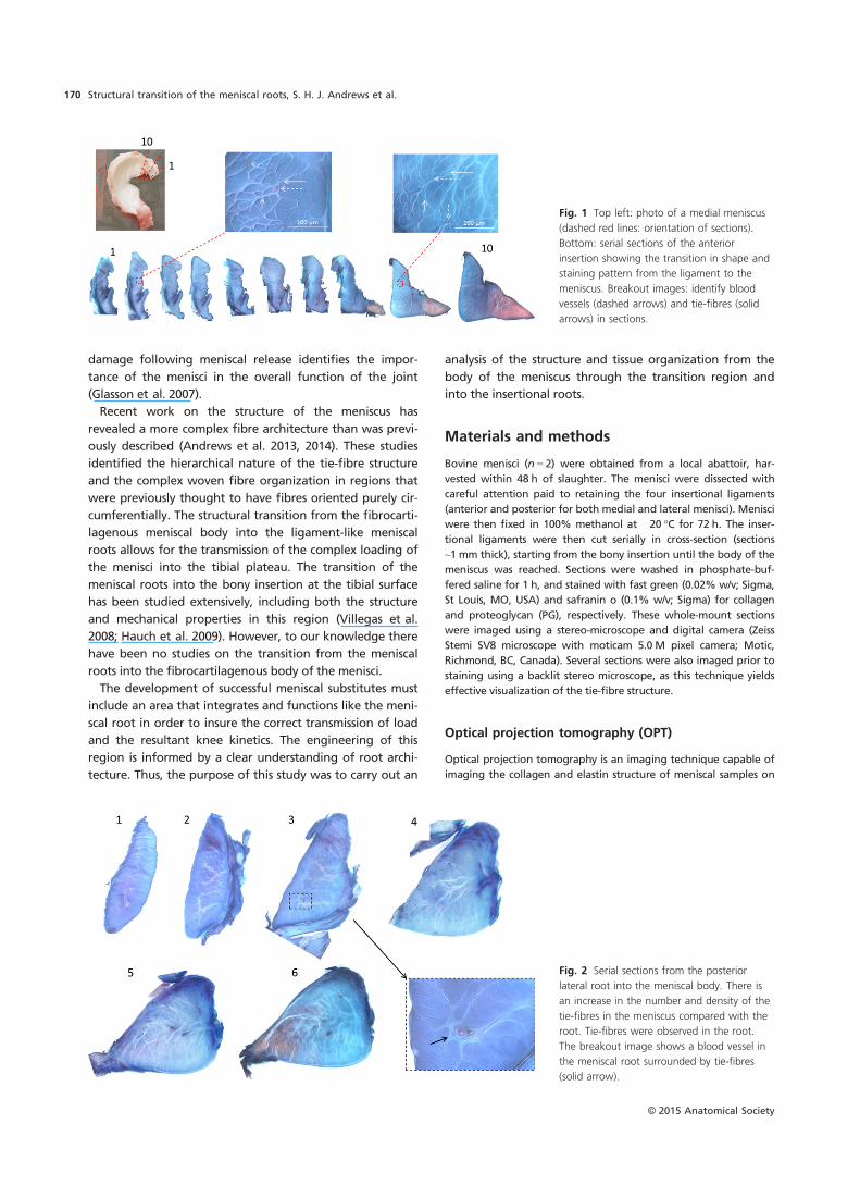
Fig. 1 Top left: photo of a medial meniscus
(dashed red lines: orientation of sections).
Bottom: serial sections of the anterior
insertion showing the transition in shape and
staining pattern from the ligament to the
meniscus. Breakout images: identify blood
vessels (dashed arrows) and tie-fibres (solid
arrows) in sections.
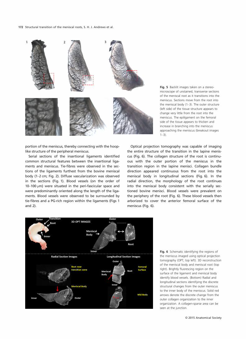
Fig. 2 Serial sections from the posterior
lateral root into the meniscal body. There is
an increase in the number and density of the
tie-fibres in the meniscus compared with the
root. Tie-fibres were observed in the root.
The breakout image shows a blood vessel in
the meniscal root surrounded by tie-fibres
(solid arrow).
© 2015 Anatomical Society
Structural transition of the meniscal roots, S. H. J. Andrews et al.170
the meso-scale (~1–10 mm) at a micro-scale resolution (5–10 lm;
Andrews et al. 2013). Hence, to understand the 3D collagen organi-
zation in this transitional region further, rabbit menisci were
obtained and imaged using OPT. The small size of rabbit menisci
allowed for imaging of the entire transitional region in one sample
using OPT. While rabbit menisci are significantly smaller than
bovine menisci, the general shape and morphology is quite similar
to bovine menisci (Proffen et al. 2012). Rabbit menisci (n = 2) were
obtained using a secondary tissue use protocol in accordance with
the Conjoint Health Research Ethics Board at the University of Cal-
gary. The menisci were dissected and immediately fixed in 100%
methanol at 4 °C for 48 h. After fixation, the samples were pre-
pared by cutting 3–4-mm sections of tissue from the insertional root
and the transitional regions both medial and lateral menisci. Speci-
mens were scanned using fluorescence OPT (Sharpe et al. 2002) on
a Bioptonics 3001M OPT scanner (Bioptonics Microscopy, Edin-
burgh, Scotland). Each tissue sample was embedded in 1.5% low-
melting-point agarose (Life Technologies, Burlington, ON, Canada).
The agarose blocks were trimmed and glued to mounts, and dehy-
drated through three washes of 100% methanol (Fisher, Ottawa,
ON, Canada) over 24 h. Specimens were then cleared for 24 h in
BABB [1 part benzyl alcohol : (Fisher) : 2 parts benzyl benzoate
(Sigma)]. Native autofluorescence was imaged using the GFP-1
channel (exciter 425 nm/40 nm; emitter LP475 nm) at a resolution
of 8–10 lm. Raw images were reconstructed into grey-scale slices
using NRECON (Skyscan NV, Kontich, Belgium). IMAGEJ (NIH open source
software) was used to create 3D images from the reconstructed
slices.
Results
Sections taken near the tibial insertion were ligament-like
morphologically, containing large collagen fascicles. In
sections approaching the meniscus, there was an increase
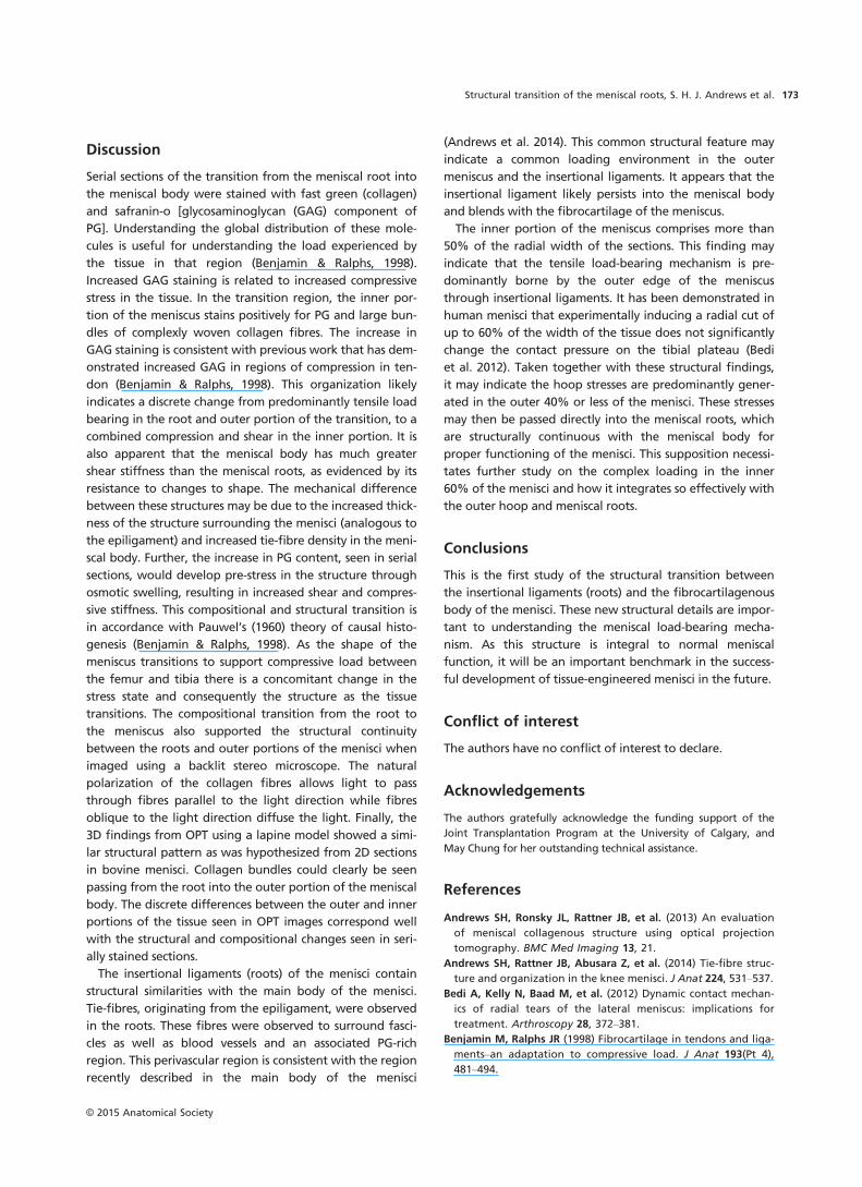
in tie-fibre size and density (Figs 1–4). Small tie-fibres
extended into the ligament from the epiligamentous struc-
ture in the outermost sections of the meniscal roots, while
large tie-fibre bundles were apparent at the meniscus tran-
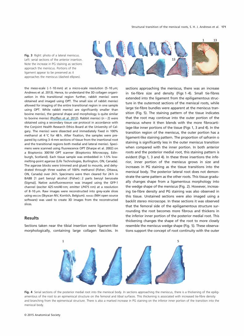
sition (Fig. 5). The staining pattern of the tissue indicates
that the root may continue into the outer portion of the
meniscus where it then blends with the more fibrocarti-
lage-like inner portions of the tissue (Figs 1, 3 and 4). In the
transition region of the meniscus, the outer portion has a
ligament-like staining pattern. The proportion of safranin o
staining is significantly less in the outer meniscus transition
when compared with the inner portion. In both anterior
roots and the posterior medial root, this staining pattern is
evident (Figs 1, 3 and 4). In these three insertions the infe-
rior, inner portion of the meniscus grows in size and
increases in PG staining as the tissue transitions into the
meniscal body. The posterior lateral root does not demon-
strate the same pattern as the other roots. This tissue gradu-
ally changes shape from a ligamentous morphology into
the wedge shape of the meniscus (Fig. 2). However, increas-
ing tie-fibre density and PG staining was also observed in
this tissue. Unstained sections were also imaged using a
backlit stereo microscope. In these sections it was observed
that the femoral side of the epiligamentous structure sur-
rounding the root becomes more fibrous and thickens in
the inferior inner portion of the posterior medial root. This
thickening changes the shape of the root to more closely
resemble the meniscus wedge shape (Fig. 5). These observa-
tions support the concept of root continuity with the outer
Fig. 3 Right: photo of a lateral meniscus.
Left: serial sections of the anterior insertion.
Note the increase in PG staining as sections
approach the meniscus. Portions of the
ligament appear to be preserved as it
approaches the meniscus (dashed ellipses).
Fig. 4 Serial sections of the posterior medial root into the meniscal body. In sections approaching the meniscus, there is a thickening of the epilig-
amentous of the root to an epimeniscal structure on the femoral and tibial surfaces. This thickening is associated with increased tie-fibre density
and branching from the epimeniscal structure. There is also a marked increase in PG staining on the inferior inner portion of the transition into the
meniscal body.
© 2015 Anatomical Society
Structural transition of the meniscal roots, S. H. J. Andrews et al. 171
portion of the meniscus, thereby connecting with the hoop-
like structure of the peripheral meniscus.
Serial sections of the insertional ligaments identified
common structural features between the insertional liga-
ments and meniscus. Tie-fibres were observed in the sec-
tions of the ligaments furthest from the bovine meniscal
body (1–2 cm; Fig. 2). Diffuse vascularization was observed
in the sections (Fig. 1). Blood vessels (on the order of
10–100 lm) were situated in the peri-fascicular space and
were predominantly oriented along the length of the liga-
ments. Blood vessels were observed to be surrounded by
tie-fibres and a PG-rich region within the ligaments (Figs 1
and 2).
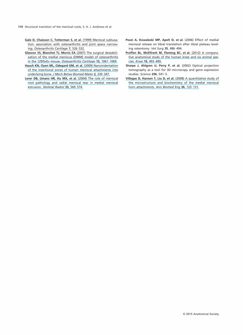
Optical projection tomography was capable of imaging
the entire structure of the transition in the lapine menis-
cus (Fig. 6). The collagen structure of the root is continu-
ous with the outer portion of the meniscus in the
transition region in the lapine menisci. Collagen bundle
direction appeared continuous from the root into the
meniscal body in longitudinal sections (Fig. 6). In the
radial direction, the morphology of the root continues
into the meniscal body consistent with the serially sec-
tioned bovine menisci. Blood vessels were prevalent on
the periphery of the root (Fig. 6). These blood vessels then
arborized to cover the anterior femoral surface of the
meniscus (Fig. 6).
Fig. 5 Backlit images taken on a stereo-
microscope of unstained, transverse sections
of the meniscal root as it transitions into the
meniscus. Sections move from the root into
the meniscal body (1–3). The outer structure
(left side) of the tissue structure appears to
change very little from the root into the
meniscus. The epiligament on the femoral
side of the tissue appears to thicken and
increase in branching into the meniscus
approaching the meniscus (breakout images
1–3).
Fig. 6 Schematic identifying the regions of
the meniscus imaged using optical projection
tomography (OPT; top left). 3D reconstruction
of the meniscal body and meniscal root (top
right). Brightly fluorescing region on the
surface of the ligament and meniscal body
identify blood vessels. (Bottom) Radial and
longitudinal sections identifying the discrete
structural changes from the outer meniscus
to the inner body of the meniscus. Solid red
arrows denote the discrete change from the
outer collagen organization to the inner
organization. A collagen-sparse area can be
seen at the junction.
© 2015 Anatomical Society
Structural transition of the meniscal roots, S. H. J. Andrews et al.172
Discussion
Serial sections of the transition from the meniscal root into
the meniscal body were stained with fast green (collagen)
and safranin-o [glycosaminoglycan (GAG) component of
PG]. Understanding the global distribution of these mole-
cules is useful for understanding the load experienced by
the tissue in that region (Benjamin & Ralphs, 1998).
Increased GAG staining is related to increased compressive
stress in the tissue. In the transition region, the inner por-
tion of the meniscus stains positively for PG and large bun-
dles of complexly woven collagen fibres. The increase in
GAG staining is consistent with previous work that has dem-
onstrated increased GAG in regions of compression in ten-
don (Benjamin & Ralphs, 1998). This organization likely
indicates a discrete change from predominantly tensile load
bearing in the root and outer portion of the transition, to a
combined compression and shear in the inner portion. It is
also apparent that the meniscal body has much greater
shear stiffness than the meniscal roots, as evidenced by its
resistance to changes to shape. The mechanical difference
between these structures may be due to the increased thick-
ness of the structure surrounding the menisci (analogous to
the epiligament) and increased tie-fibre density in the meni-
scal body. Further, the increase in PG content, seen in serial
sections, would develop pre-stress in the structure through
osmotic swelling, resulting in increased shear and compres-
sive stiffness. This compositional and structural transition is
in accordance with Pauwel’s (1960) theory of causal histo-
genesis (Benjamin & Ralphs, 1998). As the shape of the
meniscus transitions to support compressive load between
the femur and tibia there is a concomitant change in the
stress state and consequently the structure as the tissue
transitions. The compositional transition from the root to
the meniscus also supported the structural continuity
between the roots and outer portions of the menisci when
imaged using a backlit stereo microscope. The natural
polarization of the collagen fibres allows light to pass
through fibres parallel to the light direction while fibres
oblique to the light direction diffuse the light. Finally, the
3D findings from OPT using a lapine model showed a simi-
lar structural pattern as was hypothesized from 2D sections
in bovine menisci. Collagen bundles could clearly be seen
passing from the root into the outer portion of the meniscal
body. The discrete differences between the outer and inner
portions of the tissue seen in OPT images correspond well
with the structural and compositional changes seen in seri-
ally stained sections.
The insertional ligaments (roots) of the menisci contain
structural similarities with the main body of the menisci.
Tie-fibres, originating from the epiligament, were observed
in the roots. These fibres were observed to surround fasci-
cles as well as blood vessels and an associated PG-rich
region. This perivascular region is consistent with the region
recently described in the main body of the menisci
(Andrews et al. 2014). This common structural feature may
indicate a common loading environment in the outer
meniscus and the insertional ligaments. It appears that the
insertional ligament likely persists into the meniscal body
and blends with the fibrocartilage of the meniscus.
The inner portion of the meniscus comprises more than
50% of the radial width of the sections. This finding may
indicate that the tensile load-bearing mechanism is pre-
dominantly borne by the outer edge of the meniscus
through insertional ligaments. It has been demonstrated in
human menisci that experimentally inducing a radial cut of
up to 60% of the width of the tissue does not significantly
change the contact pressure on the tibial plateau (Bedi
et al. 2012). Taken together with these structural findings,
it may indicate the hoop stresses are predominantly gener-
ated in the outer 40% or less of the menisci. These stresses
may then be passed directly into the meniscal roots, which
are structurally continuous with the meniscal body for
proper functioning of the menisci. This supposition necessi-
tates further study on the complex loading in the inner
60% of the menisci and how it integrates so effectively with
the outer hoop and meniscal roots.
Conclusions
This is the first study of the structural transition between
the insertional ligaments (roots) and the fibrocartilagenous
body of the menisci. These new structural details are impor-
tant to understanding the meniscal load-bearing mecha-
nism. As this structure is integral to normal meniscal
function, it will be an important benchmark in the success-
ful development of tissue-engineered menisci in the future.
Conflict of interest
The authors have no conflict of interest to declare.
Acknowledgements
The authors gratefully acknowledge the funding support of the
Joint Transplantation Program at the University of Calgary, and
May Chung for her outstanding technical assistance.
References
Andrews SH, Ronsky JL, Rattner JB, et al. (2013) An evaluation
of meniscal collagenous structure using optical projection
tomography. BMC Med Imaging 13, 21.
Andrews SH, Rattner JB, Abusara Z, et al. (2014) Tie-fibre struc-
ture and organization in the knee menisci. J Anat 224, 531–537.
Bedi A, Kelly N, Baad M, et al. (2012) Dynamic contact mechan-
ics of radial tears of the lateral meniscus: implications for
treatment. Arthroscopy 28, 372–381.
Benjamin M, Ralphs JR (1998) Fibrocartilage in tendons and liga-
ments–an adaptation to compressive load. J Anat 193(Pt 4),
481–494.
© 2015 Anatomical Society
Structural transition of the meniscal roots, S. H. J. Andrews et al. 173
Gale D, Chaisson C, Totterman S, et al. (1999) Meniscal subluxa-
tion: association with osteoarthritis and joint space narrow-
ing. Osteoarthritis Cartilage 7, 526–532.
Glasson SS, Blanchet TJ, Morris EA (2007) The surgical destabili-
zation of the medial meniscus (DMM) model of osteoarthritis
in the 129/SvEv mouse. Osteoarthritis Cartilage 15, 1061–1069.
Hauch KN, Oyen ML, Odegard GM, et al. (2009) Nanoindentation
of the insertional zones of human meniscal attachments into
underlying bone. J Mech Behav Biomed Mater 2, 339–347.
Lerer DB, Umans HR, Hu MX, et al. (2004) The role of meniscal
root pathology and radial meniscal tear in medial meniscal
extrusion. Skeletal Radiol 33, 569–574.
Pozzi A, Kowaleski MP, Apelt D, et al. (2006) Effect of medial
meniscal release on tibial translation after tibial plateau level-
ing osteotomy. Vet Surg 35, 486–494.
Proffen BL, McElfresh M, Fleming BC, et al. (2012) A compara-
tive anatomical study of the human knee and six animal spe-
cies. Knee 19, 493–499.
Sharpe J, Ahlgren U, Perry P, et al. (2002) Optical projection
tomography as a tool for 3D microscopy and gene expression
studies. Science 296, 541–5.
Villegas D, Hansen T, Liu D, et al. (2008) A quantitative study of
the microstructure and biochemistry of the medial meniscal
horn attachments. Ann Biomed Eng 36, 123–131.
© 2015 Anatomical Society
Structural transition of the meniscal roots, S. H. J. Andrews et al.174
Related Documents