This article was published in an Elsevier journal. The attached copy is furnished to the author for non-commercial research and educational use, including for instruction at the author’s institution, sharing with colleagues and providing to institution administration. Other uses, including reproduction and distribution, or selling or licensing copies, or posting to personal, institutional or third party websites are prohibited. In most cases authors are permitted to post their version of the article (e.g. in Word or Tex form) to their personal website or institutional repository. Authors requiring further information regarding Elsevier’s archiving and manuscript policies are encouraged to visit: http://www.elsevier.com/copyright

Welcome message from author
This document is posted to help you gain knowledge. Please leave a comment to let me know what you think about it! Share it to your friends and learn new things together.
Transcript
This article was published in an Elsevier journal. The attached copy is furnished to the author for non-commercial research and
educational use, including for instruction at the author’s institution, sharing with colleagues and providing to institution administration.
Other uses, including reproduction and distribution, or selling or
licensing copies, or posting to personal, institutional or third party websites are prohibited.
In most cases authors are permitted to post their version of the
article (e.g. in Word or Tex form) to their personal website or institutional repository. Authors requiring further information
regarding Elsevier’s archiving and manuscript policies are encouraged to visit:
http://www.elsevier.com/copyright
Author's Personal Copy
58 (2007) 1070–1081
Materials CharacterizationThe staining of blue stone limestones petrographically unraveled
Roland Dreesen ⁎, Peter Nielsen, David Lagrou
Flemish Institute for Technological Research (VITO), Materials Technology, Boeretang 200, BE-2400 Mol, Belgium
Received 29 January 2007; accepted 30 March 2007
Abstract
This paper deals with the first results of a comparative petrographical study of un-aesthetically stained blue stone limestones,including Lower Carboniferous crinoidal limestones and Middle Cambrian oolitic limestones. Although it was commonly acceptedthat this staining resulted from weathering of ferroan dolomite, our study proved that it rather resulted from weathering offramboidal pyrite and subsequent dissolution of partially dedolomitized dolomite. Meteoric weathering resulted in a chemicalreaction chain including oxidation of pyrite, formation of sulphate-rich water, dissolution of dedolomitized dolomite rhombs andprecipitation of iron (hydr)oxide coatings in the intercrystalline dissolution voids. Weathering was probably accelerated by anincreased permeability, related to the presence of open stylolites and/or by initial secondary dolomitization of the host limestone.Optical microscopy of fluorescent epoxy impregnated thin sections and of polished slabs proved to be particularly useful inelucidating the above staining mechanism, especially in combination with SEM and BSEM with on-line WDS/EDS.© 2007 Elsevier Inc. All rights reserved.
Keywords: Optical microscopy; Micro-analysis; Carbonate building stones; Staining
1. Introduction
The “blue stones” are non-porous, hard limestones thatare traditionally used in Belgium and abroad as a good-quality natural dimension stone, with a more or lesspronounced natural bluish dark-grey colour. The latter“blue” colour is related to the presence of finelydisseminated organic carbon [2]. Due to mechanicalsurface treatment (“finish”) these types of rocks display abroad spectrum of colours ranging from light- tomedium-bluish-grey, over dark-grey to black (polished). Differentgood-quality Paleozoic blue stone type building stoneswere used in the past, some of which at least since Romantimes. These include the Lower Carboniferous Tournai
⁎ Corresponding author. Tel.: +32 14 33 56 56; fax: +32 14 32 11 86.E-mail addresses: [email protected] (R. Dreesen),
[email protected] (P. Nielsen), [email protected] (D. Lagrou).
1044-5803/$ - see front matter © 2007 Elsevier Inc. All rights reserved.doi:10.1016/j.matchar.2007.03.015
limestones, the Meuse Limestones and the most popular“Petit Granit” (bioclastic and oolitic wackestones tograinstones and cryptalgal–stromatolitic boundstones).However, due to decreasing quality, decreasing reservesand above all due to globalization and economically morecompetitive prices, different macroscopically analogouscandidate replacement stones recently appeared on theEuropean market. The latter originate from different partsof the world: such as Ireland (Erinstone), Vietnam (AsianBlue) and China (Tan Shan Limestone).
An important unanswered question when using thesenew materials is their physical–chemical durability andmore especially their resistance to atmospheric weathering(“ageing”) in our (wet) European climate. One relevantdurability aspect of blue stones covered in this paper is theirsusceptibility to discolouration. Besides the developmentof a natural patina (aesthetically accepted discolouration),unaesthetic and unwanted staining sometimes occur at the
Author's Personal Copy
1071R. Dreesen et al. / Materials Characterization 58 (2007) 1070–1081
surface of blue limestone-type carbonate building stoneswhen exposed to wet atmospheric conditions; these canvary from a ‘less unattractive’ or even a pleasant overallbeige patina to the appearance of unwanted yellowish orbrownish spots. Due to this phenomenon some Asian bluestones have become particularly suspect recently.
However, beige to brownish discolourations alsoappear in other blue stone types, including the indigenousBelgian blue stone (“Petit Granit”), after a few months upto a few years of exposure, in pavement stones or even infloor tiles. In outcrop (open-air quarry) a beige toyellowish-brown staining often appears within dolomi-tized parts of Paleozoic limestone beds, including theabove Lower Carboniferous limestones, when exposed towet atmospheric conditions. Moreover, at the contact withthe Cenozoic overburden a characteristically red–brownweathering colour often appears in the slightly dolomitizedPaleozoic limestones in Belgium (Namur, Dinant andVesdre Synclinoria); here it is clearly related to dissolution(palaeokarst) phenomena. This particular brownish weath-ered zone is also called “bousin” or “death stone” by localstoneworkers and quarrymen.
It was commonly accepted by field geologists thatweathering of ferroan dolomite in the dolomitizedlimestone was responsible for the unaesthetic brown toyellow superficial staining of those Paleozoic carbonaterocks. Dedolomitization of ferroan dolomite has beenreported to be responsible for the iron-stained calcite thatreplaces the ferroan dolomite [6]. A possible explanationis the fact that the concentration of ferrous iron in dolomitewould be in excess of the amount required to saturate thecalcite lattice. During dedolomitization this excess offerrous iron would be ejected and, under certain physico-chemical conditions be precipitated as colloidal ferrichydroxide that ages to iron-oxide.
In order to better understand this staining mechanism, aselection of naturally weathered (beige to brown stained)blue limestone samples taken from Belgian and Chineseblue stones, have been studied in thin section under theoptical microscope. Additionally, more sophisticatedanalytical tools such as X-ray diffraction, scanning electronmicroscopy and electron probe microanalysis, have beenused to corroborate the observed petrographical features.
2. Materials and methods
2.1. Materials
All the investigated samples were taken from buildingwharfs (kerbstones and pavement stones) in Belgium, aspart of a quality control assessment. It is, however, notclear how long the investigated stones have been exposed
to weathering during quarrying, transport and at thebuilding wharf. Discolourations of blue stone in newbuilding projects have been observed by the authors after6 months to a few years of exposure to atmospheric agents(Figs. 1A–3A).
2.2. Geological setting of the sampled building stones
The Chinese limestone comes from the southern partof the Shandong Province (Shan Tan village near thecity of Jining, Eastern China). The Belgian limestoneoriginates from Southern Belgium, more especially fromthe Condroz Basin.
The Chinese limestone is Middle Cambrian in age.During this period the Shandong area was part of the largeNorthChinaCarbonate Platform, that developed over some70 m.y. from the Early Cambrian to the Late Ordovician,extending some 1500 km east–west and 1000 km north–south [10]. The upper part of the Middle to lower UpperCambrian sequence consist of oolitic grainstones and tidal-flat lime mudstones, corresponding to either the Zhangxiaor Xuzhuang Formation.
The Belgian blue stone is Lower Carboniferous(Tournaisian Stage) in age. It is quarried in four differentareas from southern Belgium: Ourthe valley, Bocq valley,Molignée valley and the Hainaut Basin (Ecausinnes area).The studied samples have been derived from an unknownlithostratigraphical level of the so-called “Encrinite del'Ourthe” (Tn3b, Upper-Tournaisian) [5]. However,macroscopical features such as relatively darker colour,bioturbations and the relative frequency of small rugosecorals point to a possible lithostratigraphical positioningwithin the so-called “Banc Noir” level. This is alsoconfirmed by microscopical observations [4].
2.3. Methods
For the different microscopical investigations of theblue stones, specific types of thin sections were prepared:high-quality fluorescent epoxy-impregnated thin sectionsfor optical microscopy, polished thin sections withoutcover glass and/or polished rock slabs for chemicalelement mapping, and SEM/BSEM. Furthermore, selec-tive staining of the carbonates [3] has been applied tosome of the thin sections as well. Dolomite textures weredescribed using the terminology of Sibley andGregg [13].
Microscopical images (micrographs) of the thin sectionswere digitally acquired using an Axio-Cam Zeiss digitalcamera attached to a polarizing microscope. Subsequently,the images were processed with the Zeiss Axiovisionsoftware [using a specific routine especially developed forthe dolomite measurement application].
Author's Personal Copy
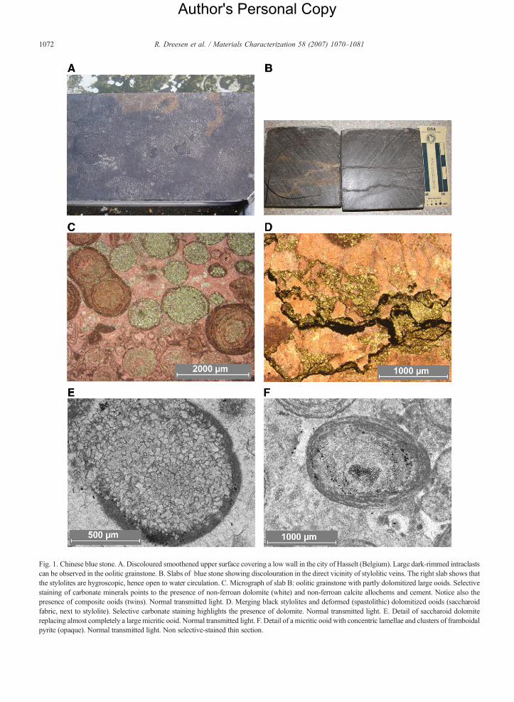
Fig. 1. Chinese blue stone. A. Discoloured smoothened upper surface covering a low wall in the city of Hasselt (Belgium). Large dark-rimmed intraclastscan be observed in the oolitic grainstone. B. Slabs of blue stone showing discolouration in the direct vicinity of stylolitic veins. The right slab shows thatthe stylolites are hygroscopic, hence open to water circulation. C. Micrograph of slab B: oolitic grainstone with partly dolomitized large ooids. Selectivestaining of carbonate minerals points to the presence of non-ferroan dolomite (white) and non-ferroan calcite allochems and cement. Notice also thepresence of composite ooids (twins). Normal transmitted light. D. Merging black stylolites and deformed (spastolithic) dolomitized ooids (saccharoidfabric, next to stylolite). Selective carbonate staining highlights the presence of dolomite. Normal transmitted light. E. Detail of saccharoid dolomitereplacing almost completely a largemicritic ooid. Normal transmitted light. F. Detail of a micritic ooid with concentric lamellae and clusters of framboidalpyrite (opaque). Normal transmitted light. Non selective-stained thin section.
1072 R. Dreesen et al. / Materials Characterization 58 (2007) 1070–1081
Author's Personal Copy
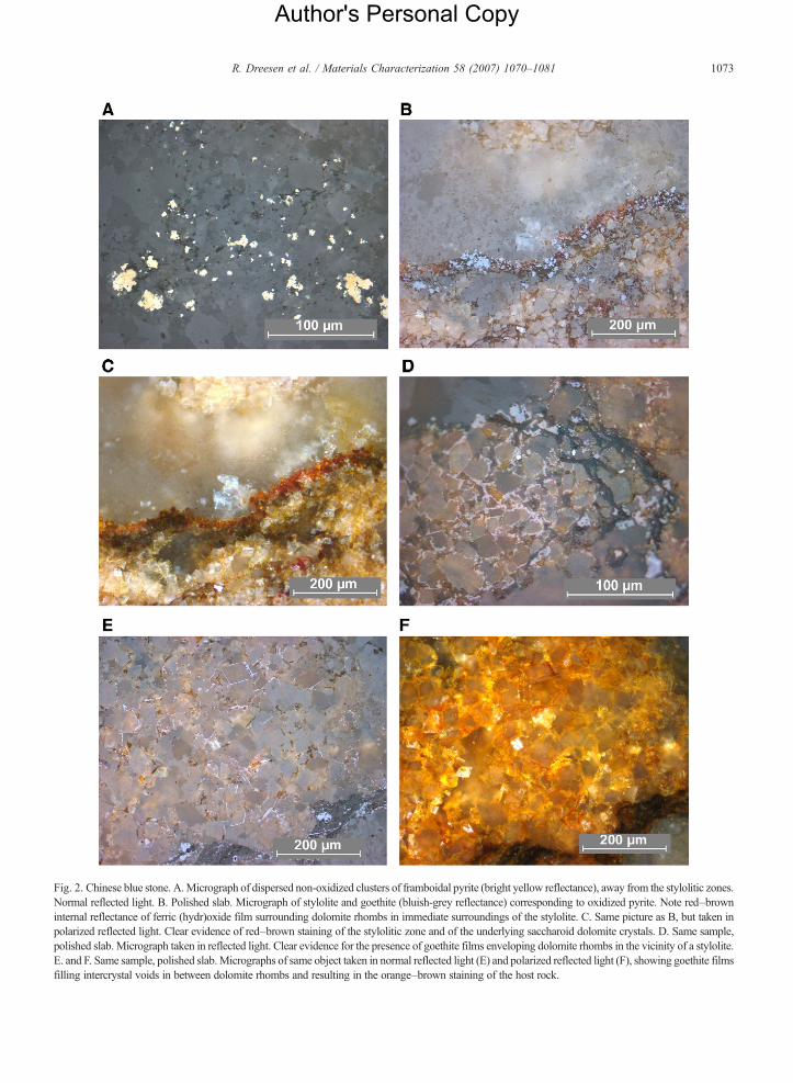
Fig. 2. Chinese blue stone. A.Micrograph of dispersed non-oxidized clusters of framboidal pyrite (bright yellow reflectance), away from the stylolitic zones.Normal reflected light. B. Polished slab. Micrograph of stylolite and goethite (bluish-grey reflectance) corresponding to oxidized pyrite. Note red–browninternal reflectance of ferric (hydr)oxide film surrounding dolomite rhombs in immediate surroundings of the stylolite. C. Same picture as B, but taken inpolarized reflected light. Clear evidence of red–brown staining of the stylolitic zone and of the underlying saccharoid dolomite crystals. D. Same sample,polished slab. Micrograph taken in reflected light. Clear evidence for the presence of goethite films enveloping dolomite rhombs in the vicinity of a stylolite.E. and F. Same sample, polished slab.Micrographs of same object taken in normal reflected light (E) and polarized reflected light (F), showing goethite filmsfilling intercrystal voids in between dolomite rhombs and resulting in the orange–brown staining of the host rock.
1073R. Dreesen et al. / Materials Characterization 58 (2007) 1070–1081
Author's Personal Copy
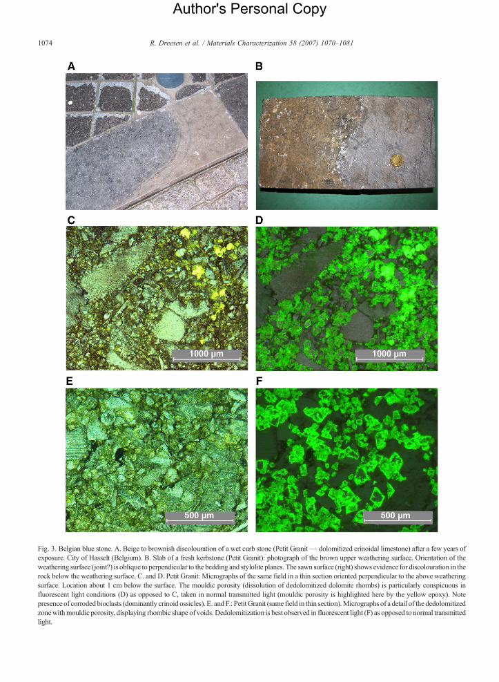
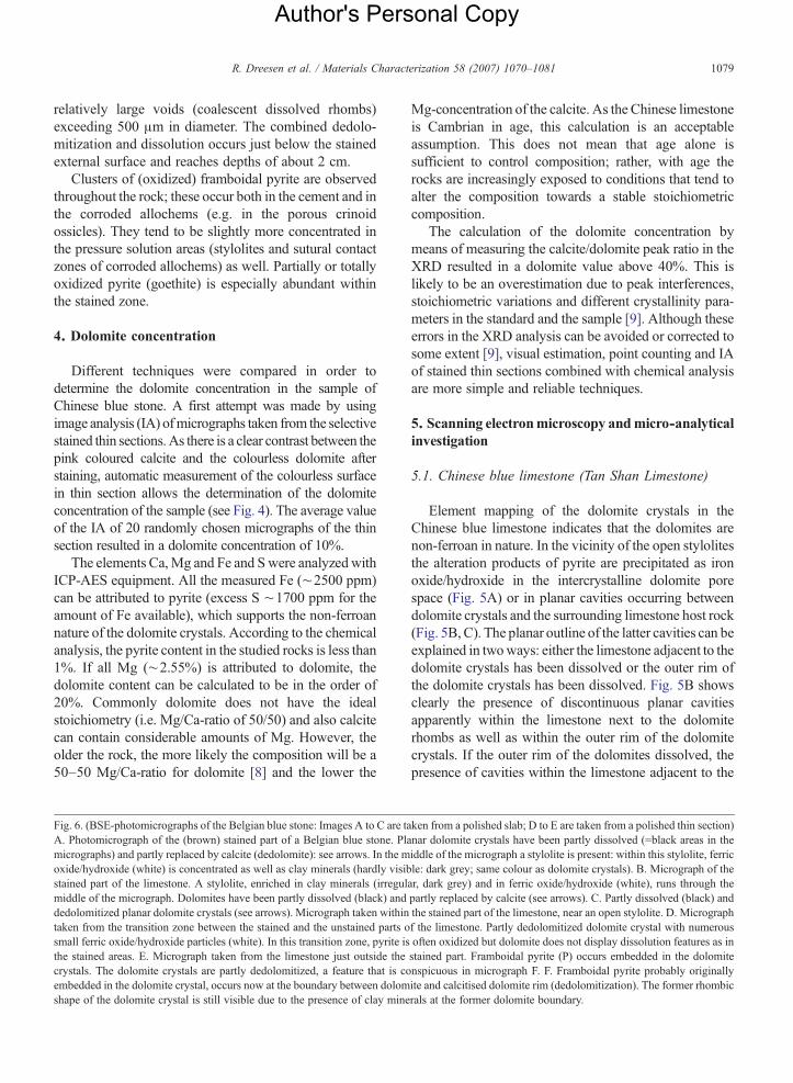
Fig. 3. Belgian blue stone. A. Beige to brownish discolouration of a wet curb stone (Petit Granit— dolomitized crinoidal limestone) after a few years ofexposure. City of Hasselt (Belgium). B. Slab of a fresh kerbstone (Petit Granit): photograph of the brown upper weathering surface. Orientation of theweathering surface (joint?) is oblique to perpendicular to the bedding and stylolite planes. The sawn surface (right) shows evidence for discolouration in therock below the weathering surface. C. and D. Petit Granit: Micrographs of the same field in a thin section oriented perpendicular to the above weatheringsurface. Location about 1 cm below the surface. The mouldic porosity (dissolution of dedolomitized dolomite rhombs) is particularly conspicuous influorescent light conditions (D) as opposed to C, taken in normal transmitted light (mouldic porosity is highlighted here by the yellow epoxy). Notepresence of corroded bioclasts (dominantly crinoid ossicles). E. and F.: Petit Granit (same field in thin section).Micrographs of a detail of the dedolomitizedzonewithmouldic porosity, displaying rhombic shape of voids. Dedolomitization is best observed in fluorescent light (F) as opposed to normal transmittedlight.
1074 R. Dreesen et al. / Materials Characterization 58 (2007) 1070–1081
Author's Personal Copy
1075R. Dreesen et al. / Materials Characterization 58 (2007) 1070–1081
Platinum-coated polished thin sections and rock slabswere used to obtain SEM and BSE (Back scatteredElectron) images with a Jeol Superprobe JXA-8621 MX.On-lineWDS/EDX systemswere used to map the elementcontent of selected parts of the studied thin-sections androck slabs. No attempt was made to quantify the elementcontent of the rock constituents.
The concentrations of Ca, Mg, Fe and S in a fewselected samples were determined by inductively coupledplasma-atomic emission spectrometry (ICP-AES), aftermicrowave destruction in an acid mixture of HNO3, HCland HF. Analytical precision at the 95% confidence leveldetermined on replicate analysis is better than 10%.
3. Petrographical analysis
3.1. Chinese blue limestone (Tan Shan Limestone)
The investigated Chinese blue stone sample was takenfrom a building wharf in Belgium (selected from a pile ofkerbstones and pavement stones). The sample displayed aconspicuous brownish staining on both sides of apressure-solution crack (stylolite) (Fig. 1B). After wetsawing of a slab from the sample, the crack remained stillwet, pointing to the fact that the stylolite was open and thatcooling water could penetrate the stone.
The Chinese blue limestone can be classified as amoderately dolomitized oolitic biosparite or dolomitizedoolitic grainstone. Themain allochems consist of relativelylarge ooids (500–1500 μm in diameter) most of which aremicritic in origin and display concentric lamellae (Fig. 1C).Besides, recrystallized sparitic ooids occur displaying afibroradiaxial fabric. Composite ooids occur as well.Peloids and large intraclasts represent only minor com-ponents. Where peloids are abundant they locally form anooidal–peloidal packstone microfabric. The intraclastsconsist of large flattened to subrounded, analogous ooliticgrainstones and porostromate (girvanellid) algal bound-stones, including several cryptalgal–bacterial components.
The carbonate cement is a clear, non-ferroan calcitecement (shown through selective staining; Dickson-method [3]) producing a coarse mosaic pattern in betweenthe allochems. Locally some patches of ferroan calcite canbe observed, most probably related to late-diageneticveinlets.
The limestone is strongly affected by pressure solution,resulting in numerous thin subhorizontal stylolites and inthe presence of strongly deformed ooids. The stylolites aregenerally thin, low in amplitude and tight. Non-solubleresidues including iron-rich sulphides (pyrite), organiccarbon and clay minerals are concentrated within the stylo-lites. Occasionally the stylolites are open, allowing water
circulation, oxidation of the sulphides and partial dissolu-tion of the carbonates and dolomite rhombs (Fig. 1D).Dolomitization is very conspicuous and irregular; sacchar-oid planar non-ferroan dolomite rhombs (20 to 50 μm indiameter) partially or even totally replace the micritic ooids(Fig. 1E) or the micritic/microsparite matrix in thepackstone areas of the grainstone. Dolomitization occurredbefore stylolitization. Concentrations of dolomite produceclear subhorizontal patches or streaks throughout thelimestone. This kind of dolomitization (equal-sized saccha-roid dolostones) results in an increase of porosity; planar orpolyhedral intercrystalline pores occur between thedolomite crystals.
Iron sulphides occur as small pyrite framboids orclusters of framboids; these are finely disseminated in thelimestone, either within the micritic ooids (Fig. 1F) or inthe recrystallized matrix (Fig. 2A). They have not beenobserved embedded in the dolomite rhombs but occur inthe intercrystalline pores and in cracks surrounding thedolomite crystals. The pyrite thus most likely post-datesdolomitization. Microscopical observations under incidentlight conditions (polished thin sections or polished slabs)clearly demonstrate the oxidation of the pyrite into goethitewithin or in the direct vicinity of the open stylolite(Fig. 2B–C). Moreover, the goethite occurs as tiny filmsaround the dolomite rhombs. Petrographical evidencepoints to migration of the goethite out of the stylolite intothe adjacent dolomitized areas (Fig. 2D). The area on bothsides of the open stylolite is stained with iron(hydr)oxides:in this particular area, a beige to orange–brown disco-louration of the limestone does occur (Fig. 2E–F).
Away from the stained areas, the finely disseminatedpyrite is still fresh and shows no evidence for oxidation.
3.2. Belgian blue stone (“Petit Granit”)
The studied sample was taken from a building wharf inBelgium (kerbstone). The sample displayed a conspicuousbeige to yellowish-brown weathering surface (Fig. 3B).The “Petit Granit” is a crinoidal limestone, comprisingnumerous bioclasts (from a few hundreds of μm diameterup tomore than1000μm): chiefly crinoid ossicles, but alsoskeletal fragments of bryozoans, brachiopods, ostracodes,rugose corals, rare calcareous algae and plurilocularforaminifera. Locally peloid-rich patches occur.
Petrographically the “Petit Granit” can be classifiedas a bioclastic wacke/packstone, locally grading into abioclastic grainstone. Quartz occurs as small sand grains(average 60 μm) dispersed in the carbonate.
The limestone underwent a strong pressure solutionresulting in the following phenomena: a conspicuous in-terpenetration of allochems (bioclast fragments), an
Author's Personal Copy
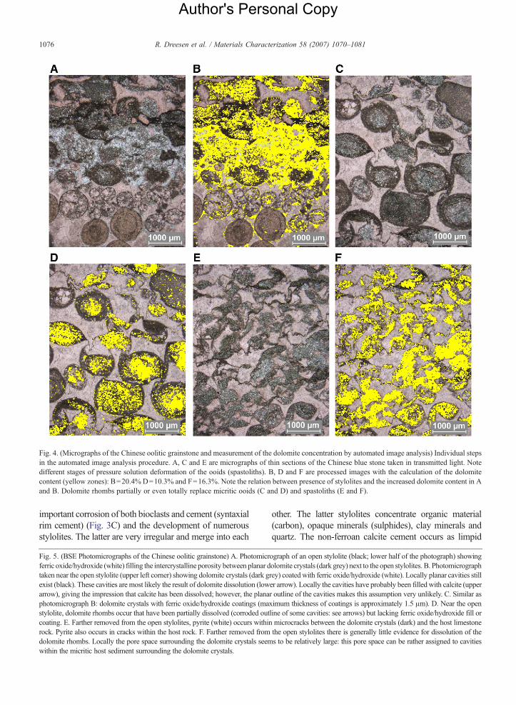
Fig. 4. (Micrographs of the Chinese oolitic grainstone and measurement of the dolomite concentration by automated image analysis) Individual stepsin the automated image analysis procedure. A, C and E are micrographs of thin sections of the Chinese blue stone taken in transmitted light. Notedifferent stages of pressure solution deformation of the ooids (spastoliths). B, D and F are processed images with the calculation of the dolomitecontent (yellow zones): B=20.4% D=10.3% and F=16.3%. Note the relation between presence of stylolites and the increased dolomite content in Aand B. Dolomite rhombs partially or even totally replace micritic ooids (C and D) and spastoliths (E and F).
1076 R. Dreesen et al. / Materials Characterization 58 (2007) 1070–1081
important corrosion of both bioclasts and cement (syntaxialrim cement) (Fig. 3C) and the development of numerousstylolites. The latter are very irregular and merge into each
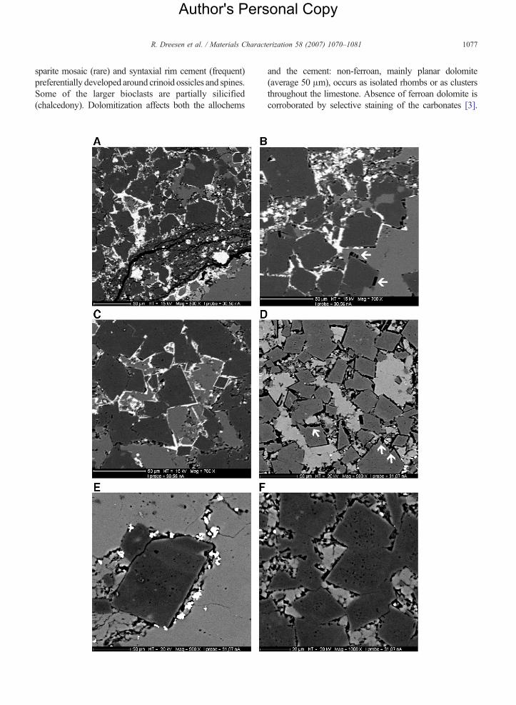
Fig. 5. (BSE Photomicrographs of the Chinese oolitic grainstone) A. Photomicrferric oxide/hydroxide (white) filling the intercrystalline porosity between planar dtaken near the open stylolite (upper left corner) showing dolomite crystals (dark gexist (black). These cavities are most likely the result of dolomite dissolution (lowarrow), giving the impression that calcite has been dissolved; however, the planaphotomicrograph B: dolomite crystals with ferric oxide/hydroxide coatings (mastylolite, dolomite rhombs occur that have been partially dissolved (corroded outcoating. E. Farther removed from the open stylolites, pyrite (white) occurs withinrock. Pyrite also occurs in cracks within the host rock. F. Farther removed fromdolomite rhombs. Locally the pore space surrounding the dolomite crystals seemwithin the micritic host sediment surrounding the dolomite crystals.
other. The latter stylolites concentrate organic material(carbon), opaque minerals (sulphides), clay minerals andquartz. The non-ferroan calcite cement occurs as limpid
ograph of an open stylolite (black; lower half of the photograph) showingolomite crystals (dark grey) next to the open stylolites. B. Photomicrographrey) coated with ferric oxide/hydroxide (white). Locally planar cavities stiller arrow). Locally the cavities have probably been filled with calcite (upperr outline of the cavities makes this assumption very unlikely. C. Similar asximum thickness of coatings is approximately 1.5 μm). D. Near the openline of some cavities: see arrows) but lacking ferric oxide/hydroxide fill ormicrocracks between the dolomite crystals (dark) and the host limestonethe open stylolites there is generally little evidence for dissolution of thes to be relatively large: this pore space can be rather assigned to cavities
Author's Personal Copy
1077R. Dreesen et al. / Materials Characterization 58 (2007) 1070–1081
sparite mosaic (rare) and syntaxial rim cement (frequent)preferentially developed around crinoid ossicles and spines.Some of the larger bioclasts are partially silicified(chalcedony). Dolomitization affects both the allochems
and the cement: non-ferroan, mainly planar dolomite(average 50 μm), occurs as isolated rhombs or as clustersthroughout the limestone. Absence of ferroan dolomite iscorroborated by selective staining of the carbonates [3].
Author's Personal Copy
1078 R. Dreesen et al. / Materials Characterization 58 (2007) 1070–1081
Except for the outer area of the sample, just beneath theweathered external surface, the dolomite rhombs are diffi-cult to observe in normal transmitted light conditions.How-ever, in fluorescent light the weathered dolomite rhombsare very conspicuous (Fig. 3C–F). Hence, in these lightconditions, dedolomitization and subsequent dissolution
phenomena can best be observed. The weathered surface isparallel to small calcite veins that are oriented perpendicularto the stylolites, pointing to their possible origin as joints.
Porosity is very low except where dissolution of thepartially dedolomitized dolomite rhombs resulted in animportant secondary porosity (mouldic porosity), with
Author's Personal Copy
1079R. Dreesen et al. / Materials Characterization 58 (2007) 1070–1081
relatively large voids (coalescent dissolved rhombs)exceeding 500 μm in diameter. The combined dedolo-mitization and dissolution occurs just below the stainedexternal surface and reaches depths of about 2 cm.
Clusters of (oxidized) framboidal pyrite are observedthroughout the rock; these occur both in the cement and inthe corroded allochems (e.g. in the porous crinoidossicles). They tend to be slightly more concentrated inthe pressure solution areas (stylolites and sutural contactzones of corroded allochems) as well. Partially or totallyoxidized pyrite (goethite) is especially abundant withinthe stained zone.
4. Dolomite concentration
Different techniques were compared in order todetermine the dolomite concentration in the sample ofChinese blue stone. A first attempt was made by usingimage analysis (IA) ofmicrographs taken from the selectivestained thin sections.As there is a clear contrast between thepink coloured calcite and the colourless dolomite afterstaining, automatic measurement of the colourless surfacein thin section allows the determination of the dolomiteconcentration of the sample (see Fig. 4). The average valueof the IA of 20 randomly chosen micrographs of the thinsection resulted in a dolomite concentration of 10%.
The elements Ca,Mg and Fe and Swere analyzed withICP-AES equipment. All the measured Fe (∼2500 ppm)can be attributed to pyrite (excess S ∼1700 ppm for theamount of Fe available), which supports the non-ferroannature of the dolomite crystals. According to the chemicalanalysis, the pyrite content in the studied rocks is less than1%. If all Mg (∼2.55%) is attributed to dolomite, thedolomite content can be calculated to be in the order of20%. Commonly dolomite does not have the idealstoichiometry (i.e. Mg/Ca-ratio of 50/50) and also calcitecan contain considerable amounts of Mg. However, theolder the rock, the more likely the composition will be a50–50 Mg/Ca-ratio for dolomite [8] and the lower the
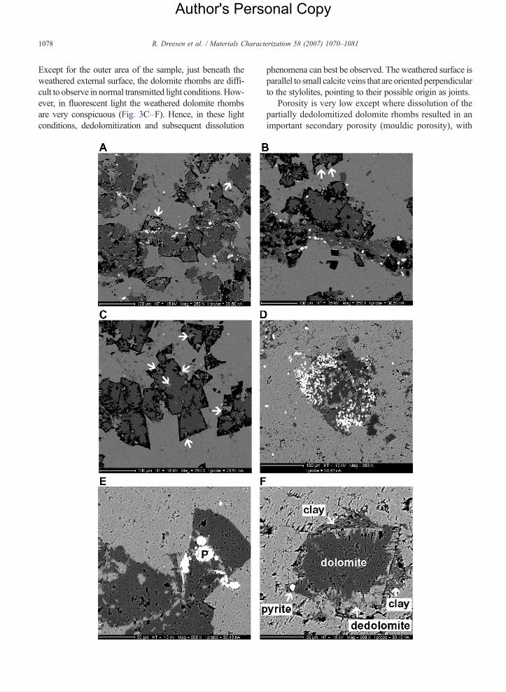
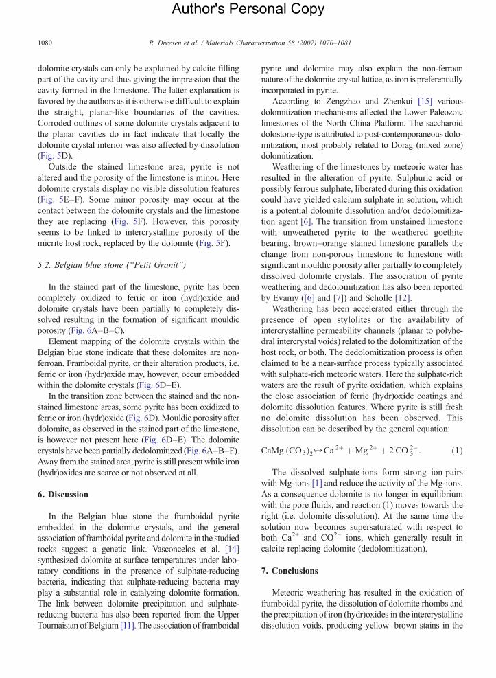
Fig. 6. (BSE-photomicrographs of the Belgian blue stone: Images A to C are taA. Photomicrograph of the (brown) stained part of a Belgian blue stone. Plamicrographs) and partly replaced by calcite (dedolomite): see arrows. In the moxide/hydroxide (white) is concentrated as well as clay minerals (hardly visibstained part of the limestone. A stylolite, enriched in clay minerals (irregulmiddle of the micrograph. Dolomites have been partly dissolved (black) anddedolomitized planar dolomite crystals (see arrows). Micrograph taken withintaken from the transition zone between the stained and the unstained parts osmall ferric oxide/hydroxide particles (white). In this transition zone, pyrite isthe stained areas. E. Micrograph taken from the limestone just outside thecrystals. The dolomite crystals are partly dedolomitized, a feature that is coembedded in the dolomite crystal, occurs now at the boundary between dolomshape of the dolomite crystal is still visible due to the presence of clay mine
Mg-concentration of the calcite. As the Chinese limestoneis Cambrian in age, this calculation is an acceptableassumption. This does not mean that age alone issufficient to control composition; rather, with age therocks are increasingly exposed to conditions that tend toalter the composition towards a stable stoichiometriccomposition.
The calculation of the dolomite concentration bymeans of measuring the calcite/dolomite peak ratio in theXRD resulted in a dolomite value above 40%. This islikely to be an overestimation due to peak interferences,stoichiometric variations and different crystallinity para-meters in the standard and the sample [9]. Although theseerrors in the XRD analysis can be avoided or corrected tosome extent [9], visual estimation, point counting and IAof stained thin sections combined with chemical analysisare more simple and reliable techniques.
5. Scanning electronmicroscopy andmicro-analyticalinvestigation
5.1. Chinese blue limestone (Tan Shan Limestone)
Element mapping of the dolomite crystals in theChinese blue limestone indicates that the dolomites arenon-ferroan in nature. In the vicinity of the open stylolitesthe alteration products of pyrite are precipitated as ironoxide/hydroxide in the intercrystalline dolomite porespace (Fig. 5A) or in planar cavities occurring betweendolomite crystals and the surrounding limestone host rock(Fig. 5B,C). The planar outline of the latter cavities can beexplained in twoways: either the limestone adjacent to thedolomite crystals has been dissolved or the outer rim ofthe dolomite crystals has been dissolved. Fig. 5B showsclearly the presence of discontinuous planar cavitiesapparently within the limestone next to the dolomiterhombs as well as within the outer rim of the dolomitecrystals. If the outer rim of the dolomites dissolved, thepresence of cavities within the limestone adjacent to the
ken from a polished slab; D to E are taken from a polished thin section)nar dolomite crystals have been partly dissolved (=black areas in theiddle of the micrograph a stylolite is present: within this stylolite, ferricle: dark grey; same colour as dolomite crystals). B. Micrograph of thear, dark grey) and in ferric oxide/hydroxide (white), runs through thepartly replaced by calcite (see arrows). C. Partly dissolved (black) andthe stained part of the limestone, near an open stylolite. D. Micrographf the limestone. Partly dedolomitized dolomite crystal with numerousoften oxidized but dolomite does not display dissolution features as instained part. Framboidal pyrite (P) occurs embedded in the dolomitenspicuous in micrograph F. F. Framboidal pyrite probably originallyite and calcitised dolomite rim (dedolomitization). The former rhombicrals at the former dolomite boundary.
Author's Personal Copy
1080 R. Dreesen et al. / Materials Characterization 58 (2007) 1070–1081
dolomite crystals can only be explained by calcite fillingpart of the cavity and thus giving the impression that thecavity formed in the limestone. The latter explanation isfavored by the authors as it is otherwise difficult to explainthe straight, planar-like boundaries of the cavities.Corroded outlines of some dolomite crystals adjacent tothe planar cavities do in fact indicate that locally thedolomite crystal interior was also affected by dissolution(Fig. 5D).
Outside the stained limestone area, pyrite is notaltered and the porosity of the limestone is minor. Heredolomite crystals display no visible dissolution features(Fig. 5E–F). Some minor porosity may occur at thecontact between the dolomite crystals and the limestonethey are replacing (Fig. 5F). However, this porosityseems to be linked to intercrystalline porosity of themicrite host rock, replaced by the dolomite (Fig. 5F).
5.2. Belgian blue stone (“Petit Granit”)
In the stained part of the limestone, pyrite has beencompletely oxidized to ferric or iron (hydr)oxide anddolomite crystals have been partially to completely dis-solved resulting in the formation of significant mouldicporosity (Fig. 6A–B–C).
Element mapping of the dolomite crystals within theBelgian blue stone indicate that these dolomites are non-ferroan. Framboidal pyrite, or their alteration products, i.e.ferric or iron (hydr)oxide may, however, occur embeddedwithin the dolomite crystals (Fig. 6D–E).
In the transition zone between the stained and the non-stained limestone areas, some pyrite has been oxidized toferric or iron (hydr)oxide (Fig. 6D). Mouldic porosity afterdolomite, as observed in the stained part of the limestone,is however not present here (Fig. 6D–E). The dolomitecrystals have been partially dedolomitized (Fig. 6A–B–F).Away from the stained area, pyrite is still present while iron(hydr)oxides are scarce or not observed at all.
6. Discussion
In the Belgian blue stone the framboidal pyriteembedded in the dolomite crystals, and the generalassociation of framboidal pyrite and dolomite in the studiedrocks suggest a genetic link. Vasconcelos et al. [14]synthesized dolomite at surface temperatures under labo-ratory conditions in the presence of sulphate-reducingbacteria, indicating that sulphate-reducing bacteria mayplay a substantial role in catalyzing dolomite formation.The link between dolomite precipitation and sulphate-reducing bacteria has also been reported from the UpperTournaisian of Belgium [11]. The association of framboidal
pyrite and dolomite may also explain the non-ferroannature of the dolomite crystal lattice, as iron is preferentiallyincorporated in pyrite.
According to Zengzhao and Zhenkui [15] variousdolomitization mechanisms affected the Lower Paleozoiclimestones of the North China Platform. The saccharoiddolostone-type is attributed to post-contemporaneous dolo-mitization, most probably related to Dorag (mixed zone)dolomitization.
Weathering of the limestones by meteoric water hasresulted in the alteration of pyrite. Sulphuric acid orpossibly ferrous sulphate, liberated during this oxidationcould have yielded calcium sulphate in solution, whichis a potential dolomite dissolution and/or dedolomitiza-tion agent [6]. The transition from unstained limestonewith unweathered pyrite to the weathered goethitebearing, brown–orange stained limestone parallels thechange from non-porous limestone to limestone withsignificant mouldic porosity after partially to completelydissolved dolomite crystals. The association of pyriteweathering and dedolomitization has also been reportedby Evamy ([6] and [7]) and Scholle [12].
Weathering has been accelerated either through thepresence of open stylolites or the availability ofintercrystalline permeability channels (planar to polyhe-dral intercrystal voids) related to the dolomitization of thehost rock, or both. The dedolomitization process is oftenclaimed to be a near-surface process typically associatedwith sulphate-rich meteoric waters. Here the sulphate-richwaters are the result of pyrite oxidation, which explainsthe close association of ferric (hydr)oxide coatings anddolomite dissolution features. Where pyrite is still freshno dolomite dissolution has been observed. Thisdissolution can be described by the general equation:
CaMg ðCO3Þ2 X Ca 2þ þMg 2þ þ 2 CO 2�3 : ð1Þ
The dissolved sulphate-ions form strong ion-pairswith Mg-ions [1] and reduce the activity of the Mg-ions.As a consequence dolomite is no longer in equilibriumwith the pore fluids, and reaction (1) moves towards theright (i.e. dolomite dissolution). At the same time thesolution now becomes supersaturated with respect toboth Ca2+ and CO2− ions, which generally result incalcite replacing dolomite (dedolomitization).
7. Conclusions
Meteoric weathering has resulted in the oxidation offramboidal pyrite, the dissolution of dolomite rhombs andthe precipitation of iron (hydr)oxides in the intercrystallinedissolution voids, producing yellow–brown stains in the
Author's Personal Copy
1081R. Dreesen et al. / Materials Characterization 58 (2007) 1070–1081
surrounding host rock. As all dolomite of the investigateddolomitized limestone samples is non-ferroan, weatheringof ferroan dolomite alone cannot explain the un-aestheticsuperficial staining of the limestones.
Open stylolites and/or intercrystalline permeabilitychannels related to the dolomitization of the host rockaccelerated the weathering process by allowing meteoricwater to penetrate the rock and oxidize the pyrite. Appar-ently, dolomitized blue limestone carbonates yieldingenough framboidal pyrite are prone to this type ofstaining. The genetic link between secondary dolomiti-zation and framboidal pyrite is particularly significant. Inthe Chinese blue limestone the concentration of dolomi-tized patches near open stylolites with an increased pyritecontent (concentrated after dissolving partially dolomi-tized micrite ooids) probably triggered the stainingprocess. In the Belgian blue stone a particular carbon-and pyrite-rich bioclast wackestone facies, in combinationwith secondary dolomitization and the presence of a jointhas triggered the weathering and subsequent staining.However, it is accepted that these are preliminarystatements that would merit further investigation.
In addition, our study has proved that the use of high-quality fluorescent epoxy impregnated thin sections ishighly recommended, and subsequent evaluation of thepetrographical observations with more-sophisticatedmicro-analytical tools is required.
References
[1] Baker PA, Kastner M. Constraints on the formation of sedimentarydolomite. Science 1981;213(4504):214–6.
[2] Belgian BuildingResearch Institute: BelgischeBlauweHardsteenof “Petit Granit” uit het Tournaisiaanse geologische tijdperk”.Technische Voorlichting 2001;220:1–58. (in Dutch).
[3] Dickson JAD. A modified staining technique for carbonates inthin section. Nature 1965;105:587.
[4] Dingelstadt C, Dreesen R. Atlas pétrographique des principalesroches calcaires et roches calcaires gréseuses utilisées dans lesmonuments de Wallonie. Tome1, Fiche 1. Petit Granit, DirectionGénérale de l'Aménagement du Territoire et du Logement,Division des Monuments, Sites et Fouilles, ISSeP, Rue du Chéra,200, B-4000 Liège. 1996. (in French).
[5] Dusar M, Hance L, editors. Acte Symposium ‘Petit Granit/’Blauwehardsteen’, Maffle, 21 oktober 1992. Bulletin van de BelgischeVereniging voor Geologie 1993;102:271–399. (in French).
[6] Evamy BD. The application of a chemical staining technique to astudy of dedolomitisation. Sedimentology1963;2:164–70.
[7] Evamy BD. Dedolomitization and the development of rhombo-hedral pores in limestones. J Sediment Petrol 1967;37:1204–15.
[8] Lumsden DN, Chimahusky JS. Relationship between dolomitenonstoichiometry and carbonate facies parameters. In: ZengerDH, Dunham JB, Ethington RL, editors. Concepts and modelsof dolomitization, Spec Publ- Soc Econ Paleontol Mineral1980;28:123–37.
[9] LumsdenDN.Discrepancy between thin-section andX-ray estimatesof dolomite in limestone. J Sediment Petrol 1979;49(2):429–36.
[10] MengX, GeM, TuckerM. Sequence stratigraphy, sea-level changesand depositional systems in the Cambro-Ordovician of the NorthChina carbonate platform. Sediment Geol 1997;114:189–222.
[11] Nielsen P, Swennen R, Dickson JAD, Fallick AE, Keppens E.Spheroidal dolomites in a Visean karst system-bacterial inducedorigin? Sedimentology 1997;44:177–95.
[12] Scholle PA. Diagenesis of deep-water carbonate turbidites, UpperCretaceous Monte Antola Flysch, Northern Apennines, Italy.J Sediment Petrol 1971;41:233–50.
[13] Sibley DF, Gregg JM. Classification of dolomite rock textures.J Sediment Petrol 1987;57:1112–4.
[14] Vasconcelos C, McKenzie JA, Bernasconi S, Grujic D, Tien AJ.Microbial mediation as a possiblemechanism for natural dolomiteformation at low temperatures. Nature 1995;377:220–2.
[15] Zengzhao F, Zhenkui J. Types and origin of dolostones in theLower Paleozoic of the North China Platform. Sediment Geol1994;93:279–90.
Related Documents


