The stability and characteristics of a DNA Holliday junction switch A.R. Mount a, , C.P. Mountford b , S.A.G. Evans a , T.-J. Su c , A.H. Buck c , P. Dickinson c , C.J. Campbell c , L.M. Keane c , J.G. Terry d , J.S. Beattie c , A.J. Walton d , P. Ghazal c , J. Crain b a School of Chemistry, The University of Edinburgh, Mayfield Road, Edinburgh EH9 3JJ, UK b School of Physics, The University of Edinburgh, Mayfield Road, Edinburgh EH9 3JZ, UK c The Scottish Centre for Genomic Technology and Informatics, The University of Edinburgh, The Chancellor's Building, 49 Little France Crescent, Edinburgh, EH16 4SB Scotland, UK d Institute of Integrated Micro and Nano Systems, Scottish Microelectronics Centre, School of Engineering and Electronics, University of Edinburgh, Edinburgh, EH9 3JF, UK Received 17 January 2006; accepted 25 March 2006 Available online 6 April 2006 Abstract A Holliday junction (HJ) consists of four DNA double helices, with a branch point discontinuity at the intersection of the component strands. At low ionic strength, the HJ adopts an open conformation, with four widely spaced arms, primarily due to strong electrostatic repulsion between the phosphate groups on the backbones. At high ionic strength, screening of this repulsion induces a switch to a more compact (closed) junction conformation. Fluorescent labelling with dyes placed on the HJ arms allows this conformational switch to be detected optically using fluorescence resonance energy transfer (FRET), producing a sensitive fluorescent output of the switch state. This paper presents a systematic and quantitative survey of the switch characteristics of such a labelled HJ. A short HJ (arm length 8 bp) is shown to be prone to dissociation at low switching ion concentration, whereas an HJ of arm length 12 bp is shown to be stable over all switching ion concentrations studied. The switching characteristics of this HJ have been systematically and quantitatively studied for a variety of switching ions, by measuring the required ion concentration, the sharpness of the switching transition and the fluorescent output intensity of the open and closed states. This stable HJ is shown to have favourable switch characteristics for a number of inorganic switching ions, making it a promising candidate for use in nanoscale biomolecular switch devices. © 2006 Elsevier B.V. All rights reserved. Keywords: Holliday junction; FRET; Biomolecular switch; DNA; Stability; Ion control 1. Introduction The structural properties that make DNA capable of conferring genetic information also enable it to behave as a scaffolding unit for DNA nanotechnology [1] and also as a processing unit for DNA-based computation [2,3]. In recent years, much research has focused on the design and implementation of DNA-based functional building blocks for the production of nanodevices [4]. Holliday junctions (HJ) and related structures [5] are likely to be particularly versatile as they can potentially act as both structural scaffolding units in nanodevices and as active elements capable of reporting on local environmental changes via ion-induced conformational transitions. The DNA Holliday structure is a junction of four double helices, first proposed in 1964 [6]. The unique topological element of the junction is a branch point at the intersection of the component strands. The junction exists in either open or closed forms, determined primarily by the strong electrostatic repulsion between the backbone phosphate groups. In low ionic strength solutions, this repulsive coulombic interaction favors an extended structure determined by maximum charge separation, an open branch point region and approximate fourfold symmetry. The open form has also been identified in certain crystal structures of the junction in complexes with recombination and repair proteins [7]. In high ionic strength solutions, screening of the repulsion between phosphate groups on the DNA backbone induces a conformational transition to a more compact, folded junction; the branch point collapses to enable pairs of double helical arms Biophysical Chemistry 124 (2006) 214 – 221 http://www.elsevier.com/locate/biophyschem Corresponding author. Tel.: +44 131 650 4747; fax: +44 131 650 4743. E-mail address: [email protected] (A.R. Mount). 0301-4622/$ - see front matter © 2006 Elsevier B.V. All rights reserved. doi:10.1016/j.bpc.2006.03.020

Welcome message from author
This document is posted to help you gain knowledge. Please leave a comment to let me know what you think about it! Share it to your friends and learn new things together.
Transcript
The stability and characteristics of a DNA Holliday junction switch
A.R. Mount a,!, C.P. Mountford b, S.A.G. Evans a, T.-J. Su c, A.H. Buck c, P. Dickinson c,C.J. Campbell c, L.M. Keane c, J.G. Terry d, J.S. Beattie c, A.J. Walton d, P. Ghazal c, J. Crain b
a School of Chemistry, The University of Edinburgh, Mayfield Road, Edinburgh EH9 3JJ, UKb School of Physics, The University of Edinburgh, Mayfield Road, Edinburgh EH9 3JZ, UK
c The Scottish Centre for Genomic Technology and Informatics, The University of Edinburgh, The Chancellor's Building,49 Little France Crescent, Edinburgh, EH16 4SB Scotland, UK
d Institute of Integrated Micro and Nano Systems, Scottish Microelectronics Centre, School of Engineering and Electronics,University of Edinburgh, Edinburgh, EH9 3JF, UK
Received 17 January 2006; accepted 25 March 2006Available online 6 April 2006
Abstract
A Holliday junction (HJ) consists of four DNA double helices, with a branch point discontinuity at the intersection of the component strands.At low ionic strength, the HJ adopts an open conformation, with four widely spaced arms, primarily due to strong electrostatic repulsion betweenthe phosphate groups on the backbones. At high ionic strength, screening of this repulsion induces a switch to a more compact (closed) junctionconformation. Fluorescent labelling with dyes placed on the HJ arms allows this conformational switch to be detected optically using fluorescenceresonance energy transfer (FRET), producing a sensitive fluorescent output of the switch state. This paper presents a systematic and quantitativesurvey of the switch characteristics of such a labelled HJ. A short HJ (arm length 8 bp) is shown to be prone to dissociation at low switching ionconcentration, whereas an HJ of arm length 12 bp is shown to be stable over all switching ion concentrations studied. The switching characteristicsof this HJ have been systematically and quantitatively studied for a variety of switching ions, by measuring the required ion concentration, thesharpness of the switching transition and the fluorescent output intensity of the open and closed states. This stable HJ is shown to have favourableswitch characteristics for a number of inorganic switching ions, making it a promising candidate for use in nanoscale biomolecular switch devices.© 2006 Elsevier B.V. All rights reserved.
Keywords: Holliday junction; FRET; Biomolecular switch; DNA; Stability; Ion control
1. Introduction
The structural properties that make DNA capable ofconferring genetic information also enable it to behave as ascaffolding unit for DNA nanotechnology [1] and also as aprocessing unit for DNA-based computation [2,3]. In recentyears, much research has focused on the design andimplementation of DNA-based functional building blocks forthe production of nanodevices [4]. Holliday junctions (HJ) andrelated structures [5] are likely to be particularly versatile asthey can potentially act as both structural scaffolding units innanodevices and as active elements capable of reporting onlocal environmental changes via ion-induced conformational
transitions. The DNA Holliday structure is a junction of fourdouble helices, first proposed in 1964 [6]. The uniquetopological element of the junction is a branch point at theintersection of the component strands. The junction exists ineither open or closed forms, determined primarily by the strongelectrostatic repulsion between the backbone phosphate groups.In low ionic strength solutions, this repulsive coulombicinteraction favors an extended structure determined bymaximum charge separation, an open branch point region andapproximate fourfold symmetry. The open form has also beenidentified in certain crystal structures of the junction incomplexes with recombination and repair proteins [7].
In high ionic strength solutions, screening of the repulsionbetween phosphate groups on the DNA backbone induces aconformational transition to a more compact, folded junction;the branch point collapses to enable pairs of double helical arms
Biophysical Chemistry 124 (2006) 214–221http://www.elsevier.com/locate/biophyschem
! Corresponding author. Tel.: +44 131 650 4747; fax: +44 131 650 4743.E-mail address: [email protected] (A.R. Mount).
0301-4622/$ - see front matter © 2006 Elsevier B.V. All rights reserved.doi:10.1016/j.bpc.2006.03.020
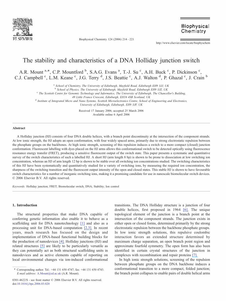
to align coaxially and cross in one of two so-called stacked-X(closed) conformations [8–10]. It is believed that the stacked-Xjunction may undergo rapid isomerisation transitions betweenthese two forms as suggested by recent single moleculemeasurements [11]. Schematic open and closed structures areshown in Fig. 1.
Several ions have now been shown to be effective promotersof junction folding. The most extensively studied are thedivalent metals Mg2+ and Ca2+, but switching at lowerconcentrations is also observed for the trivalent [Co(NH3)6]
3+
cation [10,12,13]. Structural studies of the stacked configura-tion are relatively rare though X-ray, NMR [14] and simulation[15] techniques have revealed likely binding site locations [16].Most of the interest in the structure and properties of branchedDNA intermediates arises from their importance in geneticrecombination processes [6], recognition and processing byproteins [17], viral integration [18] and DNA repair [19].
Despite being structurally well characterized [20–23] therehave been no systematic, quantitative explorations of theswitching characteristics of an HJ. In this paper we carry outsuch an exploration, with a view to assessing suitability of HJsas components in nanoscale switch devices. As there is doubtover the stability of HJ structures produced from relatively shortstrands [24], HJ stability at two different strand lengths was firstassessed using gel electrophoresis, prior to determining the
switching characteristics of the stable HJ. The issues relevant inthis context include switching ion concentration, sharpness ofthe ion-induced switching transition and fluorescence output ofswitch state. The practical issue of how ionic strength and pHcan be controlled without inducing appreciable switching isimportant for biomolecular interactions and this is alsoinvestigated.
2. Experimental
2.1. Chemicals
All solutions were prepared using deionised water from aWhatman RO 15 water purification system (resistivity!18 M! cm) using a 0.22 "m filter, and all compounds wereused as received without further purification. Hydrochloric acid(purity 99.999%), Tris-(hydroxymethyl)aminomethane (Tris,Ultrapure), Nitric acid (purity 99.99%), and spermidinetrichloride (purity 99%) were obtained from Aldrich. Lithiumchloride, sodium chloride (AnalaR) and sulfuric acid (AnalaR)were obtained from BDH, potassium chloride and calciumchloride (Laboratory Reagent Grade) were purchased fromFisons Scientific equipment. Magnesium chloride hexahydrate(purity>99.5%) and tetramethylammonium chloride (puri-ty>98%) were obtained from Fluka Chemika.
Fig. 1. Schematic of a Holliday junction. In the absence of switching cations the junction is in the open configuration with a maximally extended square-planarstructure (left). At high ionic strength, electrostatic repulsion of the phosphate groups on the backbones is reduced, resulting in helix–helix stacking and the closedconformation (right).
215A.R. Mount et al. / Biophysical Chemistry 124 (2006) 214–221
2.2. Construction of four-way junctions
A short-armed DNA-junction (SHJ) was preparedfrom a mixture of the following four pure oligonucleotides(Eurogentec):
FTGCAATCCTGAGCACA (5 "M, 1),TGTGCTCACCGAATCGGA (10 "M, 2),TCCGATTCGGACTATGCA (10 "M, 3)RTGCATAGTGATTGCA (10 "M, 4).
F is the donor Carboxyfluorescein (known commercially asFAM™) on oligonucleotide 1 and R is the acceptor Carbox-ytetramethylrhodamine (known as TAMRA™) on oligonucle-otide 4. A corresponding longer armed junction (LHJ), whichhas 4-bp extensions to each arm due to the addition ofcomplementary bases (shown in italics) to the ends of the SHJarms, was prepared from (Eurogentec):
GGCAFTGCAATCCTGAGCACATAGA (5 "M, 1),TCTATGTGCTCACCGAATCGGACCAG (10 "M, 2),CTGGTCCGATTCGGACTATGCAGCAA (10 "M, 3),TTGCRTGCATAGTGATTGCATGCC (10 "M, 4)
In this case, internal dyes were used to maintain dye positionrelative to the branch point. Each was assembled in a solution of20 mM Tris/TrisH+Cl" (pH 7.5) buffer solution containingNaCl (50 mM) and MgCl2 (5 mM), by heating to 80 °C for30 min and then allowing the solution to cool slowly in a waterbath to room temperature, which favours the thermodynami-cally most stable product. A ratio of 1 :2 :2 :2 of oligonucleotide1 :2 :3 :4 was chosen to ensure full incorporation of donorstrand into the fully assembled 4-way junctions. Typically, thefinal step in the sample preparation was to buffer exchangetwice (Microspin G25 ion exchange columns (AmershamBiosciences)), to remove the Mg2+ ions and produce HJsolutions (concentration 1 "M after dilution) in 20 mM Tris/TrisH+Cl" buffer (pH 7.5) as required for ion titration studies.100 "M tetrasodium ethylenediamine(tetraacetate) (Na4EDTA)was then added to the solution to titrate any residual Mg2+ ionsand produce HJ exclusively in the open form, as confirmed bycharacteristic and time independent steady-state fluorescencemeasurements (for titration studies, 100 "Mwas then subtractedfrom the concentration, c, of the inorganic ions which would beexpected to complex strongly with EDTA. This was confirmedby the invariance of the steady-state emission when adding aninitial 100 "M of these inorganic ions).
2.3. HJ stability
2.3.1. Gel electrophoresisFor gel electrophoresis assays, all oligonucleotide prepara-
tions were made at 10 "M in equimolar concentrations asdescribed above (except for the assembled HJ which was madeat the standard 5 "M:10 "M:10 "M:10 "M ratio, see Section2.2) and buffer exchanged twice into 45 mM Tris/TrisH+Borate(pH 8.3). MgCl2 was added to give a final concentration of
0.25 mM and 4 "l (approximately 250 ng) of each sample wasloaded as a band onto a 10% polyacrylamide gel in 45 mMTris/TrisH+Borate (pH 8.3), 0.25 mM MgCl2 electrophoresisbuffer.
Samples were loaded in the following order: Oligonucleo-tides 1, 2, 3 and 4 individually, in pairwise combinations 2+3and 3+4, as a 3-way structure 2+3+4 and as the assembled 4-way HJ structure, 1+2+3+4. Additionally, TGCAATCCT-GAGCACATTTTTFGTGCTCACCGAATCGGATTTTRTCC-GATTCGGACTATGCA (5), was also deposited as a controloligonucleotide band. This is a 60-mer, containing internalCarboxyfluorescein (F) and Carboxytetramethylrhodamine (R)labelled dT (where dT is deoxythymidine), which is expected tobe of comparable size to the fully assembled SHJ. The gel wasrun for 4.5 h at 4 °C at 6.25 V/cm and then fixed in 45 mM Tris/TrisH+Borate containing 7% (v/v) acetic acid, 10% (v/v)isopropanol for 15 min, stained in 45 mM Tris/TrisH+Boratecontaining 0.02% (w/v) stains-all dye (Aldrich), 20% (v/v)formamide, 20% (v/v) isopropanol for 30 min in the dark anddestained in Tris/TrisH+Borate containing 10% (v/v) isopropa-nol in the light for 90 min. The gel was then scanned on aflatbed scanner to capture 8-bit grey scale images.
2.3.2. gelFRET assaysgelFRET assays were performed as described elsewhere
[25]. HJ solutions (1 "M) in 45 mM Tris/Borate pH 8.3 bufferwere prepared in the presence of either 0 or 5.0 mM MgCl2 andwere loaded onto gels (10% acrylamide, 45 mM Tris/BoratepH 8.3, and 0 or 5.0 mM MgCl2 as appropriate). Gels were runat 4 °C with an electric field of 7 V/cm for 2 h, and then scannedwith a Typhoon 9400 scanner (Amersham Biosciences): laserlight at 488 nm was used to excite the donor. The emitted lightwas passed through either a 526-nm short-pass filter (for donoremission) or a 580-nm band-pass filter (for acceptor emission).Using ImageQuant software (Molecular Dynamics), the inten-sity of the signal obtained at 526 nm (represented by theintensity of the colour green) was overlaid with the intensity ofthe signal obtained at 580 nm (represented as the intensity of thecolour red) to visualize the relative difference in FRET for eachgel band.
2.4. Fluorescence spectroscopy
As in several previous HJ structural studies [20–23], theswitching of the assembled 4-way junction was detectedoptically using fluorescence resonance energy transfer (FRET)between the donor (F) and acceptor (R) fluorophores. Steady-state fluorescence emission spectra were recorded in a Starna26.5-F Far UV quartz fluorescent cell (10 mm path length)using a Jobin Yvon Spex Fluoromax spectrofluorimeter(Instruments S.A.) with a fixed excitation wavelength of476.5 nm. This wavelength was chosen to maximize donorexcitation while minimizing direct excitation of the acceptormolecule. This was confirmed by control experiments, whichshowed 1 "M short-armed oligonucleotide 4 to have peakacceptor emission of less than 5% of the peak donor emission of1 "M oligonucleotide 1 at this wavelength.
216 A.R. Mount et al. / Biophysical Chemistry 124 (2006) 214–221
2.5. Titration protocol
Titrations involved adding aliquots of a concentrated cationsolution in 20 mM Tris/TrisH+Cl" buffer to a 50 "l solution of1.0 "M HJ, prepared as in Section 2.2. The solution was thenthoroughly mixed and left to equilibrate for at least 2 min(which experiments showed was sufficient to ensure equilibra-tion) before recording the steady-state emission spectra. Theintensity of each spectrum was adjusted for the increase involume on titration to allow for the effects of dilution. For theTrisH+ titration experiment, aliquots of a concentrated (2.0 M inTris) solution of Tris/TrisH+Cl" buffer (pH 7.5) were added toachieve the desired cation concentration change, while main-taining a constant pH. Non-linear least squares curve fitting ofdata to Eqs. (7) and (8) and calculation of errors and reduced #2
values was carried out using Origin (MicroCal).
3. Results and discussion
3.1. Switch formation and integrity
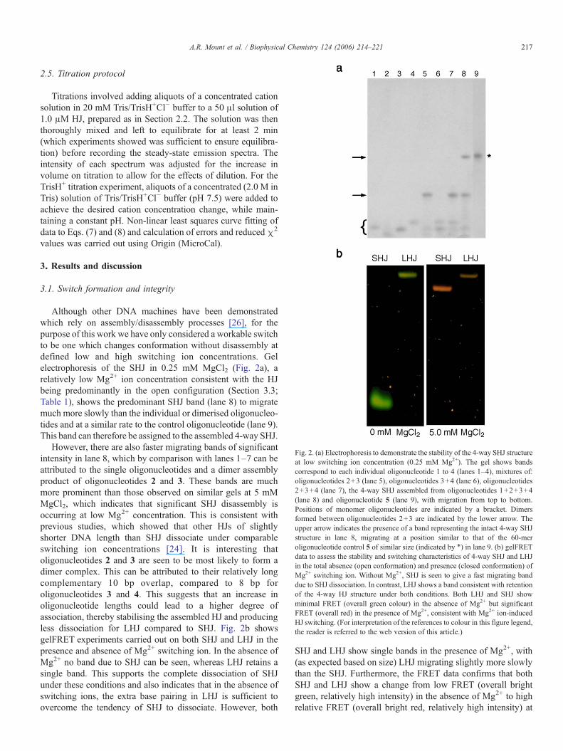
Although other DNA machines have been demonstratedwhich rely on assembly/disassembly processes [26], for thepurpose of this work we have only considered a workable switchto be one which changes conformation without disassembly atdefined low and high switching ion concentrations. Gelelectrophoresis of the SHJ in 0.25 mM MgCl2 (Fig. 2a), arelatively low Mg2+ ion concentration consistent with the HJbeing predominantly in the open configuration (Section 3.3;Table 1), shows the predominant SHJ band (lane 8) to migratemuch more slowly than the individual or dimerised oligonucleo-tides and at a similar rate to the control oligonucleotide (lane 9).This band can therefore be assigned to the assembled 4-way SHJ.
However, there are also faster migrating bands of significantintensity in lane 8, which by comparison with lanes 1–7 can beattributed to the single oligonucleotides and a dimer assemblyproduct of oligonucleotides 2 and 3. These bands are muchmore prominent than those observed on similar gels at 5 mMMgCl2, which indicates that significant SHJ disassembly isoccurring at low Mg2+ concentration. This is consistent withprevious studies, which showed that other HJs of slightlyshorter DNA length than SHJ dissociate under comparableswitching ion concentrations [24]. It is interesting thatoligonucleotides 2 and 3 are seen to be most likely to form adimer complex. This can be attributed to their relatively longcomplementary 10 bp overlap, compared to 8 bp foroligonucleotides 3 and 4. This suggests that an increase inoligonucleotide lengths could lead to a higher degree ofassociation, thereby stabilising the assembled HJ and producingless dissociation for LHJ compared to SHJ. Fig. 2b showsgelFRET experiments carried out on both SHJ and LHJ in thepresence and absence of Mg2+ switching ion. In the absence ofMg2+ no band due to SHJ can be seen, whereas LHJ retains asingle band. This supports the complete dissociation of SHJunder these conditions and also indicates that in the absence ofswitching ions, the extra base pairing in LHJ is sufficient toovercome the tendency of SHJ to dissociate. However, both
SHJ and LHJ show single bands in the presence of Mg2+, with(as expected based on size) LHJ migrating slightly more slowlythan the SHJ. Furthermore, the FRET data confirms that bothSHJ and LHJ show a change from low FRET (overall brightgreen, relatively high intensity) in the absence of Mg2+ to highrelative FRET (overall bright red, relatively high intensity) at
Fig. 2. (a) Electrophoresis to demonstrate the stability of the 4-way SHJ structureat low switching ion concentration (0.25 mM Mg2+). The gel shows bandscorrespond to each individual oligonucleotide 1 to 4 (lanes 1–4), mixtures of:oligonucleotides 2+3 (lane 5), oligonucleotides 3+4 (lane 6), oligonucleotides2+3+4 (lane 7), the 4-way SHJ assembled from oligonucleotides 1+2+3+4(lane 8) and oligonucleotide 5 (lane 9), with migration from top to bottom.Positions of monomer oligonucleotides are indicated by a bracket. Dimersformed between oligonucleotides 2+3 are indicated by the lower arrow. Theupper arrow indicates the presence of a band representing the intact 4-way SHJstructure in lane 8, migrating at a position similar to that of the 60-meroligonucleotide control 5 of similar size (indicated by !) in lane 9. (b) gelFRETdata to assess the stability and switching characteristics of 4-way SHJ and LHJin the total absence (open conformation) and presence (closed conformation) ofMg2+ switching ion. Without Mg2+, SHJ is seen to give a fast migrating banddue to SHJ dissociation. In contrast, LHJ shows a band consistent with retentionof the 4-way HJ structure under both conditions. Both LHJ and SHJ showminimal FRET (overall green colour) in the absence of Mg2+ but significantFRET (overall red) in the presence of Mg2+, consistent with Mg2+ ion-inducedHJ switching. (For interpretation of the references to colour in this figure legend,the reader is referred to the web version of this article.)
217A.R. Mount et al. / Biophysical Chemistry 124 (2006) 214–221

5 mM Mg2+, consistent with both adopting the closedconformation at 5 mM Mg2+. In the case of LHJ, this indicatesa functional switch which maintains its integrity even in theabsence of Mg2+, whereas SHJ is a switch whose assembledstability and switching is Mg2+-dependent.
3.2. LHJ switching characteristics
Fig. 3a and b shows typical LHJ fluorescence emissionspectra obtained as a function of the concentration of Mg2+ andNa+ switching ions, respectively.
At relatively low switching ion concentration, c, it is clear inboth cases that an emission peak is seen at around 517 nm. Thisis characteristic of F donor emission, which decreases inintensity with increasing c at the expense of a growing peak ataround 584 nm, characteristic of R acceptor emission andtherefore indicative of FRET. Given the strong inverse sixthpower dependence of FRET efficiency on the donor–acceptorseparation distance, this is consistent with previous observa-tions [20,22] of a growing proportion of folded (closed) HJs
which show FRET due to the effects of cation-induced folding,at the expense of open HJs which show no FRET.
The results of further measurements are summarized in Fig.4 for both monovalent, and multivalent ions where we presentthe increase of FRET signal as the ratio of the peak acceptorintensity (IA) to the donor intensity (ID). In general, consistentwith previous observations [13], we find the basic trend thatthe cation concentration threshold (the minimum concentrationrequired to actuate the switch) decreases markedly withincreasing valency. This is fully consistent with the overallnotion that the electrostatic repulsion between fixed phosphatebackbone charges must be sufficiently screened by switchingions to enable folding. It is thus clear that much larger ionconcentrations are required for monovalent ions to induceswitching and switching due to monovalent ions can beignored when multivalent ions are present. The effect of buffercation concentration on HJ conformation has not beenconsidered in previous studies; this work confirms theapplicability of TrisH+ as an inert buffer ion, maintainingconstant pH and high ionic strength, while not inducingsignificant folding at the tens of millimolar ion concentrationsrequired for solution buffering.
3.3. Assessment of LHJ switching
3.3.1. Quantitative analysis of HJ switching with inorganicions
An important feature of Fig. 3 (and all other ion titrationdata) is the significant acceptor emission intensity relative to thedonor emission intensity seen for the closed form. This isconsistent with our branch point sequence design (which isbased on the well-studied “J1” structure [27,28], which is asequence designed to cause the vast majority of the closed LHJto adopt the FRET signal conformer, maximising acceptoremission output [23]). Also significant is the presence of anisosbestic point for each of the ions at 564–565 nm, where theoverall intensity, Iiso, of the fluorescence emission is
0
1e+006
2e+006
3e+006
4e+006
5e+006
6e+006
7e+006
8e+006
9e+006
500 550 600 650 700Wavelength (nm)
0
2e+006
4e+006
6e+006
8e+006
1e+007
1.2e+007
1.4e+007
500 550 600 650 700Wavelength (nm)
Inte
nsity
(cp
s)In
tens
ity (
cps)
a
b
Fig. 3. Fluorescence emission spectra for a solution of 1 "M LHJ in 20 mMpH 7.5 Tris/TrisH+Cl" buffer (Tris buffer), titrated with increasing concentrationof (a) Mg2+ between 100 "M and 9.8 mM and (b) Na+ between 55 and 410 mM.In each case, the conversion from the open (maximum emission at 517 nm) tothe closed configuration (which shows a FRET peak with a maximum emissionat 584 nm) can clearly be seen, along with the isosbestic point at 564–565 nm.
0.2
0.3
0.4
0.5
0.6
0.7
0.8
0.9
1
1e-005 0.0001 0.001 0.01 0.1 1
FR
ET
Rat
io
Ion Concentration (M)
Fig. 4. Plots of FRET ratio versus ion concentration data obtained whentitrating the ions (!) TMA+, (") TrisH+, (#) Na+, ($) Li+, (%) K+, (&) Mg2+,(') Ca2+and (+) the spermidine ion ({H3N(CH2)3NH2(CH2)4NH3}
3+(Sp3+))into a solution of 1 "M open HJ in Tris buffer. Ion concentrations have beenadjusted for the presence of EDTA.
218 A.R. Mount et al. / Biophysical Chemistry 124 (2006) 214–221
independent of Mg2+ and Na+ ion concentration, respectively.This is strong evidence for fluorescence emission arising fromonly two states of the assembled HJ molecule (one FRETinactive, open and one FRETactive, closed), as the fluorescenceemission intensity obeys Eqs. (1) and (2) in this range ofconcentrations
I!k" # Iopen!k"xopen $ Iclosed!k"xclosed !1"
with
xopen # 1% xclosed !2"
where Iopen and Iclosed, the emission intensities of the HJsolution when entirely in the open and closed forms,respectively, are equal at 564–565 nm, and xopen, xclosed arethe mole fractions of the open and closed forms. This isconsistent with the previous postulate of two-state switching fora similar HJ system [22]. Iopen and Iclosed are related to thefundamental emission characteristics of the open and closedHJs, respectively, and must be independent of ion concentration(c). These can be obtained at all wavelengths from experimentalsteady-state emission spectra at low (c!0) and high (c!#)switching ion concentrations respectively, these conditionsbeing denoted by superscript 0 and #. At any c, independentexperimental values of xopen and xclosed can most accurately beobtained at the peak donor and acceptor wavelengths, ID and IA,respectively, by combining Eqs. (1) and (2) to give:
xopen #ID % IlDI0D % IlD
!3"
and
xclosed #IA % I0AIlA % I0A
!4"
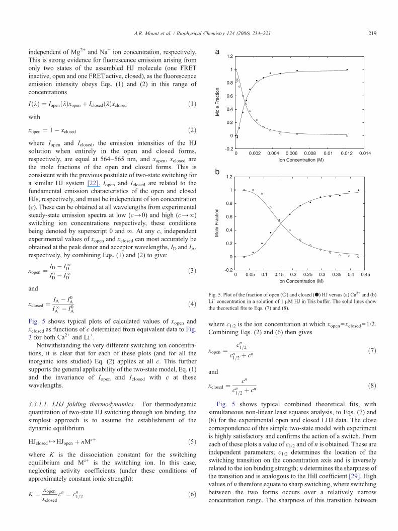
Fig. 5 shows typical plots of calculated values of xopen andxclosed as functions of c determined from equivalent data to Fig.3 for both Ca2+ and Li+.
Notwithstanding the very different switching ion concentra-tions, it is clear that for each of these plots (and for all theinorganic ions studied) Eq. (2) applies at all c. This furthersupports the general applicability of the two-state model, Eq. (1)and the invariance of Iopen and Iclosed with c at thesewavelengths.
3.3.1.1. LHJ folding thermodynamics. For thermodynamicquantitation of two-state HJ switching through ion binding, thesimplest approach is to assume the establishment of thedynamic equilibrium
HJclosed X HJopen $ nMz$ !5"
where K is the dissociation constant for the switchingequilibrium and Mz+ is the switching ion. In this case,neglecting activity coefficients (under these conditions ofapproximately constant ionic strength):
K #xopenxclosed
cn # cn1=2 !6"
where c1/2 is the ion concentration at which xopen=xclosed=1/2.Combining Eqs. (2) and (6) then gives
xopen #cn1=2
cn1=2 $ cn!7"
and
xclosed #cn
cn1=2 $ cn!8"
Fig. 5 shows typical combined theoretical fits, withsimultaneous non-linear least squares analysis, to Eqs. (7) and(8) for the experimental open and closed LHJ data. The closecorrespondence of this simple two-state model with experimentis highly satisfactory and confirms the action of a switch. Fromeach of these plots a value of c1/2 and of n is obtained. These areindependent parameters; c1/2 determines the location of theswitching transition on the concentration axis and is inverselyrelated to the ion binding strength; n determines the sharpness ofthe transition and is analogous to the Hill coefficient [29]. Highvalues of n therefore equate to sharp switching, where switchingbetween the two forms occurs over a relatively narrowconcentration range. The sharpness of this transition between
-0.2
0
0.2
0.4
0.6
0.8
1
1.2
0 0.002 0.004 0.006 0.008 0.01 0.012 0.014
Mol
e F
ract
ion
Ion Concentration (M)
-0.2
0
0.2
0.4
0.6
0.8
1
1.2
0 0.05 0.1 0.15 0.2 0.25 0.3 0.35 0.4 0.45
Mol
e F
ract
ion
Ion Concentration (M)
a
b
Fig. 5. Plot of the fraction of open (') and closed (&) HJ versus (a) Ca2+ and (b)Li+ concentration in a solution of 1 "M HJ in Tris buffer. The solid lines showthe theoretical fits to Eqs. (7) and (8).
219A.R. Mount et al. / Biophysical Chemistry 124 (2006) 214–221
the “on” and “off” states is a key characteristic that defines howclose the system is to a theoretically ideal switch.
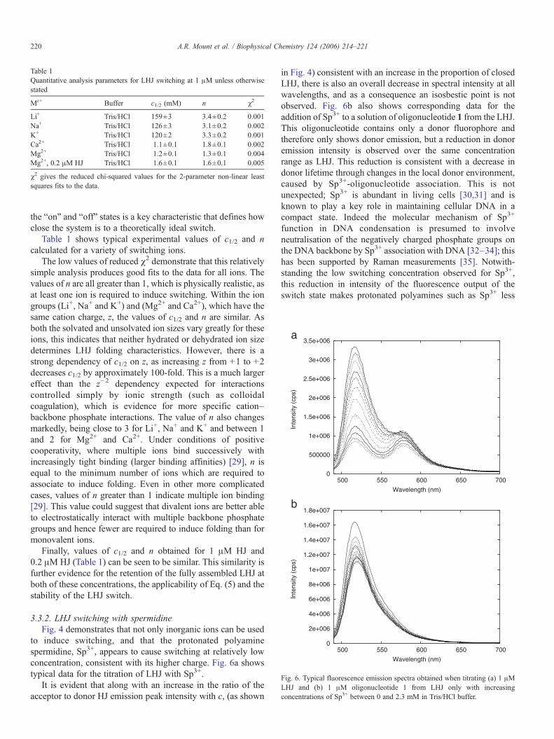
Table 1 shows typical experimental values of c1/2 and ncalculated for a variety of switching ions.
The low values of reduced !2 demonstrate that this relativelysimple analysis produces good fits to the data for all ions. Thevalues of n are all greater than 1, which is physically realistic, asat least one ion is required to induce switching. Within the iongroups (Li+, Na+ and K+) and (Mg2+ and Ca2+), which have thesame cation charge, z, the values of c1/2 and n are similar. Asboth the solvated and unsolvated ion sizes vary greatly for theseions, this indicates that neither hydrated or dehydrated ion sizedetermines LHJ folding characteristics. However, there is astrong dependency of c1/2 on z, as increasing z from +1 to +2decreases c1/2 by approximately 100-fold. This is a much largereffect than the z"2 dependency expected for interactionscontrolled simply by ionic strength (such as colloidalcoagulation), which is evidence for more specific cation–backbone phosphate interactions. The value of n also changesmarkedly, being close to 3 for Li+, Na+ and K+ and between 1and 2 for Mg2+ and Ca2+. Under conditions of positivecooperativity, where multiple ions bind successively withincreasingly tight binding (larger binding affinities) [29], n isequal to the minimum number of ions which are required toassociate to induce folding. Even in other more complicatedcases, values of n greater than 1 indicate multiple ion binding[29]. This value could suggest that divalent ions are better ableto electrostatically interact with multiple backbone phosphategroups and hence fewer are required to induce folding than formonovalent ions.
Finally, values of c1/2 and n obtained for 1 "M HJ and0.2 "M HJ (Table 1) can be seen to be similar. This similarity isfurther evidence for the retention of the fully assembled LHJ atboth of these concentrations, the applicability of Eq. (5) and thestability of the LHJ switch.
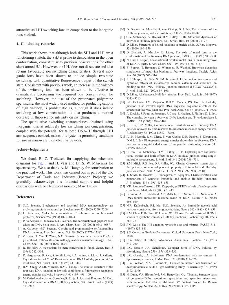
3.3.2. LHJ switching with spermidineFig. 4 demonstrates that not only inorganic ions can be used
to induce switching, and that the protonated polyaminespermidine, Sp3+, appears to cause switching at relatively lowconcentration, consistent with its higher charge. Fig. 6a showstypical data for the titration of LHJ with Sp3+.
It is evident that along with an increase in the ratio of theacceptor to donor HJ emission peak intensity with c, (as shown
in Fig. 4) consistent with an increase in the proportion of closedLHJ, there is also an overall decrease in spectral intensity at allwavelengths, and as a consequence an isosbestic point is notobserved. Fig. 6b also shows corresponding data for theaddition of Sp3+ to a solution of oligonucleotide 1 from the LHJ.This oligonucleotide contains only a donor fluorophore andtherefore only shows donor emission, but a reduction in donoremission intensity is observed over the same concentrationrange as LHJ. This reduction is consistent with a decrease indonor lifetime through changes in the local donor environment,caused by Sp3+-oligonucleotide association. This is notunexpected; Sp3+ is abundant in living cells [30,31] and isknown to play a key role in maintaining cellular DNA in acompact state. Indeed the molecular mechanism of Sp3+
function in DNA condensation is presumed to involveneutralisation of the negatively charged phosphate groups onthe DNA backbone by Sp3+ association with DNA [32–34]; thishas been supported by Raman measurements [35]. Notwith-standing the low switching concentration observed for Sp3+,this reduction in intensity of the fluorescence output of theswitch state makes protonated polyamines such as Sp3+ less
Table 1Quantitative analysis parameters for LHJ switching at 1 "M unless otherwisestated
Mz+ Buffer c1/2 (mM) n !2
Li+ Tris/HCl 159±3 3.4±0.2 0.001Na+ Tris/HCl 126±3 3.1±0.2 0.002K+ Tris/HCl 120±2 3.3±0.2 0.001Ca2+ Tris/HCl 1.1±0.1 1.8±0.1 0.002Mg2+ Tris/HCl 1.2±0.1 1.3±0.1 0.004Mg2+, 0.2 "M HJ Tris/HCl 1.6±0.1 1.6±0.1 0.005
!2 gives the reduced chi-squared values for the 2-parameter non-linear leastsquares fits to the data.
0
500000
1e+006
1.5e+006
2e+006
2.5e+006
3e+006
3.5e+006
500 550 600 650 700Wavelength (nm)
0
2e+006
4e+006
6e+006
8e+006
1e+007
1.2e+007
1.4e+007
1.6e+007
1.8e+007
500 550 600 650 700Wavelength (nm)
Inte
nsity
(cp
s)In
tens
ity (
cps)
a
b
Fig. 6. Typical fluorescence emission spectra obtained when titrating (a) 1 "MLHJ and (b) 1 "M oligonucleotide 1 from LHJ only with increasingconcentrations of Sp3+ between 0 and 2.3 mM in Tris/HCl buffer.
220 A.R. Mount et al. / Biophysical Chemistry 124 (2006) 214–221
attractive as LHJ switching ions in comparison to the inorganicions studied.
4. Concluding remarks
This work shows that although both the SHJ and LHJ are afunctioning switch, the SHJ is prone to dissociation in the openconformation, consistent with previous observations for othershort-armed HJs. However, the LHJ does not dissociate and alsoretains favourable ion switching characteristics. Several inor-ganic ions have been shown to induce simple two-stateswitching, with quantitative fluorescence output of the switchstate. Consistent with previous work, an increase in the valencyof the switching ions has been shown to be effective indramatically decreasing the required ion concentration forswitching. However, the use of the protonated polyaminespermidine, the most widely used method for producing cationsof high valency, is problematic as, although it does induceswitching at low concentration, it also produces a markeddecrease in fluorescence intensity on switching.
The quantitative switching characteristics obtained usinginorganic ions at relatively low switching ion concentration,coupled with the potential for tailored DNA-HJ through LHJarm sequence control, makes this system a promising candidatefor use in nanoscale biomolecular devices.
Acknowledgements
We thank R. Z. Troitzsch for supplying the schematicdiagrams for Fig. 1 and H. Vass and Dr. S. W. Magennis forspectroscopy. We also thank A. M. Haughey for contribution tothe practical work. This work was carried out as part of the UKDepartment of Trade and Industry (Beacon Project); wegratefully acknowledge this financial support and helpfuldiscussions with our technical monitor, Marc Bailey.
References
[1] N.C. Seeman, Biochemistry and structural DNA nanotechnology: anevolving symbiotic relationship, Biochemistry 42 (2003) 7259–7269.
[2] L. Adleman, Molecular computation of solutions to combinatorialproblems, Science 266 (1994) 1021–1024.
[3] P. Sa-Ardyen, N. Jonoska, N.C. Seeman, The construction of graphs whoseedges are DNA helix axes, J. Am. Chem. Soc. 126 (2004) 6648–6657.
[4] A. Carbone, N.C. Seeman, Circuits and programmable self-assemblingDNA structures, Proc. Natl. Acad. Sci. 99 (2002) 12577–12582.
[5] Z. Shen, H. Yan, T. Wang, N.C. Seeman, Paranemic crossover DNA: ageneralized Holliday structure with applications in nanotechnology, J. Am.Chem. Soc. 126 (2004) 1666–1674.
[6] R. Holliday, A mechanism for gene conversion in fungi, Genet. Res. 5(1964) 282–304.
[7] D. Hargreaves, D. Rice, S. Sedilinkova, P. Artymiuk, R. Lloyd, J. Rafferty,Crystal structure of E. coliRuvAwith bound DNAHolliday junction at 6 Åresolution, Nat. Struct. Biol. 5 (1998) 441–446.
[8] R.M. Clegg, A.I.H. Murchie, D.M.J. Lilley, The solution structure of thefour-way DNA junction at low-salt conditions: a fluorescence resonanceenergy transfer analysis, Biophys. J. 66 (1994) 99–109.
[9] M. Ortiz-Lombardia, A. Gonzalez, R. Eritja, J. Aymami, F. Azorn, M. Coll,Crystal structure of a DNA Holliday junction, Nat. Struct. Biol. 6 (1999)913–917.
[10] D. Duckett, A. Murchie, S. von Kitzing, D. Lilley, The structure of theHolliday junction, and its resolution, Cell 55 (1988) 79–89.
[11] S.A. McKinney, A. Declais, D.M. Lilley, T. Ha, Structural dynamics ofindividual Holliday junctions, Nat. Struct. Biol. 10 (2003) 93–97.
[12] D. Lilley, Structures of helical junctions in nucleic acids, Q. Rev. Biophys.33 (2000) 109–159.
[13] D. Duckett, A. Murchie, D. Lilley, The role of metal ions in theconformation of the four-way DNA junction, EMBO J. 9 (1990) 583–590.
[14] N. Hud, J. Feigon, Localisation of divalent metal ions in the minor grooveof DNA A-tracts, J. Am. Chem. Soc. 119 (1997) 5756–5757.
[15] B. Buuren, T. Hermann, S. Wijmenga, E. Westhof, Brownian-dynamicssimulations of metal–ion binding to four-way junctions, Nucleic AcidsRes. 30 (2002) 507–514.
[16] J.H. Thorpe, B.C. Gale, S.C.M. Teixeira, C.J. Cardin, Conformational andhydration effects of site-selective sodium, calcium and strontium ionbinding to the DNA Holliday junction structure d(TCGGTACCGA)4,J. Mol. Biol. 327 (2003) 97–109.
[17] D. Lilley, All change at Holliday junction, Proc. Natl. Acad. Sci. 94 (1997)9513–9515.
[18] B.F. Eichman, J.M. Vargason, B.H.M. Mooers, P.S. Ho, The Hollidayjunction in an inverted repeat DNA sequence: sequence effects on thestructure of four-way junctions, Proc. Natl. Acad. Sci. 97 (2000) 3971–3976.
[19] A. Declais, J. Fogg, A. Freeman, F. Coste, J. Hadden, S. Phillips, D. Lilley,The complex between a four-way DNA junction and T7 endonuclease I,EMBO J. 22 (2003) 1398–1409.
[20] P.S. Eis, D.P. Millar, Conformational distributions of a four-way DNAjunction revealed by time-resolved fluorescence resonance energy transfer,Biochemistry 32 (1993) 13852–13860.
[21] A.I.H. Murchie, R.M. Clegg, E. von Kitzing, D.R. Duckett, S. Diekmann,D.M.J. Lilley, Fluorescence energy transfer shows that the four-way DNAjunction is a right-handed cross of antiparallel molecules, Nature 341(1989) 763–765.
[22] C. Joo, S.A. McKinney, D.M.J. Lilley, T. Ha, Exploring rare conforma-tional species and ionic effects in DNA Holliday junction using single-molecule spectroscopy, J. Mol. Biol. 341 (2004) 739–751.
[23] S.M. Miick, R.S. Fee, D.P. Millar, W.J. Chazin, Crossover isomer bias isthe primary sequence-dependent property of immobilized Hollidayjunctions, Proc. Natl. Acad. Sci. U. S. A. 94 (1997) 9080–9084.
[24] T. Shida, H. Iwasaki, H. Shinagawa, Y. Kyogoku, Characterization andcomparison of synthetic immobile and mobile Holliday junctions,J. Biochem. 119 (1996) 653–658.
[25] V.R. Ramirez-Carrozzi, T.K. Kerppola, gelFRET analysis of nucleoproteincomplexes, Methods 25 (2001) 31–43.
[26] B. Yurke, A.J. Turberfield, A.P. Mills Jr., F.C. Simmel, J.L. Neumann, ADNA-fuelled molecular machine made of DNA, Nature 406 (2000)605–609.
[27] N.R. Kallenbach, R.I. Ma, N.C. Seeman, An immobile nucleic acidjunction constructed from oligonucleotides, Nature 305 (1983) 829–831.
[28] S.M. Chen, F. Heffron, W. Leupin, W.J. Chazin, Two-dimensional H NMRstudies of synthetic immobile Holliday junctions, Biochemistry 30 (1991)766–771.
[29] J.N. Weiss, The Hill equation revisited: uses and misuses, FASEB J. 11(1997) 835–841.
[30] S.S. Cohen, A Guide to Polyamines, Oxford University Press, New York,1998.
[31] C.W. Tabor, H. Tabor, Polyamines, Annu. Rev. Biochem. 53 (1983)749–790.
[32] L.C. Gosule, J.A. Schellman, Compact form of DNA induced byspermidine, Nature 259 (1976) 333–335.
[33] L.C. Gosule, J.A. Schellman, DNA condensation with polyamines: I.Spectroscopic studies, J. Mol. Biol. 121 (1978) 311–326.
[34] R.W. Wilson, V.A. Bloomfield, Counterion-induced condensation ofdeoxyribonucleic acid: a light-scattering study, Biochemistry 18 (1979)2192–2196.
[35] H. Deng, V.A. Bloomfield, J.M. Benevides, G.J. Thomas, Structure basisof polyamine-DNA recognition: spermidine and spermine interactionswith genomic B-DNAs of different GC content probed by Ramanspectroscopy, Nucleic Acids Res. 28 (2000) 3379–3385.
221A.R. Mount et al. / Biophysical Chemistry 124 (2006) 214–221
Related Documents


